The role of the medial prefrontal cortex in the acquisition, retention, and reversal of a tactile...
8
Behavioural Brain Research 236 (2013) 94–101 Contents lists available at SciVerse ScienceDirect Behavioural Brain Research j ourna l ho me pa ge: www.elsevier.com/locate/bbr Research report The role of the medial prefrontal cortex in the acquisition, retention, and reversal of a tactile visuospatial conditional discrimination task Crystal L. Shaw, Glenn D.R. Watson, Henry L. Hallock, Kathryn M. Cline, Amy L. Griffin ∗ Department of Psychology, 108 Wolf Hall, University of Delaware, Newark, DE 19716, United States h i g h l i g h t s We examined the effects of mPFC inactivation on conditional discrimination learning. Acquisition/retention of conditional discrimination task does not require the mPFC. mPFC inactivation impaired conditional discrimination reversal learning. The rat mPFC plays a crucial role in strategy selection during task reversal. a r t i c l e i n f o Article history: Received 27 January 2012 Received in revised form 1 August 2012 Accepted 16 August 2012 Available online 23 August 2012 Keywords: Medial prefrontal cortex Behavioral flexibility Reversal Conditional discrimination T-maze Muscimol Configural learning a b s t r a c t The medial prefrontal cortex (mPFC) is responsible for executive functions such as abstract rule coding, strategy switching, and behavioral flexibility; however, there is some debate regarding the extent to which mPFC is involved in reversal learning, especially in complex multisensory tasks such as conditional discrimination. Therefore, we investigated the effects of mPFC inactivation on the acquisition, retention, and reversal of a visuospatial conditional discrimination (CD) task. In experiment 1, muscimol was infused through bilateral cannulae on days 1, 2, and 3 to test the effects of mPFC inactivation on task acquisition and days 19, 20, and 21 to test the effects on retention of the task. For experiment 2, rats were trained on the CD task for 21 days with no infusions given, after which the reward contingency was reversed, with infusions given during the first six days of reversal. The results of experiment 1 showed that the muscimol and saline groups did not differ on acquisition or retention. However, experiment 2 showed that the muscimol group displayed significantly more performance errors than the control group during reversal. Compared to the control group, the muscimol group also showed a decreased tendency to use a side-bias strategy during the intermediate stages of reversal. The failure of the muscimol group to exhibit a side bias suggests that the mPFC is necessary for sampling strategies necessary for the reversal of a visuospatial CD task. © 2012 Elsevier B.V. All rights reserved. 1. Introduction The medial prefrontal cortex (mPFC) is thought to be respon- sible for higher executive functions, such as behavioral flexibility, which requires the suppression of a previously successful behavior in response to an unexpected change in the environment to favor a more suitable behavior [1,2]. Damage to the mPFC in rodents has been shown to impair performance of tasks that require behavioral flexibility, such as rule or strategy switching in spatial navigation tasks [3–6] and extinction of a conditioned response in appetitive Pavlovian conditioning [7]. Conditional discrimination (CD) learning requires a subject to learn that a particular response will be rewarded and another ∗ Corresponding author. Tel.: +1 302 831 2575; fax: +1 302 831 3645. E-mail address: [email protected] (A.L. Griffin). response not rewarded in one context and that the opposite reward contingency will be applied in a different context. This type of learning involves associating each unique context–response con- figuration with a reinforcement value (rewarded or not) and is thus considered to be a type of configural learning [8]. It has been established that the formation of configural associations depends on the hippocampal formation based on evidence that rats with hippocampal lesions cannot solve configural tasks such as negative patterning and transverse patterning [9] or visuospatial conditional discrimination [10]. However, the hippocampus is part of a larger system of anatomically-connected neural structures, including the medial prefrontal cortex (mPFC). The role of the mPFC in configu- ral learning in general and conditional discrimination learning in particular has been virtually unexplored. Moreover, the fact that the mPFC is known to be crucial for executive functioning, specifi- cally task switching, suggests that the mPFC would be particularly important for conditional discrimination reversal, in which the 0166-4328/$ – see front matter © 2012 Elsevier B.V. All rights reserved. http://dx.doi.org/10.1016/j.bbr.2012.08.024
Transcript of The role of the medial prefrontal cortex in the acquisition, retention, and reversal of a tactile...

R
To
CD
h
����
a
ARRAA
KMBRCTMC
1
swiabfltP
l
0h
Behavioural Brain Research 236 (2013) 94– 101
Contents lists available at SciVerse ScienceDirect
Behavioural Brain Research
j ourna l ho me pa ge: www.elsev ier .com/ locate /bbr
esearch report
he role of the medial prefrontal cortex in the acquisition, retention, and reversalf a tactile visuospatial conditional discrimination task
rystal L. Shaw, Glenn D.R. Watson, Henry L. Hallock, Kathryn M. Cline, Amy L. Griffin ∗
epartment of Psychology, 108 Wolf Hall, University of Delaware, Newark, DE 19716, United States
i g h l i g h t s
We examined the effects of mPFC inactivation on conditional discrimination learning.Acquisition/retention of conditional discrimination task does not require the mPFC.mPFC inactivation impaired conditional discrimination reversal learning.The rat mPFC plays a crucial role in strategy selection during task reversal.
r t i c l e i n f o
rticle history:eceived 27 January 2012eceived in revised form 1 August 2012ccepted 16 August 2012vailable online 23 August 2012
eywords:edial prefrontal cortex
ehavioral flexibilityeversal
a b s t r a c t
The medial prefrontal cortex (mPFC) is responsible for executive functions such as abstract rule coding,strategy switching, and behavioral flexibility; however, there is some debate regarding the extent towhich mPFC is involved in reversal learning, especially in complex multisensory tasks such as conditionaldiscrimination. Therefore, we investigated the effects of mPFC inactivation on the acquisition, retention,and reversal of a visuospatial conditional discrimination (CD) task. In experiment 1, muscimol was infusedthrough bilateral cannulae on days 1, 2, and 3 to test the effects of mPFC inactivation on task acquisitionand days 19, 20, and 21 to test the effects on retention of the task. For experiment 2, rats were trainedon the CD task for 21 days with no infusions given, after which the reward contingency was reversed,with infusions given during the first six days of reversal. The results of experiment 1 showed that the
onditional discrimination-mazeuscimol
onfigural learning
muscimol and saline groups did not differ on acquisition or retention. However, experiment 2 showedthat the muscimol group displayed significantly more performance errors than the control group duringreversal. Compared to the control group, the muscimol group also showed a decreased tendency to usea side-bias strategy during the intermediate stages of reversal. The failure of the muscimol group toexhibit a side bias suggests that the mPFC is necessary for sampling strategies necessary for the reversalof a visuospatial CD task.
. Introduction
The medial prefrontal cortex (mPFC) is thought to be respon-ible for higher executive functions, such as behavioral flexibility,hich requires the suppression of a previously successful behavior
n response to an unexpected change in the environment to favor more suitable behavior [1,2]. Damage to the mPFC in rodents haseen shown to impair performance of tasks that require behavioralexibility, such as rule or strategy switching in spatial navigationasks [3–6] and extinction of a conditioned response in appetitive
avlovian conditioning [7].Conditional discrimination (CD) learning requires a subject toearn that a particular response will be rewarded and another
∗ Corresponding author. Tel.: +1 302 831 2575; fax: +1 302 831 3645.E-mail address: [email protected] (A.L. Griffin).
166-4328/$ – see front matter © 2012 Elsevier B.V. All rights reserved.ttp://dx.doi.org/10.1016/j.bbr.2012.08.024
© 2012 Elsevier B.V. All rights reserved.
response not rewarded in one context and that the opposite rewardcontingency will be applied in a different context. This type oflearning involves associating each unique context–response con-figuration with a reinforcement value (rewarded or not) and isthus considered to be a type of configural learning [8]. It has beenestablished that the formation of configural associations dependson the hippocampal formation based on evidence that rats withhippocampal lesions cannot solve configural tasks such as negativepatterning and transverse patterning [9] or visuospatial conditionaldiscrimination [10]. However, the hippocampus is part of a largersystem of anatomically-connected neural structures, including themedial prefrontal cortex (mPFC). The role of the mPFC in configu-ral learning in general and conditional discrimination learning in
particular has been virtually unexplored. Moreover, the fact thatthe mPFC is known to be crucial for executive functioning, specifi-cally task switching, suggests that the mPFC would be particularlyimportant for conditional discrimination reversal, in which the
Brain
cterfl
tatumrta
2
2
hcpa
2
Ttubrva
2
a6rcfdtn
2
oTt(oobwudls
2
aoiam0((rt
C.L. Shaw et al. / Behavioural
ontext previously associated with one response–reward con-ingency is switched to the opposite context. Additionally,xamination of intermediate strategy selection, such as the tempo-ary development of a side bias, can serve as a measure of behavioralexibility in this type of task.
Our laboratory has recently developed a visuospatial CD taskhat requires a two-choice spatial discrimination in response to
multisensory conditional cue. The involvement of the mPFC inhe acquisition, retention and reversal of this type of learning isnknown. Therefore, the present study examined the necessity ofPFC integrity in the acquisition, retention (experiment 1), and
eversal (experiment 2) of a visuospatial CD task that required ratso use intramaze cues (floor inserts that varied in texture and color)s a conditional cue for goal-arm selection on a T-maze.
. Methods
.1. Subjects
Male Long–Evans Hooded rats (Harlan, Indianapolis), 3–6 months old, wereoused in standard laboratory cages maintained in a temperature and humidityontrolled colony room on a 12:12-h light/dark cycle. After a 1-week acclimationeriod, rats were kept at 90% if their free-feeding body weight and given ad libitumccess to water throughout and the experiment.
.2. Apparatus
All training was performed on a T-maze constructed of wood and painted black.he maze consisted of a central arm (117 × 6 cm), two goal arms (79 × 6 cm) andwo return arms (120 × 6 cm) (Fig. 1). A plastic cup at the end of the goal arm wassed to place a chocolate sprinkle food reward. There was a start box located at thease of the maze stem, where the rat was confined between trials. The experimentaloom was illuminated by a single 60 W bulb and surrounded by a black curtain witharious visual cues attached. Experiments 1 and 2 were carried out in the same roomnd on the same maze. The floor of the maze was covered with black contact paper.
.3. Maze acclimation and pretraining
Prior to maze acclimation, rats were handled by the experimenter for ∼10 min day for one week. Rats were then acclimated to the testing room and maze for–8 days. The first 2 acclimation sessions consisted of goal-zone training in whichats were confined to the right and left goal zone on alternating trials and learned toonsume chocolate sprinkles from reward cups. The next 4–6 sessions were 12-trialorced-run sessions in which one of the two goal arms was blocked (in a pseudoran-om sequence) so that rats learned to run up the maze stem into the open goal arm tohe reward cup, where they received a chocolate sprinkle reward. Floor inserts (seeext section) were not introduced during the pretraining stage of the experiment.
.4. Conditional discrimination (CD) training
Prior to each trial, the experimenter placed wooden floor inserts covered onne side with black plastic mesh into the central stem and both goal arms of the-maze with either the wood or mesh side facing up. Rats learned to select eitherhe left or right goal arm to obtain a food reward contingent upon the texture/colorblack mesh or bare wood) of the floor insert (Fig. 1). Half of the rats were trainedn the rule ‘left on wood/right on mesh’ and the other half was trained on the ‘rightn wood/left on mesh’ rule. During the inter-trial interval (ITI), a black woodenarricade was placed between the pedestal and the maze to obstruct the rats’ viewhile the experimenter prepared for the next trial. In order to prevent the rat fromsing auditory cues to anticipate the next trial, the experimenter flipped the inserturing every ITI. The unrewarded goal zone was sham baited on each trial. Each ITI
asted for 8–10 s. Rats were given 24 trials (12 mesh, 12 wood) in a pseudorandomequence [11].
.5. Surgery
Rats were given a pre-anesthetic subcutaneous dose of atropine (0.05 mg/kg)nd anesthetized with continuous-flow isoflurane (1.5–3% in oxygen), mountedn a stereotaxic frame. The scalp was shaved, anesthetized with a subcutaneousnjection of lidocaine and sterilized with Nolvasan® . After the skull was exposednd cleaned, four small holes were drilled near the skull ridge using a sterotaxic-ounted drill (Fine Science Tools). Bone screws (shaft length 4.0 mm, shaft diameter
.85 mm) were fixed into the holes and affixed to the skull with dental acrylicPatterson Dental). Circular holes were drilled using a 1.8-mm-diameter trephineFine Science Tools) in each hemisphere at the following coordinates: 3.0 mm ante-ior to bregma, ±1.8 mm lateral to bregma [12]. Dorsal–ventral coordinates wereaken from dura mater for more accurate placements of the cannulae. Once dura
Research 236 (2013) 94– 101 95
was removed, the exposed brain was kept moist using gel foam soaked in ster-ile saline. A 26-gauge stainless steel guide cannulae, held in a stereotaxic arm ata 14◦ angle, was lowered into the respective hemisphere 2.0 mm ventral to dura.The guide cannulae were affixed to the skull with dental acrylic. A subcutaneousinjection of Banamine (2.5 mg/kg) was given approximately 30 min prior to the endof surgery and children’s ibuprofen (20 mg/mL) was given in the drinking watertwo days postoperatively for analgesia. Rats were allowed to recover for five daysprior to behavioral training. All procedures were carried out in accordance with theUniversity of Delaware Institutional Animal Care and Use Committee.
2.6. Infusions
Muscimol, a GABAA agonist, was dissolved in sterile saline and the solutionwas infused bilaterally via a 31-gauge injector connected to a 10-�L Hamiltonsyringe by a polyethylene tube. The injector extended 1.5 mm beyond the tip of theguide cannula. The infusion volume and rate was controlled by an infusion pump(World Precision Instruments) programmed to deliver the infusate, either muscimol(0.1 �g/�L) or sterile saline, at a rate of 0.25 �L/min for 2 min, for a total volume of0.50 �L for each hemisphere. Infusion cannulae were left in place for 2 min after theinfusion to allow for diffusion. Rats were lightly anesthetized with isoflurane dur-ing each infusion and given 30 min in their home cages after the infusion to recoverfrom the anesthesia. Anesthesia was necessary due to the small size of the internalcannula and has been shown by other investigations to have no effect on subsequentbehavioral performance [6].
2.7. Histology
After completion of behavioral training, rats were anesthetized with isofluraneand infused bilaterally with 0.5 �L of a neutral red solution (dissolved in saline) fordetermination of the spread of the infusate. Rats were then given an overdose ofsodium pentobarbital (200 mg/kg, ip) and perfused using 0.9% saline followed by10% buffered formalin to fix the tissue. The brains were then removed and placedin 10% buffered formalin. After at least 24 h in formalin, the brains were placed in30% buffered sucrose solution. After sinking, the brains were frozen and sectioned(40 �m) using a cryostat. The sections were mounted on slides, stained using cresylviolet and photographed using a camera mounted on a microscope. Cannulae place-ments were verified by overlaying the photograph of the section with atlas platesfrom Paxinos and Watson [12] in Adobe Illustrator. Because it was often difficultto see the injector cannulae tracks, placements were determined from the guidecannula tracks, so the actual placement of the injectors was 1.5 mm ventral to theplacement.
2.8. Experiment 1: effects of mPFC inactivation on CD acquisition and retention
After recovery from surgery, rats were trained on the CD task for 21 days. Toexamine whether mPFC inactivation impaired CD acquisition, muscimol infusionswere given prior to training on the first 3 days of CD training. Infusions were againgiven on days 19, 20 and 21 to test the effects of mPFC inactivation on retention of theCD task. Groups of rats were counterbalanced so that half of the rats that receivedmuscimol during acquisition would receive either saline or muscimol during theretention and vice versa. To control for any effects due to anesthesia that was used forthe infusions, acquisition and retention of the CD task was measured in a third groupof rats. These rats were implanted with bilateral mPFC cannulae but did not receiveanesthesia or infusions during the 21 days of task acquisition and performance. Thisgroup of rats was then used in experiment 2 to test for effects of mPFC inactivationon CD reversal (see below). The acquisition and retention of the visuospatial CD taskwere compared across the no-infusion, saline, and muscimol groups.
2.9. Experiment 2: effects of mPFC inactivation on reversal of the CD task
Rats were trained on the CD task for 21 days without infusions and randomlyassigned to either the saline or muscimol group. Before entering the reversal phaseof the experiment, all of the rats included in experiment 2 met a criterion of 2consecutive days performing 75% correct on the CD task. Starting on day 22, thereward contingency was reversed. For instance, if the previous rule was ‘left onwood’, the rats were then trained on the rule ‘right on wood’ from days 22 to 42. Inorder to examine the effects mPFC inactivation on CD reversal, for the first six daysof reversal, infusions of either muscimol or saline were given prior to training usingthe same procedures as those described above for experiment 1.
2.10. Statistical analysis
The training sessions were grouped into 3-session (72-trial) blocks for com-parison of the learning curves across training. The number of errors was thencompared between the no-infusion, muscimol, and saline groups using mixed design
(3 group × 6 block) ANOVA for experiment 1. Also for experiment 1, choice accuracy(percentage of correct trials) was compared between the no-infusion, muscimol,and saline groups using a 3 (group) × 4 (session) mixed-design ANOVA. For bothexperiment 1 and 2, learning and reversal rates were compared between groups bycounting the number of sessions in which the choice accuracy was below chance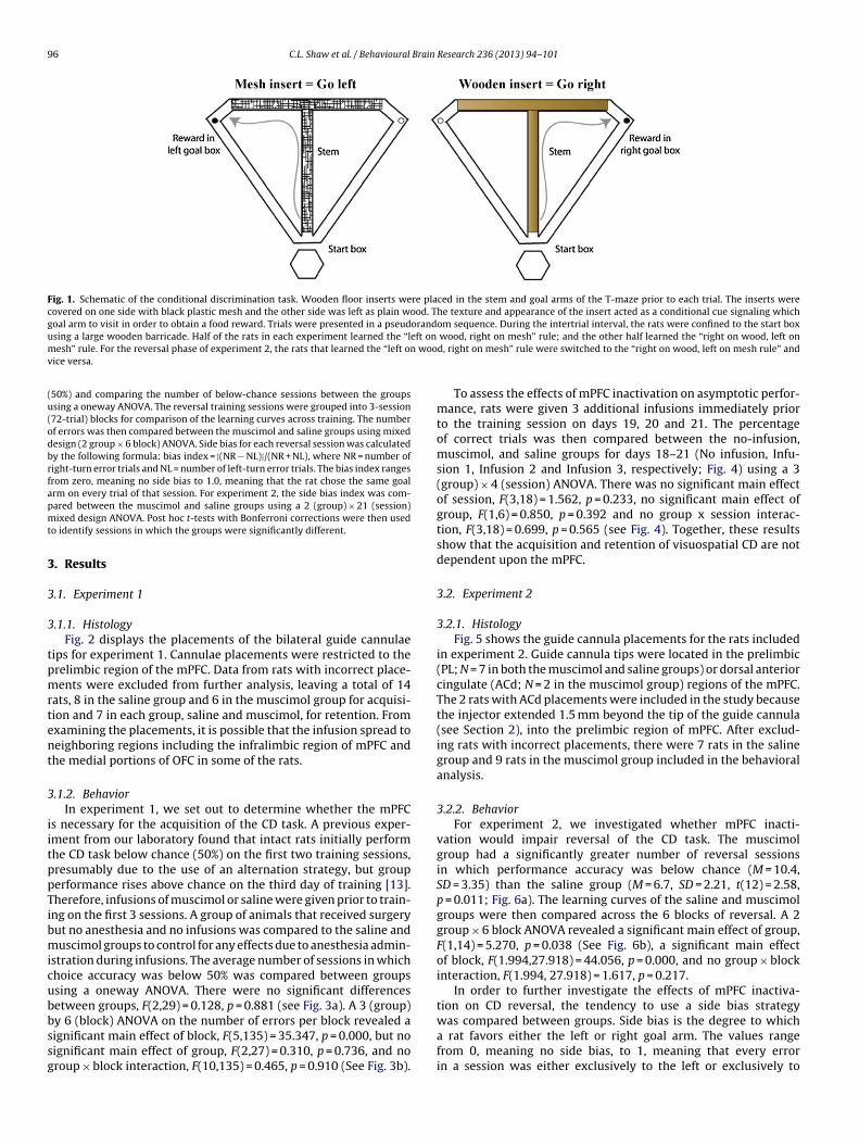
96 C.L. Shaw et al. / Behavioural Brain Research 236 (2013) 94– 101
Fig. 1. Schematic of the conditional discrimination task. Wooden floor inserts were placed in the stem and goal arms of the T-maze prior to each trial. The inserts werecovered on one side with black plastic mesh and the other side was left as plain wood. The texture and appearance of the insert acted as a conditional cue signaling whichgoal arm to visit in order to obtain a food reward. Trials were presented in a pseudorandom sequence. During the intertrial interval, the rats were confined to the start boxu eft onm woov
(u(odbrfapmt
3
3
3
tpmrtent
3
iitppTibmicubbssg
sing a large wooden barricade. Half of the rats in each experiment learned the “lesh” rule. For the reversal phase of experiment 2, the rats that learned the “left on
ice versa.
50%) and comparing the number of below-chance sessions between the groupssing a oneway ANOVA. The reversal training sessions were grouped into 3-session72-trial) blocks for comparison of the learning curves across training. The numberf errors was then compared between the muscimol and saline groups using mixedesign (2 group × 6 block) ANOVA. Side bias for each reversal session was calculatedy the following formula: bias index = |(NR − NL)|/(NR + NL), where NR = number ofight-turn error trials and NL = number of left-turn error trials. The bias index rangesrom zero, meaning no side bias to 1.0, meaning that the rat chose the same goalrm on every trial of that session. For experiment 2, the side bias index was com-ared between the muscimol and saline groups using a 2 (group) × 21 (session)ixed design ANOVA. Post hoc t-tests with Bonferroni corrections were then used
o identify sessions in which the groups were significantly different.
. Results
.1. Experiment 1
.1.1. HistologyFig. 2 displays the placements of the bilateral guide cannulae
ips for experiment 1. Cannulae placements were restricted to therelimbic region of the mPFC. Data from rats with incorrect place-ents were excluded from further analysis, leaving a total of 14
ats, 8 in the saline group and 6 in the muscimol group for acquisi-ion and 7 in each group, saline and muscimol, for retention. Fromxamining the placements, it is possible that the infusion spread toeighboring regions including the infralimbic region of mPFC andhe medial portions of OFC in some of the rats.
.1.2. BehaviorIn experiment 1, we set out to determine whether the mPFC
s necessary for the acquisition of the CD task. A previous exper-ment from our laboratory found that intact rats initially performhe CD task below chance (50%) on the first two training sessions,resumably due to the use of an alternation strategy, but grouperformance rises above chance on the third day of training [13].herefore, infusions of muscimol or saline were given prior to train-ng on the first 3 sessions. A group of animals that received surgeryut no anesthesia and no infusions was compared to the saline anduscimol groups to control for any effects due to anesthesia admin-
stration during infusions. The average number of sessions in whichhoice accuracy was below 50% was compared between groupssing a oneway ANOVA. There were no significant differencesetween groups, F(2,29) = 0.128, p = 0.881 (see Fig. 3a). A 3 (group)
y 6 (block) ANOVA on the number of errors per block revealed aignificant main effect of block, F(5,135) = 35.347, p = 0.000, but noignificant main effect of group, F(2,27) = 0.310, p = 0.736, and noroup × block interaction, F(10,135) = 0.465, p = 0.910 (See Fig. 3b).wood, right on mesh” rule; and the other half learned the “right on wood, left ond, right on mesh” rule were switched to the “right on wood, left on mesh rule” and
To assess the effects of mPFC inactivation on asymptotic perfor-mance, rats were given 3 additional infusions immediately priorto the training session on days 19, 20 and 21. The percentageof correct trials was then compared between the no-infusion,muscimol, and saline groups for days 18–21 (No infusion, Infu-sion 1, Infusion 2 and Infusion 3, respectively; Fig. 4) using a 3(group) × 4 (session) ANOVA. There was no significant main effectof session, F(3,18) = 1.562, p = 0.233, no significant main effect ofgroup, F(1,6) = 0.850, p = 0.392 and no group x session interac-tion, F(3,18) = 0.699, p = 0.565 (see Fig. 4). Together, these resultsshow that the acquisition and retention of visuospatial CD are notdependent upon the mPFC.
3.2. Experiment 2
3.2.1. HistologyFig. 5 shows the guide cannula placements for the rats included
in experiment 2. Guide cannula tips were located in the prelimbic(PL; N = 7 in both the muscimol and saline groups) or dorsal anteriorcingulate (ACd; N = 2 in the muscimol group) regions of the mPFC.The 2 rats with ACd placements were included in the study becausethe injector extended 1.5 mm beyond the tip of the guide cannula(see Section 2), into the prelimbic region of mPFC. After exclud-ing rats with incorrect placements, there were 7 rats in the salinegroup and 9 rats in the muscimol group included in the behavioralanalysis.
3.2.2. BehaviorFor experiment 2, we investigated whether mPFC inacti-
vation would impair reversal of the CD task. The muscimolgroup had a significantly greater number of reversal sessionsin which performance accuracy was below chance (M = 10.4,SD = 3.35) than the saline group (M = 6.7, SD = 2.21, t(12) = 2.58,p = 0.011; Fig. 6a). The learning curves of the saline and muscimolgroups were then compared across the 6 blocks of reversal. A 2group × 6 block ANOVA revealed a significant main effect of group,F(1,14) = 5.270, p = 0.038 (See Fig. 6b), a significant main effectof block, F(1.994,27.918) = 44.056, p = 0.000, and no group × blockinteraction, F(1.994, 27.918) = 1.617, p = 0.217.
In order to further investigate the effects of mPFC inactiva-tion on CD reversal, the tendency to use a side bias strategy
was compared between groups. Side bias is the degree to whicha rat favors either the left or right goal arm. The values rangefrom 0, meaning no side bias, to 1, meaning that every errorin a session was either exclusively to the left or exclusively to
C.L. Shaw et al. / Behavioural Brain Research 236 (2013) 94– 101 97
Fig. 2. Experiment 1: cannula tip locations. (a) Coronal sections +4.68 to +3.00 mm from bregma adopted from Paxinos and Watson [12]. Open diamonds, rats that receivedsaline for both acquisition and retention; filled squares, rats that received saline during acquisition and muscimol during retention; filled diamonds, rats that receivedmuscimol during acquisition and saline during retention; open squares, rats that received muscimol for both acquisition and retention. (b) Representative section throught ) and t
tiFgiwafsRbattusfassm
he mPFC stained with cresyl violet, showing tracks from the guide cannula (arrow
he right. A 2 (group) × 21 (session) ANOVA on the side biasndex during reversal revealed a significant main effect of session,(16.745,234.431) = 3.027, p = 0.000, no significant main effect ofroup, F(1,14) = 1.640, p = 0.221, and a significant group × sessionnteraction, F(16.745,234.431) = 1.95, p = 0.015. Post hoc t-tests
ith Bonferroni corrections revealed that the saline group showed significantly higher bias index on sessions 7 (the first infusion-ree reversal session), 8, 11, and significantly lower bias index onessions 15 and 21 than the muscimol group (p < 0.05; see Fig. 7).ats were then categorized based on whether they showed a sideias index at or above 75% on 3 or more consecutive sessions. Thisnalysis revealed 4 out of 7 rats in the saline group and no rats inhe muscimol group consistently used a side-bias strategy some-ime during the course of reversal learning. The difference in thetilization of a side bias was significantly different between thealine and muscimol groups (X2 (1) = 6.85, p = 0.008). The four ratsrom the saline group that were categorized as showing a side bias
ll showed a common pattern. Side bias was low early in rever-al, when all of the rats were following the initial rule. Starting onession 5 or 6, the 4 rats showed a robust side bias, which wasaintained between sessions 8 and 12 and dramatically droppedip of the injection cannula (arrowhead).
over subsequent sessions (see Fig. 7). In contrast to the perfor-mance results described above, the side bias index for the muscimolgroup was consistently low throughout all reversal sessions, withthe exception of the final reversal session. Together, these differ-ent patterns of side bias across reversal sessions suggest that thedevelopment of a side bias is a common strategy used by intactrats during the intermediate stages of reversal learning. The fewernumber of rats exhibiting a side bias in the muscimol group sug-gests that the mPFC may be critical for the development of the sidebias strategy and that the transient use of this strategy can facilitatereversal learning.
4. Discussion
Our results show that inactivation of the mPFC using microin-fusions of muscimol does not impair the acquisition or retention
of a visuospatial CD task. However, reversal of the task was sig-nificantly disrupted by mPFC inactivation. Interestingly, the salinegroup developed a strong side bias in the intermediate stages ofreversal learning that was not evident in the muscimol group.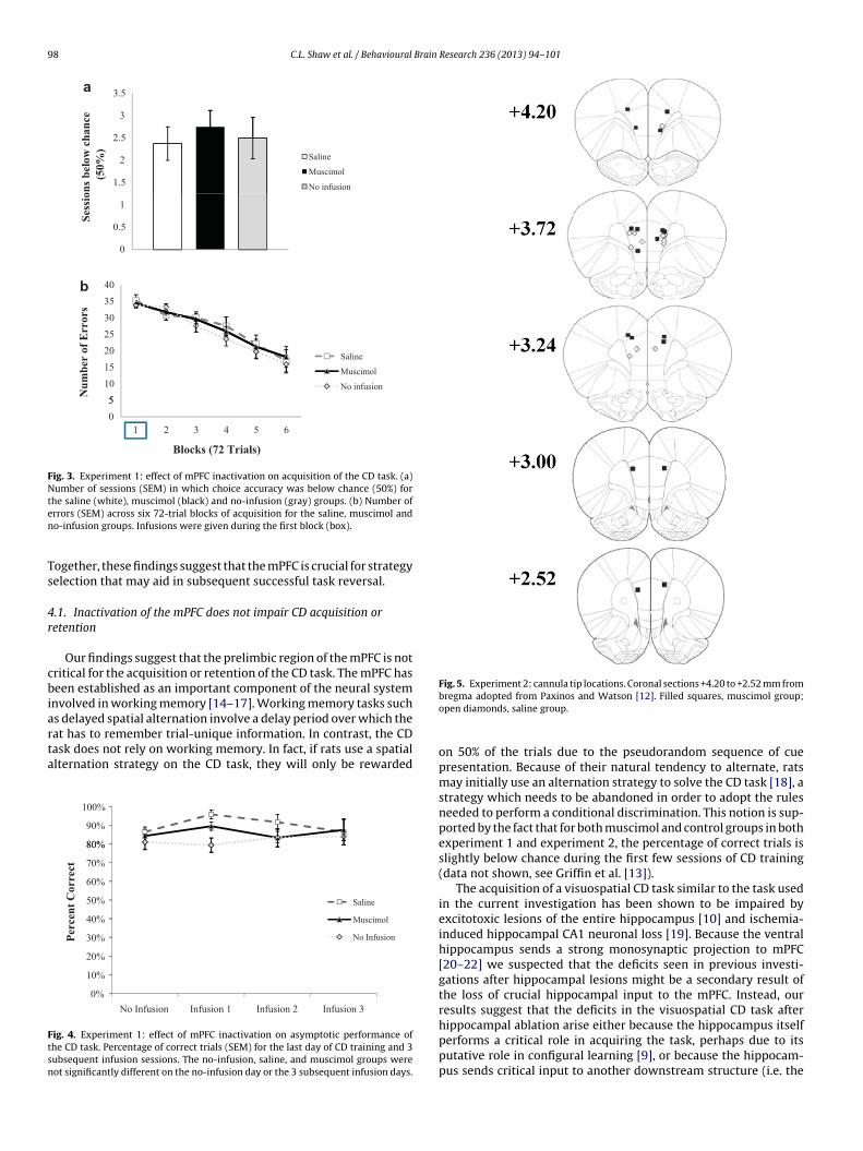
98 C.L. Shaw et al. / Behavioural Brain Research 236 (2013) 94– 101
a
3
3.5
1.5
2
2.5
(50%
)
Saline
Musci mol
No infusio n
0
0.5
1
Sess
ion
s b
elo
w c
han
ce
b
30
35
40
5
10
15
20
25
Nu
mb
er o
f E
rrors
Saline
Musci mol
No infusio n
0
5
654321
Blocks (72 Trials)
Fig. 3. Experiment 1: effect of mPFC inactivation on acquisition of the CD task. (a)Number of sessions (SEM) in which choice accuracy was below chance (50%) forten
Ts
4r
cbiarta
Ftsn
Fig. 5. Experiment 2: cannula tip locations. Coronal sections +4.20 to +2.52 mm from
he saline (white), muscimol (black) and no-infusion (gray) groups. (b) Number ofrrors (SEM) across six 72-trial blocks of acquisition for the saline, muscimol ando-infusion groups. Infusions were given during the first block (box).
ogether, these findings suggest that the mPFC is crucial for strategyelection that may aid in subsequent successful task reversal.
.1. Inactivation of the mPFC does not impair CD acquisition oretention
Our findings suggest that the prelimbic region of the mPFC is notritical for the acquisition or retention of the CD task. The mPFC haseen established as an important component of the neural system
nvolved in working memory [14–17]. Working memory tasks such
s delayed spatial alternation involve a delay period over which theat has to remember trial-unique information. In contrast, the CDask does not rely on working memory. In fact, if rats use a spatiallternation strategy on the CD task, they will only be rewarded80%
90%
100%
50%
60%
70%
80%
Saline
10%
20%
30%
40%
Percen
t C
orrect
Musci mol
No Inf usion
0%
Infusion 3Infusion 2Infusion 1No Infusion
ig. 4. Experiment 1: effect of mPFC inactivation on asymptotic performance ofhe CD task. Percentage of correct trials (SEM) for the last day of CD training and 3ubsequent infusion sessions. The no-infusion, saline, and muscimol groups wereot significantly different on the no-infusion day or the 3 subsequent infusion days.
bregma adopted from Paxinos and Watson [12]. Filled squares, muscimol group;open diamonds, saline group.
on 50% of the trials due to the pseudorandom sequence of cuepresentation. Because of their natural tendency to alternate, ratsmay initially use an alternation strategy to solve the CD task [18], astrategy which needs to be abandoned in order to adopt the rulesneeded to perform a conditional discrimination. This notion is sup-ported by the fact that for both muscimol and control groups in bothexperiment 1 and experiment 2, the percentage of correct trials isslightly below chance during the first few sessions of CD training(data not shown, see Griffin et al. [13]).
The acquisition of a visuospatial CD task similar to the task usedin the current investigation has been shown to be impaired byexcitotoxic lesions of the entire hippocampus [10] and ischemia-induced hippocampal CA1 neuronal loss [19]. Because the ventralhippocampus sends a strong monosynaptic projection to mPFC[20–22] we suspected that the deficits seen in previous investi-gations after hippocampal lesions might be a secondary result ofthe loss of crucial hippocampal input to the mPFC. Instead, ourresults suggest that the deficits in the visuospatial CD task afterhippocampal ablation arise either because the hippocampus itself
performs a critical role in acquiring the task, perhaps due to itsputative role in configural learning [9], or because the hippocam-pus sends critical input to another downstream structure (i.e. the
C.L. Shaw et al. / Behavioural Brain
12
14 *a
6
8
10
(50%
)
Sali ne
Muscimol
0
2
4
Sess
ion
s b
elo
w C
han
ce
40
50
60
70b
10
20
30
Nu
mb
er o
f E
rrors
Saline
Muscimol
0
654321
Blocks (72 Trials)
Fig. 6. Experiment 2: effects of mPFC inactivation on reversal learning. (a) Numbero(a
sC
4
itlbtt[ctm
Fctni
f sessions (SEM) below chance (50%) for the saline (white) and muscimol groupsblack). *p < 0.05. (b) Number of errors (SEM) across all 6 reversal blocks for the salinend muscimol groups. Infusions were given in the first 2 blocks of reversal (box).
triatum) that is crucial for the acquisition and/or retention of theD task.
.2. Inactivation of the mPFC disrupts reversal of the CD task
Our results show that mPFC inactivation by muscimol infusionsmpairs reversal learning, which is consistent with previous inves-igations that have found deficits in reversal learning after mPFCesions or disruptions. Although the requirement of the mPFC haseen consistently shown in studies that have used a strategy switch,here are also studies that found impairments after mPFC disrup-ion in tasks that do not require a shift in strategy. Kinoshita et al.
38] found that mPFC lesions impaired the serial reversal of a two-hoice olfactory discrimination. The mPFC was not required forhe initial reversal but was required for the second reversal. ThePFC has also been shown to be required for reversal learning
0.7
0.8
0.9
**
**
0.4
0.5
0.6
e B
ias
Ind
ex
Saline
*
0.1
0.2
0.3Sid
e
Muscimol
0
1 2 3 4 5 6 7 8 9 10 11 12 13 14 15 16 17 18 19 20 21
Sessions
ig. 7. Experiment 2: side bias during reversal learning. The side bias index wasalculated by taking the absolute value of the difference between number of leftrial errors and the number of right trial errors and dividing the result by the totalumber of errors. Infusions were given during the first 6 sessions (box). Error bars
ndicate SEM. *p < 0.05.
Research 236 (2013) 94– 101 99
when stimuli are difficult to discriminate [26]. Moreover, excito-toxic lesions of the mPFC have been shown to impair spatial reversalin the Morris Water maze [13,19] and reversal, but not acquisi-tion, of an aversively-motivated visual discrimination in a rotatingT-maze [27]. Future studies could examine whether mPFC inacti-vation results in an impairment of serial reversal learning of theCD task and examine the effects of mPFC inactivation on CD taskperformance and reversal using conditional cues that vary in dis-criminability and motivational valance.Different subregions of therodent prefrontal cortex (PFC) have been shown to mediate dis-sociable types of behavioral flexibility [28]. The two subregionsof interest for the reversal of the CD task are orbitofrontal cor-tex (OFC) and mPFC. Both the OFC and mPFC consist of largefunctional networks making many connections to a heteroge-neous mix of brain regions and are anatomically connected toone another which often results in functional overlap [28]. Recentexperiments have been parsing out the specific roles of the OFCand mPFC in learning, memory, and behavioral flexibility. Stud-ies have found a double dissociation between OFC and mPFC inreversal and extradimensional set-shifting in rats [9,10,29,30] mar-mosets [31] and mice [32], with OFC lesions causing deficits inreversal, but not set-shifting, and mPFC lesions causing deficitsin set-shifting, but not reversal. Together with previous studies,our results indicate that, with the exception of very complex dis-crimination tasks, the mPFC is only required during a shifting ofstrategy, cues, or modalities. Future studies could examine whetherthe integrity of the OFC is critical for the successful reversal of theCD task.
4.3. mPFC inactivation impairs the use of a side-bias strategy,which could be advantageous to reversal learning
Our results show that rats in the saline group were more likelythan rats in the muscimol group to use a side-bias strategy dur-ing the intermediate stages of reversal learning. Similar to resultsreported in a previous investigation in which rats switched from awin-shift strategy to a win-stay strategy in a T-maze [3], the salinegroup showed a 3-stage pattern of reversal learning, beginning withperseverative responding to the old rule, the adoption of a side-biasstrategy (in which the rat gets rewarded on half of the trials), andfinally successful reversal. The muscimol group, on the other hand,did not develop a consistent side-bias strategy at any point duringreversal learning. This lack of consistent use of a side bias strategyin the muscimol group compared to the saline group suggests thatthe mPFC is responsible for the behavioral flexibility required tosample alternative strategies when reward contingencies change.The fact that saline rats were more likely to exhibit a side bias andshowed faster learning rates suggests that the development of aside bias is an optimal (albeit not essential) intermediate strategyfor eventual reversal learning.
In a series of experiments, Delatour and Gisquet-Virrier [33–35]have demonstrated a dissociation between behavioral effects oflesions of the dorsal subregion of the mPFC (the dorsal anteriorcingulate (ACd)), and the ventral subregions of mPFC (the prelim-bic (PL) and infralimbic cortices (IL)). These experiments suggestthat the ACd, which is anatomically connected with “premotor”brain regions [36] is involved in temporal behavioral sequenc-ing, whereas the more ventral PL/IL regions, which receive strongprojections from the ventral hippocampus [21], are involved inattention and behavioral flexibility. The low number of placementsat the ACd/PL border precluded a systematic analysis of the dif-ferences in behavioral consequences of inactivation of ACd vs. PL.
However, examination of the behavioral data revealed that the 2rats in the muscimol group with ACd/PL placements performedsimilarly to the rats with PL placements during the first two-thirdsof the reversal sessions. Interestingly, these two rats showed a
1 Brain
pPstitC
mmrlbtstifiswesaaciiwWsirwtfttt
5
metm
A
t
R
[
[
[
[
[
[
[
[
[
[
[
[
[
[
[
[
[
[
[
[
[
00 C.L. Shaw et al. / Behavioural
rofound deficit (compared to the rats in the muscimol group withL placements and the saline group) in the last third of the trainingessions, with performance hovering around chance throughouthe duration of the reversal sessions. Future studies could specif-cally target the PL and ACd in separate groups to investigate ifhese mPFC subregions indeed show differential participation inD reversal.
The advantages of using the microinfusion technique over per-anent excitotoxic, radiofrequency, or aspiration lesions includeinimizing destruction of fibers of passage, the ability to use
epeated measures designs to increase statistical power, and aower likelihood of compensatory plasticity that can occur afterrain damage. These advantages are tempered by two major limi-ations. One problem is that multiple infusions in the same infusionite over the course of several days are likely to cause mechanicalissue damage. To minimize this risk, we restricted the number ofnfusions per rat to 6. In experiment 1, infusions were given in therst 3 sessions to test for acquisition deficits and the last 3 ses-ions to test for retention deficits and in experiment 2, infusionsere given in the first 6 days of CD reversal. Now that we have
stablished that the mPFC is not crucial for retention, a logical nexttep would be to inactivate the mPFC during the first 6 days ofcquisition to see if a longer period of mPFC inactivation during CDcquisition would cause disrupt learning of the CD task. Anotherhallenge inherent to the microinfusion technique is selectingnfusion parameters that manage the tradeoff between effectivelynactivating a discrete region and spreading to neighboring regions,
hich could lead to global sensorimotor or motivational deficits.e used a muscimol concentration and volume identical to those
hown by Rich and Shapiro [6] to induce deficits in strategy switch-ng. The fact that we observed a disruption in learning of theeversal suggests that the concentration and volume of muscimolas sufficient to inactivate the mPFC. Moreover, examination of
he spread of neutral red dye injected into the mPFC prior to per-usion showed approximate 1.0-mm spread of the infusate aroundhe tip of the injection cannula, as shown in previous investiga-ions [37]. Therefore, we believe that we adequately inactivatedhe mPFC.
. Conclusions
Consistent with its purported role in executive function, thePFC was shown to be crucial for both reversal learning and strat-
gy selection. This investigation sets the stage for future studieshat utilize the CD task and its reversal to further investigate brain
echanisms underlying behavioral flexibility.
cknowledgements
The authors would like to thank Monica Patel for collecting por-ions of behavioral data that were used in this manuscript.
eferences
[1] Ragozzino ME, Detrick S, Kesner RP. Involvement of the prelimbic–infralimbicareas of the rodent prefrontal cortex in behavioral flexibility for place andresponse learning. Journal of Neuroscience 1999;19:4585–894.
[2] Ragozzino ME. The contribution of the medial prefrontal cortex, orbitofrontalcortex, and dorsomedial striatum to behavioral flexibility. Annals of the NewYork Academy Science 2007;1121:355–75.
[3] Dias R, Aggleton JP. Effects of selective excitotoxic prefrontal lesions on acqui-sition of nonmatching- and matching-to-place in the T-maze in the rat:differential involvement of the prelimbic–infralimbic and anterior cingulate
cortices in providing behavioural flexibility. European Journal of Neuroscience2000;12:4457–66.[4] Ragozzino ME, Kim J, Hassert D, Minniti N, Kiang C. The contribution of therat prelimbic–infralimbic areas to different forms of task switching. BehavioralNeuroscience 2003;117:1054–65.
[
Research 236 (2013) 94– 101
[5] Salazar RF, White W, Lacroix L, Feldon J, White IM. NMDA lesions in themedial prefrontal cortex impair the ability to inhibit responses during rever-sal of a simple spatial discrimination. Behavioural Brain Research 2004;152:413–24.
[6] Rich EL, Shapiro ML. Prelimbic/infralimbic inactivation impairs memory formultiple task switches, but not flexible selection of familiar tasks. Journal ofNeuroscience 2007;27:4747–55.
[7] Griffin AL, Berry SD. Inactivation of the anterior cingulate cortex impairsextinction of rabbit jaw movement conditioning and prevents extinction-related inhibition of hippocampal activity. Learning and Memory 2005;11(5):604–10.
[8] Rudy JW, Keith JR, Georgen K. The effect of age on children’s learning ofproblems that require a configural association solution. Developmental Neu-robiology 1993;26(3):171–84.
[9] Rudy JW, Sutherland RJ. The hippocampal formation is necessary for rats tolearn and remember configural discriminations. Behavioural Brain Research1989;34(1–2):97–109.
10] Murray TK, Ridley RM. The effect of excitotoxic hippocampal lesions on simpleand conditional discrimination learning in the rat. Behavioural Brain Research1999;99:103–13.
11] Fellows BJ. Chance stimulus sequences for discrimination tasks. PsychologicalBulletin 1967;67:87–92.
12] Paxinos J, Watson C. The rat brain in stereotaxic coordinates. 5th ed. New York:Elsevier; 2004.
13] Griffin AL, Owens CB, Peters GJ, Adelman PC, Cline KM. Spatial representationsin dorsal hippocampal neurons during a tactile-visual conditional discrimina-tion task. Hippocampus 2012;22(2):299–308.
14] Aura J, Riekkinen Jr P. Blockade of NMDA receptors located at the dorsome-dial prefrontal cortex impairs spatial working memory in rats. NeuroReport1999;10:243–8.
15] Lacroix L, White I, Feldon J. Effect of excitotoxic lesions of rat medial pre-frontal cortex on spatial memory. Behavioural Brain Research 2002;133:69–81.
16] Rios Valentim Jr SJ, Gontijo AV, Peres MD, Rodrigues LC, Nakamura Palacios EM.D1 dopamine and NMDA receptors interactions in the medial prefrontal cor-tex: modulation of spatial working memory in rats. Behavioural Brain Research2009;2004:124–8.
17] Taylor CL, Latimer MP, Winn P. Impaired delayed spatial win-shiftbehaviour on the eight arm radial maze following excitotoxic lesions of themedial prefrontal cortex in the rat. Behavioural Brain Research 2003;147:107–14.
18] Dudchenko PA. An overview of the tasks used to test working mem-ory in rodents. Neuroscience and Biobehavioral Reviews 2004;28:699–709.
19] Modo M, Sowinski P, Hodges H. Conditional discrimination learning inrats with global ischaemic brain damage. Behavioural Brain Research2000;111(1–2):213–21.
20] Ferino F, Thierry A, Glowinski J. Anatomical and electrophysiological evidencefor a direct projection from Ammon’s horn to the medial prefrontal cortex inthe rat. Experimental Brain Research 1987;65(2):421–6.
21] Jay TM, Witter MP. Distribution of hippocampal CA1 and subicular efferentsin the prefrontal cortex of the rat studied by means of anterograde trans-port of Phaseolus vulgaris-leucoagglutinin. Journal of Comparative Neurology1991;313:574–86.
22] Jay TM, Thierry AM, Wiklund L, Glowinski J. Excitatory amino acid pathwayfrom the hippocampus to the prefrontal cortex. Contribution of AMPA receptorsin hippocampo–prefrontal cortex transmission. European Journal of Neuro-science 1992;4:1285–95.
26] Bussey TJ, Muir JL, Everitt BJ, Robbins TW. Triple dissociation of anterior cingu-late, posterior cingulate, and medial frontal cortices on visual discriminationtasks using a touchscreen testing procedure for the rat. Behavioral Neuro-science 1997;111:920–36.
27] Li L, Shao J. Restricted lesions to ventral prefrontal subareas block reversallearning but not visual discrimination learning in rats. Physiology and Behavior1998;65:371–9.
28] Moghaddam B, Homayoun H. Divergent plasticity of prefrontal cortex net-works. Neuropsychopharmacology 2008;33:42–55.
29] McAlonan K, Brown VJ. Orbital prefrontal cortex mediates reversal learn-ing and not attentional set shifting in the rat. Behavioural Brain Research2003;146:97–103.
30] Boulougouris V, Dalley JW, Robbins TW. Effects of orbitofrontal, infralimbicand prelimbic cortical lesions on serial spatial reversal learning in the rat.Behavioural Brain Research 2007;179:219–28.
31] Dias R, Robbins TW, Roberts AC. Dissociation in prefrontal cortex of affectiveand attentional shifts. Nature 1996;380:69–72.
32] Bissonette GB, Martins GJ, Franz TM, Harper ES, Schoenbaum G, Powell EM.Double dissociation of the effects of medial and orbital prefrontal corticallesions on attentional and affective shifts in mice. Journal of Neuroscience2008;28(44):11124–30.
33] Delatour B, Gisquet-Verrier P. Prelimbic cortex specific lesions disrupt ‘delayed-variable response tasks’ in the rat: possible interpretations. Behavioral
Neuroscience 1996;110(6):1282–98.34] Delatour B, Gisquet-Verrier P. Lesions of the prelimbic and infralimbic cor-tices in rats do no disrupt response selection but induce delay-dependentdeficits: evidence for a role in working memory? Behavioral Neuroscience1999;113(5):941–55.

Brain
[
[
[
C.L. Shaw et al. / Behavioural
35] Delatour B, Gisquet-Verrier P. Involvement of the dorsal anterior cingulate
cortex in temporal behavioral sequencing: subregional analysis of the medialprefrontal cortex in rat. Behavioural Brain Research 2001;126:105–14.36] Uylings HB, van Eden CG. Qualitative and quantitative comparison of the pre-frontal cortex in rat and in primates, including humans. Progress in BrainResearch 1990;85:31–62.
[
Research 236 (2013) 94– 101 101
37] Allen TA, Narayanan NS, Kholodar-Smith DB, Zhao Y, Laubach M, Brown TH.
Imaging the spread of reversible brain inactivations using fluorescent musci-mol. Journal of Neuroscience Methods 2008;171(1):30–8.38] Kinoshita S, Yokoyama C, Masaki D, Yamashita T, Tsuchida H, Nakatomi Y, et al.Effects of rat medial prefrontal cortex lesions on olfactory serial reversal anddelayed alternation tasks. Neuroscience Research 2008;60(2):213–8.