THE ROLE OF THE LIPID KINASE PIKFYVE IN THE ENDOSOMAL …
72
THE ROLE OF THE LIPID KINASE PIKFYVE IN THE ENDOSOMAL AND PHAGOSOMAL SYSTEMS IN MACROPHAGES by Grace Ha Eun Kim Honours Bachelor of Science University of Toronto, Ontario, Canada, 2011 A thesis presented to Ryerson University in partial fulfillment of the requirements for the degree of Master of Science in the program of Molecular Science ©Grace Ha Eun Kim 2014 Toronto, Ontario, Canada, 2014
Transcript of THE ROLE OF THE LIPID KINASE PIKFYVE IN THE ENDOSOMAL …

THE ROLE OF THE LIPID KINASE PIKFYVE IN THE ENDOSOMAL AND PHAGOSOMAL SYSTEMS IN MACROPHAGES
by
Grace Ha Eun Kim
Honours Bachelor of Science
University of Toronto, Ontario, Canada, 2011
A thesis presented to Ryerson University
in partial fulfillment of the requirements for the degree of
Master of Science
in the program of Molecular Science
©Grace Ha Eun Kim 2014
Toronto, Ontario, Canada, 2014

ii
AUTHOR'S DECLARATION FOR ELECTRONIC SUBMISSION OF A THESIS
I hereby declare that I am the sole author of this thesis. This is a true copy of the thesis,
including any required final revisions, as accepted by my examiners.
I authorize Ryerson University to lend this thesis to other institutions or individuals for the
purpose of scholarly research
I further authorize Ryerson University to reproduce this thesis by photocopying or by other
means, in total or in part, at the request of other institutions or individuals for the purpose of
scholarly research.
I understand that my thesis may be made electronically available to the public.

iii
Abstract
THE ROLE OF THE LIPID KINASE PIKFYVE IN THE ENDOSOMAL AND PHAGOSOMAL SYSTEMS IN MACROPHAGES
Grace Ha Eun Kim, Master of Science in Molecular Science, Ryerson University, 2014
Macrophages engulf pathogens into phagosomes for degradation through a process
known as phagocytosis. Nascent phagosomes progressively mature and fuse with early and late
endosomes and lysosomes to form phagolysosomes, where pathogens are degraded by hydrolytic
enzymes. Phosphatidylinositol-3-phosphate [PtdIns(3)P] and phosphatidylinositol 3,5-
bisphosphate [PtdIns(3,5)P2] are signaling lipids that recruit a unique set of effector proteins
involved in distinct stages of membrane traffic to govern the function of early and late
endosomes, respectively. Phagosome maturation requires the transient expression of PtdIns(3)P
on early phagosomal membranes. Subsequently, PtdIns(3)P can be converted to PtdIns(3,5)P2
by the lipid kinase PIKfyve. Thus, it remains unclear if the role of PtdIns(3)P in phagosome
maturation is direct and/or indirect, through the synthesis of PtdIns(3,5)P2. The role of
PtdIns(3,5)P2 in the endosomal system in macrophages also requires further investigation. My
thesis employs a pharmacological approach to address the role of PIKfyve in macrophage
biology. In general, PtdIns(3,5)P2 appears to principally coordinate the later stages of endosome
and phagosome maturation.

iv
Acknowledgements
I would like to express my deepest gratitude to my supervisor, Dr. Roberto Botelho,
for his immeasurable guidance and support. His passion for science challenged me to grow in
many aspects. Most importantly, I appreciate his effort in striving to provide me with many
opportunities to broaden my perspective of science. It has inspired me to maintain interest in my
project and take pride in making new scientific discoveries. I also thank him for our routine
meetings, which made me feel he was always available to offer help.
My sincere gratitude also goes to members of my supervisory committee, Dr. Costin
Antonescu and Dr. Jeffrey Fillingham. I would not have been able to finish my thesis without
their insight and critical feedback. I would also like to thank Dr. Debora Foster and Dr. Martina
Hausner for their willingness to participate as members of my examining committee.
I acknowledge the kind gifts from Dr. Sergio Grinstein (The Hospital for Sick
Children, Toronto, Ontario, Canada) and Dr. Kevan Shokat (University of California San
Francisco, San Francisco, California, United States), which made my thesis possible. I also thank
the Ryerson University and NSERC for their financial support.
I would like to thank the Botelho, Antonescu, and Foster lab for their unwavering love
and support. They have made my graduate experience so intimate and memorable, and I am glad
to have found such an amazing group of friends who I can share my tears and laughter with.
In particular, I would like to thank my friend and collaborator, Monica Dayam, for her persistent
help in completing this project. I would also like to thank Amra Mrakovic for her kind assistance
and support throughout my graduate studies, and Shannon Ho, Hannah Tollman, Tracy Lackraj,
Leslie Bone, and Eden Ross for always being there for me. Special thanks to Chris Choy, Camilo
Garay, and Christian Delos Santos for making my research experience fun and enjoyable.

v
Special thanks goes to my family for their loving care and encouragement. I thank my
parents for their unconditional support and prayers, and my sister, Gloria Ha Yeong Kim, and my
brother, Isaac Ha Jun Kim, for being awesome best friends.
Finally, I thank God for giving me the strength to pursue my career. It would not have
been possible to complete my thesis without my faith in him and his plans for me.
vi
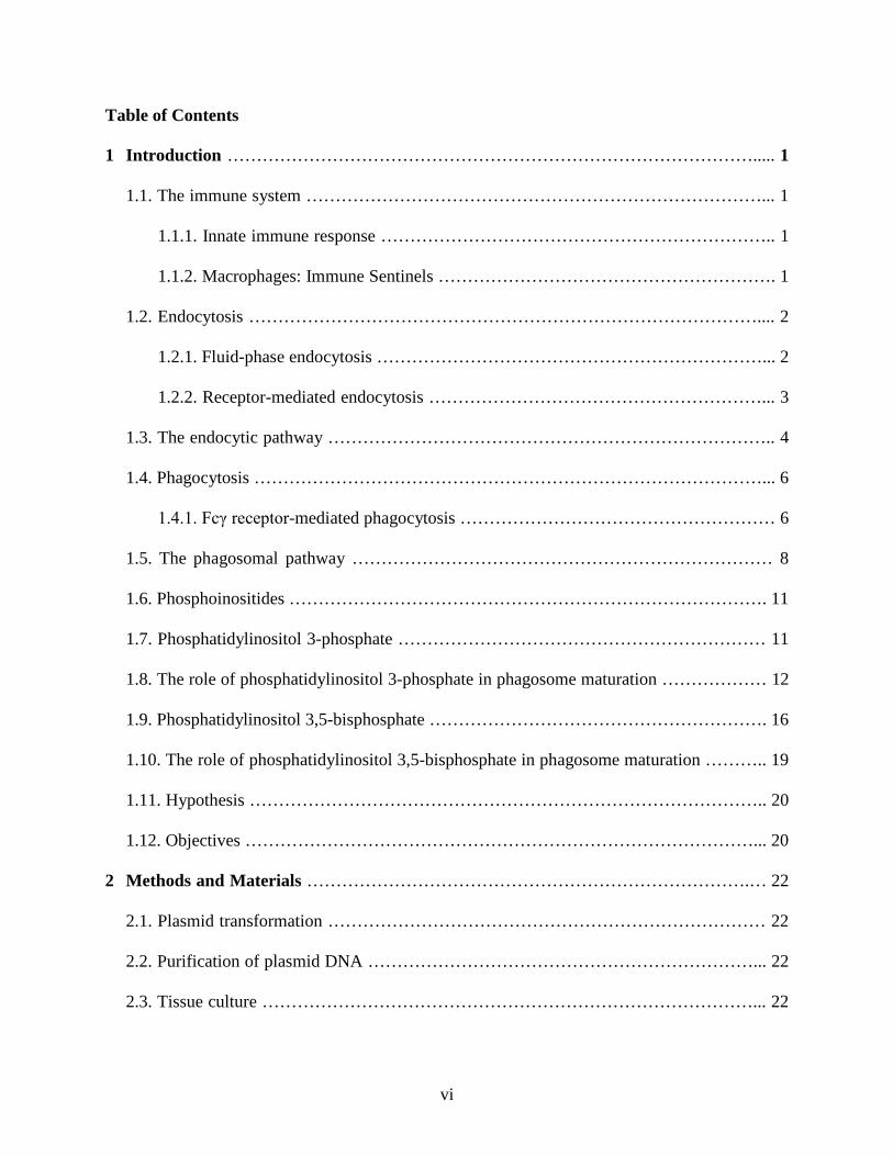
Table of Contents
1 Introduction ………………………………………………………………………………..... 1
1.1. The immune system ……………………………………………………………………... 1
1.1.1. Innate immune response ………………………………………………………….. 1
1.1.2. Macrophages: Immune Sentinels …………………………………………………. 1
1.2. Endocytosis …………………………………………………………………………….... 2
1.2.1. Fluid-phase endocytosis …………………………………………………………... 2
1.2.2. Receptor-mediated endocytosis …………………………………………………... 3
1.3. The endocytic pathway ………………………………………………………………….. 4
1.4. Phagocytosis ……………………………………………………………………………... 6
1.4.1. Fcγ receptor-mediated phagocytosis ……………………………………………… 6
1.5. The phagosomal pathway ……………………………………………………………… 8
1.6. Phosphoinositides ………………………………………………………………………. 11
1.7. Phosphatidylinositol 3-phosphate ……………………………………………………… 11
1.8. The role of phosphatidylinositol 3-phosphate in phagosome maturation ……………… 12
1.9. Phosphatidylinositol 3,5-bisphosphate …………………………………………………. 16
1.10. The role of phosphatidylinositol 3,5-bisphosphate in phagosome maturation ……….. 19
1.11. Hypothesis …………………………………………………………………………….. 20
1.12. Objectives ……………………………………………………………………………... 20
2 Methods and Materials ………………………………………………………………….… 22
2.1. Plasmid transformation ………………………………………………………………… 22
2.2. Purification of plasmid DNA …………………………………………………………... 22
2.3. Tissue culture …………………………………………………………………………... 22

vii
2.4. Pharmacological treatments ……………………………………………………………. 23
2.5. Transient transfection ……………………………………………….………………….. 23
2.6. Endocytosis Assays …………………………………………………………………….. 23
2.7. Bead binding and phagocytosis assays …………………………...……………………. 24
2.8. Lysosome labeling …………………………………….……………………………….. 25
2.9. Proteolysis assays ………………………………….…………………………………… 25
2.10. Immunofluorescence ………………………………………………………………….. 26
2.11. Confocal microscopy …………………………………………………………...…….. 26
2.12. Flow cytometry ……………………………………………………………………….. 27
2.13. Statistical Analyses …………………………………………………………………… 28
3 Results ……………………………………………………………………………………… 29
3.1. PIKfyve inhibition induces enlargement of endolysosomes in macrophages…………... 29
3.2. PIKfyve-inhibited macrophages display decelerated ability to degrade cargo molecules
despite their lysosomes remaining acidic……………………………………………….. 31
3.3. PIKfyve is required for non receptor-mediated fluid-phase endocytosis, but not LDL
endocytosis, one example of receptor-mediated endocytosis…………………...……… 34
3.4. Fluid-phase endocytosis defect observed in PIKfyve-inhibited macrophages does not
appear to be due to a recycling defect in these cells……………………………………. 36
3.5. Aggregated IgG-loaded endosome maturation is impeded in PIKfyve-inhibited
macrophages…………………………………………………………………………….. 37
3.6. An acute inhibition of PIKfyve does not affect phagocytosis by macrophages………... 39
3.7. PtdIns(3)P plays a more dominant role in phagosome maturation than PtdIns(3,5)P2…. 40

viii
4 Discussion …………………………………………………………………………………... 44
4.1. The endosomal and phagosomal systems in macrophages …………………………….. 44
4.2. The identity of the enlarged vacuoles induced by PIKfyve inhibition …….………….. 44
4.3. The role of PtdIns(3,5)P2 in endocytosis ……………………………………………….. 45
4.4. The role of PtdIns(3,5)P2 in endosome maturation …………………………………….. 46
4.5. The role of PtdIns(3,5)P2 in phagocytosis ……………………………………………… 48
4.6. The role of PtdIns(3,5)P2 in phagosome maturation……………………………………. 49
4.7. Concluding remarks …………………………………………………………………… 50
5 Future Studies …………………………………………………………………………….... 51
5.1. Acute versus chronic effects of PIKfyve inhibition …………………………………… 51
5.2. Using digestible cargo molecules to study phagosomal fission and degradation ……… 51
5.3 Verifying the role of PtdIns(3,5)P2 in phagosome maturation using other techniques …. 52
6 References ………………………………………………………………………………….. 53

ix
List of Figures
Figure Title Page
Figure 1.1 The endocytic and phagosomal pathways 9
Figure 1.2 Phosphoinositides 10
Figure 1.3 Distribution and quantification of PI(3)P during phagocytosis 13
Figure 1.4 Effects of wortmannin on phagosomal acquisition of PI(3)P, EEA1, and LAMP1 15
Figure 1.5 Synthesis of PtdIns(3)P and PtdIns(3,5)P2 16
Figure 1.6 Enlarged endolysosomes in kinase-dead PIKfyveK1831E-expressing COS7 and HEK293 cells 18 Figure 3.1 Distribution of endosomal and lysosomal markers in untreated and MF4 (6 h)-treated macrophages 30
Figure 3.2 Lysosome acidification in PIKfyve-inhibited macrophages 31
Figure 3.3 Lysosome degradation in PIKfyve-inhibited macrophages 33 Figure 3.4 Effect of PIKfyve inhibition on fluid-phase endocytosis 35
Figure 3.5 Effect of PIKfyve inhibition on LDL endocytosis, one example of receptor-mediated endocytosis 35
Figure 3.6 Effect of PIKfyve inhibition on FITC-conjugated dextran retention 36 Figure 3.7 Effect of PIKfyve inhibition on trafficking of aggregated IgG immune complexes to lysosomes 38 Figure 3.8 Effect of PIKfyve inhibition on phagocytosis 40 Figure 3.9 Effect of PIKfyve inhibition on phagosomal acquisition of LAMP1 42

x
List of Abbreviations
BSA Bovine serum albumin
CI-MPR Cation-independent mannose 6-phosphate receptor
DMEM Dulbecco's modified Eagle's medium
DNA Deoxyribonucleic acid
EEA1 Early endosome antigen 1
ESCRT Endosomal sorting complexes required for transport
FACS Fluorescence activated cell sorting
FITC Fluorescein isothiocyanate
FYVE Fab1, YOTB, Vac1, and EEA1
GFP Green fluorescent protein
IgG Immunoglobulin G
LAMP1 Lysosomal-associated membrane protein 1
LB Luria-Bertani
LDL Low density lipoprotein
MVB Multivesicular body
PFA Paraformaldehyde
PBS Phosphate buffered saline
PIKfyve FYVE finger-containing phosphoinositide kinase
PI3K Phosphatidylinositol 3-kinase
PtdIns Phosphatidylinositol
PtdInsP Phosphoinositide
PtdIns(3)P Phosphatidylinositol 3-phosphate

xi
PtdIns(3,5)P2 Phosphatidylinositol 3,5-bisphosphate
RBC Red blood cell
TfR Transferrin receptor
TGN Trans-golgi network
TRITC Tetramethylrhodamine isothiocyanate
TRPML1 Mucolipin transient receptor potential protein 1
Vps34 Vacuolar protein sorting-associated protein 34
WIPI1 WD repeat domain containing, phosphoinositide-interacting 1

1
1 Introduction
1.1 The immune system
Our body is protected by the immune system, which has several lines of defense against
infection. The most prominent feature of our immune system is the integumentary system, which
includes the skin and chemicals that are produced by the mucous membrane lining of the gut;
however, when this line of defense is penetrated, our body has an army of specialized cells that
are either part of the innate (nonspecific) or adaptive (specific) immune response that further
guard the body 1.
1.1.1 Innate immune response
Innate immune response is different from specific immune response, in that it is capable
of mounting an immediate defense response. Inflammation typically involves the release of
histamine, prostaglandins, and other “chemical alarm signals” from cells at sites of infection,
causing the nearby blood vessels to dilate and become “leaky” 1.
1.1.2 Macrophages: Immune Sentinels
Our body’s innate immune system is coordinated by a specialized group of professional
phagocytes, among which include the macrophages. Monocytes, which are undifferentiated
macrophages that circulate in the bloodstream, permeate through the walls of the leaky blood
vessels to reach the infection site. There, they differentiate into macrophages that engulf and
digest pathogens 1.
Macrophages also express a proinflammatory cytokine, interleukin-1 (IL-1), which
promotes the expression of adhesion molecules on endothelial cells that line the inner surface of

2
blood vessels. This allows for a more effective migration of leukocytes from the blood to the site
of tissue damage 2. IL-1 also causes fever (an elevation of body temperature), which helps to
enhance the phagocytic activity of leukocytes, thereby suppressing further spread of pathogens 2.
Nonetheless, the fundamental role of macrophages is to rid the body of foreign particles 3. How
do macrophages perform this task? Endocytosis and phagocytosis are common cellular processes
that macrophages employ to protect the body from pathogen invasion.
1.2 Endocytosis
Endocytosis is a process by which cells internalize a diverse range of cargo molecules,
such as fluid, solutes, plasma membrane components, extracellular–matrix components, cellular
debris, bacteria and viruses 4; however, when we discuss about endocytosis, we generally refer to
the cellular uptake of smaller molecules, such as fluid, solutes, and plasma membrane
components- e.g. receptor-ligand complexes.
1.2.1 Fluid-phase endocytosis
There are two distinct types of endocytosis; fluid-phase non-receptor mediated
endocytosis and receptor-mediated endocytosis. During fluid-phase endocytosis, macrophages
constitutively internalize surrounding fluid in a non-specific manner 5. This can occur in a
variety of ways, including clathrin-dependent and -independent endocytosis, and
macropinocytosis 5. Macropinocytosis, for example, is initiated at the ruffled border regions of
the plasma membrane that are supported by actin 6,7. It culminates with membrane ruffle closure,
which leads to the formation of large endocytic vacuoles called macropinosomes filled with a
large volume of extracellular fluid 6. It is also important to note that fluid-phase endocytosis

3
displays bidirectional traffic, in that they are either directed into the endocytic pathway (see
below) or regurgitated back to the plasma membrane. The use of such soluble markers as
horseradish peroxidase (HRP) or fluorescent dextran is common while studying fluid-phase
endocytosis 8.
Macrophages internalize an immense amount of fluid every hour- a volume equivalent to
about 30 % of the cell volume and a fractional area equivalent to about twice the cell surface area
4,9–11. Macrophages generally exhibit a higher endocytic activity than other cell types. For
example, whereas macrophages ingest approximately 3 % of its plasma membrane each minute,
the endocytic rate of fibroblasts is lower, residing at approximately 1 % per minute 12. Thus, it is
worthwhile to take a closer look at how the endosomal system functions in macrophages.
1.2.2 Receptor-mediated endocytosis
Receptor-mediated endocytosis is different from fluid-phase endocytosis, in that it
engages ligand receptors to internalize target molecules. For this reason, while fluid-phase
endocytosis is linearly dependent on marker concentration and incubation time, receptor-
mediated endocytosis reaches its saturation point when all ligand receptors are bound to its
ligand 13. Receptor-mediated endocytosis typically requires the formation of clathrin-coated pits
(CCPs) to form clathrin-coated vesicles (CCVs). Incoming receptor-ligand complexes are
assembled into nascent endocytic vesicles as the plasma membrane invaginates and pinches off
into the cytoplasm 14,15. Depending on the cargo type, some internalized molecules are recycled
back to the plasma membrane, while those that are destined for degradation continue on its path
to the lysosomes, where they are degraded by lysosomal enzymes 16,17. To illustrate, low density
lipoprotein (LDL) is internalized into cells via receptor-mediated endocytosis 15,18. LDL, which

4
carries cholesterol through the bloodstream 19, binds to LDL receptors that are heavily expressed
at sites of CCP formation 20–23. Subsequently, these CCPs pinch off to form CCVs. As CCVs
uncoat within the cell, ligand reptors and target ligands are processed by different routes (see
below) 18,24.
1.3 The endocytic pathway
Once cargo molecules are internalized via endocytosis, those destined for degradation are
transported along the endocytic pathway. There are three main endosomal compartments in the
endocytic pathway; the early and late endosomes and lysosomes 4. Internalized molecules are
first sorted into early endosomes, which has a mildly acidic luminal pH of around 6.2-6.5 25.
Endosome and lysosome acidification is generated by a proton-pumping V-type ATPase 26,
while Na+, K+-ATPase serves to buffer the effect of proton transport, primarily in early
endosomes 27,28. In some cases, the slightly acidic environment of early endosomes facilitates the
uncoupling of ligands from their cognate receptors 29. Dissociated ligands are sorted for
degradation into lysosomes, while their receptors necessary to maintain cellular homeostasis are
recycled back to the plasma membrane for further entry of ligands. Accordingly, early
endosomes are largely categorized into two distinct compartments, the sorting and recycling
endosomes 30.
As the pH is lowered and lysosomal enzymes are added, early endosomes mature to form
late endosomes or multivesicular bodies (MVBs) 4. The sequential recruitment of multiple
endosomal sorting complexes required for transport (ESCRT) mediates the formation of MVBs.
ESCRT-0 binds to ubiquitinated cargo and clusters them to the limiting membranes of early
endosomes. Subsequently, ESCRT-I and –II deform these cargo-rich regions of the early

5
endosomal membrane to form small inward budding vesicles. These vesicles are then cleaved by
ESCRT-III, giving rise to numerous intraluminal vesicles (ILVs) that reside within a larger late
endosomal MVB. The successive fusion of cargo-bearing ILV-containing MVBs with lysosomes
leads to the ultimate degradation of internalized molecules 31.
Lysosomes are the terminal stations in the endocytic pathway. They have a lower internal
pH than early endosomes (ranging from 4.7 to 4.8 in macrophages) 32, in order to activate the
lysosomal enzymes that typically have an acidic optimum pH 33.
Endosome maturation is regulated by the recruitment of various molecular markers that
help to define the identity of organelles. For example, Rab GTPases are molecular switches that
cycle between an active form in the GTP-bound state (catalyzed by a guanine nucleotide
exchange factor; GEF) and an inactive form in the GDP-bound state (stimulated by a cognate
GTPase activating protein; GAP). Rab-GTP is anchored to the cytosolic membrane of a target
organelle via its carboxy-terminal two prenyl groups 34,35. By recruiting and interacting with a
host of effector proteins, Rab GTPases play important roles in a variety of endosomal functions,
such as endosome tethering, fusion, and maturation. To illustrate, the recruitment of a tethering
factor, early endosome antigen 1 (EEA1), by the early endosomal Rab5 in its GTP-bound state is
essential for the homotypic (with other early endosomes) and heterotypic fusion (with incoming
endocytic vesicles) of early endosomes. Moreover, Rab5 replacement with the late endosome-
and lysosome-specific Rab7 is a key feature of endosome maturation 36.
Aside from Rab5 and EEA1, other molecular markers of early endosomes include the
hepatocyte growth factor-regulated tyrosine kinase substrate (Hrs), which is involved in cargo
sorting 37,38, syntaxin 13, which is essential for the recycling of plasma membrane proteins 39,
and PtdIns(3)P, a signaling lipid (see below) 40. LAMP1 41 and V-type ATPase 26 are some of the

6
common molecular markers of late endosomes and lysosomes, whereas cation-independent
mannose 6-phosphate receptor (CI-MPR) accumulation is restricted to trans-Golgi network
(TGN) and late endosomes 42,43.
1.4 Phagocytosis
Ingestion of small cargo molecules occurs through a process called endocytosis; however,
depending on the physical properties of cargo molecules, macrophages engage different modes
of cargo entry. Phagocytosis is a distinct type of endocytosis that involves the ingestion of
particles usually exceeding 0.5 μm in diameter 3,44,45. This process plays an essential role in cell
survival by enabling uptake of nutrients and eliminating cellular debris or infectious pathogens 45.
Phagocytosis is generally classified into two major groups- opsonic and non-opsonic. The latter
involves the engagement of non-opsonin receptors that bind endogenous ligands on particles that
are destined to be internalized. Mannose receptors and dectin1 that bind microbial
polysaccharides are a few examples of such receptors 45. Opsonin receptors, on the other hand,
bind exogenous ligands, or opsonin molecules, that coat the antigen surface. To illustrate,
macrophages recognize and engulf immunoglobulin G (IgG)-opsonized particles by expressing
Fcγ receptors on their cell surface that interact with the Fc portion of IgG 46.
1.4.1 Fcγ receptor-mediated phagocytosis
Fcγ receptor-mediated phagocytosis requires the expression of Fcγ receptors on the
surface of phagocytes and the recognition of IgG-opsonized particles by these receptors.
IgG is an antigen-recognizing antibody that is secreted by plasma B cells. It is a
tetrameric protein complex, consisting of two heavy- and two light chains. The amino terminal

7
ends of these chains form the two variable Fab domains that recognize and bind various antigens.
The one constant Fc domain that resides within the carboxy terminal ends of heavy chains
adheres to Fcγ receptors that are expressed on the immune cell surface 47.
Fcγ receptor is a member of the immunoglobulin receptor superfamily, and there are
currently four known classes of murine Fcγ receptors, namely FcγRI, FcγRII, FcγRIII and the
more recently identified FcγRIV 3,48–50. Each cell type of the immune system is capable of
expressing multiple Fcγ receptor isoforms on its surface, which is an indication that there are
distributional and functional redundancies between these receptors 49. Depending on which cell
type the receptor is localized to and what other receptor isoforms it is found in association with,
there are distinct differences in how phagocytosis is mediated by the host cell.
Proper internalization of IgG-coated particles involves the clustering of multiple Fcγ
receptors that recognize them. A single Fcγ receptor is incapable of maintaining its ligand-bound
state. In order to securely bind a ligand to the cell surface, multiple receptors aggregate to
increase their avidity for the ligand. The rapid lateral movement of the receptors along the
plasma membrane appears to be an important criterion for the initial stages of Fcγ receptor-
mediated phagocytosis 45. Moreover, a more recent study revealed that the probing activity of
actin-rich membrane ruffles and filopodia is required for proper binding of ligand to the Fcγ
receptors 51.
As an aside, it is important to note that the size 52 and shape 53 of the target particles play
inevitable roles in manipulating phagocytic activity. Their rigidity also influences the cells’
phagocytic efficiency 44, further complicating our current understanding of the system.

8
1.5 The phagosomal pathway
Similar to that in the endosomal system, phagocytosis is followed by the successive
transport of internalized particles along the phagosomal pathway. Once internalized, particles
that are destined for degradation reside within a phagosome. This phagosome, which originates
from the plasma membrane, progressively fuses with early and late endosomes, and eventually
with lysosomes to form the phagolysosome. At this stage, the intraphagosomal pH is lowered,
and various lysosomal enzymes get incorporated to complete the digestion of particles. The
sequential fusion of phagosomes with various endocytic organelles depends on the stage of
maturity the phagosome is at (e.g. early phagosomes with early endosomes, and late phagosomes
with late endosomes and so on), and this event allows for the active transport of endosomal
markers to the phagosomal membrane and lumen (Figure 1.1) 45.
It is fundamental to understand phagosome formation and maturation, in that pathogens
have evolved a variety of mechanisms to circumvent their degradation by the host defense
system. Mycobacterium tuberculosis arrest at the early stages of phagosome maturation, while
Salmonella spp. prevent their degradation by blocking Salmonella-containing vacuole (SCV)-
lysosome fusion at the late stages of phagosome maturation. Other species like Listeria and
Shigella interfere with the host phagosomal system by lysing the phagosome membrane and
escaping into the cytosol 45. To address some of the underlying mechanisms of pathogen
invasion, it is worthwhile to investigate how vesicular traffic is operated in macrophages.

9
© BOTELHO, R. J. & GRINSTEIN, S., 2011. Originally published in CURRENT BIOLOGY. doi: 10.1016/j.cub.2011.05.053. Figure 1.1 The endocytic and phagosomal pathways Through a process known as endocytosis, cells internalize small cargo molecules into endosomes. Early endosomes mature and acidify to become late endosomes, and late endosomes are converted to lysosomes as the pH is lowered and acid hydrolase enzymes are added. Macrophages engulf pathogens into phagosomes for degradation through a process known as phagocytosis. Newly formed phagosomes sequentially fuse with early and late endosomes and lysosomes to form phagolysosomes, where pathogens are degraded by hydrolytic enzymes. 45

10
©SASAKI, T. et al., 2009. Originally published in PROGRESS IN LIPID RESEARCH. doi: 10.1016/j.plipres.2009.06.001. Figure 1.2 Phosphoinositides PtdInsPs are phosphorylated derivatives of PtdIns that play important roles in many membrane trafficking events along the endocytic pathway. Some are also required for phagocytosis and phagosome maturation. Their precursor, PtdIns, is a negatively charged phospholipid, whose inositol ring can be phosphorylated on any of the hydroxyl groups of carbons 3, 4, and 5 to form seven PtdInsPs (a-b). Through their restricted distributions, PtdInsPs help to give membranes of different intracellular compartments their distinctive identity by facilitating the recruitment of separate subsets of effector proteins (see below). 54

11
1.6 Phosphoinositides
Phosphoinositides (PtdInsPs) play a critical role in membrane traffic along the endocytic
and phagocytic pathways. Their precursor, phosphatidylinositol (PtdIns), is a negatively charged
phospholipid, whose inositol ring can be phosphorylated on any of the hydroxyl groups of
carbons 3, 4, and 5 to form seven PtdInsPs (Figure 1.2) 54–56.
Through their restricted distributions, PtdInsPs help to give membranes of different
intracellular compartments their distinctive identity by facilitating the recruitment of separate
subsets of effector proteins. Many cytosolic proteins possess lipid-binding domains in them, such
as pleckstrin homology (PH) or phox homology (PX) domains, which allows for their
translocation to the membrane site via their interaction with the PtdInsPs. These effector proteins
then recruit a host of other proteins and elicit an appropriate signaling response that is suited for
that organelle’s purpose 57,58.
1.7 Phosphatidylinositol 3-phosphate
This study focuses on the PtdInsPs, phosphatidylinositol 3-phosphate [PtdIns(3)P] and
phosphatidylinositol 3,5-bisphosphate [PtdIns(3,5)P2] because they confer identity to endosomal
membranes. The sole member of class III PI3K, Vps34, phosphorylates PtdIns on the hydroxyl
group of its third carbon to form PtdIns(3)P 59. PtdIns(3)P ensures early endosomal identity to
those membranes that carry this PtdInsP 60,61. There are numerous lipid-binding domains that
specifically recognize and bind PtdIns(3)P . Among these modules is the FYVE (Fab1, YOTB,
Vac1, and EEA1) domain, which is preserved in a wide variety of proteins including EEA1 57,62.
EEA1 is an early endosomal marker that binds directly to PtdIns(3)P. It is required for mediating
early endosome tethering and fusion to incoming endocytic vesicles 63.

12
1.8 The role of phosphatidylinositol 3-phosphate in phagosome maturation
PtdIns(3)P is an early endosomal PtdInsP. Accordingly, PtdIns(3)P is transiently
detected on the membranes of early phagosomes 40. A construct consisting of two or more FYVE
domains of EEA1 in tandem selectively binds to PtdIns(3)P in vivo 40,60. For this reason, a green
fluorescent protein (GFP) fused to this domain (2FYVE-GFP) serves as a useful probe for
tracking the intracellular localization of PtdIns(3)P 40. Fluorescent signal, which denotes the
presence of PtdIns(3)P, is vividly apparent around the nascent phagosomes; however, as these
phagosomes progressively mature into late phagosomes and subsequently phagolysosomes, this
fluorescence is lost. As such, PtdIns(3)P acquisition is confined to early phagosomes (Figure 1.3)
40.
As confirmed by high-performance liquid chromatography (HPLC) analysis, this
transient accumulation of PtdIns(3)P on early phagosomal membranes is partially accounted for
by its de novo synthesis. Apart from the amount of PtdIns(3)P acquired via early endosome-early
phagosome fusion, PtdIns(3)P is newly synthesized at the early phagosomal membranes. In
support of this finding, it is observed that the fluorescence associated with 2FYVE-GFP that is
detected around the phagosomes is markedly higher (~50% increase) in macrophages that
phagocytosed IgG-opsonized latex beads for 12 min, compared to the basal amount detected
prior to phagocytosis (Figure 1.3) 40.

13
©VIEIRA, O. V. et al., 2001. Originally published in THE JOURNAL OF CELL BIOLOGY. doi: 10.1083/jcb.200107069. Figure 1.3 Distribution and quantification of PI(3)P during phagocytosis RAW macrophages transfected with 2FYVE-GFP (a probe of PtdIns(3)P subcellular distribution) were allowed to phagocytose IgG-opsonized red blood cells (RBCs). The number indicated at the bottom left of each panel represents the time elapsed after addition of the RBCs. Arrows point to phagosomes formed in ~4-5 min interval. DIC image (A). Newly formed phagosomes quickly acquire PtdIns(3)P (B-F). The subsequent disappearance of PtdIns(3)P (G) indicates that PtdIns(3)P is transiently expressed on early phagosomal membranes. Bar, 10 μm. HPLC quantification of PtdIns(3)P in macrophages at basal levels (control) and 12 min after phagocytosis of IgG-opsonized beads (phagocytosis). Some PtdIns(3)P are synthesized de novo during phagocytosis (H). 40

14
Wortmannin, a selective inhibitor of PI3K, is valuable in determining the distinct role of
PtdIns(3)P in phagosome maturation. An early phagosome transition to phagolysosome typically
involves the early phagosomal acquisition of EEA1, followed by the late
phagosomal/phagolysosomal acquisition of LAMP1; however, phagosomes in macrophages
pretreated with wortmannin show severely compromised EEA1 and LAMP1 acquisition. This
implies that PtdIns(3)P, although an early endosomal/phagosomal PtdInsP, is critical for the
complete maturation of early phagosomes to phagolysosomes (Figure 1.4) 40,64.

15
©VIEIRA, O. V. et al., 2001. Originally published in THE JOURNAL OF CELL BIOLOGY. doi: 10.1083/jcb.200107069. Figure 1.4 Effects of wortmannin on phagosomal acquisition of PtdIns(3)P, EEA1, and LAMP1 RAW macrophages, untreated (A, C, and open symbols in E) or pretreated with 100 nM wortmannin for 30 min (B, D, and solid symbols in E), were allowed to phagocytose IgG-opsonized beads for 7 min. The cells were transfected with 2FYVE-GFP and fixed immediately after phagocytosis (A-B) or immunostained for LAMP1 after an additional 30 min chase following phagocytosis (C-D). DIC image (inset). Bar, 10 μm. Effects of wormannin on phagosomal acquisition of EEA1 (triangles) or LAMP1(squares) were quantified after a 7 min phagocytosis pulse (as above), followed by incubation at 37℃ for the indicated chase time points. Macrophages were immunostained with antibodies to EEA1 or LAMP1 (E) 40.

16
PtdIns(3)P expression on early phagosomal membranes is only transient, and it is
reasonable to assume that this PtdInsP is converted to other lipid products over time. There are a
myriad different enzymes, lipid kinases and phosphatases, that make this happen. Members of
the myotubularin family dephosphorylate PtdIns(3)P to form PtdIns 65, whereas FYVE finger-
containing PtdInsP kinase (PIKfyve) promotes the conversion of PtdIns(3)P to PtdIns(3,5)P2 66.
In fact, Hazeki et al. identified the indispensable roles of PTEN and PIKfyve in elimination of
PtdIns(3)P from the early phagosomal membranes 67.
1.9 Phosphatidylinositol 3,5-bisphosphate
©WHITLEY, P. et al., 2009. Originally published in PLANT PHYSIOLOGY. doi: 10.1104/pp.109.146159 Figure 1.5 Synthesis of PtdIns(3)P and PtdIns(3,5)P2 The sole member of class III PI3K, Vps34, phosphorylates PtdIns on the hydroxyl group of its third carbon to form PtdIns(3)P, whereas PIKfyve (the mammalian orthologue of FAB1) promotes the conversion of PtdIns(3)P to PtdIns(3,5)P2 (see next page). 68

17
PIKfyve is a mammalian FYVE finger-containing PtdInsP kinase, which phosphorylates
PtdIns(3)P at the 5-position to form PtdIns(3,5)P2 (Figure 1.5) 66. This multi-domain enzyme has
a conserved N-terminal FYVE domain that specifically binds PtdIns(3)P 69. In conjunction with
PtdIns(3)P, this module is necessary for PIKfyve localization to late endosomes 70. For this
reason, late endosomes are thought to be the principle sites of PtdIns(3,5)P2 accumulation 58.
Consistent with this idea, PIKfyve colocalizes with a marker that cycles between TGN and MVB,
CI-MPR, whereas it is negative for such recycling endosomal markers as transferrin receptor
(TfR) and glucose transporter type IV (GLUT4) 71.
Some of the more prominent morphological changes observed in PIKfyve-inhibited cells
have already been established. To illustrate, PtdIns(3,5)P2 synthesis is important to the
maintenance of endolysosome size. Various cell lines transfected with a mutant form of PIKfyve
that is devoid of its catalytic kinase activity (PIKfyveK1831E mutant) show multiple enlarged
endolysosomes that are absent in the control (Figure 1.6). These endolysosomes that are initially
smaller in size, rounder in shape and cluster at perinuclear regions eventually fuse to grow bigger
in size, smaller in number and spaciously distribute themselves around the whole cytoplasm 72.

18
©IKONOMOV, O. C. et al., 2001. Originally published in THE JOURNAL OF BIOLOGICAL CHEMISTRY. doi: 10.1074/jbc.M101722200 Figure 1.6 Enlarged endolysosomes in kinase-dead PIKfyveK1831E-expressing COS7 and HEK293 cells COS7 cells (a-b, d-e) and HEK293 cells (c and f) were transiently transfected with HA-PIKfyveK1831E or HA-PIKfyveWT. COS7 cells were fixed and immunostained with anti-HA antibody 32 h posttransfection. HEK293 cells were visualized live 48 h posttransfection. Cells transfected with PIKfyveK1831E mutant show multiple enlarged endolysosomes that are absent in HA-PIKfyveWT-expressing cells. Bar, 10 μm. 72
Based on previous findings, there is much evidence to suggest that these endolysosomes
are derived from late endosomes of the membrane trafficking pathway 72; however, there are also
studies that suggest these endolysosomes originate from early endosomes 73,74. More research is

19
needed to unravel some of the lingering uncertainties surrounding the identity of these
endolysosomes.
At present, our knowledge of PtdIns(3,5)P2 effector proteins is quite limited. Nonetheless,
a mammalian homologue of the yeast Svp1, WD repeat domain containing, phosphoinositide-
interacting 1 (WIPI1) protein is among the best characterized 75,76. WIPI1 localizes to trans-
elements of the Golgi and peripheral endosomal membranes to assist in PtdIns(3,5)P2-mediated
endosome-to-TGN retrograde transport 76. Moreover, PIKfyve also interacts with p40, a Rab9
effector protein, that is required for the retrograde transport of CI-MPR from endosomes to the
TGN, further supporting the claim that PIKfyve is involved in this pathway 77.
Aside from these functions, PtdIns(3,5)P2 pathway is selectively activated in hyper-
osmotically stressed 3T3-L1 adipocytes 78. Furthermore, the more recently discovered roles of
PtdIns(3,5)P2 in the regulation GLUT4 exocytosis 79–81, autophagy 82,83, ion transport 84–88, and
transporter proteins like creatine transporter (CreaT) 89, and the excitatory amino acid transporter
4 (EAAT4) 90 further adds to the importance of studying this PtdInsP.
1.10 The role of phosphatidylinositol 3,5-bisphosphate in phagosome maturation
Up to date, the presence of PtdIns(3,5)P2 on phagosomes remains largely unknown 91.
Phagosome maturation involves the transient expression of PtdIns(3)P on early phagosomal
membranes. Subsequently, PtdIns(3)P may be converted to PtdIns(3,5)P2 by the lipid kinase
PIKfyve. Thus, it is unclear if the role of PtdIns(3)P in phagosome maturation is direct and/or
indirect, through the synthesis of PtdIns(3,5)P2. If PtdIns(3,5)P2 has a role to play in phagosome
maturation, then could it be that the role of PtdIns(3)P in phagosome maturation is to provide
PtdIns(3,5)P2? Or does it work in conjunction with PtdIns(3,5)P2?
20
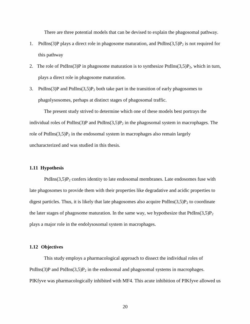
There are three potential models that can be devised to explain the phagosomal pathway.
1. PtdIns(3)P plays a direct role in phagosome maturation, and PtdIns(3,5)P2 is not required for
this pathway
2. The role of PtdIns(3)P in phagosome maturation is to synthesize PtdIns(3,5)P2, which in turn,
plays a direct role in phagosome maturation.
3. PtdIns(3)P and PtdIns(3,5)P2 both take part in the transition of early phagosomes to
phagolysosomes, perhaps at distinct stages of phagosomal traffic.
The present study strived to determine which one of these models best portrays the
individual roles of PtdIns(3)P and PtdIns(3,5)P2 in the phagosomal system in macrophages. The
role of PtdIns(3,5)P2 in the endosomal system in macrophages also remain largely
uncharacterized and was studied in this thesis.
1.11 Hypothesis
PtdIns(3,5)P2 confers identity to late endosomal membranes. Late endosomes fuse with
late phagosomes to provide them with their properties like degradative and acidic properties to
digest particles. Thus, it is likely that late phagosomes also acquire PtdIns(3,5)P2 to coordinate
the later stages of phagosome maturation. In the same way, we hypothesize that PtdIns(3,5)P2
plays a major role in the endolysosomal system in macrophages.
1.12 Objectives
This study employs a pharmacological approach to dissect the individual roles of
PtdIns(3)P and PtdIns(3,5)P2 in the endosomal and phagosomal systems in macrophages.
PIKfyve was pharmacologically inhibited with MF4. This acute inhibition of PIKfyve allowed us

21
to examine the direct roles of PtdIns(3,5)P2 rather than its potential indirect roles due to the
chronic loss of PIKfyve.
To determine the role of PtdIns(3,5)P2 in the endosomal system in macrophages, we first
strived to define the identity of enlarged endolysosomes induced by PIKfyve inhibition. We also
examined the effects of PIKfyve inhibition on endolysosomal properties, focusing on the
degradative capacity and acidic environment of lysosomes. We then characterized the effects of
PIKfyve inhibition on endocytosis (both fluid-phase and receptor-mediated endocytosis),
endosome recycling, and endosome maturation (by examining the rate of trafficking of
aggregated IgG immune complexes to lysosomes).
Our second main objective was to determine the role of PtdIns(3,5)P2 in the phagosomal
system in macrophages. In doing so, we studied the effects of PIKfyve inhibition on
phagocytosis and phagosomal acquisition of LAMP1.

22
2 Methods and Materials
2.1 Plasmid transformation
Plasmid deoxyribonucleic acids (DNAs) used in this study were GFP-2FYVE 40, GFP-
Rab5 92, red fluorescent protein (RFP)-Rab7 93, and LAMP1-mcherry. The plasmids were
transformed into DH5α competent cells [F– Φ80lacZΔM15 Δ(lacZYA-argF) U169 recA1 endA1
hsdR17 (rK–, mK+) phoA supE44 λ– thi-1 gyrA96 relA1] (Invitrogen) for DNA extraction and
purification. One microliter of plasmid DNA was added to 50 μl of DH5α competent cells and
incubated on ice for 30 min. Cells were heat shocked at 42 °C for 45 sec, followed by immediate
incubation on ice for 2 min. One millilitre of Luria-Bertani (LB) media was added to the
transformation reaction and incubated at 37 °C for 1 h. Two hundred microliters of transformed
cells were plated on LB agar plates containing the appropriate antibiotic (100 μg/ml ampicillin or
50 μg/ml kanamycin) and incubated at 37 °C overnight.
2.2 Extraction and Purification of plasmid DNA
A single colony of bacteria (see above) was inoculated in 50 ml of LB medium
containing 100 μg/ml ampicillin or 50 μg/ml kanamycin and incubated at 37 °C for 12-16 h with
shaking. The overnight bacterial culture was pelleted by centrifugation at 5000 g for 10 min at
room temperature. All plasmid DNAs were extracted and purified using E.Z.N.A.® Plasmid
Midiprep Kit (Omega Bio-Tek) as per manufacturer’s instructions.
2.3 Tissue culture
Unless indicated otherwise, RAW264.7 macrophages were cultured in Dulbecco's
modified Eagle's medium (DMEM) (1X) supplemented with high glucose, L-glutamine, sodium
23
pyruvate, and 10% heat-inactivated fetal bovine serum (FBS) (Gibco) at 37 °C with 5 % CO2.
Roswell Park Memorial Institute (RPMI) medium 1640 (1X) supplemented with L-glutamine
and 4-(2-hydroxyethyl)-1-piperazineethanesulfonic acid (HEPES) buffer (HPMI medium)
(Gibco) was used in the absence of CO2 for bead binding and live-cell imaging.
2.4 Pharmacological treatments
Prior to experiment, cells were incubated with MF4 (a gift from Dr. Kevan Shokat,
University of California San Francisco, San Francisco, California, United States) 94 at 37 °C for
2 or 6 h at a final concentration of 100 nM. Preincubation with LY294002 (Promega) was done
on ice for 30 min (during bead binding) at a final concentration of 100 μM. Drug incubation was
kept for the whole duration of the experiment.
2.5 Transient transfection
Prior to transfection, cells were seeded onto coverslips in the wells of a 6 well plate to
reach 50-60 % confluency. Plasmid DNAs were transfected into cultured cells using jetPEI®-
Macrophage (Polyplus transfection) as per manufacturer’s instructions and analyzed 24 h later.
2.6 Endocytosis Assays
Prior to endocytosis, cells were seeded into the wells of a 6-well plate to reach
confluency. After MF4 treatment for 2 h, cells were incubated with 0.1 mg/ml fluorescein
isothiocyanate (FITC)-conjugated dextran (Molecular Probes) at 37 °C for 15 min or 30 min.
Subsequently, cells were washed twice with phosphate buffered saline (PBS) to remove excess
dextran and fixed with 4 % paraformaldehyde (PFA) (MP Biomedicals) at room temperature for

24
20 min. The extent of cargo internalization was assessed using FACScalibur flow cytometer (BD
Biosciences). Likewise, BODIPY FL-conjugated LDL (10 μg/ml; Molecular Probes) was added
to cultured cells and incubated at 37 °C for 15 min or 30 min to assess receptor-mediated
endocytosis.
The uptake of aggregated IgG was assayed by heating 10 mg/ml human IgG (Sigma-
Aldrich) at 62 °C for 20 min, followed by centrifugation at 16,000 g for 10 min at room
temperature to sediment insoluble complexes. Cells were incubated with 1 mg/mL soluble
aggregated IgG at 37 °C for 15 min. Subsequently, cells were washed briefly with PBS to
remove excess aggregated IgG. Cells were fixed with 4 % PFA for 20 min, quenched with 100
mM glycine for 20 min, and permeabilized with 0.5 % Triton X-100 for 10 min. Internalized
aggregated IgG was visualized by labeling the IgG complexes with donkey anti-human Cy2, Cy3,
or Cy5-conjugated antibodies (Cedarlane Laboratories) at 1:1000 dilution for 1 h. All
aforementioned steps were performed at room temperature.
2.7 IgG-opsonized bead binding and phagocytosis assays
Crosslinked poly(styrene/divinylbenzene) (P[S/DVB]) polymer beads (Bangs
Laboratories, Inc.), 3.87 μm in mean diameter, were opsonized with 10 mg/ml human IgG in
PBS at room temperature for 1-2 h rotating. Opsonized beads were washed 3 times in PBS with
centrifugation at 2000 rpm for 1 min between each wash. To synchronize phagocytosis, beads
were bound to cells on ice. Beads were resuspended in PBS and added to the individual wells on
ice for 20 min. Subsequently, cells were washed twice with PBS to remove any unbound beads
and incubated at 37 °C for 15 min or 30 min for phagocytosis. Cells were then fixed with 4 %
PFA for 20 min, quenched with 100 mM glycine for 20 min, and stained with donkey anti-

25
human Cy2, Cy3, or Cy5-conjugated antibodies at 1:1000 dilution for 20 min to discriminate
internalized beads from externally bound beads. At least 100 phagosomes were counted per
condition to determine the phagocytic index and efficiency.
Phagocytic efficiency = number of macrophages with at least one phagosome total number of macrophages
Phagocytic index = number of phagosomestotal number of macrophages
2.8 Lysosome labeling
Lysosomes were labeled with dextran by preloading cells with 2 µM Alexa647-
conjugated dextran (Molecular Probes) at 37 °C for 1 h. Cells were then washed briefly with
PBS to remove excess dextran and incubated in dye-free media at 37 °C for a subsequent chase
period of 2 h. For labeling of lysosomes with Lysotracker Red (Molecular Probes), cells were
incubated with 1 μM Lysotracker Red at 37 °C for 5 min just prior to live-cell imaging.
2.9 Proteolysis assays
To determine the degradative capacity of lysosomes, cells were allowed to co-endocytose
10 µg/ml DQ Green BSA (Molecular Probes) and 0.1 mg/ml tetramethylrhodamine
isothiocyanate (TRITC)-conjugated dextran or 2 µM Alexa647-conjugated dextran at 37 °C for
15 min. Subsequently, cells were given a chase period of 0, 30, or 60 min. Fluorophore-
conjugated dextran was internalized to normalize for the possible difference in endocytosis
between untreated and MF4-treated cells. Cells were then fixed with 4 % PFA for 20 min and
quenched with 100 mM glycine for 20 min for microscopy. The extent of lysosomal proteolysis
was further characterized using flow cytometry by gating to isolate Alexa647-conjugated

26
dextran-positive, endocytosis-competent macrophages, then quantifying the average ratio of the
mean fluorescence of DQ Green BSA over the mean fluorescence of Alexa647-conjugated
dextran for each condition.
2.10 Immunofluorescence
Endosome and phagosome trafficking assays were performed by further incubating the
cells in DMEM (containing drug but free of TRITC-conjugated dextran, DQ Green BSA,
aggregated IgG or beads) at 37 °C for 0, 15, 30, or 60 min after 15 min of cargo internalization at
37 °C. After the chase periods, cells were fixed with 4 % PFA for 20 min, quenched with 100
mM glycine for 20 min, and permeabilized with 0.5 % Triton X-100 for 10 min or -20 °C
methanol for 5 min in the case of LAMP1 staining. Cells were washed 3 times with 0.5 % bovine
serum albumin (BSA)/PBS prior to and after incubation with rat anti-LAMP1[1D4B] antibodies
(Developmental Studies Hybridoma Bank; 1:100 dilution in PBS) at room temperature for 1 h.
This was followed by goat anti-rat antibody (Molecular Probes) staining at 1:1000 dilution in
PBS at room temperature for another hour. Finally, cells were washed and incubated with 0.5 %
BSA/PBS every 5 min for 30 min. Cells were visualized for the presence of any fluorochrome
labeled molecules using confocal microscopy.
2.11 Confocal microscopy and image processing
Cells were visualized on Zeiss LSM 510 META laser scanning microscope. This instrument is
equipped with an Argon laser (458/477/488/514 nm), a HeNe1 laser (543 nm), and a HeNe2
laser (633 nm). Plan-Apochromat 100x/1.40 oil objective was used to collect images. In order to
prevent saturated pixels during image acquisition, detector gain and amplifer offset were

27
adjusted by selecting Palette/Range Indicator. The softwares, Adobe Photoshop (v. 7.0.1, Adobe
Systems Inc.) or ImageJ (v. 1.47 bundled with 64-bit Java) were used to visualize image
collected from each fluorescent channel. Confocal fluorescent images obtained were pseudo-
color processed where necessary in order to better categorize phagosomes as being either
molecular marker-positive, partial, or negative (positive – bright, yellow fluorescent rims; partial
- weak, red fluorescent rims or half-moon-shaped incomplete fluorescent rims; negative – no
fluorescent rims). This was done using ImageJ by enabling “Fire” in Image/Look up Tables.
Colocalization analyses were done using ImageJ with the plugins, JACoP (Just Another
Colocalization Plugin) or Colocalization Indices under the Plugins function. For both plugins,
automated thresholding methods were employed.
2.12 Flow cytometry
Endocytosis of fluorochrome labeled molecules was quantified using BD FACSCalibur
flow cytometer. Samples of suspended cells in solution were run through the flow cytometer
until 10,000 events were collected per condition. Data obtained using flow cytometry was
analyzed using the softwares, Flowjo (TreeStar Inc.) or Flowing Software (Cell Imaging Core,
Turku Centre for Biotechnology, Finland). RAW macrophages were selectively gated using the
forward scatter channel (FSC)/ side scatter channel (SSC) density plot. The intrinsic
autofluorescence background in macrophages was set such that not more than 5 % of
fluorochrome-absent macrophages would be sorted as staining positive. For endocytosis and
endosome recycling assays, the mean fluorescence intensity of the macrophage population was
quantified for each condition. For lysosome proteolysis assay, a two-parameter (dual-colour
fluorescence) histogram was plotted with DQ Green BSA (FITC channel) in the x-axis and

28
Alexa647-conjugated dextran (PE channel) in the y-axis. In Alexa647-conjugated dextran-
positive subset of the macrophage population, the mean fluorescence intensity of DQ Green BSA
over the mean fluorescence intensity of Alexa647-conjugated dextran was quantified for each
condition.
2.13 Statistical Analyses
Significance of differences was calculated in Microsoft Excel using paired samples t tests.
A p value less than 0.05 was considered statistically significant.

29
3 Results
3.1 PIKfyve inhibition induces enlargement of endolysosomes in macrophages
As mentioned previously, PtdIns(3,5)P2 synthesis is important to the maintenance of
endolysosome size 72,95. Up to now, the origin of the dilated endolysosomes has never been
investigated in macrophages. Macrophages generally exhibit a more dynamic membrane
trafficking than other cell types. Accordingly, their endolysosomal trait may be different
compared to that of the previously examined cell lines.
As expected, we observed that macrophages treated with MF4 for 6 h formed multiple
enlarged vacuoles that are absent in untreated cells. To define the identity of these enlarged
vacuoles, we examined the intracellular localizations of various endosomal markers (early
endosomal markers- 2FYVE and Rab5; late endosomal markers- LAMP1 and Rab7) in PIKfyve-
inhibited macrophages using transient transfection of plasmid DNAs.
According to Figure 3.1, we observed that, in macrophages, the enlarged vacuoles
induced by PIKfyve inhibition stained positive for late endosomal markers like LAMP1 and
Rab7, whereas they did not acquire 2FYVE and Rab5, which are common early endosomal
markers. Thus, PtdIns(3,5)P2 synthesis is important to the maintenance of late endosome and
lysosome size, but not for early endosome size in macrophages.

30
Figure 3.1 Distribution of endosomal and lysosomal markers in untreated and MF4 (6 h)-treated macrophages RAW macrophages were transiently transfected with various fluorescence-conjugated endosomal markers (GFP-2FYVE, GFP-Rab5, LAMP1-mcherry, and RFP-Rab7). Subsequently, cells were treated with or without 100 nM MF4 for 6 h. 2FYVE and Rab5 are early endosomal markers. LAMP1 and Rab7 are late endosomal and lysosomal markers. Bar, 10 μm.
GFP
-2FY
VE
GFP
-Rab
5LA
MP
1-m
Che
rry
RFP
-Rab
7untreated MF4-treated

31
3.2 PIKfyve-inhibited macrophages display decelerated ability to degrade cargo molecules despite their lysosomes remaining acidic.
Seeing that the endolysosomes exhibit such a grossly enlarged morphology upon 6 h
MF4 treatment, we further explored whether the degradative capacity and acidic environment of
lysosomes were retained in PIKfyve-inhibited macrophages. To assess lysosomal acidity in
PIKfyve-inhibited macrophages, a fluorescent probe called Lysotracker Red was used. Due to its
fluorophore conjugation to a weak base, Lysotracker Red stains acidic organelles within the cell.
Lysosomes were loaded with FITC-conjugated dextran by 1 h pulse and 2 h chase. Subsequently,
cells were incubated with Lysotracker Red for 5 min just prior to live-cell imaging. Using
Pearson’s correlation coefficient to quantify colocalization of fluorescence derived from FITC-
conjugated dextran, a marker of lysosomes, and Lysotracker Red, we conclude that lysosomes
remain acidic in PIKfyve-inhibited macrophages (r=0.519±0.072; untreated macrophages
r=0.431±0.055) (Figure 3.2).
Figure 3.2 Lysosome acidification in PIKfyve-inhibited macrophages Lysosomes in RAW macrophages, untreated or treated with 100 nM MF4 for 2 h, were loaded with Alexa647-conjugated dextran by 1 h pulse and 2 h chase. Subsequently, cells were incubated with 1 μM Lysotracker Red for 5 min just prior to live-cell imaging. Colocalization of fluorescence derived from Alexa647-conjugated dextran and Lysotracker Red was observed by confocal microscopy. Bar, 10 μm.
untreatedLysotracker
MF4-treatedAlexa647 -Dextran
Merge Lysotracker Alexa647 -Dextran
Merge

32
To assess lysosomal degradation in the absence of PtdIns(3,5)P2 synthesis, macrophages
were allowed to endocytose DQ Green BSA for 15 min. DQ Green BSA proteins, which are
heavily conjugated with BODIPY FL fluorophores, are initially strongly quenched. Proteolytic
digestion of the proteins by lysosomal enzymes causes the release of isolated fluorophores,
which then fluoresce green. In order to ensure that a possible defect in lysosomal proteolytic
activity is not confounded by altered endocytosis in PtdIns(3,5)P2-depleted macrophages,
TRITC-dextran was co-endocytosed with DQ Green BSA. Lysosomal degradation was also
characterized using flow cytometry by quantifying the average ratio of the mean fluorescence of
DQ Green BSA over the mean fluorescence of Alexa647-conjugated dextran in Alexa647-
conjugated dextran-positive subset of macrophages for each condition.

33
Figure 3.3 Lysosome degradation in PIKfyve-inhibited macrophages RAW macrophages, untreated or treated with 100 nM MF4 for 2 h, were allowed to co-endocytose DQ Green BSA and TRITC-conjugated dextran (immunofluorescence) or Alexa647-conjugated dextran (flow cytometry) for 15 min, followed by the indicated chase time points. Dextran was internalized to normalize for the possible differences in fluid phase endocytosis between untreated and MF4-treated macrophages. Bar, 10 μm. (A) The average ratio of the mean fluorescence of DQ Green BSA over the mean fluorescence of Alexa647-conjugated dextran in Alexa647-conjugated dextran-positive subset of macrophages was quantified for each condition using flow cytometry. The data are means ± standard deviation of six experiments (B).
A untreatedTRITC-Dextran
0 m
in30
min
60 m
inMF4-treated
DQ Green BSA Merge TRITC-Dextran DQ Green BSA Merge
B
0
0.5
1
1.5
2
2.5
33.5
3.0
2.5
2.0
1.5
1.0
0.5
0
Lyso
som
al D
egra
datio
n In
dex
Nor
mal
ized
to D
extra
n U
ptak
e untreatedMF4-treated
0 min 30 min 60 min
*

34
Based on our confocal microscopy data, there seemed to be a notable decline in the
activity of lysosomal proteases in PIKfyve-inhibited macrophages. Typically, 30 min chase time
point was sufficient for lysosomal acquisition of degradative properties. We observed that by 30
min chase time point, there was significantly less fluorescence associated with DQ Green BSA in
PIKfyve-inhibited macrophages compared to control (Figure 3.3A). Accordingly, it was evident
that lysosomal degradation index normalized to dextran uptake was reduced in MF4-treated
macrophages at 30 min chase time point compared to that of the untreated group (n=6, p=0.0269);
however, by 60 min chase time point, lysosomal degradation was comparable to control (n=6,
p=0.3139) (Figure 3.3B), suggesting that PIKfyve-inhibited macrophages’ decelerated ability to
degrade cargo molecules is not a permanent defect and is eventually restored over time to reach
normal levels. Therefore, the degradative capacity of lysosomes appears to be retarded in
PtdIns(3,5)P2 synthesis-inhibited macrophages.
3.3 PIKfyve is required for non receptor-mediated fluid-phase endocytosis, but not LDL endocytosis, one example of receptor-mediated endocytosis
Images obtained from our lysosomal degradation assay suggested that there was reduced
TRITC-conjugated dextran uptake in PIKfyve-inhibited macrophages. For this reason, we further
investigated the role of PtdIns(3,5)P2 in fluid-phase endocytosis. Indeed, the notable decline in
the activity of lysosomal proteases in PIKfyve-inhibited macrophages observed by confocal
microscopy was later revealed to have been due to a fluid-phase endocytosis defect in these cells,
which was confirmed using flow cytometry. The uptake of FITC-conjugated dextran was
compromised in those macrophages treated with MF4 for 2 h, as early as 15 min after the onset
of dextran incubation (n=5, p=0.0246), and this effect was even more pronounced with 30 min
pulse (n=5, p=0.0081) (Figure 3.4).
35
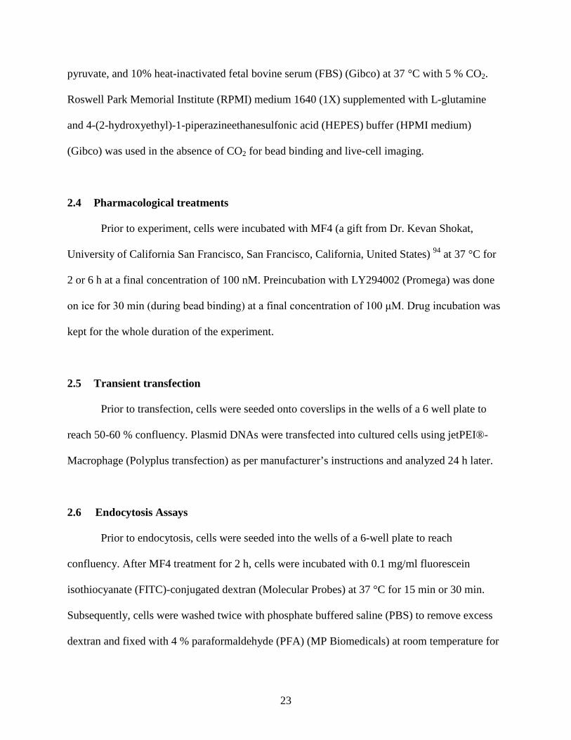
Figure 3.4 Effect of PIKfyve inhibition on fluid-phase endocytosis RAW macrophages, untreated or treated with 100 nM MF4 for 2 h, were allowed to endocytose FITC-conjugated dextran for 15 min or 30 min. The mean fluorescence intensity was quantified for each condition by flow cytometry. The data are means ± standard deviation of five experiments.
As shown above, fluid-phase endocytosis was perturbed by PIKfyve inhibition. To
determine whether or not receptor-mediated endocytosis is also affected by PIKfyve inhibition,
the same endocytosis assay was performed with a receptor-engaging cargo, LDL conjugated to
BODIPY FL fluorophores.
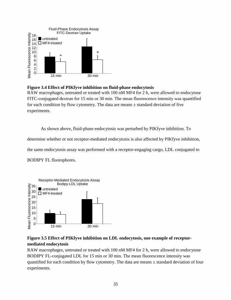
Figure 3.5 Effect of PIKfyve inhibition on LDL endocytosis, one example of receptor-mediated endocytosis RAW macrophages, untreated or treated with 100 nM MF4 for 2 h, were allowed to endocytose BODIPY FL-conjugated LDL for 15 min or 30 min. The mean fluorescence intensity was quantified for each condition by flow cytometry. The data are means ± standard deviation of four experiments.
0
5
15
20
25
30
35
Receptor-Mediated Endocytosis AssayBodipy-LDL Uptake
15 min 30 minMea
n Fl
uore
scen
ce In
tens
ity 353025201510 5 0
untreatedMF4-treated
Fluid-Phase Endocytosis AssayFITC-Dextran Uptake
15 min 30 minMea
n Fl
uore
scen
ce In
tens
ity
untreatedMF4-treated
1816141210 8 6 4 2 0
* *

36
From Figure 3.5, we conclude that PIKfyve activity, unlike its role in fluid-phase
endocytosis, is not necessary for LDL endocytosis, one example of receptor-mediated
endocytosis. The intracellular accumulation of BODIPY FL-conjugated LDL was similar
between untreated and MF4-treated macrophages at both 15 min and 30 min pulse times (n=4 for
both pulse times, p=0.0827; p=0.0513).
3.4 Fluid-phase endocytosis defect observed in PIKfyve-inhibited macrophages does not appear to be due to a recycling defect in these cells
There are two possible ways of interpreting the apparent fluid-phase endocytosis defect in
PIKfyve-inhibited macrophages. The lower accumulation of FITC-conjugated dextran may be a
result of reduced endocytic activity, but it is also possible that there is accelerated efflux of
FITC-conjugated dextran upon PIKfyve inhibition. To address this possibility, macrophages
were allowed to endocytose FITC-conjugated dextran for 15 min, followed by 0, 5, 10, 15, and
20 min chase. The amount of FITC-conjugated dextran internalized by 0 min chase time point
was normalized to 1 for each condition, and the amount recycled was measured using flow
cytometry.

37
Figure 3.6 Effect of PIKfyve inhibition on FITC-conjugated dextran retention RAW macrophages, untreated or treated with 100 nM MF4 for 2 h, were allowed to endocytose FITC-conjugated dextran for 15 min, followed by the indicated chase time points. The mean fluorescence intensity was quantified for each condition by flow cytometry. The data are means ± standard deviation of four experiments.
Our preliminary data suggested that the amount of FITC-conjugated dextran recycled was
not significantly different between untreated and MF4-treated groups (n=4 for all chase time
points; 5 min p=0.3151; 10 min p=0.1064; 15 min p=0.0577; 20 min p=0.0970) (Figure 3.6).
Therefore, the apparent fluid-phase endocytosis defect observed in PIKfyve-inhibited
macrophages is likely not due to an increase in FITC-conjugated dextran efflux. Nonetheless, it
should be noted that this experiment may require additional repetitions to provide a more
conclusive result.
3.5 Aggregated IgG-loaded endosome maturation is impeded in PIKfyve-inhibited macrophages
Endosome maturation and trafficking to lysosomes were assessed by quantifying the
colocalization of endosomal aggregated IgG with LAMP1 staining for late endosomal or
lysosomal compartments. Using untreated macrophages as a model for normal endosomal
0
0.2
0.4
0.6
0.8
1
1.2
0 min 5 min 10 min 15 min 20 min
Mea
n Fl
uore
scen
ce In
tens
ityFITC-Dextran Retention Assay
untreatedMF4-treated
1.2
1.0
0.8
0.6
0.4
0.2
00 min 5 min 10 min 15 min 20 min

38
trafficking, there appeared to be less colocalization between aggregated IgG and LAMP1 in
PIKfyve-inhibited macrophages (Figure 3.7A).
Figure 3.7 Effect of PIKfyve inhibition on trafficking of aggregated IgG immune complexes to lysosomes (A) RAW macrophages, untreated or treated with 100 nM MF4 for 2 h, were allowed to endocytose aggregated IgG for 15 min, followed by the indicated chase time points. Subsequently, cells were fixed and immunostained for LAMP1. Bar, 10 μm. (B) Colocalization of aggregated IgG and LAMP1 was quantified by calculating the normalized mean Pearson’s correlation coefficient for each condition. The data are means ± standard deviation of three experiments, at least ten cells individually analyzed per experiment.
0.000
0.500
1.000
1.500
2.000
2.500
3.000
0min 15mins 30mins 60mins
A untreatedAggregated IgG
15 m
in30
min
60 m
in
MF4-treatedLAMP1 Merge Aggregated IgG LAMP1 Merge
B Aggregated IgG-Loaded Endosome Maturation
untreatedMF4-treated
Pea
rson
Cor
rela
tion
Coe
ffici
ent
Nor
mal
ized
Mea
n
15 min 30 min 60 min0 min
3.0
2.5
2.0
1.5
1.0
0.5
0
*
* *

39
Indeed, there was a notable decline in the rate of aggregated IgG-loaded endosome
maturation in macrophages that are deficient in PtdIns(3,5)P2. Using normalized mean Pearson’s
correlation coefficient as a quantitative measure, we observed that colocalization of aggregated
IgG and LAMP1 was significantly reduced in PIKfyve-inhibited macrophages at 15 min, 30 min,
and 60 min chase time points (n=3, p=0.0194; n=3, p=0.0280; n=2, p=0.0251) (Figure 3.7B) .
These values indicate that aggregated IgG-loaded endosome maturation is impeded in PIKfyve-
inhibited macrophages.
3.6 An acute inhibition of PIKfyve does not affect phagocytosis by macrophages.
Previous studies have focused primarily on the role of PtdIns(3,5)P2 in the regulation of
endosomal membrane traffic. It is now widely accepted that PtdIns(3,5)P2 confers identity to late
endosomal membranes. The expression of PtdIns(3,5)P2 on phagosomes, however, still remains
elusive. Seeing that late endosomes frequently fuse with late phagosomes to provide them their
appropriate endosomal markers and PtdInsPs, we hypothesized that, these late phagosomes
acquire PtdIns(3,5)P2 and coordinate the later stages of phagosome maturation.
Consistent with this hypothesis, our findings on the role of PtdIns(3,5)P2 in the
phagosomal system suggested that even if PtdIns(3,5)P2 was to have an effect on the phagosomal
pathway, it would not intervene at the onset of phagocytosis, but rather, during the later stages of
phagosome maturation (see Section 3.7).

40
Figure 3.8 Effect of PIKfyve inhibition on phagocytosis RAW macrophages, untreated or treated with 100 nM MF4 for 2 h, were allowed to phagocytose IgG-opsonized beads for 15 min or 30 min. Phagocytic index of each condition was calculated, each with at least 100 phagosomes counted. The data are means ± standard deviation of three experiments.
As shown in Figure 3.8, an acute inhibition of PIKfyve did not affect the phagocytic
activity of macrophages, seeing that their phagocytic efficiency (data not shown) and index (n=3
for both pulse times; 15 min p=0.9013; 30 min p=0.9855) remained unperturbed by 2 h MF4
treatment.
3.7 PtdIns(3)P plays a more dominant role in phagosome maturation than PtdIns(3,5)P2. Typically, the conversion of nascent phagosomes into phagolysosomes involves the late
phagosomal/phagolysosomal acquisition of a late endosomal or lysosomal marker, LAMP1.
Previous studies have already demonstrated the requirement for PtdIns(3)P in phagosomal
acquisition of LAMP1 40. This observation was reproduced to, once again, confirm that
PtdIns(3)P is required for the sequential recruitment of various endosomal markers to
phagosomes. In doing so, macrophages were pretreated with LY294002, a selective inhibitor of
PI3K, for 30 min and allowed to phagocytose IgG-opsonized beads for 15 min. As described by
Vieira et al., at 60 min chase time point, when late phagosomal/phagolysosomal acquisition of
Phagocytosis Assay
0
0.5
1
1.5
2
2.5
15 min 30 min
Phag
ocyt
ic In
dex
15 min 30 min
untreatedMF4-treated
2.5
2.0
1.5
1.0
0.5
0

41
LAMP1 peaks, we observed a severe decline in the ability of LAMP1 to associate with
phagosomes in LY294002-treated macrophages compared to control (n=3, LAMP1 positive
p=0.0397) .
On the other hand, most phagosomes acquired LAMP1 in MF4-treated macrophages;
however, the intensity of fluorescence associated with phagosomal acquisition of LAMP1
seemed weaker in these cells. In order to better categorize phagosomes as being either molecular
marker-positive, partial, or negative, confocal images obtained were pseudo-color processed
using ImageJ by enabling “Fire” in Image/Look up Tables. Indeed, at 60 min chase time point,
there were significantly fewer LAMP1 positive phagosomes in PIKfyve-inhibited macrophages
(n=7, p=0.0019). Instead of acquiring bright, yellow fluorescent rims around them (strongly
positive for LAMP1), phagosomes in PIKfyve-inhibited macrophages generally acquired weaker,
red fluorescent rims or half-moon-shaped incomplete fluorescent rims around them, which we
designated as being LAMP1 partial. From this, we conclude that PtdIns(3,5)P2 synthesis
inhibition causes a moderate defect in phagosomal acquisition of LAMP1 (Figure 3.9).

42
Figure 3.9 Effect of PIKfyve inhibition on phagosomal acquisition of LAMP1 (A-B) RAW macrophages, either untreated, treated with 100 nM MF4 for 2 h, or treated with 100 μM LY294002 for 30 min, were allowed to phagocytose IgG-opsonized beads for 15 min, followed by 1 h chase. Subsequently, cells were fixed and immunostained for LAMP1. Images obtained were pseudo-color processed in order to better categorize and quantify phagosomes as being strongly positive, partial, or negative for LAMP1. Bar, 10 μm. The data are means ± standard deviation of at least three experiments per treatment.
ADIC
untre
ated
MF4
-trea
ted
LY29
4002
-trea
ted
LAMP1 Pseudo-color
B
0
10
20
30
40
50
60
70
80
90
LAMP Positive LAMP Partial LAMP Ne gative
90
80
70
60
50
40
30
20
10
0Aver
age
Per
cent
age
of P
hago
som
es
LAMP Positive LAMP Partial LAMP Negative
untreatedMF4-treatedLY294002-treated
*
*
*
*

43
PtdIns(3)P plays an important role in phagosome maturation 40. The role of PtdIns(3,5)P2
in these events, however, seemed to have less of an impact, seeing that its effect was less
pronounced than that of PtdIns(3)P. Nevertheless, PtdIns(3,5)P2 also appears to partake in
regulating the phagosomal pathway.

44
4 Discussion
4.1 The endosomal and phagosomal systems in macrophages
Macrophages protect our body from infection by invading microbes. The engulfment of
foreign pathogens through processes known as endocytosis and phagocytosis is an initial step
toward eliciting an appropriate cellular immune response. These pathogens are ultimately
degraded in the lysosomes and phagolysosomes after a series of endosome and phagosome
maturation processes.
PtdInsPs play essential roles in the traffic of membranes. For example, as described
previously, PtdIns(3)P is transiently expressed on nascent phagosomes, and this event is critical
for the successive maturation of these phagosomes 40. Many microbial pathogens take advantage
of the host endocytic and phagocytic pathways for their successful invasion. To illustrate,
Mycobacterium tuberculosis blocks phagolysosome biogenesis by secreting SapM, a lipid
phosphatase that hydrolyzes PtdIns(3)P, to inhibit phagosome maturation in macrophages 96.
For this reason, it is crucial to understand and appreciate the distinct roles of individual
PtdInsPs (more specifically, PtdIns(3)P and PtdIns(3,5)P2, since they confer identity to early and
late endosomes, respectively) in the endosomal and phagosomal systems in macrophages and
address some implications of exploiting their functions in the host immune system.
4.2 The identity of the enlarged vacuoles induced by PIKfyve inhibition
PtdIns(3,5)P2 synthesis by the lipid kinase activity of PIKfyve is required for the
maintenance of endolysosome size 72,97. PIKfyve mutants devoid of its lipid kinase activity
induce the formation of dilated endolysosomes that are absent in the wild-type. These swollen
endolysosomes are important components of the endocytic pathway because we have evidence to

45
conclude that they are derived from late endosomal compartments. To illustrate, Ikonomov et al.
displayed a substantial overlap between PIKfyveK1831E and CI-MPR around the dilated
endolysosomes in PIKfyveK1831E-expressing COS-7 cells, which implied that these
endolysosomes originated from the membranes of late endosomes and prelysosomal
compartments 72. In contrast, some reports suggest that early endosomes are enlarged in
PIKfyve-inhibited macrophages. To illustrate, fab1 mutant cells (Fab1p is the yeast equivalent of
PIKfyve) show enlarged Hrs-positive early endosomes in Drosophila melanogaster 74. For this
reason, we aimed to characterize the vacuole trait in RAW264.7 macrophages.
In this study, we conclude that PIKfyve inhibition induces the enlargement of
endolysosomes in macrophages (Figure 3.1). This supports the existing assumption that
recycling of late endosomal and prelysosomal membranes may be defected in PIKfyve-inhibited
cells 75.
4.3 The role of PtdIns(3,5)P2 in endocytosis
The role of PtdIns(3,5)P2 in the endosomal system in macrophages is noteworthy, in that
we found that fluid-phase endocytosis is significantly decreased upon PIKfyve-inhibition (Figure
3.4). Ikonomov et al. demonstrated a similar observation, where prelysosomal fluid-phase
endocytosis of HRP is inhibited in HEK293 cell line induced to express PIKfyveK1831E mutant.
Consistent with our observations, the inhibitory effect of PIKfyve inhibition on fluid-phase
endocytosis becomes more pronounced over time 98.
It is interesting to note, however, that LDL endocytosis is unperturbed by PIKfyve
inhibition (Figure 3.5). This suggests that bulk-flow endocytosis and receptor-mediated
endocytosis engage separate driving forces to trigger cargo uptake. This is not suprising, seeing

46
that Ikonomov et al. also observed a similar phenomenon, where prelysosomal fluid-phase
endocytosis of HRP is depressed, but receptor-mediated endocytosis of 125I-labeled transferrin
(125I-Tf) is unaffected by the expression of PIKfyveK1831E mutant in HEK293 cell line 98.
Likewise, the uptake of 125I-labelled EGF (125I-EGF) is also not significantly perturbed in
PIKfyve-suppressed HeLa cells 73.
Recycling of receptor ligands, such as 125I-EGF and 125I-Tf , is also not dependent on
PIKfyve activity 73,98, which corresponds to our finding that the fluid-phase endocytosis defect
observed in PIKfyve-inhibited macrophages does not appear to be a result of the recycling defect
in these cells (Figure 3.6). Although there is seemingly an inhibitory trend associated with the
role of PtdIns(3,5)P2 in recycling of fluid-phase markers, it is statistically insignificant.
Nonetheless, more research is needed to confidently conclude that endosome recycling is
unperturbed in PIKfyve-inhibited macrophages.
4.4 The role of PtdIns(3,5)P2 in endosome maturation
The fluid-phase endocytosis defect in PIKfyve-inhibited macrophages is accompanied by
a decelerated ability of macrophages to degrade cargo molecules (Figure 3.3) despite their
lysosomes remaining acidic (Figure 3.2). It is reasonable to speculate that PtdIns(3,5)P2 is
required for the efficient degradation of cargo molecules, seeing that the activity of lysosomal
hydrolases is significantly delayed upon PIKfyve inhibition. This may be due to the involvement
of PtdIns(3,5)P2 in endosome-to-TGN retrograde membrane traffic 73. The transport of newly
synthesized lysosomal hydrolases from TGN to late endosomes requires the addition of mannose
6-phosphate (M6P) groups to the N-linked oligosaccharides of these enzymes. The M6P
markers are then recognized by transport receptors like CI-MPR that shuttle between TGN and

47
late endosomes 99. For this reason, endosome-to-TGN retrograde transport of CI-MPR is
essential for the subsequent endosomal delivery of lysosomal hydrolases that bind this receptor
as means of their transport. Once in the prelysosomal compartment, these enzymes disengage
from CI-MPR in order to provide an adequate degradative environment for the maturing
prelysosome 100. In PIKfyve-suppressed cells, the steady-state TGN distribution of CI-MPR that
is observed under normal conditions 101, is disrupted, suggesting that the retrograde fission
events may be defected in the absence of PIKfyve activity 94. Likewise, a defect in cargo
trafficking between endosomes and TGN in PIKfyve-inhibited macrophages can lead to a
subsequent defect in endosomal delivery of lysosomal hydrolases, as seen by the reduction in
fluorescence associated with DQ Green BSA in MF4-treated macrophages (Figure 3.3).
Lysosome acidification, however, appears to be normal in PIKfyve-inhibited
macrophages (Figure 3.2). Interestingly, vacuole acidification is proposed to be abated in yeast
lacking Fab1p, the yeast equivalent of PIKfyve 95. These conflicting results may arise from the
presumption that PtdIns(3,5)P2 exhibits different functions in yeast versus higher eukaryotes.
Then again, more recently, my colleague, Shannon Ho, is observing that Fab1p may not be
required for yeast vacuole acidification (personal communication).
We also observe that macrophages perturbed for PIKfyve activity display retarded
trafficking of aggregated IgG immune complexes to lysosomes (Figure 3.7). Perhaps the
requirement for PIKfyve in MVB sorting accounts for this delay in membrane trafficking. Just as
an example, the lipid kinase activity of Fab1p is necessary for sorting type II integral membrane
protein carboxypeptidase S (CPS) into the vacuole lumen via the MVB pathway in yeast 102.
Likewise, PtdIns(3,5)P2 is required for MVB inward vesiculation and storage of internal vesicles
in HEK293 cells 98; however the role of PtdIns(3,5)P2 in MVB sorting for degradation is not as

48
simple as it seems, seeing that the delivery of EGFR to lysosomes and its degradation appear
unaffected by PIKfyveK1831E expression 98. Further investigation is required to address why
PtdIns(3,5)P2 imposes a kinetic delay to the transport of one receptor type and not to the other,
and which key effector proteins PtdIns(3,5)P2 interacts with in order to prompt such an effect.
Inconsistencies may depend on the cell model or method used.
4.5 The role of PtdIns(3,5)P2 in phagocytosis
As mentioned previously, phagocytosis by macrophages is unaffected by an acute
inhibition of PIKfyve (Figure 3.8). This is in accordance with Hazeki et al.’s finding, where the
rate of sheep RBC uptake was unchanged in PIKfyve-deficient cells expressing shRNA against
PIKfyve 67. Typically, 15 min pulse will assess for a possible kinetic defect in phagocytosis,
whereas extending this time interval to 30 min will assess whether or not there is a defect in the
cells’ total phagocytic capacity by looking at saturation conditions. Based on this notion, we
conclude that 2 h of PIKfyve inhibition is insufficient to induce a defect in either the rate or the
capacity of phagocytic activity by macrophages.
Nonetheless, upon prolonged inhibition of PIKfyve, it may not be surprising to predict a
role of PtdIns(3,5)P2 in phagocytosis. PtdIns(3,5)P2 is required for mucolipin transient receptor
potential protein 1 (TRPML1) activation. TRPML1, primarily localized to late endosomes and
lysosomes 103–105, is a Ca2+-permeable cation channel that releases Ca2+ in response to
PtdIns(3,5)P2 increase 88 and regulates membrane trafficking in the late endocytic pathway 106.
Among its many functions, TRPML1 is involved in regulating lysosomal exocytosis to provide
membranes necessary for phagosome biogenesis 107. During phagocytosis, plasmalemmal
pseudopods extend and fuse at the anterior tips of target particles to form nascent phagosomes.

49
This leads to increased membrane tension at the site of cargo uptake 108, and TRPML1, in
response to PtdIns(3,5)P2 elevation, acts to alleviate this tension by delivering lysosomal
membranes to the cell surface, allowing more phagocytosis to occur 107. Likewise, prolonged
PIKfyve inhibition may negatively regulate phagocytic activity by macrophages by preventing
TRPML1 activation, which is fundamental to efficient cargo uptake.
4.6 The role of PtdIns(3,5)P2 in phagosome maturation
Previous work supported by the present study unequivocally demonstrate that PtdIns(3)P
plays an important role in phagosome maturation 40,64. The direct role of PtdIns(3,5)P2 in this
pathway, however, seems less prominent, since LAMP1 appears partially on phagosomes in
PIKfyve-inhibited cells (Figure 3.9).
While it appears that PI3K is more important for phagosome maturation than PIKfyve,
PI3K inhibition deprives PIKfyve of its substrate, PtdIns(3)P; thus, a portion of the defect
induced by LY294002 treatment may be, at least in part, due to impaired PIKfyve function. The
additive effects of the lack of both PtdIns(3)P and PtdIns(3,5)P2 may have caused a more
dramatic effect in PtdIns(3)P-depleted macrophages. In agreement with this notion, the lack of
PtdIns(3,5)P2 also causes a moderate defect in phagosomal acquisition of LAMP1, which leads to
the conclusion that PtdIns(3,5)P2 may also play a contributing role in the phagosomal pathway.
As mentioned previously, PtdIns(3,5)P2 is involved in a variety of endolysosomal
functions along the endocytic pathway. These include such processes as endosome-to-TGN
retrograde transport, MVB sorting, and TRPML1 activation in the endolysosome. TRPML1
releases Ca2+ in response to PtdIns(3,5)P2 increase 88, which in turn triggers a series of
membrane fission and fusion events that is necessary to mediate lysosome-lysosome homotypic

50
fusion 109 and late endosome-lysosome 110 and phagosome-lysosome 111 heterotypic fusions. For
this reason, upon PIKfyve inhibition, the absence of PtdIns(3,5)P2 that is crucial to bind and
activate TRPML1 may decrease the level of juxtaorganellar Ca2+, thereby suppress membrane
fusion activities that are required for phagosome maturation. To illustrate, with the lack of
lysosome-lysosome homotypic fusion and late endosome-lysosome and phagosome-lysosome
heterotypic fusions, proper exchange of essential late endosomal markers between endosomal
and phagosomal compartments may be perturbed, retarding the progression of phagosomes to
phagolysosomes. Likewise, it is reasonable to speculate that PtdIns(3,5)P2 recruits multiple
effector proteins to perform its late phagosomal and phagolysosomal functions.
4.7 Concluding remarks
Endosomal phosphoinositides play inevitable roles in the traffic of membranes.
Consistent with our hypothesis, PtdIns(3,5)P2 appears to principally coordinate the later stages of
endosome and phagosome maturation. PIKfyve inhibition causes massive swelling of late
endosomes/lysosomes. This is accompanied by a decelerated ability of macrophages to degrade
cargo molecules despite their lysosomes remaining acidic. We also observe that macrophages
perturbed for PIKfyve activity have retarded trafficking of aggregated IgG immune complexes to
lysosomes. In the phagosomal system, PIKfyve-inhibited macrophages exhibit normal
phagocytic activity; however, there appears to be a moderate defect in phagosomal acquisition of
LAMP1 during the later stages of the phagosomal pathway. Overall, PtdIns(3,5)P2 plays
substantial roles in the macrophage endosomal and phagosomal systems.

51
5 Future Studies
5.1 Acute versus chronic effects of PIKfyve inhibition
In this study, much of the work focused on exploring the direct responses of macrophages
to PIKfyve inhibition. Two hours of MF4 incubation is sufficient to trigger PIKfyve inhibition;
however, to examine the effects of a prolonged loss of PtdIns(3,5)P2, different approaches are
needed. One way to address this problem is to extend the duration of MF4 pre-treatment, and
thereby inhibit PIKfyve for a longer period of time. Likewise, we can study the chronic effects of
the lack of PtdIns(3,5)P2 by using small interfering RNA (siRNA) to selectively eliminate
PIKfyve. My colleague, Monica Dayam, is currently utilizing these techniques to examine how
our current observations are modulated by prolonged inhibition of PIKfyve.
Our data suggest that prolonged inhibition of PIKfyve has an inhibitory role in
phagocytosis by macrophages. Moreover, its effect on phagosomal acquisition of LAMP1 is
even more profoundly perturbed, supporting the role of PtdIns(3,5)P2 in the phagosomal system
in macrophages (personal communication).
5.2 Using digestible cargo molecules to study phagosomal fission and degradation
One of the most obvious limitations of using an indigestible cargo model like polymer
beads is that it is difficult to observe the full life cycle of an internalized particle. Lysosomal
degradation involves a combination of multiple processes, which include particle digestion,
phagosome fission, fragmentation and disappearance. It will be worthwhile to follow phagosome
breakdown and degradation using a digestible cargo type. We are currently collaborating with
Akriti Prashar and Dr. Mauricio Terebiznik at University of Toronto Scarborough to track these
processes with digestible RBCs by conjugating them to DQ Green BSA.

52
5.3 Verifying the role of PtdIns(3,5)P2 in phagosome maturation using other techniques
Moreover, one caveat in the use of a pharmacological inhibitor MF4 is that PIKfyve may
catalyze the synthesis of two lipid products, PtdIns(3,5)P2 and phosphatidylinositol 5-phosphate
[PtdIns(5)P]. Accordingly, pharmacological inhibition of PIKfyve does not take into
consideration the accompanied synthesis inhibition of PtdIns(5)P. At present, it is unfeasible to
dissociate the effect of PtdIns(3,5)P2 synthesis inhibition from the effect of the lack of PtdIns(5)P
upon PIKfyve inhibition. Although we assume that PtdIns(3,5)P2 synthesis is inhibited upon
MF4 treatment (owing to the fact that we observe dilated endolysosomes in MF4-treated
macrophages, which is an altered phenotype associated with the absence of PtdIns(3,5)P2), it is
possible that some of the findings observed in this study may due to the absence of PtdIns(5)P
rather than PtdIns(3,5)P2. It will be valuable to employ other techniques to verify the role of
PtdIns(3,5)P2 in the endosomal and phagosomal systems in macrophages.

53
6 References
1. Raven, P. H. et al. Biology Eighth Edition. (The McGraw-Hill Companies, Inc., 2008).
2. Contassot, E., Beer, H. D. & French, L. E. Interleukin-1, inflammasomes, autoinflammation and the skin. Swiss medical weekly 142, w13590 (2012).
3. García-García, E. & Rosales, C. Signal transduction during Fc receptor-mediated phagocytosis. Journal of leukocyte biology 72, 1092–108 (2002).
4. Huotari, J. & Helenius, A. Endosome maturation. The EMBO journal 30, 3481–500 (2011).
5. Pavelka, M. & Roth, J. Functional Ultrastructure: Atlas of Tissue Biology and Pathology. (Springer Vienna, 2010).
6. Steinman, R. M. & Swanson, J. The endocytic activity of dendritic cells. The Journal of experimental medicine 182, 283–8 (1995).
7. Welliver, T. P. et al. Ruffles limit diffusion in the plasma membrane during macropinosome formation. Journal of cell science 124, 4106–14 (2011).
8. Oliver, J. M., Berlin, R. D. & Davis, B. H. Use of horseradish peroxidase and fluorescent dextrans to study fluid pinocytosis in leukocytes. Methods in enzymology 108, 336–47 (1984).
9. Besterman, J. M. & Low, R. B. Endocytosis: a review of mechanisms and plasma membrane dynamics. The Biochemical journal 210, 1–13 (1983).
10. Steinman, R. M. et al. Endocytosis and the recycling of plasma membrane. The Journal of cell biology 96, 1–27 (1983).
11. Steinman, R. M., Brodie, S. E. & Cohn, Z. A. Membrane flow during pinocytosis. A stereologic analysis. The Journal of cell biology 68, 665–87 (1976).
12. Alberts, B. et al. Molecular Biology of the Cell Fourth Edition. (Garland Science, 2002)
13. Wiley, H. S. & McKinley, D. N. Assay of growth factor stimulation of fluid-phase endocytosis. Methods in enzymology 146, 402–17 (1987).
14. Brodsky, F. M. et al. Biological basket weaving: formation and function of clathrin-coated vesicles. Annual review of cell and developmental biology 17, 517–68 (2001).
15. Kirchhausen, T. Clathrin. Annual review of biochemistry 69, 699–727 (2000).
16. Helenius, A. et al. Endosomes. Trends in Biochemical Sciences 8, 245–250 (1983).

54
17. Maxfield, F. R. & McGraw, T. E. Endocytic recycling. Nature reviews. Molecular cell biology 5, 121–32 (2004).
18. Jeon, H. & Blacklow, S. C. Structure and physiologic function of the low-density lipoprotein receptor. Annual review of biochemistry 74, 535–62 (2005).
19. Brown, M. S. & Goldstein, J. L. A receptor-mediated pathway for cholesterol homeostasis. Science (New York, N.Y.) 232, 34–47 (1986).
20. Davis, C. G. et al. The J.D. mutation in familial hypercholesterolemia: amino acid substitution in cytoplasmic domain impedes internalization of LDL receptors. Cell 45, 15–24 (1986).
21. Anderson, R. G., Goldstein, J. L. & Brown, M. S. Localization of low density lipoprotein receptors on plasma membrane of normal human fibroblasts and their absence in cells from a familial hypercholesterolemia homozygote. Proceedings of the National Academy of Sciences of the United States of America 73, 2434–8 (1976).
22. Anderson, R. G., Brown, M. S. & Goldstein, J. L. Role of the coated endocytic vesicle in the uptake of receptor-bound low density lipoprotein in human fibroblasts. Cell 10, 351–64 (1977).
23. Anderson, R. G., Goldstein, J. L. & Brown, M. S. A mutation that impairs the ability of lipoprotein receptors to localise in coated pits on the cell surface of human fibroblasts. Nature 270, 695–9
24. Goldstein, J. L. et al. Receptor-mediated endocytosis: concepts emerging from the LDL receptor system. Annual review of cell biology 1, 1–39 (1985).
25. Tjelle, T. E. et al. Isolation and characterization of early endosomes, late endosomes and terminal lysosomes: their role in protein degradation. Journal of cell science 109 ( Pt 1, 2905–14 (1996).
26. Mindell, J. A. Lysosomal acidification mechanisms. Annual review of physiology 74, 69–86 (2012).
27. Fuchs, R., Schmid, S. & Mellman, I. A possible role for Na+,K+-ATPase in regulating ATP-dependent endosome acidification. Proceedings of the National Academy of Sciences of the United States of America 86, 539–43 (1989).
28. Cain, C. C., Sipe, D. M. & Murphy, R. F. Regulation of endocytic pH by the Na+,K+-ATPase in living cells. Proceedings of the National Academy of Sciences of the United States of America 86, 544–8 (1989).

55
29. Geuze, H. J. et al. Intracellular site of asialoglycoprotein receptor-ligand uncoupling: Double-label immunoelectron microscopy during receptor-mediated endocytosis. Cell 32, 277–287 (1983).
30. Ghosh, R. N., Gelman, D. L. & Maxfield, F. R. Quantification of low density lipoprotein and transferrin endocytic sorting HEp2 cells using confocal microscopy. Journal of cell science 107 ( Pt 8, 2177–89 (1994).
31. Wollert, T. & Hurley, J. H. Molecular mechanism of multivesicular body biogenesis by ESCRT complexes. Nature 464, 864–9 (2010).
32. Ohkuma, S. & Poole, B. Fluorescence probe measurement of the intralysosomal pH in living cells and the perturbation of pH by various agents. Proceedings of the National Academy of Sciences of the United States of America 75, 3327–31 (1978).
33. Bond, J. S. & Butler, P. E. Intracellular proteases. Annual review of biochemistry 56, 333–64 (1987).
34. Calero, M. et al. Dual prenylation is required for Rab protein localization and function. Molecular biology of the cell 14, 1852–67 (2003).
35. Behnia, R. & Munro, S. Organelle identity and the signposts for membrane traffic. Nature 438, 597–604 (2005).
36. Rink, J. et al. Rab conversion as a mechanism of progression from early to late endosomes. Cell 122, 735–49 (2005).
37. Komada, M. Hrs, a Tyrosine Kinase Substrate with a Conserved Double Zinc Finger Domain, Is Localized to the Cytoplasmic Surface of Early Endosomes. Journal of Biological Chemistry 272, 20538–20544 (1997).
38. Raiborg, C. et al. Hrs sorts ubiquitinated proteins into clathrin-coated microdomains of early endosomes. Nature cell biology 4, 394–8 (2002).
39. Prekeris, R. et al. Syntaxin 13 mediates cycling of plasma membrane proteins via tubulovesicular recycling endosomes. The Journal of cell biology 143, 957–71 (1998).
40. Vieira, O. V et al. Distinct roles of class I and class III phosphatidylinositol 3-kinases in phagosome formation and maturation. The Journal of Cell Biology 155, 19–26 (2001).
41. Humphries, W. H., Szymanski, C. J. & Payne, C. K. Endo-lysosomal vesicles positive for Rab7 and LAMP1 are terminal vesicles for the transport of dextran. PloS one 6, e26626 (2011).
42. Griffiths, G. et al. The mannose 6-phosphate receptor and the biogenesis of lysosomes. Cell 52, 329–41 (1988).
56
43. Geuze, H. J. et al. Possible pathways for lysosomal enzyme delivery. The Journal of cell biology 101, 2253–62 (1985).
44. Beningo, K. A. & Wang, Y. Fc-receptor-mediated phagocytosis is regulated by mechanical properties of the target. Journal of Cell Science 115, 849–856 (2002).
45. Botelho, R. J. & Grinstein, S. Phagocytosis. Current biology : CB 21, R533–8 (2011).
46. Wines, B. D. et al. The IgG Fc contains distinct Fc receptor (FcR) binding sites: the leukocyte receptors Fc gamma RI and Fc gamma RIIa bind to a region in the Fc distinct from that recognized by neonatal FcR and protein A. Journal of immunology (Baltimore, Md. : 1950) 164, 5313–8 (2000).
47. Schroeder, H. W. & Cavacini, L. Structure and function of immunoglobulins. The Journal of allergy and clinical immunology 125, S41–52 (2010).
48. Van de Winkel, J. G. & Anderson, C. L. Biology of human immunoglobulin G Fc receptors. Journal of leukocyte biology 49, 511–24 (1991).
49. Sánchez-Mejorada, G. & Rosales, C. Signal transduction by immunoglobulin Fc receptors. Journal of leukocyte biology 63, 521–33 (1998).
50. Nimmerjahn, F. & Ravetch, J. V. Divergent immunoglobulin g subclass activity through selective Fc receptor binding. Science (New York, N.Y.) 310, 1510–2 (2005).
51. Flannagan, R. S. et al. Dynamic macrophage “probing” is required for the efficient capture of phagocytic targets. The Journal of cell biology 191, 1205–18 (2010).
52. Cannon, G. J. & Swanson, J. A. The macrophage capacity for phagocytosis. Journal of cell science 101 ( Pt 4, 907–13 (1992).
53. Donaldson, K. et al. Asbestos-stimulated tumour necrosis factor release from alveolar macrophages depends on fibre length and opsonization. The Journal of pathology 168, 243–8 (1992).
54. Sasaki, T. et al. Mammalian phosphoinositide kinases and phosphatases. Progress in Lipid Research 48, 307–343 (2009).
55. Sasaki, T. et al. The physiology of phosphoinositides. Biological & pharmaceutical bulletin 30, 1599–604 (2007).
56. Clague, M. J., Urbé, S. & de Lartigue, J. Phosphoinositides and the endocytic pathway. Experimental cell research 315, 1627–31 (2009).
57. Le Roy, C. & Wrana, J. L. Clathrin- and non-clathrin-mediated endocytic regulation of cell signalling. Nature reviews. Molecular cell biology 6, 112–26 (2005).

57
58. Di Paolo, G. & De Camilli, P. Phosphoinositides in cell regulation and membrane dynamics. Nature 443, 651–7 (2006).
59. Foster, F. M. et al. The phosphoinositide (PI) 3-kinase family. Journal of cell science 116, 3037–40 (2003).
60. Gillooly, D. J. et al. Localization of phosphatidylinositol 3-phosphate in yeast and mammalian cells. The EMBO journal 19, 4577–88 (2000).
61. Gillooly, D. J., Raiborg, C. & Stenmark, H. Phosphatidylinositol 3-phosphate is found in microdomains of early endosomes. Histochemistry and cell biology 120, 445–53 (2003).
62. Gaullier, J. M. et al. FYVE fingers bind PtdIns(3)P. Nature 394, 432–3 (1998).
63. Simonsen, A. et al. EEA1 links PI(3)K function to Rab5 regulation of endosome fusion. Nature 394, 494–8 (1998).
64. Scott, C. C. et al. Role of 3-phosphoinositides in the maturation of Salmonella-containing vacuoles within host cells. The Journal of biological chemistry 277, 12770–6 (2002).
65. Wishart, M. J. & Dixon, J. E. PTEN and myotubularin phosphatases: from 3-phosphoinositide dephosphorylation to disease. Trends in cell biology 12, 579–85 (2002).
66. Sbrissa, D. PIKfyve, a Mammalian Ortholog of Yeast Fab1p Lipid Kinase, Synthesizes 5-Phosphoinositides. EFFECT OF INSULIN. Journal of Biological Chemistry 274, 21589–97 (1999).
67. Hazeki, K. et al. Essential roles of PIKfyve and PTEN on phagosomal phosphatidylinositol 3-phosphate dynamics. FEBS letters 586, 4010–5 (2012).
68. Whitley, P., Hinz, S. & Doughty, J. Arabidopsis FAB1/PIKfyve proteins are essential for development of viable pollen. Plant physiology 151, 1812–22 (2009).
69. Shisheva, A., Sbrissa, D. & Ikonomov, O. Cloning, Characterization, and Expression of a Novel Zn2+-Binding FYVE Finger-Containing Phosphoinositide Kinase in Insulin-Sensitive Cells. Mol. Cell. Biol. 19, 623–634 (1999).
70. Sbrissa, D., Ikonomov, O. C. & Shisheva, A. Phosphatidylinositol 3-phosphate-interacting domains in PIKfyve. Binding specificity and role in PIKfyve. Endomenbrane localization. The Journal of biological chemistry 277, 6073–9 (2002).
71. Shisheva, A. et al. Localization and insulin-regulated relocation of phosphoinositide 5-kinase PIKfyve in 3T3-L1 adipocytes. The Journal of biological chemistry 276, 11859–69 (2001).

58
72. Ikonomov, O. C., Sbrissa, D. & Shisheva, A. Mammalian cell morphology and endocytic membrane homeostasis require enzymatically active phosphoinositide 5-kinase PIKfyve. The Journal of biological chemistry 276, 26141–7 (2001).
73. Rutherford, A. C. et al. The mammalian phosphatidylinositol 3-phosphate 5-kinase (PIKfyve) regulates endosome-to-TGN retrograde transport. Journal of cell science 119, 3944–57 (2006).
74. Rusten, T. E. et al. Fab1 Phosphatidylinositol 3-Phosphate 5-Kinase Controls Trafficking but Not Silencing of Endocytosed Receptors. 17, 3989–4001 (2006).
75. Dove, S. K. et al. Svp1p defines a family of phosphatidylinositol 3,5-bisphosphate effectors. The EMBO journal 23, 1922–33 (2004).
76. Jeffries, T. R. et al. PtdIns-specific MPR pathway association of a novel WD40 repeat protein, WIPI49. Molecular biology of the cell 15, 2652–63 (2004).
77. Ikonomov, O. C. et al. Active PIKfyve associates with and promotes the membrane attachment of the late endosome-to-trans-Golgi network transport factor Rab9 effector p40. The Journal of biological chemistry 278, 50863–71 (2003).
78. Sbrissa, D. & Shisheva, A. Acquisition of unprecedented phosphatidylinositol 3,5-bisphosphate rise in hyperosmotically stressed 3T3-L1 adipocytes, mediated by ArPIKfyve-PIKfyve pathway. The Journal of biological chemistry 280, 7883–9 (2005).
79. Watson, R. T. & Pessin, J. E. Bridging the GAP between insulin signaling and GLUT4 translocation. Trends in biochemical sciences 31, 215–22 (2006).
80. Ikonomov, O. C., Sbrissa, D., Dondapati, R. & Shisheva, A. ArPIKfyve-PIKfyve interaction and role in insulin-regulated GLUT4 translocation and glucose transport in 3T3-L1 adipocytes. Experimental cell research 313, 2404–16 (2007).
81. Ikonomov, O. C., Sbrissa, D. & Shisheva, A. YM201636, an inhibitor of retroviral budding and PIKfyve-catalyzed PtdIns(3,5)P2 synthesis, halts glucose entry by insulin in adipocytes. Biochemical and biophysical research communications 382, 566–70 (2009).
82. Ferguson, C. J., Lenk, G. M. & Meisler, M. H. PtdIns ( 3 , 5 ) P 2 and autophagy in mouse models of neurodegeneration. Autophagy 6, 170–171 (2010).
83. Martin, S. et al. Inhibition of PIKfyve by YM-201636 dysregulates autophagy and leads to apoptosis-independent neuronal cell death. PloS one 8, e60152 (2013).
84. Gehring, E.-M. et al. PIKfyve upregulates CFTR activity. Biochemical and biophysical research communications 390, 952–7 (2009).

59
85. Klaus, F. et al. PIKfyve-dependent regulation of the Cl- channel ClC-2. Biochemical and biophysical research communications 381, 407–11 (2009).
86. Touchberry, C. D. et al. Phosphatidylinositol 3,5-bisphosphate (PI(3,5)P2) potentiates cardiac contractility via activation of the ryanodine receptor. The Journal of biological chemistry 285, 40312–21 (2010).
87. Tsuruta, F. et al. PIKfyve regulates CaV1.2 degradation and prevents excitotoxic cell death. The Journal of cell biology 187, 279–94 (2009).
88. Dong, X. et al. PI(3,5)P(2) controls membrane trafficking by direct activation of mucolipin Ca(2+) release channels in the endolysosome. Nature communications 1, 38 (2010).
89. Strutz-Seebohm, N. et al. PIKfyve in the SGK1 mediated regulation of the creatine transporter SLC6A8. Cellular physiology and biochemistry : international journal of experimental cellular physiology, biochemistry, and pharmacology 20, 729–34 (2007).
90. Alesutan, I. S. et al. Regulation of the glutamate transporter EAAT4 by PIKfyve. Cellular physiology and biochemistry : international journal of experimental cellular physiology, biochemistry, and pharmacology 25, 187–94 (2010).
91. Steinberg, B. E. & Grinstein, S. Pathogen destruction versus intracellular survival: the role of lipids as phagosomal fate determinants. The Journal of clinical investigation 118, 2002–11 (2008).
92. Vieira, O. V et al. Modulation of Rab5 and Rab7 Recruitment to Phagosomes by Phosphatidylinositol 3-Kinase. Molecular and Cellular Biology 23, 2501–2514 (2003).
93. Huynh, K. K., Gershenzon, E. & Grinstein, S. Cholesterol accumulation by macrophages impairs phagosome maturation. The Journal of biological chemistry 283, 35745–55 (2008).
94. De Lartigue, J. et al. PIKfyve Regulation of Endosome-Linked Pathways. Traffic 10, 883–893 (2009).
95. Gary, J. D. et al. Fab1p is essential for PtdIns(3)P 5-kinase activity and the maintenance of vacuolar size and membrane homeostasis. The Journal of cell biology 143, 65–79 (1998).
96. Vergne, I. et al. Mechanism of phagolysosome biogenesis block by viable Mycobacterium tuberculosis. Proceedings of the National Academy of Sciences of the United States of America 102, 4033–4038 (2005).

60
97. Ikonomov, O. C. et al. Functional dissection of lipid and protein kinase signals of PIKfyve reveals the role of PtdIns 3,5-P2 production for endomembrane integrity. The Journal of biological chemistry 277, 9206–11 (2002).
98. Ikonomov, O. C. et al. PIKfyve Controls Fluid Phase Endocytosis but Not Recycling / Degradation of Endocytosed Receptors or Sorting of Procathepsin D by Regulating Multivesicular Body Morphogenesis. 14, 4581–4591 (2003).
99. Alberts B. et al. Molecular Biology of the Cell Fourth Edition. Molecular Biology of the Cell. 4th edition. (Garland Science, 2002).
100. Dahms, N. M., Lobel, P. & Kornfeld, S. Mannose 6-phosphate receptors and lysosomal enzyme targeting. The Journal of biological chemistry 264, 12115–8 (1989).
101. Lin, S. X. et al. Endocytosed cation-independent mannose 6-phosphate receptor traffics via the endocytic recycling compartment en route to the trans-Golgi network and a subpopulation of late endosomes. Molecular biology of the cell 15, 721–33 (2004).
102. Odorizzi, G., Babst, M. & Emr, S. D. Fab1p PtdIns(3)P 5-kinase function essential for protein sorting in the multivesicular body. Cell 95, 847–58 (1998).
103. Thompson, E. G. et al. Lysosomal trafficking functions of mucolipin-1 in murine macrophages. BMC cell biology 8, 54 (2007).
104. Pryor, P. R. et al. Mucolipin-1 is a lysosomal membrane protein required for intracellular lactosylceramide traffic. Traffic (Copenhagen, Denmark) 7, 1388–98 (2006).
105. Vergarajauregui, S. & Puertollano, R. Two di-leucine motifs regulate trafficking of mucolipin-1 to lysosomes. Traffic (Copenhagen, Denmark) 7, 337–53 (2006).
106. Cheng, X. et al. Mucolipins: Intracellular TRPML1-3 channels. FEBS letters 584, 2013–21 (2010).
107. Samie, M. et al. A TRP Channel in the Lysosome Regulates Large Particle Phagocytosis via Focal Exocytosis. Developmental Cell (2013). doi:10.1016/j.devcel.2013.08.003
108. Masters, T. A. et al. Plasma membrane tension orchestrates membrane trafficking, cytoskeletal remodeling, and biochemical signaling during phagocytosis. Proceedings of the National Academy of Sciences of the United States of America 110, 11875–80 (2013).
109. Bakker, A. C. et al. Homotypic fusion between aggregated lysosomes triggered by elevated [Ca2+]i in fibroblasts. Journal of cell science 110 ( Pt 1, 2227–38 (1997).
110. Pryor, P. R. et al. The role of intraorganellar Ca(2+) in late endosome-lysosome heterotypic fusion and in the reformation of lysosomes from hybrid organelles. The Journal of cell biology 149, 1053–62 (2000).

61
111. Jaconi, M. E. et al. Cytosolic free calcium elevation mediates the phagosome-lysosome fusion during phagocytosis in human neutrophils. The Journal of cell biology 110, 1555–64 (1990).