
The role of oocyte maturation in the ontogeny of the fertilization site in the hydrozoan Hydractinia...
10
Roux's Arch Dev Biol (1987) 196:83 92 Roux's Archives of D.evelopmental Biology Springer-Verlag 1987 The role of ooeyte maturation in the ontogeny of the fertilization site in the hydrozoan Hydractinia echinata Gary Freeman Marine Biological Laboratory, Woods Hole, Mass. and Center for Developmental Biology, Department of Zoology, University of Texas, Austin, Texas, 78712, USA Summary. In hydrozoans the sperm will fuse with the egg only at the site of polar body formation. The primary oo- cyte and maturing oocytes which have produced the first polar body cannot be fertilized even though maturing oo- cytes which have produced the first polar body attract sperm. These eggs do not acquire the ability to be fertilized until after second polar body formation. If either first or second polar body formation is inhibited or if first and second polar body formation do not take place in close proximity to each other, the fertilization site is not set up. Under normal circumstances the site of polar body forma- tion takes place at the region on the maturing oocyte sur- face nearest the site where the germinal vesicle resided in the primary oocyte. When maturing oocytes are centrifuged prior to polar body formation, the site of polar body forma- tion is frequently shifted so that it does not correspond to the site where it would be given off under normal circum- stances. Under these conditions the shifted site of polar body formation is the only site where the egg can be ferti- lized, indicating that the fertilization site is selected during oocyte maturation. Oocyte maturation in these hydrozoans is mediated by a hormone released by the somatic cells of gonophores as a consequence of bringing dark adapted gonophores into the light. The hormone acts directly on the oocyte to induce maturation. The oocyte only has to be exposed to the hor- mone for the first few minutes of the maturation process in order to complete the process of maturation. Key words: Hydrozoan - Fertilization site formation - Role of polar bodies Fertilization occurs in most animals by fusion of a sperm with an egg at any point on the egg's surface. Observations on fertilization in hydrozoans indicate that the sperm fuses with the egg at the site of polar body formation (see Free- man and Miller 1982 for a review of the literature). Experi- ments in which unfertilized eggs are cut into halves, so that the only difference between the halves is the presence or absence of the site of polar body formation, have demon- strated that the half with the site of polar body formation is the only half that can be fertilized (Freeman and Miller Dedicated to Professor N.H. Verdonk of the Rijksuniversiteit Utrecht on his 65th birthday 1982; Honegger 1983). On this basis one can infer that these eggs can only be fertilized at the site of polar body formation. In several species of hydrozoans the site of polar body formation can be distinguished from other parts of the egg surface because of its morphology or ability to bind lectins. In Hydra carnea, Hydractinia echinata and Pelmatohydra robusta there is an indentation of the cell membrane, or fertilization pit, in the egg at the site where the gametes will fuse (Freeman and Miller 1982; Honegger 1981; Noda and Kanai 1981). However, this is not a universal feature of all hydrozoan eggs (Carr6 and Sardet 1981; Freeman and Miller 1982). Miller (1981) has demonstrated that con- conavalin A binds to Orthopyxsis compressa eggs only at the site of polar body formation. This report will establish when the fertilization site is set up and try to define some of the factors that play a role in its genesis. Hydractinia has been selected for this work because it has small gonads in which individual oo- cytes can be identified and because oocyte maturation and spawning occur in a very predictable manner in response to environmental light cues. This report is divided into three parts. In part one, the process of oocyte maturation is de- scribed and a procedure is presented that can be used to induce maturation in isolated oocytes. Part two establishes the stage during oocyte maturation when eggs can first be fertilized. This has been done by exposing maturing oocytes to sperm for a short period of time and then treating them with detergent to kill any sperm that have not fused with the eggs. Mature Hydractinia eggs attract sperm (Miller 1974). This preparation has also been used to find out when during maturation eggs first attract sperm. Part three exam- ines the conditions necessary for establishing the site of fertilization. Centrifugation experiments that have been done on mature eggs indicate that the female pronucleus does not have to be at the fertilization site in order for fertilization to occur and its presence in an egg fragment that does not contain the fertilization site will not give this fragment the ability to be fertilized (Freeman and Miller 1982). However, at some time prior to or during maturation the germinal vesicle or meiotic apparatus must play a role in establishing the fertilization site because it forms where the polar bodies are given off. This relationship has been analyzed by moving the germinal vesicle or the meiotic ap- paratus to new positions in the oocyte by centrifuging gono- phores prior to and at different times during oocyte matura- tion. This part of the report also examines the effect of
-
Upload
gary-freeman -
Category
Documents
-
view
213 -
download
0
Transcript of The role of oocyte maturation in the ontogeny of the fertilization site in the hydrozoan Hydractinia...

Roux's Arch Dev Biol (1987) 196:83 92 Roux's Archives of
D.evelopmental Biology �9 Springer-Verlag 1987
The role of ooeyte maturation in the ontogeny of the fertilization site in the hydrozoan Hydractinia echinata Gary Freeman Marine Biological Laboratory, Woods Hole, Mass. and Center for Developmental Biology, Department of Zoology, University of Texas, Austin, Texas, 78712, USA
Summary. In hydrozoans the sperm will fuse with the egg only at the site of polar body formation. The primary oo- cyte and maturing oocytes which have produced the first polar body cannot be fertilized even though maturing oo- cytes which have produced the first polar body attract sperm. These eggs do not acquire the ability to be fertilized until after second polar body formation. If either first or second polar body formation is inhibited or if first and second polar body formation do not take place in close proximity to each other, the fertilization site is not set up. Under normal circumstances the site of polar body forma- tion takes place at the region on the maturing oocyte sur- face nearest the site where the germinal vesicle resided in the primary oocyte. When maturing oocytes are centrifuged prior to polar body formation, the site of polar body forma- tion is frequently shifted so that it does not correspond to the site where it would be given off under normal circum- stances. Under these conditions the shifted site of polar body formation is the only site where the egg can be ferti- lized, indicating that the fertilization site is selected during oocyte maturation.
Oocyte maturation in these hydrozoans is mediated by a hormone released by the somatic cells of gonophores as a consequence of bringing dark adapted gonophores into the light. The hormone acts directly on the oocyte to induce maturation. The oocyte only has to be exposed to the hor- mone for the first few minutes of the maturation process in order to complete the process of maturation.
Key words: Hydrozoan - Fertilization site formation - Role of polar bodies
Fertilization occurs in most animals by fusion of a sperm with an egg at any point on the egg's surface. Observations on fertilization in hydrozoans indicate that the sperm fuses with the egg at the site of polar body formation (see Free- man and Miller 1982 for a review of the literature). Experi- ments in which unfertilized eggs are cut into halves, so that the only difference between the halves is the presence or absence of the site of polar body formation, have demon- strated that the half with the site of polar body formation is the only half that can be fertilized (Freeman and Miller
Dedicated to Professor N.H. Verdonk of the Rijksuniversiteit Utrecht on his 65th birthday
1982; Honegger 1983). On this basis one can infer that these eggs can only be fertilized at the site of polar body formation.
In several species of hydrozoans the site of polar body formation can be distinguished from other parts of the egg surface because of its morphology or ability to bind lectins. In Hydra carnea, Hydractinia echinata and Pelmatohydra robusta there is an indentation of the cell membrane, or fertilization pit, in the egg at the site where the gametes will fuse (Freeman and Miller 1982; Honegger 1981; Noda and Kanai 1981). However, this is not a universal feature of all hydrozoan eggs (Carr6 and Sardet 1981; Freeman and Miller 1982). Miller (1981) has demonstrated that con- conavalin A binds to Orthopyxsis compressa eggs only at the site of polar body formation.
This report will establish when the fertilization site is set up and try to define some of the factors that play a role in its genesis. Hydractinia has been selected for this work because it has small gonads in which individual oo- cytes can be identified and because oocyte maturation and spawning occur in a very predictable manner in response to environmental light cues. This report is divided into three parts. In part one, the process of oocyte maturation is de- scribed and a procedure is presented that can be used to induce maturation in isolated oocytes. Part two establishes the stage during oocyte maturation when eggs can first be fertilized. This has been done by exposing maturing oocytes to sperm for a short period of time and then treating them with detergent to kill any sperm that have not fused with the eggs. Mature Hydractinia eggs attract sperm (Miller 1974). This preparation has also been used to find out when during maturation eggs first attract sperm. Part three exam- ines the conditions necessary for establishing the site of fertilization. Centrifugation experiments that have been done on mature eggs indicate that the female pronucleus does not have to be at the fertilization site in order for fertilization to occur and its presence in an egg fragment that does not contain the fertilization site will not give this fragment the ability to be fertilized (Freeman and Miller 1982). However, at some time prior to or during maturation the germinal vesicle or meiotic apparatus must play a role in establishing the fertilization site because it forms where the polar bodies are given off. This relationship has been analyzed by moving the germinal vesicle or the meiotic ap- paratus to new positions in the oocyte by centrifuging gono- phores prior to and at different times during oocyte matura- tion. This part of the report also examines the effect of

84
treatments that block polar body formation on the estab- lishment of the fertilization site. An abstract describing some of this work has been published (Freeman 1982).
Materials and methods
Control of spawning. Hydractinia colonies were maintained in a sea table or in a recirculatung sea water aquarium under conditions of constant illumination. These colonies were on shells utilized by hermit crabs. The colonies were fed by opening up clams 2 or 3 times a week. Ripe gono- phores were dissected off colonies prior to use. The process of dissecting gonophores off colonies will sometimes cause a small proportion of the isolated gonophores to spawn. Spawning normally occurs within 90 rain after the dissec- tion. Spawning was induced by placing gonophores in the dark for 3~4 h. They were then brought back into the light. Male gonophores usually spawned about 50 min after they were brought into the light and female gonophores spawned about 10 rain later (Ballard 1942). Two environmental fac- tors which influence the timing of oocyte maturation and spawning are: (1) the condition of the gonophores and (2) water temperature. In order to compare experiments in which these conditions varied, oocyte maturation was staged on a normalized time scale in which the shift from the dark to the light was considered time 0.0 and spawning of half the female gonophores represents time 1.0 (The sec- ond polar body is normally given off just before spawning).
Fertilization. Sperm for fertilizations were obtained by puncturing ripe male gonophores and diluting the sperm suspension appropriately. Sperm suspensions that were not highly motile were discarded. Motility appears after expo- sure to sea water for a few minutes. Sperm behavior around eggs was observed on albumin coated microscope slides using dark field illumination at magnifications of 100-160 x. The albumin prevents sperm from sticking to the slide and allows the sea water to form a large flat drop into which the eggs were placed. Sperm were added some distance from the egg so that their approach to the egg and subsequent behavior could be observed. If an egg at- tracted sperm it was moved away from the cloud of sperm to see if sperm would swim over to the egg in its new location.
In experiments in which eggs were treated with sperm, and the sperm were killed after a period of time to prevent subsequent fertilization, sodium dodecyl sulfate was used at the spermacidal agent. A 0.1% sodium dodecyl sulfate solution in sea water was made up fresh daily and diluted appropriately. The effects of various concentrations of SDS on the fertilizability of sperm were tested. This was done by adding eggs to sperm which had previously been added to various concentrations of SDS in sea water. After the eggs were incubated in the sperm-SDS solution for 5 min they were washed in three changes of normal sea water and assayed for their ability to cleave (0.002% SDS blocks fertilization, 0/50 cases cleaved, while 0.001% SDS has no effect on fertilization, 38/52 cases cleaved). If eggs were treated with sperm for 5 min and then treated with 0.002% SDS for 5 min, they cleaved (42/47 cases cleaved). In every experiment in which fertilization was scored, the sperm were always tested by adding the same concentration of sperm suspension to a batch of control eggs. In some experiments oocytes that had been previously exposed to sperm were
activated after they had completed oocyte maturation by treating them with I gg/ml of the calcium ionophore A23187 for 5 min, and then washing the eggs several times to dilute out the ionophore (Freeman and Miller 1982). The A23187 was made up as a stock solution with 1 mg ionophore/1 ml dimethyl sulfoxide and diluted prior to use.
Oocyte manipulations. Oocytes were obtained by dissecting gonophores with sharp tungsten needles. In some experi- ments oocytes and gonophores were maintained in small drops of sea water under mineral oil (Squibb). Oocyte matu- ration occurs on schedule under these conditions. Fre- quently, isolated oocytes were stained in toto with the vital dye neutral red so that they could be identified. The neutral red was made up as a 1% solution in distilled water and diluted to make a 0.025-0.05% solution in sea water just prior to use. Oocytes were stained for 2-3 min and washed in 2 changes of sea water to dilute excess stain. Stained oocytes undergo maturation after an appropriate treatment.
In some experiments one or more oocytes in a gono- phore were vitally stained with nile blue (a 1% solution in distilled water) at the point on their surface where the germinal vesicle resides. This was done by using a Singer micromanipulator to bring the open end of a fine capillary tube, filled with 2% agar containing the dye, into contact with the surface of the gonophore for a few minutes (See Novikoff 1938 for directions on how to prepare these capil- lary tubes). The stain diffuses through the epithelium of the gonophore and stains the egg locally underneath. The gonophores with locally stained oocytes were frequently centrifuged before or during oocyte maturation. This treat- ment stratifies the oocyte contents and frequently moves the oocyte germinal vesicle or meiotic apparatus to a new location in the oocyte. Gonophores or oocytes were centri- fuged for 10 rain in a Sorvall centrifuge with a SS-34 fixed angle head that was operated at speeds that generated either 5,000 or 10,000 x g. If the polar bodies were shifted to a new site the egg was cut into two fragments so that one fragment contained the new site of polar body formation and the other fragment contained the site where one initially expected the polar bodies to form. Eggs were cut into frag- ments in dishes with a 2% agar bottom. Glass needles were used as knives.
Gonophores and eggs were treated with various concen- trations of the micro filament inhibitor cytochalasin B at different points before, during and after oocyte maturation to see what effect it would have on the formation and main- tenance of the fertilization site. The concentration of the cytochalasin B stock solution was 1 mg/1 ml dimethyl sul- foxide (DMSO). It was diluted appropriately just prior to use. After the inhibitor treatment the eggs were washed in several changes of sea water, sperm were added and the eggs were assayed for fertilizability. Control experiments in which DMSO was added to gonophores and eggs at the concentrations used in the experiments had no effect on oocyte maturation or fertilizability.
Fixation. The following procedure was used to establish at a cytological level, whether or not a sperm nucleus has been incorporated into an oocyte. Oocytes were fixed in ethonal :acetic acid (3:1), embedded in JB-4 resin, sectioned serially at 4 gm, stained for DNA using the Feulgen reac- tion and counterstained with fast green. The morphology of oocytes before and during maturation was studied by
fixing gonophores in 1% osmium in cold sea water for one hour. They were embedded in Epon, sectioned at 2 ~tm and stained with methylene blue and azure II (Richardson et al. 1960).
85
Results
Oocyte maturation
In Hydractinia the sexes are separate. A sexually mature female colony has reproductive members (gonozooids) with oocytes at different stages of oogenesis. All of the oocytes in a given gonad (gonophore) develop synchronously. Only ripe oocytes will respond to an environmental light cue by undergoing maturation and spawning. With a little practice it is possible to select gonophores on the basis of the size of their oocytes that have a 75-100% probability of under- going maturation after a dark-light treatment.
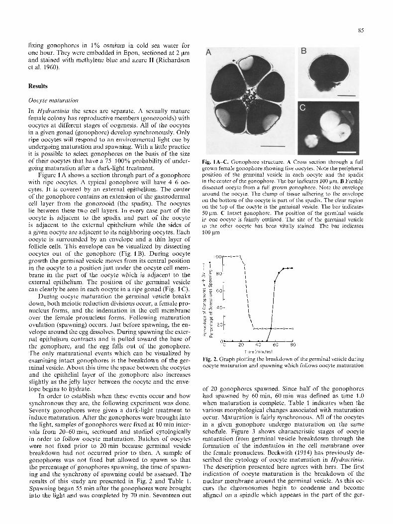
Figure l A shows a section through part of a gonophore with ripe oocytes. A typical gonophore will have 4-6 oo- cytes. It is covered by an external epithelium. The center of the gonophore contains an extension of the gastrodermal cell layer from the gonozooid (the spadix). The oocytes lie between these two cell layers. In every case part of the oocyte is adjacent to the spadix and part of the oocyte is adjacent to the external epithelium while the sides of a given oocyte are adjacent to its neighboring oocytes. Each oocyte is surrounded by an envelope and a thin layer of follicle cells. This envelope can be visualized by dissecting oocytes out of the gonophore (Fig. 1 B). During oocyte growth the germinal vesicle moves from its central position in the oocyte to a position just under the oocyte cell mem- brane in the part of the oocyte which is adjacent to the external epithelium. The position of the germinal vesicle can clearly be seen in each oocyte in a ripe gonad (Fig. 1 C).
During oocyte maturation the germinal vesicle breaks down, both meiotic reduction divisions occur, a female pro- nucleus forms, and the indentation in the cell membrane over the female pronucleus forms. Following maturation ovulation (spawning) occurs. Just before spawning, the en- velope around the egg dissolves. During spawning the exter- nal epithelium contracts and is pulled toward the base of the gonophore, and the egg falls out of the gonophore. The only maturational events which can be visualized by examining intact gonophores is the breakdown of the ger- minal vesicle. About this time the space between the oocytes and the epithelial layer of the gonophore also increases slightly as the jelly layer between the oocyte and the enve- lope begins to hydrate.
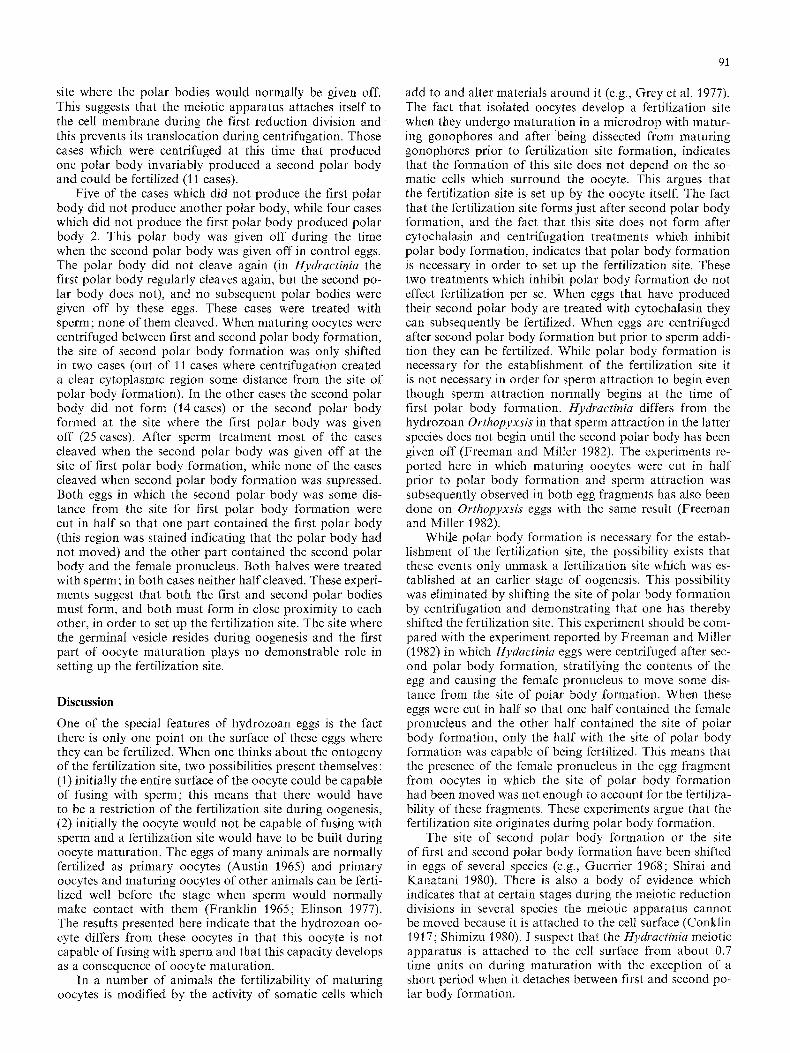
In order to establish when these events occur and how synchronous they are, the following experiment was done. Seventy gonophores were given a dark-light treatment to induce maturation. After the gonophores were brought into the light, samples of gonophores were fixed at l0 min inter- vals from 20 60 min, sectioned and studied cytologically in order to follow oocyte maturation. Batches of oocytes were not fixed prior to 20 rain because germinal vesicle breakdown had not occurred prior to then. A sample of gonophores was not fixed but allowed to spawn so that the percentage of gonophores spawning, the time of spawn- ing and the synchrony of spawning could be assessed. The results of this study are presented in Fig. 2 and Table 1. Spawning began 55 rain after the gonophores were brought into the light and was completed by 70 rain. Seventeen out
Fig. IA-C. Gonophore structure. A Cross section through a full grown female gonophore showing five oocytes. Note the peripheral position of the germinal vesicle in each oocyte and the spadix in the center of the gonophore. The bar indicates 100 jam. B Freshly dissected oocyte from a full grown gonophore. Note the envelope around the oocyte. The clump of tissue adhering to the envelope on the bottom of the oocyte is part of the spadix. The clear region on the top of the oocyte is the germinal vesicle. The bar indicates 50 jam. C Intact gonophore. The position of the germinal vesicle in one oocyte is faintly outlined. The site of the germinal vesicle in the other oocyte has been vitally stained. The bar indicates 100 jam
I O0 --o--o
o> .~ 8o
~ ~ 60
CD <9
~ 20
o~
o
60 80
Time(minutes)
Fig. 2. Graph plotting the breakdown of the germinal vesicle during oocyte maturation and spawning which follows oocyte maturation
of 20 gonophores spawned. Since half of the gonophores had spawned by 60 rain, 60 rain was defined as time 1.0 when maturation is complete. Table 1 indicates when the various morphological changes associated with maturation occur, Maturation is fairly synchronous. All of the oocytes in a given gonophore undergo maturation on the same schedule. Figure 3 shows characteristic stages of oocyte maturation from germinal vesicle breakdown through the formation of the indentation in the cell membrane over the female pronucleus. Beckwith (1914) has previously de- scribed the cytology of oocyte maturation in Hydractinia. The description presented here agrees with hers. The first indication of oocyte maturation is the breakdown of the nuclear membrane around the germinal vesicle. As this oc- curs the chromosomes begin to condense and become aligned on a spindle which appears in the part of the ger-

86
Table I. Timing of events involved in oocyte maturation
Normalized Germinal vesicle Germinal vesicle First meiotic First Second meiotic Second time scale intact breakdown apparatus in place polar body apparatus in place polar body
0.34 10 0.50 1 9 0.67 2 7 2 0.84 2 3 5 i .00 1 4
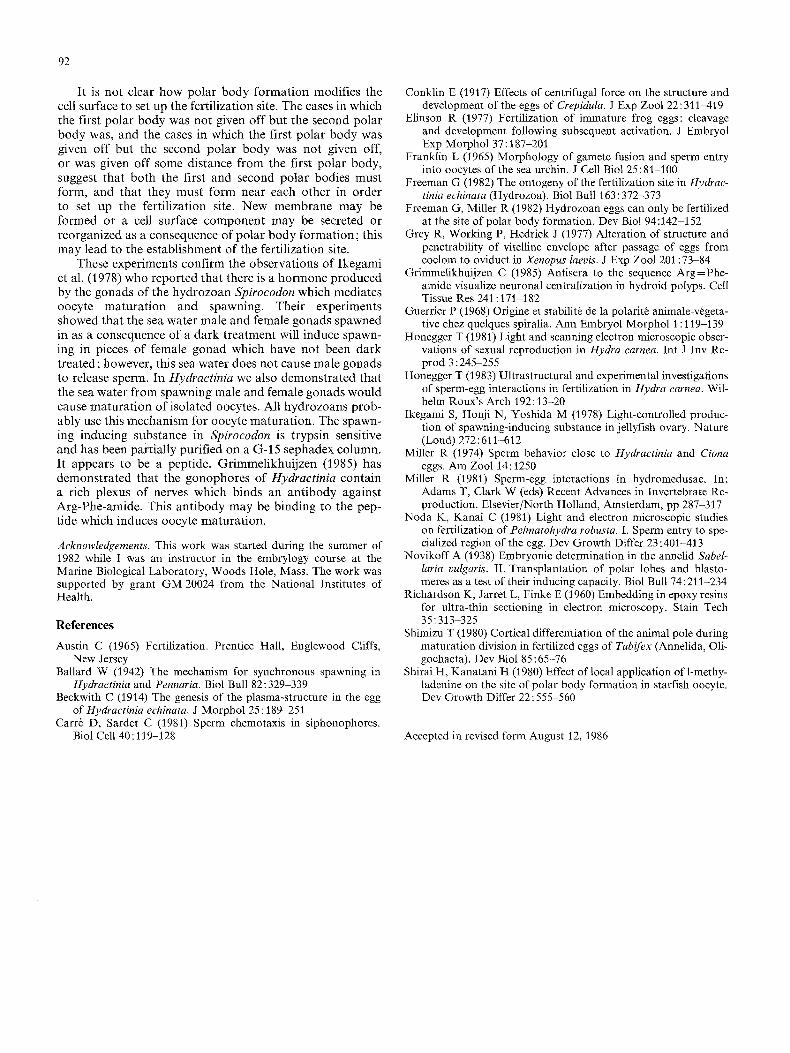
Fig. 3A-F. Different stages of oocyte maturation A Germinal vesicle breakdown (time 0.5). B Attachment of the first meiotic apparatus to the cell membrane at one pole (time 0.67). C The formation of the first polar body (time 0.84). D The period between first and second polar body formation showing the chromosomal vesicle some distance from the cell membrane and the lack of a fertilization pit. The first polar body begins at this site about 4 pm into the block (time 0.84). E Formation of the spindle for the second meiotic reduction division. The spindle is in contact with the cell membrane. Note the glancing section of the first polar body (time 1.0). F The female pronucleus and the fertilization pit (time 1.0). All photographs are at the same magnification. The bar indicates 20 gm
minal vesicle remnant closest to the cell membrane. It moves toward the cell periphery where one pole of the spindle appears to make contact with the cell membrane. There are frequently clear patches of cytoplasm which appear to be remnants of the germinal vesicle around the spindle. Following spindle contact, anaphase movements occur and the first polar body forms. No visible change in the egg surface results from first polar body formation. Following first polar body formation the spindle disappears and a small clear area with condensed chromosomes in its center appears. This area is not attached to the cell membrane. The chromosomes that remain in the egg then become aligned on a new spindle that forms and attaches to the cell membrane at one pole for the second reduction division. After second polar body formation takes place, the female pronucleus and the indentation in the egg surface over the nucleus forms. Both polar bodies form at essentially the same site; they are adjacent in freshly spawned eggs. Second polar body formation normally occurs as spawning is tak- ing place. I have seen rare cases in which second polar body fomaation does not take place until 5 or 10 min after
spawning. The first polar body divides at the time the sec- ond polar body is given off, or shortly after second polar body formation. Between first and second polar body for- mation there is a reorganization of the cytoplasmic layer just under the cell membrane. A population of granules takes up a position adjacent to the plasma membrane and a thin cytoplasmic region (ca 4 pm thick) which is relatively granule-free forms just beneath the outer granule layer (Fig. 3 F). A lightly staining layer (1 gm thick) of extracellu- lar material appears on the egg surface except at the fertil- ization pit.
The relationship between the position o f the germinal vesicle in a ripe oocyte and the place where the polar bodies are given off during maturat ion was examined by locally staining one oocyte in each of 19 gonophores just over the germinal vesicle. The dye did not stain the germinal vesicle, however, the cell membrane and cytoplasm around the germinal vesicle was stained. These gonophores were then given a dark-light treatment to induce maturat ion and spawning. In 17 cases maturat ion and spawning occurred, while in 2 cases maturation of the marked oocyte did not

Fig. 4A, B. Oocyte maturation in microdrop cultures. A Culture with 7 gonophores and 2 immature oocytes (arrow) 5 rain after removal from the dark. B Same culture 70 min later; five of the gonophores have spawned. Both photographs are at the same mag- nification. The bar indicates I mm
occur but spawning did. In every case the stain corre- sponded to the site of the germinal vesicle or the site where the polar bodies were given off. The matured eggs were treated with sperm. Fifteen of them cleaved, indicat ing that the vital dye did not inhibit fertilization.
In order to examine the ontogeny of the ferti l ization site, it was necessary to remove pr imary or matur ing oo- cytes from a gonophore and then get them to undergo or to complete maturat ion. Bal lard (1942) has claimed that, when ripe pr imary oocytes are dissected out of gonophores and subjected to a dark- l ight treatment, some of them would undergo maturat ion. I have tried to repeat this exper- iment and have not been able to. In one experiment, par t of the oocytes were dissected out of 10 gonophores and the remainder of the oocytes were left in the gonophores under the external epithelium. The isolated oocytes and their paired gonophores were then given a dark-l ight treat- ment to induce spawning. Nine out o f 10 gonophores spawned. Each of the 22 oocytes in these 9 gonophores underwent ma tu ra t ion while only one of the 16 oocytes isolated from these 9 gonophores underwent maturat ion.
The following procedure was developed for inducing matura t ion in isolated oocytes. Single gonophores or a small number of gonophores were placed in a microdrop of sea water under mineral oil and given a dark- l ight treat- ment to induce spawning. After the gonophores were brought into the light, one or two isolated oocytes that
87
had not been dark-l ight t reated were added to the micro- drop. This procedure is based on the work of Ikegami et al. (1978). These investigators showed that the sea water male or female gonads of the hydrozoan Spirocodon spawn in contains a substance which induces oocyte matura t ion and spawning in pieces of ovary that have not been subjected to a set of environmental condit ions that induce spawning. The experimental design used here differs from theirs in that they never tested the abil i ty of isolated oocytes to un- dergo matura t ion . Figure 4 shows the microdrop prepara- tion. While the size of the microdrop varies depending on the size and number of gonophores it contains, a microdrop with one female gonophore will usually have a volume of about 5 gl. The gonophores were cultured in microdrops to keep the concentrat ion of oocyte p romot ing factors high.
Three var ia tons on this experiment were done as con- trols. One control consisted of placing isolated oocytes in microdrops with feeding zooids which were dark-l ight treated. Ano the r control consisted of placing isolated oo- cytes in a microdrop and giving them a dark- l ight treat- ment. The last control consisted of placing isolated oocytes in microdrops with gonophores that were not dark-l ight treated.
The results of these experiments are presented in Table 2, The experiments show that if either female or male gono- phores are given a dark-l ight t reatment and then spawn, they will frequently induce matura t ion in oocytes that have not been dark-l ight treated.1 The oocytes that were intro- duced into the microdrop never matured in synchrony with the oocytes in the dark- l ight t reated gonophores. At the time of spawning for both male and female gonophores, the in t roduced oocytes always had intact germinal vesicles. They did not complete matura t ion until about 0.9 time units after the female gonophores spawned. This result suggests that both the male and female gonophores liberate oocyte matura t ion promot ing substance at the time of spawning.
The possibil i ty that contact between the gonophore and the oocyte media ted oocyte matura t ion was excluded by doing the following addi t ional experiment. A small sheet of dialysis tubing (Spectraphor) with a molecular weight
t Microdrop cultures were also used to test the ability of dark-light treated gonophores to induce spawning in male and female gono- phores that have not been dark-light treated. Both male and female dark-light treated gonophores induce spawning in female gonophores, but will not induce spawning in male gonophores
Table 2. Oocyte maturation in microdrop cultures
Kind of experiment Source of Number of Dark-light Spawning a Number of Number of maturation hormone experiments treatment oocytes oocytes maturing
Microdrop 5-10 gastrozoids 5 + 8 0 5 + 28 1 b
1- 2 9 gonophores 4 + + 6 0 3- 6 9 gonophores 39 + + 89 68 3- 6 ~ gonophores 8 - -- 13 1 8-15 (? gonophores 9 + + 14 14 8-16 d' gonophores 6 - - 8 0
Dialysis tubing 1020 ~ gonophores 4 + + 10 7 4- 6 2 gonophores 2 + + 6 3 dialysis tubing 3 + + 6 0
a Spawning indicates that 50% or more of the gonophores spawned b In the experiments with only isolated oocytes, the size of the microdrop was made proportionately smaller

88
GV 100 Breakdow/~
8O
c) 60 "6 g
g 4O
g_ 2 0
0 0.4 015 Oil6
p~ 2.a
Po
i
0.7 0.8 0.9 t.O Normalized Time unlit Spawning(I O)
Fig. 5. Plot showing the timing of germinal vesicle breakdown, first polar body formation and second polar body formation for 10 oocytes dissected out of maturing gonophores between 0.25 and 0.4 time units into the process of oocyte maturation. Time 1.0 was measured as the time of 50% spawning for each population of gonophores that the isolated oocytes came from
Fig. 6A, B. Maturing oocyte on a slide with sperm. A First polar body formation is just beginning, the oocyte is also just beginning to attract sperm. At this point the oocyte has been on the slide in sperm for eight minutes, B The same oocyte five minutes later surrounded by a high concentration of sperm. Both photographs are at the same magnification. The bar indicates 50 ~tm
cut off of 12,000-14,000 was placed under mineral oil. Ei- ther male or female gonophores were placed on one side of the dialysis tubing in a microdrop and given a dark-light treatment to induce spawning. When the preparation was brought into the light, oocytes that had not been dark-light treated were added to the other side of the dialysis tubing in a microdrop. These oocytes also underwent maturation (Table 2). Control experiments showed that the dialysis tub- ing had no effect on maturation or spawning.
In order for an oocyte to undergo maturation it does not have to continually be in the presence of the substance the gonophore produces. If maturing oocytes are dissected from dark treated gonophores at various times after the gonophores are brought into the light the oocytes will con- tinue to undergo maturation if they are dissected out of the gonophore after time 0.25. This is 0.25 units prior to germinal vesicle breakdown and 0.50 time units prior to first polar body formation. When oocytes which are intro- duced into a microdrop with gonophores which are under- going maturation are removed from the microdrop 10-15 min after the gonophores have spawned they will continue to undergo maturation. When oocytes are dis- sected from maturing gonophores after the critical period or removed from the source of spawning hormones, ger- minal vesicle breakdown, first polar body and second polar body formation occur on schedule (Fig. 5). If the oocyte is surrounded by an envelope it dissolves just before second polar body formation. Oocytes dissected out of maturing gonophores and oocytes treated with spawning hormone, that complete second polar body formation, can be ferti- lized (24/29 cases cleaved).
Determining the stage when maturing oocytes will attract sperm and be fertilized
The ability of large oocytes from gonophores, which have not been induced to undergo maturation, to attract sperm was tested by dissecting these oocytes out of gonophores, removing the envelope around each oocyte and studying the behavior of sperm in the vicinity of the oocyte. These oocytes have a layer of transparent jelly around them, and with time sperm get trapped in the jelly. However, there was no indication that oocytes attracted sperm (16 cases).
The ability of maturing oocytes to attract sperm was studied by dissecting oocytes out of gonophores at various times after the gonophores have been brought into the light after a prior dark treatment. The envelope was dissected off the oocyte and the behavior of sperm in the vicinity of the oocyte was studied. All oocytes examined between first and second polar body formation attracted sperm dur- ing the entire observation period (8 cases). This was mani- fest as a dense cloud of sperm around the oocyte. When oocytes were observed from prior to first polar body forma- tion through first polar body formation, oocytes began at- tracting sperm at the time of first polar body formation (11 cases). Figure 6 shows an oocyte before and after it started attracting sperm. When maturing oocytes at times 0.50-0.70 were cut in half only one-half subsequently pro- duced the first polar body. However, both halves attracted sperm (4 cases).
The fertilizability of oocytes from gonophores which had not been induced to undergo maturation and maturing oocytes with one polar body was studied in two ways. One procedure involved dissecting oocytes out of appropriate gonophores, the envelope of each kind of oocyte was re- moved and the oocytes were treated with sperm for 10 min. The oocytes were then washed, fixed and examined cytolog- ically for sperm nucleus incorporation. There were no cases in which sperm-oocyte fusion was observed. However, sperm heads were observed outside both kinds of oocytes (9 unmatured oocytes and 8 maturing oocytes with one polar body examined).
The other procedure involved treating these two classes of oocytes with sperm for 5 rain, followed by treatment with 0.002% SDS sea water to kill the sperm. The immature oocytes were then stained with neutral red so that they could be identified and induced to undergo maturation by placing them in microdrop cultures with female gonophores that were undergoing maturation. The maturing oocytes were allowed to complete maturation. They were then as- sayed to see whether or not they had been fertilized during the period when they were treated with sperm by seeing whether or not they cleaved. There were no cases which cleaved (15 primary oocytes and 11 maturing oocytes with I polar body tested). Five additional cases of each type which went through these treatments were treated with

89
2 2
Fig. 7A, B. Centrifuged oocyte. A This oocyte was marked at the site of the germinal vesicle while in the gonophore. The gonophore was induced to undergo oocyte maturation. The marked oocyte was then dissected out of the gonophore after time 0.25 and centrifuged at time 0.50. The contents of the oocyte have been stratified into three zones - an upper pigmented lipid zone, a clear cytoplasmic zone and a lower yolky zone. The photograph was taken immediately after centrifugation. The mark indicating the former position of the germinal vesicle is in the yolky zone. The bar indicates 50 ~tm. B Reconstruction based on serial sections showing the sites of polar body formation for the oocytes of a gonophore which was centrifuged just prior to (time -0.2-0) the initiation of oocytes maturation and fixed at 0.8 during maturation. In every case the polar body is in a position which reflects the change in the position of the germinal vesicle brought about by the stratification of the contents of the oocyte (compare with Fig. 1). (B1) Orientation of gonophore during centrifugation (the arrow indicates the direction of centrifugation). (B2) Sideview of reconstructed gonophore. (B3) Apical view of the same gonophore. The open and filled circle indicates the sites of polar body formation
sperm again after they completed maturation, four cases of each type cleaved. In each case cleavage was unipolar indicating that only one mitotic apparatus was present. Ten oocytes which were treated with sperm between first and second polar body formation were allowed to finish matu- ration and treated with calcium ionophore to induce egg activation. These eggs did not cleave. However, many of these eggs fragmented 2 or 3 h later. Unfertilized eggs which are treated with the concentrations of ionophore used here behave in the same way. It was clear that the calcium iono- phore had activated these eggs because they stopped attract- ing sperm (Freeman and Miller 1982). If the oocytes treated with sperm had been fertilized, subsequent treatment with an activating agent might be expected to induce a more or less normal first cleavage at the appropriate time (about 1 h after fertilization).
Conditions necessary for the establishment o f the fertilization site
The previous set of experiments has demonstrated that the egg acquires the ability to be fertilized at the time of second polar body formation. It could be that the acquisition of the ability to be fertilized is coupled to the meiotic reduction divisions. However, it is possible that the acquisition of this ability is caused by a mechanism which functions inde- pendently of the meiotic reduction divisions. These two pos- sibilities were examined by inhibiting the second meiotic reduction division with cytochalasin B.
This was done by dissecting maturing oocytes from gon- ophores prior to second polar body formation. These oo- cytes were placed in 0.004rag/m1 cytochalasin B, 0.1-0.2 time units prior to second polar body formation or allowed to undergo polar body formation and then placed in cytochalasin B. Fifteen minutes after the initiation of the cytochalasin B treatment sperm were added to the eggs and 5 rain later they were treated with 0.002% SDS to kill the sperm. The eggs were then washed in three chan- ges of sea water to dilute out the SDS and cytochalasin B. The cytochalasin B treatment inhibits second polar body formation. However, a broad cytoplasmic bulge transiently appears just under the first polar body at the time of second polar body formation. Eighteen eggs in which second polar
body formation was inhibited were fixed immediately after sperm treatment in order to see if there was cytological evidence for fertilization. These eggs contain two nuclei which result from the second meiotic reduction division, there is no indication of the indentation in egg surface that forms during the second maturation division and overlies the female pronucleus, and there was no indication of sperm nuclei in the egg. Sperm nuclei are much smaller. There was no indication that sperm behavior was altered in cyto- chalasin B. Eggs in which second polar body formation was inhibited, and eggs with the second polar body, at- tracted sperm in the presence of cytochalasin. After the cytochalasin was washed out of eggs in which second polar body formation was inhibited and from eggs which had a second polar body, these eggs were set aside to see if they would cleave. The eggs in which second polar body formation was inhibited showed no indication of forming a second polar body after the cytochalasin was washed out. These eggs did not cleave (12 cases tested). Most of the eggs that were fertilized in cytochlasin B after second polar body formation cleave (15/24 cases). However, many of these cases formed multiple cleavage furrows at the site of polar body formation at the time of first cleavage. This suggests polyspermic fertilization. The observation also sug- gests that microfilaments may play a role in the block to polyspermy in hydrozoans. Eggs from the same batches that were fertilized and subsequently treated with cytocha- lasin for 23 rain, followed by washing to remove the cyto- chalasin cleaved normally. These experiments indicate that second polar body formation is necessary in order to estab- lish the fertilization site.
Even though the fertilization site appears at the time of second polar body formation and depends on second polar body formation, it is not clear when this site is deter- mined. Since the site of polar body formation corresponds to the site where the germinal vesicle was located during a great deal of oocyte growth, the fertilization site may have been determined at an earlier time; it is also possible that no determinative events took place during this period. In order to distinguish between these possibilities a set of experiments was done in which the site of polar body forma- tion was shifted before and during oocyte maturation.
These experiments were done by marking the site of

90
Oocytes Eggs Sperm Marked Cut
Centrifugation Induce Oocyte ----~" Maturation ~....__..~
�9 , @ .
Oocytes Eggs Sperm Marked Cut
Induce Oocyte 2. Maturation Centrlfugation
Fig. 8. Diagram of the experiments designed to move the site of polar body formation (1) before and (2) during oocyte maturation. The site of the germinal vesicle in each oocycle was marked. The oocyte was then centrifuged and induced to undergo maturation (1), or induced to undergo maturation and then centrifuged (2). Centrifugation stratifies the contents of the oocyte and may move the germinal vesicle or meiotic apparatus from its normal position. In those cases in which the site of polar body formation and the stain do not coincide, the egg was cut to give a half with the polar body and a half with the stain. If the stain and the site of polar body formation coincide, the egg was also cut in half. These halves were then treated with sperm and monitored for cleav- age
the germinal vesicle in individual oocytes pr ior to oocyte matura t ion with a vital dye by staining through the external epithelium of the gonophore which contains the oocyte or by staining oocytes that had been dissected out of gono- phores. This mark indicates the site where one would expect the po la r bodies to be given off during oocyte matura t ion . Pr ior to oocyte ma tu ra t ion or at various time intervals dur-
ing oocyte matura t ion either gonophores with marked oo- cytes or isolated marked oocytes which were undergoing matura t ion in microdrop cultures, were centrifuged. Gono- phores were centrifuged in sea water. Individual oocytes were centrifuged in a mixture of Ficoll and sea water (30 grams of 400,000 M W Ficoll in 100 ml of sea water). Centr ifugat ion stratifies the contents of oocytes into 3 zones: a centripetal zone containing pigment and lipid, a central clear cytoplasmic zone and a centrifugal yolk zone (Fig. 7 A). These three zones are less well delineated in pri- mary oocytes than they are in matur ing oocytes. Since the oocytes were not oriented in the centrifugal field, the axis of strat if ication could take up any posi t ion with reference to the marked point where one expects the polar bodies to be given off. The strat if ication which is induced by cen- tr i fugation persists for about 30 rain. Depending on the or ientat ion of the oocyte the centrifugation can relocate the germinal vesicle or the meiotic appara tus to a new posi- t ion in the clear cytoplasmic region. The germinal vesicle and the meiotic appara tus end up in the clear cytoplasmic region because their density is similar to the density of the clear cytoplasmic region. Once the germinal vesicle or the meitoic appara tus has taken up a new position, polar body format ion frequently occurs at the new site (Fig. 7 B). These eggs have a mark where one expected the po la r bodies to be generated and one or both polar bodies which are fre- quently some distance from this site. (Sometimes the stain smears as a consequence of centr ifugation; these cases were discarded.) These eggs were then cut into parts so that one par t contained the marked spot and the other par t con- tained one or both polar bodies. Each of these halves was treated with sperm and ferti l ization was assayed by moni- toring cleavage. Figure 8 outlines these experiments. The results are summarized in Table 3.
When oocytes which have not undergone maturat ion, or matur ing oocytes from any time prior to 0.1 units before first polar body format ion are centrifuged, the site of polar body format ion can be shifted and the egg half which con- tains the site of polar body format ion is the only half which can be fertilized. When matur ing oocytes are centrifuged just pr ior to or during first polar body formation, either the first polar body does not form or the site of polar body format ion is not shifted even when the centrifugation creates a clear cytoplasmic zone some distance from the
Table 3. The effect of centrifugation prior to and during oocyte maturation on polar body (pb) formation and fertilization
Time of No. Polar Body Formation Site of pb's Cleavage Site of pb's Cleavage centrifugation cases coincides with different from
1st & 2nd 1st 2nd No. site ofgv Pb Non-pb site ofgv Pb Gv pb pb only pb only pb's egg egg egg egg formation fragment fragment fragment fragment
Prior to oocyte maturation
- -0 .20 /4 14
During oocyte maturation
0 -0.4 22 21 0.4-0.6 14 14 0.6-0.8 20 11 0.8 1.0 39 25
gv = germinal vesicle
0 0 0 8 6 0 6 5 0
0 0 0 12 12 0 9 7 0 0 0 0 8 6 0 6 5 0
4 5 10 9 0 1 1 0 14 23 18 0 2 0 0
91
site where the polar bodies would normally be given off. This suggests that the meiotic apparatus attaches itself to the cell membrane during the first reduction division and this prevents its translocation during centrifugation. Those cases which were centrifuged at this time that produced one polar body invariably produced a second polar body and could be fertilized (l I cases).
Five of the cases which did not produce the first polar body did not produce another polar body, while four cases which did not produce the first polar body produced polar body 2. This polar body was given off during the time when the second polar body was given off in control eggs. The polar body did not cleave again (in Hydractinia the first polar body regularly cleaves again, but the second po- lar body does not), and no subsequent polar bodies were given off by these eggs. These cases were treated with sperm; none of them cleaved. When maturing oocytes were centrifuged between first and second polar body formation, the site of second polar body formation was only shifted in two cases (out of 11 cases where centrifugation created a clear cytoplasmic region some distance from the site of polar body formation). In the other cases the second polar body did not form (14 cases) or the second polar body formed at the site where the first polar body was given off (25 cases). After sperm treatment most of the cases cleaved when the second polar body was given off at the site of first polar body formation, while none of the cases cleaved when second polar body formation was supressed. Both eggs in which the second polar body was some dis- tance from the site for first polar body formation were cut in half so that one part contained the first polar body (this region was stained indicating that the polar body had not moved) and the other part contained the second polar body and the female pronucleus. Both halves were treated with sperm; in both cases neither half cleaved. These experi- ments suggest that both the first and second polar bodies must form, and both must form in close proximity to each other, in order to set up the fertilization site. The site where the germinal vesicle resides during oogenesis and the first part of oocyte maturation plays no demonstrable role in setting up the fertilization site.
Discussion
One of the special features of hydrozoan eggs is the fact there is only one point on the surface of these eggs where they can be fertilized. When one thinks about the ontogeny of the fertilization site, two possibilities present themselves: (1) initially the entire surface of the oocyte could be capable of fusing with sperm; this means that there would have to be a restriction of the fertilization site during oogenesis, (2) initially the oocyte would not be capable of fusing with sperm and a fertilization site would have to be built during oocyte maturation. The eggs of many animals are normally fertilized as primary oocytes (Austin 1965) and primary oocytes and maturing oocytes of other animals can be ferti- lized well before the stage when sperm would normally make contact with them (Franklin 1965; Elinson 1977). The results presented here indicate that the hydrozoan oo- cyte differs from these oocytes in that this oocyte is not capable of fusing with sperm and that this capacity develops as a consequence of oocyte maturation.
In a number of animals the fertilizability of maturing oocytes is modified by the activity of somatic cells which
add to and alter materials around it (e.g., Grey et al. 1977). The fact that isolated oocytes develop a fertilization site when they undergo maturation in a microdrop with matur- ing gonophores and after "being dissected from maturing gonophores prior to fertilization site formation, indicates that the formation of this site does not depend on the so- matic cells which surround the oocyte. This argues that the fertilization site is set up by the oocyte itself. The fact that the fertilization site forms just after second polar body formation, and the fact that this site does not form after cytochalasin and centrifugation treatments which inhibit polar body formation, indicates that polar body formation is necessary in order to set up the fertilization site. These two treatments which inhibit polar body formation do not effect fertilization per se. When eggs that have produced their second polar body are treated with cytochalasin they can subsequently be fertilized. When eggs are centrifuged after second polar body formation but prior to sperm addi- tion they can be fertilized. While polar body formation is necessary for the establishment of the fertilization site it is not necessary in order for sperm attraction to begin even though sperm attraction normally begins at the time of first polar body formation. Hydractinia differs from the hydrozoan Orthopyxsis in that sperm attraction in the latter species does not begin until the second polar body has been given off (Freeman and Miller 1982). The experiments re- ported here in which maturing oocytes were cut in half prior to polar body formation and sperm attraction was subsequently observed in both egg fragments has also been done on Orthopyxsis eggs with the same result (Freeman and Miller 1982).
While polar body formation is necessary for the estab- lishment of the fertilization site, the possibility exists that these events only unmask a fertilization site which was es- tablished at an earlier stage of oogenesis. This possibility was eliminated by shifting the site of polar body formation by centrifugation and demonstrating that one has thereby shifted the fertilization site. This experiment should be com- pared with the experiment reported by Freeman and Miller (1982) in which Hydactinia eggs were centrifuged after sec- ond polar body formation, stratifying the contents of the egg and causing the female pronucleus to move some dis- tance from the site of polar body formation. When these eggs were cut in half so that one half contained the female pronucleus and the other half contained the site of polar body formation, only the half with the site of polar body formation was capable of being fertilized. This means that the presence of the female pronucleus in the egg fragment from oocytes in which the site of polar body formation had been moved was not enough to account for the fertiliza- bility of these fragments. These experiments argue that the fertilization site originates during polar body formation.
The site of second polar body formation or the site of first and second polar body formation have been shifted in eggs of several species (e.g., Guerrier 1968; Shirai and Kanatani 1980). There is also a body of evidence which indicates that at certain stages during the meiotic reduction divisions in several species the meiotic apparatus cannot be moved because it is attached to the cell surface (Conklin 1917; Shimizu 1980). I suspect that the Hydractinia meiotic apparatus is attached to the cell surface from about 0.7 time units on during maturation with the exception of a short period when it detaches between first and second po- lar body formation.
92
It is not clear how polar body format ion modifies the cell surface to set up the ferti l ization site. The cases in which the first po la r body was not given off but the second polar body was, and the cases in which the first po la r body was given off but the second po la r body was not given off, or was given off some distance from the first polar body, suggest that both the first and second polar bodies must form, and that they must form near each other in order to set up the ferti l ization site. New membrane may be formed or a cell surface component may be secreted or reorganized as a consequence of po la r body format ion; this may lead to the establishment of the ferti l ization site.
These experiments confirm the observations of Ikegami et al. (1978) who repor ted that there is a hormone produced by the gonads of the hydrozoan Spirocodon which mediates oocyte matura t ion and spawning. Their experiments showed that the sea water male and female gonads spawned in as a consequence of a dark t reatment will induce spawn- ing in pieces of female gonad which have not been dark t reated; however, this sea water does not cause male gonads to release sperm. In Hydractinia we also demons t ra ted that the sea water from spawning male and female gonads would cause ma tu ra t ion of isolated oocytes. All hydrozoans prob- ably use this mechanism for oocyte matura t ion . The spawn- ing inducing substance in Spirocodon is t rypsin sensitive and has been par t ia l ly purif ied on a G-15 sephadex column. It appears to be a peptide. Grimmelikhui jzen (1985) has demonst ra ted that the gonophores of Hydractinia contain a rich plexus of nerves which binds an an t ibody against Arg-Phe-amide. This an t ibody may be binding to the pep- tide which induces oocyte matura t ion .
Acknowledgements. This work was started during the summer of 1982 while I was an instructor in the embrylogy course at the Marine Biological Laboratory, Woods Hole, Mass, The work was supported by grant GM 20024 from the National Institutes of Health.
References
Austin C (1965) Fertilization. Prentice Hall, Englewood Cliffs, New Jersey
Ballard W (1942) The mechanism for synchronous spawning in Hydractinia and Pennaria. Biol Bull 82:329-339
Beckwith C (1914) The genesis of the plasma-structure in the egg of Hydractinia echinata. J Morphol 25:189-251
Cart+ D, Sardet C (1981) Sperm chemotaxis in siphonophores. Biol Cell 40 : 119-128
Conklin E (1917) Effects of centrifugal force on the structure and development of the eggs of Crepidula. J Exp Zool 22:311-419
Elinson R (1977) Fertilization of immature frog eggs: cleavage and development following subsequent activation. J Embryol Exp Morphol 37:187-201
Franklin L (1965) Morphology of gamete fusion and sperm entry into oocytes of the sea urchin. J Cell Biol 25:81-100
Freeman G (1982) The ontogeny of the fertilization site in Hydrac- tinia echinata (Hydrozoa). Biol Bull 163:372-373
Freeman G, Miller R (1982) Hydrozoan eggs can only be fertilized at the site of polar body formation. Dev Biol 94:142-152
Grey R, Working P, Hedrick J (1977) Alteration of structure and penetrability of vitelline envelope after passage of eggs from coelom to oviduct in Xenopus laevis. J Exp Zool 201 : 73-84
Grimmelikhuijzen C (1985) Antisera to the sequence Arg=Phe- amide visualize neuronal centralization in hydroid polyps. Cell Tissue Res 241 : 171-182
Guerrier P (1968) Origine et stabilit6 de la polarit6 animale-v6geta- tive chez quelques spiralia. Ann Embryol Morphol 1:119-139
Honegger T (1981) Light and scanning electron microscopic obser- vations of sexual reproduction in Hydra carnea. Int J Inv Re- prod 3 : 245-255
Honegger T (1983) Ultrastructural and experimental investigations of sperm-egg interactions in fertilization in Hydra carnea. Wil- helm Roux's Arch 192:13-20
Ikegami S, Honji N, Yoshida M (1978) Light-controlled produc- tion of spawning-inducing substance in jellyfish ovary. Nature (Loud) 272: 611-612
Miller R (1974) Sperm behavior close to Hydractinia and Ciona eggs. Am Zool 14:1250
Miller R (1981) Sperm-egg interactions in hydromedusae, In: Adams T, Clark W (eds) Recent Advances in Invertebrate Re- production. Elsevier/North Holland, Amsterdam, pp 28%317
Noda K, Kanai C (1981) Light and electron microscopic studies on fertilization of Pelmatohydra robusta. I. Sperm entry to spe- cialized region of the egg. Dev Growth Differ 23:401-413
Novikoff A (1938) Embryonic determination in the annelid Sabel- laria vulgaris. II. Transplantation of polar lobes and blasto- meres as a test of their inducing capacity. Biol Bull 74:211-234
Richardson K, Jarret L, Finke E (1960) Embedding in epoxy resins for ultra-thin sectioning in electron microscopy. Stain Tech 35:313 325
Shimizu T (1980) Cortical differentiation of the animal pole during maturation division in fertilized eggs of Tubifex (Annelida, Oli- gochaeta). Dev Biol 85:65-76
Shirai H, Kanatani H (1980) Effect of local application of 1-methy- ladenine on the site of polar body formation in starfish oocyte. Dev Growth Differ 22:555-560
Accepted in revised form August 12, 1986


![[PPT]26-2 Sponges - cypresswoodsbiology - homecypresswoodsbiology.wikispaces.com/file/view... · Web viewcopyright cmassengale Colonial Hydrozoan (not a single organism Tentacles](https://static.fdocuments.us/doc/165x107/5aa6232f7f8b9a7c1a8e5585/ppt26-2-sponges-cypresswoodsbiology-hom-viewcopyright-cmassengale-colonial.jpg)

