The role of magnetic resonance imaging in the management of brain metastases: diagnosis to prognosis
8
REVIEW Open Access The role of magnetic resonance imaging in the management of brain metastases: diagnosis to prognosis Rasheed Zakaria 1,2* , Kumar Das 3 , Maneesh Bhojak 3 , Mark Radon 3 , Carol Walker 1 and Michael D Jenkinson 1,4 Abstract This article reviews the different MRI techniques available for the diagnosis, treatment and monitoring of brain metastases with a focus on applying advanced MR techniques to practical clinical problems. Topics include conventional MRI sequences and contrast agents, functional MR imaging, diffusion weighted MR, MR spectroscopy and perfusion MR. The role of radiographic biomarkers is discussed as well as future directions such as molecular imaging and MR guided high frequency ultrasound. Keywords: MRI, Brain metastasis, Perfusion, Diffusion, ADC, Spectroscopy, MRS, Biomarkers Introduction Brain metastases are the most common central nervous system tumours in adults with a rising incidence due to the increased availability and utilisation of brain imaging and prolonged survival from primary cancers [1-3]. MRI is crucial in making the diagnosis, determining the best course of management, monitoring response to therapy and increasingly in trying to predict prognosis. Rather than reviewing each individual technique and its applications separately, as has been done elsewhere, the different clinical problems encountered in brain metastases are presented and the relevant MRI techniques which can be applied in each scenario addressed to give a practical summary [4-9]. Review How many brain metastases are present? Accurately identifying the number, location and size of brain metastases is important to determine which inter- ventions, if any, are appropriate for a patient. Multiple scoring systems used to predict prognosis take into account the number of lesions, for example the Recursive Partitioning Analysis or RPA classification [10-14]. With respect to detection, localisation and quantification, con- trast enhanced MR has been widely demonstrated to be superior to both enhanced CT and non-enhanced MR [15-18] as illustrated in Figure 1. The recommended gadolinium dose in this context is 0.1 mmol/kg and whilst double or triple dose administration has been suggested to increase the detection of small lesions this causes increased false positives and a higher risk of nephrogenic systemic fibrosis therefore it remains an “off label” use [19-21]. Could novel sequences and agents therefore increase detection without escalating the contrast dose? Different gadolinium based agents, all of which may have slightly differing relaxivity profiles have been compared and these studies are summarised elsewhere. At present gadobutrol appears to identify the greatest number of lesions with the greatest contrast to noise ratio whilst having a lower risk of nephrogenic systemic fibrosis (along with the other “cyclic” structured gadolinium based agents [22]). MR sequences are developing rapidly but are not always explicitly evaluated against existing protocols. Magnetisation saturation transfer (MT) imaging has been directly compared to gradient echo T1 sequences and its addition reduced by half the standard dose of contrast needed for detection leading some to advocate it over ever increasing contrast doses [23,24]. More recently, 3D T1 weighted “spoiled gradient echo” (SPGR) and T2 weighted post contrast FLAIR sequences have been shown to detect submillimetric (<3 mm) abnormalities * Correspondence: [email protected] 1 Department of Neurosurgery, The Walton Centre NHS Foundation Trust, Liverpool, UK 2 Institute of Integrative Biology, University of Liverpool, Liverpool, UK Full list of author information is available at the end of the article © 2014 Zakaria et al.; licensee BioMed Central Ltd. This is an Open Access article distributed under the terms of the Creative Commons Attribution License (http://creativecommons.org/licenses/by/4.0), which permits unrestricted use, distribution, and reproduction in any medium, provided the original work is properly credited. The Creative Commons Public Domain Dedication waiver (http://creativecommons.org/publicdomain/zero/1.0/) applies to the data made available in this article, unless otherwise stated. Zakaria et al. Cancer Imaging 2014, 14:8 http://www.cancerimagingjournal.com/content/14/1/8
-
Upload
carol-walker -
Category
Documents
-
view
213 -
download
0
Transcript of The role of magnetic resonance imaging in the management of brain metastases: diagnosis to prognosis
REVIEW Open Access
The role of magnetic resonance imaging in themanagement of brain metastases: diagnosis toprognosisRasheed Zakaria1,2*, Kumar Das3, Maneesh Bhojak3, Mark Radon3, Carol Walker1 and Michael D Jenkinson1,4
Abstract
This article reviews the different MRI techniques available for the diagnosis, treatment and monitoring of brainmetastases with a focus on applying advanced MR techniques to practical clinical problems. Topics includeconventional MRI sequences and contrast agents, functional MR imaging, diffusion weighted MR, MR spectroscopyand perfusion MR. The role of radiographic biomarkers is discussed as well as future directions such as molecularimaging and MR guided high frequency ultrasound.
Keywords: MRI, Brain metastasis, Perfusion, Diffusion, ADC, Spectroscopy, MRS, Biomarkers
IntroductionBrain metastases are the most common central nervoussystem tumours in adults with a rising incidence due tothe increased availability and utilisation of brain imagingand prolonged survival from primary cancers [1-3]. MRIis crucial in making the diagnosis, determining the bestcourse of management, monitoring response to therapyand increasingly in trying to predict prognosis. Rather thanreviewing each individual technique and its applicationsseparately, as has been done elsewhere, the different clinicalproblems encountered in brain metastases are presentedand the relevant MRI techniques which can be applied ineach scenario addressed to give a practical summary [4-9].
ReviewHow many brain metastases are present?Accurately identifying the number, location and size ofbrain metastases is important to determine which inter-ventions, if any, are appropriate for a patient. Multiplescoring systems used to predict prognosis take intoaccount the number of lesions, for example the RecursivePartitioning Analysis or RPA classification [10-14]. Withrespect to detection, localisation and quantification, con-
trast enhanced MR has been widely demonstrated to besuperior to both enhanced CT and non-enhanced MR[15-18] as illustrated in Figure 1. The recommendedgadolinium dose in this context is 0.1 mmol/kg andwhilst double or triple dose administration has beensuggested to increase the detection of small lesions thiscauses increased false positives and a higher risk ofnephrogenic systemic fibrosis therefore it remains an“off label” use [19-21].Could novel sequences and agents therefore increase
detection without escalating the contrast dose? Differentgadolinium based agents, all of which may have slightlydiffering relaxivity profiles have been compared and thesestudies are summarised elsewhere. At present gadobutrolappears to identify the greatest number of lesions withthe greatest contrast to noise ratio whilst having alower risk of nephrogenic systemic fibrosis (along withthe other “cyclic” structured gadolinium based agents[22]). MR sequences are developing rapidly but are notalways explicitly evaluated against existing protocols.Magnetisation saturation transfer (MT) imaging has beendirectly compared to gradient echo T1 sequences and itsaddition reduced by half the standard dose of contrastneeded for detection leading some to advocate it overever increasing contrast doses [23,24]. More recently,3D T1 weighted “spoiled gradient echo” (SPGR) and T2weighted post contrast FLAIR sequences have beenshown to detect submillimetric (<3 mm) abnormalities
* Correspondence: [email protected] of Neurosurgery, The Walton Centre NHS Foundation Trust,Liverpool, UK2Institute of Integrative Biology, University of Liverpool, Liverpool, UKFull list of author information is available at the end of the article
© 2014 Zakaria et al.; licensee BioMed Central Ltd. This is an Open Access article distributed under the terms of the CreativeCommons Attribution License (http://creativecommons.org/licenses/by/4.0), which permits unrestricted use, distribution, andreproduction in any medium, provided the original work is properly credited. The Creative Commons Public DomainDedication waiver (http://creativecommons.org/publicdomain/zero/1.0/) applies to the data made available in this article,unless otherwise stated.
Zakaria et al. Cancer Imaging 2014, 14:8http://www.cancerimagingjournal.com/content/14/1/8

and more sensitively assess leptomeningeal disease [25,26].The spatial resolution of the acquisition differs dependingon the indication for the scan and this has importantimplications for detection and management which needto be kept in mind. For example if a treatment is decidedupon with the multidisciplinary team and the patientbut then a surgical or stereotactic radiosurgery (SRS)“planning” scan of higher spatial resolution is obtained, thelatter will be more sensitive for detection of metastasesthan a conventional diagnostic scan and multiple, previ-ously unseen lesions may be identified which render thattreatment inappropriate [27].
Is this solitary lesion a metastasis, an abscess or a highgrade glioma?A patient presenting with no known primary cancer anda solitary ring enhancing brain lesion may be suspectedof having a brain metastasis, a primary cerebral tumoursuch as glioblastoma or a cerebral abscess, and despitecareful history and exam, misdiagnoses may still occur[28]. Distinguishing abscess from tumour is perhaps themost clinically relevant question and in this regarddiffusion-weighted MRI (DWI) has been most useful.Diffusion-weighted images detect free water movementand allow a surrogate of diffusion to be calculated foreach voxel to generate apparent diffusion coefficient (ADC)maps. A large number of observational studies have shownthat for a solitary cystic or necrotic contrast enhancinglesion, restriction of diffusion on pre-operative MRI ispredictive of abscess [29-32]. Modifications to imagingprotocols have increased sensitivity, including detectionof early capsule formation during abscess developmentand even prediction of cellularity of abscesses at higherb-values and field strengths [33,34]. However, there arepersistent cautions that some metastatic lesions mayshow restricted diffusion and mimic the appearances of
abscesses, for example two different studies have reportedthis pattern for non-small cell lung carcinoma metastases,while others have also described it for lymphoma [35-38].Distinguishing metastases from high grade glioma has
proven more difficult using DWI although in theory theregion of vasogenic oedema around metastases shouldshow greater free diffusion than the more cellular,infiltrated region around a high grade glioma. Studieshave varied in their cutoff for distinguishing the twopathologies as well as the methodology of how to takethe reading (does one use the lowest ADC value, themean of multiple measurements and which areas shouldone sample in the peri-tumoural region?) and there isno agreement about reliability [39-43]. Diffusion tensorimaging (DTI) is an enhancement of DWI with morediffusion gradients and directions used during acquisition.This allows more advanced metrics than just the averagediffusion coefficient to be calculated and disruption ofwhite matter tracts to be visualised. Again, the theorythat high grade glioma is an invasive tumour whichinfiltrates white matter tracts whereas metastases deformthem is supported but not consistently repeated by studiesexamining DTI metrics such as fractional anisotropywhich may be superior to ADC alone [44-47].Further advanced techniques have therefore been com-
bined with diffusion imaging in this context to try andincrease sensitivity, most commonly MR spectroscopy [48].Single and multivoxel magnetic resonance spectroscopy(MRS) provides information about the metabolic profileof specific regions in and around a lesion [49,50] as shownin Figure 2. Proton MRS has previously shown discrimin-atory power between high grade glioma and metastasesby measurement of the choline/NAA ratio [51,52]. Novelspectroscopic markers continue to be investigated, includ-ing lipid and macromolecule concentrations by protonMRS [53] and phosphate metabolites by 31P MRS[54].
Figure 1 A patient known to have melanoma presents with a first seizure and (A) unenhanced CT brain (32-slice scanner, 10 mm slices)taken in the emergency department shows an abnormality in the left frontal lobe with surrounding oedema – they were referred forpossible neurosurgical intervention. (B) T1 weighted MRI at 1.5 T with single dose (0.1 mmol/kg) gadolinium contrast detects this lesion but alsodelineates it further, allowing volume to be assessed accurately and in addition highlights two further areas of abnormality. After staging of thesystemic disease and discussion with oncologists and surgeons, the patient was therefore treated with stereotactic radiosurgery to all three areas.
Zakaria et al. Cancer Imaging 2014, 14:8 Page 2 of 8http://www.cancerimagingjournal.com/content/14/1/8
Further information on physiological activity can begathered by combining with MR perfusion studies. MRIperfusion permits the generation of maps of relativecerebral blood flow (rCBF) and volume (rCBV) which aremeasures of vascularity. High grade glioma pathologicallyshows neovascularisation and infiltration of surroundingbrain, hence the peritumoral rCBV is higher than for ametastasis [55,56]. Using the same dataset and differentpost processing techniques, diffusion susceptibility contrastor DSC MRI allows prediction of tissue permeability,a measure of blood-brain barrier disruption and thisaccordingly increases around a metastasis, where there isincreased capillary permeability and therefore vasogenicoedema [57,58].In summary, multimodal MR used singly or in combin-
ation has improved our ability to distinguish metastasesfrom primary cerebral tumours or abscesses. Despite theapplication of these advanced MR techniques of diffusion,perfusion and spectroscopy to solve this clinical problem[59] it remains a fact that for accessible, larger lesionssurgery is often undertaken and tissue diagnosis obtained
regardless in order to move forward treatment. The roleand evidence for MRI in surgical planning is therefore thenext area to consider.
What information does the neurosurgeon need and howcan MRI provide it?Image guided surgery allows the surgeon to plan the safestroute to the tumour preoperatively and to maximise saferesection intraoperatively. Conventional post contrast T1weighted images with thin slice protocols are generallyacquired for image guidance software registration andthe increased spatial resolution of such sequencescompared to the usual diagnostic scans (and thus po-tential to detect more lesions than first seen) has beenhighlighted in the discussion of detection. Diffusion im-aging allows images of white matter tracts and their re-lationship to tumour to be delineated. DTI acquisitions,in addition to generating quantitative parameters, canpermit tractography by estimating the directionality offibre tracts, permitting the tracking of fibre bundles in3D space. The methodology for this post processing varies
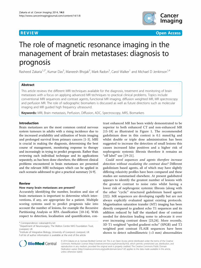
Figure 2 An elderly patient was referred with hemiparesis and suspected to have a stroke. MRI demonstrated a lesion in the left hemispherewhich on (A) T1 weighted axial image post gadolinium at 1.5 T is shown to have a solid and ring enhancing portion. (B) The associated ADC mapshows considerably reduced diffusion at the site of the solid portion of the lesion with increased diffusion due to vasogenic oedema in the whitematter surrounding the mass. (C) Single voxel proton MRS of the lesion yields an abnormal spectrum with a large lipid and lactate peak, reduced NAA,reduced Cr and slightly elevated Cho. This pointed to a metastasis, glioma or lymphoma as opposed to an abscess. There was time to optimise thepatient for surgery and begin steroid treatment before the lesion was resected and confirmed to be a renal cell carcinoma.
Zakaria et al. Cancer Imaging 2014, 14:8 Page 3 of 8http://www.cancerimagingjournal.com/content/14/1/8
and a number of non-proprietary software solutions inaddition to those supplied by manufacturers are available.Further refinements to the methodology, including useof higher order algorithms to resolve ambiguous direction-ality on voxels may further increase reliability [60]. Func-tional MR detects changes in blood oxygen level or BOLDsignal in metabolically more active areas during applicationof a stimulus or performance of a task. This is particu-larly useful to localise language areas or motor cortex.Few series have examined the role of fMRI in metastasisresection alone but in those that have significant benefitsin terms of motor recovery, preservation and thereforequality of life have been demonstrated [61]. Commerciallyavailable software can integrate functional and tractographysequences into a single merged dataset for use in theatre asshown in Figure 3, with improved outcomes for series look-ing exclusively at metastases [62,63].
Where is the original, primary cancer?In cases of multiple metastases or solitary lesions withno known primary, MRI may give useful diagnostic cluesas to the original tumour. Some primaries may have par-ticular signal characteristics even on conventional MR.For example melanoma metastases may show high signalon non-enhanced T1 sequences due to the effect ofmelanin and mucinous metastases may show low T2signal compared with the expected hyper-intensity onthis sequence. The metabolic profile has been investigatedfor metastases of differing primary tumours as well asthe surrounding brain with no abnormal spectra reportedoutside of the lesion itself. Metabolic features on MRShave shown limited value in predicting primary type.However, raised mobile lipid content has been proposedas a weak sign of a colonic origin for metastasis [64].Others have tried to use the diffusion characteristics ofparticular metastases to distinguish the primary and whilstit has been shown that ADC values are higher in well
differentiated adenocarcinoma metastases than poorlydifferentiated types this may simply reflect cellularityand could not predict the primary, only suggest a narrowerdifferential [36]. Likewise in a series with a variety ofprimary cancers including lung and breast, restricteddiffusion could not be correlated with any primary norcould ADC predict primary pathology [35]. Spectroscopywas combined with perfusion MR to show that differencesin choline-creatine ratios between metastases of lungand breast cancer correlated with differences in relativecerebral blood volume but tissue was not available forcomparison to look for a unifying pathological basis [65].In summary, MRS may be used to distinguish primary
tumour origin. However more than one advanced MRmetric may need to be combined in order to robustlygenerate models that differentiate the primary lesion andthough these studies would likely be retrospective, ideallysome image guided correlation of regions of interest onthe advanced MR with the final tissue samples is needed,as has been performed for glioma [45].
Are these brain metastases responding to chemo/radio-therapy?Beyond diagnosis, MRI may be used to monitor responseto treatment as part of clinical and radiological followup. This may be immediate, as in post-operative MRIto determine if there has been a complete resection ordelayed, i.e. has the metastasis responded chemo- orradio-therapy. In general, conventional sequences areutilized but one area of particular clinical interest infollowing cases up is ongoing or increasing enhance-ment following surgery or stereotactic radiosurgery to ametastasis and whether this represents radio-necrosisor recurrence [66]. Standard patterns of changes in brainmetastases are seen on conventional MRI with perilesionaloedema, central hypointensity on T2 weighted imagingat 2-6 months followed by blurring of the enhancement
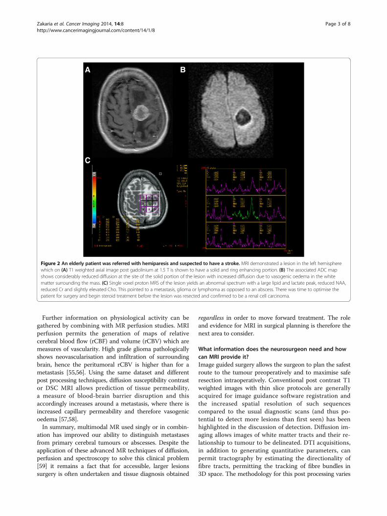
Figure 3 A patient known to have breast cancer with a manually dexterous job presented with intermittent left hand and armweakness and was found (A) to have a solitary ring enhancing lesion in the premotor area on T1W MRI with gadolinium. (B) functionalMRI performing a hand tapping and gripping task determined the location of hand function and this was used as the “seed” region of intereston a DTI scan to produce a representation of the motor tracts. (C) these were used to generate a 3D object and fused with an anatomicalplanning scan (1 mm slices) using commercially available software (StealthViz™ with StealthDTI™ by Medtronic, running on an S7 workstation) toproduce images that were used intraoperatively for image guided resection, avoiding the tracts (shown in red, with tumour rendered in green).
Zakaria et al. Cancer Imaging 2014, 14:8 Page 4 of 8http://www.cancerimagingjournal.com/content/14/1/8
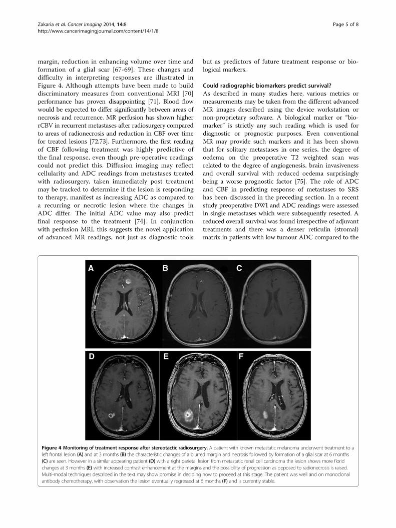
margin, reduction in enhancing volume over time andformation of a glial scar [67-69]. These changes anddifficulty in interpreting responses are illustrated inFigure 4. Although attempts have been made to builddiscriminatory measures from conventional MRI [70]performance has proven disappointing [71]. Blood flowwould be expected to differ significantly between areas ofnecrosis and recurrence. MR perfusion has shown higherrCBV in recurrent metastases after radiosurgery comparedto areas of radionecrosis and reduction in CBF over timefor treated lesions [72,73]. Furthermore, the first readingof CBF following treatment was highly predictive ofthe final response, even though pre-operative readingscould not predict this. Diffusion imaging may reflectcellularity and ADC readings from metastases treatedwith radiosurgery, taken immediately post treatmentmay be tracked to determine if the lesion is respondingto therapy, manifest as increasing ADC as compared toa recurring or necrotic lesion where the changes inADC differ. The initial ADC value may also predictfinal response to the treatment [74]. In conjunctionwith perfusion MRI, this suggests the novel applicationof advanced MR readings, not just as diagnostic tools
but as predictors of future treatment response or bio-logical markers.
Could radiographic biomarkers predict survival?As described in many studies here, various metrics ormeasurements may be taken from the different advancedMR images described using the device workstation ornon-proprietary software. A biological marker or “bio-marker” is strictly any such reading which is used fordiagnostic or prognostic purposes. Even conventionalMR may provide such markers and it has been shownthat for solitary metastases in one series, the degree ofoedema on the preoperative T2 weighted scan wasrelated to the degree of angiogenesis, brain invasivenessand overall survival with reduced oedema surprisinglybeing a worse prognostic factor [75]. The role of ADCand CBF in predicting response of metastases to SRShas been discussed in the preceding section. In a recentstudy preoperative DWI and ADC readings were assessedin single metastases which were subsequently resected. Areduced overall survival was found irrespective of adjuvanttreatments and there was a denser reticulin (stromal)matrix in patients with low tumour ADC compared to the
Figure 4 Monitoring of treatment response after stereotactic radiosurgery. A patient with known metastatic melanoma underwent treatment to aleft frontal lesion (A) and at 3 months (B) the characteristic changes of a blurred margin and necrosis followed by formation of a glial scar at 6 months(C) are seen. However in a similar appearing patient (D) with a right parietal lesion from metastatic renal cell carcinoma the lesion shows more floridchanges at 3 months (E) with increased contrast enhancement at the margins and the possibility of progression as opposed to radionecrosis is raised.Multi-modal techniques described in the text may show promise in deciding how to proceed at this stage. The patient was well and on monoclonalantibody chemotherapy, with observation the lesion eventually regressed at 6 months (F) and is currently stable.
Zakaria et al. Cancer Imaging 2014, 14:8 Page 5 of 8http://www.cancerimagingjournal.com/content/14/1/8
group average [76]. These measures could potentiallybe incorporated into the prognostic models mentionedsuch as the recursive partitioning score (RPA) or gradedprognosis assessment (GPA) score; widely validated predic-tors of survival in brain metastases patients which combineclinical information such as age, status of the primary can-cer and extracranial disease amongst others [10,77]. Furtherstandardisation of the post processing and measurementworkflow is needed, however, before MRI metrics couldbe confidently used in clinical practice.
What are the emerging directions of MRI in brainmetastases management?As cancer diagnostics and therapeutics become increas-ingly related three areas of emerging MR technology withpractical applications to brain metastases stand out. Inte-gration of cancer staging via PET-MR offers one potentialmeans of incorporating further functional data in realtime without losing all the knowledge already acquiredabout the behaviour and characteristics of metastaseson MRI [78]. Novel contrast agents including those thatcan identify molecular targets are in development inanimals and an agent based on iron oxide particles whichbinds to a vascular cell adhesion molecule common tohuman metastases has been used to visualise micro-metastases at MRI, suggesting the possibility of diagnosingand possibly treating brain metastases long before theywere previously even detected [79]. Finally the applicationof MR as an intraoperative tool for guiding minimallyinvasive therapies such as laser coagulation or high inten-sity focused ultrasound is no longer conceptual, with sev-eral small series of treated tumours including metastases[80,81]. MR technology will continue to enhance diagnosisbut is now being used to predict prognosis and beingincorporated into the treatment of metastases too [82].
ConclusionsThis article has summarised the current evidence forthe practical application of advanced MRI techniquesincluding diffusion weighted imaging, tractography, perfu-sion studies, functional MRI and MR spectroscopy incommon clinical scenarios relating to brain metastases.Previously the literature has predominantly focused onapplying these modalities to intrinsic brain tumourssuch as glioma but there appears to be an increasingrecognition of the burden of metastatic brain diseaseand the need for novel applications of MR technologyto solve the sort of practical clinical problems describedand provide more prognostic information. It may bethat this occurs by development of further conventionalsequences and better contrast media, including at amolecular level, or by combination of the existing pos-sibilities to generate multimodal datasets. Evidence ofpractical, clinical utility specific to metastases needs to
be gathered at each stage to justify the development of -and determine the best use of - these expensive resources.
AbbreviationsMRI: Magnetic resonance imaging; ADC: Apparent diffusion coefficient;ROI: Region of interest; MRS: Magnetic resonance spectroscopy;DWI: Diffusion weighted imaging; FLAIR: Fluid attenuated inversion recovery;rCBV: Relative cerebral blood volume; CBF: Cerebral blood flow;fMRI: Functional magnetic resonance imaging; RPA: Recursive partitioninganalysis; GPA: Graded prognostic assessment; SRS: Stereotactic radiosurgery;DTI: Diffusion tensor imaging; DSC: Dynamic susceptibility contrast.
Competing interestsThe authors declare that they have no competing interests.
Authors' contributionsRZ drafted the article with assistance from KD, MB and MR. MR and MBassisted with finding figures. CW, MDJ, MR significantly edited the article.All authors read and approved the final manuscript.
Author details1Department of Neurosurgery, The Walton Centre NHS Foundation Trust,Liverpool, UK. 2Institute of Integrative Biology, University of Liverpool,Liverpool, UK. 3Department of Neuroradiology, The Walton Centre NHSFoundation Trust, Liverpool, UK. 4Institute of Translational Medicine,University of Liverpool, Liverpool, UK.
Received: 6 February 2014 Accepted: 11 February 2014Published: 22 April 2014
References1. Soffietti R, Rudā R, Mutani R: Management of brain metastases. J Neurol
2002, 249:1357–1369.2. Gavrilovic IT, Posner JB: Brain metastases: epidemiology and
pathophysiology. J Neuro-Oncol 2005, 75:5–14.3. Fox BD, Cheung VJ, Patel AJ, Suki D, Rao G: Epidemiology of metastatic brain
tumors. Neurosurg Clin N Am 2011, 22(1):1–6. v.4. Fink KR, Fink JR: Imaging of brain metastases. Surg Neurol Int 2013,
4(Suppl 4):S209–S219.5. Mills SJ, Thompson G, Jackson A: Advanced magnetic resonance imaging
biomarkers of cerebral metastases. Cancer Imaging 2012, 12:245–252.6. Young RJ, Sills AK, Brem S, Knopp EA: Neuroimaging of metastatic brain
disease. Neurosurgery 2005, 57(5 SUPPL):S4-10–S4-23.7. Cha S: Neuroimaging in neuro-oncology. Neurotherapeutics 2009,
6(3):465–477.8. Kaal EC, Taphoorn MJ, Vecht CJ: Symptomatic management and imaging
of brain metastases. J Neurooncol 2005, 75(1):15–20.9. Brandão LA, Shiroishi MS, Law M: Brain tumors. A multimodality approach
with diffusion-weighted imaging, diffusion tensor imaging, magneticresonance spectroscopy, dynamic susceptibility contrast and dynamiccontrast-enhanced magnetic resonance imaging. Magn Reson ImagingClin N Am 2013, 21(2):199–239.
10. Gaspar L, Scott C, Rotman M, Asbell S, Phillips T, Wasserman T, McKenna WG,Byhardt R: Recursive partitioning analysis (RPA) of prognostic factors inthree Radiation Therapy Oncology Group (RTOG) brain metastases trials.Int J Radiat Oncol Biol Phys 1997, 37:745–751.
11. Jenkinson MD, Haylock B, Shenoy A, Husband D, Javadpour M: Managementof cerebral metastasis: evidence-based approach for surgery, stereotacticradiosurgery and radiotherapy. Eur J Cancer 2011, 47(5):649–655.
12. Nieder C, Marienhagen K, Astner ST, Molls M: Prognostic scores in brainmetastases from breast cancer. BMC Cancer 2009, 9:105.
13. Siu TL, Jeffree RL, Fuller JW: Current strategies in the surgicalmanagement of cerebral metastases: an evidence-based review. J ClinNeurosci 2011, 18(11):1429–1434.
14. Sperduto PW, Kased N, Roberge D, Xu Z, Shanley R, Luo X, Sneed PK, Chao ST,Weil RJ, Suh J, Bhatt A, Jensen AW, Brown PD, Shih HA, Kirkpatrick J, Gaspar LE,Fiveash JB, Chiang V, Knisely JP, Sperduto CM, Lin N, Mehta M: Summaryreport on the graded prognostic assessment: an accurate and facilediagnosis-specific tool to estimate survival for patients with brainmetastases. J Clin Oncol 2012, 30(4):419–425.
Zakaria et al. Cancer Imaging 2014, 14:8 Page 6 of 8http://www.cancerimagingjournal.com/content/14/1/8
15. Davis PC, Hudgins PA, Peterman SB, Hoffman JC Jr: Diagnosis of cerebralmetastases: double-dose delayed CT vs contrast-enhanced MR imaging.Am J Neuroradiol 1991, 12(2):293–300.
16. Yokoi K, Kamiya N, Matsuguma H, Machida S, Hirose T, Mori K, Tominaga K:Detection of brain metastasis in potentially operable non-small cell lungcancer: a comparison of CT and MRI. Chest 1999, 115(3):714–719.
17. Schellinger PD, Meinck HM, Thron A: Diagnostic accuracy of MRIcompared to CCT in patients with brain metastases. J Neurooncol 1999,44(3):275–281.
18. Kruger S, Mottaghy FM, Buck AK, Maschke S, Kley H, Frechen D, Wibmer T,Reske SN, Pauls S: Brain metastasis in lung cancer. Comparison ofcerebral MRI and 18F-FDG-PET/CT for diagnosis in the initial staging.Nuklearmedizin 2011, 50(3):101–106.
19. Schneider G, Kirchin MA, Pirovano G, Colosimo C, Ruscalleda J, Korves M,Salerio I, La Noce A, Spinazzi A: Gadobenate dimeglumine-enhancedmagnetic resonance imaging of intracranial metastases: Effect of dose onlesion detection and delineation. J Magn Reson Imaging 2001, 14:525–539.
20. Sze G, Johnson C, Kawamura Y, Goldberg SN, Lange R, Friedland RJ, Wolf RJ:Comparison of single- and triple-dose contrast material in the MRscreening of brain metastases. AJNR Am J Neuroradiol 1998, 19(5):821–828.
21. Yuh WTC, Tali ET, Nguyen HD, Simonson TM, Mayr NA, Fisher DJ: The effectof contrast dose, imaging time, and lesion size in the MR detection ofintracerebral metastasis. Am J Neuroradiol 1995, 16(2):373–380.
22. Anzalone N, Essig M, Lee SK, Dörfler A, Ganslandt O, Combs SE, Picozzi P:Optimizing contrast-enhanced magnetic resonance imaging characterizationof brain metastases: relevance to stereotactic radiosurgery. Neurosurgery 2013,72(5):691–701.
23. Ginsberg LE, Lang FF: Neuroradiologic screening for brain metastases–canquadruple dose gadolinium be far behind? AJNR Am J Neuroradiol 1998,19(5):829–830.
24. Haba D, Pasco Papon A, Tanguy JY, Burtin P, Aube C, Caron-Poitreau C: Use ofhalf-dose gadolinium-enhanced MRI and magnetization transfer saturationin brain tumors. Eur Radiol 2001, 11(1):117–122.
25. Chen W, Wang L, Zhu W, Xia L, Qi J, Feng D, Luo X:Multicontrast single-slab3D MRI to detect cerebral metastasis. AJR Am J Roentgenol 2012, 198(1):27–32.
26. Kakeda S, Korogi Y, Hiai Y, Ohnari N, Moriya J, Kamada K, Hanamiya M, SatoT, Kitajima M: Detection of brain metastasis at 3T: comparison among SE,IR-FSE and 3D-GRE sequences. Eur Radiol 2007, 17(9):2345–2351.
27. Nagai A, Nagai A, Shibamoto Y, Mori Y, Hashizume C, Hagiwara M,Kobayashi T: Increases in the number of brain metastases detected atframe-fixed, thin-slice MRI for gamma knife surgery planning. NeuroOncol 2010, 12(11):1187–1192.
28. Brodbelt A: Delaying emergency treatment for patients with cerebralabscesses and brain tumours: a consequence of the neuroscience MDT?Br J Neurosurg 2012, 26(3):325.
29. Desprechins B, Stadnik T, Koerts G, Shabana W, Breucq C, Osteaux M:Use of diffusion-weighted MR imaging in differential diagnosis betweenintracerebral necrotic tumors and cerebral abscesses. AJNR Am JNeuroradiol 1999, 20(7):1252–1257.
30. Ebisu T, Tanaka C, Umeda M, Kitamura M, Naruse S, Higuchi T, Ueda S, Sato H:Discrimination of brain abscess from necrotic or cystic tumors by diffusion-weighted echo planar imaging. Magn Reson Imaging 1996, 14(9):1113–1116.
31. Kim YJ, Chang KH, Song IC, Kim HD, Seong SO, Kim YH, Han MH: Brainabscess and necrotic or cystic brain tumor: discrimination with signalintensity on diffusion-weighted MR imaging. AJR Am J Roentgenol 1998,171(6):1487–1490.
32. Krabbe K, Gideon P, Wagn P, Hansen U, Thomsen C, Madsen F: MR diffusionimaging of human intracranial tumours. Neuroradiology 1997, 39:483–489.
33. Fertikh D, Krejza J, Cunqueiro A, Danish S, Alokaili R, Melhem ER:Discrimination of capsular stage brain abscesses from necrotic or cysticneoplasms using diffusion-weighted magnetic resonance imaging.J Neurosurg 2007, 106(1):76–81.
34. Tomar V, Yadav A, Rathore RK, Verma S, Awasthi R, Bharadwaj V, Ojha BK,Prasad KN, Gupta RK: Apparent diffusion coefficient with higher b-valuecorrelates better with viable cell count quantified from the cavity ofbrain abscess. AJNR Am J Neuroradiol 2011, 32(11):2120–2125.
35. Duygulu G, Ovali GY, Calli C, Kitis O, Yünten N, Akalin T, Islekel S:Intracerebral metastasis showing restricted diffusion: correlation withhistopathologic findings. Eur J Radiol 2010, 74(1):117–120.
36. Hayashida Y, Hirai T, Morishita S, Kitajima M, Murakami R, Korogi Y, Makino K,Nakamura H, Ikushima I, Yamura M, Kochi M, Kuratsu JI, Yamashita Y: Diffusion-
weighted imaging of metastatic brain tumors: comparison with histologictype and tumor cellularity. AJNR Am J Neuroradiol 2006, 27(7):1419–1425.
37. Holtas S, Geijer B, Strömblad LG, Maly-Sundgren P, Burtscher IM: A ring-enhancing metastasis with central high signal on diffusion-weighted imagingand low apparent diffusion coefficients. Neuroradiology 2000, 42(11):824–827.
38. Stadnik TW, Chaskis C, Michotte A, Shabana WM, van Rompaey K, Luypaert R,Budinsky L, Jellus V, Osteaux M: Diffusion-weighted MR imaging ofintracerebral masses: comparison with conventional MR imaging andhistologic findings. AJNR Am J Neuroradiol 2001, 22(5):969–976.
39. Bulakbasi N, Kocaoglu M, Farzaliyev A, Tayfun C, Ucoz T, Somuncu I:Assessment of diagnostic accuracy of perfusion MR imaging in primaryand metastatic solitary malignant brain tumors. AJNR Am J Neuroradiol2005, 26:2187–2199.
40. Calli C, Kitis O, Yunten N, Yurtseven T, Islekel S, Akalin T: Perfusion anddiffusion MR imaging in enhancing malignant cerebral tumors. Eur JRadiol 2006, 58(3):394–403.
41. Chiang IC, Kuo YT, Lu CY, Yeung KW, Lin WC, Sheu FO, Liu GC: Distinctionbetween high-grade gliomas and solitary metastases using peritumoral3-T magnetic resonance spectroscopy, diffusion, and perfusion imagings.Neuroradiology 2004, 46(8):619–627.
42. Lee EJ, terBrugge K, Mikulis D, Choi DS, Bae JM, Lee SK, Moon SY:Diagnostic value of peritumoral minimum apparent diffusion coefficientfor differentiation of glioblastoma multiforme from solitary metastaticlesions. AJR Am J Roentgenol 2011, 196(1):71–76.
43. Wang S, Kim S, Chawla S, Wolf RL, Knipp DE, Vossough A, O'Rourke DM,Judy KD, Poptani H, Melhem ER: Differentiation between glioblastomasand solitary brain metastases using diffusion tensor imaging. Neuroimage2009, 44(3):653–660.
44. Byrnes TJ, Barrick TR, Bell BA, Clark CA: Diffusion tensor imagingdiscriminates between glioblastoma and cerebral metastases in vivo.NMR Biomed 2011, 24(1):54–60.
45. Price SJ, Gillard JH: Imaging biomarkers of brain tumour margin andtumour invasion. Br J Radiol 2011, 84 Spec No 2:S159-67.
46. Tsuchiya K, Fujikawa A, Nakajima M, Honya K: Differentiation betweensolitary brain metastasis and high-grade glioma by diffusion tensorimaging. Br J Radiol 2005, 78(930):533–537.
47. Toh CH, Wei KC, Ng SH, Wan YL, Lin CP, Castillo M: Differentiation of brainabscesses from necrotic glioblastomas and cystic metastatic braintumors with diffusion tensor imaging. AJNR Am J Neuroradiol 2011,32(9):1646–1651.
48. Bulakbasi N, Kocaoglu M, Ors F, Tayfun C, Uçöz T: Combination ofsingle-voxel proton MR spectroscopy and apparent diffusion coefficientcalculation in the evaluation of common brain tumors. AJNR Am JNeuroradiol 2003, 24(2):225–233.
49. Pinker K, Stadlbauer A, Bogner W, Gruber S, Helbich TH: Molecular imagingof cancer: MR spectroscopy and beyond. Eur J Radiol 2012, 81(3):566–577.
50. van der Graaf M: In vivo magnetic resonance spectroscopy: basicmethodology and clinical applications. Eur Biophys J 2010, 39(4):527–540.
51. Server A, Josefsen R, Kulle B, Maehlen J, Schellhorn T, Gadmar Ø, Kumar T,Haakonsen M, Langberg CW, Nakstad PH: Proton magnetic resonancespectroscopy in the distinction of high-grade cerebral gliomas from singlemetastatic brain tumors. Acta Radiol 2010, 51(3):316–325.
52. Wong CS, Chu TYC, Ma JKF: Peri-tumoural magnetic resonance spectroscopyto differentiate solitary primary intra-axial high-grade glioma and brainmetastasis: a pilot study. J Hong Kong College Radiologists 2010, 13(4):195–198.
53. Crisi G, Orsingher L, Filice S: Lipid and macromolecules quantitation indifferentiating glioblastoma from solitary metastasis: A short-echo timesingle-voxel magnetic resonance spectroscopy study at 3 T. J ComputAssist Tomogr 2013, 37(2):265–271.
54. Ha DH, Choi S, Oh JY, Yoon SK, Kang MJ, Kim KU: Application of 31P MRspectroscopy to the brain tumors. Korean J Radiol 2013, 14(3):477–486.
55. Blasel S, Jurcoane A, Franz K, Morawe G, Pellikan S, Hattingen E: Elevatedperitumoural rCBV values as a mean to differentiate metastases fromhigh-grade gliomas. Acta Neurochir 2010, 152(11):1893–1899.
56. Law M, Cha S, Knopp EA, Johnson G, Arnett J, Litt AW: High-grade gliomasand solitary metastases: differentiation by using perfusion and protonspectroscopic MR imaging. Radiology 2002, 222:715–721.
57. Lacerda S, Law M: Magnetic resonance perfusion and permeabilityimaging in brain tumors. Neuroimaging Clin N Am 2009, 19(4):527–557.
58. Thompson G, Mills SJ, Stivaros SM, Jackson A: Imaging of brain tumors:perfusion/permeability. Neuroimaging Clin N Am 2010, 20(3):337–353.
Zakaria et al. Cancer Imaging 2014, 14:8 Page 7 of 8http://www.cancerimagingjournal.com/content/14/1/8
59. Server A, Graff BA, Døli Orheim TE, Schellhorn T, Josefsen R, Gadmar OB,Nakstad PH: Diagnostic examination performance by using microvascularleakage, cerebral blood volume, and blood flow derived from 3-T dynamicsusceptibility-weighted contrast-enhanced perfusion MR imaging in thedifferentiation of glioblastoma multiforme and brain metastasis.Neuroradiology 2011, 53(5):319–330.
60. Farquharson S, Tournier JD, Calamante F, Fabinyi G, Schneider-Kolsky M,Jackson GD, Connelly A: White matter fiber tractography: why we need tomove beyond DTI. J Neurosurg 2013, 118(6):1367–1377.
61. Walter J, Kuhn SA, Waschke A, Kalff R, Ewald C: Operative treatment ofsubcortical metastatic tumours in the central region. J Neurooncol 2011,103(3):567–573.
62. Paek SH, Audu PB, Sperling MR, Cho J, Andrews DW: Reevaluation ofsurgery for the treatment of brain metastases: review of 208 patientswith single or multiple brain metastases treated at one institution withmodern neurosurgical techniques. Neurosurgery 2005, 56(5):1021–1033.
63. Tan TC, Black PM: Image-guided craniotomy for cerebral metastases:techniques and outcomes. Neurosurgery 2007, 61(1 Suppl):349–356.discussion 356-7.
64. Chernov MF, Ono Y, Kubo O, Hori T: Comparison of 1H-MRS-detectedmetabolic characteristics in single metastatic brain tumors of differentorigin. Brain Tumor Pathol 2006, 23(1):35–40.
65. Huang BY, Kwock L, Castillo M, Smith JK: Association of choline levels andtumor perfusion in brain metastases assessed with proton MRspectroscopy and dynamic susceptibility contrast-enhanced perfusionweighted MRI. Technol Cancer Res T 2010, 9:327–337.
66. Kang TW, Kim ST, Byun HS, Jeon P, Kim K, Kim H, Lee JI: Morphological andfunctional MRI, MRS, perfusion and diffusion changes after radiosurgeryof brain metastasis. Eur J Radiol 2009, 72(3):370–380.
67. Friedman DP, Morales RE, Goldman HW: MR imaging findings afterstereotactic radiosurgery using the gamma knife. AJR Am J Roentgenol2001, 176(6):1589–1595.
68. Lunsford LD, Kondziolka D, Maitz A, Flickinger JC: Black holes, white dwarfsand supernovas: imaging after radiosurgery. Stereotact Funct Neurosurg1998, 70(Suppl 1):2–10.
69. Peterson AM, Meltzer CC, Evanson EJ, Flickinger JC, Kondziolka D: MRimaging response of brain metastases after gamma knife stereotacticradiosurgery. Radiology 1999, 211(3):807–814.
70. Dequesada IM, Dequesada IM, Quisling RG, Yachnis A, Friedman WA: Canstandard magnetic resonance imaging reliably distinguish recurrenttumor from radiation necrosis after radiosurgery for brain metastases? Aradiographic-pathological study. Neurosurgery 2008, 63(5):898–903.discussion 904.
71. Stockham AL, Tievsky AL, Koyfman SA, Reddy CA, Suh JH, Vogelbaum MA,Barnett GH, Chao ST: Conventional MRI does not reliably distinguishradiation necrosis from tumor recurrence after stereotactic radiosurgery.J Neurooncol 2012, 109(1):149–158.
72. Barajas RF, Chang JS, Sneed PK, Segal MR, McDermott MW, Cha S:Distinguishing recurrent intra-axial metastatic tumor from radiationnecrosis following gamma knife radiosurgery using dynamicsusceptibility-weighted contrast-enhanced perfusion MR imaging.AJNR Am J Neuroradiol 2009, 30(2):367–372.
73. Hoefnagels FW, Lagerwaard FJ, Sanchez E, Haasbeek CJ, Knol DL, Slotman BJ,Vandertop WP: Radiological progression of cerebral metastases afterradiosurgery: assessment of perfusion MRI for differentiating betweennecrosis and recurrence. J Neurol 2009, 256(6):878–887.
74. Goldman M, Boxerman JL, Rogg JM, Norén G: Utility of apparent diffusioncoefficient in predicting the outcome of Gamma Knife-treated brainmetastases prior to changes in tumor volume: a preliminary study.J Neurosurg 2006, 105(Suppl):175–182.
75. Spanberger T, Berghoff AS, Dinhof C, Ilhan-Mutlu A, Magerle M, Hutterer M,Pichler J, Wöhrer A, Hackl M, Widhalm G, Hainfellner JA, Dieckmann K, Marosi C,Birner P, Prayer D, Preusser M: Extent of peritumoral brain edema correlateswith prognosis, tumoral growth pattern, HIF1a expression and angiogenicactivity in patients with single brain metastases. Clin Exp Metastasis 2013,30(4):357–368.
76. Berghoff AS, Spanberger T, Ilhan-Mutlu A, Magerle M, Hutterer M, Woehrer A,Hackl M, Widhalm G, Dieckmann K, Marosi C, Birner P, Prayer D, Preusser M:Preoperative diffusion-weighted imaging of single brain metastasescorrelates with patient survival times. PLoS One 2013, 8(2):e55464.
77. Sperduto PW, Berkey B, Gaspar LE, Mehta M, Curran W: A new prognosticindex and comparison to three other indices for patients with brainmetastases: an analysis of 1,960 patients in the RTOG database. Int JRadiat Oncol Biol Phys 2008, 70(2):510–514.
78. Torigian DA, Zaidi H, Kwee TC, Saboury B, Udupa JK, Cho ZH, Alavi A: PET/MR imaging: technical aspects and potential clinical applications.Radiology 2013, 267(1):26–44.
79. Serres S, Soto MS, Hamilton A, McAteer MA, Carbonell WS, Robson MD,Ansorge O, Khrapitchev A, Bristow C, Balathasan L, Weissensteiner T,Anthony DC, Choudhury RP, Muschel RJ, Sibson NR: Molecular MRI enablesearly and sensitive detection of brain metastases. Proc Natl Acad Sci U S A2012, 109(17):6674–6679.
80. Jethwa PR, Barrese JC, Gowda A, Shetty A, Danish SF: Magnetic resonancethermometry-guided laser-induced thermal therapy for intracranialneoplasms: initial experience. Neurosurgery 2012, 71(1 Suppl Operative):133–144. 144-5.
81. Ram Z, Cohen ZR, Harnof S, Tal S, Faibel M, Nass D, Maier SE, Hadani M,Mardor Y: Magnetic resonance imaging-guided, high-intensity focusedultrasound for brain tumor therapy. Neurosurgery 2006, 59(5):949–955.discussion 955-6.
82. Monteith S, Sheehan J, Medel R, Wintermark M, Eames M, Snell J, Kassell NF,Elias WJ: Potential intracranial applications of magnetic resonance-guidedfocused ultrasound surgery. J Neurosurg 2013, 118(2):215–221.
doi:10.1186/1470-7330-14-8Cite this article as: Zakaria et al.: The role of magnetic resonance imagingin the management of brain metastases: diagnosis to prognosis. CancerImaging 2014 14:8.
Submit your next manuscript to BioMed Centraland take full advantage of:
• Convenient online submission
• Thorough peer review
• No space constraints or color figure charges
• Immediate publication on acceptance
• Inclusion in PubMed, CAS, Scopus and Google Scholar
• Research which is freely available for redistribution
Submit your manuscript at www.biomedcentral.com/submit
Zakaria et al. Cancer Imaging 2014, 14:8 Page 8 of 8http://www.cancerimagingjournal.com/content/14/1/8