The role of Gpi-anchored axonal glycoproteins in neural...
68
This is a repository copy of The role of Gpi-anchored axonal glycoproteins in neural development and neurological disorders. . White Rose Research Online URL for this paper: http://eprints.whiterose.ac.uk/108803/ Version: Accepted Version Article: Gennarini, G., Bizzoca, A., Picocci, S. et al. (3 more authors) (2017) The role of Gpi-anchored axonal glycoproteins in neural development and neurological disorders. Molecular and Cellular Neuroscienc, 81. pp. 49-63. ISSN 1095-9327 https://doi.org/10.1016/j.mcn.2016.11.006 Article available under the terms of the CC-BY-NC-ND licence (https://creativecommons.org/licenses/by-nc-nd/4.0/) [email protected] https://eprints.whiterose.ac.uk/ Reuse This article is distributed under the terms of the Creative Commons Attribution-NonCommercial-NoDerivs (CC BY-NC-ND) licence. This licence only allows you to download this work and share it with others as long as you credit the authors, but you can’t change the article in any way or use it commercially. More information and the full terms of the licence here: https://creativecommons.org/licenses/ Takedown If you consider content in White Rose Research Online to be in breach of UK law, please notify us by emailing [email protected] including the URL of the record and the reason for the withdrawal request.
Transcript of The role of Gpi-anchored axonal glycoproteins in neural...

This is a repository copy of The role of Gpi-anchored axonal glycoproteins in neural development and neurological disorders..
White Rose Research Online URL for this paper:http://eprints.whiterose.ac.uk/108803/
Version: Accepted Version
Article:
Gennarini, G., Bizzoca, A., Picocci, S. et al. (3 more authors) (2017) The role of Gpi-anchored axonal glycoproteins in neural development and neurological disorders. Molecular and Cellular Neuroscienc, 81. pp. 49-63. ISSN 1095-9327
https://doi.org/10.1016/j.mcn.2016.11.006
Article available under the terms of the CC-BY-NC-ND licence (https://creativecommons.org/licenses/by-nc-nd/4.0/)
[email protected]://eprints.whiterose.ac.uk/
Reuse
This article is distributed under the terms of the Creative Commons Attribution-NonCommercial-NoDerivs (CC BY-NC-ND) licence. This licence only allows you to download this work and share it with others as long as you credit the authors, but you can’t change the article in any way or use it commercially. More information and the full terms of the licence here: https://creativecommons.org/licenses/
Takedown
If you consider content in White Rose Research Online to be in breach of UK law, please notify us by emailing [email protected] including the URL of the record and the reason for the withdrawal request.
The role of Gpi-anchored axonal glycoproteins in neural development and
neurological disorders
Gianfranco Gennarini, Antonella Bizzoca, Sabrina Picocci, Daniela Puzzo,
Patrizia Corsi, Andrew J.W. Furley
PII: S1044-7431(16)30240-8
DOI: doi:10.1016/j.mcn.2016.11.006
Reference: YMCNE 3137
To appear in: Molecular and Cellular Neuroscience
Received date: 30 July 2016
Revised date: 10 November 2016
Accepted date: 14 November 2016
Please cite this article as: Gennarini, Gianfranco, Bizzoca, Antonella, Picocci, Sabrina,Puzzo, Daniela, Corsi, Patrizia, Furley, Andrew J.W., The role of Gpi-anchored axonalglycoproteins in neural development and neurological disorders, Molecular and CellularNeuroscience (2016), doi:10.1016/j.mcn.2016.11.006
This is a PDF file of an unedited manuscript that has been accepted for publication.As a service to our customers we are providing this early version of the manuscript.The manuscript will undergo copyediting, typesetting, and review of the resulting proofbefore it is published in its final form. Please note that during the production processerrors may be discovered which could affect the content, and all legal disclaimers thatapply to the journal pertain.

AC
CEPTED
MAN
USC
RIP
T
ACCEPTED MANUSCRIPT 1
THE ROLE OF GPI-ANCHORED AXONAL GLYCOPROTEINS IN NEURAL
DEVELOPMENT AND NEUROLOGICAL DISORDERS
Gianfranco Gennarini1*
, Antonella Bizzoca1, Sabrina Picocci
1, Daniela Puzzo
2, Patrizia Corsi
1
and Andrew J.W. Furley3
1Department of Basic Medical Sciences, Neurosciences and Sensory Organs. Medical School. University of Bari, 2Department of Biomedical and Biotechnological Sciences. University of
Catania. Italy and 3Department of Biomedical Science, University of Sheffield, Western Bank, Sheffield S10 2NT, UK.
* Corresponding author at the Department of Basic Medical Sciences, Neurosciences and Sensory Organs. Medical School. University of Bari, Italy.
* Corresponding author address: Dipartimento di Scienze Mediche di Base, Neuroscienze ed
Organi di Senso. Policlinico, Piazza Giulio Cesare 11, 70124 Bari, Italy.
Phone: +39 080 5478529. Fax: +39 080 5478417; email: [email protected]
Article type: SI: Development and Disease
Covered topics
1. Adhesion molecules and neural developmental events:
the definition of morphoregulatory molecules. 2. Families of morphoregulatory molecules.
3. Axonally-expressed adhesive glycoproteins of the Immunoglobulin supergene family.
4. The contactin family of cell adhesion molecules (CNTNs).
5. Physicochemical and molecular features of CNTN-dependent interactions. 6. The Contactin 1-2 glycoproteins.
7. Contactins expression in non-neuronal cells and the generation of action potentials.
8. Potential significance of regulated expression of Contactins 1 and 2.
9. Mechanisms which drive differential Contactin genes activation.
10. Signalling mechanisms involved in axonal adhesive glycoproteins developmental function.
11. Overall significance of Contactin 1 regulated expression.
12. Potential significance of Contactins expression in neurological disorders.
AC
CEPTED
MAN
USC
RIP
T
ACCEPTED MANUSCRIPT 2
Abstract:
This review article focuses on the Contactin (CNTN) subset of the Immunoglobulin
supergene family (IgC2/FNIII molecules), whose components share structural properties
(the association of Immunoglobulin type C2 with Fibronectin type III domains), as well as a
general role in cell contact formation and axonal growth control. IgC2/FNIII molecules
include 6 highly related components (CNTN 1-6), associated with the cell membrane via a
Glycosyl Phosphatidyl Inositol (GPI)-containing lipid tail. Contactin 1 and Contactin 2 share
~50 (49.38)% identity at the aminoacid level. They are components of the cell surface, from
which they may be released in soluble forms. They bind heterophilically to multiple partners
in cis and in trans, including members of the related L1CAM family and of the Neurexin
family Contactin-associated proteins (CNTNAPs or Casprs). Such interactions are important
for organising the neuronal membrane, as well as for modulating the growth and pathfinding
of axon tracts. In addition, they also mediate the functional maturation of axons by
promoting their interactions with myelinating cells at the nodal, paranodal and
juxtaparanodal regions. Such interactions also mediate differential ionic channels (both Na+
and K+) distribution, which is of critical relevance in the generation of the peak-shaped
action potential. Indeed, thanks to their interactions with Ankyrin G, Na+ channels map
within the nodal regions, where they drive axonal depolarization. However, no ionic
channels are found in the flanking Contactin1-containing paranodal regions, where CNTN1
interactions with Caspr1 and with the Ig superfamily component Neurofascin 155 in cis and
in trans, respectively, build a molecular barrier between the node and the juxtaparanode. In
this region K+ channels are clustered, depending upon molecular interactions with Contactin
2 and with Caspr2.

AC
CEPTED
MAN
USC
RIP
T
ACCEPTED MANUSCRIPT 3
In addition to these functions, the Contactins appear to have also a role in
degenerative and inflammatory disorders: indeed Contactin 2 is involved in
neurodegenerative disorders with a special reference to the Alzheimer disease, given its
ability to work as a ligand of the Alzheimer Precursor Protein (APP), which results in
increased Alzheimer Intracellular Domain (AICD) release in a i-secretase-dependent
manner. On the other hand Contactin-1 drives Notch signalling activation via the Hes
pathway, which could be consistent with its ability to modulate neuroinflammation events,
and with the possibility that Contactin 1-dependent interactions may participate to the
pathogenesis of the Multiple Sclerosis and of other inflammatory disorders.

AC
CEPTED
MAN
USC
RIP
T
ACCEPTED MANUSCRIPT 4
Adhesion molecules and neural developmental events: the definition of
morphoregulatory molecules.
The development of the nervous tissue requires the coordination of several
processes, including precursor proliferation and migration, neurite growth and fasciculation,
axonal pathfinding control and synaptogenesis (see for instance Gerrow and El-Husseini,
2006; Mitsuhashi and Takahashi, 2009; Borodinsky et al., 2015; Missaire and Hindges, 2015).
Most of them implicate interactions of neuronal surface glycoproteins with components of
the adjacent cell surfaces or of the extracellular matrix (Schmidt and Rathjen, 2010; Giger et
al., 2010; Barros et al., 2011; Vitriol and Zheng, 2012; Hirano and Takeichi, 2012; Frei and
Stoeckli, 2014; Petrovic and Shumker, 2015). The molecular players in such events fulfil the
operational definition of adhesion molecules since a relevant role for them is to promote
and/or stabilize cell interactions. While being of structural relevance, such interactions may
also have a more functional meaning, since cell adhesion is also a source of signals known to
modulate events involved in nervous tissue development (Ebel et al., 2014; Hildebrandt and
Dityatev, 2015), which in turn arise from the physicochemical interactions among such
molecules. On this basis, such molecules fulfil the operational definition of morphoregulatory
molecules proposed in earlier studies (Edelman, 1992; Edelman and Jones, 1997).
In this review, we consider the properties of a family of adhesion molecules whose
expression is tightly regulated during development, consistent with their roles during both
early neurogenesis and later stages of neural differentiation, and whose functions extend
beyond simple adhesion, encompassing the generation and modulation of morphoregulatory
signalling that controls the molecular composition and organisation of the cell surface
according to context. We also consider emerging evidence that changes in the expression of
these molecules underlie the genesis of specific neurological disorders.

AC
CEPTED
MAN
USC
RIP
T
ACCEPTED MANUSCRIPT 5
Families of morphoregulatory molecules.
Morphoregulatory molecules may belong to distinct families of adhesion receptors, the
main grouping being the Cadherins (Basu et al., 2015; Friedman et al., 2015; Gartner et al.,
2015), the Integrins (Gardiner, 2011) and the Immunoglobulin superfamily (IgCAM, Maness and
Schachner, 2007). Based on their interactions, these molecules may be differentially involved in
distinct aspects of neural developmental control (Yogev and Shen, 2014). Broadly speaking,
adhesion mediated by Cadherins is strong, overcoming that of other adhesion molecules (Thiery et
al., 2012). Classically, Cadherins are thought to mediate cell-cell interactions, while integrins
mediate those between cell and matrix, though this simple classification is confounded by complex
interplay among the molecules (Weber et al., 2011; Mui et al., 2016). In any case, the critical
developmental role for such molecules depends upon their ability to mediate homo-or heterophilic
interactions. For example, in the earliest developmental stages, strong homophilic calcium-
dependent cell interactions involving the function of Cadherin family components contribute to the
control of events of key ontogenetic relevance such as germ layer separation (Stepniak et al.,
2009; Hirano and Takeichi, 2012; McKeown et al., 2013; Barriga and Mayor, 2015; Hayashi and
Takeichi, 2015; Duband et al., 2015). However, it is also worth mentioning that members of the
same family may mediate the control of later developmental events as neurite growth (Gartner et
al., 2012; 2015; Hayashi et al., 2014; Stoeckli, 2014). The functional complexity of these events
and their articulated role in neural development is further supported by the evidence that
components of distinct gene families may contribute to such events, which may involve either
adhesive as the Immunoglobulin (Ig) supergene (Maness and Schachner, 2007) or repellent as the
Semaphorin (Pasterkamp et al., 2012; Jongbloets and Pasterkamp, 2014; Battistini and
Tamagnone, 2016) families components. As far as axonal growth is concerned, such a molecular
and functional complexity suggests that regulating the expression of the genes encoding such
molecules represents an aspect of critical functional relevance (Yogev and Shen, 2014).

AC
CEPTED
MAN
USC
RIP
T
ACCEPTED MANUSCRIPT 6
Axonally expressed adhesive glycoproteins of the immunoglobulin supergene family
(Igsf).
This review article focuses on those members of the Immunoglobulin supergene family
(IgSF), which are composed of Ig C2-type (IgC2) domains at their N-terminus and Fibronectin
type III (FNIII) domains grouped in tandem in the membrane-proximal region. Because of this
overall organization, those adhesive glycoproteins may be collectively denominated IgC2/FNIII
molecules, and represent a typical model for adhesive/morphoregulatory molecules expressed at
the axonal level. The IgC2 and FNIII domains have similar structural cores (Cota et al., 2001) and
likely share a common evolutionary origin. This family undergoes complex interactions with
members of the same and different families (Özkan et al., 2013) and the genomic complexity of
family members reflects organismal complexity (Vogel and Chothia, 2006), suggesting roles in
elaborating developmental complexity.
The contactin family of cell adhesion molecules (CNTNs).
Like many cell surface molecules, members of the IgC2/FNIII family can be found anchored
to the membrane via a typical transmembrane domain, or via a Glycosyl Phosphatidyl Inositol
(GPI)-containing lipid tail. In this review we focus on the latter, specifically the Contactins.
Contactins were named to reflect their preferential location at sites of cell to cell contact
(Ranscht, 1988). This subfamily comprises six different members (CNTN1-CNTN6; Shimoda and
Watanabe, 2009), which share the same overall organization, including 6 N-terminal IgC2
domains, associated with 4 C-terminal FNIII repeats, followed by a hydrophobic C-terminal
aminoacid sequence, characteristic of most GPI-linked proteins, the processing of which results in
their lipid anchorage (Low, 1989). Where it has been studied, the Contactins are secreted from
cells as well as being found at the cell surface (Ruegg et al., 1989; Furley et al., 1990; Gennarini et
al., 1991), apparently the result of specific secretion processing rather than cleavage from the cell
surface (Ruegg et al., 1989).
Contactins share in the order of 45-65% protein sequence identity (Yoshihara et al., 1995;

AC
CEPTED
MAN
USC
RIP
T
ACCEPTED MANUSCRIPT 7
Ogawa et al., 1996). The preservation of a clear overall topology of domain organisation suggests
that this family arose as the result of a gene duplication event, which occurred after divergence of
the ancestral gene from its nearest relatives, the transmembrane-bearing L1 family components,
DsCAMs and SideKicks (Yamagata and Sanes, 2012), reflected in the presence of just one
member of each of these families in Drosophila (Faivre-Sarrailh et al., 2004; Yamagata, et al.,
2002; Schmucker et al., 2000; Bieber et al., 1989).
Since the role of CNTNs 3-6 are considered elsewhere in this issue, here we focus
specifically on CNTNs 1 and 2, the first discovered and consequently most widely studied.
However, we begin with a brief overview reflecting on the general properties of the subfamily.
The Contactins, notably CNTN1 and 2, were identified initially as axonal glycoproteins expressed
during neural development that had the property of stimulating or inhibiting axonal growth
according to context (Furley et al., 1990; Gennarini et al., 1991; Durbec et al., 1992; Buttiglione et
al., 1996; 1998). The other striking feature of such molecules is in their differential developmental
profile. Indeed CNTN2 was initially named TAG-1 (Transient Axonal Glycoprotein), being
expressed relatively early in neuronal maturation (Dodd et al., 1988), when it appears to be
engaged in neuronal polarisation (Namba et al., 2014), while CNTN1 was found associated with
more mature stages (Gennarini et al., 1989; Perrin et al., 2001; Stoeckli, 2010). Subsequently, it
has become clear that while some members are expressed in most neurons at some stage (for
example, late stage expression of CNTN1 is common to most if not all neurons), others are
restricted to subsets of neurons (CNTN5, for instance, is preferentially expressed in central
auditory pathways and its loss leads to auditory deficits; Li et al., 2003). The differential
expression of these related proteins gives exquisite control of the construction of neuronal circuits,
as is beautifully shown in the case of the developing retina, where the family, together with its
close relatives Sidekicks and Dscams, is found to play a critical role in the definition of laminar
specificity, dictating both the location of cell bodies and the development of axons tracts in
specific sublaminae (Yamagata et al., 2002; 2008; Yamagata and Sanes, 2012).

AC
CEPTED
MAN
USC
RIP
T
ACCEPTED MANUSCRIPT 8
Mechanistically, laminar specificity in the retina is thought to be mediated in part by
homophilic interactions among the Contactins (and among Sidekicks and Dscams), which are
expressed by largely non-overlapping subsets of distinct retinal subpopulations and project to the
same sublaminae. Thus, differential expression of the genes encoding these homophilically
binding surface proteins allows the sorting of neurites expressing the same proteins, effectively
constituting an IgSF code for layer-specific neurite targeting (Yamagata and Sanes, 2012).
Whether this sorting is the result of selective fasciculation or selective synapse formation, or some
combination of these is unclear, as Contactins are known to be involved in both processes
(Sittaramane et al., 2009; Walsh & Doherty, 1997; Takeda et al., 2003; Murai et al., 2002).!
However, although CNTN2 and CNTN4 bind homophilically (Furley et al., 1990; Rader et
al., 1993), CNTN1, 3 and 5 do not (Yamagata & Sanes, 2012). Nonetheless, neurites expressing
the latter still project together with other neurites expressing the same Contactin, implying that
these may promote laminar specificity via heterophilic interactions. Indeed, many heterophilic
partners of Contactins have been identified, varying according to family member, but in some
cases these partners fall into families themselves. Notable among these are the related L1 family,
comprising 4 members (L1/NgCAM, NrCAM, Neurofascin and CHL1; Maness & Schachner,
2007), Amyloid Precursor Proteins (APP, APLP1, APLP2; Ma et al., 2008; Osterfield et al., 2008)
and the Contactin-associated proteins (CNTNAP/Caspr1-5; Gollan et al., 2002; 2003; Horresh et
al., 2008; 2010; Arroyo et al., 2001; Traut et al., 2006). These interactions can occur in trans or in
cis (in some cases both) and provide the means by which the GPI-anchored proteins can interact
with the cytoskeleton. The significance of these will be discussed in detail in relation to CNTN1
and 2 below.
Overall, the above data indicate an important role for the coordinated expression of
Contactins and their associated molecules in the organisation of neuronal perikaria, in the
modulation of axonal growth and in synapse formation, dependent upon homo- and heterophilic
interactions. It is clear, however, that the Contactins continue to play a role at later stages, not only

AC
CEPTED
MAN
USC
RIP
T
ACCEPTED MANUSCRIPT 9
in neurons but also in myelinating cells and other glia. Most significantly, the Contactins, in
molecular complex with L1 family members and Casprs, play a critical role in the organisation of
interactions between neurons and myelinating cells at the level of the nodal, paranodal and
juxtaparanodal regions, critically governing the positioning of both Na+ and K+ channels to the
node and juxtaparanode respectively (Falk et al., 2002; Labasque and Faivre-Sarrailh, 2010;
Buttermore et al., 2013; Salzer, 2015), a separation which is vital to the mechanisms leading to
action potential generation and conduction.
Much of the focus of the above has been on the role of Contactins in post-mitotic events,
but increasing evidence indicates that they have roles in the modulation of proliferating precursor
behaviour. This appears to take the form either of Contactins being markers on mature cells that
are detected by precursors as feedback signals to trigger or inhibit differentiation (Bizzoca et al.,
2003; Xenaki et al., 2011), or that the Contactins are themselves present in the proliferating
precursors as part of the apparatus modulating precursor behaviour (Ma et al., 2008; Bizzoca et al.,
2012; Okamoto et al., 2013).
Physicochemical and molecular features of CNTN-dependent interactions
The breadth of the known functions of Contactins is reflected in the complexity of the
molecular interactions in which they are involved, which in turn may be mapped to the different
domains from which these molecules are built. In molecular terms such complexity may be
addressed by studying the interactions of Contactin family components with their ligands, in
particular with the Receptor Protein Tyrosine Phosphatases (RPTP), mostly with their V subgroup
components RPTP ¦ and RPTP i"(Johnson and Van Vactor, 2003; Mohebiany et al., 2013;
Nikolalenko et al., 2016) and with their submolecular domains. The extracellular regions of RPTP
are built of a Carbonic Anhydrase (CA) domain, associated with Fibronectin type III domains. On
the other hand its intracellular region includes two phosphatase domains. This domain complexity
is associated with specific molecular interactions and differential expression profiles. RPTP ¦ is
expressed on glia (astrocytes and oligodendrocytes) while RPTP i"in neurons. They have also

AC
CEPTED
MAN
USC
RIP
T
ACCEPTED MANUSCRIPT 10
different binding partners: RPTP ¦"has been found as a relevant binding partner for Contactin1
while RPTP i"has been found to bind CNTN 3, 4, 5 and 6 (Bouyain and Watkins, 2010). As for
the Contactin interacting domains, in the case of CNTN1 its IgC2 domains 2, 3 and 4, adopt a
horseshoe-like conformation and are involved in binding to RPTP ¦ (Lamprianou et al., 2011).
However, for other interactions, simple division of the molecule into binding cassettes may be
involved, as is evident in the recently described binding of CNTN1 to Caspr2 (see below).
Since much of the work on the early roles of contactins, and on their roles in dictating the
distribution of ion channels in myelinated axons, relates to Contactins 1 and Contactin 2, this
review article focuses on these proteins, while the functional roles of Contactins 3 to 6 have
recently been explored (Mohebiany et al., 2014; Zuko, 2011), and will be part of a different review
article from Burbach and coworkers in this same Special Issue.
The Contactin 1-2 glycoproteins.
Contactin 1 is the name given to an axonal adhesive glycoprotein originally denominated
Contactin or F11 in chick (Ranscht, 1988; Brummendorf et al., 1989) and F3 in rodents (Gennarini
et al., 1989 a,b), while Contactin 2 was formerly denominated Transient Axonal Glycoprotein
(TAG-1) in rodents (Yamamoto et al., 1986; Dodd et al., 1988; Furley et al., 1990), Axonin1 in
chick (Ruegg et al., 1989; Zuellig et al., 1992) and TAX-1 in humans (Tsiotra et al., 1993; Hasler
et al., 1993).
Contactins 1 and 2 are closely related to each other in structural terms, sharing a nearly
50% identity at the aminoacid level (Figure 1) and a similar overall organization (Figure 2A).
Classically, both molecules have been shown to stimulate the outgrowth of neurites when
presented as substrates (Rathjen et al., 1987; Furley et al., 1990; Stoeckli et al., 1991; Gennarini et
al., 1991) or as soluble molecules (Durbec et al., 1992) and antibodies to them cause
defasciculation (e.g. Chang et al., 1987). As noted above, CNTN2 can bind homophilically to
cause cell and bead aggregation (Felsenfeld et al., 1994; Rader et al., 1993; Yamagata & Sanes,
2012), which is not true of CNTN1 which instead binds heterophilically (see Figure 2). However,

AC
CEPTED
MAN
USC
RIP
T
ACCEPTED MANUSCRIPT 11
it is unclear whether homophilic binding occurs in cis or trans (Mortl et al., 2007) and several
studies indicate that CNTN2 neurite outgrowth stimulation critically involves heterophilic
interactions with other CAMs, including ß1 integrins, L1/NgCAM and NrCAM (Kuhn et al.,
1991; Felsenfeld et al., 1994; Stoeckli et al., 1996; Lustig et al., 1999). Thus, both CNTN1 and
CNTN2 stimulate axon growth using heterophilic interactions.
Importantly, the heterophilic interactions of CNTN1 and CNTN2 are both similar and
distinct (Figure 2B), including shared partners such as L1 and NrCAM, but also interactions that
are specific to each, such as Notch with CNTN1 (Hu et al., 2003) and CNTN2 with APP (Ma et
al., 2008). In some cases, although apparently not shared, interactions are with sister molecules;
for example CNTN1 binds to Caspr1 while CNTN2 binds to Caspr2 (Poliak et al., 2003; Lu et al.,
2016), although recent data has drawn into question the exclusivity of these interactions,
suggesting that CNTN1 also binds Caspr2 (Rubio-Marrero et al., 2016). The interaction of Caspr1
with CNTN1 was found to be essential for the association of this complex with RPTPd
transport of CNTN1 to the cell surface (Bonnon et al., 2003), and therefore for myelination. The
newly reported binding of CNTN1 to Caspr2 (Rubio-Marrero et al., 2016), greatly increases the
complexity of these interactions, both in myelination and in other Contactin-dependent functions.
The functional consequences of this variety of interactions is by no means fully
understood, but it is clear that the combination of interactions involved and the relative disposition
of the components can change the functional outcome significantly. For example, while a cis
interaction between CNTN1 and NrCAM stimulates the extension of dorsal root ganglion (DRG)
sensory axons on an RPTPß substrate (Sakurai et al., 1997; 2012), cerebellar granule neuron axon
extension is inhibited by a Tenascin-R substrate for which CNTN1 is the neuronal receptor
(Pesheva et al., 1993), indicating that the role played by Contactins varies according to cellular
and molecular context. This is very much reflected in phenotypes arising from both loss of
function and gain of function experiments in vivo which will be considered below.
AC
CEPTED
MAN
USC
RIP
T
ACCEPTED MANUSCRIPT 12
Contactin 1 developmental role. Contactin 1 null mutant mice (Cntn1ko; Berglund et al.,
1999) exhibited a cerebellar phenotype in which cerebellar volume was reduced by ~25% and the
parallel fibres of granule cell neurons were misoriented, indicating an effect on axon guidance, but
not growth. There was no evidence of gross parallel fibre defasciculation, but axons within
bundles were slightly less compact, and mutant granule neurons in culture grew completely
defasciculated. Dendritic elaboration from both granule cells and from Golgi neurons were also
severely affected, although that of Purkinje neurons was not. However, the cerebellar size was
reduced and the parallel fibres were misoriented. In addition, an axonal defasciculation occurred in
mutant mice and Purkinje neuron axons exhibited aberrant morphologies. Therefore the cerebellar
circuitry was heavily perturbed in Contactin 1 null mice and functionally this morphological
phenotype resulted in severe ataxia and postnatal lethality (Berglund et al., 1999).
It seems unlikely that Cntn1 KO lethality is due simply to cerebellar dysfunction and
indeed further analysis revealed myelination defects in the peripheral (Boyle et al., 2001) nervous
system, specifically changes in the organisation of nodal and paranodal regions of the node of
Ranvier. Normally, CNTN1 is located in the PNS in the paranode, the septate-like junction that
separates the node, where the Na+ channels required to propagate the action potential are located,
from the juxtaparanode where the Shaker-type voltage-gated K+ channels (Kv1.1/1.2) required for
re-polarisation reside (Figure 3). Loss of CNTN1 widens the gap between the axolemma and the
myelin loop at the paranode and disrupts the junction resulting in the mislocalisation of the
Kv1.1/1.2 channels into the paranode and to some extent also in the node, together with a
concomitant reduction in conduction velocity. Within the paranode, CNTN1 loss leads also to loss
of its cis-binding partner Caspr1 on the axolemma and of their trans, L1-like binding partner,
Neurofascin 155 (NF-155) from the myelin loops. This is mirrored in Caspr1 null mice, which
also have defects in conduction velocity, mislocalised Kv1.1/1.2 channels and, importantly, an
absence of CNTN1 on the axolemma (Bhat et al., 2001), consistent with CNTN1 and Caspr1 being
mutually dependent for trafficking to the cell surface (Peles et al., 1997). Caspr1 mutants also

AC
CEPTED
MAN
USC
RIP
T
ACCEPTED MANUSCRIPT 13
display ataxia and have similar swellings in their Purkinje cell axons, which appears to be due to
the aberrant accumulation of mitochondria and smooth endoplasmic reticulum at the nascent
paranodes, but exhibit no defects in parallel fibre orientation (Pillai et al., 2007; 2009). Together
this indicates that although CNTN1 and Caspr1 co-operate in the organisation of the paranode,
CNTN1 functions without Caspr1 to establish parallel fibre orientation and, moreover, that the
latter defect is unlikely to contribute to the lethality seen in these mutants.
Contactin 2 developmental role. Contactin 2 expression was first characterised by antibody
staining in developing rodents, where it is seen transiently on subsets of neurons in the developing
spinal cord and brain, associated with the early stages of neuronal differentiation and axonogenesis
(Yamamoto et al., 1986; Dodd et al., 1988). Most famously it is expressed by commissural spinal
relay neurons as they extend to the floor plate, but it is turned off after these axons turn
orthogonally to ascend to the brain after crossing the midline, after which they instead express
L1/NgCAM (Dodd et al., 1988; Karagogeos et al., 1991) and CNTN1 (Perrin et al., 2001).
However, CNTN2 is also seen transiently on a number of other developing axon types, including
spinal motor and sensory axons, retinal ganglion cell axons, axons in the olfactory nerve, the
cortex, midbrain and cranial nerves (Yamamoto et al., 1986; Dodd et al., 1988). It is not on all
axons, for instance it is never seen in sympathetic neurons, on Purkinje cells or in ipsilaterally
projecting spinal relay neurons, and in some tracts, the corpus callosum for example, only subsets
of axons are labelled (Dodd et al., 1988; Yamamoto, 1986; Stottmann and Rivas, 1998). In some
regions, notably the cerebellum, it is also found on the cell bodies of migrating granule neuron
precursors (Pickford et al., 1989; Furley et al., 1990; Yamamoto et al., 1990), including on cells
which label with cell cycle markers (Xenaki et al., 2011), indicating that it can be found on
proliferating precursors.
Knockout of Cntn2 (Cntn2ko) in mice has not revealed evidence of a critical role in some
of the key systems in which it has been implicated from the above-described expression profile.
Two independent Cntn2ko mutants are each homozygous viable and display no gross phenotypes

AC
CEPTED
MAN
USC
RIP
T
ACCEPTED MANUSCRIPT 14
in the brain or spinal cord (Fukamauchi et al., 2001; Poliak et al., 2003), although a predisposition
to seizures (Fukamauchi et al., 2001) and failure to gain weight in response to high fat diet
(Buchner et al. 2012) have been reported. Extensive axon tracing analysis of commissural axon
trajectories revealed no evidence (Yeomans, Kiernan and Furley, unpublished) of the dramatic
pathfinding defects found using a variety of acute function blocking methodologies in chick
(Stoeckli & Landmesser, 1995), where CNTN2 has been implicated in interactions with midline-
expressed NrCAM in the switching of C axon sensitivities to floor plate-derived repellants
(Stoeckli et al., 1997; Pekarik et al., 2003), one of which has been identified to be Semaphorin3B
(Nawabi et al., 2010). Similarly, antisense RNA and antibody blocking techniques have suggested
a role for CNTN2 in parallel fibre alignment or extension in cerebellar granule neurons (Baeriswyl
& Stoeckli, 2008; Wang et al., 2011), yet Cntn2ko animals display only subtle effects on granule
neuron progenitor migration (Xenaki et al., 2011). While some of these differences may be due to
the species used for investigation (chick vs rodent), more likely is that compensatory mechanisms
operate in germline nulls that are not able to function in acute knockdown experiments, as has
recently been demonstrated in zebrafish (Rossi et al., 2015).
Nonetheless, Cntn2ko animals do exhibit subtle phenotypes that have given important
insight into CNTN2 function. Consistent with earlier work in chick that used function blocking
antibodies to disrupt CNTN2 function in ovo (Shiga et al., 1997; Perrin et al., 2001), Law et al.
(2008) demonstrated that nociceptive sensory fibres from the DRG enter the dorsal horn of the
spinal cord prematurely in Cntn2ko mice. Although Perrin et al. (2001) had interpreted this to
represent premature defasciculation of nociceptive fibres from the dorsal root entry zone, in vitro
analysis of DRG from Cntn2ko animals revealed these fibres to be insensitive to repellants in the
spinal cord that normally repel wild type sensory axons, including Semaphorin3A to which
Cntn2ko growth cones showed reduced sensitivity (Law et al., 2008), as had been previously
shown for growth cones lacking L1 (Castellani et al., 2000). Functionally, Cntn2ko animals also
exhibit behavioural deficits in sensori-motor gating and motor co-ordination (Savvaki et al., 2008).

AC
CEPTED
MAN
USC
RIP
T
ACCEPTED MANUSCRIPT 15
Significance of Contactin 2 interactions. Subsequent studies revealed that like L1
(Castellani et al., 2002, 2004), CNTN2 forms a complex with neuropilin1 (NRP1) on the cell
surface (Dang et al., 2012); together with PlexinA4, NRP1 is a core component of the Sema3A
receptor. However, unlike L1, CNTN2 was not required for NRP1 endocytosis upon Sema3A
binding, but instead facilitated the intracellular trafficking of NRP1 and PlexinA4 into lipid raft-
enriched vesicles away from co-endocytosed L1, which instead is trafficked into Rab11+ recycling
endosomes (Dang et al., 2012; Dang & Furley, unpublished). In the absence of CNTN2, Sema3A-
induced CRMP2 phosphorylation, a key signalling readout of PlexinA4 activation (Uchida et al.,
2005) does not occur.
There remains some uncertainty about which semaphorins are critical to the guidance of
spinal sensory axons in vivo: whereas similar defects are seen in Sema3A mutants in the central
branches of nociceptive axons in NRP1 knockouts animals (Gu et al., 2003), loss of NRP1 or of
Sema3A also affects the peripheral branches, whereas loss of CNTN2 does not (Law et al., 2008;
Liu & Halloran, 2005), suggesting that the in vivo effects of CNTN2 loss may reflect sensitivities
to other Semas, for example Sema5B (Liu et al., 2014).
The fact that CNTN2 loss in sensory axons affects just one branch of their bipolar
projection suggests the intriguing possibility that it may play a role in the establishment or
maintenance of axonal polarity. This is coherent with observations that antibodies to CNTN2
block the emergence of parallel fibres from granule neuron progenitors and inhibit radial
migration, leading to an accumulation of immature cells in the external granular layer (EGL;
Wang et al., 2011), consistent with the delayed maturation of granule neurons precursors (GNPs)
seen in Cntn2ko mice (Xenaki et al., 2011). Similar observations have been made in the
pioneering of the medial longitudinal fascicle of zebrafish (Wolman et al., 2008). Moreover,
recent data reveals that CNTN2 shRNA knockdown during cortical neurogenesis affects both
interkinetic nuclear migration of neural precursors (Okamoto et al., 2013) and the polarisation of
newly born neurons (Namba et al., 2014), depending on the timing of the knockdown. In early
AC
CEPTED
MAN
USC
RIP
T
ACCEPTED MANUSCRIPT 16
corticogenesis, CNTN2 is found restricted to the basal process of radial neural progenitors and
shRNA knockdown results in the accumulation of undifferentiated progenitors near the apical
(ventricular) surface due to a failure of knockdown cells to extend a basalward process after
apparently symmetric division at the ventricle (Okamoto et al., 2013). At later times, when such
divisions become asymmetric and give rise to neuronal progenitors, the nascent neurons fail to
polarise to generate an axon (Namba et al., 2014). In both situations, it appears that CNTN2 is
required to establish a polarised, basally-projecting process.
Given that CNTN2 is continuely endocytosed and can affect the trafficking of heterologous
surface molecules, including L1s, NRPs and Plexins (Dang et al., 2012) in addition to Casprs, this
suggests that homophilic contact with CNTN2+ early-born neurons, stabilises CNTN2 at the
surface and begins the process of recruiting the components required to establish polarity by
trapping of molecules being co-trafficked with CNTN2. Given that CNTN2/L1-mediated contacts
might also negate or reverse responses to Semaphorin3A (Castellani et al., 2000; Dang et al.,
2012), it is interesting to note that Sema3A also has a role in establishing neuronal polarity (Shelly
et al., 2011).
Finally, CNTN2 has a role in myelination, in many respects mirroring that of CNTN1.
CNTN2 also associates with a Caspr, Caspr2, and is localised to a subregion of the nodal structure,
though in this case the juxtaparanode, where it is found complexed with Kv1.1/1.2 channels
(Figure 3). Loss of CNTN2 does not lead to a breakdown of the paranode, but instead localisation
of the K+ channels to the juxtaparanode is disrupted (Poliak et al., 2003), which is mirrored in the
Caspr2 knockout (Poliak et al., 2003; Traka et al., 2003). In these studies, loss of Kv1.1/1.2
channel localisation was found to have no apparent effects on nerve conduction.
As for CNTN1 and Caspr1 at the paranode, CNTN2 and Caspr2 localisation at the
juxtaparanode is also co-dependent (Poliak et al., 2003; Traka et al., 2003). A significant
difference, however, is that in this instance an L1-like ligand for CNTN2 is not involved. Instead,

AC
CEPTED
MAN
USC
RIP
T
ACCEPTED MANUSCRIPT 17
CNTN2 expression on the enveloping glia is sufficient to localise Caspr2 and the Kv1.1/1.2
channels (Savvaki et al., 2010), although CNTN2 is also expressed on the enveloped axon.
Together, these experiments show that Contactins (and the related L1-like CAMs) have
important roles to play in the trafficking and positioning of interacting membrane components into
specific domains of the cell surface and also in the modulation of responses to heterologous
signals.
Contactins expression in non-neuronal cells and the generation of action potentials.
The studies reviewed above point to important roles for Contactins 1 and 2 in the
generation of neuronal action potentials in the peripheral nervous system, though, as noted,
CNTN2 expression is only required in the myelinating glia, not the axolemma (Savvaki et al.,
2010). In fact, glial expression of Contactins was first described for CNTN1 (Koch et al., 1997)
where it was found in oligodendrocyte-lineage cells, suggesting there may be differences between
the PNS and CNS. Indeed, while bearing some features in common in the central nervous system,
Contactin-1 was found to be expressed not only at the paranode, but also in the juxtaparanode and
in the node itself (Peles and Salzer, 2000; Boyle et al., 2001; Savvaki et al., 2010). This is
especially relevant for ion channel distribution: in the central nodes, CNTN1 was found associated
with Neurofascin 186 (Nfasc186) and NrCAM, in a complex that includes the Na+ channel d1
subunit, an interaction that is required for functional activity and expression of the latter (Kaplan
et al., 2001; Rios et al., 2000, 2003; McEwen and Isom, 2004 a, b). However, this interaction also
required submembrane interactions with Ankyrin G (AnkG; Berghs et al., 2000) and d/IV
spectrin (Bennett and Lambert, 1999; Ogawa et al., 2006; Chang et al., 2014; Boiko et al., 2001;
Komada and Soriano, 2002; Salzer, 2003; McEwen et al., 2004), which in turn are stabilized by
Tenascin R (Weber et al., 1999, Figure 3). By contrast in the peripheral nervous tissue, where
CNTN1 is not found in the node, Gliomedin was proposed to play a role in ionic channels
clustering, in particular through interactions with NrCAM (Feinberg et al., 2010, Figure 3). As we
have seen above, in the periphery instead CNTN1 plays a role in the integrity of the paranode,
AC
CEPTED
MAN
USC
RIP
T
ACCEPTED MANUSCRIPT 18
which separates the nodal Na+ channels from the flanking juxtaparanodal region where the
delayed rectifier potassium channels Kv1.1 and 1.2 are located so that a barrier between these
channels is generated (Rhodes et al., 1997; Wang et al., 1993; Poliak et al., 2003; Traka et al.,
2003; Chatzopoulou et al., 2008; Hivert et al., 2016)(see Figure 3).
These interactions are then of critical relevance in the generation of functional myelin, and
therefore of action potentials (Zhou et al., 1998; Tait et al., 2000; Buttermore et al., 2013; Gordon
et al., 2014), indicating a critical role for the regulated expression of Contactin family components
in these processes (Boyle et al., 2001; Kazarinova-Noyes et al., 2001; 2002; Freeman et al., 2016;
Buttermore et al., 2013; Salzer, 2015). In addition, a comparable organization is shared by the
axonal initial segment (AIS), in which action potentials are generated based on the overall input
reaching this region from the perikarya. This region extends about 40 om from the axon hillock
(Hedstrom and Rasband, 2006) and it includes both Na+ channels, which span the whole region,
and K+ channels, which are rather clustered in the distal juxtaparanodal region. As in the node, the
location of Na+ Channels (Nav1.2 and Nav1.6) in the AIS involves AnkG (Hedstrom et al., 2007;
Zhou et al., 1998; Gasser et al., 2012; Buttermore et al., 2013), and also Neurofascin 186 (Zonta et
al., 2011; Sherman et al., 2005). To date, there is no evidence to indicate that CNTN1 is present in
the AIS, although CNTN2 is found together with Caspr2 in the juxtaparanode of the AIS (Ogawa
et al., 2008). However, unlike in the node of Ranvier, in the AIS loss of CNTN2 (and therefore of
Caspr2) does not appear to affect the distribution of the Kv1 channels with which they normally
cluster (Duflocq et al., 2011). Together, these data indicate that the axon initial segment bears
several features in common with the node of Ranvier, including the involvement of one of the
Contactins (CNTN2), although in this instance their function is not critical, perhaps reflecting
some redundancy with other Contactins known to be expressed in glia (Cui et al., 2004; Hu et al.,
2006).
AC
CEPTED
MAN
USC
RIP
T
ACCEPTED MANUSCRIPT 19
Potential significance of regulated expression of Contactins 1 and 2.
As noted above, although Contactin 1 and Contactin 2 have distinct molecular interactions,
they also share a number of partners and properties. This suggests that their differential expression
may be a critical feature of their distinct function in development. Contactins 1 and 2 are encoded
by different genes, Cntn1 and Cntn2, mapping to human chromosomes 12q12 (Berglund and
Ranscht, 1994) and 1q32 (Tsiotra et al.,1993; Kozlov et al., 1995), respectively (15E3 and 1E4 in
mouse). As we have seen, CNTN1 is associated with later stages of neuronal differentiation and is
more generally expressed than CNTN2. This is especially evident as GNPs of the cerebellar cortex
mature: CNTN2 expression begins earliest, on premigratory precursors in the external granular
layer in developing cerebellar cortex, a subset of which, located at the boundary between the outer
and inner EGL (iEGL), are proliferating (Xenaki et al., 2011, see also Figures 4A-D). By contrast,
CNTN1 expression overlaps only with the innermost CNTN2-expressing cells (Figure 4Ab) in the
iEGL, which do not express cell cycle markers.
As an alternative approach to understanding the role of these proteins in this differentiation
process, gain of function experiments were conducted using elements of the Cntn2 regulatory
apparatus to drive Cntn1 gene expression (Bizzoca et al., 2003). The Cntn2 gene, comprising 23
exons, spans more than 40 Kbp of human chromosome 1 (1q32.1), where it lies ‘head to head’
with the gene encoding Neurofascin, its L1-like relative (Hadas et al., 2013). The basal promoter
region of the human gene maps upstream of the first non-coding exon which, together with the
11kb first intron, is sufficient to largely recapitulate the profile of the endogenous mouse Cntn2
gene in neural cell lines and transgenic animals (Kozlov et al., 1995; Bizzoca et al., 2003).
In the cerebellum, ectopic expression of CNTN1 using this promoter element, in so-called
‘TAG/F3’ transgenic mice, had a transient, but dramatic effect on cerebellar growth (Figure 5),
delaying its expansion by 25% due to an inhibition of GNP proliferation in the first postnatal week
(Bizzoca et al., 2003, 2009). Subsequent analysis of wild type GNP proliferation in culture
demonstrated that a soluble form of CNTN1 was able to suppress proliferation induced by the

AC
CEPTED
MAN
USC
RIP
T
ACCEPTED MANUSCRIPT 20
mitogen Sonic Hedgehog (SHH; Xenaki et al., 2011), which normally is produced by Purkinje
cells in vivo to support granule neuron expansion in the early postnatal period (Wallace, 1999;
Wechsler-Reya & Scott, 1999; Dahmane & Ruiz I Altaba, 1999). Suppression of SHH-induced
GNP proliferation by soluble CNTN1 was shown to be due to its binding to NrCAM (Xenaki et
al., 2011). The suppressive effect of premature CNTN1 expression in the cerebellum was
interpreted to reflect that CNTN1, which is normally expressed on maturing post-mitotic granule
neurons, may provide a feedback signal to proliferating GNPs to co-ordinate cell cycle exit and
orderly differentiation (Bizzoca et al., 2003).
However, this phenotype is not obviously consistent with the reduced cerebellar size of
Cntn1ko mutants (Berglund et al., 1999), although the reason for the latter reduction has not been
explored in detail. The knockout is a complete null, affecting expression in all cell types, not just
neurons, and so the phenotype may also be due to earlier effects on glia or progenitor cells, as
indeed may be the case for the TAG/F3 phenotype. Indeed, as we have seen, CNTN1 is expressed
in neurons and myelinating glia and, in specific circumstances, also on cells of the astroglial
lineage as shown in the case of glioblastoma (Eckerich et al., 2006) and we shall see below that
its ectopic expression in other neurogenic areas affects the behaviour of neural progenitors. It is
also likely that CNTN1 has different effects according to context and the binding partners (cis and
trans) that are present. Moreover, it is clear that CNTN1 can both elicit signals in a cell to which it
binds and generate signals in the cell in which it is expressed (Revest et al., 1999), making it
possible that ectopic expression in novel molecular environments cannot directly be equated to
loss of function.
Since the promoter element used in the TAG/F3 mice recapitulates a substantial part of
normal CNTN2 expression, ectopic expression of CNTN1 is not limited to the cerebellum
(Bizzoca et al., 2003; Bizzoca et al., 2012; Puzzo et al., 2013). In contrast to results from the
cerebellum, in the early developing cerebral cortex, ectopic CNTN1 expression from the Cntn2
regulatory region – known to be expressed in cortical neural progenitors (see above) - was found

AC
CEPTED
MAN
USC
RIP
T
ACCEPTED MANUSCRIPT 21
to promote neural precursor proliferation in the subventricular zone and reduce cell cycle exit
(Bizzoca et al., 2012). Upon closer inspection, however, it was also clear that this was
accompanied by a suppression of neurogenesis, in a manner reminiscent of the effect of activating
Notch in this region (Gaiano et al., 2000; reviewed in Imayoshi et al., 2013; Tiberi et al., 2012;
Pierfelice et al., 2011; Cau and Blader, 2009), which is discussed in detail below. Importantly,
these studies highlighted that CNTN1 itself, often assumed to be a post-mitotic marker, in fact is
normally expressed in proliferating cells of the cortical ventricular zone, though the precise
identity and state of these cells has not been determined. Thus, these transgenic experiments
demonstrate that the precise regulation of Contactin expression at all stages of neurogenesis is
critical.
Similar effects were seen in the developing hippocampus, where CNTN1 expression is
normally observed in the neurogenic region and on both granule and pyramidal neurons and on
their axonal extensions (Virgintino et al., 1999). Ectopic expression of CNTN1 in development
causes a similar suppression of neurogenesis to that seen in the cortex (Puzzo et al., 2013).
However, in the adult, where neurogenesis is known to continue (Kemperman et al., 2015) the
opposite was seen, where neurogenesis was sharply increased as a consequence of the presence of
the TAG/F3 transgene, resulting in increased hippocampal size and, in older mice, evidence of
enhanced learning and memory, including elevated long term potentiation in CA1, increased
pCREB levels (Lonze and Ginthy, 2013) and enhanced performance in spatial and object
recognition tests (Puzzo et al., 2013).
Seemingly at odds with the suppression of neurogenesis in early development, these results
suggest either that the mode of CNTN1 action changes with time just as the potential of the neural
stem/progenitor cells is known to vary, for example, in the layering of the developing cortex.
Alternatively, because the TAG/F3 transgene is expressed throughout development, it is also
possible that early expression, through its inhibition of early neurogenesis, has ‘set aside’ larger
numbers of long-lived neural stem cells that survive into the adult.

AC
CEPTED
MAN
USC
RIP
T
ACCEPTED MANUSCRIPT 22
In any event, together these observations strongly support the view that the mechanisms
which drive regulated expression of the contactins is a critical component of their precise function
in vivo. In the next section we briefly review what is known about the transcriptional regulation of
the genes encoding the Contactins.
Mechanisms which drive differential Contactin genes activation.
The regulatory regions of the mouse Cntn1 and Cntn2 genes have both been characterized.
These regions display a different level of complexity: compared to the Cntn2 gene (described
above) the mouse Cntn1 promoter was found to span a very large genomic region, overcoming
100 Kbp in size and including four alternative non-coding exons, three of which associated with
distinct alternative 5’ neuro-specific promoters, provided with a differential activation profile and
undergoing a complex splicing mechanism (De Benedictis et al., 2001; Cangiano et al., 1997;
Figure 6 A-C). The activity of the most 5’ A1 exon-associated promoter was found to undergo
only minor developmental changes, suggesting that it contributed to the basal Cntn1 gene
expression; on the other hand, the exon C1- and exon 0-associated promoters were significantly
and transiently upregulated at the end of the first postnatal week (Figure 6D). The demonstration
of multiple, alternative and developmentally-regulated promoter elements suggests that regulated
developmental activations of the Cntn1 gene implies both transcriptional and posttranscriptional
mechanisms. In turn, such an organization was found to be of critical relevance in the definition of
the Contactin 1 gene expression profile for which it carried all necessary molecular features. This
was demonstrated by combining these elements into a single Cntn1 promoter/EGFP reporter-
construct, which essentially recapitulated endogenous Cntn1 gene expression (Figure 7, see De
Benedictis et al., 2006). These data demonstrate that the Cntn1 gene does indeed breakdown into
components that are differentially regulated during development, but it remains to be seen whether
different elements control expression in different cell types (e.g. neurons vs glia, astrocytes vs
oligodendrocytes), or whether this is achieved by differential expression of common transcription
factors.
AC
CEPTED
MAN
USC
RIP
T
ACCEPTED MANUSCRIPT 23
A surprising result of this regulatory element characterisation was that mice carrying the
condensed EGFP reporter construct exhibited a behavioural circling defect typical of basal
ganglion dysfunction, which correlated with a downregulation of the endogenous CNTN1 protein
in cells in which the transgene was expressed at high level, which led to a significant
downregulation of the protein particularly in the basal ganglia of adult animals and a concomitant
increase in the generation of dopaminergic neurons (Massaro et al., 2012). Exactly how this
downregulation occurred is not understood, and whether this phenotype is directly due to CNTN1
downregulation awaits the generation of a conditional Cntn1 mutant that can be directed to the
basal ganglia, but such a phenotype at face value remains consistent with the general hypothesis
that CNTN1 exerts an inhibitory effect on developmental neurogenesis.
Signalling mechanisms involved in axonal adhesive glycoproteins developmental
function.
As noted above, (Bizzoca et al., 2012) the inhibition of neurogenesis observed as a
consequence of Contactin 1 overexpression was strongly suggestive of, and demonstrated to
correlate with Notch pathway activation, known for its ability to counteract neurogenesis
(Imayoshi et al., 2013; Tiberi et al., 2012; Pierfelice et al., 2011; Cau and Blader, 2009). Indeed,
the Contactin 1 effects on the neuronal lineage in the cerebral cortex typically reflected activation
of the canonical Notch pathway through the Hes transcription factor (Bizzoca et al., 2012). In fact,
a relevant indication of Contactin 1 ability to activate the Notch pathway was already available,
thanks to the studies from the Zhicheng Xiao (Hu et al., 2003, 2006) laboratory, who demonstrated
the Contactin 1 ability to promote oligodendrocyte differentiation, depending upon direct
interactions of the molecule with the Notch receptor. This was in contrast, however, with the
evidence that activation of Notch by its canonical ligands Delta or Serrate rather results in
inhibitory effects on oligodendrocyte differentiation and therefore on myelination, still implying
the Hes-dependent pathway activation (Wang et al., 1998; Jessen and Mirski, 2008; Woodhoo et
al., 2009), thus sharing the same effects and mechanism activated on the neuronal lineage.

AC
CEPTED
MAN
USC
RIP
T
ACCEPTED MANUSCRIPT 24
However, at the same time the evidence was achieved that Contactin-1 interactions with
oligodendrocyte Notch receptors resulted in the activation of the alternate Deltex1-dependent
pathway which, in turn, promotes oligodendrocyte commitment and differentiation (Hu et al.,
2003; 2006), thus resulting in positive effects on myelination. This indicated that Notch pathway
activation was a general consequence of Contactin 1 expression, and that this resulted in specific
and opposite effects on the neuronal and, respectively, on the oligodendrocyte lineages, depending
upon the activation of different signalling pathways.
Therefore, both Hes-1 and Deltex-1 dependent pathways were found to be activated by
CNTN1 interactions, the former being responsible for early Contactin 1 inhibitory effects on the
neuronal lineage (Bizzoca et al., 2012) and the latter for its effects in promoting oligodendrocyte
differentiation (Hu et al., 2003). This strongly indicated that Contactin 1 interactions with the
Notch receptors represents a pleiotropic effect, which, in fact, differentially contributes to the
modulation of neural developmental events.
How these differential responses are elicited in these different scenarios is not understood.
The work of Hu et al. (2003) is interpreted to reflect interactions of CNTN1 on the axolemma with
Notch on oligodendrocyte precursors, i.e. in a trans interaction. However, oligodendrocytes
themselves express CNTN1 (Haenisch et al., 2005; Çolakoğlu et al., 2014) making it possible that
cis interactions are involved, which may affect how Notch is trafficked; the trafficking pathways
taken by Notch during signalling affects both the level and type of signal generated (Yap &
Winckler, 2015). This is pertinent to the TAG/F3 experiments in the cortex since ectopic
expression of CNTN1 is induced in cells that may already express both CNTN1 and CNTN2 (it is
not clear whether this is in the same or different cells), which suggests that it may be the levels, or
the subcellular localisations of these proteins that is critical to the signalling outcome. Of
particular interest, therefore, is the suggestion that CNTN1 and CNTN2 may compete for common
receptors (Xenaki et al., 2011) and that the intracellular trafficking pathways subsequently taken
by those proteins may be altered dependent on their association with Contactins (Dang et al.,

AC
CEPTED
MAN
USC
RIP
T
ACCEPTED MANUSCRIPT 25
2012). It will be of interest in future to determine whether Notch trafficking is affected by its
association with CNTN1.
Overall significance of Contactin 1 regulated expression.
The overall idea therefore is that the Cntn1 gene represents a typical exemple of a
developmentally regulated component of the Immunoglobulin superfamily, whose expression
regulates neurogenesis in different regions of the nervous tissue and that, in doing so, distinct
signalling pathways are in turn activated. The most likely interpretation of the Contactin
developmental role is therefore that the function of this molecule mostly results from its ability to
promote signalling rather than driving adhesion per se. In turn, this supports the idea that the
molecule fulfils the definition of morphoregulatory besides of adhesive molecule (Edelman GM,
1992; Edelman and Jones, 1997).
Potential significance of Contactins expression in neurological disorders.
The use of transgenic mice models indicated that changes in the expression of Contactin
family components may affect neural developmental events. In turn, such changes may reproduce
the phenotype of specific neurological disorders, either inflammatory or degenerative in nature,
consistent with the existence of a behavioural phenotype, indicative of changes in the cerebellar
function in TAG/F3 mice (Coluccia et al., 2004). These disorders may correlate with signalling
pathways activation, which, in the case of Contactin 1, may concern either Notch or pCREB
factors-associated pathways (Hu et al., 2003; 2006; Bizzoca et al., 2012; Puzzo et al., 2013),
known to affect neural precursor proliferation/differentiation events and to be involved in either
neurodegenerative or neuroinflammatory (Dragunow, 2004; Shen, 2014; Pozueta et al., 2013; Wei
et al., 2011) disorders.
As for the latter, the most relevant one in which Contactins appear to be involved is
Multiple Sclerosis (MS), in which cell- and antibody-mediated immunity against Contactin 2 have
been demontrated (Derfuss et al., 2009; Boronat et al., 2012). As far as Contactin-1, its interaction
with Notch receptors was similarly proposed to be involved in the evolution of such disorder,

AC
CEPTED
MAN
USC
RIP
T
ACCEPTED MANUSCRIPT 26
given its ability to promote the remyelination events in the damaged nervous tissue (Aparicio et
al., 2013). As a general rule, the potential role of Contactin family components in demyelinating
disorders was suggested by the demonstration of antibodies to Contactin 1, Contactin 2 and to
further components of the nodal complex as Neurofascins, Caspr1 and Caspr2 in chronic
inflammatory demyelinating polyneuropaties (Stathopoulos et al., 2015; Miura et al., 2015,
Labasque et al., 2014, Querol et al., 2013; Lancaster and Dalmau, 2012), which supports the
hypothesis that, in such disorders, these molecules may work as autoantigens. The corresponding
autoantibodies were found to belong to either the IgG1 or IgG4 subclasses (Manso et al., 2016)
and to affect the integrity of the paranodal region (Kuwabara et al., 2015; Doppler et al., 2015),
which could contribute to specific effects observed on the conduction velocity. Besides in
demyelinating neuropathies, changes in the expression of the Cntn1 gene were also demonstrated
in some forms of familial and congenital myopathies (Compton et al., 2008).
A relevant aspect concerns the involvement of Contactins in neurodegeneration with a
special focus on Alzheimer disease. Indeed, in such disorder, reduced Contactin 2 levels were
demonstrated (Mattson and Praag, 2008), suggested to depend upon increased activity of the APP
cleaving enzyme BACE1 (Gautam et al., 2014). Indeed, this enzyme was found to recognize
Contactin 2 among its potential substrates, which, as a consequence of enzymatic cleavage,
resulted in its reduced levels at the cell surface and increased release in soluble form. Indeed,
BACE1 activity in Alzheimer neurodegeneration correlated with increased Contactin 2 delivery in
soluble form and therefore with decreased tissue levels. In turn, this could suggest that Contactin 2
levels represent a reliable indicator of neurodegeneration (Zoupi et al., 2013). As far as the tissue
counterpart of these biological effects, it may be expected that reduced levels of membrane
Contactin 2 may result in reduced effects of the molecule on nervous tissue differentiation, thus
leading to defects in neurogenesis (Denaxa et al., 2003, 2005; Ma et al., 2008) and in neuronal
function (Savvaki et al., 2008). As for the underlying molecular pathway, Contactin 2 has been
identified as a functional ligand of APP and this interaction was found to result in increased AICD

AC
CEPTED
MAN
USC
RIP
T
ACCEPTED MANUSCRIPT 27
release in a i-secretase dependent manner and, in the ventricular zone, in the activation of the
Fe65 pathway in the neural stem cells niche (Ma et al., 2008). Altogether, these data seem to
confirm the involvement of Contactin 2-associated signalling in the mechanisms leading to
neurodegeneration.
Contactins have also been widely implicated in a variety of neural and other tumours (for
review see Katidou et al., 2008). CNTN1 in particular has been said to have oncogene-like
properties (Wu et al., 2012) and plays a role in tumour metastasis in a variety of tumours, notably
in lung and prostate cancer (Su et al., 2006; Yan et al., 2016). In the former, its expression appears
to upregulate AKT, which in turn suppresses E-cadherin expression promoting disaggregation. It
has also been found in glioblastoma, where it was found to inhibit cell contacts (Eckerich et al.,
2006).
However, besides in specific neural pathologies, which recognize their basis in either
neuroinflammatory or neurodegenerative events, a role has been also proposed for Contactin
family components in neural regeneration in different systems, and, again, this has been
specifically shown in the case of Contactin 2 (Soares et al., 2005; Lin et al., 2012; Devaux et al.,
2012; Pang et al., 2012). Altogether, these data thus indicate a complex role for these molecules in
modulating the tissue and cellular events underlying neural disorders, either neuroinflammatory,
neurodegenerative in nature, but also in neurorepair events.
AC
CEPTED
MAN
USC
RIP
T
ACCEPTED MANUSCRIPT 28
References
Aparicio E, Mathieu P, Pereira Luppi M, Almeira Gubiani MF, Adamo AM. 2013. The
Notch signaling pathway: its role in focal CNS demyelination and apotransferrin-induced
remyelination. J Neurochem. 127:819-36.
Arroyo EJ, Xu T, Poliak S, Watson M, Peles E, Scherer SS. 2001. Internodal
specializations of myelinated axons in the central nervous system. Cell Tissue Res. 305:53-66
Baeriswyl T, Stoeckli ET. 2008. Axonin-1/TAG-1 is required for pathfinding of granule
cell axons in the developing cerebellum. Neural Dev. 3:7. doi: 10.1186/1749-8104-3-7.
Barriga EH, Mayor R. 2015. Embryonic cell-cell adhesion: a key player in collective
neural crest migration. Curr Top Dev Biol. 112:301-323.
Barros CS, Franco SJ, Müller U. 2011. Extracellular matrix: functions in the nervous
system. Cold Spring Harb Perspect Biol. 3:a005108.
Basu R, Taylor MR, Williams ME. 2015. The classic cadherins in synaptic specificity.
Cell Adh Migr. 9:193-201.
Battistini C, Tamagnone L. 2016. Transmembrane semaphorins, forward and reverse
signaling: have a look both ways. Cell Mol Life Sci. 73:1609-1622.
Bennett V, Lambert S. 1999. Physiological roles of axonal ankyrins in survival of
premyelinated axons and localization of voltage-gated sodium channels. J Neurocytol. 28:303-18.
Berghs S, Aggujaro D, Dirkx RJr, Maksimova E, Stabach P, Hermel JM, Zhang JP,
Philbrick W, Slepnev V, Ort T, Solimena M. 2000. BetaIV spectrin, a new spectrin localized at
axon initial segments and nodes of Ranvier in the central and peripheral nervous system. J Cell
Biol. 151:985-1002.
Berglund EO, Ranscht B. 1994. Molecular cloning and in situ localization of the human
contactin gene (CNTN1) on chromosome 12q11-q12. Genomics. 21:571-582.

AC
CEPTED
MAN
USC
RIP
T
ACCEPTED MANUSCRIPT 29
Berglund EO, Murai KK, Fredette B, Sekerková G, Marturano B, Weber L, Mugnaini E,
Ranscht B. 1999. Ataxia and abnormal cerebellar microorganization in mice with ablated contactin
gene expression. Neuron. 24:739-750.
Bhat MA, Rios JC, Lu Y, Garcia-Fresco G P, Ching W, St Martin M, Li J, Einheber S,
Chesler M, Rosenbluth J et al. 2001. Axon-glia interactions and the domain organization of
myelinated axons requires neurexin IV/Caspr/Paranodin. Neuron. 30:369-383.
Bieber AJ, Snow PM, Hortsch M, Patel NH, Jacobs JR, Traquina ZR, Schilling J,
Goodman CS. 1989. Drosophila neuroglian: a member of the immunoglobulin superfamily with
extensive homology to the vertebrate neural adhesion molecule L1. Cell. 59:447-460.
Bizzoca A, Virgintino D, Lorusso L, Buttiglione M, Yoshida L, Polizzi A, Tattoli M,
Cagiano R, Rossi F, Kozlov S, Furley A, Gennarini G. 2003. Transgenic mice expressing
F3/contactin from the TAG-1 promoter exhibit developmentally regulated changes in the
differentiation of cerebellar neurons. Development.130:29-43.
Bizzoca A, Corsi P, Gennarini G. 2009. The mouse F3/contactin glycoprotein: structural
features, functional properties and developmental significance of its regulated expression. Cell
Adh Migr. 3:53-63.
Bizzoca A, Corsi P, Polizzi A, Pinto MF, Xenaki D, Furley AJ, Gennarini G. 2012.
F3/Contactin acts as a modulator of neurogenesis during cerebral cortex development. Dev. Biol.
365:133-151.
Boiko T, Rasband MN, Levinson SR, Caldwell JH, Mandel G, Trimmer JS, Matthews G.
2001. Compact myelin dictates the differential targeting of two sodium channel isoforms in the
same axon. Neuron. 30:91-104.
Borodinsky LN, Belgacem YH, Swapna I, Visina O, Balashova OA, Sequerra EB, Tu MK,
Levin JB, Spencer KA, Castro PA, Hamilton AM, Shim S. 2015. Spatiotemporal integration of
developmental cues in neural development. Dev. Neurobiol. 75:349-359.

AC
CEPTED
MAN
USC
RIP
T
ACCEPTED MANUSCRIPT 30
Boronat A, Sepúlveda M, Llufriu S, Sabater L, Blanco Y, Gabilondo I, Solà N, Villoslada
P, Dalmau J, Graus F, Saiz A. 2012. Analysis of antibodies to surface epitopes of Contactin-2 in
multiple sclerosis. J. Neuroimmunol. 244:103-106.
Bouyain S, Watkins DJ. 2010. The protein tyrosine phosphatases PTPRZ and PTPRG bind
to distinct members of the contactin family of neural recognition molecules. Proc. Natl. Acad. Sci.
USA. 107:2443-2448.
Boyle ME, Berglund EO, Murai KK, Weber L, Peles E, Ranscht B. 2001. Contactin
orchestrates assembly of the septate-like junctions at the paranode in myelinated peripheral nerve.
Neuron. 30:385-397.
Buchner DA, Geisinger JM, Glazebrook PA, Morgan MG, Spiezio SH, Kaiyala KJ,
Schwartz MW, Sakurai T, Furley AJ, Kunze DL, Croniger CM, Nadeau JH. 2012. The proteins
CNTNAP2 and TAG1 regulate diet-induced obesity. Mamm. Genome. 23:431-442.
Brümmendorf T, Wolff JM, Frank R, Rathjen FG. 1989. Neural cell recognition molecule
F11: homology with fibronectin type III and immunoglobulin type C domains. Neuron. 2:1351-
1361.
Buttermore ED, Thaxton CL, Bhat MA. 2013. Organization and maintenance of molecular
domains in myelinated axons. J. Neurosci. Res. 91:603-622.
Cangiano G, Ambrosini M, Patruno A, Tino A, Buttiglione M, Gennarini G. 1997.
Functional organization of the promoter region of the mouse F3 axonal glycoprotein gene. Brain
Res. Mol. Brain Res. 48:279-290.
Castellani V, Chédotal A, Schachner M, Faivre-Sarrailh C, Rougon G. 2000. Analysis of
the L1-deficient mouse phenotype reveals cross-talk between Sema3A and L1 signaling pathways
in axonal guidance. Neuron. 27:237-249.
Castellani V, De Angelis E, Kenwrick S and Rougon G. 2002. Cis and trans interactions of
L1 with neuropilin-1 control axonal responses to semaphorin 3A. EMBO J. 21:6348-6357.

AC
CEPTED
MAN
USC
RIP
T
ACCEPTED MANUSCRIPT 31
Castellani V, Falk J and Rougon G. 2004. Semaphorin3A-induced receptor endocytosis
during axon guidance responses is mediated by L1 CAM. Mol. Cell. Neurosci. 26:89-100.
Cau E, Blader P. 2009. Notch activity in the nervous system: to switch or not switch?
Neural Dev. 4:36. doi: 10.1186/1749-8104-4-36.
Chang S, Rathjen F G and Raper J A. 1987. Extension of neurites on axons is impaired by
antibodies against specific neural cell surface glycoproteins. J. Cell Biol. 104:355-362.
Chang KJ, Zollinger DR, Susuki K, Sherman DL, Makara MA, Brophy PJ, Cooper EC,
Bennett V, Mohler PJ, Rasband MN. 2014. Glial ankyrins facilitate paranodal axoglial junction
assembly. Nat. Neurosci. 17:1673-1681.
Chatzopoulou E, Miguez A, Savvaki M, Levasseur G, Muzerelle A, Muriel MP, Goureau
O, Watanabe K, Goutebroze L, Gaspar P, Zalc B, Karagogeos D, Thomas JL. 2008. Structural
requirement of TAG-1 for retinal ganglion cell axons and myelin in the mouse optic nerve. J.
Neurosci. 28:7624-7636.
Charles P, Tait S, Faivre-Sarrailh C, Barbin G, Gunn-Moore F, Denisenko-Nehrbass N,
Guennoc AM, Girault JA, Brophy PJ, Lubetzki C. 2002. Neurofascin is a glial receptor for the
paranodin/Caspr-contactin axonal complex at the axoglial junction. Curr. Biol.12:217-220.
Çolakoğlu G, Bergstrom-Tyrberg U, Berglund EO, Ranscht B. 2014. Contactin-1 regulates
myelination and nodal/paranodal domain organization in the central nervous system. Proc. Natl.
Acad. Sci. U S A. 111:E394-403.
Coluccia A, Tattoli M, Bizzoca A, Arbia S, Lorusso L, De Benedictis L, Buttiglione M,
Cuomo V, Furley A, Gennarini G, Cagiano R. 2004. Transgenic mice expressing F3/contactin
from the transient axonal glycoprotein promoter undergo developmentally regulated deficits of the
cerebellar function. Neuroscience. 123:155-166.
Compton AG, Albrecht DE, Seto JT, Cooper ST, Ilkovski B, Jones KJ, Challis D, Mowat
D, Ranscht B, Bahlo M, Froehner SC, North KN. 2008. Mutations in Contactin-1, a neural
adhesion and neuromuscular junction protein, cause a familial form of lethal congenital myopathy.
AC
CEPTED
MAN
USC
RIP
T
ACCEPTED MANUSCRIPT 32
Am. J. Hum. Genet.83:714-724.
Cota E, Steward A, Fowler SB, Clarke J. 2001. The folding nucleus of a fibronectin type
III domain is composed of core residues of the immunoglobulin-like fold. J. Mol. Biol. 305:1185-
1194.
Cui X Y, Hu Q D, Tekay M, Shimoda Y, Ang BT, Nie DY, Sun L, Hu WP, Karsak, M,
Duka T et al. 2004. NB-3/Notch1 pathway via Deltex1 promotes neural progenitor cell
differentiation into oligodendrocytes. J. Biol. Chem. 279:25858-25865.
Dahmane N and Ruiz-i-Altaba A. 1999. Sonic hedgehog regulates the growth and
patterning of the cerebellum. Development 126:3089-3100.
Dang P, Smythe E and Furley A. J. 2012. TAG1 regulates the endocytic trafficking and
signaling of the semaphorin3A receptor complex. J. Neurosci. 32:10370-10382.
De Benedictis L, Polizzi A, Cangiano G, Buttiglione M, Arbia S, Storlazzi CT, Rocchi M,
Gennarini G. 2001. Alternative promoters drive the expression of the gene encoding the mouse
axonal glycoprotein F3/contactin. Brain Res. Mol. Brain Res. 95:55-74.
De Benedictis L, Bizzoca A, Corsi P, Albieri I, Consalez GG, Gennarini G. 2006.
Activation profile of the F3/Contactin gene in the developing mouse cerebellum. Mol. Cell.
Neurosci.32:403-418.
Denaxa M, Pavlou O, Tsiotra P, Papadopoulos GC, Liapaki K, Theodorakis K, Papadaki
C, Karagogeos D, Papamatheakis J. 2003. The upstream regulatory region of the gene for the
human homologue of the adhesion molecule TAG-1 contains elements driving neural specific
expression in vivo. Brain Res. Mol. Brain Res.118:91-101.
Denaxa M, Kyriakopoulou K, Theodorakis K, Trichas G, Vidaki M, Takeda Y, Watanabe
K, Karagogeos D. 2005. The adhesion molecule TAG-1 is required for proper migration of the
superficial migratory stream in the medulla but not of cortical interneurons. Dev. Biol.
288:87-99.
Derfuss T, Parikh K, Velhin S, Braun M, Mathey E, Krumbholz M, Kümpfel T,

AC
CEPTED
MAN
USC
RIP
T
ACCEPTED MANUSCRIPT 33
Moldenhauer A, Rader C, Sonderegger P, Pöllmann W, Tiefenthaller C, Bauer J, Lassmann H,
Wekerle H, Karagogeos D, Hohlfeld R, Linington C, Meinl E. 2009. Contactin-2/TAG-1-directed
autoimmunity is identified in multiple sclerosis patients and mediates gray matter pathology in
animals. Proc. Natl. Acad. Sci. U S A. 106:8302-8307.
Devaux JJ, Odaka M, Yuki N. 2012. Nodal proteins are target antigens in Guillain-Barré
syndrome. J. Peripher. Nerv. Syst. 17:62-71.!
Dodd J, Morton SB, Karagogeos D, Yamamoto M, Jessell TM. 1988. Spatial regulation of
axonal glycoprotein expression on subsets of embryonic spinal neurons. Neuron. 1:105-116.
Doppler K, Appeltshauser L, Wilhelmi K, Villmann C, Dib-Hajj SD, Waxman SG, Mäurer
M, Weishaupt A, Sommer C. 2015. Destruction of paranodal architecture in inflammatory
neuropathy with anti-Contactin-1 autoantibodies. J. Neurol. Neurosurg. Psychiatry. 86:720-728.
Dragunow M. 2004. CREB and neurodegeneration. Front. Biosci. 9:100-103.
Duband JL, Dady A, Fleury V. 2015. Resolving time and space constraints during neural
crest formation and delamination. Curr. Top. Dev. Biol. 111:27-67.
Duflocq A, Chareyre F, Giovannini M, Couraud F and Davenne M. 2011. Characterization
of the axon initial segment (AIS) of motor neurons and identification of a para-AIS and a
juxtapara-AIS, organized by protein 4.1B. BMC Biol. 9:66.
Durbec P, Gennarini G, Goridis C, Rougon G. 1992. A soluble form of the F3 neuronal cell
adhesion molecule promotes neurite outgrowth. J. Cell Biol.117:877-887.
Ebel J, Beuter S, Wuchter J, Kriebel M, Volkmer H. 2014. Organisation and control of
neuronal connectivity and myelination by cell adhesion molecule neurofascin. Adv. Neurobiol.
8:231-247.
Eckerich C, Zapf S, Ulbricht U, Müller S, Fillbrandt R, Westphal M, Lamszus K. 2006.
Contactin is expressed in human astrocytic gliomas and mediates repulsive effects. Glia. 53:1-12.
Edelman GM. 1992. Morphoregulation. Dev. Dyn.193:2-10.
Edelman GM, Jones FS. 1997. Gene regulation of cell adhesion molecules in neural

AC
CEPTED
MAN
USC
RIP
T
ACCEPTED MANUSCRIPT 34
morphogenesis. Acta Paediatr Suppl. 422:12-19.
Faivre-Sarrailh C, Banerjee S, Li J, Hortsch M, Laval M, Bhat MA. 2004. Drosophila
Contactin, a homolog of vertebrate Contactin, is required for septate junction organization and
paracellular barrier function. Development. 131:4931-4942.
Falk J, Bonnon C, Girault JA, Faivre-Sarrailh C. 2002. F3/Contactin, a neuronal cell
adhesion molecule implicated in axogenesis and myelination. Biol. Cell. 94:327-334.
Falk J, Julien F, Bechara A, Fiore R, Nawabi H, Zhou H, Hoyo-Becerra C, Bozon M,
Rougon G, Grumet M et al. 2005. Dual functional activity of semaphorin 3B is required for
positioning the anterior commissure. Neuron 48: 63-75.
Feinberg K., Eshed-Eisenbach Y, Frechter S, Amor V, Salomon D, Sabanay H, Dupree JL,
Grumet M, Brophy PJ, Shrager P, Peles E. 2010. A glial signal consisting of gliomedin and
NrCAM clusters axonal Na+ channels during the formation of nodes of Ranvier. Neuron.
65:490-502.
Freeman SA, Desmazières A, Fricker D, Lubetzki C, Sol-Foulon N. 2016. Mechanisms of
sodium channel clustering and its influence on axonal impulse conduction. Cell. Mol. Life
Sci. 73:723-35.
Frei JA, Stoeckli ET. 2014. SynCAMs extend their functions beyond the synapse. Eur. J.
Neurosci. 39:1752-1760.
Friedman LG, Benson DL, Huntley GW. 2015. Cadherin-based transsynaptic networks in
establishing and modifying neural connectivity. Curr. Top. Dev. Biol. 112:415-465.
Fukamauchi F, Aihara O, Wang YJ, Akasaka K, Takeda Y, Horie M, Kawano H, Sudo K,
Asano M, Watanabe K, Iwakura Y. 2001. TAG-1-deficient mice have marked elevation of
adenosine A1 receptors in the hippocampus. Biochem. Biophys. Res. Commun. 281:220-226.
Furley AJ, Morton SB, Manalo D, Karagogeos D, Dodd J, Jessell TM. 1990. The axonal
glycoprotein TAG-1 is an immunoglobulin superfamily member with neurite outgrowth-
promoting activity. Cell. 61:157-170.

AC
CEPTED
MAN
USC
RIP
T
ACCEPTED MANUSCRIPT 35
Gaiano N, Nye J S. and Fishell G. 2000. Radial glial identity is promoted by Notch1
signaling in the murine forebrain. Neuron. 26:395-404.
Gasser A, Ho TS, Cheng X, Chang KJ, Waxman SG, Rasband MN, Dib-Hajj SD. 2012. An
ankyrinG-binding motif is necessary and sufficient for targeting Nav1.6 sodium channels to axon
initial segments and nodes of Ranvier. J. Neurosci. 32:7232-7243.
Gardiner NJ. 2011. Integrins and the extracellular matrix: key mediators of development
and regeneration of the sensory nervous system. Dev. Neurobiol. 71:1054-1072.
Gärtner A, Fornasiero EF, Munck S, Vennekens K, Seuntjens E, Huttner WB, Valtorta F,
Dotti CG. 2012. N-cadherin specifies first asymmetry in developing neurons. EMBO J. 31:1893-
1903.
Gärtner A, Fornasiero EF, Dotti CG. 2015. Cadherins as regulators of neuronal polarity.
Cell Adh. Migr.9:175-182.
Gautam V, D'Avanzo C, Hebisch M, Kovacs DM, Kim DY. 2014. BACE1 activity
regulates cell surface contactin-2 levels. Mol. Neurodegener. 9:4. doi: 10.1186/1750-1326-9-4.
Gennarini G, Rougon G, Vitiello F, Corsi P, Di Benedetta C, Goridis C. 1989a.
Identification and cDNA cloning of a new member of the L2/HNK-1 family of neural surface
glycoproteins. J Neurosci. Res. 22:1-12.
Gennarini G, Cibelli G, Rougon G, Mattei MG, Goridis C. J Cell Biol. 1989b. The mouse
neuronal cell surface protein F3: a phosphatidylinositol-anchored member of the immunoglobulin
superfamily related to chicken contactin. 109:775-788.
Gennarini G, Durbec P, Boned A, Rougon G, Goridis C. 1991. Transfected F3/F11
neuronal cell surface protein mediates intercellular adhesion and promotes neurite outgrowth.
Neuron. 6:595-606.
Gerrow K, El-Husseini A. 2006. Cell adhesion molecules at the synapse. Front.
Biosci.11:2400-2419.

AC
CEPTED
MAN
USC
RIP
T
ACCEPTED MANUSCRIPT 36
Giger RJ, Hollis ER, Tuszynski MH. 2010. Guidance molecules in axon regeneration. Cold
Spring Harb. Perspect. Biol. 2:a001867.
Gollan L, Sabanay H, Poliak S, Berglund EO, Ranscht B, Peles E. 2002. Retention of a cell
adhesion complex at the paranodal junction requires the cytoplasmic region of Caspr. J. Cell
Biol.157:1247-1256.
Gollan L, Salomon D, Salzer JL, Peles E. 2003. Caspr regulates the processing of
Contactin and inhibits its binding to neurofascin. J. Cell Biol. 163:1213-1218.
Gordon A, Adamsky K, Vainshtein A, Frechter S, Dupree JL, Rosenbluth J, Peles E. 2014.
Caspr and Caspr2 are required for both radial and longitudinal organization of myelinated axons.
J. Neurosci. 34:14820-14826.
Gu C, Rodriguez E R, Reimert D V, Shu T, Fritzsch B, Richards L, Kolodkin A and Ginty,
D. 2003. Neuropilin-1 conveys semaphorin and VEGF signaling during neural and cardiovascular
development. Developmental Cell 5:45-57.
Hadas Y, Nitzan N, Furley A J, Kozlov S V and Klar A. 2013. Distinct Cis Regulatory
Elements Govern the Expression of TAG1 in Embryonic Sensory Ganglia and Spinal Cord. PLoS
ONE 8:e57960.
Haenisch C, Diekmann H, Klinger, M, Gennarini G, Kuwada J Y and Stuermer C A. 2005.
The neuronal growth and regeneration associated Cntn1 (F3/F11/Contactin) gene is duplicated in
fish: expression during development and retinal axon regeneration. Molecular and Cellular
Neurosciences 28:361-374.
Hasler T H, Rader C, Stoeckli E T, Zuellig R. A. and Sonderegger P. 1993. cDNA cloning,
structural features, and eucaryotic expression of human TAG-1/axonin-1. Eur. J. Biochem.
211:329-339.
Hayashi S, Inoue Y, Kiyonari H, Abe T, Misaki K, Moriguchi H, Tanaka Y, Takeichi M.
2014. Protocadherin-17 mediates collective axon extension by recruiting actin regulator
complexes to interaxonal contacts. Dev. Cell. 30:673-687.

AC
CEPTED
MAN
USC
RIP
T
ACCEPTED MANUSCRIPT 37
Hayashi S, Takeichi M. 2015. Emerging roles of protocadherins: from self-avoidance to
enhancement of motility. J. Cell Sci. 128:1455-1464.
Hedstrom KL, Rasband MN. 2006. Intrinsic and extrinsic determinants of ion channel
localization in neurons. J. Neurochem. 98:1345-1352.
Hedstrom KL, Xu X, Ogawa Y, Frischknecht R, Seidenbecher CI, Shrager P, Rasband
MN. 2007. Neurofascin assembles a specialized extracellular matrix at the axon initial segment. J
Cell Biol. 178:875-886.
Hildebrandt H, Dityatev A. 2015. Polysialic Acid in Brain Development and Synaptic
Plasticity. Top Curr Chem. 366:55-96.
Hirano S, Takeichi M. 2012. Cadherins in brain morphogenesis and wiring. Physiol. Rev.
92:597-634.
Hivert B, Pinatel D, Labasque M, Tricaud N, Goutebroze L, Faivre-Sarrailh C. 2016.
Assembly of juxtaparanodes in myelinating DRG culture: Differential clustering of the
Kv1/Caspr2 complex and scaffolding protein 4.1B. Glia. 64:840-852.
Horresh I, Poliak S, Grant S, Bredt D, Rasband MN, Peles E. 2008. Multiple molecular
interactions determine the clustering of Caspr2 and Kv1 channels in myelinated axons. J.
Neurosci. 28:14213-14222.
Horresh I, Bar V, Kissil JL, Peles E. 2010. Organization of myelinated axons by Caspr and
Caspr2 requires the cytoskeletal adapter protein 4.1B. J. Neurosci. 30:2480-2489.
Hu QD, Ang BT, Karsak M, Hu WP, Cui XY, Duka T, Takeda Y, Chia W, Sankar N, Ng
YK, Ling EA, Maciag T, Small D, Trifonova R, Kopan R, Okano H, Nakafuku M, Chiba S, Hirai
H, Aster JC, Schachner M, Pallen CJ, Watanabe K, Xiao ZC. 2003. F3/Contactin acts as a
functional ligand for Notch during oligodendrocyte maturation. Cell.115:163-175.
Hu QD, Ma QH, Gennarini G, Xiao ZC. 2006. Cross-talk between F3/contactin and Notch
at axoglial interface: a role in oligodendrocyte development. Dev Neurosci. 28:25-33.

AC
CEPTED
MAN
USC
RIP
T
ACCEPTED MANUSCRIPT 38
Imayoshi I, Shimojo H, Sakamoto M, Ohtsuka T, Kageyama R. 2013. Genetic
visualization of notch signaling in mammalian neurogenesis. Cell. Mol. Life Sci. 70:2045-2057.
Jessen KR, Mirsky R. 2008. Negative regulation of myelination: relevance for
development, injury, and demyelinating disease. Glia. 56:1552-1565.
Johnson KG, Van Vactor D. 2003. Receptor protein tyrosine phosphatases in nervous
system development. Physiol. Rev.83:1-24.
Jongbloets BC, Pasterkamp RJ. 2014. Semaphorin signalling during development.
Development. 141:3292-3297.
Kaplan MR, Cho MH, Ullian EM, Isom LL, Levinson SR, Barres BA. 2001. Differential
control of clustering of the sodium channels Na(v)1.2 and Na(v)1.6 at developing CNS nodes of
Ranvier. Neuron. 30:105-119.
Karagogeos D, Morton SB, Casano F, Dodd J, Jessell TM.1991.Developmental expression
of the axonal glycoprotein TAG-1: differential regulation by central and peripheral neurons in
vitro. Development. 112:51-67.
Katidou M., Vidaki M., Strigini M. and Karagogeos D. 2008. The immunoglobulin
superfamily of neuronal cell adhesion molecules: Lessons from animal models and correlation
with human disease. Biotechnol. J. 3:1564-1580.
Kazarinova-Noyes K, Malhotra JD, McEwen DP, Mattei LN, Berglund EO, Ranscht B,
Levinson SR, Schachner M, Shrager P, Isom LL, Xiao ZC. 2001. Contactin associates with Na+
channels and increases their functional expression. J. Neurosci. 21:7517-7525.
Kazarinova-Noyes K, Shrager P. 2002. Molecular constituents of the node of Ranvier. Mol
Neurobiol. 26:167-182.
Kemperman G, Song H, Gage FH. 2015. Neurogenesis in the Adult Hippocampus. Cold
Spring Harb Perspect Biol. Sept 1:7:a018812. doi: 10.1101/cshperspect.a018812.
Koch T, Brugger T, Bach A, Gennarini G, Trotter J. 1997. Expression of the
immunoglobulin superfamily cell adhesion molecule F3 by oligodendrocyte-lineage cells. Glia.
AC
CEPTED
MAN
USC
RIP
T
ACCEPTED MANUSCRIPT 39
19:199-212.
Komada M, Soriano P. 2002. d/IV-spectrin regulates sodium channel clustering through
ankyrin-G at axon initial segments and nodes of Ranvier. J. Cell Biol. 156:337-348.
Kozlov SV, Giger RJ, Hasler T, Korvatska E, Schorderet DF, Sonderegger P. 1995. The
human TAX1 gene encoding the axon-associated cell adhesion molecule TAG-1/axonin-1:
genomic structure and basic promoter. Genomics 30:141-148.
Kuwabara S, Misawa S, Mori M. 2015. Paranodal destruction and axo-glial dysjunction in
a subtype of CIDP with anticontaction-1 antibodies. J. Neurol. Neurosurg. Psychiatry. 86:707. doi:
10.1136/jnnp-2015-310448.
Labasque M, Faivre-Sarrailh C. 2010. GPI-anchored proteins at the node of Ranvier. FEBS
Lett. 584:1787-92.
Labasque M, Hivert B, Nogales-Gadea G, Querol L, Illa I, Faivre-Sarrailh C. 2014.
Specific Contactin N-glycans are implicated in neurofascin binding and autoimmune targeting in
peripheral neuropathies. J. Biol. Chem. 289:7907-7918.
Lancaster E, Dalmau J. 2012. Neuronal autoantigens--pathogenesis, associated disorders
and antibody testing. Nat. Rev. Neurol. 8:380-390.
Law CO, Kirby RJ, Aghamohammadzadeh S, Furley AJ. 2008. The neural adhesion
molecule TAG-1 modulates responses of sensory axons to diffusible guidance signals.
Development. 135:2361-2371.
Li H, Takeda Y, Niki H, Ogawa J, Kobayashi S, Kai N, Akasaka K, Asano M, Sudo K,
Iwakura Y. et al. 2003. Aberrant responses to acoustic stimuli in mice deficient for neural
recognition molecule NB-2. Eur. J. Neurosci. 17:929-936.
Lin JF, Pan HC, Ma LP, Shen YQ, Schachner M. 2012. The cell neural adhesion molecule
Contactin-2 (TAG-1) is beneficial for functional recovery after spinal cord injury in adult
zebrafish. PLoS One. 7:e52376.
AC
CEPTED
MAN
USC
RIP
T
ACCEPTED MANUSCRIPT 40
Liu Y, Halloran M. 2005. Central and peripheral axon branches from one neuron are
guided differentially by Semaphorin3D and transient axonal glycoprotein-1. J. Neurosci.
25:10556-10563.
Liu RQ, Wang W, Legg A, Abramyan J, O'Connor TP. 2014. Semaphorin 5B is a repellent
cue for sensory afferents projecting into the developing spinal cord. Development. 141:1940-1949.
Lonze BE, Ginty DD. 2002. Function and regulation of CREB family transcription factors
in the nervous system. Neuron 35: 605-623.
Low MG. 1989. Glycosyl-phosphatidyl inositol: a versatile anchor for cell surface
proteins. FASEB J. 3:1600-1608.
Lu Z, Reddy MV, Liu J, Kalichava A, Liu J, Zhang L, Chen F, Wang Y, Holthauzen LM,
White MA, Seshadrinathan S, Zhong X, Ren G, Rudenko G. 2016. Molecular Architecture of
Contactin-associated Protein-like 2 (CNTNAP2) and its Interaction with Contactin 2 (CNTN2). J.
Biol. Chem. pii: jbc.M116.748236.
Lustig M, Sakurai T and Grumet M. 1999. Nr-CAM promotes neurite outgrowth from
peripheral ganglia by a mechanism involving axonin-1 as a neuronal receptor. Dev. Bio. 209:340-
351.
Ma QH, Futagawa T, Yang WL, Jiang XD, Zeng L, Takeda Y, Xu RX, Bagnard D,
Schachner M, Furley AJ, Karagogeos D, Watanabe K, Dawe GS, Xiao ZC. 2008. A TAG1-APP
signalling pathway through Fe65 negatively modulates neurogenesis. Nat. Cell Biol. 10:283-94.
Maness PF, Schachner M. 2007. Neural recognition molecules of the immunoglobulin
superfamily: signaling transducers of axon guidance and neuronal migration. Nat. Neurosci.
10:19-26.
Manso C, Querol L, Mekaouche M, Illa I, Devaux JJ. 2016 Contactin-1 IgG4 antibodies
cause paranode dismantling and conduction defects. Brain. Mar 26. pii: aww062.
Massaro A, Bizzoca A, Corsi P, Pinto MF, Carratù MR, Gennarini G. 2012. Significance
of F3/Contactin gene expression in cerebral cortex and nigrostriatal development. Mol. Cell.
AC
CEPTED
MAN
USC
RIP
T
ACCEPTED MANUSCRIPT 41
Neurosci. 50:221-223.
Mattson MP, van Praag H. 2008. TAGing APP constrains neurogenesis. Nat. Cell
Biol.10:249-50.
McEwen DP, Isom LL. 2004a. Heterophilic interactions of sodium channel beta1
subunits with axonal and glial cell adhesion molecules. J. Biol. Chem.279:52744-52752.
McEwen DP, Meadows LS, Chen C, Thyagarajan V, Isom LL. 2004b. Sodium channel d1
subunit-mediated modulation of Nav1.2 currents and cell surface density is dependent on
interactions with contactin and ankyrin. J. Biol. Chem. 279:16044-16049.
McKeown SJ, Wallace AS, Anderson RB. 2013. Expression and function of cell adhesion
molecules during neural crest migration. Dev. Biol. 373:244-257.
Milev P, Maurel P, Häring M, Margolis R K and Margolis R U. 1996. TAG-1/axonin-1 is a
high-affinity ligand of neurocan, phosphacan/protein-tyrosine phosphatase-zeta/beta, and N-CAM.
J. Biol. Chem. 271, 15716-15723.
Missaire M, Hindges R. 2015. The role of cell adhesion molecules in visual circuit
formation: from neurite outgrowth to maps and synaptic specificity. Dev Neurobiol. 75:569-
83.
Miura Y, Devaux JJ, Fukami Y, Manso C, Belghazi M, Wong AH, Yuki N, CNTN1-CIDP
Study Group. 2015. Contactin 1 IgG4 associates to chronic inflammatory demyelinating
polyneuropathy with sensory ataxia. Brain. 138:1484-1491.
Mitsuhashi T, Takahashi T. 2009. Genetic regulation of proliferation/differentiation
characteristics of neural progenitor cells in the developing neocortex. Brain Dev. 31:553-557.
Mohebiany AN, Harroch S, Bouyain S. 2014. New insights into the roles of the contactin
cell adhesion molecules in neural development. Adv. Neurobiol. 8:165-94.
Mortl M, Sonderegger P, Diederichs K and Welte W. 2007. The crystal structure of the
ligand-binding module of human TAG-1 suggests a new mode of homophilic interaction. Protein
Sci 16:2174-2183.
AC
CEPTED
MAN
USC
RIP
T
ACCEPTED MANUSCRIPT 42
Mui K L, Chen C S and Assoian R K. 2016. The mechanical regulation of integrin-
cadherin crosstalk organizes cells, signaling and forces. J. Cell Sci. 129:1093-1100.
Murai K K, Misner D and Ranscht B. 2002. Contactin supports synaptic plasticity
associated with hippocampal long-term depression but not potentiation. Curr. Biol. 12:181-190.
Namba T, Kibe Y, Funahashi Y, Nakamuta S, Takano T, Ueno T, Shimada A, Kozawa S,
Okamoto M, Shimoda Y, Oda K, Wada Y, Masuda T, Sakakibara A, Igarashi M, Miyata T,
Faivre-Sarrailh C, Takeuchi K, Kaibuchi K. 2014. Pioneering axons regulate neuronal polarization
in the developing cerebral cortex. Neuron 81:814-829.
Nawabi H, Briançon-Marjollet A, Clark C, Sanyas I, Takamatsu H, Okuno T, Kumanogoh
A, Bozon M, Takeshima K, Yoshida Y. et al. 2010. A midline switch of receptor processing
regulates commissural axon guidance in vertebrates. Genes Dev. 24:396-410.
Ogawa J, Kaneko H, Masuda T, Nagata S, Hosoya H, Watanabe K. 1996. Novel neural
adhesion molecules in the Contactin/F3 subgroup of the immunoglobulin superfamily: isolation
and characterization of cDNAs from rat brain. Neurosci Lett. 218:173-176.
Ogawa Y, Schafer DP, Horresh I, Bar V, Hales K, Yang Y, Susuki K, Peles E, Stankewich
MC, Rasband MN. 2006. Spectrins and ankyrinB constitute a specialized paranodal cytoskeleton.
J Neurosci. 26:5230-5239.
Ogawa Y, Horresh I, Trimmer JS, Bredt DS, Peles E, Rasband MN. 2008. Postsynaptic
density-93 clusters Kv1 channels at axon initial segments independently of Caspr2. J Neurosci.
28:5731-5739.
Okamoto M, Namba T, Shinoda T, Kondo T, Watanabe T, Inoue Y, Takeuchi K, Enomoto
Y, Ota K, Oda K, Wada Y, Sagou K, Saito K, Sakakibara A, Kawaguchi A, Nakajima K, Adachi
T, Fujimori T, Ueda M, Hayashi S, Kaibuchi K, Miyata T. 2013. TAG-1-assisted progenitor
elongation streamlines nuclear migration to optimize subapical crowding. Nat. Neurosci. 16:1556-
1566.
AC
CEPTED
MAN
USC
RIP
T
ACCEPTED MANUSCRIPT 43
Osterfield M, Egelund R, Young L M and Flanagan JG. 2008. Interaction of amyloid
precursor protein with Contactins and NgCAM in the retinotectal system. Development.
135:1189-1199.
Özkan E, Carrillo RA, Eastman CL, Weiszmann R, Waghray D, Johnson KG, Zinn K,
Celniker SE, Garcia KC. 2013. An extracellular interactome of immunoglobulin and LRR proteins
reveals receptor-ligand networks. Cell. 154:228-239.
Pang SY, Chan KH, Mak WW, Kung MH, Lee CN, Yip EK, Ho SL. 2012. Single-nucleotide
polymorphism of transient axonal glycoprotein-1 and its correlation with clinical features
and prognosis in chronic inflammatory demyelinating polyneuropathy. J Peripher Nerv Syst.
17:72-75. !
Pasterkamp RJ. 2012. Getting neural circuits into shape with semaphorins. Nat. Rev.
Neurosci. 13:605-618.
Pekarik V, Bourikas, D, Miglino N, Joset P, Preiswerk S and Stoeckli E T. 2003. Screening
for gene function in chicken embryo using RNAi and electroporation. Nat. Biotech. 21:93-96.
Peles E, Nativ M, Lustig M, Grumet M, Schilling J, Martinez R, Plowman G and
Schlessinger J. 1997. Identification of a novel contactin-associated transmembrane receptor with
multiple domains implicated in protein-protein interactions. EMBO J. 16:978-988.
Peles E and Salzer JL. 2000. Molecular domains of myelinated axons. Curr. Opin.
Neurobiol, 10:558-565.
Perrin F E, Rathjen F and Stoeckli E. 2001. Distinct subpopulations of sensory afferents
require F11 or axonin-1 for growth to their target layers within the spinal cord of the chick.
Neuron. 30:707-723.
Pesheva P, Gennarini G, Goridis C and Schachner M. 1993. The F3/11 cell adhesion
molecule mediates the repulsion of neurons by the extracellular matrix glycoprotein J1-160/180.
Neuron 10:69-82.

AC
CEPTED
MAN
USC
RIP
T
ACCEPTED MANUSCRIPT 44
Petrovic M, Schmucker D. 2015. Axonal wiring in neural development: Target-
independent mechanisms help to establish precision and complexity. Bioessays. 37:996-1004.
Pickford LB, Mayer DN, Bolin LM, Rouse RV. 1989. Transiently expressed, neural-
specific molecule associated with premigratory granule cells in postnatal mouse cerebellum. J.
Neurocytol. 18:465-478.
Pierfelice T, Alberi L, Gaiano N. 2011. Notch in the vertebrate nervous system: an old dog
with new tricks. Neuron. 69:840-855.
Pierre K, Dupouy B, Allard M, Poulain DA, Theodosis DT. 2001. Mobilization of the cell
adhesion glycoprotein F3/contactin to axonal surfaces is activity dependent. Eur. J. Neurosci.
14:645-56.
Pillai A M, Garcia-Fresco G P, Sousa A D, Dupree J L, Philpot BD and Bhat MA 2007. No
effect of genetic deletion of contactin-associated protein (Caspr) on axonal orientation and
synaptic plasticity. J. Neurosci. Res. 85:2318-2331.
Pillai AM, Thaxton C, Pribisko AL, Cheng JG, Dupree JL, Bhat MA. 2009.
Spatiotemporal ablation of myelinating glia-specific neurofascin (Nfasc NF155) in mice reveals
gradual loss of paranodal axoglial junctions and concomitant disorganization of axonal domains. J.
Neurosci. Res. 87:1773-1793.
Poliak S, Salomon D, Elhanany H, Sabanay H, Kiernan B, Pevny L, Stewart CL, Xu X,
Chiu SY, Shrager P, Furley AJ, Peles E. 2003. Juxtaparanodal clustering of Shaker-like K+
channels in myelinated axons depends on Caspr2 and TAG-1. J. Cell Biol.162:1149-60.
Pozueta J., Lefort R., Shelanski ML. 2013. Synaptic changes in Alzheimer's disease and its
models. Neuroscience. 251:51-65.
Puzzo D, Bizzoca A, Privitera L, Furnari D, Giunta S, Girolamo F, Pinto M, Gennarini G,
Palmeri A. 2013. F3/Contactin promotes hippocampal neurogenesis, synaptic plasticity and
memory in adult mice. Hippocampus. 23:1367-1382.
Querol L, Nogales-Gadea G, Rojas-Garcia R, Martinez-Hernandez E, Diaz-Manera J,

AC
CEPTED
MAN
USC
RIP
T
ACCEPTED MANUSCRIPT 45
Suárez-Calvet X, Navas M, Araque J, Gallardo E, Illa I. 2013. Antibodies to Contactin-1 in
chronic inflammatory demyelinating polyneuropathy. Ann. Neurol. 73:370-380.
Rader C, Stoeckli E, Ziegler U, Osterwalder T, Kunz B and Sonderegger P. 1993. Cell-cell
adhesion by homophilic interaction of the neuronal recognition molecule axonin-1. Eur J Biochem
215:133-141.
Ranscht B.1988. Sequence of contactin, a 130-kD glycoprotein concentrated in areas of
interneuronal contact, defines a new member of the immunoglobulin supergene family in the
nervous system. J Cell Biol. 107:1561-1573.
Rathjen FG, Wolff JM, Frank R, Bonhoeffer F and Rutishauser U. 1987. Membrane
glycoproteins involved in neurite fasciculation. J. Cell Biol. 104:343-353.
Revest J M, Faivre-Sarrailh C, Schachner M. and Rougon G. 1999. Bidirectional signaling
between neurons and glial cells via the F3 neuronal adhesion molecule. Adv. Exp. Med.
Biol.468:309-318.
Rigato F, Garwood J, Calco V, Heck N, Faivre-Sarrailh C and Faissner A. 2002. Tenascin-
C promotes neurite outgrowth of embryonic hippocampal neurons through the alternatively
spliced fibronectin type III BD domains via activation of the cell adhesion molecule F3/contactin.
J. Neurosci. 22: 6596-6609.
Rios JC, Melendez-Vasquez CV, Einheber S, Lustig M, Grumet M, Hemperly J, Peles E,
Salzer JL. 2000. Contactin-associated protein (Caspr) and contactin form a complex that is
targeted to the paranodal junctions during myelination. J. Neurosci. 20:8354-8364.
Rios JC, Rubin M, St Martin M, Downey RT, Einheber S, Rosenbluth J, Levinson SR,
Bhat M, Salzer JL. 2003. Paranodal interactions regulate expression of sodium channel subtypes
and provide a diffusion barrier for the node of Ranvier. J. Neurosci. 23:7001-7011.
Rhodes KJ, Strassle BW, Monaghan MM, Bekele-Arcuri Z, Matos MF, Trimmer JS. 1997.
Association and colocalization of the Kvbeta1 and Kvbeta2 beta-subunits with Kv1 alpha-subunits
in mammalian brain K+ channel complexes. J. Neurosci. 17:8246-8258.

AC
CEPTED
MAN
USC
RIP
T
ACCEPTED MANUSCRIPT 46
Rossi A, Kontarakis Z, Gerri C, Nolte H, Hölper S, Krüger M and Stainier DY. 2015.
Genetic compensation induced by deleterious mutations but not gene knockdowns. Nature
524:230-233.
Rubio-Marrero EN, Vincelli G, Jeffries CM, Shaikh TR, Pakos IS, Ranaivoson FM, von
Daake S, Demeler B, De Jaco A, Perkins G, Ellisman MH, Trewhella J, Comoletti D. 2016.
Structural Characterization of the Extracellular Domain of Caspr2 and Insights into Its Association
with the Novel Ligand Contactin1. J. Biol. Chem. 291:5788-5802.
Ruegg MA, Stoeckli ET, Kuhn TB, Heller M, Zuellig R and Sonderegger P. 1989.
Purification of axonin-1, a protein that is secreted from axons during neurogenesis. EMBO J. 8,
55-63.
Sakurai T, Lustig M, Nati M, Hemperly J J, Schlessinger J, Peles E and Grumet M. 1997.
Induction of neurite outgrowth through Contactin and Nr-CAM by extracellular regions of glial
receptor tyrosine phosphatase beta. J. Cell Biol. 136:907-918.
Sakurai T. 2012. The role of NrCAM in neural development and disorders--beyond a
simple glue in the brain. Mol. Cell. Neurosci. 49:351-63.
Salzer JL. 2003. Polarized domains of myelinated axons. Neuron. 40:297-318.
Salzer JL. 2015. Schwann cell myelination. Cold Spring Harb Perspect Biol. 7:a020529.
doi: 10.1101/cshperspect.a020529.
Savvaki M, Panagiotaropoulos T, Stamatakis A, Sargiannidou I, Karatzioula P, Watanabe
K, Stylianopoulou F, Karagogeos D, Kleopa KA. 2008. Impairment of learning and memory in
TAG-1 deficient mice associated with shorter CNS internodes and disrupted juxtaparanodes.
Mol.Cell. Neurosci. 39:478-490.
Savvaki M., Theodorakis K, Zoupi L, Stamatakis A, Tivodar S, Kyriacou K,
Stylianopoulou F and Karagogeos D. 2010. The expression of TAG-1 in glial cells is sufficient for
the formation of the juxtaparanodal complex and the phenotypic rescue of TAG-1 homozygous
mutants in the CNS. J Neurosci 30:13943-13954.
AC
CEPTED
MAN
USC
RIP
T
ACCEPTED MANUSCRIPT 47
Schmidt H, Rathjen FG. 2010. Signalling mechanisms regulating axonal branching in vivo.
Bioessays. 32:977-985.
Schmucker D, Clemens JC, Shu H, Worby CA, Xiao J, Muda M, Dixon JE, Zipursky SL.
2000. Drosophila Dscam is an axon guidance receptor exhibiting extraordinary molecular
diversity. Cell 101:671-684.
Shelly M, Cancedda L, Lim B K, Popescu A T, Cheng P L, Gao H and Poo M M. 2011.
Semaphorin 3A Regulates Neuronal Polarization by Suppressing Axon Formation and Promoting
Dendrite Growth. Neuron. 71:433-446.
Shen J. 2014. Function and dysfunction of presenilin. Neurodegener. Dis. 13:61-3.
Schmidt H, Rathjen FG. 2010. Signalling mechanisms regulating axonal branching in vivo.
Bioessays. 32:977-985.
Sherman DL, Tait S, Melrose S, Johnson R, Zonta B, Court FA, Macklin WB, Meek S,
Smith AJ, Cottrell DF, Brophy PJ. 2005. Neurofascins are required to establish axonal domains for
saltatory conduction. Neuron. 48:737-742.
Shiga T, Lusti M, Grumet M and Shirai T. 1997. Cell adhesion molecules regulate
guidance of dorsal root ganglion axons in the marginal zone and their invasion into the mantle
layer of embryonic spinal cord. Developmental Biology. 192:136-148.
Shimoda Y, Watanabe K. 2009. Contactins: emerging key roles in the development and
function of the nervous system. Cell. Adh. Migr. 3:64-70.
Sittaramane V, Sawant A, Wolman M A, Maves L, Halloran M C and Chandrasekhar A.
2009. The cell adhesion molecule Tag1, transmembrane protein Stbm/Vangl2, and Lamininalpha1
exhibit genetic interactions during migration of facial branchiomotor neurons in zebrafish. Dev.
Biol. 325:363-373.
Soares S, Traka M, von Boxberg Y, Bouquet C, Karagogeos D, Nothias F. 2005.
Neuronal and glial expression of the adhesion molecule TAG-1 is regulated after peripheral
nerve lesion or central neurodegeneration of adult nervous system. Eur. J. Neurosci.

AC
CEPTED
MAN
USC
RIP
T
ACCEPTED MANUSCRIPT 48
21:1169-80.
Stathopoulos P, Alexopoulos H, Dalkas MC. 2015. Autoimmune antigenic targets at the
node of Ranvier in demyelinating disorders. Nat. Rev. Neurol. 11: 143-156.
Stepniak E, Radice GL, Vasioukhin V. 2009. Adhesive and signaling functions of
cadherins and catenins in vertebrate development. Cold Spring Harb Perspect Biol.,
doi:10.1101/cshperspect.a002949.
Stoeckli E T. 2010. Neural circuit formation in the cerebellum is controlled by cell
adhesion molecules of the Contactin family. Cell. Adh. Migr. 4, 523-526.
Stoeckli ET, Kuhn TB, Duc CO, Ruegg MA, Sonderegger P. 1991. The axonally secreted
protein axonin-1 is a potent substratum for neurite growth. J. Cell Biol. 112:449-455.
Stoeckli ET, Landmesser LT. 1995. Axonin-1, Nr-CAM, and Ng-CAM play different roles
in the in vivo guidance of chick commissural neurons. Neuron. 14:1165-1179
Stoeckli E T, Sonderegger P, Pollerberg G E and Landmesser L T. 1997. Interference with
axonin-1 and NrCAM interactions unmasks a floor-plate activity inhibitory for commissural
axons. Neuron. 18:209-221.
Stoeckli ET. 2014. Protocadherins: not just neuron glue, more too! Dev. Cell. 30:643-644.
Stottmann RW, Rivas RJ. 1998. Distribution of TAG-1 and synaptophysin in the
developing cerebellar cortex: relationship to Purkinje cell dendritic development. J. Comp. Neurol.
395:121-135.
Su J L, Yang P C, Shih J Y, Yang C Y, Wei L H, Hsieh C Y, Chou C H, Jeng Y M, Wang
M Y, Chang K J et al. 2006. The VEGF-C/Flt-4 axis promotes invasion and metastasis of cancer
cells. Cancer Cell 9:209-223.
Tait S, Gunn-Moore F, Collinson JM, Huang J, Lubetzki C, Pedraza L, Sherman DL,
Colman DR, Brophy PJ. 2000. An oligodendrocyte cell adhesion molecule at the site of assembly
of the paranodal axo-glial junction. J. Cell Biol.150:657-666.
Takeda Y, Akasaka K, Lee S, Kobayashi S, Kawano H, Murayama S, Takahashi N,

AC
CEPTED
MAN
USC
RIP
T
ACCEPTED MANUSCRIPT 49
Hashimoto K, Kano M, Asano M et al. 2003. Impaired motor coordination in mice lacking neural
recognition molecule NB-3 of the contactin/F3 subgroup. J. Neurobiol. 56:252-265.
Thiery JP, Engl W, Viasnoff V, Dufour S. 2012. Biochemical and biophysical origins of
cadherin selectivity and adhesion strength. Curr. Opin. Cell. Biol. 24:614-619.
Tiberi L, Vanderhaeghen P, van den Ameele J. 2012. Cortical neurogenesis and
morphogens: diversity of cues, sources and functions. Curr. Opin. Cell. Biol. 24:269-276.
Traka M, Goutebroze L, Denisenko N, Bessa M, Nifli A, Havaki S, Iwakura Y,
Fukamauchi F, Watanabe K, Soliven B, Girault JA, Karagogeos D. 2003. Association of TAG-1
with Caspr2 is essential for the molecular organization of juxtaparanodal regions of myelinated
fibers. J. Cell Biol.162:1161-1172.
Traut W, Weichenhan D, Himmelbauer H, Winking H. 2006. New members of the
neurexin superfamily: multiple rodent homologues of the human Caspr5 gene. Mamm. Genome.
17:723-31.
Tsiotra PC, Karagogeos D, Theodorakis K, Michaelidis TM, Modi WS, Furley AJ, Jessell
TM, Papamatheakis J. 1993. Isolation of the cDNA and chromosomal localization of the gene
(TAX1) encoding the human axonal glycoprotein TAG-1. Genomics. 18:562-567.
Uchida Y, Ohshima T, Sasaki Y, Suzuki H, Yanai S, Yamashita N, Nakamura F, Takei K,
Ihara Y, Mikoshiba K. et al. 2005. Semaphorin3A signalling is mediated via sequential Cdk5 and
GSK3beta phosphorylation of CRMP2: implication of common phosphorylating mechanism
underlying axon guidance and Alzheimer’s disease. Genes Cells 10:165-179.
Virgintino D, Ambrosini M, D'Errico P, Bertossi M, Papadaki C, Karagogeos D, Gennarini
G. 1999. Regional distribution and cell type-specific expression of the mouse F3 axonal
glycoprotein: a developmental study. J. Comp. Neurol. 413:357-372.
Vitriol EA, Zheng JQ. 2012. Growth cone travel in space and time: the cellular ensemble
of cytoskeleton, adhesion, and membrane. Neuron.73:1068-1081.
Vogel C, Chothia C. 2006. Protein family expansions and biological complexity. PLoS
AC
CEPTED
MAN
USC
RIP
T
ACCEPTED MANUSCRIPT 50
Comput. Biol. 2:e48. Epub 2006 May 26.
Wallace V A. 1999. Purkinje-cell-derived Sonic hedgehog regulates granule neuron
precursor cell proliferation in the developing mouse cerebellum. Curr. Biol. 9:445-448.
Walsh F S and Doherty P. 1997. Neural cell adhesion molecules of the immunoglobulin
superfamily: role in axon growth and guidance. Annu. Rev. Cell. Dev. Biol. 13:425-456.
Wang H, Kunkel DD, Martin TM, Schwartzkroin PA, Tempel BL. 1993. Heteromultimeric
K+ channels in terminal and juxtaparanodal regions of neurons. Nature 365:75-79.
Wang S, Sdrulla AD, diSibio G, Bush G, Nofziger D, Hicks C, Weinmaster G, Barres BA.
1998. Notch receptor activation inhibits oligodendrocyte differentiation. Neuron. 21:63-75.
Wang W, Karagogeos D, Kilpatrick DL. 2011. The effects of Tag-1 on the maturation of
mouse cerebellar granule neurons. Cell Mol. Neurobiol. 31:351-356.
Waterhouse AM, Procter JB, Martin DMA, Clamp M and Barton GJ. 2009.
"Jalview Version 2 - a multiple sequence alignment editor and analysis
workbench" Bioinformatics 25:1189-1191.
Weber P, Bartsch U, Rasband MN, Czaniera R, Lang Y, Bluethmann H, Margolis RU,
Levinson SR, Shrager P, Montag D, Schachner M. 1999. Mice deficient for tenascin-R display
alterations of the extracellular matrix and decreased axonal conduction velocities in the CNS. J.
Neurosci.19:4245-4262.
Weber G F, Bjerke M A and DeSimone DW. 2011. Integrins and cadherins join forces to
form adhesive networks. J. Cell. Sci. 124:1183-1193.
Wechsler-Reya R J and Scott M P. 1999. Control of neuronal precursor proliferation in the
cerebellum by Sonic Hedgehog. Neuron. 22:103-114.
Wei Z, Chigurupati S, Arumugam TV, Jo DG, Li H, Chan SL. 2011. Notch activation
enhances the microglia-mediated inflammatory response associated with focal cerebral ischemia.
Stroke. 42:2589-2594.
Wolman MA, Sittaramane VK, Essner JJ, Yost HJ, Chandrasekhar A, Halloran MC. 2008.

AC
CEPTED
MAN
USC
RIP
T
ACCEPTED MANUSCRIPT 51
Transient axonal glycoprotein-1 (TAG-1) and laminin-alpha1 regulate dynamic growth cone
behaviors and initial axon direction in vivo. Neural Dev. doi: 10.1186/1749-8104-3-6.
Woodhoo A, Alonso MB, Droggiti A, Turmaine M, D'Antonio M, Parkinson DB, Wilton
DK, Al-Shawi R, Simons P, Shen J, Guillemot F, Radtke F, Meijer D, Feltri ML, Wrabetz L,
Mirsky R, Jessen KR. 2009. Notch controls embryonic Schwann cell differentiation, postnatal
myelination and adult plasticity. Nat. Neurosci.12:839-847.
Wu HM, Cao W, Ye D, Ren G X, Wu Y N and Guo W. 2012. Contactin 1 (CNTN1)
expression associates with regional lymph node metastasis and is a novel predictor of prognosis in
patients with oral squamous cell carcinoma. Mol. Med. Rep. 6:265-270.
Xenaki D, Martin IB, Yoshida L, Ohyama K, Gennarini G, Grumet M, Sakurai T, Furley
AJ. 2011. F3/contactin and TAG1 play antagonistic roles in the regulation of sonic hedgehog-
induced cerebellar granule neuron progenitor proliferation. Development. 138:519-529.
Yamagata M, Weiner JA, Sanes JR. 2002. Sidekicks: synaptic adhesion molecules that
promote lamina-specific connectivity in the retina. Cell. 110:649-660.
Yamagata M, Sanes JR. 2008. Dscam and Sidekick proteins direct lamina-specific synaptic
connections in vertebrate retina. Nature. 451:465-469.
Yamagata M, Sanes JR. 2012. Expanding the Ig superfamily code for laminar specificity
in retina: expression and role of Contactins. J. Neurosci. 32:14402-14414.
Yamamoto M, Hassinger L, Crandall JE.1990. Ultrastructural localization of stage-specific
neurite-associated proteins in the developing rat cerebral and cerebellar cortices. J. Neurocytol.
19:619-627.
Yamamoto M, Boyer A M, Crandall J E, Edwards M and Tanaka H. 1986. Distribution of
stage-specific neurite-associated proteins in the developing murine nervous system
recognized by a monoclonal antibody. J. Neurosci. 6:3576-3594.

AC
CEPTED
MAN
USC
RIP
T
ACCEPTED MANUSCRIPT 52
Yan J, Ojo D, Kapoor A, Lin X, Pinthus J H, Aziz T, Bismar T A, Wei F, Wong N, De
Melo J. et al. 2016. Neural Cell Adhesion Protein CNTN1 Promotes the Metastatic Progression of
Prostate Cancer. Cancer Res. 76:1603-1614.
Yap C C and Winckler B. 2015. Adapting for endocytosis: roles for endocytic sorting
adaptors in directing neural development. Front. Cell Neurosci. 9:119.
Ye H, Tan Y L, Ponniah S, Takeda Y, Wang S Q, Schachner M, Watanabe K, Pallen C J and
Xiao Z. 2008. Neural recognition molecules CHL1 and NB-3 regulate apical dendrite orientation
in the neocortex via PTP alpha. EMBO J. 27, 188-200.
Yogev S, Shen K. 2014. Cellular and molecular mechanisms of synaptic specificity. Annu.
Rev. Cell Dev. Biol. 30:417-437.
Yoshihara Y, Kawasaki M, Tamada A, Nagata S, Kagamiyama H and Mori K. 1995.
Overlapping and differential expression of BIG-2, BIG-1, TAG-1, and F3: four members of an
axon-associated cell adhesion molecule subgroup of the immunoglobulin superfamily. J.
Neurobiol. 28:51-69.
Yan J, Ojo D, Kapoor A, Lin X, Pinthus JH, Aziz T, Bismar TA, Wei F, Wong N, De Melo
J, Cutz JC, Major P, Wood G, Peng H, Tang D. 2016. Neural Cell Adhesion Protein CNTN1
Promotes the Metastatic Progression of Prostate Cancer. Cancer Res. 76:1603-1614.
Zeng L, D’Alessandri L, Kalousek M B, Vaughan L and Pallen CJ. 1999. Protein tyrosine
phosphatase alpha (PTPalpha) and Contactin form a novel neuronal receptor complex linked to the
intracellular tyrosine kinase fyn. J. Cell Biol. 147, 707-714.
Zhou D, Lambert S, Malen PL, Carpenter S, Boland LM, Bennett V. 1998. AnkyrinG is
required for clustering of voltage-gated Na+ channels at axon initial segments and for normal
action potential firing. J. Cell Biol. 143:1295-1304.
Zonta B, Desmazieres A, Rinaldi A, Tait S, Sherman D L, Nolan MF and Brophy P J.
2011. A Critical Role for Neurofascin in Regulating Action Potential Initiation through
Maintenance of the Axon Initial Segment. Neuron. 69:945-956.
AC
CEPTED
MAN
USC
RIP
T
ACCEPTED MANUSCRIPT 53
Zoupi L, Markoullis K, Kleopa KA, Karagogeos D. 2013. Alterations of juxtaparanodal
domains in two rodent models of CNS demyelination. Glia. 61:1236-1249.
Zuellig RA, Rader C, Schroeder A, Kalousek MB, Von Bohlen und Halbach F,
Osterwalder T, Inan C, Stoeckli ET, Affolter HU, Fritz A, Hafen H and Sonderegger P. 1992. The
axonally secreted cell adhesion molecule, axonin-1. Primary structure, immunoglobulin-like and
fibronectin-type-III-like domains and glycosyl-phosphatidylinositol anchorage. Eur. J. Biochem.
204:453-463.
Zuko A, Bouyain S, van der Zwaag B, Burbach JP. 2011. Contactins: structural aspects in
relation to developmental functions in brain disease. Adv. Protein Chem. Struct. Biol. 2011;
84:143-180.
AC
CEPTED
MAN
USC
RIP
T
ACCEPTED MANUSCRIPT 54
Figure legend
Figure 1:
Alignment of Contactin 1 and Contactin 2 aminoacid sequences by using the CLC
sequence viewer 7.6.1, scored by the Jalview 2.9.0b2 software (Waterhouse et al., 2009).
Figure 2:
A: Overall organization of the Contactin 1 and Contactin 2 axonal glycoproteins, showing
the Immunoglobulin type C2 domains in the N-terminal, and the Fibronectin type III repeats in the
C terminal, premembrane regions as well as the GPI attachment.
B: Schematic summary of the interactions of CNTN1 and CNTN2, illustrating that
some of them are held in common while others are distinct. Interacting molecules are
grouped into families where relevant, structural similarities are indicated by icon colour and
shape. Interactions between some of these and the CNTNs is promiscuous (e.g. with the L1-
like family), while others appear to be specific (e.g. with the Casprs). CNTNs 3-6 are also
shown with their known interactions, while the whole Caspr family is also shown to
illustrate that further interactions with CNTNs seem likely, but have not been documented.
In these cases, molecules with no known interactions with CNTN1 or CNTN2 are shown in
lighter colours. Abbreviations (with refs not in main text): NCAM, neural cell adhesion
molecule (Milev et al., 1996); NRP, neuropilin (Falk et al., 2005); L1, L1CAM; CHL1, Close
Homolog of L1 (Ye et al., 2008); Nf, neurofascin; Nr, NrCAM or NgCAM-related CAM (Falk et
al., 2005); APP, Amyloid Precursor Protein; APLP, APP-like protein; RPTP, Receptor-like
Protein Tyrosine Phosphatase; Phosphacan, splice variant of RPTPく (Milev et al., 1996);
Tenascin (Rigato et al., 2002); PTPg, Protein Tyrosine Phosphatase (Zeng et al., 1999); Kv1,
Shaker-type, Potassium voltage-gated channel.

AC
CEPTED
MAN
USC
RIP
T
ACCEPTED MANUSCRIPT 55
Figure 3:
Molecular organization of the Nodes of Ranvier, of the paranode and of the juxtapananodal
regions in both central and peripheral nervous tissue. In central nodes (CNS), contactin-1
(CNTN1) associates in cis with Neurofascin186 (NF-186) and with NrCAM as well as with
Na+ channels d1 subunit (Nav). In addition, it undergoes submembrane interaction with
Ankyrin G and with dIV spectrin, stabilized by Tenascin R. In peripheral neurons (PNS),
Contactin-1 is not expressed in the nodal region.
In the paranode, axons and myelinating glia interact via the Contactin1/Caspr1 binary
complex with glial Neurofascin 155 (Charles, et al., 2002). This region separates nodal Na+
channels from K+ channels, restricted to the juxtaparanode by their interactions with the
Contactin2/Caspr2 molecular complex. Delayed rectifier potassium channels Kv1.1 and 1.2 are
clustered in the same region based on their interaction with such a complex. In the peripheral
nervous tissue Gliomedin is involved in NrCAM clustering with ionic channels.
Figure 4:
A. Immunohistochemical localization of the Contactin 2 (TAG1 red, a) and Contactin 1
(F3, green, b) glycoproteins in postnatal day 5 cerebellar cortex. c shows a merged picture,
counterstained with DAPI. IGL, Inner Granular Layer; ML, Molecular layer. EGL, External
Germinal Layer; Scale bar in c: 40 om.
B. Double stain of Contactin 2 (TAG-1) with either Ki-67 (a) or PCNA (b) proliferation
markers in postnatal day 5 cerebellar cortex. Arrowheads point to elements displaying different
levels of double immunostainings.
C,D: primary cerebellar cultures stained with Ki67 (C, red), Contactin 2 (TAG1) (C, blue)
and Tuj1 (C, green) or with Contactin 2 (TAG1, D Red) and BrdU antibodies (D, green). Asterisk
indicates Ki67+/TAG1
-, arrowhead Ki67+/TAG1
+ and arrow Ki67-/TAG1
+ granule cells.

AC
CEPTED
MAN
USC
RIP
T
ACCEPTED MANUSCRIPT 56
Figure 5:
A: Organization of the TAX-1 gene promoter/Contactin 1 cDNA construct. The Contactin
1 regulatory region, incuding the 5’ flanking exons A1, 0, B and C1, and the associated alternative
promoter elements was replaced for by the human TAX-1 gene regulatory region, which included
the 5’flanking region, the first two exons as well as the intervening intron of the TAX-1 gene,
fused to the Contactin 1 cDNA.
B. Phenotype of the postnatal day 6 cerebellum from either wild type (WT) and TAG/F3
transgenic mice, demonstrated by Contactin 1 immunostaining. Note the relevant reduction of the
cerebellar size. egl, external granular layer; igl, inner granular layer; PCl, Purkinje cells layer;
WM, white matter; CP, Choroid Plexus. Scale bar: 200 たm (from Bizzoca et al., 2009).
Figure 6:
A. Exon intron organization of the 5’ region from the Cntn1 gene, including the regions
surrounding exons A1, 0, and C1, whose 5’ ends correspond to the Cntn1 gene transcription start
sites. Exon 1 includes the translation initiation site (ATG). The size of the first 3 introns is also
reported.
B. C. The 5' flanking exons of the Cntn1 gene are shown in B, which undergo the complex
splicing events shown in C, resulting in a high level of complexity of the Contactin 1 mRNA.
D. Relative developmental utilization of the Contactin1 5’ exons A1, 0, B and C1 as
determined by densitometric scan of RT/PCR amplification profiles obtained by using exon-
specific primers (from De Benedictis et al., 2001).
Figure 7:
Map of the Cntn1 promoter/EGFP reporter construct (A) and, in B, its expression in developing
cerebellar cortex at postnatal days 0 (P0) and 8 (P8). egl, external granular layer; igl, inner
granular layer; PCl, Purkinje cells layer; WM, white matter. Scale bars: P0, P8 = 200 たm (insets
20 たm).

AC
CEPTED
MAN
USC
RIP
T
ACCEPTED MANUSCRIPT 57
Acknowledgements
This study was supported by grants from the Italian Ministry of University, from Regione Puglia
(D.A.R.E. project, Proalifun Project) and from grants of the Bari University.

AC
CEPTED
MAN
USC
RIP
T
ACCEPTED MANUSCRIPT 58
Figure 1

AC
CEPTED
MAN
USC
RIP
T
ACCEPTED MANUSCRIPT 59
Figure 2

AC
CEPTED
MAN
USC
RIP
T
ACCEPTED MANUSCRIPT 60
Figure 3
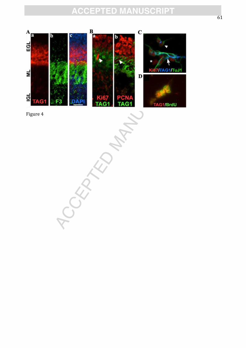
AC
CEPTED
MAN
USC
RIP
T
ACCEPTED MANUSCRIPT 61
Figure 4
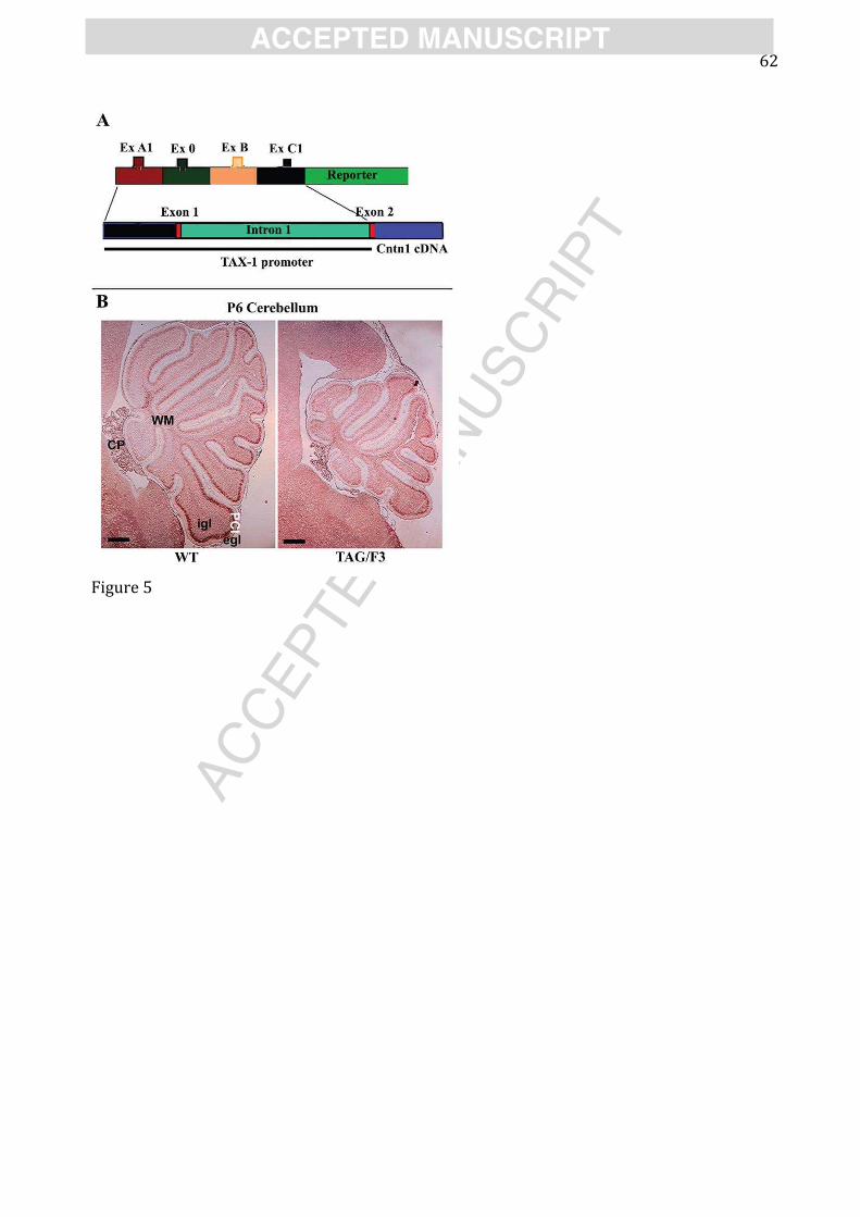
AC
CEPTED
MAN
USC
RIP
T
ACCEPTED MANUSCRIPT 62
Figure 5

AC
CEPTED
MAN
USC
RIP
T
ACCEPTED MANUSCRIPT 63
Figure 6
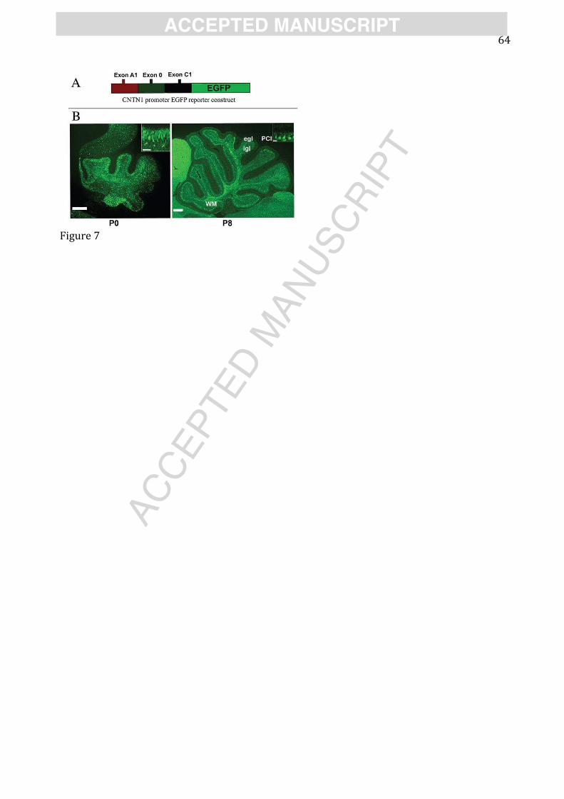
AC
CEPTED
MAN
USC
RIP
T
ACCEPTED MANUSCRIPT 64
Figure 7

AC
CEPTED
MAN
USC
RIP
T
ACCEPTED MANUSCRIPT 65
Graphical Abstract 1. Adhesion molecules in neural development. 2. Morphoregulatory molecules.
3. Immunoglobulin superfamily adhesive glycoproteins.
4. Contactins (CNTN).
5. Contactins 1-2 and their interactions.
6. Contactins genes differential regulation.
7. Contactins expression and the action potentials generation.
8. Signalling mechanisms in axonal adhesive glycoproteins function.
9. Significance of Contactin 1 regulated expression in development.
10. Significance of Contactins expression in neurological disorders.
AC
CEPTED
MAN
USC
RIP
T
ACCEPTED MANUSCRIPT 66
Highlights: 1.This manuscripts deals with the properties of the GPI anchored adhesive glycoproteins expressed at the axonal level. The main focus is on two components of the Contactin Family (CNTN1 and CNTN2). 2. The developmental significance of the regulated expression of these molecules is mostly described in the attempt to explore their significance in both development of the nervous tissue and in some neurological disorders.