The Role of COX-2 in Cell Proliferation and Cell Death in Human Malignancies
21
Hindawi Publishing Corporation International Journal of Cell Biology V olume 2010, Article ID 215158, 21 pages doi:10.1155/2010/215158 Review Article Th eRol e of Cycl oo xy genase-2 in Ce ll Pro li fe rati on and Ce ll Death in Huma n Ma lign ancies Cyril Sobol ewski , 1 Clau dia Cere lla, 1 Mari o Dica to, 1 Lina Ghibe lli, 2 and Mar c Diederic h 1 1 Laboratoire de Biologie Mol ´ eculaire et Cellulaire du Cancer, H ˆ opital Kirchberg, 9 rue Edward Steichen, 2540 Luxembourg, Luxembourg 2 Dipartimento di Biologia, Universit ` a di Roma di Roma Tor Vergata, Via Ricerca Scientifica snc, 00133 Rome, Italy Correspondence should be addressed to Marc Diederich, [email protected] Received 16 July 2009; Accepted 18 De cember 2009 Academic Editor: Simone Fulda Copyright © 2010 Cyril Sobolewski et al. This is an open access article distributed under the Creative Commons Attribution License, which permits unrestricted use, distribution, and reproduction in any medium, provided the original work is properly cited. It is well admitted that the link between chronic inflammation and cancer involves cytokines and mediators of inflammatory pathways, which act during the diff erent steps of tumorigenesis. The cyclooxygenases (COXs) are a family of enzymes, which catalyze the rate-limiting step of prostaglandin biosynthesis. This family contains three members: ubiquitously expressed COX- 1, which is involved in homeostasis; the inducible COX-2 isoform, which is upregulated during both inflammation and cancer; and COX-3, expressed in brain and spinal cord, whose functions remain to be elucidated. COX-2 was described to modulate cell proliferation and apoptosis mainly in solid tumors, that is, colorectal, breast, and prostate cancers, and, more recently, in hematological malignancies. These findings prompt us to analyze here the e ff ects of a combination of COX-2 inhibitors together with diff erent clinically used therapeutic strategies in order to further improve the efficiency of future anticancer treatments. COX- 2 modulation is a promising field investigated by many research groups. 1. Intr oduct ion: Infl ammation and Ca nc er ar e Linke d Infl ammati on is the maj or rea ctio n of nat ura l immuni ty with the goal to defend the organism against pathogens. It can be induced upon bacterial infections by compounds including lipopolysaccharides, as well as by viruses, which are detected by Toll-like receptors (TLRs), expr essed by immune cells like macrophages. Besides, inflammation can be triggered by physical injuries (i.e., UV) or chemical compounds (i.e., reactive oxygen species) [ 1]. The activation of specific recep- tors triggers intracellular signals (i.e., NFκB, p38, or MAPKs- medi ated) , which regulatepro-inflammatory cytokine expr e- ssi on, suc h as int erl euk in 1 bet a (IL1 β), tumor nec ros is fac tor alpha (TNFα), interleukin 6 (Il6), together with chemokines and cel l adh esion pro tei ns [1], in turn, leading to the recruitment and the activation of immune cells. Sev era l dis eas es are ass oci ate d to chr oni c inflamma- tion, suc h as ost eoa rthri tis , Cro hn ’ s dis ease, and cancer [2]. Although the first evidence of a connection between inflammation and cancer dates back to more than a century ago [3], only recently, this link has been further investigated, thus evid encing tha t the inc ide nce of sev era l can cer s is tightly associated to inflammation such as colon, breast, and prostate cancers [4–6]. This hypothesis is supported by the findings that the tumor microenvironment is characterized by the infiltration with di ff erent types of immune cells ( i.e., dendritic cells, lymphocytes, and macrophages) responsible for the release of cytokines [ 1]. The role of these cytokines in tumor incidence has been established in many studies. For exa mpl e, the overe xpr ession of TNFα in transge nic mice bearing a lung tumor is associated with an increase of the size of the tumor [ 7]. Moreover, a chronic intake of nonsteroidal antiinflammatory drugs (NSAIDs) leads to a sig nifican t red ucti on in the inc ide nce of suc h tumors. Colorectal cancer (CRC), which remains an important cause of death in the indus tria lized world, is one of the most characterized types of tumor that benefits from treatment by NSAIDs [8]. Interestingly, chronic use of aspirin is reported to reduce the relative risk of CRC by about 50% [ 9]. Familial adenomatous polyposis, an inherited form of colon cancer, is characterized by the development of preneoplastic polyps.
Transcript of The Role of COX-2 in Cell Proliferation and Cell Death in Human Malignancies

8/2/2019 The Role of COX-2 in Cell Proliferation and Cell Death in Human Malignancies
http://slidepdf.com/reader/full/the-role-of-cox-2-in-cell-proliferation-and-cell-death-in-human-malignancies 1/21
Hindawi Publishing CorporationInternational Journal of Cell Biology Volume 2010, Article ID 215158, 21 pagesdoi:10.1155/2010/215158
Review ArticleThe Role of Cyclooxygenase-2 in Cell Proliferation and CellDeath in Human Malignancies
Cyril Sobolewski,1 Claudia Cerella,1 Mario Dicato,1 Lina Ghibelli,2 and Marc Diederich1
1 Laboratoire de Biologie Mol eculaire et Cellulaire du Cancer, H opital Kirchberg, 9 rue Edward Steichen, 2540 Luxembourg, Luxembourg
2 Dipartimento di Biologia, Universit a di Roma di Roma Tor Vergata, Via Ricerca Scientifica snc, 00133 Rome, Italy
Correspondence should be addressed to Marc Diederich, [email protected] 16 July 2009; Accepted 18 December 2009
Academic Editor: Simone Fulda
Copyright © 2010 Cyril Sobolewski et al. This is an open access article distributed under the Creative Commons AttributionLicense, which permits unrestricted use, distribution, and reproduction in any medium, provided the original work is properly cited.
It is well admitted that the link between chronic inflammation and cancer involves cytokines and mediators of inflammatory pathways, which act during the diff erent steps of tumorigenesis. The cyclooxygenases (COXs) are a family of enzymes, whichcatalyze the rate-limiting step of prostaglandin biosynthesis. This family contains three members: ubiquitously expressed COX-1, which is involved in homeostasis; the inducible COX-2 isoform, which is upregulated during both inflammation and cancer;and COX-3, expressed in brain and spinal cord, whose functions remain to be elucidated. COX-2 was described to modulatecell proliferation and apoptosis mainly in solid tumors, that is, colorectal, breast, and prostate cancers, and, more recently, inhematological malignancies. These findings prompt us to analyze here the eff ects of a combination of COX-2 inhibitors togetherwith diff erent clinically used therapeutic strategies in order to further improve the efficiency of future anticancer treatments. COX-2 modulation is a promising field investigated by many research groups.
1. Introduction: Inflammation andCancer are Linked
Inflammation is the major reaction of natural immunity withthe goal to defend the organism against pathogens. It can beinduced upon bacterial infections by compounds includinglipopolysaccharides, as well as by viruses, which are detectedby Toll-like receptors (TLRs), expressed by immune cells
like macrophages. Besides, inflammation can be triggeredby physical injuries (i.e., UV) or chemical compounds (i.e.,reactive oxygen species) [1]. The activation of specific recep-tors triggers intracellular signals (i.e., NFκB, p38, or MAPKs-mediated), which regulate pro-inflammatory cytokine expre-ssion, such as interleukin 1 beta (IL1 β), tumor necrosis factoralpha (TNFα), interleukin 6 (Il6), together with chemokinesand cell adhesion proteins [1], in turn, leading to therecruitment and the activation of immune cells.
Several diseases are associated to chronic inflamma-tion, such as osteoarthritis, Crohn’s disease, and cancer[2]. Although the first evidence of a connection betweeninflammation and cancer dates back to more than a century
ago [3], only recently, this link has been further investigated,thus evidencing that the incidence of several cancers istightly associated to inflammation such as colon, breast, andprostate cancers [4–6]. This hypothesis is supported by thefindings that the tumor microenvironment is characterizedby the infiltration with diff erent types of immune cells (i.e.,dendritic cells, lymphocytes, and macrophages) responsiblefor the release of cytokines [1]. The role of these cytokines
in tumor incidence has been established in many studies.For example, the overexpression of TNFα in transgenicmice bearing a lung tumor is associated with an increaseof the size of the tumor [7]. Moreover, a chronic intakeof nonsteroidal antiinflammatory drugs (NSAIDs) leads toa significant reduction in the incidence of such tumors.Colorectal cancer (CRC), which remains an important causeof death in the industrialized world, is one of the mostcharacterized types of tumor that benefits from treatment by NSAIDs [8]. Interestingly, chronic use of aspirin is reportedto reduce the relative risk of CRC by about 50% [9]. Familialadenomatous polyposis, an inherited form of colon cancer,is characterized by the development of preneoplastic polyps.

8/2/2019 The Role of COX-2 in Cell Proliferation and Cell Death in Human Malignancies
http://slidepdf.com/reader/full/the-role-of-cox-2-in-cell-proliferation-and-cell-death-in-human-malignancies 2/21
2 International Journal of Cell Biology
At the molecular level, this disease is caused with a mutationof a tumor suppressor gene called Adenomatous polyposiscoli (APC). It has been shown that the use of NSAIDs, likesulindac, as a chemopreventive treatment, is able to decreasethe incidence of polyp formation [10]. Similar results wereobtained with celecoxib [11], which is now approved by the
Food and Drug Administration’s Oncologic Drugs Advisory Committee as an adjuvant in FAP therapy.A body of evidence indicates a role for inflammation
in the development/modulation of diff erent steps of cancerprogression. Inflammation may play a role in tumor initia-tion by triggering the production of reactive oxygen species(ROS), responsible for DNA damage, thus increasing therate of mutations [12]. It may also be implicated in tumorpromotion, where inflammation triggers the secretion of growth factors, such as the epithelial (EGF) and fibroblastgrowth factors (FGF). These, in turn, favor the proliferationof initiated tumor cells by determining an imbalance betweencell proliferation and cell death stimuli [6], due to theactivation of diff erent cell survival pathways [7].
Besides, the diff erent cytokines produced during inflam-mation (i.e., TNFα, IL1 β, IL6, and IL8) can also activateseveral survival pathways, thus leading to an escape of tumorcells from cell death. Well known is the case of TNFα,produced by tumor and immune cells, which leads to thesurvival of cancer cells by the upregulation of antiapoptoticproteins, that is, Bcl-2 [13–15], via the activation of thenuclear factor kappa B (NFκB) [16]. The modulation of pro-survival pathways or anti-apoptotic proteins makes theexpression/activation of such proinflammatory mediatorsalso a determining factor in chemoresistance. A constitu-tive activation of such proinflammatory factors has beenfrequently found in many cancers, such as hepatocellularcarcinoma [17], prostate cancer [18], as well as chronic andacute myeloid leukemia [19], where it is frequently associatedwith a bad prognosis. In these instances, the modulationof Bcl-2 anti-apoptotic family members has been frequently shown [13–15, 20].
Amongst the diff erent mediators of inflammation, thecyclooxygenases (COXs) clearly appear to be implicated incancer. This review focuses on COX-2, the inducible form,normally induced and implicated in inflammation, andintends to analyze what is currently known about the linkbetween COX-2 and cancer, in terms of eff ects on cell pro-liferation and cell death. In this view, we will focus ourattention on studies analyzing the eff ects of COX-2 inhibitors
on cancer cells, when used alone as well as in combinationwith therapeutic approaches, including radiotherapy, chem-otherapeutic agents, and photodynamic therapy. Finally, wewill consider the relevance of COX-2-independent eff ects.
2. The Cyclooxygenase Enzyme Family
Cyclooxygenases (or prostaglandin H synthases), commonly referred to as COXs, are a family of myeloperoxidases locatedat the luminal side of the endoplasmic reticulum and nuclearmembrane [21], which catalyze the rate-limiting step of prostaglandin biosynthesis from arachidonic acid [21]. Theseenzymes act by two coupled reactions. The first one is the
conversion of arachidonic acid released from the plasmamembrane by phospholipase A2 to prostaglandin G2 by the cyclooxygenase activity. The second reaction is mediatedby the peroxidase activity and leads to the conversionof prostaglandin G2 to prostaglandin H2. Then, diff erentsynthases convert prostaglandin H2 to prostaglandin D2,
F2α, E2, I2, and thromboxane A2 (Figure 1).Prostanoids (prostaglandins and thromboxanes) areimmediately released from the cells, where it is believedthat they act locally in an autocrine and paracrine mannerthrough diff erent receptors activating diff erent intracellularpathways still to be completely elucidated (Figure 1) [22].Prostaglandins, specifically, are important for physiologicalfunctions like vasodilatation (PGD2, PGE2, PGI2), gastriccytoprotection (PGI2), maintenance of renal homeostasis,and platelet aggregation. Besides, prostaglandins play amajor role in mediating fever (PGE2), pain sensitivity, andinflammation [21].
So far, three isoforms of COXs have been identified.Cyclooxygenase-1 (COX-1) is a glycoprotein of 71kDa, whichis constitutively expressed in diff erent tissues. COX-1 isencoded by a gene on chromosome 9 and plays a role in tissuehomeostasis by modulating several cellular processes rangingfrom cell proliferation to angiogenesis or platelet aggregationdue to thromboxane production [21].
Cyclooxygenase-2 (COX-2) is the inducible isoform,which is regulated by growth factors and diff erent cytokinessuch as IL1 β, IL6, or TNFα [23], therefore overexpressedduring inflammation. The COX-2 gene is located on chro-mosome 1 and its promoter displays an NFκB responseelement as well as other cytokine-dependent (i.e., IL6)response elements [21]. The protein shows a 60% homology with COX-1 [24]; in addition, COX-2 presents a C-terminalextension and a diff erent binding site for NSAIDs, whichmakes COX-2 a preferential target compared to COX-1, thusbeing specifically inhibited at lower doses [25].
Finally, COX-3 has been identified as a splice variant of COX-1, and it is present mainly in brain and spinal cord[26, 27]. Currently, the role of COX-3 is not known. Somepieces of evidence suggest a possible role in pain sensitivity,based on studies focused on the mechanism of action of acetaminophen (paracetamol), recently evoked as a selectiveinhibitor of COX-3 [28]. However, this hypothesis is debatedbecause other findings argue that acetaminophen targets atthe same time COX-2 [29].
3. COX-2 As a Tumor Promoter and a GoodCandidate for Cancer Therapy
Overexpression of COX-2 has been detected in a numberof tumors, such as colorectal breast as well as pancreaticand lung cancers [2, 30–32], where it correlates with apoor prognosis. Moreover, overexpression of COX-2 hasbeen reported in hematological cancer models such asRAJI (Burkitt’s lymphoma) and U937 (acute promonocyticleukemia) [33, 34] as well as in patient’s blast cells [32, 34].Data suggested that COX-2 may play a role in diff erent stepsof cancer progression, by increasing proliferation of mutatedcells [30], thus favoring tumor promotion as well as by

8/2/2019 The Role of COX-2 in Cell Proliferation and Cell Death in Human Malignancies
http://slidepdf.com/reader/full/the-role-of-cox-2-in-cell-proliferation-and-cell-death-in-human-malignancies 3/21
International Journal of Cell Biology 3
Arachidonic acid
COX-2
COX-2
NSAIDs, COX-2 selectiveinhibitorsPGG2
PGH2
Isomerases
PGF2α PGD2 PGJ2 PGE2 PGI2
FP receptor DP receptor PPARγ EP 1,2,3, 4 IP receptor
PPARδ
Figure 1: Metabolism of arachidonic acid by COX-2 and receptors implicated in response to prostaglandins (according to Chandrasekharan etal. [21]). Prostaglandins act through diff erent receptors to mediate their eff ects. PGE2 is able to bind four receptors (EP1, 2, 3, and 4). Thesereceptors do not possess the same ligand affinity and their expression is tissuedependent. The diff erent receptors are associated with diff erentintracellular pathways. Most of these receptors are localized in the plasma membrane but nuclear receptors PPARγ can also bind PGJ2.Abbreviation: COX-2, cyclooxygenase-2; PG, prostaglandin; FP, prostaglandin F receptor; DP, prostaglandin D receptor; EP, prostaglandin Ereceptor; IP, prostaglandin I receptor; PPAR, peroxisome proliferator-activated receptor; NSAIDs, nonsteroidal anti-inflammatory drugs.
aff ecting programmed cell death and aff ecting the efficacy of anticancer therapies [35–39] to be, finally, implicated inmetastasis formation, for example, by aff ecting apoptosisinduced by loss of cell anchorage (anoikis) [40].
COX-2 induction or overexpression is associated with anincreased production of PGE2, one of the major products of COX-2 which is known to modulate cell proliferation, celldeath, and tumor invasion in many types of cancer includingcolon, breast, and lung. Prostaglandin E2 acts throughdiff erent membrane receptors called EP receptors (EP1, EP2,EP3, and EP4) [41]. These receptors are all located onthe cell surface and characterized by seven-transmembranedomains, and rhodopsin-type G protein-coupled receptors,but trigger diff erent signaling pathways. Thus, it is knownthat EP1 signaling acts through phospholipase C/inositoltriphosphate signaling, leading to intracellular mobilizationof calcium. EP2 and EP4 receptors are coupled with G
proteins which activate adenylate cyclase, leading to anincrease of intracellular cAMP [41]. cAMP is then able toactivate kinases such as protein kinase A (PKA) or PI3K forexample, and also GSK3 leading to an activation of β-catenin,a pathway regulating cell proliferation [42, 43]. In contrary to EP2 and EP4, EP3 is coupled with Gi protein, leadingto an inhibition of adenylate cyclase, and thus a decreaseof cAMP inside the cells [41]. The diff erential expressionof these diff erent receptors according to the cell type may explain the diverse and antagonist eff ects of PGE2 describedin literature.
Until now, there are multiple evidences about the roleof PGE2 in tumorigenesis in some cancers. These evidences
are mostly described for adherent tumors while this linkis poorly understood for hematopoietic malignancies suchas leukemia or lymphoma. Indeed, several papers havereported that PGE2 is the most important prostaglandin
produced during colorectal carcinogenesis [44]. Moreover, itis known that the level of PGE2 increases in a size-dependentmanner in Familial Adenomatous Polyposis (FAP) patients[45], suggesting a correlation between tumor growth andprostaglandin biosynthesis. Tumorigenesis is characterizedby a disequilibrium between cell proliferation and cell death.PGE2 is able to inhibit apoptosis in human colon cancercells. It has been demonstrated that PGE2 can upregulatethe level of the anti-apoptotic protein Bcl-2 in HCA-7 cells(adenocarcinoma), which produce significant amounts of PGE2. This paper described a modulation of the MAPKpathway that precedes the upregulation of Bcl-2 [46]. PGE2can mediate its eff ect through EGF receptor, leading to
MAPK activation. The ability of PGE2 to modulate tumorprogression in colorectal cell has been shown in othermodels of colon cancer such as HT-29 cells that expressEP receptors. In this cell type, PGE2 is associated with anincrease of cAMP through EP4 receptor. The eff ect can bereversed by L-161982, an antagonist of EP4 [47]. Moreover,PGE2 transactivates EGFR by triggering the release of amphiregulin, a well-known EGFR ligand [48]. SC-236, aninhibitor of COX-2, is able to inhibit cell proliferation of HT-29 cells and this eff ect is greater in combination with anamphiregulin neutralizing antibody [47]. In this cell line, theexpression of amphiregulin is correlated to the expression of COX-2.

8/2/2019 The Role of COX-2 in Cell Proliferation and Cell Death in Human Malignancies
http://slidepdf.com/reader/full/the-role-of-cox-2-in-cell-proliferation-and-cell-death-in-human-malignancies 4/21
4 International Journal of Cell Biology
The transactivation of EGFR by PGE2 can lead also to AKTactivation, which is a well-known survival pathway [49].This eff ect was well described in a study by Tessner et al.[50] demonstrating that 16,16-dimethyl PGE2 (dmPGE2)inhibits radiation-induced apoptosis in the mouse intestinalepithelium. Using HCT-116 cell line as a model to reflect
the eff
ect on mouse small intestine, it has been shownthat the anti-apoptotic eff ect of dmPGE2, which is knownto bind EP2, was tightly related to AKT phosphorylationthrough activation of EGFR and leads to an inhibition of Bax translocation in mitochondria, an important step forapoptosis [51].
PGE2 modulates also tumor growth of lung cancer. Thiseff ect has been described by Yamaki et al. [52] showingthat PGE2 activates Src kinase in A549 cells, leading toan induction of cell growth. These cells express EP3 thatactivates Src (sarcoma) kinase. This study has demonstratedthat the activation of Src leads to an activating phosphory-lation of STAT3, a transcription factor known to regulatecyclin D1 transcription, an important positive regulatorof cell proliferation. Apoptosis can be inhibited becauseSTAT3 regulates the transcription of Bcl-XL, a well-knownanti-apoptotic protein [53]. Moreover, Src phosphorylatesp27, a protein known to inhibit cell cycle progressionespecially at the G1/S transition [54]. However, it has beenrecently shown that this protein plays a dual role as theunphosphorylated form of p27 inhibits the cell cycle, andthus cell proliferation. If phosphorylation occurs on T157and T198 by PI3K (phosphoinositide 3-kinase), it triggerscell cycle transition by stabilizing the cyclin D1/cdk4 complex[55]. Thus phosphorylation of S10 appears to be importantfor other phosphorylation steps and it has been hypothesizedthat Src kinase can play this role [55]. Moreover, it isknown that phosphorylation of p27 is responsible also forits degradation by the proteasome [56]. All together thesedata suggest that PGE2 increases cell proliferation via p27phosphorylation through EP4 receptors.
Nonsmall lung cancer is characterized by a Ras mutationcorrelated with a poor prognosis [57]. Activation of Ras leadsto an upregulation of COX-2 resulting in increased PGE2production [58]. PGE2 increases cell proliferation of A549cells (adenocarcinoma) and this eff ect is associated with anactivation of Ras pathway via EP4 receptor. In this case, PGE2mediates its eff ect by the release of amphiregulin, the mostabundant ligand in A549 cells [59]. EGFR activation leads toactivation of MAPK pathway that regulates cell proliferation
by transactivating several oncogenes such as c-myc [60].PGE2 is also important for tumor invasion. A study by
Ma et al. [61] described that PGE2 can increase the numberof metastasis. This eff ect has been demonstrated in a modelin which murine mammary tumor cells 66.1 were injectedin syngenic immune competent BALB/CByJ mice. All thesecell lines express EP1, 2, 3, and 4. The use of EP4 antagonists(AH23848 and AH6809) decreased surface tumor coloniesand reduced tumor invasion. Another study has revealed thatPGE2 increases the level of VEGF in granuloma [62]. VEGFis an important factor of angiogenesis, and thus of tumorprogression by enhancing the vascularization of the tumors[63].
Alltogether these data together suggest that PGE2 and,thus, COX-2 play an important role in tumor progressionby enhancing cell proliferation, cell survival, and tumorinvasion. The diversity of PGE2 receptors and their diff erentsignaling pathways suggest that the protumorigenic eff ectof PGE2 depends on the cell type and the type of receptor
expressed. Until now, many signaling pathways associatedwith tumor progression are linked to PGE2 and this couldexplain why the use of COX-2 inhibitors is a good strategy in cancer therapy. However, the signaling pathways of EPreceptors are not completely characterized and their preciseroles in the diff erent cancers remain to be elucidated before aclinical application.
COXs may be targets of several compounds that may inhibit their functions. Combination of such preferential orselective COX-2 inhibitors with anti-cancer agents already used in clinics were tested with the goal to improve theefficiency of anti-cancer protocols.
COX-2 is the preferential target of several NSAIDs(Figure 2) [64, 65]. Historically, NSAIDs used for clinical andanti-inflammatory purposes were represented by the nonse-lective COX-2 inhibitors, to which belong aspirin, sulindacacid and, more recently, agents such as nimesulide, ibuprofenand naproxen. As their definition well reflects, this firstgeneration of NSAIDs may aff ect both main COXs isoforms,even if preferentially COX-2 (see above). Their mechanismsof action are not all completely elucidated, complicated by the fact that diff erent agents seem to act in diff erent ways.For example, diff erent NSAIDs bind the active site of COX-2. Commonly, binding occurs by a reversible competitiveinhibition (i.e., ibuprofen, naproxen, and indomethacin).In contrast, aspirin is able to acetylate the active site of COX at a serine residue, leading to an irreversible inhibition(see Figure 2, summarizing the classification of COX-2inhibitors mentioned in this review). Considerable sideeff ects generated by the interference with homeostatic func-tions modulated by COX-1 include increased incidence of gastrointestinal hemorrhage and ulceration upon chronic orlong-time intake [66]. A novel generation of COX-2-selectiveinhibitors NSAIDs termed “Coxibs” was then developed.These compounds promised to be much less gastrotoxic.They act as competitive inhibitors of the active site of COX-2 and present indeed a higher specificity. However,concerns related to a long-time/chronic intake of thesedrugs raised quite soon, following some clinical reports,suggest a correlation between an increased risk of myocardial
infarction and their consumption [67]. This has lead tothe voluntary withdrawal of some of these agents, that is,rofecoxib and valdecoxib [68], and drastic regulatory advicesregarding the use of the other ones, thus opening a discussionon the real benefits versus side eff ects of their use in clinics.Consequently, studies focused on the use of traditionalversus COX-2-selective NSAIDs, frequently associated to theelaboration of economical models, have been performed inthese latest years, with the aim to evaluate the real riskstogether with the costeff ectiveness and, possibly, identify classes of users/patients where regular NSAIDs intake may bebeneficial. Although, further analyses need to be performed,a number of reports suggest that Coxibs may really increase

8/2/2019 The Role of COX-2 in Cell Proliferation and Cell Death in Human Malignancies
http://slidepdf.com/reader/full/the-role-of-cox-2-in-cell-proliferation-and-cell-death-in-human-malignancies 5/21
International Journal of Cell Biology 5
O
O
O
O
S
OO
F
S
F
O
O
O
O
N N
O
N
S
OO
H
SO
O
NN
CH
S
OO
O
O
S
O
N
O
O
O
SCH
O
O
N
O
CF
S
O
O
N
N
O
OO
S
S
F
S
O
O
S
O O
ON
S
O
S
O
O
HOH3C H3C
COOHCOOH
COOH
H3C
H3
3
3
CH3
C
H3C
H3C
H3C
H3
3
3
C
HC
H3C
Cl
Cl
Cl
NH
OH
OH
N
2
H3CO
CF3
NH2
2
2
NH
NO2
NH
2NH
CFBr
OH
NH
NH
NO
O
O
OH
N
Aspirin
IbuprofenSulindac sulphone
Sulindac sulphide
Diclofenac
Nabumetone
Naproxen
Indomethacine Piroxicam
CelecoxibRofecoxib
ValdecoxibNS-398
Cay10404
SC-236
DUP697
Meloxicam Nimesulide
Figure 2: COX-2 inhibitor classification. COX-2 is the target of many compounds. COX-2 inhibitors described in this review are classifiedaccording to their ability to inhibit COX-2: nonselective (green), selective (pale blue), and preferential (grey).

8/2/2019 The Role of COX-2 in Cell Proliferation and Cell Death in Human Malignancies
http://slidepdf.com/reader/full/the-role-of-cox-2-in-cell-proliferation-and-cell-death-in-human-malignancies 6/21
6 International Journal of Cell Biology
cardiovascular risks only in patients presenting a positivity to other cardiovascular factor risks, as high blood pressureand altered lipid metabolism [69–73]. These results suggestthat their use should be limited to patients with a low riskof cardiovascular complications after analysis of multiplebiomarkers [Chaiamnuay et al., 2006, clinical reviews].
Therefore, the future perspective in the pharmacologicaluse of preferential versus selective COX-2 inhibitors is theidentification of a panel of interesting biomarkers, helpingin defining individual biological risk factors and limiting theuse of a specific class of COX-2 inhibitors to the appropriateresponders [74, 75]. This approach will have a considerableimplication in therapy as well as in chemoprevention of inherited forms of colon cancer.
It is interesting to mention that recent alternativeapproaches have been considered. Strillacci et al. [76] andChan et al. suggested RNA interference using adenoviralvehicles. Moreover, other selective COX-2 inhibitors havebeen developed and experimentally used: SC-558 [35] ,DUP-697 [77], SC-58125 [78], and NS-398 [8]. Some of them induce an irreversible inhibition. This is the case forNS-398, which acts by inducing a conformational changeof COX-2 [25] (Figure 2). Another strategy discussed inliterature could be the use of EP receptor antagonists. Indeed,it has been demonstrated that EP antagonists can decreasecell proliferation and cell invasion [47, 61, 79]. This could bea more specific strategy that could limit the other side eff ectsof classic COX-2 inhibitors.
4. COX-2 As a Regulator of Cell Proliferation
Cell cycle is regulated by diff erent serine-threonine kinaseproteins called cyclin-dependent kinase (Cdk). These pro-teins regulate the diff erent steps of cell cycle progressionby phosphorylating many substrates (i.e., nuclear lamins)[54]. These proteins are regulated by phosphorylation anddephosphorylation. Thus, Cdks can be activated by phos-phatases such as CDC25C (cell division cycle 25 homolog C ) for CDK1 or kinase like CAK (Cdk activating kinase).The activity of cdks is also regulated by cyclins, which formheterodimers with cdks leading to an activation of Cdks by conformational change [54, 80].
Cell cycle is under the control of other factors, implicatedin the regulation of cell cycle transition. These regulatory mechanisms form checkpoints where the cell cycle can bestopped after cellular damage in order to allow repair and
to maintain cellular integrity or, alternatively, to eliminatemutated and potentially dangerous cells. The INK4 family (p16, p15, p18, and p19) and the Cip/Kip family (p21, p27,and p57) [54, 80, 81] are key regulators of G1/S transition.For example, after DNA damage, p53, a tumor suppres-sor gene, activates transcription of p21, which inhibitscyclin E phosphorylation leading to hypophosphorylationof retinoblastoma protein (pRb) [81]. INK4 family inhibitsCdk4 and Cdk6, whereas Cip/Kip family inhibits all Cdks.Retinoblastoma protein needs to be phosphorylated in orderto release transcription factor E2F activating genes involvedin the S phase-like PCNA (proliferating cell nuclear antigen)[82]. p53 is also important for the regulation of the G2/M
transition, which requires activation of the cyclin B-cdk2complex. This complex accumulates during the previous stepof the cell cycle but is inactivated by a phosphorylationat tyrosine 15 and threonine 14 by Wee 1 and Myt 1.These phosphate groups are removed by the phosphataseCDC25A when cells enter mitosis. In the case of DNA
damages, p53 is activated and increases the level of p21that is directly inhibiting cdk2. Moreover, 14-3-3 protein,a transcriptional target of p53, leads to a sequestration of cdk2 in the cytoplasm [83]. Other mechanisms involved inthe regulation of the G2 checkpoint or the mitotic spindlecheckpoint are reviewed by Stewart et al. [54].
Cancer cells are characterized by deregulation of the cellcycle via alteration of cell cycle controllers (cyclins) and cellcycle regulators (p53) [54], resulting in a perturbation of cellcycle checkpoints.
Currently, there is evidence that prostaglandins producedby COX-2 intervene in tumor cell proliferation as NSAIDsand selective COX-2 inhibitors inhibit proliferation of diff erent cancer cell types expressing COX-2 [30]. NS-398,a COX-2 specific inhibitor, was described to reduce cellproliferation of MC-26 cell line, a highly invasive mouseCRC cell model expressing constitutively COX-2 [8]. Thiseff ect was associated with a reduction of cyclin D level, akey protein involved in G1-S transition [54], and PCNA,thus increasing the processivity of DNA polymerase [82].NS-398 and COX-2 specific inhibitor nabumetone reducedcell proliferation of U937 (acute promonocytic leukemia)and ML1 (human myeloblastic leukemia), thus leading to anaccumulation in G0/G1 phase [33]. Interestingly, meloxicamwas also able to downregulate PCNA and cyclin A in HepG2cell line (hepatocellular carcinoma cells), leading to aninhibition of the cell proliferation and an accumulation of the cells in G0/G1 phase of cell cycle [84]. Alternatively,the link between COX-2 and CRC has been demonstratedby the fact that prostaglandin E2 (PGE2) derivating fromCOX-2-mediated arachidonic acid metabolism increased theproliferation of colorectal cancer cells [85].
The inhibitory eff ect of NSAIDs on cell proliferationof CRC has been also observed in ovarian cancer. Indeed,treatment of OVCAR-3 tumors xenotransplanted in nu/numice (nude mice) with aspirin and piroxicam (NSAIDs) andthe selective COX-2 inhibitor meloxicam led to a reductionof tumor growth [86].
It has been estimated that 40% of breast cancers showan overexpression of COX-2, which is associated with a bad
prognosis [5]. Indomethacin (NSAIDs), celecoxib, rofecoxiband nimesulide have been shown to able to inhibit cell prolif-eration of these cells [5]. Moreover, prostaglandins were ableto increase cell proliferation of hormonal-dependent breastcancer by increasing transcription of CYP19 aromataseimplicated in estrogen biosynthesis [87].
Several studies revealed that inhibition of COX-2 by celecoxib in Burkitt’s lymphoma cell lines RAJI, BjAB,(Epstein-Barr virusnegative), and BL41 led to a reductionof cell proliferation [34]. NS-398 and celecoxib were able toreduce proliferation of pancreatic cancer cell line, Panc-1 ina dose-dependent manner [88]. Treatment with celecoxib of these cells implanted into nude mice led to a reduction of

8/2/2019 The Role of COX-2 in Cell Proliferation and Cell Death in Human Malignancies
http://slidepdf.com/reader/full/the-role-of-cox-2-in-cell-proliferation-and-cell-death-in-human-malignancies 7/21
International Journal of Cell Biology 7
Growth factors
p21Cip1
p27kip1
Cyclin B
CDC25
Cyclin B
CDK1
CDK1
Wee1G2
M
NS-398 (MC-26)meloxicam(HepG2)
PCNA S
Meloxicam(HepG2) Cyclin A
CDK2
p21p27
E2F1/2,PCNA
cyclin A/EPol α · · ·
AAAAA E2F
p21,p27
CDK2
Cyclin E
pRB
p21Cip1
p53
Genotoxicstress
Celecoxib(K562)DUP-697(K562)p15, p16, p18, p19G1
E2F 1-3
pRB
CDK 4/6
Cyclin D1
E2F 1-3
NS-398 (MC26)Celecoxib (K562)DUP-697 (K562)
P
P P
P
P PP
Figure 3: E ff ects of COX-2 inhibitors on cell proliferation. Cell cycle is divided into diff erent steps: G1, S, G2, and M (mitosis). This processis regulated by cyclin proteins, which activate cyclin-dependent kinase (cdk) and phosphatase (i.e., CDC25) or kinase like cyclin-dependentkinase inhibitors such as p16, p15, p18, p19, p21, and p27 [ 54]. Selective COX-2 inhibitors are able to modulate some cell cycle checkpoints.In this picture, some examples of this link have been shown for diff erent cell types: MC26, colorectal cancer; HepG2, hepatocellularcarcinoma; K562, chronic myeloid leukemia. Cdk; cyclin-dependent kinase; pRb, retinoblastoma protein; PCNA, proliferating cell nuclearantigen.
the volume of the tumor [88]. Other studies have shown thatcelecoxib is able to reduce cell proliferation of the chronicmyeloid leukemia (CML) cell line K562, which expressesCOX-2 at the mRNA and protein level [89]. This eff ectwas accompanied by an accumulation of cells in G0/G1.Moreover, the inhibition of cell proliferation was correlatedto a downregulation of cyclin D1, cyclin E, and pRb and theupregulation of p16 and p27 [89]. Similar results were foundon this cell type with the other selective COX-2 inhibitorDUP-697 [77]. Diff erent eff ects are recapitulated in Figure 3.
5. Implication of COX-2 in Cell Death
5.1. Apoptosis. Apoptosis (type I cell death) is importantfor the development and maintenance of tissue homeostasisof multicellular organisms [90, 91]. This active form of cell death is characterized by the occurrence of typicalcell alterations including plasma membrane blebbing, cellshrinkage, chromatin condensation and nuclear fragmen-tation, and, finally, formation of apoptotic bodies, whichcan be phagocyted by macrophages [92]. Deregulation of apoptosis is linked to several pathophysiological disorders,including autoimmune disorders, Alzheimer’s disease, andcancer [93].
Two major cascades of intracellular events are commonly involved in mediating apoptosis (Figure 4). The intrinsic
pathway, also called the mitochondrial or stress-inducedapoptotic pathway, is activated in response to damagingstresses, such as DNA damage. Typical hallmarks of this path-way are mitochondrial outer membrane permeabilization(MOMP), accompanied by a collapse of the mitochondrialmembrane potential [51]. These events lead to the releaseof cytochrome c into the cytosol, which is an indispensablecomponent of the apoptosome, the death complex formedalso by APAF-1, and procaspase-9. Once recruited, thisprotease is cleaved to its activated form (caspase-9) to furtheractivate the executor caspase-3 and, finally, to finalize theapoptotic program.
Alternatively, the extrinsic, or physiological, apoptotic
pathway (Figure 4) can be triggered upon binding of specificligands to death receptors characterized by the presence of a death eff ector domain [94]. Ligands include cytokines,such as TNFα, tumor necrosis factor-related apoptosis-inducing ligand-induced apoptosis (TRAIL), or FAS. Afterbinding, death inducing silencing complex (DISC) is formed.The DISC is composed by the adaptors proteins TRADD(TNF receptor-associated death domain) and FADD (Fas-associated death domain) and is able to recruit and activatepro-caspase-8. Finally, caspase-8 activates caspase-3 in orderto trigger the final steps of apoptosis (Figure 4).
Cross-talks between the two pathways take place. Theextrinsic apoptotic pathway can activate the intrinsic

8/2/2019 The Role of COX-2 in Cell Proliferation and Cell Death in Human Malignancies
http://slidepdf.com/reader/full/the-role-of-cox-2-in-cell-proliferation-and-cell-death-in-human-malignancies 8/21
8 International Journal of Cell Biology
SulindacindomethacineSC-236 (HT29)
celecoxib (MG-63)DR5 clustering in
cholesterol-rich domainby DUP-697 in HT29 cells
Increase of TRAIL receptorby NS-398 and CAY10404 in
SK-Hep 1 and HLE cells
TNFα,TRAIL,
FasL
GSH depletion
Bax
Bak
Cyt
Cyt
Bax
Bak
Bak
Bax
BakBcl-2Bak
CytCyt
CytDiablo
Bax
PI3K/PKB
Bcl-2
BadBad
14-3-3
P T
P
Bcl-xL
Diclofenac(HT29; HCT-15)
NS-398CAY10404
(SK-Hep; HLE)
NFkB
Activationtranscription Bax, Bid,
casp-9
Celecoxib(neuroblastoma)
NS-398CAY10404
(SK-Hep1; HLE)
Tap 73
Apoptosome
LAPDiablo
IAPs
Bcl-2
Bcl-xL
APAF
Cyt
Bid
ProCasp9
DNA damage
Lamin AGelsolin
β-catenin
Casp-3Casp-9
Casp-8
DUP-697(K562)
Pro-casp-8
D E D
D E D D
I S C
F A D D
F A D D
DD
Cyt
Cyt
Cyt
CytCyt
Cyt
Cyt
Figure 4: E ff ects of COX-2 inhibitors on apoptosis. Apoptosis can be mainly mediated by two pathways: the mitochondrial, intrinsic, orstress-induced apoptosis, which is activated in response to damaging stresses and the extrinsic pathway, triggered by the binding of ligandsto specific death receptors [51]. COX-2 inhibitors are able to modulate stress-induced apoptosis as well as extrinsic apoptosis in several celltypes. In this picture, some examples of these interaction discussed in the text are presented for diff erent cell types: LNCaP, prostate cancer;K562, chronic myeloid leukemia; HT29, colorectal cancer; SK-Hep 1 and HLE, human hepatocarcinoma cells; HepG2, hepatocarcinoma;Be17402, hepatocarcinoma; SMMC-7402, hepatocarcinoma; MG-63, osteosarcoma. Abbreviation: AIF: apoptosis-inducing factor; Bcl-2, Bcell lymphoma 2; Bid, Bcl-2 interacting domain; Casp, caspase; Cyt, cytochrome C; DD, death domain; DED, death eff ector domain; DISC,death-inducing silencing complex; PI3K/PKB, phosphatidyl inositol-3 kinase/protein kinase B; FADD, Fas-associated death domain; GSH,glutathione; PTP, transition permeability pore; TNF, tumor necrosis factor; TRAIL, TNF-related-inducing-apoptosis-ligand.
pathway via truncation of the BH3-only protein Bid (t-Bid) by caspase-8. t-Bid interacts with mitochondria, by favoring the activation of the proapoptotic Bcl-2 family members Bak and Bax, thus leading to MOMP and caspase-9 activation [51, 95] (Figure 4). The intrinsic apoptoticpathway may, in turn, activate caspase-8, downstream tocaspase-3 [96] (Figure 4). Cross-talks represent an important
strategy of amplification loops carried out by dying cells toensure/potentiate cell death.
5.1.1. Involvement of COX-2 in Intrinsic Apoptosis. Whencells are damaged by a variety of chemicals or physical stress(i.e., reactive oxygen species, UV, and ionizing radiation),they undergo apoptosis by triggering the intrinsic apoptoticpathway (Figure 4). This pathway may be associated with aredox disequilibrium, mediated by depletion of glutathione(GSH) [94, 97, 98], required for the activation and transloca-tion to mitochondria of the Bcl-2 pro-apoptotic member Bax[98], which, in turn, forms complexes (oligomers) mediatingMOMP and cytochrome c release. As Bax, Bak may play the
same role [99]. In contrast to Bax, Bak is already presentat the surface of mitochondria, normally sequestered in itsactive monomeric form by the Bcl-2 anti-apoptotic membersBcl-xL and Mcl-1 (see Burlacu for a general overview of the Bcl-2 family members modulation involved in Bax/Bakactivation [51]).
Apoptosis is regulated in order to maintain tissue home-
ostasis. This regulation implicates protein-protein inter-actions, with some of them counteracting apoptosis. Inthis view, the interaction between Bcl-2 family pro- andantiapoptotic members represents a crucial and delicate step.Bcl-2 is the best described member of this family preventingBax activation [51]. Bax can form also a complex with theanti-apoptotic protein Bcl-xL [53] and Mcl-1 [14]. Similarly,Bak activity is monitored by the anti-apoptotic members Bcl-xL and Mcl-1 [51]. The interaction between Bax/Bak and theBcl-2 family anti-apoptotic members is carefully regulated by the BH-3-only proteins. Another carefully regulated down-stream checkpoint of the apoptotic pathway is the activationof caspases. Inhibitor of apoptosis (IAP) family, by directly

8/2/2019 The Role of COX-2 in Cell Proliferation and Cell Death in Human Malignancies
http://slidepdf.com/reader/full/the-role-of-cox-2-in-cell-proliferation-and-cell-death-in-human-malignancies 9/21
International Journal of Cell Biology 9
interacting with caspases (i.e., XIAP, survivin [100]) controlsand prevents their activity once cleaved. IAPs monitoringfunction can be, in turn, counteracted by the pro-apoptoticSMAC/DIABLO, a mitochondrial heterodimer, which isreleased from mitochondria when MOMP is aff ected [101].This interaction favors the induction of apoptosis.
Imbalance between cell proliferation and apoptosisobserved in cancer can be tightly related to an alteredfunction of pro-apoptotic proteins as well as to an up-regulation of anti-apoptotic proteins (i.e., Bcl-2 or IAPs)or a downregulation of tumor suppressor genes (i.e., p53).In addition, the activation of prosurvival pathways (i.e.,PI3K/Akt) may be implicated upstream. Inflammation cancontribute to this imbalance via cytokines secreted in thetumor microenvironment able to activate survival path-ways. For example, TNFα can induce NFκB, leading toan inhibition of apoptosis [38]. COX-2 seems also to play a role in this process because it is known that COX-2inhibition is correlated to an increase of apoptosis in severalcancer models. NS-398 downregulated Bcl-2 expression in anandrogen-sensitive human prostate cancer cell line LNCaPthat exhibited a high constitutive level of COX-2 [102].Similar results have been observed in human colorectalcancer cells (HCA-7 cell line which expresses COX-2) wherePGE2 was able to inhibit apoptosis induced by SC58125,a selective COX-2 inhibitor, and increase Bcl-2 expression[46]. Diff erent mechanisms are supposed to explain howCOX-2 inhibitors may trigger apoptosis. In a number of studies, COX-2 inhibition was linked to a concomitantincrease of intracellular arachidonic acid. In HT-29 humancolon adenocarcinoma cell this accumulation led to theinduction of apoptosis [103]. The arachidonic acid-inducedapoptosis was inhibited by Bcl-2 transfection, indicatinga role of arachidonic acid in aff ecting Bcl-2 intracellularlevels [103]. Accumulation of arachidonic acid can aff ectapoptosis by mediating an increase of pro-apoptotic intra-cellular ceramides caused by activation of sphingomyelinase[104, 105]. Sulindac sulphide, a metabolite of sulindac, alsoactivates sphingomyelinase and enhances the ceramide levelin the two human colorectal carcinoma cell lines HCT116and SW480 [106].
COX-2 reduces pro-apoptotic nitric oxide (NO) levels incancer cells downstream of prostaglandin production [30](see Brune et al. [107] for an overview on nitric oxide rolein apoptosis). Chang et al. reported that PGE2 preventedapoptosis induced by NGF (nerve growth factor) withdrawal
by increasing the level of dynein light chain, an inhibitorof neuronal NO synthase in pheochromocytoma of therat adrenal medulla PC12 cells, thus leading to decreasedintracellular NO levels [108].
More recently, connections between COX-2 inhibitorsand p53 family members have been described. For example,celecoxib was shown to able to modulate diff erent isoforms of p73, a p53 family member in neuroblastoma cell lines [109].p73 encodes many isoforms with diff erent roles. Tap73 ispro-apoptotic and contains a transactivation domain. Thisisoform is considered as a tumor suppressor gene becauseit seems to be involved in cell cycle regulation as well asin apoptosis induction [109, 110]. In contrast, DeltaNp73
is anti-apoptotic and lacks the transactivation domain.DeltaNp73 is overexpressed in neuroblastoma, leading tochemotherapy resistance [109]. It has been shown thatcelecoxib was able to upregulate Tap73 and downregulateDeltaNp73. These data suggest the useof COX-2 inhibitors asp73 modulators in order to improve efficiency of chemother-
apy [110].The apoptotic eff ect of COX-2 inhibitors has been alsoobserved for other tumor cell types, such as in the chronicmyeloid leukemia model K562 where DUP-697 inducedapoptosis by cell cycle arrest and caspase-8 activation [77].
COX-2 inhibitors can also activate prosurvival pathways.The PI3K/Akt pathway is a survival pathway, frequently activated in cancer cells [49]. PI3K produces PIP3 (phos-phatidylinositol 3,4,5 triphosphate) that activates PDK1(pyruvate deshydrogenase kinase). This protein phosphory-lates and activates PKB (protein kinase B), which, in turn, isresponsible for the phosphorylation of several targets playinga modulator function in apoptosis. An anti-cancer eff ect of celecoxib due to the inhibition of Akt signaling [111] wasobserved in a gastric cancer model. Celecoxib triggered alsoapoptosis in osteosarcoma cells (MG-63) through down-regulation of Bcl-2, survivin and PI3K (phosphoinositide 3-kinase) pathway [112]. Similarly, Hsu et al. [113] found thatinhibition of Akt phosphorylation by celecoxib in prostatecancer models (LNCaP and PC3 cell lines which expressconstitutively COX-2) led to apoptosis, but in this casewithout aff ecting Bcl-2 level.
The PI3K pathway is negatively regulated by PTEN(phosphatase and TENsin homolog), which converts PIP3in PIP2, preventing PKB activation and Bad phosphory-lation/sequestration. Thus PTEN is considered as a tumorsuppressor gene. It has been shown that NS-398 was able toincrease the level of PTEN in human gastric carcinoma cellline MKN45 [114].
One of the PKB targets is Bad, a BH3-only member [51].The nonphosphorylated form of Bad plays a pro-apoptoticrole, by binding Bcl-xL or Bcl-2 and, thus, preventing theirinteractions with Bak and Bax. The activation of PI3K/Aktpathway may lead to the phosphorylation of Bad, whichis consequently sequestrated in the cytoplasm by 14-3-3 protein and, in this way, inhibited in its pro-apoptoticfunction [51]. It has been reported that sulindac sulphone,indomethacine, and SC-236 were able to induce apoptosisvia Bad activation, by inhibiting 14-3-3 expression in adose- and time-dependent manner in HT-29 cells [115].
This eff ect was tightly related to PPARδ . It is known,indeed, that 14-3-3 protein contains PPRE recognized andbound by PPARδ [115]. COX-2 can mediate the synthesis of prostaglandin I2, which can bind and activate PPARδ [41].Thus, it has been suggested that the inhibition of COX-2,leading to a decrease of PGI2, impaired PPARδ activation,which, in turn, was responsible for a downregulation of 14-3-3 protein, thus allowing Bad to play its pro-apoptoticfunctions [115].
NFκB is a most important transcription factor involvedin survival by enhancing transcription of anti-apoptoticproteins such as Bcl-2 [14, 15]. Sulindac inhibits NFκB intwo colon cancer cell lines (human colon adenocarcinoma

8/2/2019 The Role of COX-2 in Cell Proliferation and Cell Death in Human Malignancies
http://slidepdf.com/reader/full/the-role-of-cox-2-in-cell-proliferation-and-cell-death-in-human-malignancies 10/21
10 International Journal of Cell Biology
HCT-15 and HT29 cell lines) [116]. Similar results wereobtained with diclofenac, which was able to inhibit nuclearaccumulation of NFκB [117]. In the same study, PGE2 wasdemonstrated to increase the transcriptional activity of NFκBp65/p50 dimer in CACO-2 cells (human epithelial colorectaladenocarcinoma cells), transfected with a luciferase con-
struct containing NFκB response elements [117].It is known that activation of prostaglandin receptorsinduces an increased cAMP level which in turn can activateprotein kinase A (PKA) [58]. Studies have suggested thatPKA, like PKB, phosphorylates Bad [118], leading to itssequestration and inhibition of apoptosis. Some of the pro-or anti-apoptotic mediators aff ected by COX-2 inhibitors areschematized in Figure 4.
5.1.2. Implication of COX-2 in Extrinsic Apoptotic Cell Signal-ing Mechanisms. Studies reported that COX-2 inhibitors arealso associated with a sensitization of tumor cells to extrinsicapoptosis. Thus, DUP-697 sensitized HT29 colon cancercell line to TRAIL-induced apoptosis. This eff ect was dueto an accumulation of arachidonic acid inside the cells,which activates sphingomyelinase, triggering a clustering of death receptor (DR) 5 receptors in ceramide and cholesterol-rich domains [119]. Alternatively, the expression of COX-2 has been frequently associated with a modulation of theexpression of death receptors, thus leading to an upstreamcontrol of the extrinsic apoptotic pathway. Tang et al. [120]showed that COX-2 overexpression in human colon cancerscells led to an inhibition of DR5 expression and a resistanceto TRAIL-induced apoptosis. Accordingly, COX-2 specificinhibitors, NS-398 and CAY10404, are sensitizing humanhepatocarcinoma cells (SK-Hep1 and HLE) to TRAIL-induced apoptosis. This eff ect was due to an upregulationof TRAIL receptors (TRAIL R2/DR5 and TRAIL-R1/DR4),together with an ability of the compounds to induce a down-regulation of the anti-apoptotic proteins survivin (IAP) andBcl-xL [121]. In hepatocellular carcinoma models (HepG2,Bel7402, and SMMC-7402), Li et al. [84] showed that COX-2 inhibition with meloxicam led to an upregulation of Fas-mediated apoptosis. In vivo studies performed on transgenicmice constitutively expressing human COX-2 confirmed anincreased resistance to Fas-induced apoptosis in liver, asshown by the preservation of liver architecture in COX-2-expressing mice compared to wild type [122]. Similarly,another study performed on human extrahepatic bile duct
carcinoma cell line showed that COX-2 induction led to theinhibition of Fas-induced apoptosis, whereas the inhibitionof COX-2 with NS-398 in cytokine-treated cells exacerbatedapoptosis induced by CH-11, an agonist of Fas receptor[123].
AKT pro-survival pathway may play a role also inthe modulation of extrinsic apoptosis. The human gastriccarcinoma cell line MKN45, which expresses COX-2, wassensitized to Fas-induced apoptosis by NS-398. The COX-2 inhibitor, indeed, was able to increase the level of PTEN,leading to a decrease of Akt phosphorylation and activationof Bad [114]. Some eff ects of COX-2 inhibitors on extrinsicapoptosis are summarized in Figure 4.
Altogether, these results encourage the perspective thatCOX-2 inhibitors could be used in future as a therapeuticstrategy to sensitize tumor cells to apoptosis induced by physiological stimuli.
5.2. Involvement of COX-2 in Other Types of Cell Death
5.2.1. Anoikis. Anoikis is a form of apoptosis mediated by theloss of cell anchorage. This pathway plays a fundamental roleduring development and maintenance of tissue homeostasisby killing damaged cells or detached cells in order to main-tain tissue architecture. For example, the inner endodermalcells undergo anoikis after the loss of anchorage to the matrixduring development [124]. It is known also that intestinalepithelial cells loose anchorage when located at the luminalsurface, leading to anoikis [42]. As a form of apoptosis,anoikis is dependent on caspase activation and cytochromec release by mitochondria and is regulated by Bcl-2 family members [42].
It has been shown that anoikis is prevented in cancercells, thus favoring tumor progression with the formationof metastasis [42]. Accordingly, modulation of anoikis isconsidered a promising target for anti-cancer strategies.
Cell anchorage is due to cell-cell and cell-matrix interac-tions. Cell-cell interactions are mainly mediated by integrinswhich are transmembrane receptors located at the cellsurface and composed of alpha and beta chains [125]. Many intracellular signals can act downstream to integrins, which,correctly switched on, can ensure cell survival. Some of themare mediated by kinases such as Focal-adhesion-kinase (Fak)or integrin-linked kinase (ILK) [42]. Fak is phosphorylatedupon integrin adhesion, leading to activation of other sig-naling pathways like PI3K, MAPK. ILK is a serine/threoninekinase that directly phosphorylates PKB.
Together with cell-cell and cell-matrix interactions, para-crine factors could be important for the regulation of anoikis.It has been shown that E-cadherin (epithelial cadherin)can activate COX-2 [23]. It is possible that prostaglandinsproduced by COX-2, which act in an autocrine and aparacrine manner, favor cell survival. A study from Joseph etal. [126] showed that PGE2 inhibited anoikis in IEC-18 cells(rat intestine ileum cells). This eff ect was suggested to be dueto cAMP signaling because prostaglandin E2 receptors arecoupled to adenylate cyclase, which converts AMP to cAMP[126].
Other studies demonstrated that COX-2 inhibits anoikis
via activation of PI3K/Akt pathway, as the case of a humanbladder cancer cell line expressing COX-2 [40]. A linkbetween COX-2 and anoikis has been described, further-more, in uterine endometrial carcinoma [127]. COX-2 isover-expressed in this type of cancer and this is associatedwith tumor aggressiveness. In addition, a recent report basedon HEC-1B and RL95-2 (two human endometrial cancercell lines) showed that the treatment of these cells withhepatocyte growth factor (HGF) led to an up-regulation of COX-2. Hepatocyte growth factor interacts with its tyrosinekinase receptor c-Met. This interaction is responsible fortumor progression. Overexpression of HGF/c-Met has beendescribed in diff erent tumors such as breast cancer [128]

8/2/2019 The Role of COX-2 in Cell Proliferation and Cell Death in Human Malignancies
http://slidepdf.com/reader/full/the-role-of-cox-2-in-cell-proliferation-and-cell-death-in-human-malignancies 11/21
International Journal of Cell Biology 11
as well as head and neck cancer [129], also in endometrialcarcinoma [130]. It has been demonstrated that HGFinhibited anoikis and treatment of HEC-1B and RL95-2cells with the COX-2 selective inhibitor meloxicam preventedHGF-mediated anoikis resistance [127]. Similar results wereobtained in head and neck squamous cell carcinoma [131].
Altogether these data suggest that COX-2 may be impli-cated in the inhibition of anoikis and that COX-2 inhibitorsmay play a role in inhibiting tumor progression (metastasis),by sensitizing tumor cell to anoikis.
5.2.2. Autophagy. Autophagy is a process triggering cellsto degrade intracellular constituents, ranging from proteinsup to entire organelles. It represents an important processensuring the turnover of long-lived cellular components,which can be activated also by stress conditions like nutrientstarvation in order to avoid cell death. The process startswith the formation of doubled membrane-bound vacuolescorresponding to autophagosomes that entrap parts of the
cytoplasm or organelles (i.e., mitochondria). Then, thesestructures are fused with lysosomes (autolysosomes), thusleading to the degradation of the intracellular parts previ-ously enclosed. Together with apoptosis, when exacerbated,autophagy contributes to the modulation of homeostasis, by eliminating damaged and potentially dangerous cells (type IIcell death) [132]. However, the relationship between apopto-sis and autophagy is currently still poorly understood [132]because in some cases autophagy permits an adaptation of the cells to stress (i.e., nutrient starvation), thus counteract-ing apoptosis, whereas, in other cases, autophagy is a processtriggering downstream apoptosis [132]. Indeed, similarstimuli can induce both apoptosis or autophagy [132].
This process is implicated in pathologies such as Alz-heimer’s disease and cancer, suggesting a promising field intherapy. By considering that COX-2 is supposed to play arole in apoptosis and a link between apoptosis and autophagy exists, it is conceivable that COX-2 plays a role also in thisprocess. Currently, not many studies aimed at investigatinga possible link between COX-2 and autophagy have beenpublished. Nevertheless, one study revealed that sulindacsulphide (NSAIDs) induced apoptosis of the colon cancerHT29 cell line. This eff ect was increased by treatment of the cells with 3 methyl-adenine, a well-known inhibitorof autophagy [133]. Moreover, the extent of apoptosis inQ204L cells (a clone of HT-29 cells in which 3 methyl-adenine-sensitive autophagic sequestration is impaired) was
less than in HT29. These data suggest that autophagy candelay sulindac sulphide-induced apoptosis [133].
6. COX-2 Inhibitors in Cancer Therapy
Despite the latest progress in cancer research and the diff er-ent strategies to kill cancer cells, several tumors are resistantto conventional therapeutics treatment (i.e., radiotherapy,chemotherapy, and photodynamic therapy).
COX-2 inhibitors play an important role in cancerprevention. Indeed, the chronic intake of NSAIDs is ableto consistently reduce the appearance and incidence of many types of cancer as described in Familial Adenomatous
Polyposis (for celecoxib) [134, 135] and also in breast cancer[136]. This property of COX-2 inhibitors could be useful forpatients with a high risk to develop cancer such as peoplewith Li-Fraumeni syndrome, for example [137]. The factthat many reports in literature suggest that COX-2 inhibitorsare responsible for an inhibition of cell proliferation and
apoptosis induction in a number of diff
erent cancer cellmodels prompts to consider a possible use of COX-2inhibitors in future therapeutical protocols, administeredalone as well as in combination with anti-cancer clinicalprotocols in order to improve tumor cell death.
6.1. COX-2 Inhibitors in Combination with Radiotherapy.Radiation therapy is a common treatment used for thetreatment of solid tumors, such as breast, prostate, colorectal,and lung cancers. It is known that the anti-cancer propertiesof ionizing radiation are due to pleiotropic mechanisms.Radiation leads to the formation of DNA doubled-strandbreaks in proliferating cells, which triggers the activation of DNA damage pathways (i.e., p53), followed by the inductionof apoptosis [36]. The importance of Bcl-2 family membersduring apoptosis [51] suggests that prosurvival proteins (i.e.,Bcl-2, Bcl-xL) play an important role in radioprotection of tumor cells. The NFκB pathway seems to be implicated,being required in regulating expression of the anti-apoptoticBcl-2 family members like Bcl-xL [36]. Moreover, it is wellestablished that NFκB regulates the level of COX-2, suggest-ing that COX-2 may play a role in radiotherapy resistance[21]. Similarly, nimesulide could increase radiation efficiency in nonsmall cell lung cancer in vivo (nude mice) and invitro (A549 cell line) as shown by Grimes et al. [138]. Thiseff ect was due to a down-regulation of MnSOD (superoxidedismutase containing manganese (Mn) and localized inmitochondria), a primary antioxidant protein and survivin,an anti-apoptotic protein (IAPS family member). These twoproteins are regulated by NFκB. It is well known that duringradiation therapy NFκB can be upregulated due to reactiveoxygen species release and inflammation (i.e., PGE2). Thisreport suggests that nimesulide may act on NFκB to inhibitMnSOD and survivin.
Melanoma is known to be very resistant to conven-tional radiotherapy and chemotherapy. Irradiation of twomelanoma cell lines WM35 and LU1205 in the presence of NS-398, a selective COX-2 inhibitor, strongly exacerbated theG2/M arrest as well as the induction in apoptosis. Accord-ingly, the down-regulation of COX-2 by RNA interference in
these cell lines was followed by an upregulation of p53 andG2/M arrest [36], thus confirming that the eff ect of NS-398is due to its role on COX-2 inhibition.
Other studies have shown that the radiosensitivity of PC3(human prostate carcinoma cells) and Hela (human cervicalcarcinoma cells) was enhanced after silencing of COX-2 by siRNA. NS-398 was able to increase radiosensitivity of PC3cells expressing COX-2, but not in PC3 silenced for COX-2.In contrast, NS-398 enhanced radiosensitivity of Hela cells,irrespective to the level of COX-2 [37].
However, combination of COX-2 inhibitors with radi-ation therapy can also lead to a reduction of efficiency of the radiotherapy. In one report, it has been shown that the
8/2/2019 The Role of COX-2 in Cell Proliferation and Cell Death in Human Malignancies
http://slidepdf.com/reader/full/the-role-of-cox-2-in-cell-proliferation-and-cell-death-in-human-malignancies 12/21
12 International Journal of Cell Biology
selective COX-2 inhibitor nimesulide decreased radiationefficiency of two head-and-neck cancer cells lines (SCC9 andSCC25) which are COX-2 positive [139]. This suggests thatthe sensitization of tumor cells to radiation might be strongly dependent on tumor cell type.
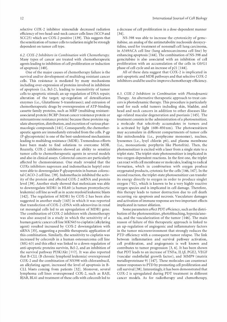
6.2. COX-2 Inhibitors in Combination with Chemotherapy.Many types of cancer are treated with chemotherapeuticagents leading to inhibition of cell proliferation or inductionof apoptosis [140].
One of the major causes of chemotherapy failure is thesurvival and/or development of multidrug resistant cancercells. This resistance is mediated by many mechanismsincluding over-expression of proteins involved in inhibitionof apoptosis (i.e, Bcl-2), leading to insensitivity of tumorcells to apoptotic stimuli; an up-regulation of DNA repair;alteration of the target; up-regulation of detoxificationenzymes (i.e., Glutathione S-transferases); and extrusion of chemotherapeutic drugs by overexpression of ATP-bindingcassette family proteins, such as MRP (multidrug resistant-associated protein) BCRP (breast cancer resistance protein ormitoxantrone resistance protein) because these proteins reg-ulate absorption, distribution, and excretion of various phar-macologic compounds [141]. Consequently, the chemother-apeutic agents are immediately extruded from the cells. P-gp(P-glycoprotein) is one of the best-understood mechanismsleading to multidrug resistance (MDR). Tremendous eff ortshave been made to find solutions to overcome MDR.Recently, COX-2 inhibitors showed an ability to sensitizetumor cells to chemotherapeutic agents in several modelsand also in clinical assays. Colorectal cancers are particularly aff ected by chemoresistance. One study revealed that theCOXs inhibitors naproxen and indomethacin heptyl esterwere able to downregulate P-glycoprotein in human colorec-tal CACO-2 cell line. [39]. Indomethacin inhibited the activ-ity of the protein and aff ected COX-2 mRNA and proteinlevel [39]. Another study showed that meloxicam was ableto downregulate MDR1 in HL60 (a human promyelocyticleukemia) cell line as well as in acute myeloid leukemic blasts[142]. The regulation of MDR1 by COX-2 has been alsosuggested in another study [143] in which it was reportedthat transfection of COX-2 cDNA with adenovirus in renalrat mesangial cells led to an upregulation of MDR1 gene.The combination of COX-2 inhibitors with chemotherapy was also assayed in a study in which the sensitivity of ahuman gastric cancer cell line MKN45 to cisplatin (alkylating
agent) resulted increased by COX-2 downregulation withsiRNA [35], suggesting a possible therapeutic application of this combination. Similarly, the sensitivity to cisplatin wasincreased by celecoxib in a human osteosarcoma cell line(MG-63) and this eff ect was linked to a down-regulation of anti-apoptotic proteins survivin, Bcl-2, and an inhibition of the survival pathway PI3K/Akt [112]. It was also reportedthat B-CLL (B chronic lymphoid leukemia) overexpressedCOX-2 and the combination of NS398 with chlorambucil,an alkylating agent, increased the level of apoptosis in B-CLL blasts coming from patients [32]. Moreover, severallymphoma cell lines overexpressed COX-2, such as RAJI,BJAB, BL41 and treatment of these cells with celecoxib led to
a decrease of cell proliferation in a dose-dependent manner[34].
NS-398 was able to increase the cytotoxicity of gemc-itabine, an analog of the antimetabolite nucleoside deoxycy-tidine, used for treatment of nonsmall cell lung carcinoma,in A549ACA cell line (lung adenocarcinoma cell line) by
enhancing apoptosis [144]. The combination of NS-398 andgemcitabine is also associated with an inhibition of cellproliferation with an accumulation of the cells in G0/G1phase of cell cycle and an increase of p21 [144].
All of these data suggest that COX-2 is implicated inanti-apoptotic and MDR pathways and that selective COX-2inhibitors could be used to improve chemotherapy efficiency.
6.3. COX-2 Inhibitors in Combination with Photodynamic Therapy. An alternative therapeutic approach to treat can-cers is photodynamic therapy. This procedure is particularly used for such solid tumors including skin, bladder, andhead and neck cancers in addition to other diseases likeage-related macular degeneration and psoriasis [145]. Thetreatment consists in the administration of a photosensitizer,a molecule that selectively accumulates in tumors andis activated by light (600–850 nm). The photosensitizersmay accumulate in diff erent compartments of tumor cellslike mitochondria (i.e., porphycene monomer), nucleus,lysosomes (i.e., lysyl chlorin p6), and plasma membrane(i.e., monocationic porphyrin like Photofrin). Then, thephotosensitizer is excited with a laser from a single state to atriplet state. The triplet-state photosensitizer is implicated intwo oxygen-dependent reactions. In the first one, the tripletcan react with cell membrane or molecules, leading to radicalformation, which in combination with oxygen produceoxygenated products, cytotoxic for the cells [146, 147]. In thesecond reaction, the triplet-state photosensitizer can transferits energy directly to oxygen in order to produce singletoxygen (102), which is known to be a very highly reactiveoxygen species and is implicated in cell damage. Therefore,this therapy leads to tumor destruction due to cell deathoccurring via apoptosis and necrosis. Vasculature damagesand activation of immune response are two important eff ectsimplicated in tumor ablation.
Some parameters aff ect PDT efficiency, such as the distri-bution of the photosensitizer, photobleaching, hypoxia/ano-xia, and the vascularization of the tumor [146]. The mainreason of failure of this therapeutic approach is linked to
an up-regulation of angiogenic and inflammatory factorsin the tumor microenvironment that strongly reduces thePTD efficiency with a consequent tumor relapse. The linkbetween inflammation and survival pathway activation,cell proliferation, and angiogenesis is well known andcontributes to tumor progression [3, 6]. It has been shownthat PDT leads to an increase of TNFα, IL1 β, PGE2, VEGF(vascular endothelial growth factor), and MMP9 (matrixmetalloproteinase 9) [147]. These molecules can counteracttumor responses to PTD by promoting cell proliferation andcell survival [38]. Interestingly, it has been demonstrated thatCOX-2 is upregulated during PDT treatment in diff erentcancer models. As for radiotherapy and chemotherapy,

8/2/2019 The Role of COX-2 in Cell Proliferation and Cell Death in Human Malignancies
http://slidepdf.com/reader/full/the-role-of-cox-2-in-cell-proliferation-and-cell-death-in-human-malignancies 13/21
International Journal of Cell Biology 13
this suggests COX-2 as a possible target to increase PDTefficiency.
Indeed, celecoxib has been proved to aff ect the Photofrin-induced PDT in in vitro and in vivo studies performedon a mouse mammary carcinoma BA cell line [148]. Invitro, celecoxib and NS-398 increase PDT-induced apoptosis.
These results were correlated with caspase-3 and PARPcleavage and Bcl-2 degradation. In vivo, the photosensi-tization by COX-2 inhibitors was not due to apoptosisexacerbation. Interestingly, celecoxib and NS-398 decreasePDT-induced apoptosis but were also able to decrease thelevel of angiogenic factors such as TNFα, IL1 β, PGE2, VEGF,and MMP9 [148].
Upon chlorin-induced PDT, COX-2 was found up-regu-lated 25-fold in mouse mammary carcinoma RIF cells[149]. This up-regulation was associated with an increaseof PGE2 level in the tumor microenvironment. WhenRIF cells were transplanted in CH3/HeJ mice, for in vivostudies, PDT similarly induced an increase of COX-2 andPGE2. These eff ects were prevented by NS-398. Here, it wasdemonstrated that PDT induced vascular endothelial growthfactor expression (VEGF) and this increase was attenuated by treating mice with NS-398, meaning that COX-2 might play a role also in angiogenesis. In consequence of these eff ects,the combination of COX-2 inhibitors with PDT resulted inan increased efficiency of tumor treatment.
Possible correlation between COX-2 level and resistanceto PDT has been also investigated in Hela (human cervixcarcinoma cells) and T24 (human transitional cell carcinomaof the urinary bladder) cells [150]. It has been reportedthat in PDT induced by hypericin, a natural photosensitizerwhich accumulates in endoplasmic reticulum and Golgiapparatus, an increase of PGE2 levels occurred. Hypericininduces apoptosis by triggering the release of cytochromec after light excitation through a process requiring theactivation of p38 MAPK, which it is known to induce anup-regulation of COX-2 [23, 151]. The increase in PGE2levels was prevented by the use of a p38 MAPK inhibitor(PD169316). Moreover, the impairment of p38 MAPK wasassociated with an increase in the susceptibility of tumorcells to PDT. However, COX-2 inhibitors did not lead to thesame eff ect, meaning that COX-2 was not involved in PDTresistance in this model.
In contrast to the study of Ferrario et al. [148, 149],a report from Makowski et al. [152] has revealed thatrofecoxib, NS-398, and nimesulide were unable to potentiate
PDT in C-26 cells (poorly diff erentiated colon adenocarci-noma cell line) in vitro. However, chronic exposition of micebearing C-26 cells to nimesulide potentiated PDT. These datasuggest that COX-2 inhibitors may indirectly potentiate PDT.
It is known that vasculature damages are importantfor PDT efficiency and that COX-2 inhibitors act as anti-angiogenic factors [153]. It has been hypothesized that theseantiangiogenic eff ects could be responsible for the anti-tumor eff ect.
Currently, the link between COX-2 and PDT efficiency is not well characterized. Some studies have revealed animprovement of efficiency with COX-2 inhibitors whereasother reports have demonstrated no direct eff ects. In any
case, this eff ect may be cell-type dependent as for chemother-apy or radiotherapy.
7. Inhibition of COX-2 Expression by Natural Compounds
Synthetic cyclooxygenase-2 inhibitors hold promise for can-cer chemoprevention; however, recent toxicity problems sug-gest that new strategies are needed. Natural compounds withthe potential to inhibit key cell signaling pathways includingCOX-2 gained much attention over the last regarding yearswhether they are used alone or in combination with existingchemotherapeutic agents.
Recently, Bhui et al. demonstrated that Bromelain, apharmacologically active compound present in pineapple(Ananas cosmosus), leads to a marked inhibition of COX-2expression and inactivation of NFκB. Bromelain treatmentinduces up-regulation of p53 and Bax and subsequentactivation of caspase-3 and caspase-9 with a decrease in Bcl-2
expression [154]. Furthermore bromelain induces apoptosis-related proteins along with inhibition of NFκB -driven COX-2 expression by blocking the MAPK and Akt/protein kinase Bsignaling in DMBA-TPA-induced mouse skin tumors [155].
Curcumin, a naturally occurring polyphenol from Cur-cuma longa, was described to act as an antiinflammatory and antiproliferative agent by causing downregulation of COX-2 in cervical cancer. Curcumin-mediated apoptosis inthese cells is initiated by up-regulation of pro-apoptoticBax, AIF, release of cytochrome c, and downregulation of anti-apoptotic Bcl-2, Bcl-xL in HeLa and SiHa cell lines.This onset of apoptosis was accompanied by an increase incaspase-3 and -9 activity, suggesting the role of mitochondria
in curcumin-mediated apoptotic cell death as described by M. Singh and N. Singh [156]. Marın et al., furthermore,concluded that curcumin inhibits NFκB activity as well asthe expression of its downstream target genes, and also selec-tively induces apoptosis of melanoma cells but not normalmelanocytes [157]. In addition, curcumin-induced apoptosiswas also associated with the activation of caspase-3 andcaspase-9, and the degradation of PARP. Curcumin decreasedthe expression levels of COX-2 mRNA and protein withoutcausing significant changes in COX-1 levels, which wascorrelated with the inhibition of prostaglandin E(2) synthesis[158]. In BV-2 microglial cells, curcumin and analogs wereshown to inhibit LPS-induced COX-2 expression; analogs
identified as more potent than curcumin in the screeningassay were also more potent than curcumin in preventingCOX-2 expression [159].
Coumarin (1,2-benzopyrone) is a naturally occurringfragrant compound found in numerous plants and spices.Results obtained with human nonsmall cell lung cancer A549cells suggest that downregulation of Bcl-xL, COX-2, andMAP kinase pathway and up-regulation of p53, Akt, andNFκB pathway are involved in the underlying molecularmechanism of apoptosis induction as suggested by Goel et al.[160].
Suh et al. concluded that the plant flavonoid fisetin indu-ces apoptosis and suppresses the growth of colon cancer cells

8/2/2019 The Role of COX-2 in Cell Proliferation and Cell Death in Human Malignancies
http://slidepdf.com/reader/full/the-role-of-cox-2-in-cell-proliferation-and-cell-death-in-human-malignancies 14/21
14 International Journal of Cell Biology
Radiotherapy
- Nimesulide in nonsmall cell lung cancer in vivo and in vitro
(A549 cell line)
- Ns-398 in melanoma cell line (WM35 and LU1205)
- siRNA COX-2 in human prostate carcinoma (PC3)
and human cervical carcinoma
COX-2inhibitors
Chemotherapy - Naproxen/indomethacin downregulate
P-glycoprotein in human colorectal cell line (CACO-2)- COX-2 siRNA/cisplatin in human gastric cancer cellline (MKN 45)
- Celecoxib/cisplatin in human osteosarcoma cell line(MG-63)
- NS-398/chlorambucil in B-CLL- NS-398/gemcitabine in nonsmall cell lung carcinoma
cell line (A549)
Photodynamic therapy
- Celecoxib and NS-398 in photofrin-induced-
photodynamic therapy in mouse mammary
carcinoma cell line (BA)
- NS-398 in mouse mammary carcinoma (RIF)
Figure 5: COX-2 inhibition in cancer therapy.
by inhibition of COX-2- and Wnt/EGFR/NFκB-signalingpathways [161].
Sulforaphane (SFN) is a biologically active compoundextracted from cruciferous vegetables, and presents potentanti-cancer and anti-inflammatory activities by suppressionof NFkB-dependent genes involved in anti-apoptotic signal-ing (IAP-1, IAP-2, XIAP, Bcl-2, and Bcl-xL), cell proliferation
(c-Myc, COX-2, and cyclin D1), and metastasis (VEGF andMMP-9) as published by Moon et al. [162].
Nontoxic apigenin can suppress anti-apoptotic pathwaysinvolving NFκB activation including cFLIP and COX-2 exp-ression as demonstrated by Xu et al. [88]. According to Namet al., DA-6034, a synthetic derivative of flavonoid Eupatilin,strongly enhanced apoptosis and inhibited the expressionof COX-2 and phospho-IKKalpha in inflammation-relatedcolon cancer models [163].
EGCG from green tea was described to attenuate theAR, to downregulate IGF-1, to modulate COX-2 expression,and to decrease MAPK signaling leading to the reductionin cell proliferation and induction of apoptosis in prostate
cancer without toxicity [164]. Interestingly, combination of EGCG and COX-2 inhibitor NS-398 enhanced cell growthinhibition, apoptosis induction, expression of Bax, pro-caspase-6, and pro-caspase-9, and PARP cleavage, inhibitionof PPAR gamma, and inhibition of NFκB compared with theadditive eff ects of the two agents alone, suggesting a possiblesynergism. In vivo, combination treatment with green teapolyphenols and COX-2 inhibitor celecoxib resulted inenhanced tumor growth inhibition, lowering of prostate-specific antigen levels, lowering of IGF-I levels, and circu-lating levels of serum IGF-1 binding protein-3 comparedwith results of single-agent treatment. Accordingly, Adhamiet al. postulate the efficiency of synergistic and/or additive
eff ects of combinatorial chemopreventive agents and furtherunderscore the need for rational design of human clinicaltrials involving such natural compounds [165].
Pandey et al. published that butein inhibited the expres-sion of the NFκB-regulated gene products involved in anti-apoptosis (IAP2, Bcl-2, and Bcl-xL), proliferation (cyclin D1and c-Myc), and invasion (COX-2 and MMP-9). Suppression
of these gene products correlated with enhancement of theapoptosis induced by TNF and chemotherapeutic agents, andinhibition of cytokine-induced cellular invasion. This groupclearly demonstrates that antitumor and anti-inflammatory activities assigned to butein may be mediated in part throughthe direct inhibition of IKK, leading to the suppression of theNFκB activation pathway [166].
Hostanska et al. used human colon COX-2-positive HT29 and COX-2-negative HCT 116 or lung COX-2 proficientA 549 and low COX-2 expressing SW2 cells and showed thatwillow bark extract BNO 1455 and its fractions inhibit thecell growth and promote apoptosis in human colon and lungcancer cell lines irrespective of their COX selectivity [167].
8. COX-2 Independent Effects
It is currently well known that several selective COX-2inhibitors inhibit cell proliferation and induce apoptosisindependently of COX-2. Celecoxib is particularly known tohave these COX-2-independent eff ects, which were reviewedby Grosch et al. [68]. Indeed, celecoxib was able to directly bind and inhibit PKB/Akt, which plays an important rolein cell proliferation and in apoptosis. Concerning cell cycle,PKB is able to phosphorylate cdk inhibitors, such as p21 andp27, leading to PCNA activation [168, 169]. Furthermore,PKB can also activate several cyclin-cdk complexes and

8/2/2019 The Role of COX-2 in Cell Proliferation and Cell Death in Human Malignancies
http://slidepdf.com/reader/full/the-role-of-cox-2-in-cell-proliferation-and-cell-death-in-human-malignancies 15/21
International Journal of Cell Biology 15
induce E2F factor in some cases [68], stimulating cell prolif-eration. Besides, PKB inhibits apoptosis, by phosphorylatingthe pro-apoptotic protein Bad and by inhibiting caspase-9cleavage [51].
The COX-2-independent eff ects concern also the extrin-sic apoptotic pathway. Indeed, we discussed that selective
COX-2 inhibitors, such as NS-398, celecoxib, and meloxi-cam, are able to modulate the sensitivity of several tumorcells to Fas- and TRAIL-induced apoptosis. It has beendiscussed that this modulation could be due to COX-2-independent eff ects. In fact, NS-398 and nimesulide wereable to promote TNF and TRAIL-induced apoptosis of D98and H21 Hela cell lines [170]. In D98, COX-2 is inactive.Moreover, prostaglandin E2 readdition was not able to revertthe sensitization eff ect. In the same report, it has been shownthat NS-398 was able to promote apoptosis induced by TNFin MCF-7 cell line (human breast adenocarcinoma cells),which again does not express COX-2 [170].
A report from Ryan et al. [171] demonstrated thatSC58125 and CAY10404, two selective COX-2 inhibitors,were able to decrease intracellular content of GSH inmalignant human B-cells. This eff ect was accompanied by an increase of reactive oxygen species production. Indeed,GSH is the most important intracellular nonprotein thiolantioxidant defense against free radicals, meaning that it pro-tects the cells from cellular damages. The GSH depletion wascorrelated in this study with a reduced survival for these cells.
The fact that many studies imply an association betweenCOX-2 inhibition and apoptosis induction or cell prolifera-tion inhibition, without assessing whether COX-2 activity iseff ectively decreased, suggests caution in the interpretationof the data. This is confirmed by the observation thatdiff erent COX-2 inhibitors may trigger apoptosis in the samecancer cell model by modulating diff erent mechanisms. Forexample, celecoxib [113] induced apoptosis by an inhibitionof Akt phosphorylation in prostate cancer cells COX-2-positive LNCaP without aff ecting Bcl-2 level. In contrast, astudy by Liu et al. [102] revealed that NS-398 in the samecell line was able to induce apoptosis but down-regulation of Bcl-2. These results suggest a possible COX-2-independenteff ect and strongly recommends considering in parallel otherexperimental strategies to ascertain the eff ective role of COX-2 in human malignancies [33], such as methodologies basedon RNA interference or antisense oligonucleotides. Studieshave already suggested these alternative methods. It has beenshown that the sensitivity of a human gastric cancer cell
line MKN45 to cisplatin (alkylating agent) was increased by COX-2 downregulation with siRNA [35].
9. Conclusion
A number of studies suggest that COX-2 inhibition may lead to an inhibition of cell proliferation in diff erent cancertypes. The fact that COX-2 inhibition may per se triggerapoptosis of tumor cells and/or sensitize them to cytotoxictreatments is an indication that COX-2 may be a goodtarget in cancer therapy, in order to improve the efficiency of tumor cell death and to reduce tumor progression (seeFigure 5 for a synthesis). Accordingly, the combination of
selective COX-2 inhibitors with radiotherapy or diff erentchemotherapeutics revealed a sensitization to apoptosis.This eff ect was also observed with several agents inducingapoptosis in a physiological way, thus suggesting that COX-2inhibitors used in combination with death receptors agonistsmight be a novel approach to elicit apoptosis of cancer cells.
However, the fact that COX-2 inhibitors can mediate theireff ects by COX-2-independent mechanisms suggests cautionin the interpretation of the data.
Nowadays, selective COX-2 inhibitors have beenincluded in several clinical assays. Some of them eff ectively increase the efficiency of radiotherapy and chemotherapy [172]. For example, celecoxib is in a clinical phase II assay incombination with Paclitaxel, carboplatin, and radiotherapy for patients with inoperable stage IIIA/B nonsmall cell lungcancer [172]. These clinical assays confirm that COX-2inhibition may be a promising field in cancer treatment.However, the selective COX-2 inhibitors are responsible forside eff ects, including an increasing risk of cardiovascularcomplications [67, 68]. It is hoped that other methods toinhibit COX-2 will be developed. To this purpose, RNAinterference using vehicle (i.e., adenovirus) as well as naturalcompounds were suggested by some studies [35, 76], asalternative and promising strategy.
Conflicts of Interest
The authors have no actual or potential conflict of interest.
Acknowledgments
C. Sobolewski. and C. Cerella are recipients of doctoral
and postdoctoral Televie gtrants, respectively. Research atthe Laboratoire de Biologie Moleculaire et Cellulaire duCancer (LBMCC) is financially supported by “RechercheCancer et Sang” foundation, by the “Recherches ScientifiquesLuxembourg” association, by “Een Haerz fir kriibskrankKanner” association, by the Action Lions “Vaincre le Can-cer”association and by Televie Luxembourg. The authors areparticularly grateful to Fonds National pour la Recherche(FNR) Luxembourg, for additional financial support pro-vided for this publication.
References
[1] V. I. Kulinsky, “Biochemical aspects of inflammation,” Bio-chemistry , vol. 72, no. 6, pp. 595–607, 2007.
[2] R. N. DuBois, S. B. Abramson, L. Croff ord, et al., “Cyclooxy-genase in biology and disease,” FASEB Journal , vol. 12, no. 12,pp. 1063–1073, 1998.
[3] F. Balkwill and A. Mantovani, “Inflammation and cancer:back to Virchow?” The Lancet , vol. 357, no. 9255, pp. 539–545, 2001.
[4] L. K. Dennis, C. F. Lynch, and J. C. Torner, “Epidemiologicassociation between prostatitis and prostate cancer,” Urology ,vol. 60, no. 1, pp. 78–83, 2002.
[5] L. R. Howe, “Inflammation and breast cancer. Cyclooxy-genase/prostaglandin signaling and breast cancer,” Breast Cancer Research, vol. 9, no. 4, p. 210, 2007.

8/2/2019 The Role of COX-2 in Cell Proliferation and Cell Death in Human Malignancies
http://slidepdf.com/reader/full/the-role-of-cox-2-in-cell-proliferation-and-cell-death-in-human-malignancies 16/21
16 International Journal of Cell Biology
[6] L. M. Coussens and Z. Werb, “Inflammation and cancer,” Nature, vol. 420, no. 6917, pp. 860–867, 2002.
[7] M. Suganuma, S. Okabe, M. W. Marino, A. Sakai, E. Sueoka,and H. Fujiki, “Essential role of tumor necrosis factor α(TNF-α) in tumor promotion as revealed by TNF-α-deficientmice,” Cancer Research, vol. 59, no. 18, pp. 4516–4518, 1999.
[8] M. Yao, E. C. Lam, C. R. Kelly, W. Zhou, and M. M.
Wolfe, “Cyclooxygenase-2 selective inhibition with NS-398suppresses proliferation and invasiveness and delays livermetastasis in colorectal cancer,” British Journal of Cancer , vol.90, no. 3, pp. 712–719, 2004.
[9] R. N. DuBois and W. E. Smalley, “Cyclooxygenase, NSAIDs,and colorectal cancer,” Journal of Gastroenterology , vol. 31,no. 6, pp. 898–906, 1996.
[10] A. L. Eisinger, S. M. Prescott, D. A. Jones, and D. M.Staff orini, “The role of cyclooxygenase-2 and prostaglandinsin colon cancer,” Prostaglandins and Other Lipid Mediators,vol. 82, no. 1–4, pp. 147–154, 2007.
[11] G. Steinbach, P. M. Lynch, R. K. S. Phillips, et al., “The eff ectof celecoxib, a cyclooxygenase-2 inhibitor, in familial adeno-matous polyposis,” The New England Journal of Medicine, vol.
342, no. 26, pp. 1946–1952, 2000.[12] S. P. Hussain and C. C. Harris, “Inflammation and cancer:
an ancient link with novel potentials,” International Journal of Cancer , vol. 121, no. 11, pp. 2373–2380, 2007.
[13] S. Cristofanon, F. Morceau, A. I. Scovassi, M. Dicato, L.Ghibelli, and M. Diederich, “Oxidative, multistep activationof the noncanonical NF-κB pathway via disulfide Bcl-3/p50complex,” FASEB Journal , vol. 23, pp. 45–57, 2009.
[14] R. Chen, A. B. Alvero, D. A. Silasi, and G. Mor, “Inflam-mation, cancer and chemoresistance: taking advantage of the toll-like receptor signaling pathway,” American Journal of Reproductive Immunology , vol. 57, no. 2, pp. 93–107, 2007.
[15] M. D’Alessio, C. Cerella, C. Amici, et al., “Glutathionedepletion up-regulates Bcl-2 in BSO-resistant cells,” FASEB
Journal , vol. 18, no. 13, pp. 1609–1611, 2004.[16] W. W. Lin and M. Karin, “A cytokine-mediated link between
innate immunity, inflammation, and cancer,” Journal of Clinical Investigation, vol. 117, no. 5, pp. 1175–1183, 2007.
[17] E. Pikarsky, R. M. Porat, I. Stein, et al., “NF-κB functionsas a tumour promoter in inflammation-associated cancer,”
Nature, vol. 431, no. 7007, pp. 461–466, 2004.
[18] S. T. Palayoor, M. Y. Youmell, S. K. Calderwood, C. N.Coleman, and B. D. Price, “Constitutive activation of IκBkinase α and NF-κB in prostate cancer cells is inhibited by ibuprofen,” Oncogene, vol. 18, no. 51, pp. 7389–7394, 1999.
[19] U. Kordes, D. Krappmann, V. Heissmeyer, W. D. Ludwig, andC. Scheidereit, “Transcription factor NF-κB is constitutively activated in acute lymphoblastic leukemia cells,” Leukemia,
vol. 14, no. 3, pp. 399–402, 2000.[20] G. Tricot, “New insights into role of microenvironment in
multiple myeloma,” The Lancet , vol. 355, no. 9200, pp. 248–250, 2000.
[21] N. V. Chandrasekharan and D. L. Simmons, “The cyclooxy-genases,” Genome Biology , vol. 5, no. 9, article 241, 2004.
[22] S. Narumiya and G. A. FitzGerald, “Genetic and pharmaco-logical analysis of prostanoid receptor function,” Journal of Clinical Investigation, vol. 108, no. 1, pp. 25–30, 2001.
[23] R. G. Ramsay, D. Ciznadija, M. Vanevski, and T. Man-tamadiotis, “Transcriptional regulation of cyclo-oxygenaseexpression: three pillars of control,” International Journal of Immunopathology and Pharmacology , vol. 16, no. 2,supplement, pp. 59–67, 2003.
[24] B. Hinz and K. Brune, “Cyclooxygenase-2–10 years later,” Journal of Pharmacology and Experimental Therapeutics, vol.300, no. 2, pp. 367–375, 2002.
[25] R. G. Kurumbail, A. M. Stevens, J. K. Gierse, et al.,“Structuralbasis for selective inhibition of cyciooxygenase-2 by anti-inflammatory agents,” Nature, vol. 384, no. 6610, pp. 644–648, 1996.
[26] F. H. Sarkar, S. Adsule, Y. Li, and S. Padhye, “Back to thefuture: COX-2 inhibitors for chemoprevention and cancertherapy,” Mini-Reviews in Medicinal Chemistry , vol. 7, no. 6,pp. 599–608, 2007.
[27] B. Kis, J. A. Snipes, T. Isse, K. Nagy, and D. W. Busija,“Putative cyclooxygenase-3 expression in rat brain cells,”
Journal of Cerebral Blood Flow and Metabolism, vol. 23, no.11, pp. 1287–1292, 2003.
[28] N. V. Chandrasekharan, H. Dai, K. L. T. Roos, et al., “COX-3, a cyclooxygenase-1 variant inhibited by acetaminophenand other analgesic/antipyretic drugs: cloning, structure, andexpression,” Proceedings of the National Academy of Sciences of the United States of America, vol. 99, no. 21, pp. 13926–13931,2002.
[29] B. Hinz, O. Cheremina, and K. Brune, “Acetaminophen(paracetamol) is a selective cyclooxygenase-2 inhibitor inman,” FASEB Journal , vol. 22, no. 2, pp. 383–390, 2008.
[30] Y. Cao and S. M. Prescott, “Many actions of cyclooxygenase-2 in cellular dynamics and in cancer,” Journal of Cellular Physiology , vol. 190, no. 3, pp. 279–286, 2002.
[31] A. Ristimaki, A. Sivula, J. Lundin, et al., “Prognostic sig-nificance of elevated cyclooxygenase-2 expression in breastcancer,” Cancer Research, vol. 62, no. 3, pp. 632–635, 2002.
[32] P. Secchiero, E. Barbarotto, A. Gonelli, et al., “Potentialpathogenetic implications of cyclooxygenase-2 overexpres-sion in B chronic lymphoid leukemia cells,” American Journal of Pathology , vol. 167, no. 6, pp. 1599–1607, 2005.
[33] Y. Nakanishi, R. Kamijo, K. Takizawa, M. Hatori, and M.Nagumo, “Inhibitors of cyclooxygenase-2 (COX-2) sup-pressed the proliferation and diff erentiation of humanleukaemia cell lines,” European Journal of Cancer , vol. 37, no.12, pp. 1570–1578, 2001.
[34] T. Wun, H. McKnight, and J. M. Tuscano, “Increasedcyclooxygenase-2 (COX-2): a potential role in the pathogene-sis of lymphoma,” Leukemia Research, vol. 28, no. 2, pp. 179–190, 2004.
[35] M. W. Chan, C. Y. Wong, A. S. Cheng, et al., “Targeted inhi-bition of COX-2 expression by RNA interference suppressestumor growth and potentiates chemosensitivity to cisplatinin human gastric cancer cells,” Oncology Reports, vol. 18, no.6, pp. 1557–1562, 2007.
[36] G. E. Johnson, V. N. Ivanov, and T. K. Hei, “Radiosensiti-zation of melanoma cells through combined inhibition of
protein regulators of cell survival,” Apoptosis, vol. 13, no. 6,pp. 790–802, 2008.
[37] S. T. Palayoor, M. J. Arayankalayil, A. Shoaibi, and C.N. Coleman, “Radiation sensitivity of human carcinomacells transfected with small interfering RNA targeted againstcyclooxygenase-2,” Clinical Cancer Research, vol. 11, no. 19,pp. 6980–6986, 2005.
[38] M. Philip, D. A. Rowley, and H. Schreiber, “Inflammation asa tumor promoter in cancer induction,” Seminars in Cancer Biology , vol. 14, no. 6, pp. 433–439, 2004.
[39] A. Zrieki, R. Farinotti, and M. Buyse, “Cyclooxygenaseinhibitors down regulate P-glycoprotein in human colorectalcaco-2 cell line,” Pharmaceutical Research, vol. 25, no. 9, pp.1991–2001, 2008.

8/2/2019 The Role of COX-2 in Cell Proliferation and Cell Death in Human Malignancies
http://slidepdf.com/reader/full/the-role-of-cox-2-in-cell-proliferation-and-cell-death-in-human-malignancies 17/21
International Journal of Cell Biology 17
[40] E. M. Choi, S. J. Kwak, Y. M. Kim, et al., “COX-2 inhibitsanoikis by activation of the PI-3K/Akt pathway in humanbladder cancer cells,” Experimental and Molecular Medicine,vol. 37, no. 3, pp. 199–203, 2005.
[41] R. M. Breyer, C. K. Bagdassarian, S. A. Myers, and M.D. Breyer, “Prostanoid receptors: subtypes and signaling,”
Annual Review of Pharmacology and Toxicology , vol. 41, pp.
661–690, 2001.[42] J. Grossmann, “Molecular mechanisms of “detachment-
induced apoptosis—anoikis”,” Apoptosis, vol. 7, no. 3, pp.247–260, 2002.
[43] J. R. Woodgett, “Regulation and functions of the glycogensynthase kinase-3 subfamily,” Seminars in Cancer Biology , vol.5, no. 4, pp. 269–275, 1994.
[44] N. K. Boughton Smith, C. J. Hawkey, and B. J. R. Whittle,“Biosynthesis of lipoxygenase and cyclo-oxygenase productsfrom [14C]-arachidonic acid by human colonic mucosa,”Gut , vol. 24, no. 12, pp. 1176–1182, 1983.
[45] V. W. Yang, J. M. Shields, S. R. Hamilton, et al., “Size-dependent increase in prostanoid levels in adenomas of patients with familial adenomatous polyposis,” Cancer
Research, vol. 58, no. 8, pp. 1750–1753, 1998.[46] H. Sheng, J. Shao, J. D. Morrow, R. D. Beauchamp, and R.
N. DuBois, “Modulation of apoptosis and Bcl-2 expressionby prostaglandin E2 in human colon cancer cells,” Cancer Research, vol. 58, no. 2, pp. 362–366, 1998.
[47] G. A. Doherty, S. M. Byrne, E. S. Molloy, et al.,“Proneoplasticeff ects of PGE2 mediated by EP4 receptor in colorectalcancer,” BMC Cancer , vol. 9, article 207, 2009.
[48] L. Damstrup, S. K. Kuwada, P. J. Dempsey, et al., “Amphireg-ulin acts as an autocrine growth factor in two humanpolarizing colon cancer lines that exhibit domain selectiveEGF receptor mitogenesis,” British Journal of Cancer , vol. 80,no. 7, pp. 1012–1019, 1999.
[49] Q. B. She, D. B. Solit, Q. Ye, K. E. O’Reilly, J. Lobo, and
N. Rosen, “The BAD protein integrates survival signalingby EGFR/MAPK and PI3K/Akt kinase pathways in PTEN-deficient tumor cells,” Cancer Cell , vol. 8, no. 4, pp. 287–297,2005.
[50] T. G. Tessner, F. Muhale, T. E. Riehl, S. Anant, and W. F. Sten-son, “Prostaglandin E2 reduces radiation-induced epithelialapoptosis through a mechanism involving AKT activationand Bax translocation,” Journal of Clinical Investigation, vol.114, no. 11, pp. 1676–1685, 2004.
[51] A. Burlacu, “Regulation of apoptosis by Bcl-2 family pro-teins,” Journal of Cellular and Molecular Medicine, vol. 7, no.3, pp. 249–257, 2003.
[52] T. Yamaki, K. Endoh, M. Miyahara, et al., “Prostaglandin E 2
activates Src signaling in lung adenocarcinoma cell via EP3,”
Cancer Letters, vol. 214, no. 1, pp. 115–120, 2004.[53] L. P. Billen, C. L. Kokoski, J. F. Lovell, B. Leber, and D. W.
Andrews, “Bcl-XL inhibits membrane permeabilization by competing with Bax,” PLoS Biology , vol. 6, no. 6, article e147,2008.
[54] Z. A. Stewart, M. D. Westfall, and J. A. Pietenpol, “Cell-cycledysregulation and anticancer therapy,” Trends in Pharmaco-logical Sciences, vol. 24, no. 3, pp. 139–145, 2003.
[55] M. D. Larrea, J. Liang, T. Da Silva, et al., “Phosphorylation of p27Kip1 regulates assembly and activation of cyclin D1-Cdk4,”
Molecular and Cellular Biology , vol. 28, no. 20,pp. 6462–6472,2008.
[56] M. Pagano, S. W. Tam, A. M. Theodoras, et al., “Role of theubiquitin-proteasome pathway in regulating abundance of
the cyclin-dependent kinase inhibitor p27,” Science, vol. 269,no. 5224, pp. 682–685, 1995.
[57] J. L. Bos, “ras Oncogenes in human cancer: a review,” Cancer Research, vol. 49, no. 17, pp. 4682–4689, 1989.
[58] C. Tsatsanis, A. Androulidaki, M. Venihaki, and A. N.Margioris, “Signalling networks regulating cyclooxygenase-2,” International Journal of Biochemistry and Cell Biology , vol.
38, no. 10, pp. 1654–1661, 2006.[59] T. Yano, G. Zissel, J. Muller-Qernheim, S. Jae Shin, H. Satoh,
and T. Ichikawa, “Prostaglandin E2 reinforces the activationof Ras signal pathway in lung adenocarcinoma cells via EP3,”FEBS Letters, vol. 518, no. 1–3, pp. 154–158, 2002.
[60] M. R. Junttila and J. Westermarck, “Mechanisms of MYCstabilization in human malignancies,” Cell Cycle, vol. 7, no.5, pp. 592–596, 2008.
[61] X. Ma, N. Kundu, S. Rifat, T. Walser, and A. M. Fulton,“Prostaglandin E receptor EP4 antagonism inhibits breastcancer metastasis,” Cancer Research, vol. 66, no. 6, pp. 2923–2927, 2006.
[62] M. Majima, I. Hayashi, M. Muramatsu, J. Katada, S.Yamashina, and M. Katori, “Cyclo-oxygenase-2 enhances
basic fibroblast growth factor-induced angiogenesis throughinduction of vascular endothelial growth factor in rat spongeimplants,” British Journal of Pharmacology , vol. 130,no. 3,pp.641–649, 2000.
[63] J. Folkman, “Tumor angiogenesis: therapeutic implications,”The New England Journal of Medicine, vol. 285, no. 21, pp.1182–1186, 1971.
[64] G. A. Green, “Understanding NSAIDs: from aspirin to COX-2,” Clinical Cornerstone, vol. 3, no. 5, pp. 50–59, 2001.
[65] S. H. Ferreira, S. Moncada, and J. R. Vane, “Indomethacinand aspirin abolish prostaglandin release from the spleen,”
Nature, vol. 231, no. 25, pp. 237–239, 1971.[66] S. Pasa, K. Bayan, M. Kucukoner, et al., “The eff ects of
nonsteroidal anti-inflammatory drugs on platelet functionand severity of upper gastrointestinal haemorrhage,” Journal of Thrombosis and Thrombolysis, vol. 28, no. 1, pp. 83–89,2009.
[67] M. Hudson, E. Rahme, H. Richard, and L. Pilote, “Risk of congestive heart failure with nonsteroidal antiinflammatory drugs and selective cyclooxygenase 2 inhibitors: a classeff ect?” Arthritis Care and Research, vol. 57, no. 3, pp. 516–523, 2007.
[68] S. Grosch, T. J. Maier, S. Schiff mann, and G. Geisslinger,“Cyclooxygenase-2 (COX-2)-independent anticarcinogeniceff ects of selective COX-2 inhibitors,” Journal of the National Cancer Institute, vol. 98, no. 11, pp. 736–747, 2006.
[69] W. B. White, G. Faich, A. Whelton, et al., “Comparison of thromboembolic events in patients treated with celecoxib,a cyclooxygenase-2 specific inhibitor, versus ibuprofen or
diclofenac,” American Journal of Cardiology , vol. 89, no. 4, pp.425–430, 2002.
[70] M. E. Farkouh, F. W. A. Verheugt, S. Ruland, et al., “Acomparison of the blood pressure changes of lumiracoxibwith those of ibuprofen and naproxen,” Journal of Clinical Hypertension, vol. 10, no. 8, pp. 592–602, 2008.
[71] A. Whelton, J. G. Fort, J. A. Puma, D. Normandin, A. E. Bello,and K. M. Verburg, “Cyclooxygenase-2-specific inhibitorsand cardiorenal function: a randomized, controlled trial of celecoxib and rofecoxib in older hypertensive osteoarthritispatients,” American Journal of Therapeutics, vol. 8, no. 2, pp.85–95, 2001.
[72] T. Juhlin, S. Bjorkman, and P. Hoglund, “Cyclooxygenaseinhibition causes marked impairment of renal function in

8/2/2019 The Role of COX-2 in Cell Proliferation and Cell Death in Human Malignancies
http://slidepdf.com/reader/full/the-role-of-cox-2-in-cell-proliferation-and-cell-death-in-human-malignancies 18/21
18 International Journal of Cell Biology
elderly subjects treated with diuretics and ACE-inhibitors,”European Journal of Heart Failure, vol. 7, no. 6, pp. 1049–1056, 2005.
[73] S. Z. Zhao, M. W. Reynolds, J. Lefkowith, A. Whelton,and F. M. Arellano, “A comparison of renal-related adversedrug reactions between rofecoxib and celecoxib, based onthe World Health Organization/Uppsala Monitoring Centre
safety database,” Clinical Therapeutics, vol. 23, no. 9, pp.1478–1491, 2001.
[74] T. P. van Staa, L. Smeeth, I. Persson, J. Parkinson, and H.G. M. Leufkens, “What is the harm—benefit ratio of Cox-2 inhibitors?” International Journal of Epidemiology , vol. 37,no. 2, pp. 405–413, 2008.
[75] B. D. De Smet, A. M. Fendrick, J. G. Stevenson, and S. J.Bernstein, “Over and under-utilization of cyclooxygenase-2selective inhibitors by primary care physicians and specialists:the tortoise and the hare revisited,” Journal of General Internal
Medicine, vol. 21, no. 7, pp. 694–697, 2006.
[76] A. Strillacci, C. Griff oni, E. Spisni, M. C. Manara, andV. Tomasi, “RNA interference as a key to knockdownoverexpressed cyclooxygenase-2 gene in tumour cells,” British
Journal of Cancer , vol. 94, no. 9, pp. 1300–1310, 2006.[77] H. L. Peng, G. S. Zhang, J. H. Liu, F. J. Gong, and R. J. Li,
“Dup-697, a specific COX-2 inhibitor, suppresses growth andinduces apoptosis on K562 leukemia cells by cell-cycle arrestand caspase-8 activation,” Annals of Hematology , vol. 87, no.2, pp. 121–129, 2008.
[78] H. Sheng, J. Shao, S. C. Kirkland, et al., “Inhibition of human colon cancer cell growth by selective inhibition of cyclooxygenase-2,” Journal of Clinical Investigation, vol. 99,no. 9, pp. 2254–2259, 1997.
[79] H. Amano, I. Hayashi, H. Endo, et al., “Host prostaglandinE2-EP3 signaling regulates tumor-associated angiogenesisand tumor growth,” Journal of Experimental Medicine, vol.197, no. 2, pp. 221–232, 2003.
[80] H. Hochegger, S. Takeda, and T. Hunt, “Cyclin-dependentkinases and cell-cycle transitions: does one fit all?” NatureReviews Molecular Cell Biology , vol. 9, no. 11, pp. 910–916,2008.
[81] D. Cobrinik, “Pocket proteins and cell cycle control,” Onco- gene, vol. 24, no. 17, pp. 2796–2809, 2005.
[82] Z. Kelman, “PCNA: structure, functions and interactions,”Oncogene, vol. 14, no. 6, pp. 629–640, 1997.
[83] W. R. Taylor and G. R. Stark, “Regulation of the G2/Mtransition by p53,” Oncogene, vol. 20, no. 15, pp. 1803–1815,2001.
[84] J. Li, X. Chen, X. Dong, Z. Xu, H. Jiang, and X. Sun, “SpecificCOX-2 inhibitor, meloxicam, suppresses proliferation andinduces apoptosis in human HepG2 hepatocellular carci-
noma cells,” Journal of Gastroenterology and Hepatology , vol.21, no. 12, pp. 1814–1820, 2006.
[85] M. D. Castellone, H. Teramoto, B. O. Williams, K. M. Druey,and J. S. Gutkind, “Prostaglandin E2 promotes colon cancercell growth through a Gs-axin- β-catenin signaling axis,”Science, vol. 310, no. 5753, pp. 1504–1510, 2005.
[86] B. Xin, Y. Yokoyama, T. Shigeto, and H. Mizunuma, “Anti-tumor eff ect of non-steroidal anti-inflammatory drugs onhuman ovarian cancers,” Pathology and Oncology Research,vol. 13, no. 4, pp. 365–369, 2007.
[87] C. Falandry, P. A. Canney, G. Freyer, and L. Y. Dirix, “Role of combination therapy with aromatase and cyclooxygenase-2inhibitors in patients with metastatic breast cancer,” Annalsof Oncology , vol. 20, no. 4, pp. 615–620, 2009.
[88] X. F. Xu, C. G. Xie, X. P. Wang, et al., “Selective inhibition of cyclooxygenase-2 suppresses the growth of pancreatic cancercells in vitro and in vivo,” Tohoku Journal of Experimental
Medicine, vol. 215, no. 2, pp. 149–157, 2008.[89] G. S. Zhang, D. S. Liu, C. W. Dai, and R. J. Li, “Antitumor
eff ects of celecoxib on K562 leukemia cells are mediated by cell-cycle arrest, caspase-3 activation, and downregulation of
Cox-2 expression and are synergistic with hydroxyurea orimatinib,” American Journal of Hematology , vol. 81, no. 4, pp.242–255, 2006.
[90] S. Dimmeler, K. Breitschopf, J. Haendeler, and A. M. Zeiher,“Dephosphorylation targets Bcl-2 for ubiquitin-dependentdegradation: a link between the apoptosome and the protea-some pathway,” Journal of Experimental Medicine, vol. 189,no. 11, pp. 1815–1822, 1999.
[91] G. Majno and I. Joris, “Apoptosis, oncosis, and necrosis: anoverview of cell death,” American Journal of Pathology , vol.146, no. 1, pp. 3–15, 1995.
[92] J. F. Fullard, A. Kale, and N. E. Baker, “Clearance of apoptoticcorpses,” Apoptosis, vol. 14, no. 8, pp. 1029–1037, 2009.
[93] S. Shimohama, “Apoptosis in Alzheimer’s disease—an
update,” Apoptosis, vol. 5, no. 1, pp. 9–16, 2000.[94] S. Coppola and L. Ghibelli, “GSH extrusion and the
mitochondrial pathway of apoptotic signalling,” Biochemical Society Transactions, vol. 28, no. 2, pp. 56–61, 2000.
[95] D. C. Henshall, D. P. Bonislawski, S. L. Skradski, J. Q. Lan,R. Meller, and R. P. Simon, “Cleavage of bid may amplify caspase-8-induced neuronal death following focally evokedlimbic seizures,” Neurobiology of Disease, vol. 8, no. 4, pp.568–580, 2001.
[96] Y. J. Cha,H. S.Kim, H.Rhim,B. E.Kim, S.W. Jeong, and I.K.Kim, “Activation of caspase-8 in 3-deazaadenosine-inducedapoptosis of U-937 cells occurs downstream of caspase-3 and caspase-9 without Fas receptor-ligand interaction,”Experimental and Molecular Medicine, vol. 33, no. 4, pp. 284–292, 2001.
[97] L. Ghibelli, C. Fanelli, G. Rotilio, et al., “Rescue of cells fromapoptosis by inhibition of active GSH extrusion,” FASEB
Journal , vol. 12, no. 6, pp. 479–486, 1998.[98] M. D’Alessio, M. De Nicola, S. Coppola, et al., “Oxidative
Bax dimerization promotes its translocation to mitochondriaindependently of apoptosis,” FASEB Journal , vol. 19, no. 11,pp. 1504–1506, 2005.
[99] L. Zhou and D. C. Chang, “Dynamics and structure of theBax-Bak complex responsible for releasing mitochondrialproteins during apoptosis,” Journal of Cell Science, vol. 121,no. 13, pp. 2186–2196, 2008.
[100] I. Tamm, Y. Wang, E. Sausville, et al., “IAP-family proteinsurvivin inhibits caspase activity and apoptosis induced by Fas (CD95), Bax, caspases, and anticancer drugs,” Cancer
Research, vol. 58, no. 23, pp. 5315–5320, 1998.[101] J. Chai, C. Du, J. W. Wu, S. Kyin, X. Wang, and Y. Shi,
“Structural and biochemical basis of apoptotic activation by Smac/DIABLO,” Nature, vol. 406, no. 6798, pp. 855–862,2000.
[102] X. H. Liu, S. Yao, A. Kirschenbaum, and A. C. Levine,“NS398, a selective cyclooxygenase-2 inhibitor, inducesapoptosis and down-regulates Bcl-2 expression in LNCaPcells,” Cancer Research, vol. 58, no. 19, pp. 4245–4249, 1998.
[103] Y. Cao, A. T. Pearman, G. A. Zimmerman, T. M. McIntyre,and S. M. Prescott, “Intracellular unesterified arachidonicacid signals apoptosis,” Proceedings of the National Academy of Sciences of the United States of America , vol. 97, no. 21, pp.11280–11285, 2000.

8/2/2019 The Role of COX-2 in Cell Proliferation and Cell Death in Human Malignancies
http://slidepdf.com/reader/full/the-role-of-cox-2-in-cell-proliferation-and-cell-death-in-human-malignancies 19/21
International Journal of Cell Biology 19
[104] S. Jayadev, C. M. Linardic, and Y. A. Hannun, “Identificationof arachidonic acid as a mediator of sphingomyelin hydrol-
ysis in response to tumor necrosis factor α,” The Journal of Biological Chemistry , vol. 269, no. 8, pp. 5757–5763, 1994.
[105] B. J. Pettus, C. E. Chalfant, and Y. A. Hannun, “Ceramide inapoptosis: an overview and current perspectives,” Biochimicaet Biophysica Acta, vol. 1585, no. 2-3, pp. 114–125, 2002.
[106] T. A. Chan, P. J. Morin, B. Vogelstein, and K. W. Kin-zler, “Mechanisms underlying nonsteroidal antiinflamma-tory drug-mediated apoptosis,” Proceedings of the National
Academy of Sciences of the United States of America, vol. 95,no. 2, pp. 681–686, 1998.
[107] B. Brune, A. von Knethen, and K. B. Sandau, “Nitricoxide (NO): an eff ector of apoptosis,” Cell Death andDi ff erentiation, vol. 6, no. 10, pp. 969–975, 1999.
[108] Y. W. E. Chang, R. Jakobi, A. McGinty, M. Foschi, M. J. Dunn,and A. Sorokin, “Cyclooxygenase 2 promotes cell survival by stimulation of dynein light chain expression and inhibitionof neuronal nitric oxide synthase activity,” Molecular andCellular Biology , vol. 20, no. 22, pp. 8571–8579, 2000.
[109] L. M. S. Lau, J. K. Wolter, J. T. M. L. Lau, et al., “Cyclooxy-
genase inhibitors diff erentially modulate p73 isoforms inneuroblastoma,” Oncogene, vol. 28, no. 19, pp. 2024–2033,2009.
[110] M. S. Irwin and F. D. Miller, “p73: regulator in cancer andneural development,” Cell Death and Di ff erentiation, vol. 11,supplement 1, pp. S17–S22, 2004.
[111] N. Kim, C. H. Kim, D. W. Ahn, et al., “Anti-gastric cancereff ects of celecoxib, a selective COX-2 inhibitor, throughinhibition of Akt signaling,” Journal of Gastroenterology andHepatology , vol. 24, no. 3, pp. 480–487, 2009.
[112] B. Liu, Z. l. Shi, J. Feng, and H. M. Tao, “Celecoxib,a cyclooxygenase-2 inhibitor, induces apoptosis in humanosteosarcoma cell line MG-63 via down-regulation of PI3K/Akt,” Cell Biology International , vol. 32, no. 5, pp. 494–
501, 2008.[113] A. L. Hsu, T. T. Ching, D. S. Wang, X. Song, V. M. Rangnekar,
and C. S. Chen, “The cyclooxygenase-2 inhibitor celecoxibinduces apoptosis by blocking Akt activation in humanprostate cancer cells independently of Bcl-2,” The Journal of Biological Chemistry , vol. 275, no. 15, pp. 11397–11403, 2000.
[114] S. Honjo, M. Osaki, T. D. Ardyanto, T. Hiramatsu, N. Maeta,and H. Ito, “COX-2 inhibitor, NS398, enhances fas-mediatedapoptosis via modulation of the PTEN-Akt pathway inhuman gastric carcinoma cell lines,” DNA and Cell Biology ,vol. 24, no. 3, pp. 141–147, 2005.
[115] J. Y. Liou, D. Ghelani, S. Yeh, and K. K. Wu, “Nonsteroidalanti-inflammatory drugs induce colorectal cancer cell apop-tosis by suppressing 14-3-3ε,” Cancer Research, vol. 67, no. 7,
pp. 3185–3191, 2007.[116] Y. Yamamoto, M. J. Yin, K. M. Lin, and R. B. Gaynor,
“Sulindac inhibits activation of the NF-κB pathway,” The Journal of Biological Chemistry , vol. 274, no. 38, pp. 27307–27314, 1999.
[117] B. Poligone and A. S. Baldwin, “Positive and negative regula-tion of NF-κB by COX-2: roles of diff erent prostaglandins,”The Journal of Biological Chemistry , vol. 276, no. 42, pp.38658–38664, 2001.
[118] M. K. Park, Y. J. Kang, Y. M. Ha, et al., “EP 2 receptoractivation by prostaglandin E2 leads to induction of HO-1via PKA and PI3K pathways in C6 cells,” Biochemical andBiophysical Research Communications, vol. 379, no. 4, pp.1043–1047, 2009.
[119] S. Martin, D. C. Phillips, K. Szekely-Szucs, L. Elghazi, F.Desmots, and J. A. Houghton, “Cyclooxygenase-2 inhibi-tion sensitizes human colon carcinoma cells to TRAIL-induced apoptosis through clustering of DR5 and concen-trating death-inducing signaling complex components intoceramide-enriched caveolae,” Cancer Research, vol. 65, no. 24,pp. 11447–11458, 2005.
[120] R. Tang, A.-M. Faussat, J.-Y. Perrot, et al., “Zosuquidarrestores drug sensitivity in P-glycoprotein expressing acutemyeloid leukemia (AML),” BMC Cancer , vol. 8, article 51,2008.
[121] Y. Yamanaka, K. Shiraki, T. Inoue, et al., “COX-2 inhibitorssensitize human hepatocellular carcinoma cells to TRAIL-induced apoptosis,” International Journal of Molecular
Medicine, vol. 18, no. 1, pp. 41–47, 2006.
[122] M. Casado, B. Molla, R. Roy, et al., “Protection againstFas-induced liver apoptosis in transgenic mice expressingcyclooxygenase 2 in hepatocytes,” Hepatology , vol. 45, no. 3,pp. 631–638, 2007.
[123] U. C. Nzeako, M. E. Guicciardi, J. H. Yoon, S. F. Bronk,and G. J. Gores, “COX-2 inhibits Fas-mediated apoptosis
in cholangiocarcinoma cells,” Hepatology , vol. 35, no. 3, pp.552–559, 2002.
[124] S. M. Frisch and E. Ruoslahti, “Integrins and anoikis,”Current Opinion in Cell Biology , vol. 9, no. 5, pp. 701–706,1997.
[125] F. G. Giancotti and E. Ruoslahti, “Integrin signaling,” Science,vol. 285, no. 5430, pp. 1028–1032, 1999.
[126] R. R. Joseph, E. Yazer, Y. Hanakawa, and A. W. Stadnyk,“Prostaglandins and activation of AC/cAMP prevents anoikisin IEC-18,” Apoptosis, vol. 10, no. 6, pp. 1221–1233, 2005.
[127] S. Kanayama, Y. Yamada, R. Kawaguchi, Y. Tsuji, S. Haruta,and H. Kobayashi, “Hepatocyte growth factor inducesanoikis resistance by up-regulation of cyclooxygenase-2expression in uterine endometrial cancer cells,” Oncology
Reports, vol. 19, no. 1, pp. 117–122, 2008.[128] L. Beviglia, K. Matsumoto, C. S. Lin, B. L. Ziober, and R. H.
Kramer, “Expression of the c-Met/HGF receptor in humanbreast carcinoma: correlation with tumor progression,” Inter-national Journal of Cancer , vol. 74, no. 3, pp. 301–309, 1997.
[129] G. Dong, Z. Chen, Z. Y. Li, N. T. Yeh, C. C. Bancroft, and C.Van Waes, “Hepatocyte growth factor/scatter factor-inducedactivation of MEK and PI3K signal pathways contributes toexpression of proangiogenic cytokines interleukin-8 and vas-cular endothelial growth factor in head and neck squamouscell carcinoma,” Cancer Research, vol. 61, no. 15, pp. 5911–5918, 2001.
[130] S. Wagatsuma, R. Konno, S. Sato, and A. Yajima, “Tumorangiogenesis, hepatocyte growth factor, and c-Met expression
in endometrial carcinoma,” Cancer , vol. 82, no. 3, pp. 520–530, 1998.
[131] Q. Zeng, L. K. McCauley, and C. Y. Wang, “Hepatocytegrowth factor inhibits anoikis by induction of activatorprotein 1-dependent cyclooxygenase-2: implication in headand neck squamous cell carcinoma progression,” The Journal of Biological Chemistry , vol. 277, no. 51, pp. 50137–50142,2002.
[132] M. C. Maiuri, E. Zalckvar, A. Kimchi, and G. Kroemer,“Self-eating and self-killing: crosstalk between autophagy and apoptosis,” Nature Reviews Molecular Cell Biology , vol.8, no. 9, pp. 741–752, 2007.
[133] C. Bauvy, P. Gane, S. Arico, P. Codogno, and E. Ogier-Denis,“Autophagy delays sulindac sulfide-induced apoptosis in the

8/2/2019 The Role of COX-2 in Cell Proliferation and Cell Death in Human Malignancies
http://slidepdf.com/reader/full/the-role-of-cox-2-in-cell-proliferation-and-cell-death-in-human-malignancies 20/21
20 International Journal of Cell Biology
human intestinal colon cancer cell line HT-29,” Experimental Cell Research, vol. 268, no. 2, pp. 139–149, 2001.
[134] P. M. Lynch, “COX-2 inhibition in clinical cancer preven-tion,” Oncology , vol. 15, no. 3, supplement 5, pp. 21–26, 2001.
[135] N. Arber, “Cyclooxygenase-2 inhibitors in colorectal cancerprevention: point,” Cancer Epidemiology Biomarkers andPrevention, vol. 17, no. 8, pp. 1852–1857, 2008.
[136] B. Arun and P. Goss, “The role of COX-2 inhibition in breastcancer treatment and prevention,” Seminars in Oncology , vol.31, supplement 7, pp. 22–29, 2004.
[137] D. W. Bell, J. M. Varley, T. E. Szydlo, et al., “Heterozygousgerm line hCHK2 mutations in Li-Fraumeni syndrome,”Science, vol. 286, no. 5449, pp. 2528–2531, 1999.
[138] K. R. Grimes, G. W. Warren, F. Fang, Y. Xu, and W. H. St Clair,“Cyclooxygenase-2 inhibitor, nimesulide, improves radiationtreatment against non-small cell lung cancer both in vitroand in vivo,” Oncology Reports, vol. 16, no. 4, pp. 771–776,2006.
[139] C. Czembirek, C. Eder-Czembirek, B. M. Erovic, et al.,“The cyclooxygenase-2 inhibitor nimesulide, a nonsteroidalanalgesic, decreases the eff ect of radiation therapy in head-
and-neck cancer cells,” Strahlentherapie und Onkologie, vol.185, no. 5, pp. 310–317, 2009.
[140] B. A. Chabner and T. G. Roberts Jr., “Chemotherapy and thewar on cancer,” Nature Reviews Cancer , vol. 5, no. 1, pp. 65–72, 2005.
[141] Y. A. Luqmani, “Mechanisms of drug resistance in cancerchemotherapy,” Medical Principles and Practice, vol. 14,supplement 1, pp. 35–48, 2005.
[142] U. Puhlmann, C. Ziemann, G. Ruedell, et al., “Impact of thecyclooxygenase system on doxorubicin-induced functionalmultidrug resistance 1 overexpression and doxorubicin sen-sitivity in acute myeloid leukemic HL-60 cells,” Journal of Pharmacology and Experimental Therapeutics, vol. 312, no. 1,pp. 346–354, 2005.
[143] V. A. Patel, M. J. Dunn, and A. Sorokin, “Regulation of MDR-1 (P-glycoprotein) by cyclooxygenase-2,” The Journal of Biological Chemistry , vol. 277, no. 41, pp. 38915–38920,2002.
[144] X. J.Chen, W. Xiao, X. Qu, and S. Y. Zhou, “NS-398 enhancesthe efficacy of gemcitabine against lung adenocarcinomathrough up-regulation of p21WAF1 and p27KIP1 protein,”
Neoplasma, vol. 55, no. 3, pp. 200–204, 2008.[145] T. J. Dougherty, “An update on photodynamic therapy
applications,” Journal of Clinical Laser Medicine and Surgery ,vol. 20, no. 1, pp. 3–7, 2002.
[146] D. E. Dolmans, D. Fukumura, and R. K. Jain, “Photodynamictherapy for cancer,” Nature Reviews Cancer , vol. 3, no. 5, pp.380–387, 2003.
[147] T. J. Dougherty, C. J. Gomer, B. W. Henderson, et al.,
“Photodynamic therapy,” Journal of the National Cancer Institute, vol. 90, pp. 889–905, 1998.
[148] A. Ferrario, A. M. Fisher, N. Rucker, and C. J. Gomer,“Celecoxib and NS-398 enhance photodynamic therapy by increasing in vitro apoptosis and decreasing in vivoinflammatory and angiogenic factors,” Cancer Research, vol.65, no. 20, pp. 9473–9478, 2005.
[149] A. Ferrario, K. Von Tiehl, S. Wong, M. Luna, and C.J. Gomer, “Cyclooxygenase-2 inhibitor treatment enhancesphotodynamic therapy-mediated tumor response,” Cancer Research, vol. 62, no. 14, pp. 3956–3961, 2002.
[150] N. Hendrickx, C. Volanti, U. Moens, et al., “Up-regulation of cyclooxygenase-2 and apoptosis resistance by p38 MAPK inhypericin-mediated photodynamic therapy of human cancer
cells,” The Journal of Biological Chemistry , vol. 278, no. 52, pp.52231–52239, 2003.
[151] M. Lasa, K. R. Mahtani, A. Finch, G. Brewer, J. Saklatvala,and A. R. Clark, “Regulation of cyclooxygenase 2 mRNA sta-bility by the mitogen-activated protein kinase p38 signalingcascade,” Molecular and Cellular Biology , vol. 20, no. 12, pp.4265–4274, 2000.
[152] M. Makowski, T. Grzela, J. Niderla, et al., “Inhibition of cyclooxygenase-2 indirectly potentiates antitumor eff ects of photodynamic therapy in mice,” Clinical Cancer Research,vol. 9, no. 14, pp. 5417–5422, 2003.
[153] M. Tsujii, S. Kawano, S. Tsuji, H. Sawaoka, M. Hori, and R.N. DuBois, “Cyclooxygenase regulates angiogenesis inducedby colon cancer cells,” Cell , vol. 93, no. 5, pp. 705–716, 1998.
[154] K. Bhui, S. Prasad, J. George, and Y. Shukla, “Bromelaininhibits COX-2 expression by blocking the activation of MAPK regulated NF-kappa B against skin tumor-initiationtriggering mitochondrial death pathway,” Cancer Letters, vol.282, no. 2, pp. 167–176, 2009.
[155] N. Kalra, K. Bhui, P. Roy, et al., “Regulation of p53, nuclearfactor κB and cyclooxygenase-2 expression by bromelain
through targeting mitogen-activated protein kinase pathway in mouse skin,” Toxicology and Applied Pharmacology , vol.226, no. 1, pp. 30–37, 2008.
[156] M. Singh and N. Singh, “Molecular mechanism of curcumininduced cytotoxicity in human cervical carcinoma cells,”
Molecular and Cellular Biochemistry , vol. 325, no. 1-2, pp.107–119, 2009.
[157] Y. E. Marın, B. A. Wall, S. Wang, et al., “Curcumin downregu-lates the constitutive activity of NF-κB and induces apoptosisin novel mouse melanoma cells,” Melanoma Research, vol. 17,no. 5, pp. 274–283, 2007.
[158] C. Park, D. O. Moon, I. W. Choi, et al., “Curcumininduces apoptosis and inhibits prostaglandin E2 productionin synovial fibroblasts of patients with rheumatoid arthritis,”International Journal of Molecular Medicine, vol. 20, no. 3, pp.365–372, 2007.
[159] W. M. Weber, L. A. Hunsaker, A. M. Gonzales, et al., “TPA-induced up-regulation of activator protein-1 can be inhibitedor enhanced by analogs of the natural product curcumin,”Biochemical Pharmacology , vol. 72, no. 8, pp. 928–940, 2006.
[160] A. Goel, A. K. Prasad, V. S. Parmar, B. Ghosh, and N. Saini,“Apoptogenic eff ect of 7,8-diacetoxy-4-methylcoumarin and7,8-diacetoxy-4-methylthiocoumarin in human lung adeno-carcinoma cell line: role of NF-κB, Akt, ROS and MAP kinasepathway,” Chemico-Biological Interactions, vol. 179, no. 2-3,pp. 363–374, 2009.
[161] Y. Suh, F. Afaq, J. J. Johnson, and H. Mukhtar, “A plantflavonoid fisetin induces apoptosis in colon cancer cellsby inhibition of COX2 and Wnt/EGFR/NF-κB-signaling
pathways,” Carcinogenesis, vol. 30, no. 2, pp. 300–307, 2009.[162] D. O. Moon, M. O. Kim, S. H. Kang, Y. H. Choi, and G. Y.
Kim, “Sulforaphane suppresses TNF-α-mediated activationof NF-κB and induces apoptosis through activation of reactive oxygen species-dependent caspase-3,” Cancer Letters,vol. 274, no. 1, pp. 132–142, 2009.
[163] S. Y. Nam, J. S. Kim, J. M. Kim, et al., “DA-6034, a deriva-tive of flavonoid, prevents and ameliorates dextran sulfatesodium-induced colitis and inhibits colon carcinogenesis,”Experimental Biology and Medicine, vol. 233, no. 2, pp. 180–191, 2008.
[164] C. E. Harper, B. B. Patel, J. Wang, I. A. Eltoum, and C.A. Lamartiniere, “Epigallocatechin-3-gallate suppresses early stage, but not late stage prostate cancer in TRAMP mice:

8/2/2019 The Role of COX-2 in Cell Proliferation and Cell Death in Human Malignancies
http://slidepdf.com/reader/full/the-role-of-cox-2-in-cell-proliferation-and-cell-death-in-human-malignancies 21/21
International Journal of Cell Biology 21
mechanisms of action,” Prostate, vol. 67, no. 14, pp. 1576–1589, 2007.
[165] V. M. Adhami, A. Malik, N. Zaman, et al., “Combinedinhibitory eff ects of green tea polyphenols and selectivecyclooxygenase-2 inhibitors on the growth of human prostatecancer cells both in vitro and in vivo,” Clinical Cancer Research, vol. 13, no. 5, pp. 1611–1619, 2007.
[166] M. K. Pandey, S. K. Sandur, B. Sung, G. Sethi, A. B.Kunnumakkara, and B. B. Aggarwal, “Butein, a tetrahy-droxychalcone, inhibits nuclear factor (NF)-κB and NF-κB-regulated gene expression through direct inhibition of IκBαkinase β on cysteine 179 residue,” The Journal of Biological Chemistry , vol. 282, no. 24, pp. 17340–17350, 2007.
[167] K. Hostanska, G. Jurgenliemk, G. Abel, A. Nahrstedt, andR. Saller, “Willow bark extract (BNO1455) and its fractionssuppress growth and induce apoptosis in human colon andlung cancer cells,” Cancer Detection and Prevention, vol. 31,no. 2, pp. 129–139, 2007.
[168] J. Liang and J. M. Slingerland, “Multiple roles of thePI3K/PKB (Akt) pathway in cell cycle progression,” Cell Cycle,vol. 2, no. 4, pp. 339–345, 2003.
[169] L. Rossig, A. S. Jadidi, C. Urbich, C. Badorff , A . M .Zeiher, and S. Dimmeler, “Akt-dependent phosphorylationof p21Cip1 regulates PCNA binding and proliferation of endothelial cells,” Molecular and Cellular Biology , vol. 21, no.16, pp. 5644–5657, 2001.
[170] G. Totzke, K. Schulze-Osthoff , and R. U. Janicke,“Cyclooxygenase-2 (COX-2) inhibitors sensitize tumorcells specifically to death receptor-induced apoptosisindependently of COX-2 inhibition,” Oncogene, vol. 22, no.39, pp. 8021–8030, 2003.
[171] E. P. Ryan, T. P. Bushnell, A. E. Friedman, I. Rahman,and R. P. Phipps, “Cyclooxygenase-2 independent eff ects of cyclooxygenase-2 inhibitors on oxidative stress and intracel-lular glutathione content in normal and malignant humanB-cells,” Cancer Immunology, Immunotherapy , vol. 57, no. 3,pp. 347–358, 2008.
[172] R. Mutter, B. Lu, D. P. Carbone, et al., “A Phase II study of celecoxib in combination with paclitaxel, carboplatin, andradiotherapy for patients with inoperable stage IIIA/B non-small cell lung cancer,” Clinical Cancer Research, vol. 15, no.6, pp. 2158–2165, 2009.