Spinal Cord, Spinal Nerves A&P lab Dr. Kandula. Anatomy of Spinal Cord.

1
The role of complement in spinal cord injury
PhD dissertation
Fei Qiao
Doctoral School of Basic Medicine
Semmelweis University
Mentor: Stephen Tomlinson, PhD, Professor and Vice Chair of the Department
of Microbiology/Immunology of the Medical University
of South Carolina, USA
Supervisor: Béla Merkely, MD, PhD, DSc, Professor and director of the
Department of Heart Center at the Semmelweis University
Official reviewers:
László Kőhidai, MD, Ph.D
Krisztián Papp, MD, Ph.D
Head of the Final Examination Committee:
Ferenc Horkay, MD, PhD, DSc
Members of the Final Examination Committee:
Zoltán Prohászka, MD, PhD, DSc
Zoltán Járai, MD, PhD
2011, Budapest

2
Table of Contents
LIST OF ABBREVIATIONS ……………………………………………………….5
1. INTRODUCTION ………………………………………………………………....7
1.1. BACKGROUND AND SIGNIFICANCE ......................................................................7
1.1.1. Spinal cord injury ……………………………………………………………......7
1.1.2. Complement system and neuroinflammation …………………………......….7
1.2. OVERVIEW OF THE COMPLEMENT SYSTEM AND EFFECTOR
MECHANISMS ……………………………………………………………………..8
1.3. THE COMPLEMENT SYSTEM IN NEURODEGENERATION …………….9
1.4. WHY IT IS IMPORTANT TO STUDY THE ROLE OF COMPLEMENT
IN SPINAL CORD INJURY? ……………………………………………………...10
1.4.1. Complement activation in the classical and the alternative pathway.........12
1.4.2. Complement activation in the lectin pathway………………………………...14
1.4.3. The membrane attack pathway (MAP), the terminal pathway ………….....14
1.5. COMPLEMENT INHIBITORS AS THERAPEUTIC AGENTS ……………15
2. OBJECTIVES …………………………………………………………………….17
2.1. TO DEVELOP A NEUROPROTECTIVE STRATEGY BASED ON
ATTENUATING COMPLEMENT-DEPENDENT SECONDARY DAMAGE
AFTER SCI ……………………………………………………………………….18
2.2. TO INVESTIGATE THE NEUROPROTECTIVE EFFECT OF A NOVEL
TARGETED COMPLEMENT INHIBITOR. …………………………………... 18
3. METHODS AND MATERIALS ………………………………………………....20
3.1. MICE …………………………………………………………………….........20
3.2. COMPLEMENT INHIBITOR- CR2-Crry ......................................................20
3.3. ANTI-fB ANTI-BODY......................................................................................21
3.4. SPINAL CORD INJURY SURGERY AND ANTIBODY TREATMENT ......21
3.4.1. Treatment ...................................................................................................21
3.4.2. SCI model ……………………………………………………………………… 22
3.4.2a. Instruments………………………………………………………………..........22
3
3.4.2b. Anaesthesia …………………………………………………………………… 23
3.4.2c. Surgery and care ………………………………………………………………23
3.5. LOCOMOTOR FUNCTION ANALYSIS ……………………………………23
3.5.1. The Basso, Beattie and Bresnahan (BBB) rating scale …………………...24
3.5.2. The Basso Mouse Scale (BMS)..……………………………………………...27
3.6. HISTOPATHOLOGICAL ANALYSIS ……………………………………...29
3.7. NEUTROPHIL AND MACROPHAGE INFILTRATION …………………...30
3.8. COMPLEMENT DEPOSITION ……………………………………………....30
3.9. STATISTICAL ANALYSIS ………………………………………………….31
4. RESULTS ………………………………………………….………………….…32
4.1. RECOVERY OF LOCOMOTOR FUNCTION POST-TRAUMATIC
INJURY …..............................................................................................................32
4.1.1 Effect of C3 deficiency and of complement inhibition on locomotor recovery
following SCI………………………………………………………………………….32
4.1.2 Effect of fB, CD59 deficiency and alternative complement pathway
inhibition on locomotor recovery following SCI …………………………………34
4.2. EFFECT OF COMPLEMENT DEFICIENCY AND COMPLEMENT
INHIBITION ON TISSUE DAMAGE AND DEMYELINATION AFTER SCI...36
4.2.1. Macroscopic images of spinal cords isolated at 72 hours postinjury …..36
4.2.2. Effect of C3 deficiency and of complement inhibition on the extent of tissue
destruction following SCI …………………………………………………………....38
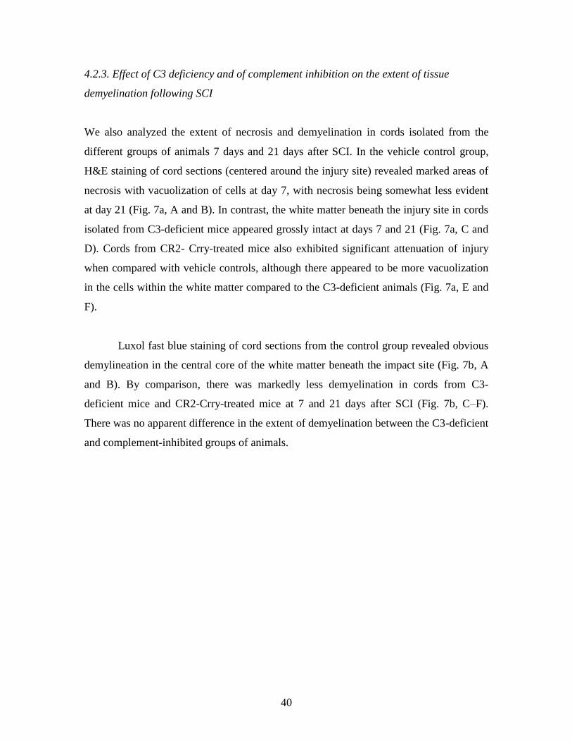
4.2.3. Effect of C3 deficiency and of complement inhibition on the extent of
tissue demyelination following SCI ……………………………………….40
4.2.4. Histopathology of spinal cord sections after SCI …………………..……..42
4.2.5. Quantitative assessment of histopathological inflammation and injury..44
4.2.6. Morphometric analysis of tissue sparing 3 days after injury ………..….44
4.3. EXPRESSION OF NEUTROPHILS AND MACROPHAGE INFILTRATION
AFTER SPINAL CORD INJURY ………………………………………..…….46
4.3.1a. Effect of C3 deficiency and of complement inhibition on the neutrophil
infiltration following SCI…………………………………………………………..46

4
4.3.1b. Effect of fB and CD59 deficiency and of alternative pathway complement
inhibition on the neutrophil infiltration following SCI ……………………..…48
4.3.2. Expression of macrophage infiltration after spinal cord injury ……..49
4.4. TARGETING AND BIODISTRIBUTION OF THERAPEUTICALLY
ADMINISTERED CR2- Crry …………………………………….…………….50
4.5. DEPOSITION OF COMPLEMENT ACTIVATION ……………………….53
4.5.1. Time course of complement activation 1, 24 and 72 hours after SCI ...53
4.5.2. Complement deposition at epicenter of injury 24 h post-SCI ………….54
4.5.3. Quantitative assessment of complement deposition post-SCI …………56
4.6. HISTOLOGICAL ANALYSIS OF RECOVERY ……………………..……58
5. DISCUSSION ……………………………………………………………………...61
5.1 ROLE OF C3 AND COMPLEMENT INHIBITORY PROTEIN CR2-CRRY
DURING SCI……………………………………………………………………62
5.2 ROLE OF FB THROUGH ALTERNATIVE PATHWAY DURING SCI …….65
5.3 ROLE OF CD59 THROUGH TERMINAL COMPLEMENT PATHWAY
DURING SCI …………………………………………………………………...66
6. SUMMARY AND CONCLUSIONS…………………………………………..…..68
7. ABSTRACT………………………………………………………………..……......70
8. ABSTRACT IN HUNGARIAN (ÖSSZEFOGLALÁS)………………………......72
9. BIBLIOGRAPHY…………………………………………………………………..73
10. PUBLICATIONS OF PH.D. CANDIDATE .........................................................90
1. LIST OF PUBLICATION RELATED TO THESIS...............................................90
Peer reviewed articles …………………………………………………………………...90
2. OTHER PUBLICATIONS………………………………………………………..91
Peer reviewed articles…………………………………………………………………...91
11. ACKNOWLEDGEMENT………………………………………………………...94

5
List of abbreviations
SCI, spinal cord injury
CP, classical pathway
AP, alternative pathway
MBL, mannose-binding lectin
MAP, membrane attack pathway
CNS, central nervous system
MAC, membrane attack complex
fB, factor B
CR1, complement receptor 1
DAF, decay-accelerating factor
MCP, membrane cofactor protein
TBI, traumatic brain injury
AD, Alzheimer’s disease
Wt, Wild-type
C3-/-, C3-deficient

6
fB-/-, fB-deficient
CD59-/-, CD59-deficient
PBS, phosphatebuffered saline
AfB, anti-fB antibody
SCR, short consensus repeat
BBB, Basso,Beattie,Bresnahan rating scale
BMS, Basso Mouse Scale

7
1. INTRODUCTION 1.1. Background and significance
1.1.1. Spinal cord injury
Spinal cord injury (SCI) is characterized by an initial traumatic injury phase, followed
closely by secondary events that result in edema, ischemia, excitotoxicity, and
inflammation(1). The mechanisms of secondary injury are not well defined, but it is clear
that inflammatory processes play a significant role in functional recovery(2, 3). While the
initial traumatic injury is difficult to guard against, the subsequent inflammatory cascade
represents a therapeutic target for SCI. The only clinical therapy accepted currently for
acute SCI is methylprednisolone, a therapy that has yielded disappointing results, with
the data from clinical trails being contradictory and inconclusive(4-6).
1.1.2. Complement system and neuroinflammation
Complement is important for host defense against pathogens and is an effector
mechanism for both innate and adaptive immune responses. Complement is also involved
in immune homeostasis mechanisms including the catabolism of immune complexes and
clearance of apoptotic cells. Under certain disease conditions in which inappropriate or
excessive complement activation occurs, complement control mechanisms breakdown or
are overcome and complement causes tissue injury. Complement play a key role in the
pathogenesis of many inflammatory and ischemic disease conditions. Following primary
injury to the spinal cord, a complex cascade of pathophysiologic processes occur that
result in secondary injury and progressive degenerative effects that can determine the
extent of recovery. The mechanisms involved in secondary tissue injury are not well
defined, but inflammation and ischemia are considered to be key components. Evidence
indicates an important, albeit not well defined, role for complement in the
8
pathophysiological processes that occur following both traumatic brain and spinal cord
injury (2, 6-10)(2, 6-10)(2, 6-10)[2, 6-10].
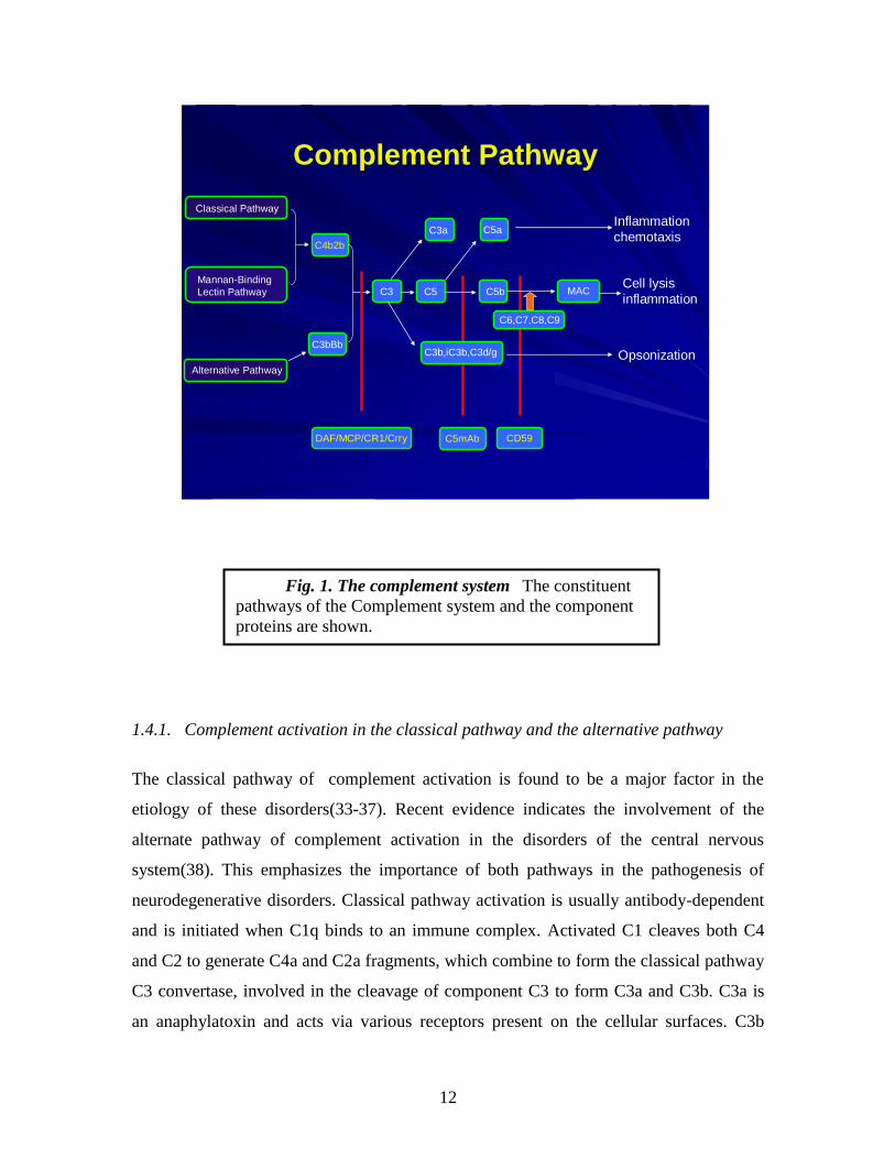
1.2. Overview of the complement system and effector mechanisms
In order to appreciate the role of complement in spinal cord injury, it is essential to first
understand the basics of the system. The complement system (Fig. 1), a major component
of the innate immune response, is activated in many neurodegenerative diseases (11, 12).
The complement system is a self amplifying cascade of proteases(13). Activation results
in 1) attraction and activation of phagocytes(14, 15); 2) opsonisation; and 3) formation of
the membrane attack complex (MAC or C5b-9)(16). The complement system consists of
nearly 30 proteins involved in non-specific immune response and the role of various
complement components in the pathophysiology of several disorders has been well
studied in the past two decades. The complement system is activated in several disorders,
particularly in response to foreign proteins, polysaccharides, cell surface components of
microbial origin, or toxic proteins associated with the pathogenesis of several disorders.
These foreign proteins activate the complement system by various pathways. The
complement system is activated by the three major pathways, the classical pathway (CP),
alternative pathway (AP), and lectin-mediated pathway. Activation of one of the
pathways may lead to activation of the other pathway. In the central nervous system
(CNS), the two main pathways found to be activated are the classical and the alternative
pathway(17, 18). Some complement factors, including C1q (initiator of the classical
complement pathway), C1r, C1s, C3 and C4 are locally produced in the CNS (19-22),
whereas others derive from the blood. Activated complement can trigger an adaptive
immune response, involving antigen presenting cells, T-cells and B-cells. The classical
pathway is normally antibody dependent whereas the alternative pathway is usually
antibody independent. Activation of complement by any pathway leads to the formation
of a C3 convertase that cleaves C3 into C3a and C3b, with the latter binding covalently to
the activating surface and participating in the formation of additional C3 convertase
complexes (amplification loop). C3 convertases also participate in the formation of C5
convertase, which cleaves C5 to yield C5a and C5b. Formation of C5b initiates the
terminal complement pathway resulting in the sequential assembly of complement

9
proteins C6, C7, C8 and (C9)n to form the cytolytic membrane attack complex (MAC or
C5b-9). Cleavage fragments of C3 covalently bound to activating surfaces act as ligands
for receptors on immune effector cells. C3a and C5a are chemotactic and can activate
leukocytes. Formation of the MAC on an activating cell surface can cause direct cell lysis.
Host cells are normally protected from complement by membrane-bound inhibitors of the
complement activation (inhibit C3 convertase formation) and of the terminal complement
pathway (inhibit MAC formation). Membrane inhibitors of complement activation are
complement receptor 1 (CR1), decay-accelerating factor (DAF) and membrane cofactor
protein (MCP). Rodents express an additional inhibitor of complement activation termed
Crry that is a structural and functional homolog of human CR1 (23, 24). Control of the
terminal pathway and MAC formation in cell membranes occurs through the activity of
CD59 that binds to C8 and C9 in the assembling MAC. The complement anaphylatoxins
are responsible for the various proinflammatory events in the CNS and chemotaxis of the
immunocompetent cells (25).
1.3. The complement system in neurodegeneration
The complement system is known to perform a wide range of functions in the human
body. It forms an essential component of the host immune system and is associated with
the clearance of molecules of foreign origin as well as the elimination of invading
pathogens from the body. It plays an important role in adaptive immunity(26). However,
it is nonspecific in action and unable to distinguish between self and non-self. Under
normal conditions, it is strictly regulated by complement regulatory molecules. However,
in neuroinflammatory disorders, the complement regulatory molecules fail to control the
activated complement components. These activated complement components then act as
a double-edged sword and are responsible for the degeneration of neurons(27). The
devastating roles of the complement components in neurodegenerative disorders are well
documented. Spinal cord injury (SCI )(6), traumatic brain injury (TBI)(28), Alzheimer’s
disease (AD)(29), multiple sclerosis (MS)(30, 31), myasthenia gravis(32) and
Parkinson’s disease (PD)(33) are examples of a few disorders in which activated
complement components play an important role. Not only do these disorders arise out of

10
dysfunction in the normal metabolic machinery, but also neuroinflammatory disorders
associated with microbial infections show involvement of complement components.
1.4. Why it is important to study the role of complement in spinal cord injury?
An impact to the spinal cord results in a primary injury, but the impact also triggers a
series of downstream events that lead to secondary injury of tissue and the progressive
degeneration of the spinal cord. Therapeutic interventions that minimize secondary injury
will improve functional recovery after traumatic injury. Inflammation plays a key role in
secondary injury following spinal cord injury (SCI). Inflammation is a rapid immune
response that can be initiated by infection or tissue damage. An important component of
an inflammatory response is the complement system, a collection of blood proteins that
form part of the immune system and that can be activated by injured cells and tissues.
Activation of complement amplifies the inflammatory response and produces molecules
that can be directly toxic or that can recruit and activate cells of the immune system to
produce toxic molecules. Inhibiting the complement system has been shown to be an
effective therapy for inflammatory disease in various animal models, and some
complement inhibitors are in clinical trials.
We have shown that complement deficient mice and normal mice treated with a
complement inhibitor are protected from neuronal injury following SCI, and that they
have significantly improved functional scores over time compared to control mice. These
data indicate an important role for complement in secondary injury, and indicate
complement inhibition will reduce inflammation and provide neuroprotection following
spinal cord injury. However, complement-dependent mechanisms involved in secondary
injury following SCI are not known, and there remain concerns regarding the clinical
application of the currently available systemic (body-wide) complement inhibitors with
regard to their safety and efficacy. Complement activation products are important for host
defense and immune maintenance mechanisms, and systemic complement inhibition can
compromise the protective and beneficial roles of complement. This will not be optimal

11
in patients at risk of infection, and urinary tract infection is a frequent complication
during initial and ongoing medical rehabilitation after SCI.
We propose to develop a neuroprotective strategy based on attenuating
complement-dependent secondary damage after SCI using a novel and validated
approach. The strategy involves the targeting of complement inhibitors to sites of
complement activation and injury, and we have shown the approach to be highly effective
in vitro and in mouse models of SCI and other inflammatory disease conditions. The
targeting strategy enhanced the activity and protective effect of complement inhibitors by
10-20 fold compared to untargeted counterparts. Furthermore, untargeted, but not
targeted complement inhibitors systemically inhibited complement and increased
susceptibility to infection in a mouse model.
We propose to fully characterize our targeted complement inhibitors in a mouse
model of SCI. In addition to therapeutic endpoints, we will use different types of
complement inhibitor to investigate complement-dependent disease mechanisms. We will
determine relationships between the generation of different complement activation
products and other molecules associated with inflammation in order to gain a better
understanding of the mechanisms of secondary tissue injury following SCI. These
preclinical studies will establish the guidelines necessary for the translation of this
therapy to the clinic using recombinant human proteins.
12
ComplementComplement PathwayPathway
Classical Pathway
C3 MAC
C6,C7,C8,C9
C3a C5aInflammation
chemotaxis
Cell lysis
inflammation
C3b,iC3b,C3d/g OpsonizationAlternative Pathway
Mannan-Binding
Lectin Pathway
C4b2b
C3bBb
DAF/MCP/CR1/Crry
C5 C5b
C5mAb CD59
1.4.1. Complement activation in the classical pathway and the alternative pathway
The classical pathway of complement activation is found to be a major factor in the
etiology of these disorders(33-37). Recent evidence indicates the involvement of the
alternate pathway of complement activation in the disorders of the central nervous
system(38). This emphasizes the importance of both pathways in the pathogenesis of
neurodegenerative disorders. Classical pathway activation is usually antibody-dependent
and is initiated when C1q binds to an immune complex. Activated C1 cleaves both C4
and C2 to generate C4a and C2a fragments, which combine to form the classical pathway
C3 convertase, involved in the cleavage of component C3 to form C3a and C3b. C3a is
an anaphylatoxin and acts via various receptors present on the cellular surfaces. C3b
Fig. 1. The complement system The constituent
pathways of the Complement system and the component
proteins are shown.
13
combines with C4a and C2a to form C5 convertase, which leads to the formation of C5a
and C5b. The former is an anaphylatoxin and the latter is involved in the production of
terminal complement components, C6 to C9. C5b, with these terminal complement
components, results in the formation of MAC(39, 40). The alternate pathway also results
in the formation of complement anaphylatoxins and MAC, but activated through C3
component, requires factor B to form C3 convertase and C5 convertase(40).
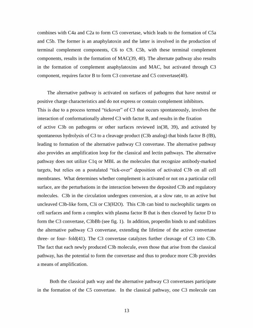
The alternative pathway is activated on surfaces of pathogens that have neutral or
positive charge characteristics and do not express or contain complement inhibitors.
This is due to a process termed “tickover” of C3 that occurs spontaneously, involves the
interaction of conformationally altered C3 with factor B, and results in the fixation
of active C3b on pathogens or other surfaces reviewed in(38, 39), and activated by
spontaneous hydrolysis of C3 to a cleavage product (C3b analog) that binds factor B (fB),
leading to formation of the alternative pathway C3 convertase. The alternative pathway
also provides an amplification loop for the classical and lectin pathways. The alternative
pathway does not utilize C1q or MBL as the molecules that recognize antibody-marked
targets, but relies on a postulated “tick-over” deposition of activated C3b on all cell
membranes. What determines whether complement is activated or not on a particular cell
surface, are the perturbations in the interaction between the deposited C3b and regulatory
molecules. C3b in the circulation undergoes conversion, at a slow rate, to an active but
uncleaved C3b-like form, C3i or C3(H2O). This C3b can bind to nucleophilic targets on
cell surfaces and form a complex with plasma factor B that is then cleaved by factor D to
form the C3 convertase, C3bBb (see fig. 1). In addition, properdin binds to and stabilizes
the alternative pathway C3 convertase, extending the lifetime of the active convertase
three- or four- fold(41). The C3 convertase catalyzes further cleavage of C3 into C3b.
The fact that each newly produced C3b molecule, even those that arise from the classical
pathway, has the potential to form the convertase and thus to produce more C3b provides
a means of amplification.
Both the classical path way and the alternative pathway C3 convertases participate
in the formation of the C5 convertase. In the classical pathway, one C3 molecule can

14
covalently bind C4b in the classical pathway C3 convertase, C4b2a, to form C4b2a3b, the
classical pathway C5 convertase. Similarly, the addition of activated C3b to the
alternative pathway C3 convertase, C3bBb, forms C3bBbC3b, the alternative pathway C5
convertase. Both C5 convertases can cleave C5 to yield C5a and C5b. C5a has powerful
proimflammatory and chemotactic properties(42).
1.4.2. Complement activation in the lectin pathway
The lectin pathway is activated when mannose binding protein (MBL) or ficolins bind to
conserved carbohydrate structures.
1.4.3. The membrane attack pathway (MAP), the terminal pathway
The terminal complement components formed by the cleavage of C5 either by the
classical or the alternate pathway result in the formation of membrane attack complex
(MAC). The MAC formation is regulated by complement defense protein CD59,
deficiency of which results in neurodegenerative diseases(43). The neurotoxic effects of
MAC on neuronal cells are time and concentration dependent. Mechanisms like free
radical generation, cytokines, eicasonoids, and increased permeability to Na+ and Ca2+
ions induced by MAC might be responsible for the observed effect(44). Apart from their
lytic properties, recent findings suggest that both cytolytically active and inactive forms
of terminal complement complexes are involved in the accumulation of leukocytes into
the cerebrospinal fluid and endothelial cell activation. At sublytic concentration, it not
only protects host cells but also stimulates protein biosynthesis. The role of MAC, at
sublytic and lytic concentration, is outlined by Wurzner(45). This explains the
concentration-dependent dual role of MAC. At lytic concentration, it acts as a
neurodegenerative agent and at sublytic concentration it acts as a neuroprotective agent.
Marked neurodegeneration in CNS disorders, even in the presence of the MAC, indicates
the failure of regulation of MAC components and the need to restrict its activity to

15
sublytic concentration by proper regulation of the pathways leading to MAC formation.
Thus, regulating the complement pathways leading to its generation can control
formation of MAC(46).
All pathways converge at C3 activation with the subsequent cleavage of C5. During
this process, the anaphylatoxins C3a and C5a are generated, and C5 cleavage initiates the
terminal complement pathway that culminates in the formation of the membrane attack
complex (MAC). The MAC can be directly cytolytic and can stimulate the production of
proinflammatory molecules when deposited in cell membranes at sublytic concentrations
(for a review of the complement system, see Ref (47)).Cleavage of C5 is the final
enzymatic step of the complement system. C5b bound to the convertase sequentially
binds C6 and C7. The C5b67 complex is released from the convertase and associates
with adjacent membrane via a labile hydrophobic binding site in the C6 component. The
C5b67 complex stably associates with the membrane, then binds C8, and finally, multiple
copies (up to 12) of C9. MAC induces concentration-dependent neuronal cell death and
changes in membrane permeability to Na+, K+ and Ca2+, release of cytokines,
eicosanoids, and reactive-free radicals. These changes occur at sublytic concentrations of
MAC(44). MAC is also responsible for the demyelination of neurons in demyelinated
forms of certain disorders(48). Upon binding to C5b-8, C9 unfolds and inserts in the
membrane. The recruitment of additional C9 molecules, membrane attack complexes,
MACs, form a functional pore in the cell membrane through which ions and small
molecules can pass, bringing about osmotic lysis of the cell.
1.5. Complement inhibitors as therapeutic agents
Although, inflammation plays an important role in CNS disorders, no currently available
anti-inflammatory agent offers significant neuroprotection in such disorders(40). Various
types of complement inhibitory proteins are currently under investigation for therapy of
inflammatory and ischemic disease (reviewed in (49-52)(49-52)(49-52)[49-52]).
Interventional studies with soluble complement inhibitors in rodent models of traumatic
brain injury, and studies using transgenic mice with astrocyte-targeted expression soluble
Crry indicate complement plays an important role in inflammation and secondary tissue

16
damage (28, 53, 54)(28, 53, 54)(28, 53, 54)[28, 53, 54]. Clinical studies have shown
elevated levels of complement proteins in cerebrospinal fluid from patients with
traumatic brain injury, and a recent study demonstrated that a viral complement control
protein modulates inflammation following spinal cord injury in rats (2, 55, 56)(2, 55,
56)(2, 55, 56)[2, 55, 56].
Virtually all complement inhibitory strategies reported to date depend on systemic
complement inhibition. We have shown that the targeting of complement inhibitors to
sites of complement activation and disease significantly improves their efficacy and
obviates the need for systemic inhibition. One strategy to target complement inhibitors is
to link them to antibody fragments, and we have shown that antibody-linked complement
inhibitors (DAF, Crry and CD59) are significantly more effective than untargeted
counterparts in vitro and in an animal model of tubulointerstitial disease(57-59)(57-
59)(57-59)[57-59]. Significantly, for CD59 to function effectively, targeting to the site of
complement activation is a requirement (57)(57)(57)[57]. Due to considerations of
species selective activity, Crry is a relevant C3 inhibitor for studies in mice.
In this study we propose to use a targeting strategy based on a fragment of
complement receptor 2 (CR2) to target complement inhibitors specifically to sites of
complement activation. Natural ligands for CR2 are iC3b, C3dg and C3d, cell-bound
breakdown fragments of C3 that are deposited on activating surfaces(60, 61)(60, 61)(60,
61)[60, 61]. These C3 ligands are relatively long lived and are present in high
concentrations at sites of complement activation. We have shown that human CR2-DAF
and CR2-CD59 bind to C3-coated targets and are at least 20-fold more potent than their
untargeted counterparts at providing protection from complement (58)(58)(58)[58] (see
attached manuscript). We recently characterized CR2-Crry in a mouse model of intestinal
ischemia/reperfusion injury (IRI) and demonstrated that CR2-Crry was 10-fold more
effective than Crry-Ig, an untargeted systemic counterpart. Furthermore, CR2-Crry,
unlike Crry-Ig, was therapeutically effective at a dose that did not result in systemic
complement inhibition and that did not increase susceptibility to infection.
17
2. Objectives
Traumatic spinal cord injury initiates a cascade of pathophysiological events that cause
secondary injury and determine the extent of functional recovery. Although the processes
that occur following SCI are complex, inflammation is considered to play a key role in
the progressive degenerative events that take place, especially within the lesion penumbra.
The complement system plays a key role in the pathogenesis of many inflammatory and
ischemic conditions, and recent evidence indicates it also plays an important role in
secondary SCI. Various systemic complement inhibitors are currently under therapeutic
investigation as anti-inflammatory agents, but there remain concerns regarding their
efficacy and safety. Complement activation products are important for host defense and
immune homeostasis mechanisms, and systemic complement inhibition can compromise
the protective and immunomodulatory roles of complement. In this context, CNS injury
has been shown to be immunosuppressive, and further immune suppression by systemic
complement inhibition may not be optimal in patients at risk of infection (urinary tract
infection is a frequent complication during initial and ongoing medical rehabilitation after
SCI).
The goals of the present study were (1) To investigate the dynamics of
complement activation and its role in the development of SCI in mice. (2) To determine
in vivo relationships between the generation of different complement activation products
with cytokine production, adhesion molecule expression, leukocyte infiltration and
activation, and injury. (3) To investigate the neuroprotective effect of a novel targeted
complement inhibitor and develop a neuroprotective strategy based on attenuating
complement-dependent secondary damage after SCI.

18
2.1. To develop a neuroprotective strategy based on attenuating complement-dependent
secondary damage after SCI
We have developed a strategy to target complement inhibitors to sites of complement
activation and injury, and have shown that targeted complement inhibitors are 10-20 fold
more effective in vitro and in an experimental models of ischemia and reperfusion injury
compared to conventional systemic approaches to inhibit complement. We hypothesize
that our novel strategy to target complement inhibitors to the site of SCI will improve
bioavailability, obviate the need to systemically inhibit complement, and provide a safe
and highly efficacious therapy. We propose to investigate our hypothesis in a mouse
model of SCI. Targeted complement inhibition will be achieved by the use of soluble
recombinant chimeric molecules consisting of a targeting moiety linked to a complement
inhibitor. Complement inhibitors will be mouse Crry (inhibits early in the complement
pathway) or CD59 (inhibits late in pathway). The targeting moiety will be a fragment of
complement receptor 2 (CR2) that binds to long lived degradation products of C3 that are
deposited at sites of complement activation. In addition to therapeutic endpoints, we will
utilize the targeted complement inhibitors that function at different points in the
complement cascade to investigate disease mechanisms in a clinical setting.
2.2. To investigate the neuroprotective effect of a novel targeted complement
inhibitor.
To investigate the neuroprotective effect of two complement inhibitory strategies, a novel
targeted complement inhibitor (CR2-Crry) which targets specifically to sites of
complement activation and does not require systemic complement inhibition and an anti-
fB antibody that suppresses the alternative pathway of complement activation. For the
therapeutic studies we used mouse CR2-Crry, a complement inhibitory fusion protein that
functions at the C3 level. The complement inhibitor, Crry, is targeted specifically to sites
of complement activation by means of the complement receptor 2 (CR2)-targeting
moiety(62). CR2 is a member of the C3-binding protein family, and its natural ligands are
cleavage fragments of C3 that become deposited at sites of complement activation.
Targeted complement inhibition has been shown to provide significant benefits over
19
systemic (untargeted) inhibition in terms of efficacy and host susceptibility to
infection(63). The use of mouse Crry is appropriate when studying the effects of
complement inhibition in mice, because complement inhibitors display different degrees
of species selectivity. Crry is a structural and direct functional analog of human
complement receptor 1 (CR1), and data obtained with Crry in rodents will likely translate
in functional terms to the use of CR1 in humans.
As discussed in section 5.2, in the present study, we show that fB-deficient mice
have an improved outcome after SCI. The level of protection afforded to fB-deficient
mice was similar to that seen in our previous study using C3-deficient mice(64). These
data indicate a dependence on the alternative pathway for complement-mediated SCI.
Together with the previous findings described above using B cell and C1q-deficient mice,
it is likely that the alternative pathway plays a critical role in amplifying antibody-
mediated classical pathway initiated complement activation. In this context, reduced
pathology and improved locomotor recovery in fB-deficient mice was associated with
significantly reduced C3 and MAC deposition compared to wt control mice, even though
the classical (and lectin) pathway is intact in fB-deficient mice. A similar dependence on
both antibody-mediated lectin pathway activation and alternative pathway activation has
been described in models of intestine ischemia and reperfusion injury(65-67). In a more
clinically relevant paradigm, we demonstrated that specific inhibition of the alternative
pathway with anti-fB mAb was also protective against SCI, and to a similar level as fB
deficiency.

20
3. METHODS AND MATERIALS
3.1. Mice
Female wild-type C57BL/6 and C57Bl/6 C3-deficient (C3-/-) mice were obtained from
Jackson Laboratories (Bar Harbor, ME). Breeding pairs of fB-deficient (fB-/-) mice on
C57BL/6 background were generated as described (68) and provided by Dr. J. Thurman
(University of Colorado Health Sciences Center, Denver, CO) and a breeding colony
established. In mice, there are two genes encoding CD59; CD59a is widely expressed and
is the primary regulator of the MAC in mice, whereas CD59b expression is limited to the
testis and, at very low levels, bone marrow (69). In this study, CD59a-deficient mice on
C57BL/6 background were used and were generated as described (70). fB and CD59
deficiency was confirmed by genotyping. Mice weighing between 18 22 g (6 8 weeks
old) were used in experiments. All mice were fed on standard laboratory food and given
tap water ad libitum with a light dark cycle of 12 hours.
3.2. Complement Inhibitor-CR2-Crry
The fusion protein CR2-Crry was produced and purified as described previously
(71). In brief, a cDNA construct of the recombinant fusion protein was prepared by
joining the mouse CR2 sequence encoding the four N-terminal short consensus repeat
(SCR) units (residues 1–257 of mature protein, National Center for Biotechnology
Information Gen-Bank, accession number M35684) to sequences encoding extracellular
regions of mouse Crry. The Crry sequence used encoded residues 1–319 of the mature
protein (National Center for Biotechnology Information GenBank, accession number
NM013499). To join CR2 to Crry, linking sequences encoding (GGGGS)2 were used.
The recombinant protein was expressed in NSO cells and purified by anti-Crry affinity
chromatography as described (71). CR2-Crry has a circulatory half-life in C57BL/6 mice
of 8 hours (71).

21
3.3. Anti-fB anti-body
The isolation and characterization of anti-fB mAb 1379 used in these studies was
described previously, and the mAb effectively inhibits the mouse alternative pathway(72).
The mAb was generously provided by Drs. V. M. Holers, J. M. Thurman (University of
Colorado Health Sciences Center, Denver, CO), and G. S. Gilkeson (Medical University
of South Carolina).
3.4. Spinal cord injury surgery and antibody treatment
3.4.1. Treatment
Wild-type (wt) mice were randomized into sham (laminectomy, no SCI damage),
vehicle control (phosphatebuffered saline [PBS]), and CR2-Crry treatment and anti-fB
mAb treatment groups. Other groups consisted of C3-deficient (C3-/-), fB-deficient (fB-/-)
and CD59a-deficient (CD59-/-) mice. For CR2-Crry treatment were administered a
single dose of 0.25 mg of CR2-Crry by tail vein injection. All other animals received
intravenous injections of phosphate buffered saline. For anti-fB mAb treatment, wt mice
were randomized into four groups, with mice in each group receiving an intravenous (tail
vein) injection of 100 µl PBS vehicle control or 2 mg anti-fB mAb in 100µl PBS at 1 and
12 hours after surgery, 12 and 24 hours after surgery, or 24 and 36 hours after surgery.
The 2mg dose used was based on previous studies characterizing the therapeutic effect of
this Ab in mouse models of inflammation (72-74).

22
3.4.2. SCI model
3.4.2a. Instruments
Fig. 2. Instruments of the spinal cord injury surgery.

23
3.4.2b. Anaesthesia
Mice were anesthetized with ketamine/xylazine anesthesia (80 mg/kg/10 mg/kg,
i.p.) Animals were breathing spontaneously, and body temperature was maintained using
a heat mat for the duration of the experiment.
3.4.2c. SCI Surgery
Mice subjected to laminectomy at the level of the 12th thoracic vertebra (T12),
followed by contusion-induced SCI using the NYU weight drop impactor as described
previously(64). After injury, the muscles and the subcutaneous tissue were closed in
layers, the skin was closed with metal wound clips (World precision instruments), and
mice were given 1 ml of saline per day for 3 days to compensate for loss of blood and
dehydration. Bladders were expressed before testing, and each mouse was evaluated for 4
5 minutes on the day before surgery, immediately after surgery, and then manual bladder
expression was performed twice daily until full bladder function was observed. Sham
mice received a dorsal laminectomy without impact injury. There was less than 5%
surgical mortality, and zero mortality of mice that recovered from surgery. Groups of
mice were sacrificed at 1, 3, 7 and 21 days after injury, and spinal cords were isolated for
analysis.
3.5. Locomotor Function Analysis
Open-field observations of locomotor function recovery were independently scored by
two observers blinded to experimental groups using the Basso, Beattie and Bresnahan
(BBB) rating scale developed for rats(75), but later adapted by others for mice(76-79) or
Basso Mouse Scale (BMS)(80).

24
3.5.1. The Basso, Beattie and Bresnahan (BBB) rating scale
TABLE 1
Basso, Beattie, and Bresnahan Locomotor Rating Scale
0 No observable hindlimb (HL) movement
1 Slight movement of one or two joints, usually the hip and/or knee
2 Extensive movement of one joint or extensive movement of one joint and slight
movement of one other joint
3 Extensive movement of two joints
4 Slight movement of all three joints of the HL
5 Slight movement of two joints and extensive movement of the third
6 Extensive movement of two joints and slight movement of the third
7 Extensive movement of all three joints of the HL
8 Sweeping with no weight support or plantar placement of the paw with no weight
support
9 Plantar placement of the paw with weight support in stance only (i.e., when stationary)
or occasional, frequent, or consistent weight-supported dorsal stepping and no plantar
stepping
10 Occasional weight-supported plantar steps; no FL–HL coordination
11 Frequent to consistent weight-supported plantar steps and no FL–HL coordination
12 Frequent to consistent weight-supported plantar steps and occasional FL–HL
coordination
13 Frequent to consistent weight-supported plantar steps and frequent FL–HL
coordination
14 Consistent weight-supported plantar steps, consistent FL–HL coordination, and
predominant paw position during locomotion is rotated (internally or externally)
when it makes initial contact with the surface as well as just before it is lifted off at
the end of stance; or frequent plantar stepping, consistent FL–HL coordination, and
occasional dorsal stepping

25
15 Consistent plantar stepping and consistent FL–HL coordination and no toe clearance
or occasional toe clearance during forward limb advancement; predominant paw
position is parallel to the body at initial contact
16 Consistent plantar stepping and consistent FL–HL coordination during gait and toe
clearance occurs frequently during forward limb advancement; predominant paw
position is parallel at initial contact and rotated at lift off
17 Consistent plantar stepping and consistent FL–HL coordination during gait and toe
clearance occurs frequently during forward limb advancement; predominant paw
position is parallel at initial contact and lift off
18 Consistent plantar stepping and consistent FL–HL coordination during gait and toe
clearance occurs consistently during forward limb advancement; predominant paw
position is parallel at initial contact and rotated at lift off
19 Consistent plantar stepping and consistent FL–HL coordination during gait, toe
clearance occurs consistently during forward limb advancement, predominant paw
position is parallel at initial contact and lift off, and tail is down part or all of the
time
20 Consistent plantar stepping and consistent coordinated gait, consistent toe clearance,
predominant paw position is parallel at initial contact and lift off, and trunk
instability; tail consistently up
21 Consistent plantar stepping and coordinated gait, consistent toe clearance,
predominant paw position is parallel throughout stance, and consistent trunk
stability; tail consistently up
Note.
Slight: Partial joint movement through less than half the range of joint motion.
Extensive: Movement through more than half of the range of joint motion.
Sweeping: Rhythmic movement of HL in which all three joints are extended and then
fully flex and extend again; animal is usually sidelying and plantar surface of paw may or
may not contact the ground; no weight support across the HL is evident.
26
No weight support: No contraction of the extensor muscles of the HL during plantar
placement of the paw; or no elevation of the hindquarter.
Weight support: Contraction of the extensor muscles of the HL during plantar
placement of the paw; or, elevation of the hindquarter.
Plantar stepping: The paw is in plantar contact with weight support and then the HL is
advanced forward and plantar contact with weight support is reestablished.
Dorsal stepping: Weight is supported through the dorsal surface of the paw at some
point in the step cycle.
FL–HL coordination: For every FL step a HL step is taken and the HLs alternate.
Occasional: Less than or equal to half; #50%.
Frequent: More than half but not always; 51–94%. Consistent: Nearly always or always;
95–100%. Trunk instability: Lateral weight shifts which cause waddling from side to side
or a partial collapse of the trunk.
The BBB locomotor rating scale is an open-field 21 point evaluation and is rated
according to categories describing the quality of joint movements, the trunk, abdomen,
and paw placement, stepping, trunk stability, and tail position. Animals were assessed
preoperatively, on the day of surgery, and then daily postoperatively by an observer
blinded to animal treatment. Changes in BBB score during spinal cord injury between
vehicle control, C3-deficient, CR2-Crry-treated, and control animals were determined by
analysis of variance with repeated measures using Scheff’s test for posthoc comparisons.
A P value of less than 0.05 was considered statistically significant. All data were
subjected to statistical analysis using Statview Analysis Software (version 5; SAS
Institute Inc., Cary, NC).

27
3.5.2. The Basso Mouse Scale (BMS)
TABLE 2
BASSO MOUSE SCALE (BMS) [80
0 No ankle movement
1 Slight ankle movement
2 Extensive ankle movement
3 Plantar placing of the paw with or without weight support -OROccasional,
frequent or consistent dorsal stepping but no plantar stepping
4 Occasional plantar stepping
5 Frequent or consistent plantar stepping, no coordination -ORFrequent
or consistent plantar stepping, some coordination, paws rotated at initial contact and
lift off (R/R)
6 Frequent or consistent plantar stepping, some coordination, paws parallel at initial
contact (P/R, P/P) -ORFrequent or consistent plantar stepping, mostly coordinated,
paws rotated at initial contact and lift off (R/R)
7 Frequent or consistent plantar stepping, mostly coordinated, paws parallel at initial
contact and rotated at lift off (P/R)-ORFrequent or consistent plantar stepping,
mostly coordinated, paws parallel at initial contact and lift off (P/P), and severe
trunk instability
8 Frequent or consistent plantar stepping, mostly coordinated, paws parallel at initial
contact and lift off (P/P), and mild trunk instability -ORFrequent or consistent
plantar stepping, mostly coordinated, paws parallel at initial contact and lift off (P/P),
and normal trunk stability and tail down or up & down
28
9 Frequent or consistent plantar stepping, mostly coordinated, paws parallel at initial
contact and lift off (P/P), and normal trunk stability and tail always up.
Slight: Moves less than half of the ankle joint excursion.
Extensive: Moves more than half of the ankle joint excursion.
Plantar placing: Paw is actively placed with both the thumb and the last toe of the paw
touching the ground.
Weight support: (dorsal or plantar): The hindquarters must be elevated enough that the
hind end near the base of the tail is raised off of the surface and the knees do not touch
the ground during the step cycle.
Stepping: (dorsal or plantar): Weight support at lift off, forward limb advancement and
re-establishment of weight support at initial contact.
Occasional: Stepping less than or equal to half of the time moving forward.
Frequent: Stepping more than half the time moving forward.
Consistent: Plantar stepping all of the time moving forward with less than 5 missed steps
(due to medial placement at initial contact, butt down, knee down, skiing, scoliosis,
spasms or dragging) or dorsal steps.
Coordination: For every forelimb step a hindlimb step is taken and the hindlimbs
alternate during an assessable pass. For a pass to be assessable, a mouse must move at a
consistent speed and a distance of at least 3 body lengths. Short or halting bouts are not
assessable for coordination. At least 3 assessable passes must occur in order to evaluate
coordination. If less than 3 passes occur then the mouse is scored as having no
coordination.
Some coordination: Of all assessable passes (a minimum of 3), most of them are not
coordinated.
Most coordination: Of all assessable passes (a minimum of 3), most of them are
coordinated.
Paw position: Digits of the paw are parallel to the body (P), turned out away from the
body (external rotation: E) or turned inward toward midline (internal rotation; I).
Severe trunk instability: Severe trunk instability occurs in two ways.

29
(1) The hindquarters show severe postural deficits such as extreme lean, pronounced
waddle and/or near collapse of the hindquarters predominantly during the test. Or
(2) Five or more of any of the following events stop stepping of one or both hindlimbs
• Haunch hit: the side of hindquarters rapidly contacts the ground
• Spasms: sustained muscle contraction of the hindlimb which appears to immobilize
the limb in a flexed or extended position
• Scoliosis: lateral deviation of the spinal column to appear “C” shaped instead of
straight
Mild trunk instability: Less than 5 events listed above and some sway in the
hindquarters. Mild trunk instability is scored when the pelvis and haunches
predominantly dip, rock, or tilt from side-to-side (tilt). If the tail is up, the swaying of the
pelvis and/or haunches produces side-to-side movements of the distal third of the tail
which also indicates mild trunk instability (side tail).
Normal trunk stability: No lean or sway of the trunk, and the distal third of the tail is
steady and unwavering during locomotion. No severe postural deficits or events and less
than 5 instances of mild instability.
3.6. Histopathological Analysis
Spinal cords were removed at 1, 3, 7 and 21 days postinjury for histological analysis.
Immediately after sacrifice, mice were perfused transcardially with PBS followed by 4%
paraformaldehyde in PBS. Spinal cords were then removed, placed into 4%
paraformaldehyde/PBS, and either cryoprotected in 30% sucrose for 48 hours before
storing at -80°C or placed in formalin and processed to paraffin for histological analysis.
For histological assessment, sections of spinal cord were stained with hematoxylin and
eosin (H&E) or luxol fast blue (LFB) as previously described (Clark G: Staining
Procedures. Baltimore, MD: Williams and Wilkins, 1981, pp 111–129) and (81, 82)
Histopathological damage was assessed quantitatively by an independent reviewer
blinded to the experimental groups. H&E and LFB sections were scored from 0 to 3 for
the presence and intensity of inflammatory cell infiltration, neuronal vacuolation, and
hemorrhage (where 0, is no evidence and 3 severe). Scores were then expressed as a

30
cumulative score of 0–9. When assessing recovery at 21 days after injury, the extent of
demyelination, graded between 0–3 (with 0 being no evidence of demyelination) was
added for a cumulative score of 0–12. To further quantify spinal cord injury,
morphometric analyses was conducted to determine the degree of tissue sparing after
injury. Transverse sections of spinal cord were stained with H&E, and the cross-sectioned
area of spinal cord was measured at 150 μm increments extending 2mm either side of the
injury epicenter using Zeiss Axiovision image analysis software (Carl Zeiss, Oberkochen,
Germany). Measurements were averaged for animals in each group at each time point as
previously described (83). All assessments were performed in a blinded fashion.
3.7. Neutrophil and Macrophage Infiltration
The presence of infiltrating neutrophils and macrophages was assessed using
immunohistochemistry on frozen spinal cord sections. Standard immunohistochemical
methods were used as previously described(71). Neutrophils and macrophages were
identified by anti-mouse Gr-1 and Mac-3 (BD Biosciences), respectively. Neutrophils
and macrophages were quantified at the spinal cord injury epicenter, defined as the
section exhibiting maximal tissue damage. The total number of neutrophils and
macrophages were quantified using computerized image analysis methods, as previously
described(84, 85). Results are expressed as the number of neutrophils/macrophages per
mm2. Specificity of immunostaining was confirmed by both the use of isotype control
antibody and by the omission of primary antibody.
3.8. Complement Deposition
Spinal cord cryosections were fixed in cold acetone for 5 minutes and then washed in
running water followed by PBS. Sections were then incubated for 1 hour at room
temperature with either anti-mouse C3 fluorescein isothiocyanate (FITC) (Dako, Ely,
UK), mouse anti-mouse fB mAb 1379 (see above), rabbit anti rat C9 that is cross reactive
with mouse C9(86), or rat anti-mouse CD59 mAb 7A6(87). For C9 and CD59
visualization, donkey anti-rabbit FITC and donkey anti-rat FITC antibodies were used.

31
For fB visualization, we used a mouse on mouse staining kit and protocol from Vector
laboratories (Burlingame, CA). Sections were then counterstained with DAPI (Thermo
Scientific, Rockford, IL) for nuclear detail. Sections were then coverslipped and analyzed
for fluorescence intensity using a Zeiss LSM5 Confocal microscope. Fluorescence
intensity for each complement component was scored on a scale of 0 3, where 0 is no
staining, 1, mild, 2, moderate, and 3, intense. All observations were made by an observer
blinded to group identities.
3.9. Statistical Analysis
All data are presented as Mean±SEM or mean±SD as indicated. Analyses were
performed using Statview Analysis Software (version 5; SAS Institute Inc., Cary, NC) or
SPSS 13.0 for Windows. Statistical significance between groups was determined by two-
way analysis of variance with Bonferroni/ Dunn’s corrected post hoc t-tests. For
Locomotor functional analysis, repeated measures of analysis of variance was used to
determine differences between the groups. P <0.05 was considered statistically
significant.

32
4. RESULTS
4.1. Recovery of Locomotor Function Post-Traumatic Injury
4.1.1 Effect of C3 Deficiency and of Complement Inhibition on Locomotor Recovery
following SCI
To investigate the role of complement in SCI, we induced contusion injury to the spinal
cord in wt mice and in mice deficient in C3, a central protein of the complement system
and common for all pathways of activation. Following injury, locomotor recovery was
assessed using the modification of the BBB rating scale(75). All animals had a BBB
score of 21 pre-injury and a score of 0 immediately after injury, with bilateral hindlimb
paralysis (Fig. 3). Two days after injury, and every day thereafter through the termination
of the study at day 21, the C3-deficient mice had a significantly improved BBB score
compared to the wild-type controls (P < 0.001) (Fig. 3). By day 21 after injury, the C3-
deficient mice showed a near normal BBB score of 19.6 ± 1.2 (P< 0.001), whereas the
BBB score for wild-type mice was only 11.5 ± 2.14 which was significantly lower than
that of C3-deficient mice (P< 0.001). These data indicate that C3 plays an important role
in the posttraumatic events that affect functional recovery. Next, we determined whether
C3 blockade, using an intravenously administered inhibitor previously shown to target to
sites of complement activation, is a feasible posttraumatic therapeutic approach for
improving functional recovery. Using the same spinal cord paradigm, a group of mice
were treated with a single intravenous injection of 0.25 mg CR2-Crry at 1 hour after SCI.
As with the C3-deficient mice, the CR2-Crry-treated mice had a significantly improved
BBB score compared to shamoperated controls at all time points from day 2 following
traumatic injury (P<0.001) (Figure 3). The C3-deficient mice appeared to have a better
outcome than the CR2-Crry-treated mice, but the difference was not significant.

33
Fig. 3 Combined BBB locomotor scores post-SCI within sham, vehicle control, C3-deficient,
and CR2-Crry groups. Note significant improvement in BBB score at day 3, 7, and 21 in both
the C3-deficient and CR2-Crry groups when compared to vehicle controls (P = 0.001) (n = 12).
The values are expressed as mean ± SE.

34
4.1.2 Effect of fB and CD59 Deficiency and of alternative complement pathway
inhibition on Locomotor Recovery following SCI
Contusion injury to the spinal cord was induced in mice deficient in fB or CD59, in wt
untreated mice, and in wt mice subsequently treated with anti-fB mAb or PBS (vehicle).
Locomotor function was assessed by the BMS scale, and all mice subjected to contusion
injury exhibited a BMS score of 0 immediately after injury. Over the course of 21 days,
locomotor function significantly improved in fB-/- mice and wt mice treated with anti-fB
mAb at 1 and 12 hours after SCI compared to wt and PBS treated controls (Fig. 4). There
were no significant differences in BMS scores between fB-/- mice and antifB–treated
(1/12 hours) mice over the course of the experiment. When anti-fB mAb was
administered at 12 and 24 hours after SCI, there was a trend toward improved locomotor
function when compared to controls, but the difference did not reach significance (Fig. 4).
We also administered anti-fB mAb at 24 and 36 hours post-SCI, but there was no
difference in functional recovery compared to PBS treated mice (data not shown). Unless
otherwise stated, all data shown below for anti-fB treated mice was obtained using a 1
and 12 hour post-SCI treatment schedule. In contrast to the improvement seen in fB-/-
mice and anti-fB (1/12 hours) treated mice, recovery of locomotor function was
significantly impaired in CD59-/-mice compared to wt and vehicle treated controls. There
was no difference in functional recovery between PBS (vehicle) treated mice and
untreated wt mice after SCI.

35
0
1
2
3
4
5
6
7
8
9
pre 1 3 5 7 9 11 13 15 17 19 21
Days after injury
BM
S s
core
(0
-9)
ShamwtfB-/-Vehical Anti-fB(1/12h)Anti-fB(12/24h)CD59-/-
Fig. 4. Locomotor recovery after SCI in complement deficient and inhibited mice. Open-field 10-point BMS scores were recorded for 21 days after contusion-induced SCI
in the indicated groups of mice. Anti-fB mAb treatment group received an i.v. injection of
2 mg at 1 and 12 or 12 and 24 hours after injury. BMS scores are significantly higher for
fB-/- mice and anti-fB mAb (1/12 hours)-treated mice compared to wt or vehicle (PBS)-
treated mice from day 3 postinjury. No significant difference between vehicle controls
and anti-fB mAb (12/24 hours) treated mice. BMS scores are significantly lower in
CD59-/- mice compared to wt and vehicle control mice from day 11 after injury (P < 0.
01). Mean ± SE, n = 8 - 10.

36
4.2. Effect of Complement Deficiency and Complement Inhibition on Tissue Damage
And Demyelination after SCI
4.2.1. Macroscopic images of spinal cords isolated at 72 hours postinjury.
Spinal cord contusion results in a primary hemorrhage, inflammation, and loss/damage of
neurons. We determined the effect of fB deficiency, fB inhibition (1/12 hours), or CD59
deficiency on spinal cord tissue injury by macroscopic examination of spinal cords and
by assessment of histological changes within the spinal cords postinjury. At 72 hours
after injury, macroscopic examination of control spinal cords demonstrated marked
indentation at the injury site with evidence of hemorrhage. These features were markedly
reduced in fB-/- and anti-fB mAb treated animals, but appeared exacerbated in spinal
cords from CD59-/- mice (Fig. 5).

37
Vehicle
fB-/-
Anti-fB
Fig. 5. Macroscopic images of spinal cords isolated at 72 hours postinjury. Mice received anti-fB mAb or PBS (vehicle) at 1 and 12 hours after injury.
Representative images shown, n = 6 8 per group.
CD59-/-

38
4.2.2 Effect of C3 Deficiency and of Complement Inhibition on the Extent of Tissue
Destruction following SCI
To determine whether C3 deficiency or complement inhibition with CR2-Crry attenuated
overall spinal cord tissue damage, we determined the cross-sectioned area of spinal cords
at 100 μm increments extending 2 mm either side of the initial injury impact site.
Measurements were made using spinal cords isolated from C3-deficient mice and mice
treated with CR2-Crry or vehicle control (PBS). At 24 hours after injury, the profile of
tissue damage was similar in both C3-deficient and CR2-Crrytreated groups (Fig. 6A). In
the control group, there was a clear trend toward increased injury compared to the C3-
deficient/inhibited groups, but by 24 hours after SCI the difference did not reach
statistical significance at the injury site or on either side of the injury site. Comparable
relative profiles were obtained for the three groups of animals at 72 hours after SCI (Fig.
6B). Seven days after injury, however, there was significantly more tissue sparing at and
around the injury site in C3-deficient mice and in mice treated with CR2-Crry compared
to vehicle control mice (Fig. 6C). There was no difference in tissue sparing between C3-
deficient and CR2-Crrytreated mice at 7 days after SCI.

39
4.2.3. Effect of C3 deficiency and of complement inhibition on the extent of tissue
demyelination following SCI
Fig. 6. Tissue sparing as assessed by analyzing the cross-sectional area of spinal
cords removed from vehicle controls, C3-deficient, and CR2-Crrytreated animals at
24 hours (A), 72 hours (B), and 7 days after injury (C). Measurements were made
from histological sections taken at 100-μm increments extending 2 mm either side of
the injury site. No significant difference in tissue sparing was evident at 24 and 72
hrs. Significant tissue sparing was noted in CR2-Crry and C3-/- animals compared to
vehicle controls at day 7 (P = 0.002). Mean ± SD, n = 4.
40
4.2.3. Effect of C3 deficiency and of complement inhibition on the extent of tissue
demyelination following SCI
We also analyzed the extent of necrosis and demyelination in cords isolated from the
different groups of animals 7 days and 21 days after SCI. In the vehicle control group,
H&E staining of cord sections (centered around the injury site) revealed marked areas of
necrosis with vacuolization of cells at day 7, with necrosis being somewhat less evident
at day 21 (Fig. 7a, A and B). In contrast, the white matter beneath the injury site in cords
isolated from C3-deficient mice appeared grossly intact at days 7 and 21 (Fig. 7a, C and
D). Cords from CR2- Crry-treated mice also exhibited significant attenuation of injury
when compared with vehicle controls, although there appeared to be more vacuolization
in the cells within the white matter compared to the C3-deficient animals (Fig. 7a, E and
F).
Luxol fast blue staining of cord sections from the control group revealed obvious
demylineation in the central core of the white matter beneath the impact site (Fig. 7b, A
and B). By comparison, there was markedly less demyelination in cords from C3-
deficient mice and CR2-Crry-treated mice at 7 and 21 days after SCI (Fig. 7b, C–F).
There was no apparent difference in the extent of demyelination between the C3-deficient
and complement-inhibited groups of animals.
41
Fig. 7a. H&E-stained sections of spinal cord
centered on the injury site at days 7 and 21
after injury. A B: vehicle control. C D: C3-
deficient animals. E F: CR2-Crry-treated
animals. Original magnification, ×100.
Fig. 7b. Luxol fast blue stained section of
spinal cord centered on the injury site at days
7 and 21 after injury. A B: vehicle control.
C D: C3-deficient animals. E F: CR2-Crry-
treated animals. Original magnification,
×100.

42
4.2.4. Histopathology of spinal cord sections after SCI
We next analyzed spinal cords microscopically at the injury epicenter using H&E stain to
assess the extent of inflammatory cell infiltrate, neuronal vacuolation and hemorrhage.
Sections were prepared from cords isolated at 24 and 72 hours postinjury, and the
sections were graded for a total cumulative score of 0–9 (see Materials and Methods).
Injury to spinal cords was evident in all groups at both 24 and 72 hours (Fig. 8).

43
Fig. 8. Histopathology of spinal cord sections after SCI. A-H: Transverse sections
from the epicenter of injury were prepared from spinal cords isolated at 24 and 72
hours after injury and stained with H&E. Mice received anti-fB mAb or PBS (vehicle)
at 1 and 12 hours after injury. Representative images shown, n = 6 per group.
44
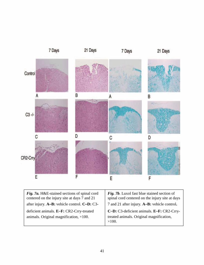
No significant differences were noted at 24 hours postinjury, but at 72 hours there
was significantly less damage seen in fB-/- mice and anti-fB mAb - treated mice
compared to control mice. In contrast, damage was significantly exacerbated in CD59-/-
mice compared to controls 72 hours after injury.
4.2.5. Quantitative assessment of histopathological inflammation and injury
Representative images of spinal cord sections postinjury are shown in Fig. 8, with
quantification of data shown in Fig. 9. Histologically, control and CD59-/- spinal cord
sections demonstrated evidence of hemorrhage, pronounced inflammation, neuronal cell
vacuolation, and demyelination, and while these features existed in fB-/-mice and anti-fB
mAb - treated mice, they were markedly reduced.
4.2.6. Morphometric analysis of tissue sparing 3 days after injury
Fig. 9. Quantitative assessment of histopathological inflammation and injury. H&E-
stained sections from the epicenter of injury were scored from 0 to 3 for the presence and
intensity of inflammatory cell infiltration, neuronal vacuolation, and hemorrhage (0 = no
evidence, 3 = severe). Scores were then expressed as a cumulative score of 0-9. Assessments
were made from spinal cords isolated at 24 and 72 hours postinjury. Mice received anti-fB
mAb or PBS (vehicle) at 1 and 12 hours after injury.
*P < 0.01, **P < 0.001 compared to vehicle control. Mean ± SD, n = 6 per group.
45
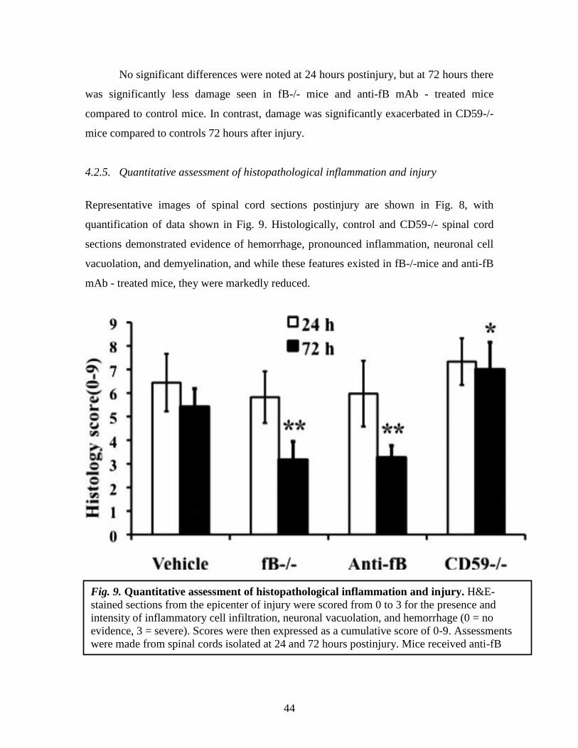
To further quantify the impact of complement deficiency or inhibition on SCI, we
analyzed tissue destruction by determining the cross-sectional area of spinal cords at
150µm increments extending 2 mm either side of the initial injury impact site. In accord
with our subjective histological assessments, there was significantly increased tissue
sparing in fB-deficient and fB-inhibited mice, and significantly less tissue sparing in
CD59-/- mice, when compared to control mice (Fig. 10).
Fig. 10. Morphometric analysis of tissue sparing 3 days after injury. The cross-
sectional area of H&E stained spinal cord sections was measured at 150 µm increments
extending 2 mm either side of the injury site. There was a significant level of tissue sparing
in fB-/- mice and anti-fB-treated mice compared with vehicle control (PBS)-treated mice,
and a significant decrease in tissue sparing in CD59-/- mice compared with wt (at injury site
and out to 1.2 mm each side of the injury site, P < 0.01). Mice received anti-fB mAb or PBS
at 1 and 12 hours after injury. Mean ± SD n = 6 per group.

46
4.3. Expression of Neutrophils and Macrophage Infiltration after Spinal Cord Injury
After SCI, the infiltration and activation of neutrophils and macrophages is considered to
play an important role in the propagation of inflammation and tissue damage. Neutrophils
are typically the first leukocytes to arrive at the injury site, while macrophages/microglia
appear later but, unlike neutrophils, persist in the spinal cord(88). Complement activation
products provide chemotactic and activating signals for neutrophils and macrophages, as
well as induce the expression of inflammatory cytokines/chemokines and adhesion
molecules. Using complement deficient and inhibited mice, we investigated the influence
of the alternative and terminal complement pathways in neutrophil and macrophage
infiltration after SCI. Inflammatory cell influx was evaluated by immunohistochemical
analysis using Gr-1, a marker antibody for neutrophils, and Mac-3, a marker antibody for
microglia/ macrophages. Neutrophils and macrophages were quantified at the site of
injury in mice from all groups at 1 and 3 or 7 days post-SCI.
4.3.1a. Effect of C3 deficiency and of complement inhibition on the Neutrophil
Infiltration following SCI
Infiltration of neutrophils is thought to be a significant factor in the development of
secondary injury in spinal cords following traumatic injury, and complement activation
products can provide activating and chemotactic signals and up-regulate expression of
adhesion molecules. We therefore investigated the presence of neutrophils at the site of
injury in animals from all three groups at 24 hours, 72 hours, and 7 days after injury.
Neutrophil infiltration was most pronounced within the first 24 hours after injury in all
groups, with declining numbers present at 72 hours and 7 days (Fig. 11). At all time
points, however, neutrophil infiltration was significantly inhibited in both C3-deficient
and CR2-Crry-treated mice (P<0.001). There was no significant difference between
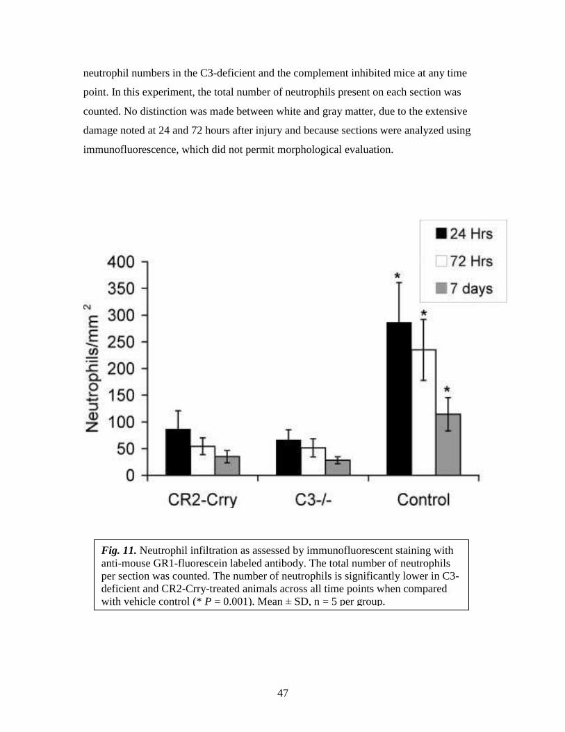
47
neutrophil numbers in the C3-deficient and the complement inhibited mice at any time
point. In this experiment, the total number of neutrophils present on each section was
counted. No distinction was made between white and gray matter, due to the extensive
damage noted at 24 and 72 hours after injury and because sections were analyzed using
immunofluorescence, which did not permit morphological evaluation.
Fig. 11. Neutrophil infiltration as assessed by immunofluorescent staining with
anti-mouse GR1-fluorescein labeled antibody. The total number of neutrophils
per section was counted. The number of neutrophils is significantly lower in C3-
deficient and CR2-Crry-treated animals across all time points when compared
with vehicle control (* P = 0.001). Mean ± SD, n = 5 per group.
48
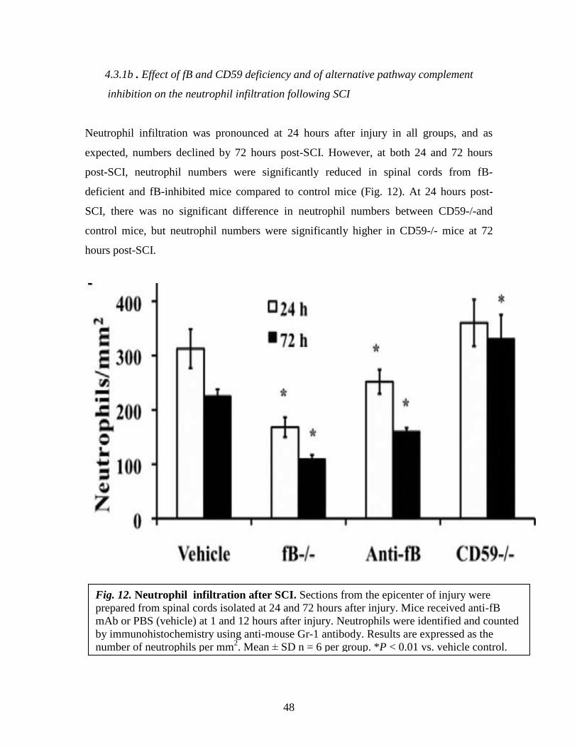
4.3.1b . Effect of fB and CD59 deficiency and of alternative pathway complement
inhibition on the neutrophil infiltration following SCI
Neutrophil infiltration was pronounced at 24 hours after injury in all groups, and as
expected, numbers declined by 72 hours post-SCI. However, at both 24 and 72 hours
post-SCI, neutrophil numbers were significantly reduced in spinal cords from fB-
deficient and fB-inhibited mice compared to control mice (Fig. 12). At 24 hours post-
SCI, there was no significant difference in neutrophil numbers between CD59-/-and
control mice, but neutrophil numbers were significantly higher in CD59-/- mice at 72
hours post-SCI.
Fig. 12. Neutrophil infiltration after SCI. Sections from the epicenter of injury were
prepared from spinal cords isolated at 24 and 72 hours after injury. Mice received anti-fB
mAb or PBS (vehicle) at 1 and 12 hours after injury. Neutrophils were identified and counted
by immunohistochemistry using anti-mouse Gr-1 antibody. Results are expressed as the
number of neutrophils per mm2. Mean ± SD n = 6 per group. *P < 0.01 vs. vehicle control.

49
4.3.2. Expression of Macrophage Infiltration after Spinal Cord Injury
Not surprisingly, there was no difference in the number of macrophages between any of
the groups at 24 hours post-SCI, in accord with previous data on the dynamics of
macrophage infiltration following SCI. At 72 hours post-SCI, macrophage numbers were
increased in all groups, but numbers were significantly lower in fB-deficient and fB-
inhibited mice compared to control mice (Fig. 13). In CD59-/- mice at 72 hours post-SCI,
macrophage numbers were the same as in control mice, and this is in contrast to the
increased neutrophil infiltration seen in CD59-/- mice.
These data indicate an important role for the alternative pathway in neutrophil and
macrophage infiltration after SCI, and together with the above data demonstrate that the
infiltration of these leukocytes is associated with tissue injury.
Fig. 13. Macrophage infiltration after SCI. Sections from the epicenter of injury were
prepared from spinal cords isolated at 24 and 72 hours after injury. Mice received anti-fB
mAb or PBS (vehicle) at 1 and 12 hours after injury. Macrophages were identified and
counted by immunohistochemistry using anti-mouse Mac-3 antibody. Results are expressed
as the number of macrophages per mm2. Mean ± SD n = 6 per group. *P < 0.01 vs. vehicle
control.

50
In this study, the total number of neutrophils and macrophages present on each
section was counted. No distinction was made between white and gray matter, due to the
extensive damage noted at 24 and 72 hours postinjury in some groups and because
sections were analyzed using immunohistochemistry, which did not permit
morphological evaluation.
4.4. Targeting and Biodistribution of Therapeutically Administered CR2-Crry
We recently demonstrated that intravenously administered CR2-Crry targets sites of
complement activation in a mouse model of complement-dependent intestine ischemia
and reperfusion injury(63). The microenvironment of the spinal cord is different, and
access of macromolecules and inflammatory cells is restricted by the bloodspinal cord
barrier. On damage, however, the bloodspinal cord barrier becomes temporarily more
permeable, and this may account for the access of intravenously administered CR2-Crry
to its targeting ligand (C3) within the spinal cord. To support the concept that CR2-Crry
functions by targeting specifically to the spinal cord following injury, we assessed the
tissue distribution of 125
I-labeled CR2-Crry in normal control mice and in mice subjected
to SCI. 125
I-labeled CR2-Crry was injected intravenously 1 hour after SCI, as per
therapeutic protocol, and biodistribution determined 12 hours later. In control mice (no
SCI), CR2-Crry was distributed primarily in the blood, with tissue localization restricted
to the liver and kidney and, to a lesser extent, the heart (Fig. 14). The location of CR2-
Crry within the kidney and liver is likely associated with the nonspecific clearance of the
protein. Of note, no 125
I-labeled CR2-Crry was detected in the CNS tissues of the spinal
cord or brain, indicating that under normal physiological conditions CR2-Crry cannot
pass the blood-brain/spinal cord barrier. In contrast, there was significant localization of
125I-labeled CR2-Crry within the spinal cord of injured mice. In the spinal cord, levels of
CR2-Crry was highest at the site of injury but was also detected at sites rostral and caudal
to the site of injury (Fig. 14A). The absence of 125
I-labeled CR2-Crry in brain tissue of
injured mice is also supportive of the specific targeting of CR2-Crry. To further
investigate the specific targeting of CR2-Crry to the injured spinal cord, we performed
immunofluorescent staining with an anti- CR2 antibody. CR2 is expressed conservatively

51
and found primarily on B cells and dendritic cells. Therefore, the presence of CR2
immunoreactivity within the spinal cord was deemed to be indicative of localization of
recombinant CR2-Crry protein, and indeed no immunoreactivity for CR2 antibody could
be detected in sham controls or SCI control (untreated) animals (not shown). CR2
immunoreactivity was, however, seen in CR2-Crrytreated animals when sections of
spinal cord were assessed 12 hours after SCI. Staining was seen primarily within the
white matter around the injury site and morphologically appeared to stain
oligodendrocytes, fiber tracts, and vascular structures (Fig. 14B). This distribution of
CR2 corresponds to the distribution of deposited C3 in injured spinal cords reported
above (Figure 1), and is a further indication of the C3 targeting specificity of CR2- Crry.
Staining was also noted, to a lesser degree, in the dorsal horns of the gray matter with
immunolocalization seen in cells with neuronal morphology (Fig.14C).

52
Fig. 14. A: Biodistribution of CR2-Crry following spinal cord injury. 125
I CR2-
Crry was injected into normal C57Bl/6 mice (no injury) and C57Bl/6 animals 1 hour
following spinal cord injury. Tissue distribution was assessed 12 hours after initial
injury in major organs and tissues of the central nervous system. B:
Immunolocalization of CR2-Crry binding with an anti-CR2 Ab. Positive
immunoflourescence is noted in the white matter beneath the injury site with a pattern
similar to that observed in C3 stained sections (arrows). C: CR2 immunoreactivity
seen in neuronal cells within the dorsal horn of the gray matter (arrows). Images are
taken under oil immersion using ×63objective and are representative of n = 4.
53
4.5. Deposition of Complement Activation
To correlate post-traumatic inflammation and injury with complement activation, we exa
mined spinal cord sections for deposited C3, fB, and C9 (MAC) by immunofluorescence
microscopy.
4.5.1. Time Course of Complement Activation 1, 24 and 72 hours after SCI
The presence of C3, deposition of which marks a site of complement activation by any
pathway, was assessed in mice that had undergone SCI and in sham laminectomy
controls. No staining for C3 was observed in sham operated- animals in any
compartments of the spinal cord. In contrast, C3 deposition was evident following SCI in
spinal cords harvested at 1 hour, 2 hours, 4 hours, 12 hours, and 24 hours after injury. At
1 hour, 2 hours, and 4 hours post-SCI, C3 deposition was centered to the white matter of
the injury site and within the ventral horns of the gray matter (Fig. 15 A). At later time
points of 12 hours and 24 hours, C3 staining was evident in surviving white matter, with
staining also present throughout the gray matter and extending into the ventral and dorsal
horns (Fig. 15 B). By day 3 after injury, complement deposition was almost undetectable,
with no C3 staining evident at the injury site (Fig. 15 C and D). This result is different
from that reported for complement deposition in the rat spinal cord following injury, in
which complement deposition was evident for up to 42 days after injury(10). An
additional apparent difference in the mouse model was that, at all time points, spinal cord
sections 10 mm rostral and caudal to the injury site showed a much reduced C3 staining
pattern compared to sections taken from the injury site. Complement deposition was seen
up to 20 mm from the injury site in rats, with no apparent decrease in immunoreactivity
and with increasing distance from the site of injury(6). However, this apparent difference
is likely a consequence of animal size and differences in size of impact injury required to
produce an equivalent condition.

54
4.5.2. Complement deposition at epicenter of injury 24 h post-SCI
Representative immunofluorescence images showing complement deposition, as well as
CD59 expression, at 24 hours after injury are shown in Fig. 16. There was no evidence of
C3, fB, or C9 staining in spinal cords from sham operated mice, and expression of CD59
was similar in wt control treated and fB-/- mice and absent in CD59-/- mice (Fig. 16).
Fig. 15. Complement activation as shown by C3 deposition at the site of spinal
cord injury. 1 hour (A), 24 hours (B), and 72 hours (C). C3 deposition is marked
by green fluorescent staining of the polyclonal anti-mouse C3 antibody in spinal
cords harvested at different time points after injury (n= 3). In contrast
laminectomy sham controls lack immunoreactivity for C3 (D).
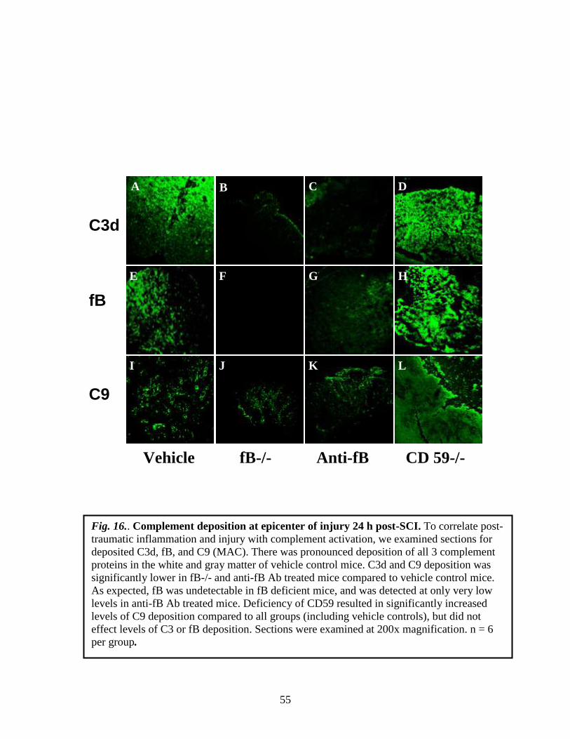
55
C3d fB C9
Vehicle fB-/- Anti-fB CD 59-/-
I
H G
C
F E
D
L K J
A B
Fig. 16.. Complement deposition at epicenter of injury 24 h post-SCI. To correlate post-
traumatic inflammation and injury with complement activation, we examined sections for
deposited C3d, fB, and C9 (MAC). There was pronounced deposition of all 3 complement
proteins in the white and gray matter of vehicle control mice. C3d and C9 deposition was
significantly lower in fB-/- and anti-fB Ab treated mice compared to vehicle control mice.
As expected, fB was undetectable in fB deficient mice, and was detected at only very low
levels in anti-fB Ab treated mice. Deficiency of CD59 resulted in significantly increased
levels of C9 deposition compared to all groups (including vehicle controls), but did not
effect levels of C3 or fB deposition. Sections were examined at 200x magnification. n = 6
per group.
B

56
4.5.3. Quantitative assessment of complement deposition post-SCI
In agreement with previous data obtained using mice(64) (for C3 deposition) and rats(6)
(for fB and C9 deposition), there was pronounced deposition of all three complement
proteins in the white and gray matter of control mice at 24 hours postinjury. C3 and C9
deposition was significantly lower in fB-deficient and fB inhibited mice compared to
control mice. As expected, fB was undetectable in fB-deficient mice and was detected at
only very low levels in fB-inhibited mice. Deficiency of CD59 resulted in significantly
increased levels of C9 deposition compared to all groups (including controls) but did not
effect levels of C3 or fB deposition (Fig. 17A).
Similar relative profiles for C3, fB, and C9 deposition were seen at 72 hours post-
SCI in all groups, although C3 and fB levels were lower than at 24 hours after SCI (Fig.
17B). C9 levels were similar at 24 and 72 hours after SCI (Fig. 17 A and B).

57
A
Fig. 17. Quantitative assessment of complement deposition post-SCI. The deposition
of C3 (C3d), fB, and C9 (MAC) was analyzed at 24 hours (A) and 72 hours (B) after
injury in spinal cord sections from the epicenter of injury. Mice received anti-fB mAb
or PBS (vehicle) at 1 and 12 hours after injury. Analysis was performed using
immunofluorescence microscopy, and sections scored for fluorescence intensity on a 4
point scale (see Materials and Methods). Mean ± SD, n = 6. *P < 0.01.
58
In this study, we detected C3 deposition at 72 hours post-SCI, although at low
levels. This is in contrast to our previous report that C3 was undetectable in mouse spinal
cords 72 hours after injury, and we attribute this difference to the use of a different
detection antibody (see Materials and Methods).
4.6. Histological Analysis of Recovery
Data presented above show that after SCI, fB-deficient mice and fB-inhibited mice
(when treated at 1 and 12 hours after SCI) have a significantly improved outcome
compared with controls in terms of inflammation, tissue injury, and functional locomotor
recovery. To determine the extent of histological recovery, we analyzed spinal cords 21
days after the initial impact injury. H&E and LFB staining of cord sections from wt and
PBS treated controls revealed demylineation in the central core of the white matter
beneath the impact site and evidence of vacuolation with some necrosis with
inflammatory cells still present (Fig. 18 A and B.). By comparison, at 21 days after SCI,
there was markedly less demyelination and inflammation and no evidence of necrosis in
cords from fB deficient mice and mice treated with anti-fB mAb at 1 and 12 hours after
SCI. There was no apparent difference in the extent of demyelination between the fB-
deficient and fB-inhibited (1/12 hours) groups. In contrast, CD59-/- mice exhibited a
serious lack of structural organization, abundant inflammatory cell infiltration, scaring,
vacuolation, and no obvious myelin structure. Also, in agreement with locomotor
recovery data, when anti-fB mAb was administered 12 and 24 hours after SCI (or 24 and
36 hours, not shown), there was no significant improvement in histological evidence of
recovery when compared to wt and PBS treated controls.

59
21 Days H & E LFB
wt
fB-/-
Anti-fB
(1/12h)
Anti-fB
(12/24h)
CD59-/-
A
A B
C D
E F
G H
I J

60
B
Fig. 18. Histopathology of spinal cord sections 21 days after injury. A: Images of transverse
sections from spinal cords at the epicenter of injury. Sections stained with either H&E or
LFB. Representative images, n = 6 per group. B: Quantitative assessment of histopathological
inflammation, injury, and demyelination. H&E-stained sections were scored on a 0 3 scale
for the presence and intensity of inflammatory cell infiltration, neuronal vacuolation, and
hemorrhage. LFB-stained sections scored for demyelination (0 = no evidence, 3 = severe).
Anti-fB mAb treatment groups received injections at 1 and 12, or 12 and 24 hours after
injury. Vehicle control group (PBS) received injections at 1 and 12 hours after injury. Scores
were then expressed as a cumulative score of 0-12. Mean ± SD, n = 6 per group. *P < 0.001
and **P<0.0001 vs. vehicle control and wt.
61
5. DISCUSSION
There are approximately 10,000 new serious spinal cord injuries each year, with a
disproportionate number of injuries occurring in young people. Following the initial
trauma, there is a secondary injury cascade that results in increased cell death, cavitation,
and demyelination that is detrimental to functional recovery. Disruption of the blood-
brain barrier and the triggering of an inflammatory response are important components of
secondary SCI injury, and while the initial trauma is difficult to guard against, secondary
inflammatory events represent potential therapeutic targets.
Post-traumatic inflammation and secondary tissue damage after SCI is
characterized by, among other things, complement activation, expression of inflammatory
cytokines and chemokines, the up-regulation of adhesion molecules, and the infiltration
of neutrophils and macrophages. Complement activation products play known roles in
promoting the expression of these inflammatory molecules and the recruitment and
activation of leukocytes.
Although the damaging effects of inflammation in SCI are well documented, there
is a paucity of information concerning the mechanisms that control this process. Recent
studies using rat models of SCI have highlighted a potential role for the complement
system in the development of spinal cord damage. (6) have described the deposition of
complement components C1q, factor B, C4, and C5b-9, as well as complement inhibitors
factor H and clusterin within neurological tissue of rats following SCI(6). These data
implicate complement in the development of secondary injury, but whether complement
is an effector or a bystander in this inflammatory milieu is unclear from these studies.
Complement is an important component of host defense, and the classical and lectin
pathways provide important contributions to defense against some pathogens. Thus,
compared with C3 inhibition, alternative pathway inhibition would be less generally
immunosuppressive and may have less potential for increasing the incidence of sepsis or
infection. Complement is also implicated in neuroprotective and regenerative
mechanisms. Activated microglia and macrophages have been shown to be important for
neuron protection and axon growth in animal models(89), and complement activation

62
products, in particular C5a, can recruit and activate these cells. A recent study
demonstrated that the treatment of rats 14 days post-SCI with a C5a receptor antagonist
reduced locomotor recovery and myelination(90). Complement also participates in
phagocytic clearance of apoptotic cells and cellular debris via their opsonization with
C1q and C3 activation products, and interfering with this clearance pathway can impair
axon growth (89, 91). Thus, compared with total complement blockade, allowing
activation of the classical (or lectin) pathway while inhibiting alternative pathway
amplification could feasibly improve the balance between the neurodegenerative and
neuroprotective effects of complement inhibition.
5.1 Role of C3 and complement inhibitory protein CR2-Crry during SCI
In the current study, the functional significance of complement deposition was addressed
using C3-deficient animals and a targeted complement inhibitory protein, CR2-Crry. We
first confirmed that complement activation occurred within injured spinal cords of mice,
as has been shown for rats(10), by immunostaining for C3, a protein common to all
complement activation pathways. Levels of deposited C3 peaked at 24 hours after injury,
which was similar to that reported in the rat, but a notable difference was that whereas
complement could be detected 42 days post- SCI in rats, C3 deposition was undetectable
3 days post- SCI in mice. These differences are probably due to species differences, but
we cannot exclude other factors such as antibody affinity or antibody detection methods.
The cell types associated with C3 deposition within spinal cord lesions were not
specifically assessed in this study, although Anderson et al6 have shown that complement
is deposited on oligodendrocytes, neurons, and axons in injured rat spinal cords. In a
previous study on the use of a complement inhibitor in a rat model of SCI, vaccinia virus
complement control protein (VCP) was infused directly into the spinal cord over a 10-
minute period following contusion induced SCI. Vaccinia virus complement control
protein is a functional analog of CR1. It was shown that VCP reduced leukocyte
infiltration, qualitatively reduced cord destruction and had some effect on locomotor
63
recovery(56, 92). The authors observed an improvement in recovery within the first week
and an overall improvement over the 6-week duration of the experiment. While these
observations did not reach significance, they highlighted a potential for the use of
exogenous complement regulatory proteins in spinal cord injury therapy.
In another study, rats were treated with sCR1 following SCI, and treated rats had
less leukocyte infiltration and improved motor function(93). In this previous study, motor
function was assessed using a nonstandard scale, rendering cross comparison difficult,
but functional improvements in inhibitor- treated rats were not great. The rats received an
injection of sCR1 1 hour after injury and daily thereafter. In the current study, mice were
treated with a single intravenous injection of a targeted complement inhibitor, CR2-Crry,
at 1 hour after traumatic injury, and this was sufficient to protect the spinal cord to a
statistically similar level to that seen in C3-deficient animals. In the CR2- Crry-treated
mice, improvements in neurological recovery were observed early after injury and
continued throughout the duration of the experiment, with the difference between
controls and treated animals being marked and statistically significant. CR2-Crry
biodistribution and C3 staining, together with CR2 immunolocalization studies confirm
the presence of the CR2-binding ligand (C3 breakdown fragments) in cell types
associated with neurological injury, and demonstrate that CR2- Crry traffics across the
blood-spinal cord barrier to sites of complement activation. Since sCR1 and VCP have a
similar activity spectrum to Crry, it is likely that untargeted Crry would have some
similar therapeutic effect in this model. However, the efficacy of untargeted Crry depends
on systemic complement inhibition and a significantly higher dose of inhibitor, and this
would render the host susceptible to infection, a relevant and important issue for SCI.
While the data presented show that complement deficiency or inhibition provides
protection from the secondary effects of initial spinal cord contusion injury, the
mechanism(s) of complement activation and complement- dependent injury are
incompletely characterized. The inflammatory response following SCI is marked by the
infiltration of monocytes/macrophages and neutrophils, and data from studies showing
that adhesion molecule blockade or neutrophil ablation is protective, indicate that these
cells play a key role in secondary destructive processes (81, 83, 94, 95). Thus, our data
64
suggest that the protective effect of C3 deficiency/inhibition is due, at least in part, to its
effect on neutrophil trafficking. Two complement activation products downstream of C3
cleavage are C3a and C5a, both of which have leukocyte chemotactic and activating
properties. C5a can also upregulate expression of the adhesion molecules P- and E-
selectin, intercellular adhesion molecule-1 and vascular adhesion molecule-1 on
endothelial cells (96-99). Of note, C5a is implicated in brain cryoinjury, because injury
and neutrophil extravasation is reduced in C5-deficient mice and C5a receptor antagonist-
treated mice(100).
In the context of relating our findings to human therapy, there are three human
membrane inhibitors of complement activation that function at the C3 level; decay
accelerating factor, membrane cofactor protein, and CR1. In rodents, Crry appears to
function as the principal membrane inhibitor of C3 activation, and soluble (untargeted)
Crry has been used widely for studying complement inhibition in rodent models of
disease. Because complement inhibitors display different degrees of species selectivity,
the use of Crry is appropriate when studying the effects of complement inhibition in
rodents. Crry is a structural and direct functional analog of human CR1, and data
obtained with Crry in rodents can be expected to translate to CR1 in humans.
Nevertheless, soluble forms of CR1 have been investigated in the clinic, but with
generally disappointing results. However, a similar targeting strategy to that described
here could be applied to CR1 or other human complement inhibitors with the goal of
increasing efficacy. Effective therapy with a single intravenous injection, as demonstrated
in the current model, would allow for early inhibitor delivery on patient presentation.
Complement provides an important host defense mechanism, and an additional advantage
of CR2-mediated targeting of complement inhibitors is a reduced level of systemic
immunosuppression at therapeutically effective doses. Patients with spinal cord injuries
are at increased risk of infection, in particular urinary tract infection. We have previously
shown that the dose of CR2-Crry that was used in the experiments reported here (0.25
mg), minimally effects systemic complement levels and does not increase host resistance
to infection(101).

65
5.2 Role of fB through alternative pathway during SCI
In the present study, we show that fB-deficient mice have an improved outcome after SCI.
The level of protection afforded to fB-deficient mice was similar to that seen in our study
using C3-deficient mice. These data indicate a dependence on the alternative pathway for
complement-mediated SCI. Together with the previous findings described above using B
cell and C1q-deficient mice, it is likely that the alternative pathway plays a critical role in
amplifying antibody-mediated classical pathway initiated complement activation. In this
context, reduced pathology and improved locomotor recovery in fB-deficient mice was
associated with signifi- cantly reduced C3 and MAC deposition compared to wt control
mice, even though the classical (and lectin) pathway is intact in fB-deficient mice. A
similar dependence on both antibody-mediated lectin pathway activation and alternative
pathway activation has been described in models of intestine ischemia and reperfusion
injury(65-67). In a more clinically relevant paradigm, we demonstrated that specific
inhibition of the alternative pathway with anti-fB mAb was also protective against SCI,
and to a similar level as fB deficiency. Selective inhibition of the alternative pathway
while leaving the classical and lectin pathways intact has potential benefits over
inhibiting all pathways at the C3 activation step.
In the current study, we show that compared with control treated mice, there are
significantly reduced neutrophil numbers in fB-deficient and fB-inhibited mice at both
time points post-SCI. We similarly showed reduced macrophage/ microglia numbers in
fB-deficient or fB-inhibited mice compared with controls at 72 hours postinjury, whereas
it was previously shown that C1q deficiency was (interestingly) associated with increased
macrophage infiltration(102). Together, these data indicate a key role for the alternative
pathway in neutrophil and macrophage infiltration after SCI.
As discussed above, for reasons relating to the protective 3068 Qiao et al AJP
December 2010, Vol. 177, No. 6 roles of complement that are relevant for SCI patients,
there are potential benefits of selectively inhibiting the alternative pathway of
complement to reduce secondary damage and improve functional recovery after SCI.

66
5.3 Role of CD59 through terminal complement pathway during SCI
The MAC can be directly cytolytic and may contribute directly to neuronal injury and
demyelination but can also induce the production of pro-inflammatory mediators,
including adhesion molecules on endothelial cells, when deposited at sublytic
concentrations. Indeed, studies indicate an important role for the MAC in rodent models
of experimental allergic encephalomyelitis (EAE), a demyelinating disease that mimics
multiple sclerosis. In rodent models of EAE, injury and myelin loss is attenuated in
animals deficient in C5 and C6 and increased in animals deficient in CD59a, an inhibitor
of MAC formation(28, 48, 103-106). A contradictory finding was reported, however, in
a rat model of immunological demyelination in which intraspinal infusions of IgG
antibodies were administered in the presence of serum deficient in either C3, C4, C5, C6,
or factor B(107). Deficiency in serum C3 and C4, but not C5, C6, or factor B was
protective against demylineation, suggesting a role for the classic pathway of
complement activation and no role for the MAC (C4 is a classic pathway protein, factor
B is an alternative pathway protein, and C6 is a terminal pathway protein involved in
MAC formation(107)).
The exacerbated injury and impaired recovery of locomotor function in CD59a-
deficient mice, together with increased deposits of the MAC (C9) in these mice compared
to wt control treated mice, indicate an important contribution of the terminal pathway and
the MAC to secondary neuronal injury after trauma. Furthermore, significant functional
deficit persisted in CD59-deficient mice at 21 days after injury compared with controls. It
is possible that neoepitope expression and cell lysis resulting from enhanced MAC
formation in CD59-deficient mice could result in enhanced complement activation.
However, while there were significantly increased deposits of C9 in CD59-
deficient mice at both 24 and 72 hours post-SCI compared with wt treated control mice,
CD59 deficiency had little effect on C3 or fB deposition. Previous studies using CD59a-
deficient mice have similarly implicated the MAC in spinal cord demyelination and
axonal injury in murine acute experimental allergic encephalomyelitis(106), as well as in
axonal damage during Wallerian degeneration (peripheral nerve injury)(108). The
alternative and terminal complement pathways have also been implicated in causing
67
neuropathology in a different model of traumatic neuroinflammation, traumatic brain
injury. In these previous studies, CD59a deficiency impaired neurological outcome and
increased neuronal cell death and brain tissue destruction compared to wt mice(109),
It is interesting that unlike fB deficiency, CD59 deficiency did not significantly effect
the number of macrophages at 72 hours post injury. Neutrophils, on the other hand, were
present in significantly higher numbers in CD59-deficient mice compared to wt treated
controls at 72 hours after injury. The MAC can influence cellular infiltration when
deposited in sublethal quantities in cell membranes by inducing the expression of
adhesion molecules, cytokines, and chemokines, including IL-8(98, 110-114). Why there
is an apparent difference in CD59-deficient mice on neutrophil and macrophage
infiltration at 72 hours post injury (compared to controls) is not clear, but a consequence
could be the protraction of neutrophil mediated inflammatory events and a reduction in
macrophage- mediated protective events. In addition to showing that the complement
inhibitor CD59 plays an important role in providing protection against SCI, the data have
potential clinical implications.
However, the MAC plays a very limited known role in host defense and immune
homeostatic/repair mechanisms, and because our data indicate that the MAC is a
significant contributor to pathology in SCI, selective inhibition of the terminal pathway
could potentially have even greater benefit over blocking complement at an early step in
any pathway.

68
6. Summary and conclusions
An impact to the spinal cord results in a primary injury, but the impact also triggers a
series of downstream events that lead to secondary injury of tissue and the progressive
degeneration of the spinal cord. Therapeutic interventions that minimize secondary injury
will improve functional recovery after traumatic injury. Inflammation plays a key role in
secondary injury following spinal cord injury (SCI). Inflammation is a rapid immune
response that can be initiated by infection or tissue damage. An important component of
an inflammatory response is the complement system, a collection of blood proteins that
form part of the immune system and that can be activated by injured cells and tissues.
Activation of complement amplifies the inflammatory response and produces molecules
that can be directly toxic or that can recruit and activate cells of the immune system to
produce toxic molecules. Inhibiting the complement system has been shown to be an
effective therapy for inflammatory disease in various animal models, and some
complement inhibitors are in clinical trials.
We have shown that complement deficient mice and normal mice treated with a
complement inhibitor are protected from neuronal injury following SCI, and that they
have significantly improved functional scores over time compared to control mice. These
data indicate an important role for complement in secondary injury, and indicate
complement inhibition will reduce inflammation and provide neuroprotection following
spinal cord injury. However, complement-dependent mechanisms involved in secondary
injury following SCI are not known, and there remain concerns regarding the clinical
application of the currently available systemic (body-wide) complement inhibitors with
regard to their safety and efficacy. Complement activation products are important for host
defense and immune maintenance mechanisms, and systemic complement inhibition can
compromise the protective and beneficial roles of complement. This will not be optimal
in patients at risk of infection, and urinary tract infection is a frequent complication
during initial and ongoing medical rehabilitation after SCI.
We propose to develop a neuroprotective strategy based on attenuating
complement-dependent secondary damage after SCI using a novel and validated
approach. The strategy involves the targeting of complement inhibitors to sites of
complement activation and injury, and we have shown the approach to be highly effective

69
in vitro and in mouse models of SCI and other inflammatory disease conditions. The
targeting strategy enhanced the activity and protective effect of complement inhibitors by
10-20 fold compared to untargeted counterparts. Furthermore, untargeted, but not
targeted complement inhibitors systemically inhibited complement and increased
susceptibility to infection in a mouse model.
We propose to fully characterize our targeted complement inhibitors in a mouse
model of SCI. In addition to therapeutic endpoints, we will use different types of
complement inhibitor to investigate complement-dependent disease mechanisms. We will
determine relationships between the generation of different complement activation
products and other molecules associated with inflammation in order to gain a better
understanding of the mechanisms of secondary tissue injury following SCI. These
preclinical studies will establish the guidelines necessary for the translation of this
therapy to the clinic using recombinant human proteins.

70
7. Abstract
An inflammatory cascade that is initiated following traumatic spinal cord injury (SCI) is
thought to cause secondary injury and adversely impact functional recovery Complement
is implicated in the inflammatory response and the secondary neuronal damage that
occurs after traumatic spinal cord injury (SCI). Complement can be activated by the
classical, lectin, or alternative pathways, all of which share a common terminal pathway
that culminates in formation of the cytolytic membrane attack complex (MAC).
Here, we investigated the role of the alternative and terminal complement pathways and
role of complement in the secondary injury process occurring following acute contusion
SCI in mice.
The dynamics of complement activation post-SCI were investigated by assessing
the deposition of C3 in spinal cords. C3 deposition occurred within 1 h post-SCI at the
injury site, with C3 staining extending into the ventral and dorsal horns by 12 h. C3
deposition was almost undetectable at 3 days after SCI. Mice deficient in C3 had a
significantly improved locomotor score compared to wild type controls and regained
almost full locomotor function by day 21 after injury.
To further investigate the role of complement in SCI, we determined the effect of
fB and CD59 deficiency on SCI. Mice deficient in fB had significantly improved
locomotor scores compared to wt controls, although recovery was not as complete as in
C3 deficient mice (difference was significant, p<0.01). CD59 deficient mice had
significantly worse locomotor scores compared to wt controls.
Wild type mice were also treated with CR2-Crry, an inhibitor of mouse
complement activation that targets to sites of complement activation. A single
intravenous injection of CR2-Crry 1 h post-injury significantly improved functional
recovery and reduced tissue injury and demyelination compared to PBS treated controls
and to a similar extent to that seen in C3-deficient animals. CR2-Crry specifically
targeted to the injured spinal cord in a distribution pattern corresponding to that seen for
deposited C3.
Together, these data indicate an important role for complement in the secondary
injury process following SCI, with a significant role for both the alternative pathway and

71
the membrane attack complex in causing tissue injury. The feasibility of targeted
complement inhibition as a therapeutic strategy for improving post-traumatic recovery.

72
8. ABSTRACT IN HUNGARIAN (ÖSSZEFOGLALÁS)
A traumát követő gerincvelő sérülés (spinal cord injury - SCI) során az aktiválódó
gyulladásos kaszkád másodlagos károsodást okoz és hátrányosan befolyásolja a
gyógyulást. Ismert, hogy a komplement rendszer befolyásolja a gyulladásos választ és a
neuronális károsodást, amely a trauma kapcsán kialakult gerincvelő-sérülésnél
jelentkezik. A komplement rendszer három útvonalon keresztül aktiválódhat (klasszikus,
lektin ill. alternatív út); az aktiváció egy közös terminális útvonalon keresztül a
membránkárosító-komplex (membrane attack complex - MAC) kialakulásához vezet.
Jelen munkánkban egerekben vizsgáltuk a komplement kaszkád alternatív- és terminális
útvonalának jelentősségét az akut kontúziót követő másodlagos gerincvelő-károsodásban.
A SCI utáni komplement-aktiváció dinamikáját a gerincvelőben lévő C3 depozitumok
meghatározásával követtük. A depozitumok már a sérülést követő 1 órán belül
kimutathatóak voltak, 12 óra után az elülső és hátsó szarv felé terjedtek, végül 3 nap
múlva már nem voltak jelen. Megfigyeltük, hogy a C3 deficiens egereknek jelentősen
jobb volt mozgásszervi státusza a vad típusú kontroll egerekhez képest és 21 nappal a
sérülés után teljesen visszanyerték mozgásszervi funkcióikat.
A komplement rendszer szerepét tovább vizsgálva SCI-ben, meghatároztuk a fB és CD59
deficiencia hatását. Az fB hiányos egerek mozgásszervi score-ja szignifikánsan jobb volt
a vad típushoz képest, bár a gyógyulás nem volt olyan mértékű, mint C3 deficiencia
esetében (p<0,01). A CD59 hiányos egerek jelentősen rosszabb mozgásszervi score-ral
rendelkeztek a vad típushoz képest. A vad típusú egereket a komplementrendszer
aktivációját lokálisan gátló CR2-Crry-vel kezeltük. A C3-deficiens állatoknál
tapasztaltakhoz hasonlóan, a CR2-Crry egyszeri intravénás alkalmazása 1 órával a
sérülést követően jelentős mértékben javította a funkcionális gyógyulást, csökkentette a
szöveti sérülést és a demyelinizációt a PBS-sel kezelt kontroll csoporthoz képest. CR2-
Crry kifejezetten a sérült gerincvelőt célozza meg a C3 depozitumoknak megfelelően.
Ezek az adatok együttesen a komplement rendszer kiemelt szerepét mutatják a gerincvelő
sérülés másodlagos károsodásában. Ebben meghatározó szerepe van mind az alternatív
útvonalnak, mind pedig a MAC kialakulásának. Terápiás lehetőségként felmerül a
komplement rendszer gátlása a poszt-traumás gyógyulás javításában.

73
9. Bibliography
1. Fiore, C., Inman, D.M., Hirose, S., Noble, L.J., Igarashi, T., and Compagnone,
N.A. (2004). Treatment with the neurosteroid dehydroepiandrosterone promotes
recovery of motor behavior after moderate contusive spinal cord injury in the
mouse. J Neurosci Res 75:391-400.
2. Kwon, B.K., Tetzlaff, W., Grauer, J.N., Beiner, J., and Vaccaro, A.R. (2004).
Pathophysiology and pharmacologic treatment of acute spinal cord injury. Spine J
4:451-464.
3. Taoka, Y., Okajima, K., Uchiba, M., Murakami, K., Kushimoto, S., Johno, M.,
Naruo, M., Okabe, H., and Takatsuki, K. (1997). Role of neutrophils in spinal
cord injury in the rat. Neuroscience 79:1177-1182.
4. Hall, E.D., and Springer, J.E. (2004). Neuroprotection and acute spinal cord
injury: a reappraisal. NeuroRx 1:80-100.
5. Rebhun, J., Madorsky, J.G., and Glovsky, M.M. (1991). Proteins of the
complement system and acute phase reactants in sera of patients with spinal cord
injury. Ann Allergy 66:335-338.
6. Anderson, A.J., Robert, S., Huang, W., Young, W., and Cotman, C.W. (2004).
Activation of complement pathways after contusion-induced spinal cord injury. J
Neurotrauma 21:1831-1846.
7. Hausmann, O.N. (2003). Post-traumatic inflammation following spinal cord
injury. Spinal Cord 41:369-378.

74
8. Carlson, G.D., and Gorden, C. (2002). Current developments in spinal cord injury
research. Spine J 2:116-128.
9. Mautes, A.E., Weinzierl, M.R., Donovan, F., and Noble, L.J. (2000). Vascular
events after spinal cord injury: contribution to secondary pathogenesis. Phys Ther
80:673-687.
10. Anderson, A.J., Najbauer, J., Huang, W., Young, W., and Robert, S. (2005).
Upregulation of complement inhibitors in association with vulnerable cells
following contusion-induced spinal cord injury. J Neurotrauma 22:382-397.
11. Goldknopf, I.L., Sheta, E.A., Bryson, J., Folsom, B., Wilson, C., Duty, J., Yen,
A.A., and Appel, S.H. (2006). Complement C3c and related protein biomarkers in
amyotrophic lateral sclerosis and Parkinson's disease. Biochem Biophys Res
Commun 342:1034-1039.
12. Zipp, F., and Aktas, O. (2006). The brain as a target of inflammation: common
pathways link inflammatory and neurodegenerative diseases. Trends Neurosci
29:518-527.
13. Tomlinson, S. (1993). Complement defense mechanisms. Curr Opin Immunol
5:83-89.
14. Frank, M.M., and Fries, L.F. (1991). The role of complement in inflammation and
phagocytosis. Immunol Today 12:322-326.
15. Schraufstatter, I.U., Trieu, K., Sikora, L., Sriramarao, P., and DiScipio, R. (2002).
Complement c3a and c5a induce different signal transduction cascades in
endothelial cells. J Immunol 169:2102-2110.

75
16. Bhakdi, S., and Tranum-Jensen, J. (1991). Complement lysis: a hole is a hole.
Immunol Today 12:318-320; discussion 321.
17. Kawamata, T., Akiyama, H., Yamada, T., and McGeer, P.L. (1992). Immunologic
reactions in amyotrophic lateral sclerosis brain and spinal cord tissue. Am J
Pathol 140:691-707.
18. Sta, M., Sylva-Steenland, R.M., Casula, M., de Jong, J.M., Troost, D., Aronica, E.,
and Baas, F. Innate and adaptive immunity in amyotrophic lateral sclerosis:
evidence of complement activation. Neurobiol Dis 42:211-220.
19. de Jonge, R.R., van Schaik, I.N., Vreijling, J.P., Troost, D., and Baas, F. (2004).
Expression of complement components in the peripheral nervous system. Hum
Mol Genet 13:295-302.
20. Lobsiger, C.S., Boillee, S., and Cleveland, D.W. (2007). Toxicity from different
SOD1 mutants dysregulates the complement system and the neuronal regenerative
response in ALS motor neurons. Proc Natl Acad Sci U S A 104:7319-7326.
21. Padilla-Docal, B., Dorta-Contreras, A.J., Bu-Coifiu-Fanego, R., and Rey, A.R.
(2009). CSF/serum quotient graphs for the evaluation of intrathecal C4 synthesis.
Cerebrospinal Fluid Res 6:8.
22. Tsuboi, Y., and Yamada, T. (1994). Increased concentration of C4d complement
protein in CSF in amyotrophic lateral sclerosis. J Neurol Neurosurg Psychiatry
57:859-861.
23. Kim, Y.U., Kinoshita, T., Molina, H., Hourcade, D., Seya, T., Wagner, L.M., and
Holers, V.M. (1995). Mouse complement regulatory protein Crry/p65 uses the

76
specific mechanisms of both human decay-accelerating factor and membrane
cofactor protein. Journal of Experimental Medicine 181:151-159.
24. Takizawa, H., Okada, N., and Okada, H. (1994). Complement inhibitor of rat cell
membrane resembling mouse Crry/p65. J.Immunol. 152:3032-3038.
25. Bal-Price, A., and Brown, G.C. (2001). Inflammatory neurodegeneration
mediated by nitric oxide from activated glia-inhibiting neuronal respiration,
causing glutamate release and excitotoxicity. J Neurosci 21:6480-6491.
26. Barrington, R., Zhang, M., Fischer, M., and Carroll, M.C. (2001). The role of
complement in inflammation and adaptive immunity. Immunol Rev 180:5-15.
27. Gasque, P., Neal, J.W., Singhrao, S.K., McGreal, E.P., Dean, Y.D., Van, B.J., and
Morgan, B.P. (2002). Roles of the complement system in human
neurodegenerative disorders: pro-inflammatory and tissue remodeling activities.
Mol Neurobiol 25:1-17.
28. Sewell, D.L., Nacewicz, B., Liu, F., Macvilay, S., Erdei, A., Lambris, J.D.,
Sandor, M., and Fabry, Z. (2004). Complement C3 and C5 play critical roles in
traumatic brain cryoinjury: blocking effects on neutrophil extravasation by C5a
receptor antagonist. J Neuroimmunol 155:55-63.
29. Tacnet-Delorme, P., Chevallier, S., and Arlaud, G.J. (2001). Beta-amyloid fibrils
activate the C1 complex of complement under physiological conditions: evidence
for a binding site for A beta on the C1q globular regions. J Immunol 167:6374-
6381.
77
30. Sanders, M.E., Koski, C.L., Robbins, D., Shin, M.L., Frank, M.M., and Joiner,
K.A. (1986). Activated terminal complement in cerebrospinal fluid in Guillain-
Barre syndrome and multiple sclerosis. J Immunol 136:4456-4459.
31. Krakauer, M., Schaldemose Nielsen, H., Jensen, J., and Sellebjerg, F. (1998).
Intrathecal synthesis of free immunoglobulin light chains in multiple sclerosis.
Acta Neurol Scand 98:161-165.
32. Romi, F., Kristoffersen, E.K., Aarli, J.A., and Gilhus, N.E. (2005). The role of
complement in myasthenia gravis: serological evidence of complement
consumption in vivo. J Neuroimmunol 158:191-194.
33. McGeer, P.L., and McGeer, E.G. (2004). Inflammation and neurodegeneration in
Parkinson's disease. Parkinsonism Relat Disord 10 Suppl 1:S3-7.
34. Daly, J.t., and Kotwal, G.J. (1998). Pro-inflammatory complement activation by
the A beta peptide of Alzheimer's disease is biologically significant and can be
blocked by vaccinia virus complement control protein. Neurobiol Aging 19:619-
627.
35. Fonseca, M.I., Zhou, J., Botto, M., and Tenner, A.J. (2004). Absence of C1q leads
to less neuropathology in transgenic mouse models of Alzheimer's disease. J
Neurosci 24:6457-6465.
36. Shen, Y., Lue, L., Yang, L., Roher, A., Kuo, Y., Strohmeyer, R., Goux, W.J., Lee,
V., Johnson, G.V., Webster, S.D., Cooper, N.R., Bradt, B., and Rogers, J. (2001).
Complement activation by neurofibrillary tangles in Alzheimer's disease.
Neurosci Lett 305:165-168.

78
37. Head, E., Azizeh, B.Y., Lott, I.T., Tenner, A.J., Cotman, C.W., and Cribbs, D.H.
(2001). Complement association with neurons and beta-amyloid deposition in the
brains of aged individuals with Down Syndrome. Neurobiol Dis 8:252-265.
38. Holers, V.M., and Thurman, J.M. (2004). The alternative pathway of complement
in disease: opportunities for therapeutic targeting. Mol Immunol 41:147-152.
39. Muller-Eberhard, H.J. (1988). Molecular organization and function of the
complement system. Annu Rev Biochem 57:321-347.
40. Kulkarni, A.P., Kellaway, L.A., and Kotwal, G.J. (2005). Herbal complement
inhibitors in the treatment of neuroinflammation: future strategy for
neuroprotection. Ann N Y Acad Sci 1056:413-429.
41. Fearon, D.T., Daha, M.R., Weiler, J.M., and Austen, K.F. (1976). The natural
modulation of the amplification phase of complement activation. Transplant Rev
32:12-25.
42. Feldbush, T.L., Hobbs, M.V., Severson, C.D., Ballas, Z.K., and Weiler, J.M.
(1984). Role of complement in the immune response. Fed Proc 43:2548-2552.
43. Yang, L.B., Li, R., Meri, S., Rogers, J., and Shen, Y. (2000). Deficiency of
complement defense protein CD59 may contribute to neurodegeneration in
Alzheimer's disease. J Neurosci 20:7505-7509.
44. Shen, Y., Halperin, J.A., Benzaquen, L., and Lee, C.M. (1997). Characterization
of neuronal cell death induced by complement activation. Brain Res Brain Res
Protoc 1:186-194.
79
45. Wurzner, R. (2003). Deficiencies of the complement MAC II gene cluster (C6, C7,
C9): is subtotal C6 deficiency of particular evolutionary benefit? Clin Exp
Immunol 133:156-159.
46. Kulkarni, A.P., Kellaway, L.A., Lahiri, D.K., and Kotwal, G.J. (2004).
Neuroprotection from complement-mediated inflammatory damage. Ann N Y
Acad Sci 1035:147-164.
47. Dunkelberger, J.R., and Song, W.C. Complement and its role in innate and
adaptive immune responses. Cell Res 20:34-50.
48. Mead, R.J., Singhrao, S.K., Neal, J.W., Lassmann, H., and Morgan, B.P. (2002).
The membrane attack complex of complement causes severe demyelination
associated with acute axonal injury. J Immunol 168:458-465.
49. Lambris, J.D., and Holers, V.M., editors. 2000. Therapeutic interventions in the
complement system. Totowa, New Jersey: Humana Press. 259 pp.
50. Asghar, S.S., and Pasch, M.C. (2000). Therapeutic inhibition of the complement
system. Y2K update. Front Biosci 5:E63-81.
51. Quigg, R.J. (2002). Use of complement inhibitors in tissue injury. Trends Mol
Med 8:430-436.
52. Sahu, A., and Lambris, J.D. (2000). Complement inhibitors: a resurgent concept
in anti-inflammatory therapeutics. Immunopharmacology 49:133-148.
53. Rancan, M., Morganti-Kossmann, M.C., Barnum, S.R., Saft, S., Schmidt, O.I.,
Ertel, W., and Stahel, P.F. (2003). Central nervous system-targeted complement

80
inhibition mediates neuroprotection after closed head injury in transgenic mice. J
Cereb Blood Flow Metab 23:1070-1074.
54. Kaczorowski, S.L., Schiding, J.K., Toth, C.A., and Kochanek, P.M. (1995). Effect
of soluble complement receptor-1 on neutrophil accumulation after traumatic
brain injury in rats. J Cereb Blood Flow Metab 15:860-864.
55. Kossmann, T., Stahel, P.F., Morganti-Kossmann, M.C., Jones, J.L., and Barnum,
S.R. (1997). Elevated levels of the complement components C3 and factor B in
ventricular cerebrospinal fluid of patients with traumatic brain injury. J
Neuroimmunol 73:63-69.
56. Reynolds, D.N., Smith, S.A., Zhang, Y.P., Lahiri, D.K., Morassutti, D.J., Shields,
C.B., and Kotwal, G.J. (2003). Vaccinia virus complement control protein
modulates inflammation following spinal cord injury. Ann N Y Acad Sci
1010:534-539.
57. Zhang, H.-F., Yu, J., Bajwa, E., Morrison, S.L., and Tomlinson, S. (1999).
Targeting of functional antibody-CD59 fusion proteins to a cell surface.
J.Clin.Invest. 103:55-66.
58. Song, H., He, C., Knaak, C., Guthridge, J.M., Holers, V.M., and Tomlinson, S.
(2003). Complement receptor 2-mediated targeting of complement inhibitors to
sites of complement activation. J Clin Invest 111:1875-1885.
59. He, C., Imai, M., Song, H., Quigg, R.J., and Tomlinson, S. (2005). Complement
inhibitors targeted to the proximal tubule prevent injury in experimental nephrotic
syndrome and demonstrate a key role for C5b-9. J Immunol 174:5750-5757.

81
60. Law, S.K., Fearon, D.T., and Levine, R.P. (1979). Action of the C3b-inactivator
on the cell-bound C3b. J Immunol 122:759-765.
61. Seya, T., and Nagasawa, S. (1985). Limited proteolysis of complement protein
C3b by regulatory enzyme C3b inactivator: isolation and characterization of a
biologically active fragment, C3d,g. J Biochem (Tokyo) 97:373-382.
62. Takizawa, H., Okada, N., and Okada, H. (1994). Complement inhibitor of rat cell
membrane resembling mouse Crry/p65. J Immunol 152:3032-3038.
63. Kuhn, P.L., and Wrathall, J.R. (1998). A mouse model of graded contusive spinal
cord injury. J Neurotrauma 15:125-140.
64. Qiao, F., Atkinson, C., Song, H., Pannu, R., Singh, I., and Tomlinson, S. (2006).
Complement plays an important role in spinal cord injury and represents a
therapeutic target for improving recovery following trauma. Am J Pathol
169:1039-1047.
65. Stahl, G.L., Xu, Y., Hao, L., Miller, M., Buras, J.A., Fung, M., and Zhao, H.
(2003). Role for the alternative complement pathway in ischemia/reperfusion
injury. Am J Pathol 162:449-455.
66. Zhang, M., Takahashi, K., Alicot, E.M., Vorup-Jensen, T., Kessler, B., Thiel, S.,
Jensenius, J.C., Ezekowitz, R.A., Moore, F.D., and Carroll, M.C. (2006).
Activation of the lectin pathway by natural IgM in a model of
ischemia/reperfusion injury. J Immunol 177:4727-4734.
67. Huang, Y., Qiao, F., Atkinson, C., Holers, V.M., and Tomlinson, S. (2008). A
novel targeted inhibitor of the alternative pathway of complement and its
therapeutic application in ischemia/reperfusion injury. J Immunol 181:8068-8076.

82
68. Thurman, J.M., Ljubanovic, D., Edelstein, C.L., Gilkeson, G.S., and Holers, V.M.
(2003). Lack of a functional alternative complement pathway ameliorates
ischemic acute renal failure in mice. J Immunol 170:1517-1523.
69. Baalasubramanian, S., Harris, C.L., Donev, R.M., Mizuno, M., Omidvar, N.,
Song, W.C., and Morgan, B.P. (2004). CD59a is the primary regulator of
membrane attack complex assembly in the mouse. J Immunol 173:3684-3692.
70. Holt, D.S., Botto, M., Bygrave, A.E., Hanna, S.M., Walport, M.J., and Morgan,
B.P. (2001). Targeted deletion of the CD59 gene causes spontaneous intravascular
hemolysis and hemoglobinuria. Blood 98:442-449.
71. Atkinson, C., Song, H., Lu, B., Qiao, F., Burns, T.A., Holers, V.M., Tsokos, G.C.,
and Tomlinson, S. (2005). Targeted complement inhibition by C3d recognition
ameliorates tissue injury without apparent increase in susceptibility to infection. J
Clin Invest 115:2444-2453.
72. Thurman, J.M., Kraus, D.M., Girardi, G., Hourcade, D., Kang, H.J., Royer, P.A.,
Mitchell, L.M., Giclas, P.C., Salmon, J., Gilkeson, G., and Holers, V.M. (2005).
A novel inhibitor of the alternative complement pathway prevents
antiphospholipid antibody-induced pregnancy loss in mice. Mol Immunol 42:87-
97.
73. Taube, C., Thurman, J.M., Takeda, K., Joetham, A., Miyahara, N., Carroll, M.C.,
Dakhama, A., Giclas, P.C., Holers, V.M., and Gelfand, E.W. (2006). Factor B of
the alternative complement pathway regulates development of airway
hyperresponsiveness and inflammation. Proc Natl Acad Sci U S A 103:8084-8089.
74. Fortier, A.H., and Falk, L.A. (2001). Isolation of murine macrophages. Curr
Protoc Immunol Chapter 14:Unit 14 11.

83
75. Basso, D.M., Beattie, M.S., and Bresnahan, J.C. (1996). Graded histological and
locomotor outcomes after spinal cord contusion using the NYU weight-drop
device versus transection. Exp Neurol 139:244-256.
76. Noble, L.J., Donovan, F., Igarashi, T., Goussev, S., and Werb, Z. (2002). Matrix
metalloproteinases limit functional recovery after spinal cord injury by
modulation of early vascular events. J Neurosci 22:7526-7535.
77. Mikami, Y., Toda, M., Watanabe, M., Nakamura, M., Toyama, Y., and
Kawakami, Y. (2002). A simple and reliable behavioral analysis of locomotor
function after spinal cord injury in mice. Technical note. J Neurosurg 97:142-147.
78. Wells, J.E., Rice, T.K., Nuttall, R.K., Edwards, D.R., Zekki, H., Rivest, S., and
Yong, V.W. (2003). An adverse role for matrix metalloproteinase 12 after spinal
cord injury in mice. J Neurosci 23:10107-10115.
79. Wells, J.E., Hurlbert, R.J., Fehlings, M.G., and Yong, V.W. (2003).
Neuroprotection by minocycline facilitates significant recovery from spinal cord
injury in mice. Brain 126:1628-1637.
80. Basso, D.M., Fisher, L.C., Anderson, A.J., Jakeman, L.B., McTigue, D.M., and
Popovich, P.G. (2006). Basso Mouse Scale for locomotion detects differences in
recovery after spinal cord injury in five common mouse strains. J Neurotrauma
23:635-659.
81. Carlson, S.L., Parrish, M.E., Springer, J.E., Doty, K., and Dossett, L. (1998).
Acute inflammatory response in spinal cord following impact injury. Exp Neurol
151:77-88.

84
82. Spooner, G.H., Reed, C.S., and Clark, G. (1980). Staining myelin, elastic fibers
and nuclei with iron hematoxylin. Stain Technol 55:185-189.
83. Gonzalez, R., Glaser, J., Liu, M.T., Lane, T.E., and Keirstead, H.S. (2003).
Reducing inflammation decreases secondary degeneration and functional deficit
after spinal cord injury. Exp Neurol 184:456-463.
84. Genovese, T., Esposito, E., Mazzon, E., Di Paola, R., Caminiti, R., Bramanti, P.,
Cappelani, A., and Cuzzocrea, S. (2009). Absence of endogenous interleukin-10
enhances secondary inflammatory process after spinal cord compression injury in
mice. J Neurochem 108:1360-1372.
85. Genovese, T., Mazzon, E., Di Paola, R., Cannavo, G., Muia, C., Bramanti, P., and
Cuzzocrea, S. (2005). Role of endogenous ligands for the peroxisome
proliferators activated receptors alpha in the secondary damage in experimental
spinal cord trauma. Exp Neurol 194:267-278.
86. Matsuo, S., Nishikage, H., Yoshida, F., Nomura, A., Piddlesden, S.J., and Morgan,
B.P. (1994). Role of CD59 in experimental glomerulonephritis in rats. Kidney Int
46:191-200.
87. Harris, C.L., Hanna, S.M., Mizuno, M., Holt, D.S., Marchbank, K.J., and Morgan,
B.P. (2003). Characterization of the mouse analogues of CD59 using novel
monoclonal antibodies: tissue distribution and functional comparison.
Immunology 109:117-126.
88. Profyris, C., Cheema, S.S., Zang, D., Azari, M.F., Boyle, K., and Petratos, S.
(2004). Degenerative and regenerative mechanisms governing spinal cord injury.
Neurobiol Dis 15:415-436.
85
89. Alexander, J.K., and Popovich, P.G. (2009). Neuroinflammation in spinal cord
injury: therapeutic targets for neuroprotection and regeneration. Prog Brain Res
175:125-137.
90. Beck, K.D., Nguyen, H.X., Galvan, M.D., Salazar, D.L., Woodruff, T.M., and
Anderson, A.J. Quantitative analysis of cellular inflammation after traumatic
spinal cord injury: evidence for a multiphasic inflammatory response in the acute
to chronic environment. Brain 133:433-447.
91. Alexander, J.J., Anderson, A.J., Barnum, S.R., Stevens, B., and Tenner, A.J.
(2008). The complement cascade: Yin-Yang in neuroinflammation--neuro-
protection and -degeneration. J Neurochem 107:1169-1187.
92. Reynolds, D.N., Smith, S.A., Zhang, Y.P., Mengsheng, Q., Lahiri, D.K.,
Morassutti, D.J., Shields, C.B., and Kotwal, G.J. (2004). Vaccinia virus
complement control protein reduces inflammation and improves spinal cord
integrity following spinal cord injury. Ann N Y Acad Sci 1035:165-178.
93. Li, L.M., Zhu, Y., and Fan, G.Y. (2005). Effects of recombinant sCR1 on the
immune inflammatory reaction in acute spinal cord injury tissue of rats. Chin J
Traumatol 8:49-53.
94. de Castro, R., Jr., Hughes, M.G., Xu, G.Y., Clifton, C., Calingasan, N.Y., Gelman,
B.B., and McAdoo, D.J. (2004). Evidence that infiltrating neutrophils do not
release reactive oxygen species in the site of spinal cord injury. Exp Neurol
190:414-424.
95. Saville, L.R., Pospisil, C.H., Mawhinney, L.A., Bao, F., Simedrea, F.C., Peters,
A.A., O'Connell, P.J., Weaver, L.C., and Dekaban, G.A. (2004). A monoclonal
86
antibody to CD11d reduces the inflammatory infiltrate into the injured spinal cord:
a potential neuroprotective treatment. J Neuroimmunol 156:42-57.
96. Oatway, M.A., Chen, Y., Bruce, J.C., Dekaban, G.A., and Weaver, L.C. (2005).
Anti-CD11d integrin antibody treatment restores normal serotonergic projections
to the dorsal, intermediate, and ventral horns of the injured spinal cord. J Neurosci
25:637-647.
97. Collard, C.D., Agah, A., Reenstra, W., Buras, J., and Stahl, G.L. (1999).
Endothelial nuclear factor-kappaB translocation and vascular cell adhesion
molecule-1 induction by complement: inhibition with anti-human C5 therapy or
cGMP analogues. Arterioscler Thromb Vasc Biol 19:2623-2629.
98. Foreman, K.E., Vaporciyan, A.A., Bonish, B.K., Jones, M.L., Johnson, K.J.,
Glovsky, M.M., Eddy, S.M., and Ward, P.A. (1994). C5a-induced expression of
P-selectin in endothelial cells. J Clin Invest 94:1147-1155.
99. Mulligan, M.S., Schmid, E., Till, G.O., Hugli, T.E., Friedl, H.P., Roth, R.A., and
Ward, P.A. (1997). C5a-dependent up-regulation in vivo of lung vascular P-
selectin. J Immunol 158:1857-1861.
100. Vaporciyan, A.A., Mulligan, M.S., Warren, J.S., Barton, P.A., Miyasaka, M., and
Ward, P.A. (1995). Up-regulation of lung vascular ICAM-1 in rats is complement
dependent. J Immunol 155:1442-1449.
101. Atkinson, C., Song, H., Lu, B., Qiao, F., Burns, T.A., Holers, V.M., Tsokos, G.C.,
and Tomlinson, S. (2005). Targeted complement inhibition by C3d recognition
ameliorates tissue injury without apparent increase in susceptibility to infection. J
Clin Invest 115:2444-2453.

87
102. Galvan, M.D., Luchetti, S., Burgos, A.M., Nguyen, H.X., Hooshmand, M.J.,
Hamers, F.P., and Anderson, A.J. (2008). Deficiency in complement C1q
improves histological and functional locomotor outcome after spinal cord injury.
J Neurosci 28:13876-13888.
103. Vaporciyan, A.A., Mulligan, M.S., Warren, J.S., Barton, P.A., Miyasaka, M.,
Tamatani, T., and Ward, P.A. (1995). Up-regulation of lung vascular ICAM-1 in
rats is complement dependent. J.Immunol. 155:1442-1150.
104. Rus, H., Cudrici, C., and Niculescu, F. (2005). C5b-9 complement complex in
autoimmune demyelination and multiple sclerosis: dual role in neuroinflammation
and neuroprotection. Ann Med 37:97-104.
105. Tran, G.T., Hodgkinson, S.J., Carter, N., Killingsworth, M., Spicer, S.T., and Hall,
B.M. (2002). Attenuation of experimental allergic encephalomyelitis in
complement component 6-deficient rats is associated with reduced complement
C9 deposition, P-selectin expression, and cellular infiltrate in spinal cords. J
Immunol 168:4293-4300.
106. Mead, R.J., Neal, J.W., Griffiths, M.R., Linington, C., Botto, M., Lassmann, H.,
and Morgan, B.P. (2004). Deficiency of the complement regulator CD59a
enhances disease severity, demyelination and axonal injury in murine acute
experimental allergic encephalomyelitis. Lab Invest 84:21-28.
107. Dyer, J.K., Bourque, J.A., and Steeves, J.D. (2005). The role of complement in
immunological demyelination of the mammalian spinal cord. Spinal Cord 43:417-
425.

88
108. Ramaglia, V., King, R.H., Morgan, B.P., and Baas, F. (2009). Deficiency of the
complement regulator CD59a exacerbates Wallerian degeneration. Mol Immunol
46:1892-1896.
109. Stahel, P.F., Flierl, M.A., Morgan, B.P., Persigehl, I., Stoll, C., Conrad, C.,
Touban, B.M., Smith, W.R., Beauchamp, K., Schmidt, O.I., Ertel, W., and
Leinhase, I. (2009). Absence of the complement regulatory molecule CD59a leads
to exacerbated neuropathology after traumatic brain injury in mice. J
Neuroinflammation 6:2.
110. Schonermark, M., Deppisch, R., Riedasch, G., Rother, K., and Hansch, G.M.
(1991). Induction of mediator release from human glomerular mesangial cells by
the terminal complement components C5b-9. Int Arch Allergy Appl Immunol
96:331-337.
111. Kilgore, K.S., Shen, J.P., Miller, B.F., Ward, P.A., and Warren, J.S. (1995).
Enhancement by the complement membrane attack complex of tumor necrosis
factor-alpha-induced endothelial cell expression of E-selectin and ICAM-1. J
Immunol 155:1434-1441.
112. Kilgore, K.S., Flory, C.M., Miller, B.F., Evans, V.M., and Warren, J.S. (1996).
The membrane attack complex of complement induces interleukin-8 and
monocyte chemoattractant protein-1 secretion from human umbilical vein
endothelial cells. Am J Pathol 149:953-961.
113. Saadi, S., Holzknecht, R.A., Patte, C.P., and Platt, J.L. (2000). Endothelial cell
activation by pore-forming structures: pivotal role for interleukin-1alpha.
Circulation 101:1867-1873.
89
114. Dobrina, A., Pausa, M., Fischetti, F., Bulla, R., Vecile, E., Ferrero, E., Mantovani,
A., and Tedesco, F. (2002). Cytolytically inactive terminal complement complex
causes transendothelial migration of polymorphonuclear leukocytes in vitro and in
vivo. Blood 99:185-192.

90
10. PUBLICATIONS OF PH.D. CANDIDATE
1. List of publication related to the thesis
Peer reviewed articles
1) Atkinson C, Song H, Lu B, Qiao F, Burns TA, Holers VM, Tsokos GC, Tomlinson S:
Targeted complement inhibition by C3d recognition ameliorates tissue injury without
apparent increase in susceptibility to infection. The Journal of clinical investigation
2005, 115(9):2444-2453.
2) Qiao F, Atkinson C, Song H, Pannu R, Singh I, Tomlinson S: Complement plays an
important role in spinal cord injury and represents a therapeutic target for improving
recovery following trauma. The American journal of pathology 2006, 169(3):1039
1047.
3) Huang Y, Qiao F, Atkinson C, Holers VM, Tomlinson S: A novel targeted inhibitor of
the alternative pathway of complement and its therapeutic application in
ischemia/reperfusion injury. J Immunol 2008, 181(11):8068-8076.
4) He S, Atkinson C, Qiao F, Cianflone K, Chen X, Tomlinson S: A complement-
dependent balance between hepatic ischemia/reperfusion injury and liver regeneration
in mice. The Journal of clinical investigation 2009, 119(8):2304-2316.
5) Qiao F, Atkinson C, Kindy MS, Shunmugavel A, Morgan BP, Song H, Tomlinson S:
The alternative and terminal pathways of complement mediate post-traumatic spinal
cord inflammation and injury. The American journal of pathology, (2010)
177(6):3061-3070.
91
2. Other publications
Peer reviewed articles
1) Ishibashi Y, Takahashi M, Isomatsu Y, Qiao F, Iijima Y, Shiraishi H, Simsic JM,
Baicu CF, Robbins J, Zile MR, and Cooper G 4th. Role of microtubules versus myosin
heavy chain isoforms in contractile dysfunction of hypertrophied murine cardiocytes. Am
J Physiol Heart Circ Physiol. (2003) Sep; 285(3): H 1270- 85 .
2) Cheng GM, Qiao F, Thomas N Gallien, Dhandapani Kuppuswamy and George
Cooper,IV. Inhibition of Beta-Adrenergic Receptor Trafficking in Adult Cardiocytes by
MAP4 Decoration of Microtubules. Am J Physiol Heart Circ Physiol. 2005 Mar;
288(3):H1193-202. Epub (2004) Nov 4.
3) Huang Y, Qiao F, Abagyan R, Hazard S, Tomlinson S: Defining the CD59-C9 binding
interaction. J Biol Chem. (2006) Sep 15;281(37):27398-404.
4) Atkinson C, Zhu H, Qiao F, Varela JC, Yu J, Song H, Kindy MS, Tomlinson S:
Complement-dependent P-selectin expression and injury following ischemic stroke. J
Immunol (2006), 177(10):7266-7274.
5) Song H, Qiao F, Atkinson C, Holers VM, Tomlinson S: A complement C3 inhibitor
specifically targeted to sites of complement activation effectively ameliorates collagen-
induced arthritis in DBA/1J mice. J Immunol (2007), 179(11):7860-7867.
6) Atkinson C, Qiao F, Song H, Gilkeson GS, Tomlinson S: Low-dose targeted
complement inhibition protects against renal disease and other manifestations of
autoimmune disease in MRL/lpr mice. J Immunol (2008), 180(2):1231-1238.
7) Rohrer B, Long Q, Coughlin B, Wilson RB, Huang Y, Qiao F, Tang PH,
Kunchithapautham K, Gilkeson GS, Tomlinson S: A targeted inhibitor of the alternative

92
complement pathway reduces angiogenesis in a mouse model of age-related macular
degeneration. Invest Ophthalmol Vis Sci. 2009 Jul; 50(7):3056-64.
8) He S, Atkinson C, Qiao F, Chen X, Tomlinson S: Ketamine-xylazine-acepromazine
compared with isoflurane for anesthesia during liver transplantation in rodents. J Am
Assoc Lab Anim Sci, (2010) Jan;49(1):45-51.
9) Zhang C, Xu Y, Jia L, Yang Y, Wang Y, Sun Y, Huang L, Qiao F, Tomlinson S, Liu
X, Zhou Y, Song H. A new therapeutic strategy for lung tissue injury induced by
influenza with CR2 targeting complement inhibitor. Virol J. (2010) Feb 9;7:30.
10) Jia L, Xu Y, Zhang C, Wang Y, Chong H, Qiu S, Wang L, Zhong Y, Liu W, Sun Y,
Qiao F, Tomlinson S, Song H, Zhou Y, He Y. A novel trifunctional IgG-like bispecific
antibody to inhibit HIV-1 infection and enhance lysis of HIV by targeting activation of
complement. Virol J. (2010) Jun 29;7:142.
11) Atkinson C, He S, Morris K, Qiao F, Casey S, Goddard M, Tomlinson S: Targeted
complement inhibitors protect against posttransplant cardiac ischemia and reperfusion
injury and reveal an important role for the alternative pathway of complement activation.
J Immunol, 185(11):7007-7013.
12) Sekine H, Kinser TT, Qiao F, Martinez E, Paulling E, Ruiz P, Gilkeson GS,
Tomlinson S: The benefit of targeted and selective inhibition of the alternative
complement pathway for modulating autoimmunity and renal disease in MRL/lpr mice.
Arthritis and rheum, (2011); 63(4):1076-1085.
13) Sun S, Wang H, Zhao G, An Y, Guo Y, Du L, Song H, Qiao F, Yu H, Wu X,
Atkinson C, Jiang S, Tomlinson S, Zhou Y. Complement inhibition alleviates paraquat-
induced acute lung injury. Am J Respir Cell Mol Biol. (2011), 45(4):834-842.

93
14) Sun, S., Guo, Y., Zhao, G., Zhou, X., Li, J., Hu, J., Yu, H., Chen, Y., Song, H., Qiao,
F., Xu, G., Yang, F., Wu, Y., Tomlinson, S., Duan, Z., and Zhou, Y. Complement and the
alternative pathway play an important role in LPS/D-GalN-induced fulminant hepatic
failure. PloS one, (2011); 6(11):e26838.
15) Elvington M, Huang Y, Morgan BP, Qiao F, van Rooijen N, Atkinson C, Tomlinson
S: A targeted complement-dependent strategy to improve the outcome of mAb therapy,
and characterization in a murine model of metastatic cancer. Blood, 119(25):6043-6051.
16) Schepp-Berglind J, Atkinson C, Elvington M, Qiao F, Mannon P, Tomlinson S:
Complement-dependent injury and protection in a murine model of acute dextran sulfate
sodium-induced colitis. J Immunol, 188(12):6309-6318.

94
11. Acknowledgement
I would like to thank from the bottom of heart:
Dr. Stephen Tomlinson, PhD., Professor and Vice Chair of the Department of
Microbiology/Immunology at the Medical University of South Carolina, USA. My
mentor, who helped and supported my research work with great enthusiasm.
Dr. Carl Atkinson, PhD, Associate professorprofessor of the Department of
Microbiology/Immunology at the Medical University of South Carolina, USA, who
supported my research work with great enthusiasm.
I would like to thank Dr. Béla Merkely, MD, PhD, DSc, Professor and director of the
Department of Heart Center at the Semmelweis University, Budapest, Hungary, for
helping me continuously along my career.
I am grateful to Dr. Gábor Széplaki, MD, PhD, Professor at Department of Heart Center
at the Semmelweis University, Budapest, Hungary for a lot of helpful discussion about
our research.
I would like to thank my colleagues at the Medical University of South Carolina for their
help and the friendly atmosphere.
Finally, I would like to dedicate my work to my Mom, my husband and my daughter,.
Without their encouragement I wouldn’t have finished this work.