
The role of capillary transit time heterogeneity in...
18
INVITED REVIEW The role of capillary transit time heterogeneity in myocardial oxygenation and ischemic heart disease Leif Østergaard • Steen Buus Kristiansen • Hugo Angleys • Jørgen Frøkiær • J. Michael Hasenkam • Sune Nørhøj Jespersen • Hans Erik Bøtker Received: 18 February 2014 / Revised: 30 March 2014 / Accepted: 31 March 2014 Ó The Author(s) 2014. This article is published with open access at Springerlink.com Abstract Ischemic heart disease (IHD) is characterized by an imbalance between oxygen supply and demand, most frequently caused by coronary artery disease (CAD) that reduces myocardial perfusion. In some patients, IHD is ascribed to microvascular dysfunction (MVD): microcir- culatory disturbances that reduce myocardial perfusion at the level of myocardial pre-arterioles and arterioles. In a minority of cases, chest pain and reductions in myocardial flow reserve may even occur in patients without any other demonstrable systemic or cardiac disease. In this topical review, we address whether these findings might be caused by impaired myocardial oxygen extraction, caused by capillary flow disturbances further downstream. Myocar- dial blood flow (MBF) increases approximately linearly with oxygen utilization, but efficient oxygen extraction at high MBF values is known to depend on the parallel reduction of capillary transit time heterogeneity (CTH). Consequently, changes in capillary wall morphology or blood viscosity may impair myocardial oxygen extraction by preventing capillary flow homogenization. Indeed, a recent re-analysis of oxygen transport in tissue shows that elevated CTH can reduce tissue oxygenation by causing a functional shunt of oxygenated blood through the tissue. We review the combined effects of MBF, CTH, and tissue oxygen tension on myocardial oxygen supply. We show that as CTH increases, normal vasodilator responses must be attenuated in order to reduce the degree of functional shunting and improve blood-tissue oxygen concentration gradients to allow sufficient myocardial oxygenation. Theoretically, CTH can reach levels such that increased metabolic demands cannot be met, resulting in tissue hypoxia and angina in the absence of flow-limiting CAD or MVD. We discuss these predictions in the context of MVD, myocardial infarction, and reperfusion injury. Keywords Microvascular dysfunction (MVD) Á Ischemic heart disease (IHD) Á Microcirculation Á Oxygen transport Á Myocardial blood flow (MBF) Á Capillary transit time heterogeneity (CTH) Á Reperfusion injury Á Myocardial capillaries Á Glycocalyx Á Connexins Á Pericyte Introduction Ischemic heart disease (IHD) can be viewed as an imbal- ance between oxygen supply and demand [41]. The con- dition is most frequently caused by reductions in the blood L. Østergaard Department of Neuroradiology, Aarhus University Hospital, Building 10G, Nørrebrogade 44, 8000 Aarhus C, Denmark L. Østergaard (&) Á H. Angleys Á S. N. Jespersen Center of Functionally Integrative Neuroscience and MINDLab, Aarhus University, Building 10G, Nørrebrogade 44, 8000 Aarhus C, Denmark e-mail: leif@cfin.dk S. B. Kristiansen Á H. E. Bøtker Department of Cardiology, Aarhus University Hospital, Brendstrupgaardsvej 100, 8200 Aarhus N, Denmark J. Frøkiær Department of Nuclear Medicine and PET-Center, Aarhus University Hospital, Brendstrupgaardsvej 100, 8200 Aarhus N, Denmark J. Michael Hasenkam Department of Cardiothoracic and Vascular Surgery, Aarhus University Hospital, Brendstrupgaardsvej 100, 8200 Aarhus N, Denmark S. N. Jespersen Department of Physics and Astronomy, Aarhus University, Ny Munkegade 120, 8000 Aarhus C, Denmark 123 Basic Res Cardiol (2014) 109:409 DOI 10.1007/s00395-014-0409-x
Transcript of The role of capillary transit time heterogeneity in...

INVITED REVIEW
The role of capillary transit time heterogeneityin myocardial oxygenation and ischemic heart disease
Leif Østergaard • Steen Buus Kristiansen •
Hugo Angleys • Jørgen Frøkiær • J. Michael Hasenkam •
Sune Nørhøj Jespersen • Hans Erik Bøtker
Received: 18 February 2014 / Revised: 30 March 2014 / Accepted: 31 March 2014
� The Author(s) 2014. This article is published with open access at Springerlink.com
Abstract Ischemic heart disease (IHD) is characterized
by an imbalance between oxygen supply and demand, most
frequently caused by coronary artery disease (CAD) that
reduces myocardial perfusion. In some patients, IHD is
ascribed to microvascular dysfunction (MVD): microcir-
culatory disturbances that reduce myocardial perfusion at
the level of myocardial pre-arterioles and arterioles. In a
minority of cases, chest pain and reductions in myocardial
flow reserve may even occur in patients without any other
demonstrable systemic or cardiac disease. In this topical
review, we address whether these findings might be caused
by impaired myocardial oxygen extraction, caused by
capillary flow disturbances further downstream. Myocar-
dial blood flow (MBF) increases approximately linearly
with oxygen utilization, but efficient oxygen extraction at
high MBF values is known to depend on the parallel
reduction of capillary transit time heterogeneity (CTH).
Consequently, changes in capillary wall morphology or
blood viscosity may impair myocardial oxygen extraction
by preventing capillary flow homogenization. Indeed, a
recent re-analysis of oxygen transport in tissue shows that
elevated CTH can reduce tissue oxygenation by causing a
functional shunt of oxygenated blood through the tissue.
We review the combined effects of MBF, CTH, and tissue
oxygen tension on myocardial oxygen supply. We show
that as CTH increases, normal vasodilator responses must
be attenuated in order to reduce the degree of functional
shunting and improve blood-tissue oxygen concentration
gradients to allow sufficient myocardial oxygenation.
Theoretically, CTH can reach levels such that increased
metabolic demands cannot be met, resulting in tissue
hypoxia and angina in the absence of flow-limiting CAD or
MVD. We discuss these predictions in the context of MVD,
myocardial infarction, and reperfusion injury.
Keywords Microvascular dysfunction (MVD) � Ischemic
heart disease (IHD) � Microcirculation � Oxygen transport �Myocardial blood flow (MBF) � Capillary transit time
heterogeneity (CTH) � Reperfusion injury � Myocardial
capillaries � Glycocalyx � Connexins � Pericyte
Introduction
Ischemic heart disease (IHD) can be viewed as an imbal-
ance between oxygen supply and demand [41]. The con-
dition is most frequently caused by reductions in the blood
L. Østergaard
Department of Neuroradiology, Aarhus University Hospital,
Building 10G, Nørrebrogade 44, 8000 Aarhus C, Denmark
L. Østergaard (&) � H. Angleys � S. N. Jespersen
Center of Functionally Integrative Neuroscience and MINDLab,
Aarhus University, Building 10G, Nørrebrogade 44,
8000 Aarhus C, Denmark
e-mail: [email protected]
S. B. Kristiansen � H. E. Bøtker
Department of Cardiology, Aarhus University Hospital,
Brendstrupgaardsvej 100, 8200 Aarhus N, Denmark
J. Frøkiær
Department of Nuclear Medicine and PET-Center,
Aarhus University Hospital, Brendstrupgaardsvej 100,
8200 Aarhus N, Denmark
J. Michael Hasenkam
Department of Cardiothoracic and Vascular Surgery,
Aarhus University Hospital, Brendstrupgaardsvej 100,
8200 Aarhus N, Denmark
S. N. Jespersen
Department of Physics and Astronomy, Aarhus University,
Ny Munkegade 120, 8000 Aarhus C, Denmark
123
Basic Res Cardiol (2014) 109:409
DOI 10.1007/s00395-014-0409-x
supply to the heart muscle due to coronary artery disease
(CAD). Accordingly, patients who suffer from episodes of
chest pain typically display obstructive atherosclerotic
lesions in their epicardial arteries and reductions in coro-
nary flow reserve (CFR)—the myocardial blood flow
(MBF) response to physiological (reactive hyperemia) and
pharmacological vasodilators [43]. In some patients, chest
pain develops in the absence of significant CAD. It is now
recognized that disturbances in the myocardial microcir-
culation can be the source of IHD in some patients; a
condition referred to as microvascular dysfunction (MVD)
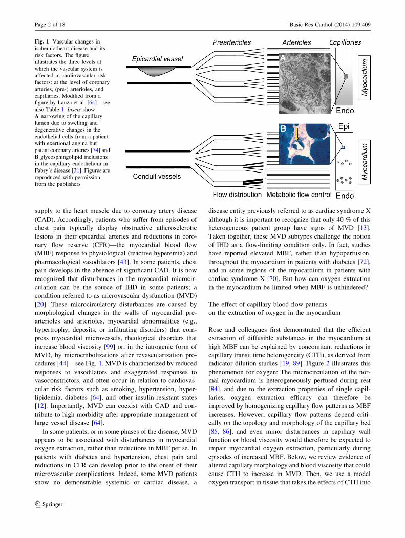
[20]. These microcirculatory disturbances are caused by
morphological changes in the walls of myocardial pre-
arterioles and arterioles, myocardial abnormalities (e.g.,
hypertrophy, deposits, or infiltrating disorders) that com-
press myocardial microvessels, rheological disorders that
increase blood viscosity [99] or, in the iatrogenic form of
MVD, by microembolizations after revascularization pro-
cedures [44]—see Fig. 1. MVD is characterized by reduced
responses to vasodilators and exaggerated responses to
vasoconstrictors, and often occur in relation to cardiovas-
cular risk factors such as smoking, hypertension, hyper-
lipidemia, diabetes [64], and other insulin-resistant states
[12]. Importantly, MVD can coexist with CAD and con-
tribute to high morbidity after appropriate management of
large vessel disease [64].
In some patients, or in some phases of the disease, MVD
appears to be associated with disturbances in myocardial
oxygen extraction, rather than reductions in MBF per se. In
patients with diabetes and hypertension, chest pain and
reductions in CFR can develop prior to the onset of their
microvascular complications. Indeed, some MVD patients
show no demonstrable systemic or cardiac disease, a
disease entity previously referred to as cardiac syndrome X
although it is important to recognize that only 40 % of this
heterogeneous patient group have signs of MVD [13].
Taken together, these MVD subtypes challenge the notion
of IHD as a flow-limiting condition only. In fact, studies
have reported elevated MBF, rather than hypoperfusion,
throughout the myocardium in patients with diabetes [72],
and in some regions of the myocardium in patients with
cardiac syndrome X [70]. But how can oxygen extraction
in the myocardium be limited when MBF is unhindered?
The effect of capillary blood flow patterns
on the extraction of oxygen in the myocardium
Rose and colleagues first demonstrated that the efficient
extraction of diffusible substances in the myocardium at
high MBF can be explained by concomitant reductions in
capillary transit time heterogeneity (CTH), as derived from
indicator dilution studies [19, 89]. Figure 2 illustrates this
phenomenon for oxygen: The microcirculation of the nor-
mal myocardium is heterogeneously perfused during rest
[84], and due to the extraction properties of single capil-
laries, oxygen extraction efficacy can therefore be
improved by homogenizing capillary flow patterns as MBF
increases. However, capillary flow patterns depend criti-
cally on the topology and morphology of the capillary bed
[85, 86], and even minor disturbances in capillary wall
function or blood viscosity would therefore be expected to
impair myocardial oxygen extraction, particularly during
episodes of increased MBF. Below, we review evidence of
altered capillary morphology and blood viscosity that could
cause CTH to increase in MVD. Then, we use a model
oxygen transport in tissue that takes the effects of CTH into
Fig. 1 Vascular changes in
ischemic heart disease and its
risk factors. The figure
illustrates the three levels at
which the vascular system is
affected in cardiovascular risk
factors: at the level of coronary
arteries, (pre-) arterioles, and
capillaries. Modified from a
figure by Lanza et al. [64]—see
also Table 1. Insets show
A narrowing of the capillary
lumen due to swelling and
degenerative changes in the
endothelial cells from a patient
with exertional angina but
patent coronary arteries [74] and
B glycosphingolipid inclusions
in the capillary endothelium in
Fabry’s disease [31]. Figures are
reproduced with permission
from the publishers
Page 2 of 18 Basic Res Cardiol (2014) 109:409
123

account [52] to predict which clinical findings would be
expected to result from a gradual increase of CTH. The
oxygen transport model is described in detail in [52], and
its application to cerebral ischemia in [79].
Regulation of capillary blood flow
The heterogeneity of capillary transit times during rest and
their homogenization towards high flow rates may be a
passive effect of capillary bed topology and morphology
[85, 86]. The capillary wall, however, also contains con-
tractile pericytes [90] which form the functional unit of the
capillariomotor system; a mechanism that seemingly
ensures the redistribution of erythrocytes along capillary
paths according to the regional oxygen needs of the tissue
[61]. Indeed, retinal pericytes constrict in response to high
oxygen tension but relax in response to lactate and low pH
[118], possibly providing a mechanism by which pericytes
MBF
MM
RO
2max
erythrocyteswhite blood cell
C
a
b
c
B
A
Fig. 2 The classical flow-diffusion equation for oxygen. The classi-
cal flow-diffusion equation curve (C) shows the maximum amount of
oxygen that can diffuse from capillaries to tissue for a given perfusion
rate, under the assumption that all erythrocytes pass through the tissue
capillaries at identical velocities, as indicated in A. This assumption is
rarely considered, but the heterogeneous distribution of capillary
flows shown in B shows why this assumption is important: because of
the shape of the curve in C, it is an inherent property of the classical
flow-diffusion that it overestimates tissue oxygenation if capillary
flows are heterogeneously distributed [89]. This is seen by using the
curve, which is accurate for individual capillaries, to determine the
net tissue oxygen availability resulting from the individual flows in
case B. The resulting net tissue oxygen availability is the weighted
average of the oxygen availabilities for the two flows, labeled b in the
plot. Note that the resulting tissue oxygen availability will always be
less than that of the homogenous case, labeled a. In fact, capillary
flows are heterogeneous in normal, resting tissue, but homogenize
during hyperemia. Label c shows a condition of higher flow, with
homogenous capillary flow. Note how homogenization ? hyperemia
(b ? c) provides a larger increase in tissue oxygenation than
hyperemia with homogenous capillary flow (a ? c) as assumed by
the classical flow-diffusion equation. The hindered capillary passage
indicated in the figure is the sum of preexisting age- or risk-factor-
related changes, and ischemia-related changes such as altered blood–
cell interactions with the endothelial surface properties (cell adhesion,
loss of glycocalyx, and so forth), and/or external edema pressure.
Modified from [79]
Basic Res Cardiol (2014) 109:409 Page 3 of 18
123

can redistribute capillary flows according to local cellular
metabolic needs in a continuous manner during rest [118].
Pericytes are embedded in the capillary basement mem-
brane and have been studied extensively in muscle tissue
[15]. Organ perfusion studies suggest that pericytes from
skeletal muscle, but not myocardial pericytes, constrict
upon exposure to angiotensin, norepinephrine, and vaso-
pressin [103]. Unlike pericytes from skeletal muscle tissue,
however, myocardial pericytes are often found in close
relation to myocardial nerve terminals [102]. The way in
during which central innervation and local vasoactive sig-
nals regulate pericyte tone in vivo, however, remains
poorly understood. Studies of retinal capillaries suggest
that pericytes react to intrinsic signaling in much the same
way as smooth muscle cells: Pericyte constrictions have
hence been observed in response to mechanical stretch and
exposure to angiotensin II (via AT1 receptors) [55] and
endothelin-1 (via ETA receptors) [91], by a Ca?? depen-
dent mechanism [25]. Meanwhile, pericytes relax in
response to adenosine [68], ATP [56], and nitric oxide
(NO) [37, 38], as well as to cholinergic [117] and adren-
ergic (via b2 receptors) [25] stimulation. Importantly,
recent studies show that cerebral pericytes control blood
flow, while ischemia and oxidative stress cause irreversible
constrictions of cerebral pericytes [39, 120].
The luminal surface of the capillary endothelium is
covered by a 0.5-lm-thick carbohydrate-rich matrix, the
glycocalyx [111], which affects the passage of blood cells
through the capillary bed [93]. Electrostatic interactions
between erythrocytes and glycocalyx [112] and slow pas-
sage of plasma in relation to this endothelial surface layer
[60] reduce capillary hematocrit to 20–50 % of that found
in the systemic circulation, and disruption of the glycoca-
lyx therefore causes capillary hematocrit to approach that
of the systemic circulation [18, 23]. The glycocalyx con-
stitutes a fluid barrier in the vascular system [105, 106],
and glycocalyx degradation is hence associated with
myocardial edema [105], and possibly capillary compres-
sion. The glycocalyx is degraded by exposure to direct
oxidative stress and oxidized lipoproteins [18, 21, 110], to
acute hyperglycemia [77], and ischemia [21, 51]. Disrup-
tion of the glycocalyx alters the normal blood flow
responses and hence the normal relation between blood
flow and metabolism in the myocardium [108, 109]. While
glycocalyx disruption and disturbed capillary flow patterns
often occur in parallel, it is unclear whether these changes
are causally related [22].
Endothelial cells throughout the vascular system are
electrically and metabolically coupled to each other, and to
nearby smooth muscle cells, via gap junctions composed of
so-called connexins [50]. The extent to which pericytes are
also involved in this efficient vascular signaling is uncer-
tain, but this rapid, bidirectional signaling pathway via gap
junctions seemingly ensures efficient coordination of vessel
function across the microvascular bed [27, 94, 95].
Changes in capillary morphology and blood viscosity
in MVD
At the level of myocardial capillaries, conditions that
predispose to MVD are associated with changes in capil-
lary wall morphology, including basement membrane
thickening, endothelial swelling, or compression by the
surrounding tissue—see Fig. 1 and Table 1. These changes
are observed in capillaries that appear to have been per-
fused and would hence be expected to disturb—rather than
to block—the capillary distribution of erythrocytes. Simi-
larly, increased blood viscosity and abnormal blood cell
adhesion to the capillary endothelium would be expected to
disturb the regulation of capillary flow patterns, rather than
to block myocardial perfusion. As illustrated by Fig. 2, the
dimensions of white blood cell (WBC) and erythrocyte
exceed the average capillary diameter. Experimental stud-
ies have shown that capillary flow patterns are sensitive to
the size, viscosity, number, and endothelial adhesion of
blood cells, and undergo profound changes during infec-
tions [69] and as part of the low-grade vascular inflam-
mation that accompany many cardiovascular risk factors
[69, 110].
Table 1 Changes in capillary morphology in MVD risk factors
Risk factor Changes in capillary
morphology
Reference(s)
Dilated and
hypertrophic
cardiomyopathy
(human)
Irregular capillary diameters.
Frequent narrowing of the
capillary lumen due to
thickened endothelial cells
*20 % of capillaries
affected
[74, 100]
Diabetes (human) Thickening of basement
membrane
[34]
SVD without CAD
(human)
Reduced capillary diameters.
Frequent narrowing of the
capillary lumen due to
thickened endothelial cells.
*50 % of capillaries
affected
[74]
Chagas disease
(human)
Basement membrane
thickening
[33]
Fabry’s disease
(animal model)
Glycosphingolipid inclusions
in the capillary
endothelium
[31]
Smoking Nicotine up-regulates the
expression of adhesion
molecules in the capillary
endothelium and increases
leukocyte rolling
[2, 122]
Page 4 of 18 Basic Res Cardiol (2014) 109:409
123
Relation between MBF, CTH, oxygen tension,
and the extraction of oxygen
The relation between blood flow (measured in mL blood
per 100 mL tissue per minute) through tissue, and its
access to oxygen, is traditionally derived from the classical
flow-diffusion equation [88] depicted by the curve in
Fig. 2. While this equation is only accurate for single
capillaries, or in tissue with homogenously perfused cap-
illaries, the complexity of describing systems with multiple
capillaries and transit times has thus far prevented the study
of tissue oxygenation changes in response to increasing
CTH. Figure 3A describes our first attempt to develop a
model of tissue oxygenation that incorporates the effects
CTH by assuming a distribution of capillary transit times
[52]. We used the accepted gamma variate function,
according to which CTH is introduced as a single param-
eter, namely the standard deviation of capillary transit
times. Figure 4 illustrates how MBF and myocardial tissue
oxygen tension (PtO2) must be adapted in order to maintain
myocardial oxygenation in response to a gradual increase
in CTH (top row). Below, we describe the tissue oxygen-
ation model [52] and the origin of these predictions in
greater detail, and then discuss the model and its properties
in relation to existing biophysical models of myocardial
oxygen transport.
Figure 5 illustrates the properties of the extended flow-
diffusion model, based on the hemodynamics and the
metabolic demands of the myocardium [52]. For conve-
nience, we summarized myocardial hemodynamics both in
terms of MBF (secondary x-axis) and the mean transit time
(MTT) for blood as it passes through myocardial capillaries
(primary x-axis). According to the central volume theorem
[98] MTT equals the myocardial capillary blood volume
(CBV), which we set to 7 % [8], divided by MBF. The
contour plot in Fig. 5A shows the maximum oxygen
extraction fraction (OEFmax) that can be achieved for any
combination of CTH and MTT (or MBF) for a fixed tissue
oxygen tension (PtO2) of 20 mmHg. The value of OEFmax
that corresponds to a given location in the (MBF, CTH)
plane is most easily derived from the OEFmax values
indicated on the two nearest iso-contours.
The diagonal arrow in Fig. 5A indicates a doubling of
MBF from 100 mL/100 mL/min to 200 mL/100 mL/min
at a constant OEFmax of 55 %, and constant PtO2. In the
human left ventricle, OEF values range 65–75 % during
rest [71], while 4–5 fold increases in myocardial oxygen
consumption during heavy exercise are met by increases in
MBF, and to a small extent in OEF [26, 42, 46]. In the right
ventricle, animal studies suggest that OEF is\50 % during
rest, while increases in OEF account for the majority of the
increase in oxygen availability during heavy exercise [40].
Accordingly, the arrow in Fig. 5A illustrates how left
ventricle can maintain constant OEFmax during several-fold
increases in MBF without reductions in myocardial oxygen
tension [28]. Similarly, a concomitant increase in OEF can
be achieved at fixed PtO2 by larger reductions in CTH
during the MBF increase. The horizontal component of the
arrow (indicated as a horizontal dashed line in Fig. 5A)
indicates the OEFmax change that would result from a
vasodilation alone, assuming that the CTH and PtO2
remained fixed during the increase in MBF. Note that if
CTH cannot be reduced, OEFmax decreases as MBF
increases, owing to the poor extraction of oxygen from
capillaries with very short transit times. This model prop-
erty is consistent with the original observations by Rose
and colleagues, namely that CTH must be reduced at high
MBF in order to explain the efficient extraction of solutes
by the myocardium during vasodilation [19, 89].
Net oxygen extraction capacity in the myocardium,
MMRO2max: capillary recruitment
Figure 5B and C shows contour plots of the maximum
myocardial metabolic rate of oxygen (MMRO2max, mea-
sured in mL O2/100 mL tissue/min) that can be supported
for any combination of MTT and CTH, again for a fixed
tissue oxygen tension of 20 mmHg. These figures are
derived from 5A by multiplying its OEFmax values by a
typical arterial oxygen concentration (19 mL/100 mL), and
MBF. In Fig. 5B, we assumed that all myocardial capil-
laries are perfused, i.e., absence of capillary recruitment
(opening of previously un-perfused capillaries) at high flow
rates. In Fig. 5C, we assumed that capillaries were
recruited as a function of increasing MBF. Specifically,
CBV was set to increase as the 0.38th power of MBF,
implying a 30 % increase in CBV as MBF increases from
100 to 200 mL/100 mL/min, and a 52 % CBV increase
when MBF triples. Note that, with this degree of capillary
recruitment, a doubling of MBF at constant CTH (hori-
zontal arrow in Fig. 5C) improves tissue oxygen avail-
ability as much as doubling MBF with a parallel CTH
reduction without recruitment (oblique arrow in Fig. 5B).
This effect is discussed further below.
The extent to which hyperemia is accompanied by
capillary recruitment in the heart remains uncertain. Both a
reduction of CTH, and capillary recruitment, increase the
effective capillary surface area for solute extraction [52],
and classical indicator dilution studies have therefore not
been able to prove or disprove the existence of recruitable
capillaries—see discussion in Reference [29]. The oxygen
extraction fraction (OEF) in the myocardium is constant, or
even increases, as MBF increases [28]. According to
Fig. 5A, this observation implies that hyperemia is
accompanied by either a CTH reduction along the oblique
lines in Fig. 5A–C, or by constant CTH and MTT. The
Basic Res Cardiol (2014) 109:409 Page 5 of 18
123
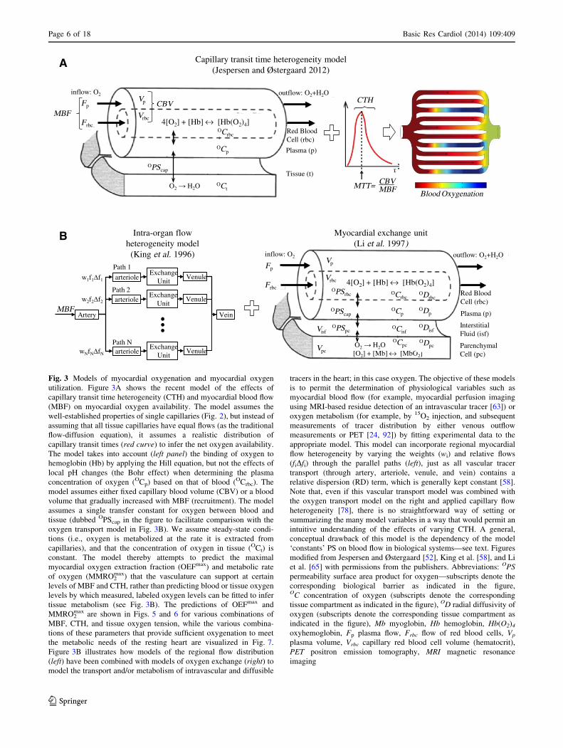
arterioleExchange
UnitVenule
Artery Vein
Path 1
w1f1 1
arterioleExchange
UnitVenule
Path 2w2f2 2
arterioleExchange
UnitVenule
Path NwNfN N
MBF
Intra-organ flow heterogeneity model
(King et al. 1996) inflow: O2
Vpc
4[O2] + [Hb] ↔ [Hb(O2)4]
ODp
[O2] + [Mb] ↔ [MbO2]O2 H2O
ODisf
ODrbc
OPScap
Visf
Vrbc
Vp
outflow: O2+H2O
Red Blood Cell (rbc)
Plasma (p)
InterstitialFluid (isf)
ParenchymalCell (pc)
Fp
Frbc
OPSpc
OPSrbc OCrbc
OCp
OCisf
OCpcODpc
Myocardial exchange unit (Li et al. 1997)
Capillary transit time heterogeneity model (Jespersen and Østergaard 2012)
MTT=CBVMBF
CTH
Blood Oxygenation
4[O2] + [Hb] ↔ [Hb(O2)4]Vrbc
Vp
outflow: O2+H2O
Red Blood Cell (rbc)
Plasma (p)
inflow: O2
Frbc
Fp
OCrbc
OCp
OCt
OPScapTissue (t)
MBF
CBV
O2 H2O
B
A
Fig. 3 Models of myocardial oxygenation and myocardial oxygen
utilization. Figure 3A shows the recent model of the effects of
capillary transit time heterogeneity (CTH) and myocardial blood flow
(MBF) on myocardial oxygen availability. The model assumes the
well-established properties of single capillaries (Fig. 2), but instead of
assuming that all tissue capillaries have equal flows (as the traditional
flow-diffusion equation), it assumes a realistic distribution of
capillary transit times (red curve) to infer the net oxygen availability.
The model takes into account (left panel) the binding of oxygen to
hemoglobin (Hb) by applying the Hill equation, but not the effects of
local pH changes (the Bohr effect) when determining the plasma
concentration of oxygen (OCp) based on that of blood (OCrbc). The
model assumes either fixed capillary blood volume (CBV) or a blood
volume that gradually increased with MBF (recruitment). The model
assumes a single transfer constant for oxygen between blood and
tissue (dubbed OPScap in the figure to facilitate comparison with the
oxygen transport model in Fig. 3B). We assume steady-state condi-
tions (i.e., oxygen is metabolized at the rate it is extracted from
capillaries), and that the concentration of oxygen in tissue (OCt) is
constant. The model thereby attempts to predict the maximal
myocardial oxygen extraction fraction (OEFmax) and metabolic rate
of oxygen (MMRO2max) that the vasculature can support at certain
levels of MBF and CTH, rather than predicting blood or tissue oxygen
levels by which measured, labeled oxygen levels can be fitted to infer
tissue metabolism (see Fig. 3B). The predictions of OEFmax and
MMRO2max are shown in Figs. 5 and 6 for various combinations of
MBF, CTH, and tissue oxygen tension, while the various combina-
tions of these parameters that provide sufficient oxygenation to meet
the metabolic needs of the resting heart are visualized in Fig. 7.
Figure 3B illustrates how models of the regional flow distribution
(left) have been combined with models of oxygen exchange (right) to
model the transport and/or metabolism of intravascular and diffusible
tracers in the heart; in this case oxygen. The objective of these models
is to permit the determination of physiological variables such as
myocardial blood flow (for example, myocardial perfusion imaging
using MRI-based residue detection of an intravascular tracer [63]) or
oxygen metabolism (for example, by 15O2 injection, and subsequent
measurements of tracer distribution by either venous outflow
measurements or PET [24, 92]) by fitting experimental data to the
appropriate model. This model can incorporate regional myocardial
flow heterogeneity by varying the weights (wi) and relative flows
(fiDfi) through the parallel paths (left), just as all vascular tracer
transport (through artery, arteriole, venule, and vein) contains a
relative dispersion (RD) term, which is generally kept constant [58].
Note that, even if this vascular transport model was combined with
the oxygen transport model on the right and applied capillary flow
heterogeneity [78], there is no straightforward way of setting or
summarizing the many model variables in a way that would permit an
intuitive understanding of the effects of varying CTH. A general,
conceptual drawback of this model is the dependency of the model
‘constants’ PS on blood flow in biological systems—see text. Figures
modified from Jespersen and Østergaard [52], King et al. [58], and Li
et al. [65] with permissions from the publishers. Abbreviations: OPS
permeability surface area product for oxygen—subscripts denote the
corresponding biological barrier as indicated in the figure,OC concentration of oxygen (subscripts denote the corresponding
tissue compartment as indicated in the figure), OD radial diffusivity of
oxygen (subscripts denote the corresponding tissue compartment as
indicated in the figure), Mb myoglobin, Hb hemoglobin, Hb(O2)4
oxyhemoglobin, Fp plasma flow, Frbc flow of red blood cells, Vp
plasma volume, Vrbc capillary red blood cell volume (hematocrit),
PET positron emission tomography, MRI magnetic resonance
imaging
Page 6 of 18 Basic Res Cardiol (2014) 109:409
123

maintenance of constant MTT would imply that capillaries
are recruited such that CBV increases in proportion to both
MBF and the net myocardial oxygen utilization. Indicator
dilution studies suggest, however, that relative CBV
increases are only half as big as the corresponding
increases in myocardial oxygen consumption [29]. There-
fore, it seems that a reduction of CTH, but not capillary
recruitment, is necessary to explain the observed coupling
between MBF and myocardial oxygen metabolism. Below,
we therefore limit our description of the extended flow-
diffusion model to include its features in the absence of
capillary recruitment.
Stage 1 Ensuring myocardial oxygenation as CTH
increases: adaptations to small increases in CTH
Using CTH as a parameter that summarizes the effects of
disturbed capillary flow patterns, we can now analyze the
effects of ‘capillary disease’ on myocardial oxygenation,
isolated from the effects that MVD may have on arterial
and arteriolar patency, and thereby MBF and MTT. We
refer to increases in CTH, and the accompanying inability
to reduce CTH (homogenize capillary flow patterns), as
capillary dysfunction below. Figure 4 provides an over-
view of the dynamics changes in MBF, PtO2, OEFmax, and
MMRO2max that follow from a gradual increase in CTH.
For small increases in CTH, the parallel reduction in
OEFmax can be compensated by increases in MBF to meet
the metabolic needs of the myocardium. The prediction
that early changes in capillary morphology give rise to less
efficient oxygen extraction is consistent with findings of
reduced oxygen extraction fractions (OEF) in the myo-
cardium of rats with streptozotocin-induced diabetes [82],
in diabetic patients [4], and in patients with microvascular
angina [13], as well as with findings of increased regional
MBF during rest in patients with diabetes and syndrome X
[14, 70, 72].
Stage 2 Adapting MBF, and blood flow responses,
to larger increases in CTH
Perhaps the most critical property of capillary dysfunction
is that as MTT decreases (MBF increases), vasodilation
may fail as a means of increasing the availability of oxygen
Fig. 4 Changes in MBF and
tissue oxygen tension that must
accompany increasing levels of
capillary dysfunction in order to
maintain tissue oxygen
availability. The figure displays
the adaptations of MBF and
PtO2 that are necessary in order
to maintain tissue oxygen
availability as CTH levels
gradually increase over time.
The gradual reduction in MFR
is indicated in the lower panel.
In the infarction-prone state,
minor reductions in MBF or
increases in CTH are predicted
to result in symptoms as
MMRO2max approaches the
actual, metabolic needs of the
tissue. Abbreviations: MBF
myocardial blood flow, PtO2
tissue oxygen tension, CTH
capillary transit time
heterogeneity, MMRO2max
maximum myocardial metabolic
rate of oxygen
Basic Res Cardiol (2014) 109:409 Page 7 of 18
123
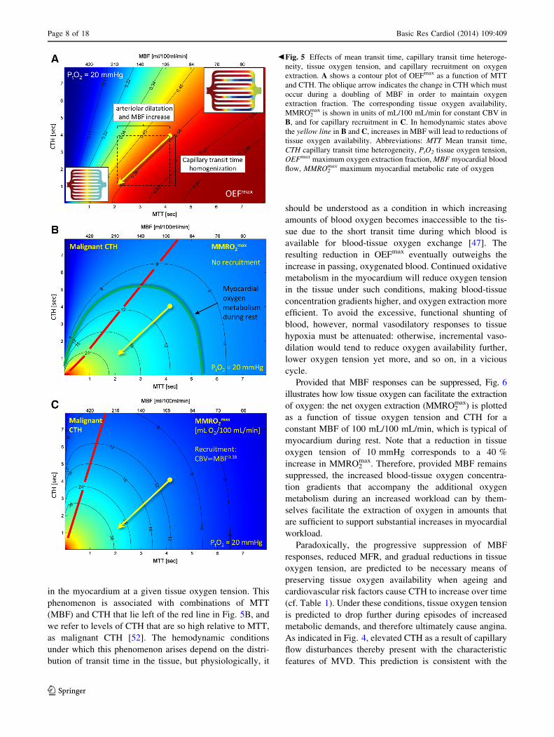
in the myocardium at a given tissue oxygen tension. This
phenomenon is associated with combinations of MTT
(MBF) and CTH that lie left of the red line in Fig. 5B, and
we refer to levels of CTH that are so high relative to MTT,
as malignant CTH [52]. The hemodynamic conditions
under which this phenomenon arises depend on the distri-
bution of transit time in the tissue, but physiologically, it
should be understood as a condition in which increasing
amounts of blood oxygen becomes inaccessible to the tis-
sue due to the short transit time during which blood is
available for blood-tissue oxygen exchange [47]. The
resulting reduction in OEFmax eventually outweighs the
increase in passing, oxygenated blood. Continued oxidative
metabolism in the myocardium will reduce oxygen tension
in the tissue under such conditions, making blood-tissue
concentration gradients higher, and oxygen extraction more
efficient. To avoid the excessive, functional shunting of
blood, however, normal vasodilatory responses to tissue
hypoxia must be attenuated: otherwise, incremental vaso-
dilation would tend to reduce oxygen availability further,
lower oxygen tension yet more, and so on, in a vicious
cycle.
Provided that MBF responses can be suppressed, Fig. 6
illustrates how low tissue oxygen can facilitate the extraction
of oxygen: the net oxygen extraction (MMRO2max) is plotted
as a function of tissue oxygen tension and CTH for a
constant MBF of 100 mL/100 mL/min, which is typical of
myocardium during rest. Note that a reduction in tissue
oxygen tension of 10 mmHg corresponds to a 40 %
increase in MMRO2max. Therefore, provided MBF remains
suppressed, the increased blood-tissue oxygen concentra-
tion gradients that accompany the additional oxygen
metabolism during an increased workload can by them-
selves facilitate the extraction of oxygen in amounts that
are sufficient to support substantial increases in myocardial
workload.
Paradoxically, the progressive suppression of MBF
responses, reduced MFR, and gradual reductions in tissue
oxygen tension, are predicted to be necessary means of
preserving tissue oxygen availability when ageing and
cardiovascular risk factors cause CTH to increase over time
(cf. Table 1). Under these conditions, tissue oxygen tension
is predicted to drop further during episodes of increased
metabolic demands, and therefore ultimately cause angina.
As indicated in Fig. 4, elevated CTH as a result of capillary
flow disturbances thereby present with the characteristic
features of MVD. This prediction is consistent with the
b Fig. 5 Effects of mean transit time, capillary transit time heteroge-
neity, tissue oxygen tension, and capillary recruitment on oxygen
extraction. A shows a contour plot of OEFmax as a function of MTT
and CTH. The oblique arrow indicates the change in CTH which must
occur during a doubling of MBF in order to maintain oxygen
extraction fraction. The corresponding tissue oxygen availability,
MMRO2max is shown in units of mL/100 mL/min for constant CBV in
B, and for capillary recruitment in C. In hemodynamic states above
the yellow line in B and C, increases in MBF will lead to reductions of
tissue oxygen availability. Abbreviations: MTT Mean transit time,
CTH capillary transit time heterogeneity, PtO2 tissue oxygen tension,
OEFmax maximum oxygen extraction fraction, MBF myocardial blood
flow, MMRO2max maximum myocardial metabolic rate of oxygen
Page 8 of 18 Basic Res Cardiol (2014) 109:409
123

findings of reduced MFR in some cases of MVD and car-
diac syndrome X [11, 64] and with the finding that
reductions in MFR are more frequent in patients with small
vessels disease [13], in whom changes in capillary mor-
phology are more severe [74]—see Table 1. One might
expect that reduced tissue oxygen tension would elicit
angiogenesis and hence a ‘physiological’ recruitment of
new vessels to improve tissue oxygenation. It should be
kept in mind, however, that existing capillaries are the
main source of the elevated CTH. The formation of new,
low-resistance capillary paths might in fact be suspected to
increase arteriolo-venular shunting rather than to improve
tissue oxygenation [80].
In Fig. 4, we illustrated the hypothesized improvement
in OEFmax that results from suppression of MBF. We
indicated a curve that eventually exceeds the OEF of
healthy, left ventricle myocardium. The extent to which a
relatively hypoxic myocardium can facilitate OEF values
that are above those of normal myocardium remains poorly
understood. Messer et al. [71] found reduced OEF
(N = 20, 66 % compared to 70 % in controls) in patients
with coronary artery disease, but these patients were able to
progressively increase OEF after 3 (73 %) and 7 (75 %)
min of exercise, without any increase in lactate levels.
These findings are consistent with the notion that relative
myocardial hypoxia may increase OEF to supranormal
values. Similar findings were reported by Holmberg et al.
[46] who measured smaller MBF increase during exercise
in patients with coronary insufficiency than in controls
(222 vs. 271 %), but correspondingly larger increases in
OEF in the patients (76 vs. 66 % in normal controls).
Stage 3 Critical increases in CTH: angina
and proneness for acute cardiac syndrome
If CTH continues to increase, the largest MMRO2max that can
be achieved by the tissue circulation gradually falls toward the
actual metabolic needs of the resting myocardium—see
Fig. 4. The green, three-dimensional surface on Fig. 7 cor-
responds to the combinations of MTT, CTH, and PtO2, that
give rise to a tissue oxygen availability equal to the metabolic
rate of oxygen of the myocardium during rest, MMRO2 =
10 mL O2/100 mL/min [1]. The surface is formed by joining
the 10 mL/100 mL/min iso-contours (cf. Figure 5B) for PtO2
values between 0 and 25 mmHg. The resulting green half-
cone therefore contains hemodynamic conditions that can
support metabolic needs of the myocardium during rest. The
red plane marks the boundary to malignant CTH.
Assuming that PtO2 is kept constant at 25 mmHg, the
point labeled A indicates the critical level of capillary
dysfunction for this PtO2 in myocardium: if CTH increased
Fig. 6 Myocardial oxygen availability without reactive hyperemia, at
normal MBF. MMRO2max is plotted as a function of tissue oxygen
tension and CTH for constant MBF (100 mL/100 mL/min). Note that
a reduction in tissue oxygen tension of 10 mmHg correspond to a
50 % increase in MMRO2max. Therefore, if MBF remains suppressed,
the increased blood-tissue oxygen concentration gradients that
accompany the increased oxygen metabolism during increased
workload can facilitate the extraction of oxygen in amounts that are
sufficient to support additional energy needs of the tissue. Abbrevi-
ations: MMRO2max maximum myocardial metabolic rate of oxygen,
CTH capillary transit time heterogeneity, MBF myocardial blood flow
Myocardialmetabolismduring rest
MalignantCTH threshold reduced
blood supply
140
84105
7060
210
MBF [ml/100ml/min]
[mm
Hg]
CTH [sec] [sec]
420
Fig. 7 Metabolic thresholds. The green surface indicates combina-
tions of MTT (MBF is shown on the upper x-axis), CTH, and PtO2
that provide sufficient oxygen to meet the metabolic rate of the
myocardium during rest. The red plane marks the boundary, left of
which vasodilation reduces tissue oxygen availability (malignant
CTH). The blue arrows indicate the principal ways in which
myocardial oxygen availability can be reduced in disease: by
reductions in MBF (by CAD), increases in CTH (capillary dysfunc-
tion), and combinations thereof—see text. Abbreviations: CTH
capillary transit time heterogeneity, MBF myocardial blood flow,
MTT mean transit time, PtO2 = tissue oxygen tension, CAD coronary
artery disease
Basic Res Cardiol (2014) 109:409 Page 9 of 18
123
beyond this value, oxygen availability would no longer be
able to meet the metabolic needs of resting myocardium.
As described above, the tissue oxygen tension will gradu-
ally fall as oxygen availability approaches the metabolic
needs of the tissue, and the figure illustrates how the more
efficient extraction permits the maintenance of oxygen
availability for a wider range of MBF and CTH values: the
green half-cone becomes wider towards its base, indicating
that at relative tissue hypoxia, myocardial oxygenation can
be maintained across a wider range of CTH and (lower)
MBF values.
If CTH increases further, it reaches a critical limit
(broken line parallel to the MTT axis at PtO2 = 0), at
which tissue oxygen tension is negligible and MTT maxi-
mizes MMRO2max (Labeled B). Note that, at this point, the
metabolic needs of tissue cannot be supported unless MTT
is prolonged to a threshold of approximately 5 s, corre-
sponding to MBF = 84 mL/100 mL/min. In other words,
the gradual reduction in tissue oxygen availability owing to
progressive capillary dysfunction (increase in CTH) can be
compensated for by gradual reductions in tissue oxygen
tension and in resting MBF.
Figure 7 also allows us to analyze myocardial oxygena-
tion after a sudden reduction in MBF, for example as a result
of the partial obstruction of an upstream epicardial artery.
Myocardial oxygen availability remains above the metabolic
demands of resting myocardium until MTT exceeds 8 s,
corresponding to an MBF threshold of 53 mL/100 mL/min,
provided that CTH is negligible or moderate (Label C).
Triggers of myocardial infarction and angina
in conditions of high CTH
The model of myocardial oxygen availability presented
here predicts that not only reductions in MBF, but also
increases in CTH, can trigger a critical lack of oxygen and
thereby angina and/or acute cardiac syndrome. Further-
more, increasing capillary dysfunction is predicted to
reduce tissue oxygen reserves towards that of the myo-
cardium during rest, in a gradual fashion. Theoretically,
minor reductions in MBF or slight increases in CTH are
therefore sufficient to trigger reductions in tissue oxygen
tension, and thereby angina and systolic dysfunction. In
principle, angina is therefore predicted to be either ‘arte-
rial’ in origin (triggered by reductions in MBF, for example
due to an atherosclerotic stenosis or small vessel disease),
or ‘capillary’, triggered primarily by elevated CTH. In the
latter case, MBF is predicted to be moderately reduced,
having adapted itself to maximize myocardial oxygenation,
cf. Figs. 4 and 7. Therefore, tissue affected by either a
chronic or a sudden increase in CTH is predicted to appear
hypoperfused when compared to tissue with unaffected by
CTH, even in the absence of any vessel stenosis.
The extent to which episodes of elevated CTH can
trigger angina or systolic dysfunction remains unclear.
Dehydration, and infections accompanied by elevated vis-
cosity due to an increased neutrophil count and endothelial
adhesion [69], would be expected to elevate CTH and
thereby cause critical reduction in myocardial oxygen
availability. The seasonal variation in cardiovascular
deaths [96] has indeed been linked to increased neutrophil
count in relation to winter respiratory infections [116]. This
mechanism might contribute to the hypothesized relation
between acute infections and cardiovascular deaths, and
between chronic infections and the development IHD [67].
Tissue injury during myocardial ischemia: the predicted
effect of capillary flow disturbances
The extended flow-diffusion equation predicts that tissue
oxygen availability in hypoperfused tissue is affected by
the extent of the MBF reduction, as well as CTH. While the
level of hypoperfusion would be expected to remain con-
stant until successful recanalization therapy, changes in
capillary flow patterns during the ischemic period could
cause tissue oxygen availability to deteriorate. The extent
of tissue damage during the ischemic period may therefore
depend on factors other than the vascular occlusion, and
potentially, be modified prior to hospitalization. Capillary
flows are indeed known to undergo profound changes
during experimental ischemia owing to capillary plugging,
endothelial damage, capillary leakage, pericapillary edema,
and reductions in local flow [32].
Reperfusion injury: the putative roles of capillary
occlusions and capillary compression/constriction
Successful normalization of MBF after episodes of myo-
cardial ischemia is often accompanied by reperfusion
injury (RI) which accounts for up to 50 % of myocardial
damage in animal models, and may account for a propor-
tion of the deaths and cardiac failures that occur in humans,
in spite of optimal recanalization therapy [119].
The extended flow-diffusion equation predicts that in
order for tissue reperfusion to restore myocardial oxygen
availability to its pre-ischemic level, both MBF and CTH
must be restored to their pre-ischemic values. This pre-
diction re-emphasizes the notion that recanalization must
be accompanied by the reversal of any capillary plugging,
pericyte constriction, endothelial swelling, and edema-
driven capillary compression that may have evolved during
the ischemic/hypoxic period, as well as any capillary flow
disturbances that may have resulted from the lysis or
mechanical removal of upstream clots [45]. If not, any
residual capillary obstructions, or elevated CTH across
perfused capillaries, would be predicted to prevent optimal
Page 10 of 18 Basic Res Cardiol (2014) 109:409
123
re-oxygenation, and possibly to attenuate MBF or MFR to
optimize myocardial oxygen availability, cf. Figs. 4 and 7.
Both endothelial edema and granulocyte entrapments per-
sist after myocardial reperfusion [119] and would hence be
expected to cause elevated CTH levels and result in
incomplete re-oxygenation. The passage of plasma through
the microcirculation is predicted to be less disturbed by
capillary changes than that of erythrocytes, due to their size
and capillary adhesion properties. The prediction that
capillary flow disturbances contribute to reperfusion injury
is therefore consistent with the finding that the use of ‘free’
hemoglobin as an oxygen carrying perfusate reduces
reperfusion injury [16].
The malignant CTH phenomenon also implies that, if
CTH is not immediately restored upon reperfusion, OEFmax
will remain low for short MTT, and the sudden restoration
of flow through fully dilated arteries and arterioles could
therefore, paradoxically, cause immediate, severe tissue
hypoxia and tissue damage. On the other hand, the increase
in perfusion pressure after recanalization would be expec-
ted to augment the dilation of capillaries and help restore
homogenous capillary flows. Reperfusion injury is greatly
reduced in animal models if perfusion and perfusion
pressure is gradually increased to pre-ischemic values over
a few minutes, as opposed to uncontrolled, hyperemic
reperfusion [76, 81]. Similarly, post-conditioning (episodes
of interrupted flow after recanalization) seemingly
improves tissue salvage, electrical function and outcome in
patients [97]. The extent to which this beneficial effect is
related to the restoration of capillary patency and CTH
remains unclear.
Discussion
The extended flow-diffusion equation may help us widen
our understanding of IHD and MVD from being conditions
characterized by limited MBF, to also take the effects of
elevated CTH on myocardial oxygen availability into
account. In the resulting understanding of the etiopatho-
genesis of IHD and MVD, angina and acute coronary
syndrome are preceded not only by atherosclerosis and/or
small vessel disease, but also by changes in capillary
morphology or blood viscosity that cause CTH to increase.
The resulting changes in oxygen extraction efficacy are
predicted to require adaptations of both resting MBF and
MBF responses during exercise to meet the metabolic
needs of myocardial oxygen metabolism. In fact, a gradual
increase in CTH is predicted to require adaptations that are
consistent with initial findings of elevated resting MBF and
of gradual reductions in CFR in MVD risk factors. The
extended flow-diffusion equation predicts that heart failure
is the result of reductions in MMRO2max to levels below the
metabolic needs of the myocardium, either as a result of
result of reductions in MBF (the traditional understanding
of IHD), as a result of elevated CTH levels, or both.
Importantly, the equation predicts that both the restoration
of MBF, and of capillary flow patterns, represent key
aspects of reducing ischemic damage and reperfusion
injury after myocardial ischemia. Therapeutic means of
maintaining capillary perfusion, both prior to hospitaliza-
tion and in relation to recanalization, could therefore prove
to be important in the management of acute cardiac
syndrome.
Can capillary dysfunction elicit endothelial dysfunction
and attenuate MBF responses?
The endothelial dysfunction and reduced CFR observed in
patients with cardiovascular risk factors, IHD, and MVD,
are predicted by our model to represent necessary adapta-
tions to increasing CTH as hyperemia gradually fails to
increase myocardial oxygen availability. The ways by
which capillary dysfunction can attenuate upstream vaso-
dilation (endothelial dysfunction), however, remain
unclear. The coordination of microvascular function is
controlled in large parts by efficient, bidirectional signaling
among endothelial cells, who act as metabolic ‘sensors’
[27, 50, 94, 95]. The extent to which connexins are
involved in endothelial dysfunction in relation to tissue
hypoxia has only recently been studied [87]. A defining
feature of capillary dysfunction is that tissue hypoxia
gradually develops as vasodilation can no longer support
the metabolic needs of the tissue during exercise. Such
reductions in tissue oxygen tension would be expected to
lead to the activation of hypoxia-inducible transcription
factors (HIF), which in turn initiates a number of adapta-
tions in tissue to better tolerate low oxygen levels. One of
these is the up-regulation of nicotinamide adenine dinu-
cleotide phosphate (NADPH) oxidase (NOX) levels [123].
NOX is a major source of superoxide and free radicals in
the vasculature [9]. Endothelial dysfunction is indeed
mediated by elevated levels of superoxide anions in the
vessel wall, and parallel depletion of nitric oxide (NO). As
a consequence, capillary dysfunction could, in principle,
elicit upstream endothelial dysfunction via a hypoxia-sen-
sitive mechanism. Of note, the up-regulation of hypoxia-
inducible factor type 1 HIF-1 also leads to the initiation of
inflammatory processes via activation of nuclear factor jB
(NF-jB) [30]. Therefore, tissue inflammation may be an
additional, obligatory companion to capillary dysfunc-
tion—see Fig. 4.
Long-term ROS exposure of artery and arteriole walls to
ROS and low NO are known to cause remodeling and
thickening of the vessel walls [104]. With more prolonged
oxidative damage, vascular smooth muscle cells may
Basic Res Cardiol (2014) 109:409 Page 11 of 18
123

degenerate and develop abnormal focal constrictions that
result in additional narrowing of their luminal diameters.
These adaptations tend to attenuate flow and flow respon-
ses, and may therefore, paradoxically, protect tissue from
hypoxic episodes caused by increases in blood flow. We
have speculated that vascular ROS production may have
another, more severe side-effect: ROS and peroxynitrate
are likely to reach capillaries immediately downstream,
where peroxynitrate can cause further damage to the cap-
illary wall, while both increased ROS levels [120] and
reduced NO levels [37, 38] are believed to cause pericyte
constriction. As a consequence, endothelial dysfunction
may result in abnormal constriction of additional capillar-
ies, further elevating CTH, and thereby further exacerbate
the detrimental lack of oxygen in a vicious cycle.
Implications for the prevention and management
of IHD
Elevated superoxide levels in vessels and tissue are likely
to deplete capillary NO levels in the myocardium, while
tissue hypoxia causes a lack of substrate for NO production
via tissue NO-synthases. Means of restoring capillary NO
levels or preventing the constriction of capillary pericytes
may therefore prove cardioprotective in IHD, and during
acute cardiac syndrome. This is consistent with findings
that nitroglycerine appears to reduce tissue hypoxia by
altering microvascular flow patterns, but without changing
overall vascular resistance [53]. Tissue nitrite stores can be
reduced to NO during ischemia or hypoxia without the
need of tissue oxygen as a substrate. The administration of
nitrite during cardiac ischemia has indeed been shown to be
cardioprotective in animal studies, and circulating levels of
nitrite are thought to be related to cardiovascular risk [17].
Green leafy vegetable is a major dietary source of nitrite in
humans [66], and the intake of these has been shown to be
inversely related to the incidence of myocardial infarctions
[49].
Biophysical modeling of myocardial oxygenation
and oxygen consumption
Oxygen transport models have been used extensively in the
attempt to understand the relation between MBF and
metabolism, and to derive myocardial oxygen metabolism
based on dynamic recordings of [15O] in tissue or in venous
blood [24, 65, 115] see Fig. 3B. Myocardial perfusion is
very heterogeneous when measured at a millimeter scale, a
phenomenon termed micro-heterogeneity to distinguish it
from regional and transmural flow differences [3, 5–7, 36,
57, 92, 107]. To account for flow heterogeneity within the
tissue volume being modeled, oxygen transport models
therefore typically include flow distribution and vascular
dispersion terms which can then be calibrated by measured
relative dispersion (RD) values from tissue samples [58]—
see Fig. 3B. The figure also shows the capillary-tissue
oxygen exchange and oxygen utilization model developed
by Li et al. [65], in which the binding of oxygen to
hemoglobin and myoglobin is accounted for. This model
has been utilized to fit dynamic 15O2 data in isolated hearts,
and the predicted myocardial oxygen utilization shown to
be in good agreement with direct measurements using
Fick’s principle [92]. We have previously used the model
in Fig. 3B (left panel) to model capillary flow heteroge-
neity, assigning different numerical weights and fractional
flows to the parallel paths in Fig. 3B [78]. With its large
number of parameters, however, this exhaustive model
does not lend itself to an intuitive analysis of how MBF,
CTH, and tissue oxygen levels, respectively, permit a given
myocardial oxygen utilization.
The extended flow-diffusion equation models oxygen
availability with as few parameters as possible in order to
examine how blood flow, CTH and tissue oxygen tension,
combined, can support a given metabolic demand, while
permitting intuitive visualization of these limitations in
three-dimensional plots such as Fig. 7. Similar to the
approach of Li et al., who used a lagged normal density
function to describe flow heterogeneity, we used a gamma
variate function to describe transit time heterogeneity, in
order to parameterize CTH by a single parameter—in this
case the standard deviation of transit times. Similarly, we
incorporated the binding of oxygen to hemoglobin in
blood. Myocardial capillaries are organized in a complex,
interconnected three-dimensional network [8, 54]. While
spatially distributed models such as that of Li et al. requires
assumptions on interstitial oxygen diffusion within this
complex topology, and the degree of oxygen binding to
myoglobin, we assumed a uniform interstitial oxygen ten-
sion. By having tissue oxygen tension as our model’s third
variable (see Fig. 7), the model clarifies the physiological
distinction between myocardial ischemia (low blood flow)
and myocardial hypoxia, which is directly related to IHD
symptoms. As shown in Fig. 3, neither model directly
models the interconnectedness of myocardial capillaries, a
feature better captured by network models [115]. Kiyooka
et al. imaged epicardial capillary flow patterns in dogs and
found a significant increase in the diameter of, and blood
flow through, cross-connecting capillaries during reactive
hyperemia [59]. The flow through dilated cross-connecting
capillaries would be expected to reduce flow differences
among parallel capillary paths, and hence to facilitate the
homogenization of capillary flow patterns during reactive
hyperemia. According to our model and the observations
by Rose et al. [89], this topological feature of the myo-
cardial capillary bed thus seems to facilitate the mainte-
nance of constant oxygen extraction fraction across a wide
Page 12 of 18 Basic Res Cardiol (2014) 109:409
123

range of flow values in the heart. More complex features of
the myocardial microcirculation, such as its compression
during systole, may also be of relevance to future bio-
physical models of myocardial oxygen transport [35].
Capillary recruitment
The classical flow-diffusion equation [88], as well as more
complex models such as that of Li et al. [65], predict that
the extraction of diffusible substances is limited by blood
flow and the capillary permeability (P) multiplied by their
surface area (S). Assuming that capillary permeability is
constant for a given molecule, these models therefore
imply that the net extraction of a given substance can be
increased either by increasing blood flow, or by capillary
recruitment (opening of previously closed capillaries to
increase capillary surface area) [62]. Applying the oxygen
transport model of Li et al. to dynamic 15O2 data,
Schwanke et al. [92] found that satisfactory data fits could
only be achieved by assuming that PS for oxygen increases
linearly with MBF during a threefold increase in flow, and
a sixfold increase in oxygen utilization. Although a slight
increase in capillary blood volume is observed during
hyperemia, such an increase in the number of perfused
capillaries is contradicted by observations [59]. The
extended flow-diffusion equation offers a crucial advan-
tage over existing models by offering an explanation to
this ‘recruitment paradox’: the physiological effect of
CTH is to modify the extent to which diffusible tracers can
exchange with tissue through the capillary wall, and
thereby their ‘apparent’ capillary surface area, even for a
fixed, ‘physical’ PS. We showed that the ‘apparent’ PS one
would observe in the heart is given by PS = -MBF�ln(1-
OEFmax) [52]. Provided that myocardial metabolism and
hemodynamics are coupled, so that OEF = OEFmax, our
model therefore predicts that apparent PS for oxygen in the
myocardium (where OEF is remarkably constant) increa-
ses linearly with MBF, as Schwanke et al. [92] observed
and had to assume in order to fit experimental data to their
model.
Capillary transit time heterogeneity versus myocardial
flow heterogeneity
The CTH phenomenon, and the potential ‘oxygen loss’ due
to functional shunting through capillary paths with short
transit times, has received much less attention [47, 89] than
myocardial flow heterogeneity on the millimeter scale and
beyond [3, 5–7, 36, 57, 92, 121] due to the initial suspicion
that areas of low perfusion were prone to ischemic damage.
Myocardial micro-heterogeneity is typically reported as the
standard deviation of flow values within a certain tissue
volume, and remains relatively constant (30 % of the
resting flow) across tissue volume sizes and species [6].
The heterogeneity in flow appears to be linked to the
topology of the vascular tree [48], and detailed studies of
the oxygen supply–demand balance within the myocar-
dium suggest that metabolic needs are in fact met [124] in
spite of the low perfusion values. The relation between
flow heterogeneity and the regulation of arterial and arte-
riolar tone is reviewed in greater detail in Ref. [101]. We
speculate that CTH, and thereby OEFmax, is actively reg-
ulated, possibly via pericyte dilation [39], to meet the
metabolic needs of the tissue for a given MBF and tissue
oxygen tension. This notion is consistent with the obser-
vation that flow heterogeneity is paralleled by considerable
heterogeneity in the oxygen saturation of small myocardial
veins [114], with high OEF at low flow rates [92]. Simi-
larly, metabolic needs in the right ventricle are seemingly
met to some extent by increased OEF rather than MBF
[40].
Measurement of CTH, MTT, and capillary recruitment
in the myocardium
We hypothesize that the model parameters of the extended
flow-diffusion model (MTT, CTH, and tissue oxygen ten-
sion) hold information about myocardial oxygen avail-
ability that cannot be gleaned from MBF measurements
alone. The coronary microcirculation can be assessed by a
number of techniques, albeit direct observation of capillary
flow patterns in the myocardium itself remains challenging
[59, 83]. Myocardial blood transit time characteristics can
in principle be determined by routine cardiac MRI, CT, or
ultrasound ‘bolus tracking’ perfusion measurements using
contrast agents with little or no first-pass extraction [63,
113]. We have shown that this approach permits reliable
retrieval of MTT and CTH as parametric maps at the
typical signal-to-noise ratio of clinical perfusion MRI in
the brain [75]. Transit time distributions retrieved by this
approach are characteristics of individual voxels, which for
state-of-the-art cardiac perfusion MRI are
1.7 9 1.9 9 10 mm [73]. Given the heterogeneity of
myocardial flows over short distance scales, the stability of
such measurements should be carefully evaluated to ensure
that they reflect CTH of uniformly perfused tissue. Quali-
tatively, increased CTH would be expected to result in
delayed tissue-clearance of contrast medium during
angiographic procedures. This phenomenon is indeed
observed in some patients suspected of MVD [10].
Perspectives
The presence of age- or risk-factor-related changes in
capillary morphology constitutes a key difference between
Basic Res Cardiol (2014) 109:409 Page 13 of 18
123
human IHD and the animal models that are typically used
in the study of myocardial ischemia and reperfusion injury.
Given the putative effects of capillary flow disturbances
described here, this difference could affect the extent to
which cardioprotective strategies developed in animal
models with normal regulation of CTH translate into suc-
cessful human therapies. We speculate that the use of
animal models with capillary changes characteristic of
human disease (spontaneously hypertensive rats, diabetic
animals, pharmacological degradation of the glycocalyx,
and so forth) may provide more realistic models of human
disease.
This review re-emphasizes the importance of biophysi-
cal modeling as a means of understanding the relation
between myocardial blood supply and the metabolic needs
of the myocardium. Combining such models with cutting-
edge imaging techniques, we speculate that studies of the
importance of capillary flow distributions in MVD, myo-
cardial ischemia, and reperfusion injury may be within
reach in both animal models and humans. Meanwhile, we
must develop a thorough understanding of capillary func-
tion, including that of the glycocalyx, of endothelial cells,
and of the regulation and pharmacological modulation of
pericyte tone.
Acknowledgments This work was supported by the Danish
National Research Foundation (LØ, SNJ), the Danish Ministry of
Science, Technology and Innovation’s University Investment Grant
(LØ, SNJ), The VELUX foundation (ARCADIA, LØ, HA), the
Danish Research Council (HEB, 11-108354), and The Danish Stra-
tegic Research Council (HEB, 11-1115818).
Conflict of interest The authors declare that they have no conflict
of interest.
Open Access This article is distributed under the terms of the
Creative Commons Attribution License which permits any use, dis-
tribution, and reproduction in any medium, provided the original
author(s) and the source are credited.
References
1. Agostini D, Iida H, Takahashi A, Tamura Y, Henry Amar M,
Ono Y (2001) Regional myocardial metabolic rate of oxygen
measured by O2-15 inhalation and positron emission tomogra-
phy in patients with cardiomyopathy. Clin Nucl Med 26:41–49.
doi:10.1097/00003072-200101000-00010. http://journals.lww.
com/nuclearmed/Fulltext/2001/01000/Regional_Myocardial_
Metabolic_Rate_of_Oxygen.10
2. Albaugh G, Bellavance E, Strande L, Heinburger S, Hewitt CW,
Alexander JB (2004) Nicotine induces mononuclear leukocyte
adhesion and expression of adhesion molecules, VCAM and
ICAM, in endothelial cells in vitro. Ann Vasc Surg 18:302–307.
doi:10.1007/s10016-004-0030-9
3. Austin RE Jr, Aldea GS, Coggins DL, Flynn AE, Hoffman JI
(1990) Profound spatial heterogeneity of coronary reserve.
Discordance between patterns of resting and maximal
myocardial blood flow. Circ Res 67:319–331. doi:10.1161/01.
RES.67.2.319
4. Baldi JC, Aoina JL, Oxenham HC, Bagg W, Doughty RN (2003)
Reduced exercise arteriovenous O2 difference in type 2 diabetes.
J Appl Physiol 94:1033–1038. doi:10.1152/japplphysiol.00879.
2002
5. Bassingthwaighte JB, Beard DA, Li Z (2001) The mechanical
and metabolic basis of myocardial blood flow heterogeneity.
Basic Res Cardiol 96:582–594. doi:10.1007/s003950170010
6. Bassingthwaighte JB, King RB, Roger SA (1989) Fractal nature
of regional myocardial blood flow heterogeneity. Circ Res
65:578–590. doi:10.1161/01.RES.65.3.578
7. Bassingthwaighte JB, Malone MA, Moffett TC, King RB, Chan
IS, Link JM, Krohn KA (1990) Molecular and particulate
depositions for regional myocardial flows in sheep. Circ Res
66:1328–1344. doi:10.1161/01.RES.66.5.1328
8. Bassingthwaighte JB, Yipintsoi T, Harvey RB (1974) Micro-
vasculature of the dog left ventricular myocardium. Microvasc
Res 7:229–249. doi:10.1016/0026-2862(74)90008-9
9. Bedard K, Krause KH (2007) The NOX family of ROS-gener-
ating NADPH oxidases: physiology and pathophysiology.
Physiol Rev 87:245–313. doi:10.1152/physrev.00044.2005
10. Beltrame JF, Limaye SB, Wuttke RD, Horowitz JD (2003)
Coronary hemodynamic and metabolic studies of the coronary
slow flow phenomenon. Am Heart J 146:84–90. doi:10.1016/
S0002-8703(03)00124-8
11. Botker HE (2001) Vascular and metabolic abnormalities in
patients with angina pectoris and normal coronary angiograms.
Dan Med Bull 48:1–18
12. Botker HE, Moller N, Ovesen P, Mengel A, Schmitz O, Orskov
H, Bagger JP (1993) Insulin resistance in microvascular angina
(syndrome X). Lancet 342:136–140. doi:10.1016/0140-
6736(93)91344-L
13. Botker HE, Sonne HS, Bagger JP, Nielsen TT (1997) Impact of
impaired coronary flow reserve and insulin resistance on myo-
cardial energy metabolism in patients with syndrome X. Am J
Cardiol 79:1615–1622. doi:10.1016/S0002-9149(97)00209-9
14. Bottcher M, Botker HE, Sonne H, Nielsen TT, Czernin J (1999)
Endothelium-dependent and -independent perfusion reserve and
the effect of L-arginine on myocardial perfusion in patients with
syndrome X. Circulation 99:1795–1801. doi:10.1161/01.CIR.99.
14.1795
15. Bruns RR, Palade GE (1968) Studies on blood capillaries: I.
General organization of blood capillaries in muscle. J Cell Biol
37:244–276. doi:10.1083/jcb.37.2.244
16. Burkhoff D, Lefer DJ (2005) Cardioprotection before revascu-
larization in ischemic myocardial injury and the potential role of
hemoglobin-based oxygen carriers. Am Heart J 149:573–579.
doi:10.1016/j.ahj.2004.06.028
17. Calvert JW, Lefer DJ (2009) Myocardial protection by nitrite.
Cardiovasc Res 83:195–203. doi:10.1093/cvr/cvp079
18. Constantinescu AA, Vink H, Spaan JA (2001) Elevated capillary
tube hematocrit reflects degradation of endothelial cell glyco-
calyx by oxidized LDL. Am J Physiol Heart Circ Physiol
280:H1051–7. http://ajpheart.physiology.org/content/280/3/
H1051.full-text.pdf
19. Cousineau D, Rose CP, Lamoureux D, Goresky CA (1983)
Changes in cardiac transcapillary exchange with metabolic
coronary vasodilation in the intact dog. Circ Res 53:719–730.
doi:10.1161/01.RES.53.6.719
20. Crea F, Camici PG, Bairey Merz CN (2013) Coronary micro-
vascular dysfunction: an update. Eur Heart J. doi:10.1093/eur
heartj/eht513
21. Czarnowska E, Karwatowska-Prokopczuk E (1995) Ultrastruc-
tural demonstration of endothelial glycocalyx disruption in the
Page 14 of 18 Basic Res Cardiol (2014) 109:409
123

reperfused rat heart. Involvement of oxygen free radicals. Basic
Res Cardiol 90:357–364. doi:10.1007/BF00788496
22. Damon DH, Duling BR (1987) Are physiological changes in
capillary tube hematocrit related to alterations in capillary per-
fusion heterogeneity? Int J Microcirc Clin Exp 6:309–319
23. Desjardins C, Duling BR (1990) Heparinase treatment suggests
a role for the endothelial cell glycocalyx in regulation of cap-
illary hematocrit. Am J Physiol 258:H647–54. http://ajpheart.
physiology.org/content/258/3/H647.full-text.pdf
24. Deussen A, Bassingthwaighte JB (1996) Modeling [15O]oxygen
tracer data for estimating oxygen consumption. Am J Physiol
270:H1115–30. http://ajpheart.physiology.org/content/270/3/
H1115.full-text.pdf
25. Diaz-Flores L, Gutierrez R, Madrid JF, Varela H, Valladares F,
Acosta E, Martin-Vasallo P, Diaz-Flores L,Jr (2009) Pericytes.
Morphofunction, interactions and pathology in a quiescent and
activated mesenchymal cell niche. Histol Histopathol
24:909–969. http://hh.um.es/pdf/Vol_24/24_7/Diaz-Flores-24-
909-969-2009.pdf
26. Dole WP, Nuno DW (1986) Myocardial oxygen tension deter-
mines the degree and pressure range of coronary autoregulation.
Circ Res 59:202–215. doi:10.1161/01.RES.59.2.202
27. Duling BR, Berne RM (1970) Propagated vasodilation in the
microcirculation of the hamster cheek pouch. Circ Res
26:163–170. doi:10.1161/01.RES.26.2.163
28. Duncker DJ, Bache RJ (2008) Regulation of coronary blood
flow during exercise. Physiol Rev 88:1009–1086. doi:10.1152/
physrev.00045.2006
29. Duran WN, Marsicano TH, Anderson RW (1977) Capillary
reserve in isometrically contracting dog hearts. Am J Physiol
233:H276–81. http://ajpheart.physiology.org/content/233/2/
H276.full-text.pdf
30. Eltzschig HK, Carmeliet P (2011) Hypoxia and inflammation.
N Engl J Med 364:656–665. doi:10.1056/NEJMra0910283
31. Eng CM, Banikazemi M, Gordon RE, Goldman M, Phelps R,
Kim L, Gass A, Winston J, Dikman S, Fallon JT, Brodie S,
Stacy CB, Mehta D, Parsons R, Norton K, O’Callaghan M,
Desnick RJ (2001) A phase 1/2 clinical trial of enzyme
replacement in fabry disease: pharmacokinetic, substrate clear-
ance, and safety studies. Am J Hum Genet 68:711–722. doi:10.
1086/318809
32. Engler RL, Dahlgren MD, Peterson MA, Dobbs A, Schmid-
Schonbein GW (1986) Accumulation of polymorphonuclear
leukocytes during 3-h experimental myocardial ischemia. Am J
Physiol 251:H93–100. http://ajpheart.physiology.org/content/
251/1/H93.full-text.pdf
33. Ferrans VJ, Milei J, Tomita Y, Storino RA (1988) Basement
membrane thickening in cardiac myocytes and capillaries in
chronic Chagas’ disease. Am J Cardiol 61:1137–1140. doi:10.
1016/0002-9149(88)90148-8
34. Fischer VW, Barner HB, Leskiw ML (1979) Capillary basal
laminar thickness in diabetic human myocardium. Diabetes
28:713–719. doi:10.2337/diab.28.8.713
35. Fokkema DS, VanTeeffelen JW, Dekker S, Vergroesen I, Re-
itsma JB, Spaan JA (2005) Diastolic time fraction as a deter-
minant of subendocardial perfusion. Am J Physiol Heart Circ
Physiol 288:H2450–H2456. doi:10.1152/ajpheart.00790.2004
36. Grattan MT, Hanley FL, Stevens MB, Hoffman JI (1986)
Transmural coronary flow reserve patterns in dogs. Am J Physiol
250:H276–83. http://ajpheart.physiology.org/content/250/2/
H276.full-text.pdf
37. Haefliger IO, Anderson DR (1997) Oxygen modulation of
guanylate cyclase-mediated retinal pericyte relaxations with
3-morpholino-sydnonimine and atrial natriuretic peptide. Invest
Ophthalmol Vis Sci 38:1563–1568. http://iovs.org/content/38/8/
1563.full.pdf
38. Haefliger IO, Zschauer A, Anderson DR (1994) Relaxation of
retinal pericyte contractile tone through the nitric oxide-cyclic
guanosine monophosphate pathway. Invest Ophthalmol Vis Sci
35:991–997. http://iovs.org/content/35/3/991.full.pdf
39. Hall CN, Reynell C, Gesslein B, Hamilton NB, Mishra A,
Sutherland BA, O’Farrell FM, Buchan AM, Lauritzen M, Att-
well D (2014) Capillary pericytes regulate cerebral blood flow in
health and disease. Nature. doi:10.1038/nature13165
40. Hart BJ, Bian X, Gwirtz PA, Setty S, Downey HF (2001) Right
ventricular oxygen supply/demand balance in exercising dogs.
Am J Physiol Heart Circ Physiol 281:H823–30. http://ajpheart.
physiology.org/content/281/2/H823.full-text.pdf
41. Hearse DJ (1994) Myocardial ischaemia: can we agree on a
definition for the 21st century? Cardiovasc Res 28:1737–1744.
doi:10.1093/cvr/28.12.1737
42. Heiss HW, Barmeyer J, Wink K, Hell G, Cerny FJ, Keul J,
Reindell H (1976) Studies on the regulation of myocardial blood
flow in man. I.: training effects on blood flow and metabolism of
the healthy heart at rest and during standardized heavy exercise.
Basic Res Cardiol 71:658–675. doi:10.1007/BF01906411
43. Heusch G (2010) Adenosine and maximum coronary vasodila-
tion in humans: myth and misconceptions in the assessment of
coronary reserve. Basic Res Cardiol 105:1–5. doi:10.1007/
s00395-009-0074-7
44. Heusch G, Kleinbongard P, Bose D, Levkau B, Haude M,
Schulz R, Erbel R (2009) Coronary microembolization: from
bedside to bench and back to bedside. Circulation
120:1822–1836. doi:10.1161/CIRCULATIONAHA.109.888784
45. Heusch G, Kleinbongard P, Skyschally A (2013) Myocardial
infarction and coronary microvascular obstruction: an intimate,
but complicated relationship. Basic Res Cardiol 108:380. doi:10.
1007/s00395-013-0380-y
46. Holmberg S, Serzysko W, Varnauskas E (1971) Coronary cir-
culation during heavy exercise in control subjects and patients
with coronary heart disease. Acta Med Scand 190:465–480.
doi:10.1111/j.0954-6820.1971.tb07462.x
47. Honig CR, Odoroff CL (1981) Calculated dispersion of capillary
transit times: significance for oxygen exchange. Am J Physiol
240:H199–208. http://ajpheart.physiology.org/content/240/2/
H199.full-text.pdf
48. Huo Y, Kaimovitz B, Lanir Y, Wischgoll T, Hoffman JI, Kassab
GS (2009) Biophysical model of the spatial heterogeneity of
myocardial flow. Biophys J 96:4035–4043. doi:10.1016/j.bpj.
2009.02.047
49. Iqbal R, Anand S, Ounpuu S, Islam S, Zhang X, Rangarajan S,
Chifamba J, Al-Hinai A, Keltai M, Yusuf S, INTERHEART
Study Investigators (2008) Dietary patterns and the risk of acute
myocardial infarction in 52 countries: results of the INTER-
HEART study. Circulation 118:1929–1937. doi:10.1161/CIR
CULATIONAHA.107.738716
50. Isakson BE, Damon DN, Day KH, Liao Y, Duling BR (2006)
Connexin40 and connexin43 in mouse aortic endothelium: evi-
dence for coordinated regulation. Am J Physiol Heart Circ
Physiol 290:H1199–H1205. doi:10.1152/ajpheart.00945.2005
51. Ishiharajima S, Aida T, Nakagawa R, Kameyama K, Sugano K,
Oguro T, Asano G (1986) Early membrane damage during
ischemia in rat heart. Exp Mol Pathol 44:1–6. doi:10.1016/0014-
4800(86)90027-4
52. Jespersen SN, Østergaard L (2012) The roles of cerebral blood
flow, capillary transit time heterogeneity and oxygen tension in
brain oxygenation and metabolism. J Cereb Blood Flow Metab
32:264–277. doi:10.1038/jcbfm.2011.153
53. Kanatsuka H, Eastham CL, Marcus ML, Lamping KG (1992)
Effects of nitroglycerin on the coronary microcirculation in
normal and ischemic myocardium. J Cardiovasc Pharmacol
19:755–763
Basic Res Cardiol (2014) 109:409 Page 15 of 18
123
54. Kassab GS, Fung YC (1994) Topology and dimensions of pig
coronary capillary network. Am J Physiol 267:H319–25. http://
ajpheart.physiology.org/content/267/1/H319.full-text.pdf
55. Kawamura H, Kobayashi M, Li Q, Yamanishi S, Katsumura K,
Minami M, Wu DM, Puro DG (2004) Effects of angiotensin II
on the pericyte-containing microvasculature of the rat retina.
J Physiol 561:671–683. http://ncbi.nlm.nih.gov/pmc/articles/
PMC1665385/pdf/tjp0561-0671.pdf
56. Kawamura H, Sugiyama T, Wu DM, Kobayashi M, Yamanishi
S, Katsumura K, Puro DG (2003) ATP: a vasoactive signal in
the pericyte-containing microvasculature of the rat retina.
J Physiol 551:787–799. doi:10.1113/jphysiol.2003.047977
57. King RB, Bassingthwaighte JB, Hales JR, Rowell LB (1985)
Stability of heterogeneity of myocardial blood flow in normal
awake baboons. Circ Res 57:285–295. doi:10.1161/01.RES.57.
2.285
58. King RB, Raymond GM, Bassingthwaighte JB (1996) Modeling
blood flow heterogeneity. Ann Biomed Eng 24:352–372. doi:10.
1007/BF02660885
59. Kiyooka T, Hiramatsu O, Shigeto F, Nakamoto H, Tachibana H,
Yada T, Ogasawara Y, Kajiya M, Morimoto T, Morizane Y,
Mohri S, Shimizu J, Ohe T, Kajiya F (2005) Direct observation
of epicardial coronary capillary hemodynamics during reactive
hyperemia and during adenosine administration by intravital
video microscopy. Am J Physiol Heart Circ Physiol 288:H1437–
H1443. doi:10.1152/ajpheart.00088.2004
60. Klitzman B, Duling BR (1979) Microvascular hematocrit and
red cell flow in resting and contracting striated muscle. Am J
Physiol 237:H481–90. http://ajpheart.physiology.org/content/
237/4/H481.full-text.pdf
61. Krogh A (1967) Nobel Lecture: a contribution to the physiology
of the capillaries. Elsevier Publishing Company, Amsterdam
62. Krogh A (1919) The supply of oxygen to the tissues and the
regulation of the capillary circulation. J Physiol (London)
52:457–474. http://ncbi.nlm.nih.gov/pmc/articles/PMC1402718/
pdf/jphysiol01760-0067.pdf
63. Kroll K, Wilke N, Jerosch-Herold M, Wang Y, Zhang Y, Bache
RJ, Bassingthwaighte JB (1996) Modeling regional myocardial
flows from residue functions of an intravascular indicator. Am J
Physiol 271:H1643–55. http://ajpheart.physiology.org/content/
271/4/H1643.full-text.pdf
64. Lanza GA, Crea F (2010) Primary coronary microvascular
dysfunction: clinical presentation, pathophysiology, and man-
agement. Circulation 121:2317–2325. doi:10.1161/CIRCULA
TIONAHA.109.900191
65. Li Z, Yipintsoi T, Bassingthwaighte JB (1997) Nonlinear model
for capillary-tissue oxygen transport and metabolism. Ann
Biomed Eng 25:604–619. doi:10.1007/BF02684839
66. Lidder S, Webb AJ (2012) Vascular effects of dietary nitrate (as
found in green leafy vegetables & beetroot) via the nitrate-
nitrite-nitric oxide pathway. Br J Clin Pharmacol. doi:10.1111/j.
1365-2125.2012.04420.x
67. Manolakis A, Kapsoritakis AN, Potamianos SP (2007) A review
of the postulated mechanisms concerning the association of
Helicobacter pylori with ischemic heart disease. Helicobacter
12:287–297. doi:10.1111/j.1523-5378.2007.00511.x
68. Matsugi T, Chen Q, Anderson DR (1997) Adenosine-induced
relaxation of cultured bovine retinal pericytes. Invest Ophthal-
mol Vis Sci 38:2695–2701. http://iovs.org/content/38/13/2695.
full.pdf
69. Mazzoni MC, Schmid-Schonbein GW (1996) Mechanisms and
consequences of cell activation in the microcirculation. Car-
diovasc Res 32:709–719. doi:10.1016/0008-6363(96)00146-0
70. Meeder JG, Blanksma PK, van der Wall EE, Willemsen AT,
Pruim J, Anthonio RL, de Jong RM, Vaalburg W, Lie KI (1997)
Coronary vasomotion in patients with syndrome X: evaluation
with positron emission tomography and parametric myocardial
perfusion imaging. Eur J Nucl Med 24:530–537. doi:10.1007/
BF01267685
71. Messer JV, Wagman RJ, Levine HJ, Neill WA, Krasnow N,
Gorlin R (1962) Patterns of human myocardial oxygen extrac-
tion during rest and exercise. J Clin Invest 41:725–742. doi:10.
1172/JCI104531
72. Meyer C, Schwaiger M (1997) Myocardial blood flow and
glucose metabolism in diabetes mellitus. Am J Cardiol 80:94A–
101A. doi:10.1016/S0002-9149(97)00463-3
73. Morton G, Chiribiri A, Ishida M, Hussain ST, Schuster A, In-
dermuehle A, Perera D, Knuuti J, Baker S, Hedstrom E,
Schleyer P, O’Doherty M, Barrington S, Nagel E (2012)
Quantification of absolute myocardial perfusion in patients with
coronary artery disease: comparison between cardiovascular
magnetic resonance and positron emission tomography. J Am
Coll Cardiol 60:1546–1555. doi:10.1016/j.jacc.2012.05.052
74. Mosseri M, Schaper J, Admon D, Hasin Y, Gotsman MS, Sa-
poznikov D, Pickering JG, Yarom R (1991) Coronary capillaries
in patients with congestive cardiomyopathy or angina pectoris
with patent main coronary arteries. Ultrastructural morphometry
of endomyocardial biopsy samples. Circulation 84:203–210.
doi:10.1161/01.CIR.84.1.203
75. Mouridsen K, Østergaard L, Christensen S, Jespersen SN (2011)
Reliable Estimation of Capillary Transit Time Distributions at
Voxel-Level using DSC-MRI. In: Proceedings of the Interna-
tional Society for Magnetic Resonance in Medicines 19th
Annual Meeting and Exhibition: 3915. http://cds.ismrm.org/pro
tected/11MProceedings/files/3915.pdf
76. Musiolik J, van Caster P, Skyschally A, Boengler K, Gres P,
Schulz R, Heusch G (2010) Reduction of infarct size by gentle
reperfusion without activation of reperfusion injury salvage
kinases in pigs. Cardiovasc Res 85:110–117. doi:10.1093/cvr/
cvp271
77. Nieuwdorp M, Mooij HL, Kroon J, Atasever B, Spaan JA, Ince
C, Holleman F, Diamant M, Heine RJ, Hoekstra JB, Kastelein
JJ, Stroes ES, Vink H (2006) Endothelial glycocalyx damage
coincides with microalbuminuria in type 1 diabetes. Diabetes
55:1127–1132. doi:10.2337/diabetes.55.04.06.db05-1619
78. Østergaard L, Chesler DA, Weisskoff RM, Sorensen AG, Rosen
BR (1999) Modeling cerebral blood flow and flow heterogeneity
from magnetic resonance residue data. J Cereb Blood Flow
Metab 19:690–699. doi:10.1097/00004647-199906000-00013
79. Østergaard L, Jespersen SN, Mouridsen K, Mikkelsen IK,
Jonsdottir KY, Tietze A, Blicher JU, Aamand R, Hjort N, I-
versen NK, Cai C, Dupont K, Simonsen CZ, Weitzel-Muders-
bach PV, Modrau B, Nagenthiraja K, Ribe LR, Hansen MB,
Bekke SL, Dahlman MG, Puig J, Pedraza S, Serena J, Cho TH,
Siemonsen S, Thomalla G, Fiehler J, Nighoghossian N,
Andersen G (2013) The Role of the Cerebral Capillaries in
Acute Ischemic Stroke: The extended Penumbra Model. J Cereb
Blood Flow Metab 33:635–648. doi:10.1038/jcbfm.2013.18
80. Østergaard L, Tietze A, Nielsen T, Drasbek KR, Mouridsen K,
Jespersen SN, Horsman MR (2013) The Relationship between
tumor blood flow, angiogenesis, tumor hypoxia, and aerobic
glycolysis. Cancer Res 73:5618–5624. doi:10.1158/0008-5472.
CAN-13-0964
81. Peng CF, Murphy ML, Colwell K, Straub KD (1989) Controlled
versus hyperemic flow during reperfusion of jeopardized
ischemic myocardium. Am Heart J 117:515–522. doi:10.1016/
0002-8703(89)90723-0
82. Penpargkul S, Schaible T, Yipintsoi T, Scheuer J (1980) The
effect of diabetes on performance and metabolism of rat hearts.
Circ Res 47:911–921. doi:10.1161/01.RES.47.6.911
83. Pries AR, Habazettl H, Ambrosio G, Hansen PR, Kaski JC,
Schachinger V, Tillmanns H, Vassalli G, Tritto I, Weis M, de Wit
Page 16 of 18 Basic Res Cardiol (2014) 109:409
123

C, Bugiardini R (2008) A review of methods for assessment of
coronary microvascular disease in both clinical and experimental
settings. Cardiovasc Res 80:165–174. doi:10.1093/cvr/cvn136
84. Pries AR, Secomb TW (2009) Origins of heterogeneity in tissue
perfusion and metabolism. Cardiovasc Res 81:328–335. doi:10.
1093/cvr/cvn318
85. Pries AR, Secomb TW, Gaehtgens P (1996) Relationship
between structural and hemodynamic heterogeneity in micro-
vascular networks. Am J Physiol 270:H545–53. http://ajpheart.
physiology.org/content/270/2/H545.full-text.pdf
86. Pries AR, Secomb TW, Gaehtgens P (1995) Structure and
hemodynamics of microvascular networks: heterogeneity and
correlations. Am J Physiol 269:H1713–22. http://ajpheart.phy
siology.org/content/269/5/H1713.full-text.pdf
87. Rath G, Saliez J, Behets G, Romero-Perez M, Leon-Gomez E,
Bouzin C, Vriens J, Nilius B, Feron O, Dessy C (2012) Vascular
hypoxic preconditioning relies on TRPV4-dependent calcium
influx and proper intercellular gap junctions communication.
Arterioscler Thromb Vasc Biol 32:2241–2249. doi:10.1161/
ATVBAHA.112.252783
88. Renkin EM (1985) B. W. Zweifach Award lecture. Regulation
of the microcirculation. Microvasc Res 30:251–263
89. Rose CP, Goresky CA (1976) Vasomotor control of capillary
transit time heterogeneity in the canine coronary circulation.
Circ Res 39:541–554. doi:10.1161/01.RES.39.4.541
90. Rouget CMB (1879) Sur la contractilite capillaires sanguins.
Compt Rend Acad Sci Paris 88:916–918
91. Schonfelder U, Hofer A, Paul M, Funk RH (1998) In situ
observation of living pericytes in rat retinal capillaries. Micro-
vasc Res 56:22–29. doi:10.1006/mvre.1998.2086
92. Schwanke U, Deussen A, Heusch G, Schipke JD (2000) Het-
erogeneity of local myocardial flow and oxidative metabolism.
Am J Physiol Heart Circ Physiol 279:H1029–35. http://ajpheart.
physiology.org/content/279/3/H1029.full-text.pdf
93. Secomb TW, Hsu R, Pries AR (1998) A model for red blood cell
motion in glycocalyx-lined capillaries. Am J Physiol
274:H1016–H1022
94. Segal SS, Duling BR (1989) Conduction of vasomotor responses
in arterioles: a role for cell-to-cell coupling?. Am J Physiol
256:H838–45. http://ajpheart.physiology.org/content/256/3/
H838.full-text.pdf
95. Segal SS, Duling BR (1986) Flow control among microvessels
coordinated by intercellular conduction. Science 234:868–870.
http://jstor.org/stable/1698093
96. Sheth T, Nair C, Muller J, Yusuf S (1999) Increased winter
mortality from acute myocardial infarction and stroke: the effect
of age. J Am Coll Cardiol 33:1916–1919. doi:10.1016/S0735-
1097(99)00137-0
97. Staat P, Rioufol G, Piot C, Cottin Y, Cung TT, L’Huillier I,
Aupetit JF, Bonnefoy E, Finet G, Andre-Fouet X, Ovize M
(2005) Postconditioning the human heart. Circulation
112:2143–2148. doi:10.1161/CIRCULATIONAHA.105.558122
98. Stewart GN (1893) Researches on the circulation time in organs
and on the influences which affect it. Parts I–III. J Physiol
15:1–89. http://jp.physoc.org/content/15/1-2/1.full.pdf
99. Strauer BE (1990) Significance of coronary circulation in
hypertensive heart disease for development and prevention of
heart failure. Am J Cardiol 65:34G–41G. doi:10.1016/0002-
9149(90)90957-3
100. Takemura G, Takatsu Y, Fujiwara H (1998) Luminal narrowing of
coronary capillaries in human hypertrophic hearts: an ultrastructural
morphometrical study using endomyocardial biopsy specimens.
Heart 79:78–85. http://heart.bmj.com/content/79/1/78.full.pdf
101. Tiefenbacher CP, Chilian WM (1998) Heterogeneity of coro-
nary vasomotion. Basic Res Cardiol 93:446–454. doi:10.1007/
s003950050114
102. Tilton RG, Kilo C, Williamson JR (1979) Pericyte-endothelial
relationships in cardiac and skeletal muscle capillaries. Micro-
vasc Res 18:325–335. doi:10.1016/0026-2862(79)90041-4
103. Tilton RG, Kilo C, Williamson JR, Murch DW (1979) Differ-
ences in pericyte contractile function in rat cardiac and skeletal
muscle microvasculatures. Microvasc Res 18:336–352. doi:10.
1016/0026-2862(79)90042-6
104. Touyz RM, Schiffrin EL (2004) Reactive oxygen species in
vascular biology: implications in hypertension. Histochem Cell
Biol 122:339–352. doi:10.1007/s00418-004-0696-7
105. van den Berg BM, Vink H, Spaan JA (2003) The endothelial
glycocalyx protects against myocardial edema. Circ Res
92:592–594. doi:10.1161/01.RES.0000065917.53950.75
106. van Haaren PM, VanBavel E, Vink H, Spaan JA (2003)
Localization of the permeability barrier to solutes in isolated
arteries by confocal microscopy. Am J Physiol Heart Circ
Physiol 285:H2848–H2856. doi:10.1152/ajpheart.00117.2003
107. van Horssen P, van den Wijngaard JP, Brandt MJ, Hoefer IE,
Spaan JA, Siebes M (2014) Perfusion territories subtended by
penetrating coronary arteries increase in size and decrease in
number toward the subendocardium. Am J Physiol Heart Circ
Physiol 306:H496–H504. doi:10.1152/ajpheart.00584.2013
108. VanTeeffelen JW, Brands J, Jansen C, Spaan JA, Vink H (2007)
Heparin impairs glycocalyx barrier properties and attenuates
shear dependent vasodilation in mice. Hypertension
50:261–267. doi:10.1161/HYPERTENSIONAHA.107.089250
109. VanTeeffelen JW, Dekker S, Fokkema DS, Siebes M, Vink H,
Spaan JA (2005) Hyaluronidase treatment of coronary glyco-
calyx increases reactive hyperemia but not adenosine hyperemia
in dog hearts. Am J Physiol Heart Circ Physiol 289:H2508–
H2513. doi:10.1152/ajpheart.00446.2005
110. Vink H, Constantinescu AA, Spaan JA (2000) Oxidized lipo-
proteins degrade the endothelial surface layer: implications for
platelet-endothelial cell adhesion. Circulation 101:1500–1502.
doi:10.1161/01.CIR.101.13.1500
111. Vink H, Duling BR (1996) Identification of distinct luminal
domains for macromolecules, erythrocytes, and leukocytes
within mammalian capillaries. Circ Res 79:581–589. doi:10.
1161/01.RES.79.3.581
112. Vink H, Wieringa PA, Spaan JA (1995) Evidence that cell
surface charge reduction modifies capillary red cell velocity-flux
relationships in hamster cremaster muscle. J Physiol 489 (Pt
1):193–201. http://ncbi.nlm.nih.gov/pmc/articles/PMC1156803/
pdf/jphysiol00304-0191.pdf
113. Vogel R, Indermuhle A, Reinhardt J, Meier P, Siegrist PT,
Namdar M, Kaufmann PA, Seiler C (2005) The quantification of
absolute myocardial perfusion in humans by contrast echocar-
diography: algorithm and validation. J Am Coll Cardiol
45:754–762. doi:10.1016/j.jacc.2004.11.044
114. Weiss HR, Sinha AK (1978) Regional oxygen saturation of
small arteries and veins in the canine myocardium. Circ Res
42:119–126
115. Wieringa PA, Stassen HG, Van Kan JJ, Spaan JA (1993) Oxy-
gen diffusion in a network model of the myocardial microcir-
culation. Int J Microcirc Clin Exp 13:137–169
116. Woodhouse PR, Khaw KT, Plummer M, Foley A, Meade TW
(1994) Seasonal variations of plasma fibrinogen and factor VII
activity in the elderly: winter infections and death from car-
diovascular disease. Lancet 343:435–439. doi:10.1016/S0140-
6736(94)92689-1
117. Wu DM, Kawamura H, Sakagami K, Kobayashi M, Puro DG
(2003) Cholinergic regulation of pericyte-containing retinal
microvessels. Am J Physiol Heart Circ Physiol 284:H2083–
H2090. doi:10.1152/ajpheart.01007.2002
118. Yamanishi S, Katsumura K, Kobayashi T, Puro DG (2006)
Extracellular lactate as a dynamic vasoactive signal in the rat
Basic Res Cardiol (2014) 109:409 Page 17 of 18
123

retinal microvasculature. Am J Physiol Heart Circ Physiol
290:H925–H934. doi:10.1152/ajpheart.01012.2005
119. Yellon DM, Hausenloy DJ (2007) Myocardial reperfusion injury.
N Engl J Med 357:1121–1135. doi:10.1056/NEJMra071667
120. Yemisci M, Gursoy-Ozdemir Y, Vural A, Can A, Topalkara K,
Dalkara T (2009) Pericyte contraction induced by oxidative-
nitrative stress impairs capillary reflow despite successful
opening of an occluded cerebral artery. Nat Med 15:1031–1037.
doi:10.1038/nm.2022
121. Yipintsoi T, Dobbs WA Jr, Scanlon PD, Knopp TJ, Bassi-
ngthwaighte JB (1973) Regional distribution of diffusible tracers
and carbonized microspheres in the left ventricle of isolated dog
hearts. Circ Res 33:573–587. doi:10.1161/01.RES.33.5.573
122. Yong T, Zheng MQ, Linthicum DS (1997) Nicotine induces
leukocyte rolling and adhesion in the cerebral microcirculation
of the mouse. J Neuroimmunol 80:158–164. doi:10.1016/S0165-
5728(97)00151-3
123. Yuan G, Khan SA, Luo W, Nanduri J, Semenza GL, Prabhakar
NR (2011) Hypoxia-inducible factor 1 mediates increased
expression of NADPH oxidase-2 in response to intermittent
hypoxia. J Cell Physiol 226:2925–2933. doi:10.1002/jcp.22640
124. Zuurbier CJ, van Iterson M, Ince C (1999) Functional hetero-
geneity of oxygen supply-consumption ratio in the heart. Car-
diovasc Res 44:488–497. doi:10.1016/S0008-6363(99)00231-X
Page 18 of 18 Basic Res Cardiol (2014) 109:409
123


![Capillary thermostatting in capillary electrophoresis · Capillary thermostatting in capillary electrophoresis ... 75 µm BF 3 Injection: ... 25-µm id BF 5 capillary. Voltage [kV]](https://static.fdocuments.us/doc/165x107/5c176ff509d3f27a578bf33a/capillary-thermostatting-in-capillary-electrophoresis-capillary-thermostatting.jpg)
