The role of arthroscopy in the diagnosis and operative treatment of joint injuries.
51
The role of arthroscopy in the diagnosis and operative treatment of joint injuries
-
Upload
maximilian-horton -
Category
Documents
-
view
223 -
download
5
Transcript of The role of arthroscopy in the diagnosis and operative treatment of joint injuries.

The role of arthroscopy in the diagnosis and operative
treatment of joint injuries
General principles
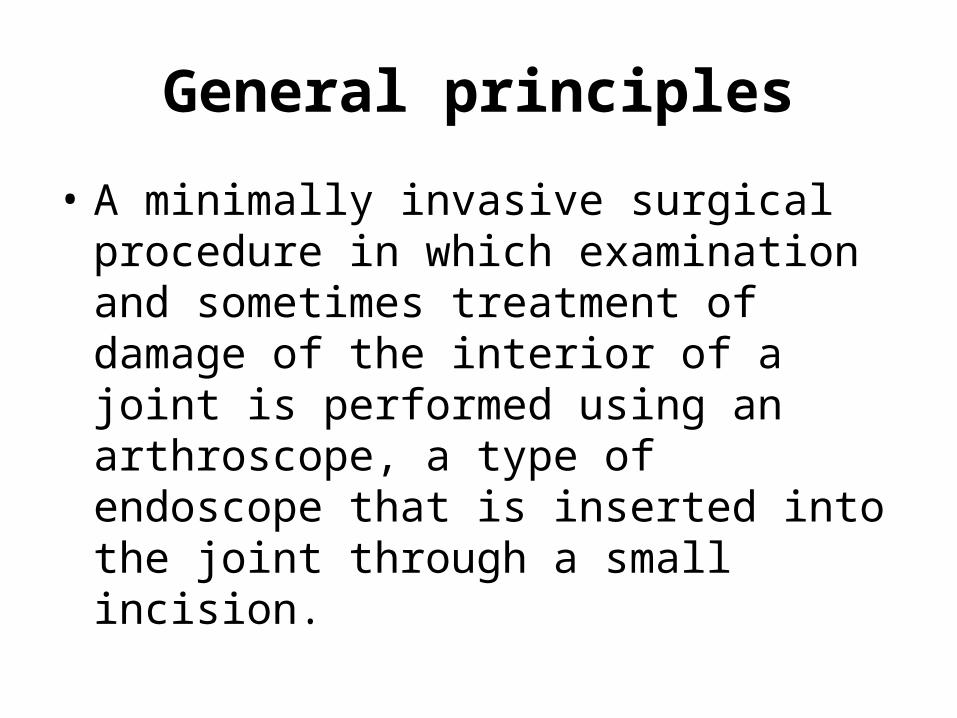
• A minimally invasive surgical procedure in which examination and sometimes treatment of damage of the interior of a joint is performed using an arthroscope, a type of endoscope that is inserted into the joint through a small incision.

General principles
• It is technically possible to perform arthroscopic examination of almost every joint in the human body.
• The joints most commonly examined and treated by arthroscopy are the knee, shoulder, elbow, wrist, ankle, foot and hip.
General principles
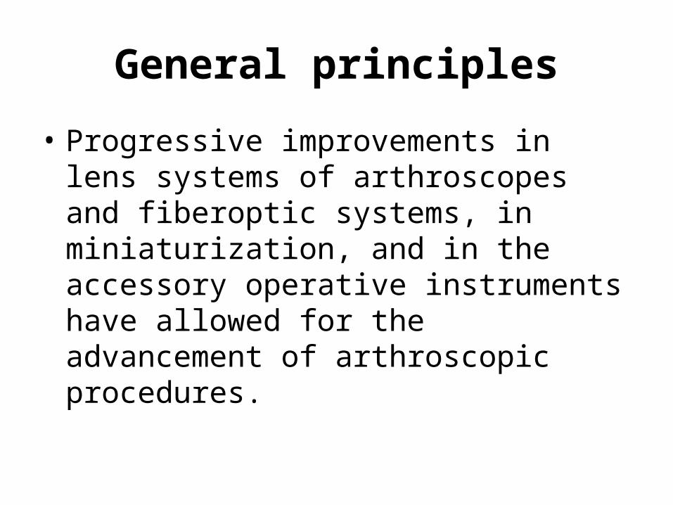
• Progressive improvements in lens systems of arthroscopes and fiberoptic systems, in miniaturization, and in the accessory operative instruments have allowed for the advancement of arthroscopic procedures.

Instruments and equipment
• Arthroscope
• Fiberoptic light sources
• Television cameras
• Accessory instruments

Instruments and equipment: Arthroscope
• Optical instrument
• Three basic optical systems used in rigid arthroscopes: 1) classic thin lens system, 2) rod-lens system, 3) graded index (GRIN) lens system.


Instruments and equipment: Arthroscope
• Optical characteristics:– Diameter– Angle of inclination– Field of view

Arthroscope: Diameter
• Arthroscopes vary in diameter from 1.7 to 7 mm.
• The most common size is 4mm
• Smaller scopes (1.9 and 2.7mm) are useful for smaller and tighter joints (wrist, ankle)

Arthroscope: Angle of inclination
• The angle between the axis of the arthroscope and a line perpendicular to the surface of the lens.
• Varies from 0-120 degrees
• Most commonly use 25 and 30 degree scopes
• 70 and 90 degree scopes are useful in seeing around corners

Angle of inclination

Arthroscope: field of view
• Field of view refers to the viewing angle encompassed by the lens and varies according to the type of arthroscope
• Wide viewing angles make orientation much easier
• Rotation of the forward oblique viewing arthroscopes (25 and 30 degree) allows a much larger area of the joint to be observed

Instruments and equipment: Fiberoptic light sources
• A fiberoptic cable consists of a bundle of specially prepared glass fibers encased in a protective sheath
• One end of the cable is attached to a light source far from the operative field
• The other end is attached to the arthroscope which is surrounded by fiberoptic fibrils
Instruments and equipment: television cameras
• Small cameras have been developed that are connected directly to the arthroscope
• Have the possibility for recording

Instruments and equipment: accessory instruments
• Basic kit:– Arthroscopes, 30 and 70 degree– Probe– Scissors– Basket forceps– Grasping forceps– Arthroscopic knives– Motorized meniscus cutter and shaver– Electrosurgical and laser instruments– Miscellaneous equipment

Care and sterilization of instruments: scopes and cables
• Fiberoptics do not tolerate steam autoclaving• The best method is sterilization using gas
(ethylene oxide) but this takes hours• Another method is by a low-temperature
sterilization process (Steris) and is done in 30 minutes
• Both are sporicidal and bactericidal• Use of cold disinfection (nonsporicidal) in
activated glutaraldehyde between successive procedures (10 minutes)
Care and sterilization of instruments: other instruments
• Knives, graspers, basket forceps and cannulas should be sterilized by steam autoclaving after each procedure
Irrigation systems
• Irrigation and distension of the joint are necessary in all arthroscopic procedures
• Use of normal saline or lactated Ringer solution
• Continuous irrigation keeps fluid clear for optimal viewing as well as maintains hydrostatic pressure and distension within the joint
Tourniquet
• Tourniquet is applied and inflated as needed during arthroscopic procedures of the knee, ankle, elbow and other distal joints
• For minimization of intraarticular bleeding• Many now forego this• Contraindicated in patients with previous
thrombophlebitis or peripheral vascular disease

Leg holders
• Permits application of stress primarily to open the posteromedial compartment for better viewing, manipulation of the meniscus and posterior horn meniscal surgery, especially in tight knees

Anesthesia
• Diagnostic arthroscopy can be performed with the patient under local, regional (spinal or epidural) or general anesthesia
• Some intraarticular operative procedures can be performed using regional and local anesthetics

Advantages of arthroscopy
• Reduced postoperative morbidity, fast return to work• Smaller incisions• Less intense inflammatory response• Improved thoroughness of diagnosis• Absence of secondary effects• Reduced hospital cost• Reduced complication rate• Improved follow-up examination• Possibility of performing surgical procedures that are
difficult or impossible to perform through open arthrotomy (ex:meniscus trimming)

Disadvantages of arthroscopy
• Very few• Requires working through small portals with
delicate instruments: not every surgeon has the temperament for this!
• Tight confines of intraarticular space make maneuvering difficult
• Time-consuming when surgeon is not experienced
• Specialized equipment is extensive and expensive

The Knee
• The knee is the joint in which arthroscopy has its greatest diagnostic and intraarticular surgical application
• Has allowed for the evaluation of the accuracy of clinical examination, laboratory tests, x-rays, MRI and other diagnostic tools
• It is a diagnostic aid used in conjunction with a good history, complete physical examination and appropriate roentgenograms
The Knee
• Can use arthroscopy as a purely diagnostic procedure
• Can use arthroscopy as the essential initial step before proceeding to operative arthroscopy
• Can use arthroscopy before an open arthrotomy
The Knee: keys to success
• Adequate light
• Adequate distension of the joint
• Precise localization of the portals of entry of the arthroscope and accessory instruments

The Knee: standard portals
• Anterolateral, anteromedial, posteromedial and superolateral

The Knee: arthroscopic examination
• Systematic approach– Suprapatellar pouch and patellofemoral joint– Medial gutter– Medial compartment– Intercondylar notch– Posteromedial compartment– Lateral compartment– Lateral gutter and posterolateral compartment

The Knee: diagnostic examination
• What can we see with the scope?– Medial meniscus– Lateral meniscus– Anterior and posterior cruciate ligaments– Condition of cartilage
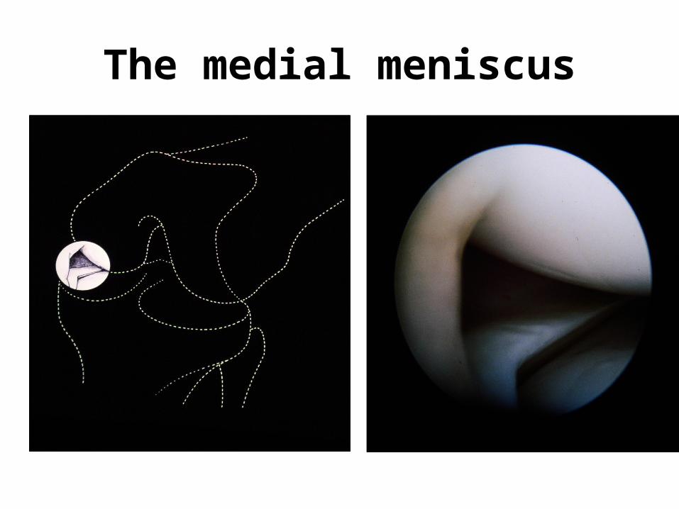
The medial meniscus
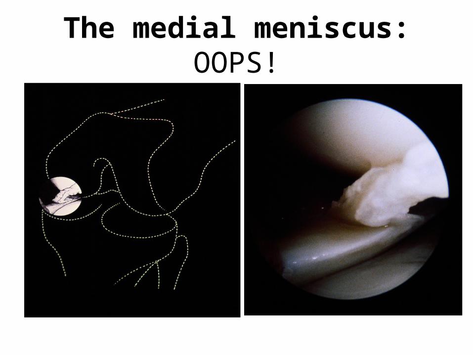
The medial meniscus: OOPS!
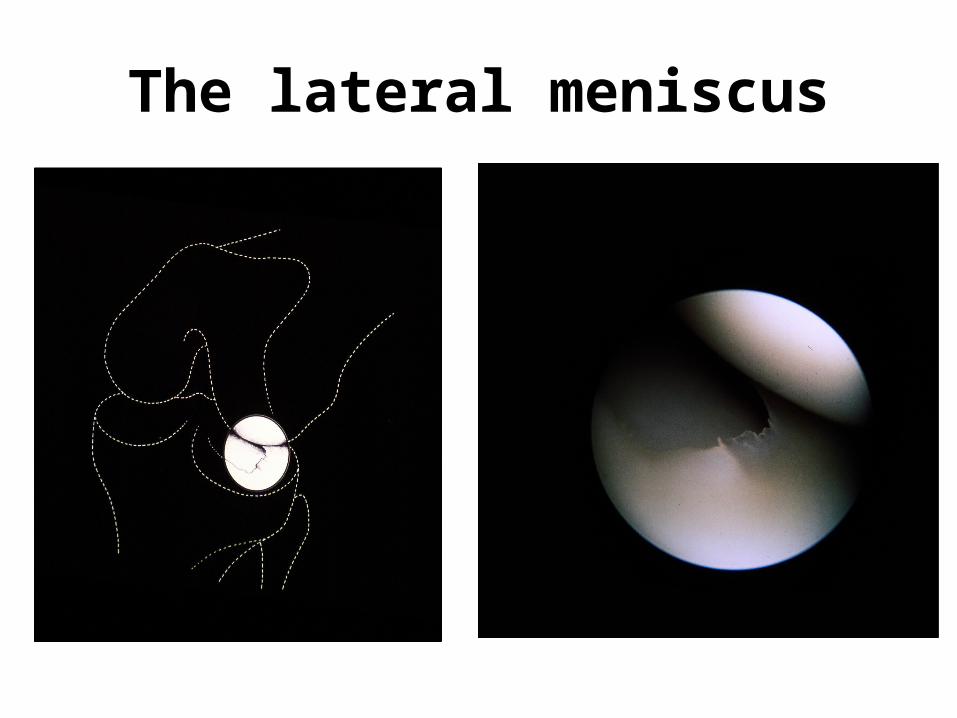
The lateral meniscus
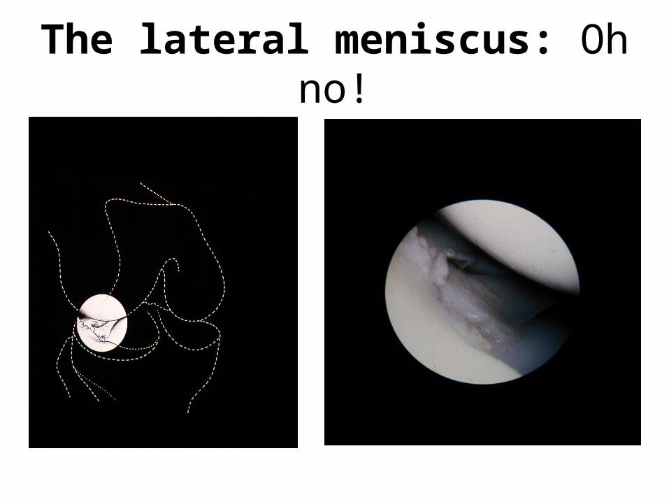
The lateral meniscus: Oh no!

Lesions of the ACL: fresh

Lesion of the ACL: not so fresh

Condition of the cartilage: patellofemoral arthrosis

Condition of the cartilage: osteochondritis dissecans

Arthroscopy: operative treatment
• Not only can arthroscopy be used for diagnosis of certain conditions in joints, it can also be a method of operative treatment.

Operative treatment: the knee
• Repair of meniscal tears– Excision– Suture
Operative treatment: other disorders
• Loose bodies in the knee joint• Synovial plicae of knee• Osteochondritis dissecans (drilling or
screw fixation• Bone grafting• Cruciate ligament reconstruction• Synovectomy• Debridement of osteoarthrosis

The Ankle
• Arthroscopy allows for the inspection and palpation of the articular surface of the joint, stress examination of ligamentous structures and performance of operative procedures– Less postoperative morbidity– Cosmetically pleasing– Earlier rehabilitation and return to function

The Ankle
• Arthroscopic examination
• Soft tissue procedures
• Bony procedures

The Ankle: soft tissue procedures
• Synovectomy
• Debridement for septic arthritis
• Reconstruction for ligamentous ankle injuries

The Ankle: bony procedures
• Drilling, excision, pinning or osteochondral transfer for osteochondritis dissecans
• Removal of osteophytes for anterior impingement syndrome
• Ankle arthrodesis

The Hip
• Difficult to treat with arthroscopy because of sphericity of the head and the inaccessibility of certain areas
• Has advanced over the past decade

The Hip
• Most common indications:– Labral symptoms– Buckling– Locking– Falling episodes– Persistent inguinal pain unresponsive to
conservative treatment
The Shoulder
• Diagnostic arthroscopy:– Identify posterior loose bodies– Evaluate primary rotator cuff impingement– Determine quality of rotator cuff– Debridement of calcific tendinitis lesions or
septic arthritis– Manipulation of frozen shoulder

The Shoulder
• Surgical procedures:– Removal of loose bodies– Synovectomy– Drainage and debridement– Repair of labral tears– Repair of biceps tendon lesions– Reconstruction for anterior instability

The Shoulder: Villous synovitis in labrum tear- refixation was not
technically possible

Sooooo….
• As you can see, arthroscopy can be used for a variety of procedures in a variety of joints
• Arthroscopy is a modern alternative to open surgery and will probably advance in use as technology advances as well

Thank you for your attention!!!