The Right Track - Exactech, Inc. · one-half year results on 1,526 arthroplasties he performed...
13
It’s not just a road we’re on, it’s a trail we’re blazing. The Right Track
Transcript of The Right Track - Exactech, Inc. · one-half year results on 1,526 arthroplasties he performed...

It’s not just a road we’re on, it’s a trail we’re blazing.
The Right Track

Evolution, Not Revolution 2 Congruency 4 Patella-Femoral Articulation 6 Tibial-Polyethylene Stability 8 Net Compression Molded Polyethylene 10 PCL Preservation 12 Non-Modular Constrained 14 Revision Precision 16 Instrumentation 18 A Great Day in the O.R. 20
Optetrak Design team in cooperation with Hospital
for Special Surgery, New York
Albert Burstein, PhD Donald Bartel, PhD Ivan Gradisar, MD
Gary Miller, PhDWilliam Murray, MD
William Petty, MD
A comprehensive knee system that addresses your concerns for contact stress, patellar tracking, polyethylene wear, joint stability and bone preservation. Streamlined instrumentation lets you work quickly and efficiently.
All from a company that’s committed to improving the quality of life for individuals by maintaining their activity and independence.
This is the Optetrak® story.
An approach to total knee arthroplasty that has improved clinical outcomes for patients around the world.

The design behind the Optetrak knee
system has been evolving for more than a
quarter of a century. Its lineage began with
a concept developed at Hospital for Special
Surgery in New York. Successive designs,
guided by clinical and laboratory data,
demonstrated 91-99 percent long-term implant survival rates.1-8
Exactech‘s team of surgeons and bio-engineers built on this solid
foundation, progressively improving implants and instruments. Today,
surgeons are documenting a continuing record of excellent Optetrak
clinical results.9,10,11
You say you want a revolution? Sure, there are many
choices out there, but there‘s nothing like the
confidence that comes from five generations of
proven design. With evolution, not revolution,
you can count on the test of time.
Optetrak Design DevelOpment timeline
DuOCONDyLAR, 1971 DuOPATELLAR, 1974 INSALL/BuRSTEIN POSTERIOR STABILIzED
(I/B PS), 1978
INSALL/BuRSTEIN PS II (I/B II), 1988
OPTETRAk, 1994TOTAL CONDyLAR (TC), 1974
Evolution, Not Revolution.
2
How far can your knee system trace its roots?
3
Optetrak CliniCal results Surgeons around the world have documented excellent results with the Optetrak knee system. Dr. Ivan Gradisar reported excellent eight and one-half year results on 1,526 arthroplasties he performed using Optetrak. Independent, non-surgical investigators evaluated patients at two and five years after surgery using standardized testing tools. Hospital for Special Surgery scores improved from 88 at two years to 91 at five years (Figure 1). Ninety-four percent of the patients reported that the results of their surgery could be best described as excellent, very good or good.10
The rate of re-operation for any reason was extremely low (1.4 percent). No re-operations were required for either design-related problems, component fault or failure, patellar or tibial-femoral instability, for insufficient motion or for repair or release of collateral or posterior soft tissue (Figure 2).
Dr. Raymond Robinson also reported excellent results after a five-year study of his patients. Despite reduced ratings due to obesity (55 percent of the patients had body mass index greater than 30), 90 percent of the patients were rated good or excellent on both Knee Society and HSS scores. Using aseptic revision of any component as an end point, 99 percent of the implants were predicted to survive at 93 months.11
Optetrak builds on a strong lineage of proven designs. The current generation was introduced to orthopaedic surgeons in 1994. Building on the original technology licensed from Hospital for Special Surgery, Exactech has enhanced the system with unique improvements while preserving the proven aspects preceeding designs. In addition, a complete system including the Posterior Stabilized, Cruciate Retaining and Constrained Condylar knee was developed with integrated instrumentation.
Origins Of the Optetrak knee systemThe Optetrak design team recognized that surgeons are often reluctant to experiment with “new” knee designs that do not have long-term clinical results. For this reason, under the close direction of Albert Burstein, PhD, the Optetrak design team, in cooperation with engineers at Hospital for Special Surgery and an extensive team of clinical evaluators, developed a knee design based closely on the clinically successful Total Condylar, Insall/Burstein (I/B) and Insall/Burstein II® (I/B II) knees.
94%
1,526 cases, 5-year follow-up
■ Excellent, Very Good,Good
■ Fair■ Poor
94%
4% 2%
Figure 1. Optetrak Patient Satisfaction10
5-year follow-up
Ivan A. Gradisar, MD
Raymond P. Robinson, MD
8.5-year follow-up
99%
1%
Figure 2. Primary Total Knee Arthroplasties
■ Re-operation Rate
1.4%
98.6%
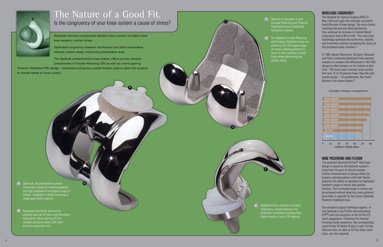
Optetrak’s femoral components address a key concern of today’s total
knee surgeon: contact stress.
Optimized congruency between the femoral and tibial components
reduces contact stress, improving polyethylene wear.
The Optetrak comprehensive knee system offers primary femoral
components in Cruciate Retaining (CR) as well as a bone-sparing
Posterior Stabilized (PS) design. Cemented and porous coated fixation options allow the surgeon
to choose based on bone quality.
Is the congruency of your knee system a cause of stress?The Nature of a Good Fit.
A
C
BSpherical, all-polyethylene patellar component allows for interchangeability and high congruence throughout range of motion. Available in either three-peg or single-peg fixation options.
A
Enhanced cam/spine mechanism controls femoral roll-back and minimizes subluxation. Bone-sparing PS box changes proportionately with each femoral component size.
B
Optetrak is available in both Cruciate Retaining and Posterior Stabilized porous-coated and non-porous options.
C
The Optetrak Cruciate Retaining and Posterior Stabilized knees can achieve a full 125-degree range of motion, allowing patients to return to their activities of daily living while maintaining low contact stress.
D
54
BOne preserving high flexiOnThe patented Optetrak Hi-Flex® total knee design is based on the Optetrak system’s more than 10 years of clinical success. Further enhancements in design allow the properly selected patient with high flexion potential the ability to capitalize on Optetrak’s excellent range of motion and patellar function. This increased range of motion can be achieved without resecting more posterior bone than is required for the classic Optetrak Posterior Stabilized knee.
The standard surgical technique applies, or use Optetrak‘s Low Profile Instrumentation (LPI®) with the exception of the Hi-Flex PS notch preparation. Following the Femoral Finishing Guide resections, the corresponding sized Hi-Flex PS Notch Guide is used. Hi-Flex femoral trials, as well as Hi-Flex tibial insert trials, are also required.
inCreasing COngruenCyThe Hospital for Special Surgery (HSS) in New York built upon the clinically successful Insall/Burstein II knee design. By more closely matching femoral and tibial geometries, they achieved an increase in medial/lateral congruency from 0.94 to 0.96. This new knee technology optimized the conformity, stability and kinematics without increasing the stress at the prosthesis-bone interface.12
In 1995, Bartel, Rawlinson, Burstein, Ranawat and Flynn conducted additional finite element analysis to compare the differences in the HSS design to other designs on the market at that time. The result was a contact stress profile that was 15 to 20 percent lower than the next lowest design – it’s predecessor, the Insall/Burstein II as shown below.13
Optetrak Hi-Flex maintains excellent congruency, contact pressure and dislocation resistance including high flexion angles of up to 145 degrees.
E
E
D
Contact stress comparison
0CONTACT STRESS (MPa)
10 20 30 40 50 60
Knee System B
Knee System A
Knee System C
Knee System D
Knee System E
Knee System F
I/B PSI I ®
Optetrak®

From the earliest designs, surgeons have faced challenges with patello-femoral
articulation. The Optetrak team of surgeons and bio-engineers have addressed
this concern by improving on the clinically successful Insall/Burstein II. The
result: a knee system that reduces dislocation, subluxation, tilt and patellar clunk.
Optetrak’s contoured femoral flange, the smooth shape in the sagittal plane and
a deep femoral groove are design features that reduce strain in retinaculum, allowing for more natural
patellar tracking from extension to flexion.
Get in the groove. Optetrak has proven successful in significantly reducing lateral retinacular release
rates and the incidence of peripatellar fibrosis.9,14 With a strong lineage, streamlined instrumentation and a
proven femoral design, Optetrak keeps your patella on the right track.
Does the patella in your knee system follow the path of least resistance?
Let Nature Take Its Course.
6 7
Thomas P. Sculco, MD
Raymond P. Robinson, MD
0%
80%
0%
50%
Optetrak Decreases Lateral Release Rates 9,14
Two surgeons, > 2,000 cases
■ I/B II ■ Optetrak
57% decrease 77% decrease
DeCreasing lateral retinaCular releaseFrom the earliest knee designs, surgeons have faced challenges with patello-femoral articulation. Optetrak has proven successful in significantly reducing lateral retinacular release rates and the incidence of peripatellar fibrosis.
Many factors have been implicated as causes of patello-femoral complications after total knee arthroplasty. One of these factors is seen in female patients or in patients with valgus deformities, whose muscular forces and other soft tissues tend to pull the patella more laterally. Optetrak’s wide femoral groove (A) and patented patellar design allow the patient’s patella to track naturally (either medially or laterally) during flexion and extension. This design feature has also proven successful in significantly reducing lateral retinacular release rates and the incidence of peripatellar fibrosis.15
Unlike newer knee systems, Optetrak offers the confidence you can only get from excellent, long-standing clinical results.
A
B
More than three decades ago when the design team first put ideas on paper, they took into account the obvious anatomical differences between narrow, smaller, typically female knees and wider, larger knee dimensions typically seen in men. This is a fact that some other knee system designs are just now addressing.
Some implant systems are based simply on an average size of women‘s and men‘s knees combined. Optetrak‘s design team optimized the aspect ratio of its femoral components so every component in every size covers diverse distal femoral morphologies, regardless of gender, without overhang and subsequent soft tissue irritation or alterations in the knee biomechanics.
A
Bthe "genDer" issue
A
B
C
Retinacular tissue stretch and patellar component distances16 produced by other implant designs
(shown in red) compared to the Optetrak (green).
Optetrak
A Wide patellar groove (superior region only) provides less constraint to allow for excellent patellar tracking.
Debulked anterior femoral flange reduces tension in lateral retinaculum and incidence of lateral release.
B

Backside wear threatens the function and longevity of total knee replacements.
Optetrak’s tibial components target backside wear—minimizing polyethylene
debris and the risk of component disassociation.
Optetrak’s modular tibial components feature a rock-solid locking mechanism
with three design elements that keep tibial inserts in place. A continuous
peripheral rim around the tray, posterior feet that couple with precision undercuts in the tray and a central
mushroom provide a barrier to insert motion and prevent lift-off.
Optetrak also offers all-polyethylene and molded, metal-
backed tibial components—the ultimate in tibial
polyethylene stability.
How solid is your knee’s tibial insert?
maintaining COntaCt Plenty of knee systems provide tibial up- and down-sizing, but at a cost: increased contact stress. Optetrak tackled this problem where the stress occurs between the femoral component and the tibial insert.
Optetrak maintains its excellent congruency regardless of tibial sizing. The femoral component and the polyethylene insert are a matched pair, with three interchangeable tibial trays to choose from for each femoral size.
Locked in Tight.
Modular tibial components are available in cemented fin, porous fin or cemented trapezoidal options.
A
Patented press-in caps provide for polyethylene support to prevent cold flow, as well as debris migration.
B
Undercut cement pockets on the cemented tibial trays allow for a mechanical interlock to provide for excellent stability of the components.
D
All-polyethylene tibial components maximize poly thickness and are available in either Cruciate Retaining or Posterior Stabilized.
E
Molded, metal-backed tibial components feature a cemented finned stem and are Posterior Stabilized.
F
A
B
C
D
F
E
C The modular tibial components feature a three-part locking mechanism, which prevents tibial insert motion and disassociation.
Retrieved Optetrak insert demonstrates minimal wear with no measurable material loss.
Tibial Up- and Down-Sizing
3F/2T
3F/3T
3F/4T
8 9
Optetrak‘s sOliD resultsRecent studies documenting the backside wear of polyethylene inserts call into question the stability of locking mechanisms in some modu-lar tibial components.17,18 In contrast, indica-tors on Optetrak inserts substantiate its locking mechanism’s ability to reduce backside wear.19

Two main drivers affect polyethylene performance in total knee replacement: design and
materials. With its strong lineage, Optetrak’s polyethylene tibial inserts benefit from
optimized congruency and low contact stress.
Improvements in materials further enhance Optetrak’s excellent performance. Its tibial inserts
feature net compression molded polyethylene—no machining is performed on the articulating
surface. The result? Less wear debris and less pitting than machined tibial inserts.20
The Optetrak net compression molded tibial inserts demonstrated an 83 percent reduction in wear rate (top) and 52 percent less damaged area (above) than I/B II machined, extruded tibial inserts.20
Net Molded, Not Machined.Is the polyethylene in your knee system wearing on you?
Damage Area
Machined IB/II
Molded Exactech Optetrak
The articular surface of the net molded tibial insert is never machined. The result is a smooth finish, free of machine lines.
A
Precise machining of non-articular surfaces establishes the final thickness and completes the fine details of the locking mechanism to ensure an exacting fit with modular tibial trays.
B
The mold used in the net compression molding process forms the surface of the corresponding tibial insert. Each insert is individually produced to ensure the complete and consistent consolidation of resin.
C
A
Pressure applied
Resin
Heating
Cooling
ProducinG net comPression molded Polyethylene
This process is a variation on the traditional molding scheme in
which a small mold cavity representing the exact complex shape of
the part is created. A precisely calibrated amount of resin is placed
in a mold that is heated and cooled in a computer-controlled press.
This yields the exact shape of the finished tibial insert’s articular
surface with exceptional uniformity of material properties.
B
10 11
a material DifferenCe in perfOrmanCeIn a knee simulation study comparing wear rates of net compression molded polyethylene to machined, sheet-molded polyethylene, the Optetrak net compression molded inserts demonstrated volumetric wear of 1.46 mg/MC. That corresponds to an 83 percent reduction in wear rate and 52 percent less damaged area than I/B II extruded tibial inserts.20 That’s approximately six times less wear than the traditional I/B II design. This is achieved without sacrificing critical mechanical properties such as fracture toughness.
Through the careful blending of design and materials, Exactech’s Optetrak total knee system continues to advance the longevity of knee arthroplasty.
Average Wear Rates
01020304050607080
0 1 2 3 4 5 6
● Molded Exactech Optetrak■
Cycles (millions)
Ave
rage
Wea
r (m
g)
(1.46mg/MC volumetric wear)
Machined I/B II
83% Lower Wear than I/B II
Balanced crosslinkinG
Optetrak’s net compression molded polyethylene is sterilized with
gamma radiation (2.5-4.0 Mrad) in a vacuum. While the molecular
chains of net molded polyethylene are moderately crosslinked due
to the irradiation process in the absence of oxygen molecules, this
material retains all of its mechanical properties (yield strength,
fatigue strength and fracture resistance), avoiding the generation of
free radicals. This balances the equation between wear, mechanical
properties and oxidation.21
C

12 13
More than 75 percent of tibial resections lead to at least partial compromise of the posterior
cruciate ligament (PCL) in the hands of experienced orthopaedic surgeons.22 To truly retain
the function of the PCL, one must pay meticulous attention to preserving its anatomy and
avoiding releasing PCL fibers – either directly by surgical releases, or indirectly by increasing
the slope of the tibial resection.
Exactech is pleased to offer an innovative approach to total knee arthroplasty designed
to enhance the precision of the tibial resection and to preserve the integrity of the PCL. The Optetrak CR Slope®
patent-pending design enables surgeons to plan and perform PCL-retaining total knee arthroplasty based on the
anatomical integrity of the posterior cruciate ligament. User-friendly instrumentation and three different sloped inserts
accommodate balancing the flexion and extension gaps.
Preserving your Environment.Do you ever release the PCL or re-cut more bone to balance tight flexion gaps?
oPtetrak cr sloPe is desiGned to:23
• Identifyandprotecttheanatomicalintegrityof the PCL
• Providepreciseandreproducibleboneresections
• OptimizethePCLtensioninapredictablemanner
• Allowintra-operativetensioningadjustmentof the PCL without releasing the PCL, cutting additional tibia slope or downsizing the femur
• Balanceflexion/extensiongapsindependently
• Accommodatevariabilityinpatients’anatomies
• Restorekneejointstabilitythroughouttherange of motion
Design ratiOnaleThe Optetrak CR Slope design team conducted a meticulous MRI study with two goals in mind: (1) to consistently identify the origins of the posterior cruciate ligament (PCL) in both the femur and the tibia and (2) to define the resulting “joint space” depending of the posterior slope of the proximal tibial cut.24,25 The study revealed that a reference point at the base of the ligament’s tibial attachment points could be consistently identified and measured over a variety of knee sizes and geometries. Additionally, the vertical distance between the PCL femoral insertion and the planned proximal tibial cuts was measured. The data revealed that if the surgeon performed the proximal tibial cut according to the natural posterior slope , the resultant tibial cut could be too thin (less than 9mm) not leaving sufficient joint space for the implant components. If the surgeon needed to increase the slope of the proximal tibial resection to open up the flexion space, the PCL integrity was most often compromised.
The results of the study led to the development of unique instrumentation and optimized tibial inserts that comprise the Optetrak CR Slope system. The Posterior Cruciate Referencing Technique was developed, to allow the surgeon to identify and reference the PCL by consistently protecting it intra-operatively. Using the PCL as the reference point, a traditional tibial cut is made according to a neutral slope . Trials and inserts with increased posterior angulation (CR Slope + and CR Slope ++) were added to the standard CR in order to reproduce the natural slope of the tibia. These inserts allow for balance of the flexion gap, reducing the need for soft tissue releases, partial PCL releases or additional tibial bone cuts.26-30
nOteUse CR Slope inserts only with Optetrak CR Symmetric or Asymmetric femoral components. CR Slope inserts are compatible with Optetrak finned or trapezoid tibial components.
Three insert options allow surgeons the ability to balance flexion and extension gaps, eliminating the need to release the PCL fibers, cut additional tibial slope or downsize the femur.
D
Direct compression molded polyethylene minimizes wear and polyethylene debris.20
A
Debulked anterior femoral flange reduces tension in lateral retinaculum and incidence of lateral release.16
C
B Wide patellar groove (superior region only) is designed to provide less constraint to allow for excellent patellar tracking.
A
C
STD Slope
Slope + Slope ++
D
B
oPerative technique
Femoral preparation is performed using the general Optetrak
CR operative technique. The tibia is prepared by referencing
the tibial insertion of the PCL rather than the tibial plateau. The
AdjustablePCLStylus,aNo-TouchPCLRetractorandinserttrials
(CR Standard, CR Slope + and CR Slope ++) are used in addition
to Optetrak Classic or Low Profile Instrumentation.Vertical distance between the femoral origin of the PCL and a planned natural slope tibial cut
Vertical distance between the femoral origin of the PCL and a planned neutral slope tibial cut
A
B
AB
pCL insersion
point
pCL insersion
point
A
B

Optetrak's Non-Modular Constrained knee is designed to deliver the stability of a constrained condylar without the bone sacrifice required for femoral stem placement.
What do you do when you need a little more stability than a posterior stabilized knee,
but good bone quality doesn’t demand stems or augments? The answer is typically a
constrained condylar prosthesis with its added femoral resection and preparation of
the intra-medullary canal. That is, unless you choose Optetrak.
The Optetrak knee system’s Non-Modular Constrained (NMC)
prosthesis is a solution that uncouples ligament stability from bone issues. Its unique
“box” design provides the same varus/valgus constraint as a constrained condylar and
greater resistance to subluxation than a posterior stabilized knee, all in a bone-sparing
technique. In fact, patients with severe valgus deformity have demonstrated Knee Society
score improvement from 46 to 86 points post-operatively with the Optetrak NMC.31
That’s constraint without compromise.
Are you sacrificing femoral bone to address ligament instability?
Stability Without Sacrifice.
NMC femoral components, in conjunc-tion with Constrained Condylar inserts, restore stability to the joint without the need for stems or augments on the femoral side.
A
In addition to the constraint that the NMC offers, increased jumping height is allowed as compared to a posterior stabilized total knee.
B
Tibial trays are modular and require that a stem extension be used with the Constrained Condylar tibial insert.
C
F
D
E
C
B
Spherical, all-polyethylene patellar component allows for interchangeability and high congruence throughout range of motion. Available in either three-peg or single-peg fixation options.
D
Debulked anterior femoral flange reduces tension in lateral retinaculum and incidence of lateral release.
F
E Wide patellar groove (superior region only) provides less constraint to allow for excellent patellar tracking.
A
14 15
experienCe exaCteCh‘s COre COmpetenCiesThe NMC features the same outstanding con-gruency, range of motion, patellar tracking and polyethylene wear as the Cruciate Retaining and Posterior Stabilized systems.
In addition, highly controlled tolerances between the box and Constrained Condylar spine pro-vide for ± 1.5 degrees of varus/valgus and ± 2 degrees of rotational constraint. This constraint has proven to be bio-mechanically and clinically effective for soft tissue deficiencies.

COnstraintThe constraint of the Optetrak Constrained Condylar knee design is ± 2 degrees in rotation and ± 1.5 degrees in varus/valgus. This constraint has proven to be bio-mechanically and clinically effective for soft tissue deficiencies.
The Optetrak Constrained Condylar knee system’s Joint Line Referencing Guide takes the guesswork out of maintaining the joint line after the primary implant is removed.
A
When a constrained tibial insert is used, a spine stiffener screw enhances the integrity of the spine and stabilizes the insert, tray and stem extension.
C
Tibial augments are available in 5, 8 and 11mm thicknesses. One-half as well as bone-sparing one-third options can replace tibial deficiencies.
E
Stem extensions are common to both the femoral and tibial components and can be cemented or press-fit. Multiple stem diameters and lengths offer solutions to the most difficult revision surgeries.
B
The CC femoral component offers a 2, 5, or 7 degree valgus angle to accommodate a wide range of patients.
D
Independent distal and posterior augmen-tation blocks provide maximum flexibility for reconstructing deficient femoral bone.
F
D
F
E
C
B
B
A
There is nothing routine about revision knee surgery. It is an art. You never know
what you’ll finduntilyougetthere.Restoringthejointline,augmentinglostbone
and providing stabilizing constraint involve guesswork, estimation and frustration.
The Optetrak Constrained Condylar (CC) knee system addresses the art of revision with the science of
precision.Itsinstrumentationallowsthesurgeontoreproducethejointline.
If augmentation is required, a wide range of femoral and tibial solutions are available. The CC’s unique
“box” design provides stability with excellent varus/valgus constraint. Restoration, flexibility, stability and
simplicity—that’s Optetrak‘s formula for revision precision.
Is your technique for revision knee surgery based on precision or approximation?
Revision Precision.
prOximal tiBial COverageOffset Tibial Trays allow optimal coverage of the tibial plateau while maintaining the integrity of a fixed tibial tray. The proximal offset design offers advantages over offset stems, lessening the chance of cortical impingement.
An anatomic study was conducted in conjunction with Hospital for Special Surgery to gain a thorough understanding of the position of the tibial intra-medullary canal with respect to the center of the tibial plateau.32 It was determined that the optimal offset was medial/lateral and increased with increasing tibial size. The Optetrak Offset Tibial Tray incorporates this data into its design. Along with the standard tibial options, the offset tibial trays provide seven tibial options per femur.
16 17
BeyOnD a shaDOW Of a DOuBtOptetrak‘s Joint Line Referencing Guide takes the guesswork out of maintaining the joint line after the primary implant is removed. The surgeon can reproduce–without elevating–the joint line or adjust it if necessary.
The CC features the same outstanding congruency, range of motion, patellar tracking and polyethylene wear as the cruciate retaining and posterior stabilized systems.

Master the Balancing Act.Do your instruments support your technique?
Revision Joint Line Reference and Distal Femoral Cutting GuideThe Joint Line Referencing Guide allows the surgeon to duplicate, or adjust if needed, the joint line of primary knee implants. Augmentation can be accommodated via captured cutting slots if bone loss is present.
Tibial InstrumentationSurgeons have varying instrument preferences for preparing the tibia. From extra-medullary to intra-medullary, with posterior slope or without, fixed or adjustable, Optetrak has the special instruments you need to achieve reproducible and accurate results.
HSS InstrumentationIf the surgeon prefers to start with an anterior rough cut, Optetrak offers a special set of femoral instruments designed in conjunction with Hospital for Special Surgery. The HSS instruments have evolved as a continuation of the previous knee systems developed at HSS with improvements and increased reliability.
A
18 19
Low Profile Instrumentation
Optetrak’s Low Profile Instrumentation (LPI®) supports Exactech’s philosophy on total joint replacement: to improve patient outcomes. These instruments give you flexibility to adjust your incision and manage soft tissue to meet the needs of each individual patient, while using your own proven surgical technique.
B
A Ligament Balancing Options
Plenty of surgeons tackle ligament balancing on their own, using anatomic landmarks or assuming an average 3 degrees of external rotation. With the patented Optetrak Ligament Balancing System, the patient‘s soft tissue determines the ideal external rotation of the femoral component, taking ligament balancing from a guess to a science.
Balancing your needs for flexibility and accuracy. Leveraging
exact results with efficient use of O.R. time. These are the
Optetrak instrument systems.
A single instrument set serves both cruciate retaining and
posterior stabilized knees. The same streamlined
approach supports revisions, where minimal additional trays are
required. From the Mauldin Multi-tool which can be used for nearly
adozendifferentfunctions,toligamentbalancing,tojointline
referencing, our instrument trays are full of sleek solutions.
Easy to use and multifunctional. Durable and accurate
for reproducible results. Optetrak‘s instrument systems
strike the balance between art and science.
instrumentatiOn OptiOns
B

A Great Day in the O.R.®The commitment that defines the Exactech brand.
Founded by an orthopaedic surgeon and bio-medical engineer,
Exactech is committed to making every day a great day in the
O.R.—for the surgeon, the O.R. staff and above all, for the patient.
Alongwithinnovativeimplantsandinstrumentationfortotaljoint
replacement, Exactech provides pre-primary, bone cement and biological solutions to meet
your needs throughout the entire case.
Experience Exactech. A Great Day in the O.R.®Bone Cement
Cemex® bone cement features a unique low monomer formula that has been clinically proven in Europe for more than two decades.34 Available in a self-contained delivery system or hand mix options, the Cemex family of products are designed to offer surgeons and operating room personnel simplicity, safety and reliability in bone cement.
D
20
Pre-Primary Total Joint Solutions
The Optetrak unicondylar knee system provides for bone preservation through proper ligament tensioning. Its biomechanically-inspired articular geometry and exclusive net-molded polyethylene facilitate restoration of knee function. The OsteoTrac® High Tibial Osteotomy (HTO) Plating System provides adjustable length through a patented design. It allows the surgeon to affix the HTO plate to the osteotomy site before creating/opening the wedge.
A
Biologic Solutions
Exactech is shaping the future of bone repair. Its full scope of biologic materials features demineralized bone matrix in a thermoplastic carrier, with or without cortical cancellous bone chips. For a bone graft that doesn’t wash away, is 100 percent tested and terminally sterilized, Exactech Biologics are the natural choice.
B
Accelerate® Platelet Concentrating System
Platelet rich plasma (PRP) gel has global applications in a variety of surgical procedures, including total joint replacement, bone repair and facial cosmetic and reconstruction surgery. Its ability to speed healing and improve patient outcomes has been well documented.33 This easy-to-use Accelerate Concentrating System provides a fast and convenient method for processing PRP in the operating room from a small amount of patient’s blood.
C
InterSpace® Knee Spacer
InterSpace® knee is a pre-formed, articulating, partial load-bearing structure comprised of Gentamicin-impregnated Cemex PMMA bone cement. It maintains joint space, allows limited mobility and provides for predictable, consistent antibiotic release locally.
F
Revision Equipment
The AcuDriver® Automated Osteotome System complements Exactech’s total joint product line, offering the surgeon efficient, effective instrumentation for removing the components that need replacement in revision hip and revision knee surgery. The AcuDriver system consists of an air-driven impact hand piece, a wide variety of osteotome attachments and a fiber optic illuminator that enhances visualization in the femoral canal.
E

U.S. patents 5,732,992; 5,688,281; 5,910,143; 6,193,723 B; 5,725,580; 4,928,992; 5,702,458. Additional U.S. and foreign patents pending.
Optetrak is a registered trademark of Exactech. IB/II and I/B PSII are registered trademarks of the Hospital for Special Surgery. Opteform and Optefil are processed by RTI Biologics and distributed in the U.S. by Exactech. Cemex is a registered trademark of Tecres S.p.A., Italy, and distributed in the U.S. by Exactech.
352-377-1140 1-800-EXACTECHwww.exac.com
©20
10 E
xact
ech,
Inc.
• IS
O 13
485
Certi
fied
712-01-21 Rev. D Optetrak Main Brochure 0410
References1. Ranawat CS, Flynn WF Jr, Saddler S, Hansraj KK, Maynard MJ. Long-term results of the total condylar knee arthroplasty: A 15-year survivorship study. Clin Orthop. 1993;286:94-102.
2. Gill GS, Joshi AB, Mills DM. Total condylar knee arthroplasty: 16- to 21-year results. Clin Orthop. 1999;367:210-5.
3. Insall JN, Lachiewicz PF, Burstein AH. The posterior stabilized condylar prosthesis: a modification of the Total Condylar design. Two-to four-year clinical experience. J Bone Joint Surg. 1982;64-A:1317-23.
4. Stern SH, Insall JN. Posterior stabilized prosthesis: results after follow-up of nine to 12 years. J Bone Joint Surg. 1992;74-A(7): 980-6.
5. Aglietti P, Buzzi R, De Felice R, Giron F. The Insall/Burstein total knee replacement in osteoarthritis: a 10-year minimum follow-up. J Arthroplasty. 1999;14(5):560-5.
6. Scuderi GR, Insall JN, Windsor RE, Moran MC. Survivorship of cemented knee replacements. J Bone Joint Surg Br. 1989;71(5):798-803.
7. Vince KG, Insall JN, Kelly MA. The total condylar prosthesis:10- to 12-year results of a cemented knee replacement. J Bone Joint Surg Br. 1989;71(5):793-7.
8. Font-Rodriquez DE, Scuderi GR, Insall JN. Survivorship of cemented total knee arthroplasty. Clin Orthop. 1997;(345): 79-86.
9. Robinson RP. Comparison of clinical results of the third, fourth, and fifth generations of the Hospital for Special Surgery prosthetic knee implant. Presented at the Pennsylvania Orthopaedic Society, Fall 1999. Farmington, PA.
10. Edwards J, Gradisar I Jr, Nadaud M, Kovacik M, Askey M. Eight and one-half year clinical experience with the Optetrak total knee prosthesis. Presented at the American Academy of Orthopaedic Surgeons. February 2004.
11. Robinson RF. Five-year follow-up of primary Optetrak posterior stabilized total knee arthroplasties in osteoarthritis. J Arthroplasty. 2005 Oct;20(7):927-31.
12. Cottrell JM, Townsend E, Lipman J, Sculco TP, Wright TM. Bearing surface design changes affect contact patterns in total knee arthroplasty. Clin Orthop Relat Res. 2007 November; 464:127-31.
13. Bartel D, Rawlinson J, Burstein H, Ranawat C, Flynn W Jr. Stresses in polyethylene components of contemporary total knee reploacements. Clin Orthop. 1995;317:76-82.
14. Sculco TP. The significance of patellar clunk: how loud the sound! Presented at Current Concepts in Joint Replacement, Winter 1999.
15. Engh G. Gender specific knee designs: Solutions for a non-existent problem. Presented at Current Concepts in Joint Replacement, Spring 2007. Session VIII.
16. Petty RW. Caveats in patello-femoral design. Presented at the 10th Annual Meeting, Current Concepts in Joint Replacement, Orlando, FL. 1994.
17. Engh GA, Lounici S, Rao AR, Collier MB. In vivo deterioration of tibial baseplate locking mechanisms in contemporary modular total knee components. J Bone Joint Surg. 2001;83-A:1660-5.
18. Li S, Scuderi G, Furman BD, Bhattacharyya S, Schmieg JJ, Insall JN. Assessment of backside wear from the analysis of 55 retrieved tibial inserts. Clin Orthop. 2002;(404):75-82.
19. Jayabalan P, Furman B, Cottrell J, Wright T. Backside
wear in modern total knee design. HSSJ.2007;3:30-4.
20. Furman BD, Lai S, Li S. A comparison of knee simulator wear rates between directly molded and extruded UHMWPE. Presented at Society for Biomaterials, 2001.
21. Li S, Burstein AH. Ultra-high molecular weight polyethylene: the material and its use in joint implants. J Bone Joint Surg Am. 1994 Jul;76(7):1080-90.
22. Shannon FJ, Cronin JJ, Cleary MS, Eustace SJ, O’Byrne JM. The posterior cruciate ligament-preserving total knee replacement: do we 'preserve' it? J Bone Joint Surg Br. 2007 Jun; 89(6):766-71.
23. Covall DJ, Stulberg BN, Mabrev J, Burstein AH, Angibaud LD, Smith K, Zadzilka JD. Introducing a New Technique for Improving Predictability in Cruciate-retaining Total Knee Arthroplasty: The Posterior Cruciate Referencing Technique. Techniques in Knee Surgery. 2009 Dec; 8(4):271-275.
24. Mariani PP, Margheritini F. The Posterior Cruciate Ligament, pp 11 – 37. Timeo Editore, Bologna 2007. 22. Data on file at Exactech, Inc.
25. Data on file at Exactech, Inc.
26. Fantozzi S, Benedetti MG, Leardini A, Banks SA, Capello A, Assirelli D, Catani F. Fluoroscopic and Gait Analysis of the Functional Performance in Stair Ascent of two Total Knee Replacement Designs. Gait and Posture. 17(2003) 225-234.
27. Catani F, Fantozzi S, Ensini A, Leardini A, Moschella D, Giannini S. Influence of Tibial Component Posterior Slope on In Vivo Knee Kinematics in Fixed-Bearing Total Knee Arthroplasty. J Orthop Res. 2006 Apr;24(4):581-7.
28. Yoshiya S, Matsui N, Komistek R, Dennis D, Mahfouz M, Kurosaka, M. In Vivo Kinematic Comparison of Posterior Cruciate-Retaining and Posterior Stabilized Total Knee Arthroplasties under Passive and Weight-bearing Conditions. J Arthroplasty. Vol. 20 No. 6 2005.
29. Scott RD, Chmell MJ. Balancing the posterior cruciate ligament during cruciate-retaining fixed and mobile-bearing total knee arthroplasty: description of the pull-out lift-off and slide-back tests. J Arthroplasty. 2008 Jun;23(4):605-8.
30. Bellemans J, Robijns F, Duerinckx J, Banks S, Vandennuecker H. The influence of tibial slope on maximal flexion after total knee arthroplasty. Knee Surg Sports Traumatol Arthrosc. (2005) 13: 193–196.
31. Anderson J, Baldini A, MacDonald J, Pellicci P, Sculco T. Primary constrained condylar knee arthroplasty without stem extensions for the valgus knee. Clin Orthop. 2006;442:199-203.
32. Cornell CN, Yarger C, Solan P, Halley DK, Sculco TP. Variations in the relative position of the tibial plateau and the tibial medullary canal: an anatomic survey of patients undergoing total knee arthroplasty. Presented at AAOS 2003.
33. Gardner MJ, Demetrakopoulos D, Klepchick PR, Mooar PA. The efficacy of autologous platelet gel in pain control and blood loss in total knee arthroplasty: an analysis of the haemoglobin, narcotic requirement and range of motion. Int Orthop. 2007 Jun; 31(3):309-13.
34. Nivbrant B, Kärrholm J, Röhrl S, Hassander H, Wesslén B. Bone cement with reduced proportion of monomer in total hip arthroplasty: preclinical evaluation and randomized study of 47 cases with 5 years' follow-up. Acta Orthop Scand. 2001 December;72(6):572-84.