The Resolution of Rayless Goldenrod (Isocoma pluriflora ...
7
27 The Resolution of Rayless Goldenrod (Isocoma pluriflora) Poisoning in Goats Bryan L. Stegelmeier 1* , T. Zane Davis 1 , Benedict T. Green 1 , Stephen T. Lee 1 , and Jeffery O. Hall 2 1 USDA-ARS Poisonous Plant Research Laboratory, Logan, UT 2 Utah Veterinary Diagnostic Laboratory, Logan, UT *Corresponding author: Bryan L. Stegelmeier, [email protected] Abstract Rayless goldenrod (Isocoma pluriflora) occasionally poisons livestock causing myocardial and skeletal muscle degeneration and necrosis. The objectives of this study were to describe the resolution of the clinical and pathological changes of rayless goldenrod poisoning in goats. Eight goats were gavaged for 7 days with ground rayless goldenrod to obtain benzofuran ketone dosages of 40 mg/kg BW/day. After treatment, three goats were euthanized and the other goats were allowed to recover for 10 days and 2, 4, 8, and 16 weeks. After 6 days of treatment, all the treated animals were reluctant to move, stood with an erect stance, and became exercise intolerant. Serum enzymes such as AST, ALT, LDH, and CK had elevated activities indicative of muscle damage. Animals quickly recovered and showed few clinical signs at 2 weeks post treatment. Histologically, poisoned goats developed severe skeletal myodegeneration and necrosis characterized by myocyte swelling and hypereosinophilia, clumping and aggregation of myofibers, and myocyte disruption with extensive perimysial edema and inflammation. This degeneration and necrosis persisted with increased inflammation in the goats that were euthanized at 10 days and 2 weeks post treatment. In animals that were allowed to recover for 4 and 8 weeks, there was progressively less degeneration, necrosis, and inflammation with more edema, regeneration, and fibrosis. After 16 weeks there was edema and mild fibrosis. These findings indicate that rayless goldenrod poisoning causes skeletal muscle necrosis that continues to resolve 3 months after exposure. Though the remaining lesions are minimal, complete resolution could take many additional months. Keywords: goats, Isocoma pluriflora, rayless goldenrod Introduction Rayless goldenrod or jimmyweed [Isocoma pluriflora (Torr. & A. Gray) Greene (Asteraceae) previously Isocoma wrightii (A. Gray) Rydb and Happlopappus heterophyllus (A. Gray) S.F. Blake] is found and sporadically poisons livestock in Arizona, Colorado, New Mexico, and Texas (figure 1). All species are probably susceptible, but poisoning has only been reported in horses, cattle, pigs, sheep, goats, and humans (Burrows and Tyrl 2001). First described as “alkali disease” in the early 1900s, rayless goldenrod poisoning was erroneously associated with drinking saline or alkaline water (Marsh and Roe 1921, Marsh 1926). Clinically, poisoned livestock are depressed, anorexic, and reluctant to move. Some poisoned animals may develop tachypnea and tachycardia with ascites and hydrothorax. Ultimately, most develop violent trembling when forced to move. Both the skeletal muscle and myocardial lesions are due to diffuse and severe muscle degeneration and necrosis with subsequent fibrosis and atrophy (Stegelmeier et al. 2010). Poisoning by
Transcript of The Resolution of Rayless Goldenrod (Isocoma pluriflora ...

27
The Resolution of Rayless Goldenrod (Isocoma pluriflora) Poisoning in Goats Bryan L. Stegelmeier1*, T. Zane Davis1, Benedict T. Green1, Stephen T. Lee1, and Jeffery O. Hall2
1USDA-ARS Poisonous Plant Research Laboratory, Logan, UT 2Utah Veterinary Diagnostic Laboratory, Logan, UT *Corresponding author: Bryan L. Stegelmeier, [email protected] Abstract Rayless goldenrod (Isocoma pluriflora) occasionally poisons livestock causing myocardial and skeletal muscle degeneration and necrosis. The objectives of this study were to describe the resolution of the clinical and pathological changes of rayless goldenrod poisoning in goats. Eight goats were gavaged for 7 days with ground rayless goldenrod to obtain benzofuran ketone dosages of 40 mg/kg BW/day. After treatment, three goats were euthanized and the other goats were allowed to recover for 10 days and 2, 4, 8, and 16 weeks. After 6 days of treatment, all the treated animals were reluctant to move, stood with an erect stance, and became exercise intolerant. Serum enzymes such as AST, ALT, LDH, and CK had elevated activities indicative of muscle damage. Animals quickly recovered and showed few clinical signs at 2 weeks post treatment. Histologically, poisoned goats developed severe skeletal myodegeneration and necrosis characterized by myocyte swelling and hypereosinophilia, clumping and aggregation of myofibers, and myocyte disruption with extensive perimysial edema and inflammation. This degeneration and necrosis persisted with increased inflammation in the goats that were euthanized at 10 days and 2 weeks post treatment. In animals that were allowed to recover for 4 and 8 weeks, there was progressively less degeneration, necrosis, and inflammation with more edema, regeneration, and fibrosis. After 16 weeks there was edema and mild fibrosis. These findings indicate that rayless goldenrod poisoning causes skeletal muscle necrosis that continues to resolve 3 months after exposure. Though the remaining lesions are minimal, complete resolution could take many additional months. Keywords: goats, Isocoma pluriflora, rayless goldenrod Introduction Rayless goldenrod or jimmyweed [Isocoma pluriflora (Torr. & A. Gray) Greene (Asteraceae) previously Isocoma wrightii (A. Gray) Rydb and Happlopappus heterophyllus (A. Gray) S.F. Blake] is found and sporadically poisons livestock in Arizona, Colorado, New Mexico, and Texas (figure 1). All species are probably susceptible, but poisoning has only been reported in horses, cattle, pigs, sheep, goats, and humans (Burrows and Tyrl 2001). First described as “alkali disease” in the early 1900s, rayless goldenrod poisoning was
erroneously associated with drinking saline or alkaline water (Marsh and Roe 1921, Marsh 1926). Clinically, poisoned livestock are depressed, anorexic, and reluctant to move. Some poisoned animals may develop tachypnea and tachycardia with ascites and hydrothorax. Ultimately, most develop violent trembling when forced to move. Both the skeletal muscle and myocardial lesions are due to diffuse and severe muscle degeneration and necrosis with subsequent fibrosis and atrophy (Stegelmeier et al. 2010). Poisoning by

Stegelmeier et al.: Resolution of rayless goldenrod toxicity in goats
28
Figure 1. Rayless goldenrod (Isocoma pluriflora) from near Pecos, TX. It is an erect, 30-to-120-cm-tall, bushy perennial that arises from a woody rootstalk. It is unbranched or sparsely branched with alternate, linear leaves. It has between 7 and 15 yellow flowers that form heads with flat clusters of the tips of the stems. It commonly grows in alkaline and gypsic soils in riparian zones along river valleys, drainage areas, or dry plains in southern Colorado, Texas, New Mexico, and Arizona. rayless goldenrod usually occurs during fall and winter, when frosts may make rayless more palatable, when other forages have been depleted, or when snow makes alternative forages less accessible.
In 1930 tremetol, a straw yellow thick oil, was isolated from rayless goldenrod (Couch 1927, 1929, 1930). Recent work demonstrated that tremetol from rayless goldenrod is a mixture of benzofuran ketones including tremetone, dehydrotremetone, and 3-oxyangeloyl-tremetone (Zalkow et al. 1962, Lee et al. 2009). As these toxins or one of their metabolites may be excreted in milk, secondary poisoning of nursing neonates with reduced or no apparent maternal toxicity is of concern, and clinical cases have been reported (Burrows and Tyrl 2001).
The objectives of this study are to characterize and describe the resolution of rayless goldenrod and correlate these lesions with the clinical and biochemical-induced lesions to better predict the permanent sequelae of sublethal poisoning. Materials and Methods Plant Material Rayless goldenrod was collected in Pecos City, TX (06˚42.656' N / 34˚74.847' E). The plant was taxonomically identified as rayless goldenrod (Isocoma pluriflora; Intermountain Herbarium at Utah State University, Logan, UT vouchers 250012 and 250014). The plant was air-dried, and 1 day
prior to the beginning of the study, it was ground to pass through a 2.38 mm screen and thoroughly mixed to insure homogeneity. Concentrations of the benzofuran ketone compounds (tremetone, dehydrotremetone, and 3-oxyangeloyl-tremetone) were determined using previously described techniques (Lee et al. 2009). The relative proportions of benzofuran ketones in the dosed material were 12.5% tremetone, 32% dehydrotremetone, and 55.5% 3-oxyangeloyl-tremetone. Animals Eight yearling female Spanish goats weighing about 30 kg were trained to lead and to run on a treadmill for 3 weeks before the start of the study. The day before the initial dosing, all animals were weighed, bled by jugular venipuncture, and exercised on a treadmill while their electrocardiograms (ECGs) were monitored and recorded. For 7 consecutive days the goats were dosed intraruminally using an oral speculum and a 1-cm-diameter gastric tube with ground rayless goldenrod that contained 0.21% benzofuran ketones to obtain dosages of 40 mg benzofuran ketones/kg BW/day. Three control goats from the same herd were also trained and given ground alfalfa/grass hay using the same dosing method. Water and alfalfa hay were available ad libitum to all animals throughout the study. Throughout the study, all animals were monitored and exercised, and serum was collected weekly. After 7 days of dosing, the goats were randomized, and three treatment goats and the three control goats were euthanized and necropsied. The remaining animals were necropsied on day 10 and weeks 2, 4, 8, and 16 after treatment. At necropsy, samples of left lateral retro-ocular, tongue, masseter, superficial pectoral, triceps, intercostal, longissimus dorsi, semitendinosus, diaphragm, biceps femoris, biceps brachii, quadriceps femoris, gluteus medius, psoas major, adductor ,and semimembranosus skeletal muscles were collected, attached to wooden tongue depressors, and fixed in 10% neutral buffered formalin. The heart was opened, cleaned with water, examined, fixed intact, and later sectioned to examine portions of right atrium, right papillary muscle, right ventricular wall, septum, left atrium, left papillary muscle, and left ventricular wall. Other tissues including brain, spinal cord, lung, liver, kidneys, adrenal gland, urinary bladder, thyroid gland, lymph node, esophagus, rumen, omasum, abomasum, duodenum, pancreas, jejunum, ileum, cecum, and colon were collected, fixed, and prepared for microscopic examination. Tissues were
IJPPR, vol. 2, Fall 2012
29
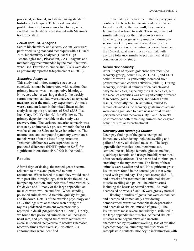
processed, sectioned, and stained using standard histologic techniques. To better demonstrate proliferation of fibrous connective tissue, select skeletal muscle slides were stained with Masson’s trichrome stain. Serum and ECG Analyses Serum biochemistry and electrolyte analyses were performed using standard techniques with a Hitachi 7180 biochemistry analyzer (Hitachi High Technologies Inc., Pleasanton, CA). Reagents and methodology recommended by the manufacturers were used. Exercise tolerance and ECGs were done as previously reported (Stegelmeier et al. 2010). Statistical Analyses This study had limited sample sizes so our conclusions must be interpreted with caution. Our primary interest was in comparative histology. However, when n was larger than 2, the weekly serum biochemical data were analyzed as repeated measures over the multi-day experiment. Animals were a random factor in the mixed linear model analysis using the procedures of SAS (SAS Inst. Inc., Cary, NC; Version 9.1 for Windows). The primary dependent variable in the study was recovery time. The variance-covariance matrix was chosen by an interactive process wherein the best fit was based on the Schwarz Bayesian criterion. The unstructured and compound symmetry covariance models were often the best fitting structures. Treatment differences were separated using predicted difference (PDIFF option in SAS) for significant interactions (P < 0.05) in the model. Results After 5 days of dosing, the treated goats became reluctant to move and preferred to remain recumbent. When forced to stand, they would stand with post-like, straight legs, their backs flexed in a humped up position, and their tails flexed vertically. On days 6 and 7, many of the large appendicular muscles were swollen and firm. When standing, poisoned animals would tremble, quickly fatigue, and lie down. Details of the exercise physiology and ECG findings similar to those seen during the rayless goldenrod treatment were previously reported in detail (Stegelmeier et al. 2010). Briefly, we found that poisoned animals had an increased heart rate, and prolonged times were required for exercise-induced tachycardia to resolve (increased recovery times after exercise). No other ECG abnormalities were identified.
Immediately after treatment, the recovery goats continued to be reluctant to rise and move. When forced to walk on the treadmill, they quickly fatigued and refused to walk. These signs were of similar intensity for the first recovery week; however, they progressively improved during the second week. Improvement was slower for the remaining portion of the entire recovery phase, and the 16-week goat was clinically normal, with exercise tolerance similar to pretreatment at the conclusion of the study. Serum Biochemistry After 7 days of rayless goldenrod treatment (no recovery group), serum CK, AST, ALT, and LDH activities were all significantly increased from pretreatment and control activities (table 1). During recovery, individual animals often had elevated enzyme activities, especially the CK activities, but the mean of activities was not significantly different than control goats. However, individual animal results, especially the CK activities, tended to remain elevated as the recovery goats improved and were once again able to have near normal treadmill performances and recoveries. By 8 and 16 weeks post treatment both remaining animals had enzyme activities similar to pretreatment. Necropsy and Histologic Studies Necropsy findings of the goats necropsied immediately after dosing included swelling and pallor of nearly all skeletal muscles. The large appendicular muscles (semimembranosus, semitendinosus, biceps femoris, gluteus medius, quadriceps femoris, and triceps brachii) were most often severely affected. The hearts had minimal pale streaking in the myocardium. The livers of these animals were swollen and red. No significant gross lesions were found in the control goats that were dosed with ground hay. The goats necropsied 1, 2, and 4 weeks after treatment had minimal skeletal muscle swelling and pallor. The other tissues including the hearts appeared normal. Animals necropsied on weeks 8 and 16 were grossly normal.
Histologic studies of goats that were euthanized and necropsied immediately after dosing demonstrated extensive monophasic degeneration and necrosis of skeletal muscle (figure 2). The lesions were most severe and widely distributed in the large appendicular muscles. Affected skeletal muscles were degenerative and necrotic characterized by myofiber swelling, loss of striation, hypereosinophilia, clumping and disruption of sarcoplasmic contents, monocytic inflammation with
Stegelmeier et al.: Resolution of rayless goldenrod toxicity in goats
30
Table 1. Selected mean serum biochemical data from recovering goats dosed with rayless goldenrod to obtain benzofuran ketone dosages of 40 mg/kg BW/day for 7 days* PretreatmentΙΙ No Recovery 1 Week 2 Weeks 3 Weeks 4 Weeks Creatinine Kinase (CK < 350 U/LΙ)
215±90a
11,952±8497b
748±447a
481±296a
330±99a
777±38a
Aspartate aminotransferase (AST < 125 U/LΙ)
130±51a
4306±1869b
1418±421a
581±509a
338±267a
351±36a
Alanine amminotransferase (ALT < 55 U/LΙ)
43±7a
452±205b
470±100a
193±62a
107±35a
87±19a
Lactate dehydrogenase (LDH < 1560 U/LΙ)
1112±307a
9665±2961b
3650±721c
1960±1057ac
1438±1052a
1920±1021ac
* Data are reported as means ± standard deviation. Different means (P < 0.05) between groups are indicated with superscript letters. Results from animals after 8 and 16 weeks of recovery are not included as there were insufficient numbers for statistical comparison and the activities were similar to pre-treatment measurements.
Ι Estimates of normal range were determined as 2 SD from mean values of controls and pretreatment samples. These ranges are laboratory and assay specific.
ΙΙ Mean and standard deviation of animals on initial treatment day.
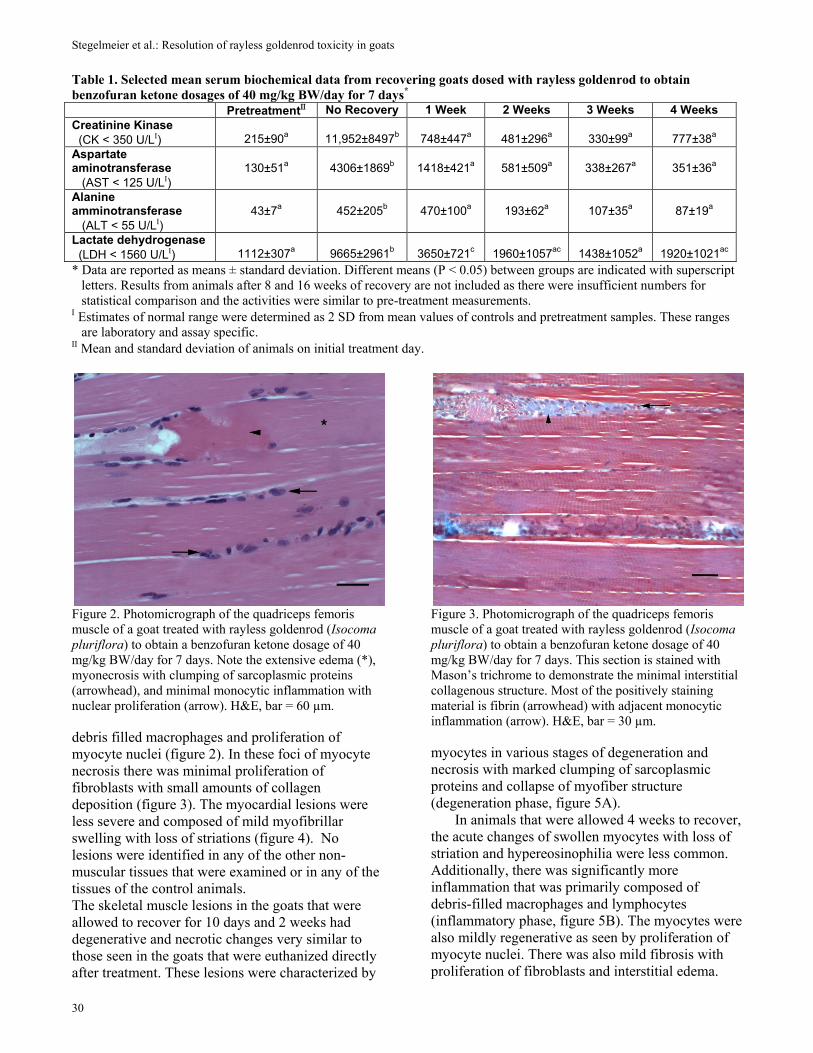
Figure 2. Photomicrograph of the quadriceps femoris muscle of a goat treated with rayless goldenrod (Isocoma pluriflora) to obtain a benzofuran ketone dosage of 40 mg/kg BW/day for 7 days. Note the extensive edema (*), myonecrosis with clumping of sarcoplasmic proteins (arrowhead), and minimal monocytic inflammation with nuclear proliferation (arrow). H&E, bar = 60 µm. debris filled macrophages and proliferation of myocyte nuclei (figure 2). In these foci of myocyte necrosis there was minimal proliferation of fibroblasts with small amounts of collagen deposition (figure 3). The myocardial lesions were less severe and composed of mild myofibrillar swelling with loss of striations (figure 4). No lesions were identified in any of the other non-muscular tissues that were examined or in any of the tissues of the control animals. The skeletal muscle lesions in the goats that were allowed to recover for 10 days and 2 weeks had degenerative and necrotic changes very similar to those seen in the goats that were euthanized directly after treatment. These lesions were characterized by
Figure 3. Photomicrograph of the quadriceps femoris muscle of a goat treated with rayless goldenrod (Isocoma pluriflora) to obtain a benzofuran ketone dosage of 40 mg/kg BW/day for 7 days. This section is stained with Mason’s trichrome to demonstrate the minimal interstitial collagenous structure. Most of the positively staining material is fibrin (arrowhead) with adjacent monocytic inflammation (arrow). H&E, bar = 30 µm. myocytes in various stages of degeneration and necrosis with marked clumping of sarcoplasmic proteins and collapse of myofiber structure (degeneration phase, figure 5A).
In animals that were allowed 4 weeks to recover, the acute changes of swollen myocytes with loss of striation and hypereosinophilia were less common. Additionally, there was significantly more inflammation that was primarily composed of debris-filled macrophages and lymphocytes (inflammatory phase, figure 5B). The myocytes were also mildly regenerative as seen by proliferation of myocyte nuclei. There was also mild fibrosis with proliferation of fibroblasts and interstitial edema.
IJPPR, vol. 2, Fall 2012
31
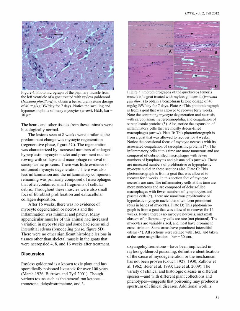
Figure 4. Photomicrograph of the papillary muscle from the left ventricle of a goat treated with rayless goldenrod (Isocoma pluriflora) to obtain a benzofuran ketone dosage of 40 mg/kg BW/day for 7 days. Notice the swelling and hypereosinophilia of many myocytes (arrow). H&E, bar = 30 µm. The hearts and other tissues from these animals were histologically normal.
The lesions seen at 8 weeks were similar as the predominant change was myocyte regeneration (regenerative phase, figure 5C). The regeneration was characterized by increased numbers of enlarged hyperplastic myocyte nuclei and prominent nuclear rowing with collapse and macrophage removal of sarcoplasmic proteins. There was little evidence of continued myocyte degeneration. There was also less inflammation and the inflammatory component remaining was primarily composed of macrophages that often contained small fragments of cellular debris. Throughout these muscles were also small foci of fibroblast proliferation and extracellular collagen deposition.
After 16 weeks, there was no evidence of myocyte degeneration or necrosis and the inflammation was minimal and patchy. Many appendicular muscles of this animal had increased variation in myocyte size and some had some mild interstitial edema (remodeling phase, figure 5D). There were no other significant histologic lesions in tissues other than skeletal muscle in the goats that were necropsied 4, 8, and 16 weeks after treatment. Discussion Rayless goldenrod is a known toxic plant and has sporadically poisoned livestock for over 100 years (Marsh 1926, Burrows and Tyrl 2001). Though various toxins such as the benzofuran ketones—tremetone, dehydrotremetone, and 3-
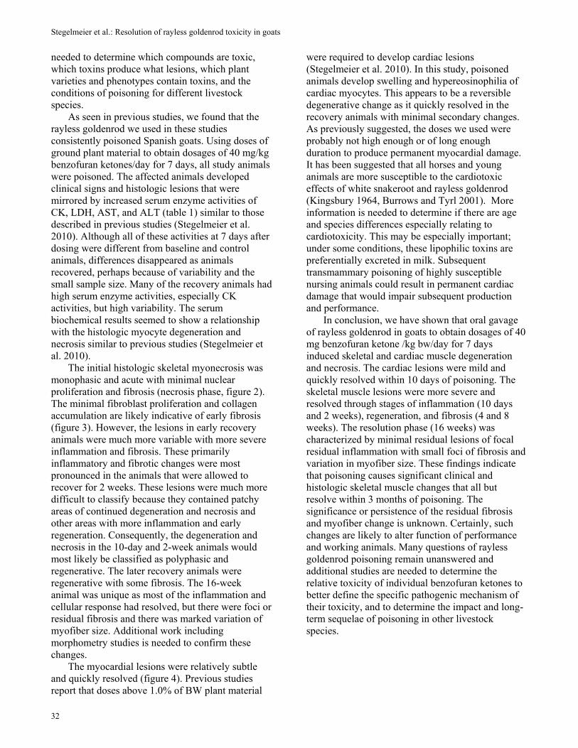
Figure 5. Photomicrographs of the quadriceps femoris muscle of a goat treated with rayless goldenrod (Isocoma pluriflora) to obtain a benzofuran ketone dosage of 40 mg/kg BW/day for 7 days. Plate A: This photomicrograph is from a goat that was allowed to recover for 2 weeks. Note the continuing myocyte degeneration and necrosis with sarcoplasmic hypereosinophilia, and coagulation of sarcoplasmic proteins (*). Also, notice the expansion of inflammatory cells that are mostly debris-filled macrophages (arrow). Plate B: This photomicrograph is from a goat that was allowed to recover for 4 weeks. Notice the occasional focus of myocyte necrosis with its associated coagulation of sarcoplasmic proteins (*). The inflammatory cells at this time are more numerous and are composed of debris-filled macrophages with fewer numbers of lymphocytes and plasma cells (arrow). There are increased numbers of proliferative or hyperplastic myocyte nuclei in these sections also. Plate C: This photomicrograph is from a goat that was allowed to recover for 8 weeks. In this section foci of myocyte necrosis are rare. The inflammatory cells at this time are more numerous and are composed of debris-filled macrophages with fewer numbers of lymphocytes and plasma cells (*). There are numerous proliferative or hyperlastic myocyte nuclei that often form prominent rows in bands of myocytes. Plate D: This photomicro-graph is from a goat that was allowed to recover for 16 weeks. Notice there is no myocyte necrosis, and small clusters of inflammatory cells are rare (not pictured). The myocytes are variably sized, and most have prominent cross-striation. Some areas have prominent interstitial edema (*). All sections were stained with H&E and taken at the same magnification—bar = 30 µm. oxyangeloyltremetone—have been implicated in rayless goldenrod poisoning, definitive identification of the cause of myodegeneration or the mechanism has not been proven (Couch 1927, 1930; Zalkow et al. 1962; Beier et al. 1993; Lee et al. 2009). The variety of clinical and histologic disease in different species—and with different plant collections and phenotypes—suggests that poisoning may produce a spectrum of clinical diseases. Additional work is
Stegelmeier et al.: Resolution of rayless goldenrod toxicity in goats
32
needed to determine which compounds are toxic, which toxins produce what lesions, which plant varieties and phenotypes contain toxins, and the conditions of poisoning for different livestock species.
As seen in previous studies, we found that the rayless goldenrod we used in these studies consistently poisoned Spanish goats. Using doses of ground plant material to obtain dosages of 40 mg/kg benzofuran ketones/day for 7 days, all study animals were poisoned. The affected animals developed clinical signs and histologic lesions that were mirrored by increased serum enzyme activities of CK, LDH, AST, and ALT (table 1) similar to those described in previous studies (Stegelmeier et al. 2010). Although all of these activities at 7 days after dosing were different from baseline and control animals, differences disappeared as animals recovered, perhaps because of variability and the small sample size. Many of the recovery animals had high serum enzyme activities, especially CK activities, but high variability. The serum biochemical results seemed to show a relationship with the histologic myocyte degeneration and necrosis similar to previous studies (Stegelmeier et al. 2010).
The initial histologic skeletal myonecrosis was monophasic and acute with minimal nuclear proliferation and fibrosis (necrosis phase, figure 2). The minimal fibroblast proliferation and collagen accumulation are likely indicative of early fibrosis (figure 3). However, the lesions in early recovery animals were much more variable with more severe inflammation and fibrosis. These primarily inflammatory and fibrotic changes were most pronounced in the animals that were allowed to recover for 2 weeks. These lesions were much more difficult to classify because they contained patchy areas of continued degeneration and necrosis and other areas with more inflammation and early regeneration. Consequently, the degeneration and necrosis in the 10-day and 2-week animals would most likely be classified as polyphasic and regenerative. The later recovery animals were regenerative with some fibrosis. The 16-week animal was unique as most of the inflammation and cellular response had resolved, but there were foci or residual fibrosis and there was marked variation of myofiber size. Additional work including morphometry studies is needed to confirm these changes.
The myocardial lesions were relatively subtle and quickly resolved (figure 4). Previous studies report that doses above 1.0% of BW plant material
were required to develop cardiac lesions (Stegelmeier et al. 2010). In this study, poisoned animals develop swelling and hypereosinophilia of cardiac myocytes. This appears to be a reversible degenerative change as it quickly resolved in the recovery animals with minimal secondary changes. As previously suggested, the doses we used were probably not high enough or of long enough duration to produce permanent myocardial damage. It has been suggested that all horses and young animals are more susceptible to the cardiotoxic effects of white snakeroot and rayless goldenrod (Kingsbury 1964, Burrows and Tyrl 2001). More information is needed to determine if there are age and species differences especially relating to cardiotoxicity. This may be especially important; under some conditions, these lipophilic toxins are preferentially excreted in milk. Subsequent transmammary poisoning of highly susceptible nursing animals could result in permanent cardiac damage that would impair subsequent production and performance.
In conclusion, we have shown that oral gavage of rayless goldenrod in goats to obtain dosages of 40 mg benzofuran ketone /kg bw/day for 7 days induced skeletal and cardiac muscle degeneration and necrosis. The cardiac lesions were mild and quickly resolved within 10 days of poisoning. The skeletal muscle lesions were more severe and resolved through stages of inflammation (10 days and 2 weeks), regeneration, and fibrosis (4 and 8 weeks). The resolution phase (16 weeks) was characterized by minimal residual lesions of focal residual inflammation with small foci of fibrosis and variation in myofiber size. These findings indicate that poisoning causes significant clinical and histologic skeletal muscle changes that all but resolve within 3 months of poisoning. The significance or persistence of the residual fibrosis and myofiber change is unknown. Certainly, such changes are likely to alter function of performance and working animals. Many questions of rayless goldenrod poisoning remain unanswered and additional studies are needed to determine the relative toxicity of individual benzofuran ketones to better define the specific pathogenic mechanism of their toxicity, and to determine the impact and long-term sequelae of poisoning in other livestock species.

IJPPR, vol. 2, Fall 2012
33
Acknowledgments The authors thank Ed Knoppel, Joseph Jacobson, and Katie Lott for their assistance in animal care and laboratory technical expertise. References Beier, R.C., J.O. Norman, J.C. Reagor, M.S. Rees, and B.P. Mundy. 1993. Isolation of the major component in white snakeroot that is toxic after microsomal activation: possible explanation of sporadic toxicity of white snakeroot plants and extracts. Natural Toxins 1:286-293.
Burrows, G.E., and R.J. Tyrl, eds. 2001. Toxic Plants of North America. Iowa State Press, Ames, IA.
Couch, J.F. 1927. The toxic constituent of richweed or white snakeroot (Eupatorium urticaefolium). Journal of Agricultural Research 35:547-576.
Couch, J.F. 1929. Tremetol, the compound that produces “trembles” (milk sickness). Journal of the American Chemical Society 51:3617-3619.
Couch, J.F. 1930. The toxic constituents of rayless goldenrod. Journal of Agricultural Research 40:649-658.
Kingsbury, J.M. 1964. Poisonous Plants of the United States and Canada. Prentice-Hall, Inc., Englewood Cliffs, NJ.
Lee, S.T., T.Z. Davis, D.R. Gardner, B.L. Stegelmeier, and T.J. Evans. 2009. Quantitative method for the measurement of three benzofuran ketones in rayless goldenrod (Isocoma pluriflora) and white snakeroot (Ageratina altissima) by high-performance liquid chromatography (HPLC). Journal of Agriculture and Food Chemistry 57:5639-5643.
Marsh, C.D. 1926. Rayless goldenrod (Aplopappus heterophyllus) as a poisonous plant. United States Department of Agriculture Departmental Bulletin 1391:1-24.
Marsh, C.D., and G.C. Roe. 1921. The “alkali disease” of livestock in the Pecos Valley. United States Department of Agriculture Departmental Bulletin 180:3-8.
Stegelmeier, B.L., T.Z. Davis, B.T. Green, S.T. Lee, and J.O. Hall. 2010. Experimental rayless goldenrod (Isocoma pluriflora) toxicosis in goats. Journal of Veterinary Diagnostic Investigation 22:570-577.
Zalkow, L.H., N. Burke, G. Cabat, and E.A. Rula. 1962. Toxic constituents of rayless goldenrod. Journal of Medicinal and Pharmaceutical Chemistry 91:1342-1351.
Submitted: 5/20/2011 Revised: 2/29/2012
Accepted: 5/31/2012