THE REQUIREMENT FOR LYMPHOCYTE FUNCTION-
18
THE REQUIREMENT FOR LYMPHOCYTE FUNCTION- ASSOCIATED ANTIGEN 1 IN HOMOTYPIC LEUKOCYTE ADHESION STIMULATED BY PHORBOL ESTER BY ROBERT ROTHLEIN AND TIMOTHY A. SPRINGER From the Laboratory of Membrane Immunochemistry, Dana-Farber Cancer Institute, Harvard Medical School, Boston, Massachusetts 02115 Lymphocyte activation is accompanied by increased adhesiveness and motility. Although specific antigen may be used to stimulate increased adhesiveness, stimulated lymphocytes show a generalized increased adherence to cells lacking specific antigen. Cells cultured in the MLR acquire the ability to adhere to a wide variety of tumor cell types. There is no MHC restriction in this adhesion, although species specificity has been shown (1). Adhesion of lymphocytes to one another can also be measured as cluster formation, i.e., aggregation. After autologous MLR or periodate stimulation, 5-35% of the viable lymphocytes are found in clusters (2). Lymphocytes isolated from clusters by vigorous vortexing are found to readily reaggregate. Cluster formation is also induced by phorboi esters. Within 15 rain, human PBL show uropod formation, and within 30 min exhibit hairy surface projections, ruffled membranes, and begin aggregating (3). Similar aggregation is seen with monocytes (3) and some leukocyte cell lines, including EBV-transformed B lymphocytes (4). Both B lymphocytes and the Lyt- 2 + and Lyt-2- subsets of T lymphocytes have been shown to participate in cluster formation (2). Despite the obvious relevance of changes in lymphocyte adhesive- ness to immune cell interactions, the cell surface molecules mediating adhesive- ness by stimulated lymphocytes have not been defined. Previous studies have shown that mAbs to the lymphocyte function-associated 1 (LFA-1)I molecule inhibit the effector-target adhesion step of CTL-mediated killing (5), and a wide variety of other adhesion-dependent leukocyte functions, including antigen-specific T-B cell cooperation, antigen-specific and mitogen proliferative responses (6), natural killing, and antibody-dependent cellular cy- totoxicity by K cells and granulocytes (7). Lymphocytes from patients with genetic deficiency of LFA-1 show diminished activity in adhesion-dependent functional assays (8-10). These findings suggested that LFA-1 cooperates with a number of different specific cell surface receptors in heterotypic cell-cell interactions. LFA-1 is a leukocyte cell surface glycoprotein with an c~L subunit of 180,000 Mr and/3 subunit of 95,000 Mr. Here we have investigated the role of LFA-1 in the increased adhesiveness of phorbol ester-stimulated leukocytes. Phorbol esters are analogues of diacylglyc- This work was supported by Council for Tobacco Researchgrant 1307, National Institutes of Health grants CA31798 and AI05877, and Dr. Rothlein's National Institutes of Health fellowshipAI06963. J Abbreviation used in this paper: LFA-1, lymphocyte function-associatedantigen 1. 1 132 J. ExP. MED. © The RockefellerUniversityPress - 0022-1007/86/05/1132/18 $1.00 Volume 163 May 1986 1132-1149
Transcript of THE REQUIREMENT FOR LYMPHOCYTE FUNCTION-

T H E R E Q U I R E M E N T F O R L Y M P H O C Y T E F U N C T I O N -
A S S O C I A T E D A N T I G E N 1 IN H O M O T Y P I C L E U K O C Y T E
A D H E S I O N S T I M U L A T E D BY P H O R B O L E S T E R
BY ROBERT ROTHLEIN AND TIMOTHY A. SPRINGER
From the Laboratory of Membrane Immunochemistry, Dana-Farber Cancer Institute, Harvard Medical School, Boston, Massachusetts 02115
Lymphocyte activation is accompanied by increased adhesiveness and motility. Although specific antigen may be used to stimulate increased adhesiveness, stimulated lymphocytes show a generalized increased adherence to cells lacking specific antigen. Cells cultured in the MLR acquire the ability to adhere to a wide variety of tumor cell types. There is no MHC restriction in this adhesion, although species specificity has been shown (1). Adhesion of lymphocytes to one another can also be measured as cluster formation, i.e., aggregation. After autologous MLR or periodate stimulation, 5-35% of the viable lymphocytes are found in clusters (2). Lymphocytes isolated from clusters by vigorous vortexing are found to readily reaggregate. Cluster formation is also induced by phorboi esters. Within 15 rain, human PBL show uropod formation, and within 30 min exhibit hairy surface projections, ruffled membranes, and begin aggregating (3). Similar aggregation is seen with monocytes (3) and some leukocyte cell lines, including EBV-transformed B lymphocytes (4). Both B lymphocytes and the Lyt- 2 + and Lyt-2- subsets of T lymphocytes have been shown to participate in cluster formation (2). Despite the obvious relevance of changes in lymphocyte adhesive- ness to immune cell interactions, the cell surface molecules mediating adhesive- ness by stimulated lymphocytes have not been defined.
Previous studies have shown that mAbs to the lymphocyte function-associated 1 (LFA-1)I molecule inhibit the effector-target adhesion step of CTL-mediated killing (5), and a wide variety of other adhesion-dependent leukocyte functions, including antigen-specific T-B cell cooperation, antigen-specific and mitogen proliferative responses (6), natural killing, and antibody-dependent cellular cy- totoxicity by K cells and granulocytes (7). Lymphocytes from patients with genetic deficiency of LFA-1 show diminished activity in adhesion-dependent functional assays (8-10). These findings suggested that LFA-1 cooperates with a number of different specific cell surface receptors in heterotypic cell-cell interactions. LFA-1 is a leukocyte cell surface glycoprotein with an c~L subunit of 180,000 Mr and/3 subunit of 95,000 Mr.
Here we have investigated the role of LFA-1 in the increased adhesiveness of phorbol ester-stimulated leukocytes. Phorbol esters are analogues of diacylglyc-
This work was supported by Council for Tobacco Research grant 1307, National Institutes of Health grants CA31798 and AI05877, and Dr. Rothlein's National Institutes of Health fellowship AI06963.
J Abbreviation used in this paper: LFA-1, lymphocyte function-associated antigen 1.
1 132 J. ExP. MED. © The Rockefeller University Press - 0022-1007/86/05/1132/18 $1.00 Volume 163 May 1986 1132-1149
ROTHLEIN AND SPRINGER 1133
erol, an intracellular second messenger which mediates activation or differentia- tion o f lymphocytes and monocytes and can mimic the effects o f certain physio- logic stimuli (11-13) . We repor t that antibodies to LFA-1 specifically inhibit phorbo l es te r - s t imula ted homotypic aggregat ion by B cell, T cell and monocyt ic cell lines, and by PBL. Studies with cell lines f rom individuals genetically deficient in LFA-1 conf i rm these findings, and show that while LFA-1 mediates homotypic cell interactions, this process is not media ted by like-like interactions be tween LFA-1 molecules. T h e cellular physiology o f aggregat ion has been investigated by techniques including time-lapse videomicroscopy. T h e results show that LFA- 1 is a molecule of general impor tance in phorbol es te r - s t imula ted adhesion by lymphocytes and monocyt ic cells.
M a t e r i a l s a n d M e t h o d s Cell Lines. JY EBV-transformed B lymphoblastoid cells were maintained in RPMI
1640 medium supplemented with 10% FCS and 50 #g/ml gentamycin (Gibco Laborato- ries, Grand Island, NY) (complete medium). EBV-transformed B-cell lines were established and maintained as previously described (14) using peripheral blood mononuclear cells from normal individuals, patients who genetically lack membrane LFA-1, and heterozy- gote relatives of these patients (8).
mAb and F(ab')2 and Fab' Fragments. The mouse IgG1 TS1/22 and TS1/18 mAb to the human LFA-1 aL and/3 subunits, respectively (15), and the TS2/9 anti-LFA-3 mAb (16) were obtained as previously described. The species-specific anti-HLA-A,B mAb W6/32 has been described (17). A panel of 115 putatively anti-myeloid mAbs that reacted with many different types of surface antigens, some of which were on lymphoid cells, was obtained in the Second International Workshop on Human Leukocyte Differentiation (l 8). F(ab')2 of TS1/22 were made using preactivated thiol-free papain as described by Parham et al. (19). Fab' were made using cysteine reduction of F(ab')~ (19).
Qualitative Aggregation Assay. Cells lines were washed two times with RPMI 1640 containing 5 mM Hepes buffer (Sigma Chemical Co., St. Louis) and resuspended to a concentration of 2 × 10⁶ cells/ml. Added to flat-bottomed, 96-well microtiter plates (No. 3596; Costar, Cambridge, MA) were 50 #l of appropriate mAb supernatant or 50 #1 of complete medium with or without purified mAb, 50 #1 of complete medium containing 200 ng/ml of PMA (Sigma Chemical Co.) where necessary, and 100 /~1 of cells at a concentration of 2 x I 0" cells/ml in complete medium. This yielded a final concentration of 50 ng/ml PMA and 2 X 10 ~ cells/well. Cells were allowed to settle spontaneously and the degree of aggregation was scored at indicated time points. Scores ranged from 0 to 5+ where 0 indicated that essentially no cells were in clusters; 1+ indicated <10% of the cells were in aggregates; 2+ indicated that <50% of the cells were aggregated; 3+ indicated that up to 100% of the cells were in small, loose clusters; 4+ indicated that up to 100% of the cells were aggregated in larger clusters; and 5+ indicated that 100% of the cells were in large, very compact aggregates.
Quantitative Aggregation Assay. Reagents and cells were added to 5 ml polystyrene tubes (No. 2054; Falcon Labware, Oxnard, CA) in the same order as above. Tubes were placed in a rack on a gyratory shaker at 37°C. After 1 h at 200 rpm (unless otherwise specified), 10 tA of the cell suspension were placed on a hemocytometer and the number of free ceils was quantitated. Percent aggregation was determined by the following equation: Percent aggregation = 100 x [1 - (number of free cells)]/(number of input cells), where number of input cells is the number of cells per ml in a control tube containing only cells and complete medium that was not incubated, and number of free cells equals the number of nonaggregated cells per ml from experimental tubes.
FACS. JY cells were washed three times with RPMI 1640 and stained with mAb using indirect immunofluorescence according to Kfirzinger et al. (20). As control, a nonspecific IgG was substituted in the first step. FITC conjugated goat anti-mouse IgG with heavy and light chain reactivity (Zymed) was the indirect reagent. Cells were analyzed on an

1134 PHORBOL-STIMULATED LEUKOCYTE ADHESION
Epics V flow cytometer. Results are expressed as linear fluorescence intensity after subtraction of control fluorescence (specific linear fluorescence).
Capping Experiments. Cells were preincubated with PMA at 37°C for 5 min before staining where appropriate. Cells were then stained by indirect immunofluorescence in test tubes (No. 2054; Falcon Labware) using the same protocol as above while held on ice or at 4 °C throughout the staining and washing protocol. Following the final wash, cells were resuspended to a concentration of 107/ml in complete medium that was preheated to 37°C, and the tubes were incubated at 37°C. At the prescribed time points, 100 t~i of cell suspensions were collected and fixed with an equal volume of 2% paraformaldehyde on ice to prevent further capping. Cells showing fluorescence on less than 50% of the membrane periphery were considered to have capped. In coaggregation experiments, cells were stained with 6-carboxyfluorescein diacetate (Behring Diagnostics, San Diego, CA) (21).
Time-Lapse Video Microscopy Recording. Cells were placed in a microtiter plate as in the qualitative assay. The well was sealed with clear tape and the plate was put on ice for 15 min while the cells settled to the bottom of the well. The plate was then placed on an inverted microscope (Carl Zeiss, Thornwood, NY) equipped with a Zeiss video recording camera, and hot air was blown around the plate to maintain a temperature of about 37°C. Time-lapse recordings were made at a rate of 1 frame/s.
Resul ts
PMA Stimulates Aggregation of JY Cells. Cells f rom the EBV-transformed line JY were found to aggregate in response to PMA using two different assays. A qualitative assay in which JY cells were allowed to aggregate on the bot tom of a 96-well microt i ter plate showed that a l though some adhesion of JY cells occur red in the absence of PMA, in its presence much larger clusters formed, and cluster format ion was more rapid and reproducible (Fig. 1 A and data not shown). Time- lapse video recording showed that JY cells on the bot tom of microt i ter wells were motile and exhibited active membrane ruffl ing and pseudopodia movement . Contact between the pseudopodia o f neighbor ing cells often resulted in cell-cell adherence. I f adherence was sustained, the region o f cell contact moved to the uropod. Contact could be maintained despite vigorous cell movements and tugging o f the cells in opposite directions. T h e pr imary difference between PMA- t reated and unt rea ted cells appeared to be in the stability o f these contacts once they were formed. With PMA, clusters of cells developed, growing in size as additional cells adhered at their periphery. At first clusters were f lat tened out along the bot tom of the well with a thickness of only one or a few cells. As the clusters grew in size, there was a dramatic change in the shape o f the cluster as most of the cells lifted of f the bot tom of the well and the cluster assumed the shape o f a roughly spherical ball o f cells. T~is change suggests that cell-cell adhesivity is s t ronger than cell-substrate adhesivity. After cluster formation, vigorous cell membrane ruffling continued, with little change in the relative position of individual cells within the cluster. In video timeqapse, this gave the cluster the appearance o f a heaving mass.
As a second means of measuring adhesion, a quantitative assay was used in which cell suspensions were shaken at 200 rpm for 2 h, t ransferred to a hemocytometer , and cells not in aggregates were enumerated . In the absence o f PMA, 42% (SD = 20%, n = 6) o f JY cells were in aggregates af ter 2 h, while JY cells incubated under identical conditions with 50 ng/ml o f PMA had 87% (SD = 8%, n = 6) o f the cells in aggregates. Kinetic studies of aggregation showed

ROTHLEIN AND SPRINGER 1135
FIGURE 1. Photomicrographs of aggregation and its inhibition by anti-LFA-1. JY cells were incubated with PMA (A) or with PMA + anti-LFA-1 (B) in the qualitative assay for 30 rain.
that PMA enhanced the rate and magnitude of aggregation at all time points tested (Fig. 2).
Anti-LFA-1 mAbs Inhibit PMA-induced Aggregation of JY Cells. Since LFA-1 has been implicated in lymphocyte adhesion (9), we looked at its role in the PMA induced aggregation of JY cells. The effects of anti-LFA-1 mAb on PMA induced-JY cell aggregation was assessed in both the quantitative and qualitative assays.
Anti-LFA-1 a chain (TS1/22) and /3 chain (TS1/18) mAb inhibited PMA- induced aggregation of JY cells by 90 and 85%, respectively (Fig. 3). In contrast, anti-LFA-3 (TS2/9) and anti-HLA (W6/32) mAb had no effect on aggregation. These findings were confirmed in the qualitative assay, mAb directed against either the a or/3 subunit of LFA-1 inhibited the formation of aggregates of JY cells assayed with and without PMA. One representative experiment is shown in Fig. 1, which shows inhibition of phorbol ester-stimulated aggregation by anti- LFA-1 /3 mAb. In a blind screening of a panel of 115 mAbs directed to many different types of leukocyte antigens, only and all seven mAb with anti-LFA-1 a or/3 chain reactivity inhibited PMA-induced aggregation (Table I).
To determine whether inhibition of aggregation was independent of effects mediated by the Fc portion or by antibody bivalency, F(ab')2 and Fab' fragments of the TS1/22 anti-LFA-la mAb were tested. Both fragments inhibited PMA- induced JY cell aggregation (Fig. 4). Since about 10 times more Fab' fragment than F(ab')~ fragment was required to completely inhibit aggregation, the purity

1136 PHORBOL-STIMULATED LEUKOCYTE ADHESION
100"
80'
60'
N 40'
20"
30 60 90 120
Ttmm (-,t nl
FIGURE 2. Kinetics of aggregation. Aggregation in the absence (X) or presence of 50 ng/ml PMA (O) was determined in the quantitative assay.
SOOT
80'
i 50 '
c
m
~- 4 0
N
20
i 0 u none ant 1 -LFA- t ant l -LFA- t ant 1 -LFA-3 ant 1 -HLA
alpha chain beta chain
Nonoclonel antibody
FIGURE 3. Anti-LFA-1 mAb specifically inhibits aggregation. The indicated mAbs (50 pl of tissue culture supernatant) were added at the initiation of quantitative aggregation assays. Values are one of six representative experiments.
of the preparations was examined to rule out the presence of residual F(ab')2 in the Fab' preparation. Non-reducing SDS-PAGE of 0.5 pg of F(ab')2 (Fig. 4, lane 1) and 5.0 pg of Fab' (Fig. 4, lane 2) showed no contamination of the Fab'

ROTHLEIN AND SPRINGER
TAnLE I mAb That Inhibit PMA-induced Aggregation
mAb inhibiting Specificity* aggregation
TS1/22 LFA-1 (a) MHM24 LFA-1 (a) TS1/18 LEA-l, Mac-l, p150,95 (~3) MHM23 LFA-1, Mac-l, p150,95 (/~) 60.3 LFA-I, Mac-l, p150,95 CLB-54 LFA-I, Mac-l, p150,95
115 mAb in the myeloid panel of the Second International Leukocyte Workshop were tested blind at a final ascites dilution of 1:150 in the qualitative PMA-stimulated JY aggregation assay. Inhibitory mAb (re- ducing aggregation from 5+ to 1+ or -) are shown.
* Sanchez-Madrid et al. (16), Springer and Anderson (19), and Hildreth et al. (38).
1137
FIGURE 4. F(ab')~ (X) and Fab' (O) fragments of LFA-1 mAb inhibit aggregation. TS1/22 mAb fragments were included in the quantitative aggregation assay. Inset: SDS 10% PAGE under nonreducing conditions of 0.5 ug E(ab')~ (lane 1) and 5 #g Fab' (lane 2) of TS1/22 LFA-1 mAb.
p repara t ion by F(ab')2. This shows that monova len t Fab ' LFA-1 m A b can inhibit aggregat ion, and suggested that the differ ing inhibi tory activities o f F(ab')z and Fab ' f r agments is due to differences in avidity between bivalent and monova len t f ragments .
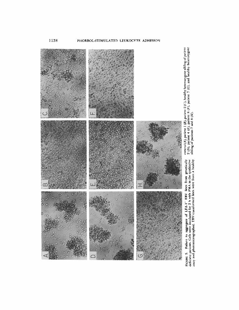
EBV-transformed Cells from Patients Lacking LFA-1, Mac-l, and p150,95 Do Not Self-aggregate. As a second, independen t means o f testing the functional impor- tance of LFA-1, PMA-st imula ted aggregat ion o f LFA-1- cells was examined. EBV- t rans formed B lymphoblas toid cell lines have been established in ou r labora tory f rom patients who are genetically deficient in surface expression o f the LFA-1, Mac- l , and p150,95 glycoproteins (14). Lines established f rom severely and modera te ly deficient patients expressed ~<0 .1 and 3% of normal amounts o f LFA-1, respectively. Lines were established at the same t ime f rom
S.
00
O
O
> ,q
Z7
O
C3
-r
o Z
FI(;
UR
E 5
. F
ailu
re
to
aggr
egat
e of
LF
A-1
- E
BV
li
nes
from
ge
neti
call
y de
fici
ent
pati
ents
. C
ells
wer
e in
cuba
ted
for
2 h
wit
h P
MA
in
the
qual
itat
ive
assa
y an
d ph
otom
icro
grap
hed.
E
BV
-tra
nsfo
rmed
lin
es w
ere
from
a h
ealt
hy
cont
rol
(A),
pati
ent
1 (B
), pa
tien
t 2
(C),
heal
thy
hete
rozy
gote
sib
ling
of p
atie
nt
2 (D
), pa
tien
t 4
(E),
pati
ent
8 (F
), p
atie
nt 7
(G
), an
d he
alth
y he
tero
zygo
te
sibl
ing
of p
atie
nts
7 an
d 8
(H).

100
ROTH LEIN AND SPRINGER 1139
80
4o
N
20
normal pat ient I pattent 2 pottent 4 pat~ant 5 ~'nV tronoforamd O-cel lo
FIGURE 6. Coaggregation between LFA-1- and LFA-1 + cells. Carboxyfluorescein diacetate- labeled EBV-transformed cells (104) as designated in the figure were mixed with 105 unlabeled autologous cells (solid bars) orJY cells (open bars) in the presence of PMA. After 1.5 h in the qualitative assay, the labeled cells in aggregates or free were enumerated using an inverted fluorescent microscope (> 100 fl+ cells were counted). The percentage of fluorescent (fl+) cells in aggregates is shown. One representative experiment of two is shown.
healthy, heterozygote relatives and from normal subjects. The latter lines express LFA-1, but not the related Mac-1 or p150,95 molecules (15). There was little if any aggregation of LFA-1- EBV-transformed cells f rom two different severely deficient patients (Fig. 5, B and C) or three different moderately deficient patients (Fig. 5, E-G). In contrast, EBV-transformed LFA-1 + cells from healthy heterozygote siblings (Fig. 5, D and H) and a healthy control (Fig. 5A) aggregated strongly.
Cells from LFA-l-deficient Patients Aggregate with JY Cells. It was of interest to determine whether LFA-l-deficient lymphoblasts, which could not aggregate with themselves, could coaggregate with LFA-1 + cells. Fluorescein diacetate- labeled normal or patient cells were mixed in a 1:10 ratio with autologous or JY cells, and the percentage of fluorescein-labeled cells in aggregates was determined (Fig. 6). An LFA-1 + cell line aggregated well with itself and with JY. LFA-1- deficient EBV lines showed little self-aggregation, confirming the results in Fig. 5. However, LFA-1- patient cells coaggregated well with LFA-1 + JY cells.
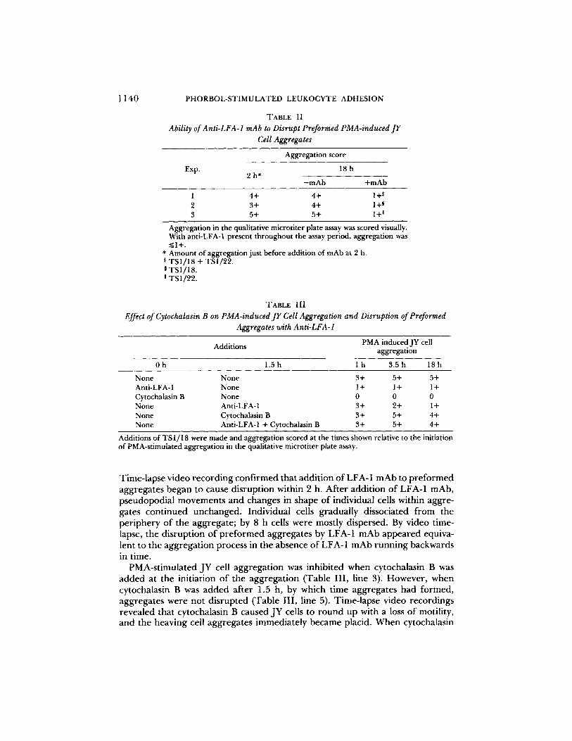
Anti-LFA-1 Can Disrupt Preformed Aggregates. To determine whether LFA-1 was important only in forming aggregates, or also in their maintenance, LFA-1 mAb were added to preformed aggregates (Table II). As shown above, there was strong aggregation of JY cells after 2 h in the presence o f PMA (3+ to 5+ aggregation in the qualitative assay). When anti-LFA-1 was added to wells at this time, and aggregation was assayed 16 h later, we found it was strongly disrupted.
1 1 4 0 PHORBOL-STIMULATED LEUKOCYTE ADHESION
TABLE II Ability of Anti-LFA-1 mAb to Disrupt Preformed PMA-induced JY
Cell Aggregates
Aggregation score
Exp. 18 h 2 h*
-mAb +mAb
1 4+ 4+ 1+* 2 3+ 4+ 1+ ~ 3 5+ 5+ 1+ a
Aggregation in the qualitative microtiter plate assay was scored visually. With anti-LFA-1 present througbout the assay period, aggregation was <_1+.
* Amount of aggregation just before addition of mAb at 2 h. * TSI/18 + TS1/22. 0 TSI/18. m TS1/22.
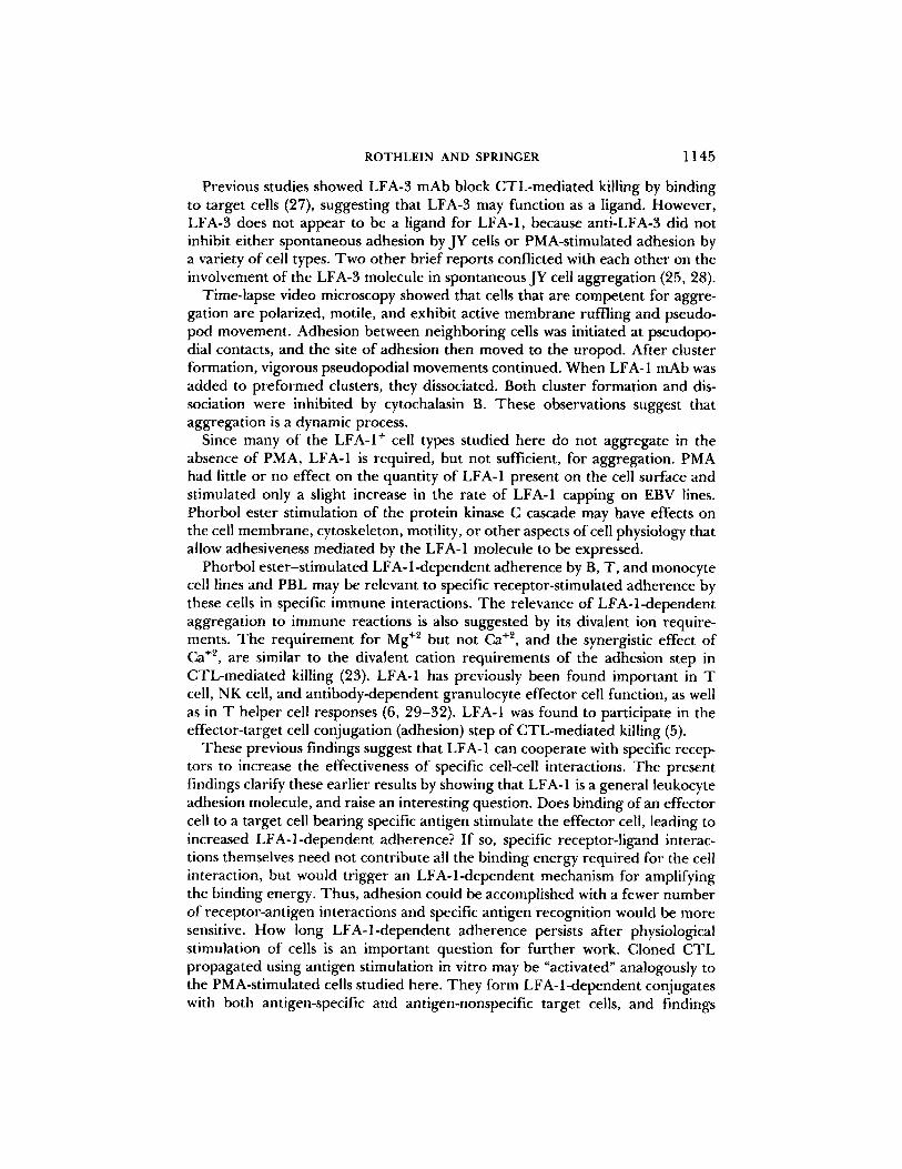
TABLE III Effect of Cytochalasin B on PMA-induced JY Cell Aggregation and Disruption of Preformed
Aggregates with Anti-LFA-1
Additions PMA induced JY cell aggregation
Oh 1.5h l h 3.5h 18h
None None 3+ 5+ 5+ Anti-LFA- 1 None 1 + 1 + 1 + Cytochalasin B None 0 0 0 None Anti-LFA-1 3+ 2+ 1 + None Cytochalasin B 3+ 5+ 4+ None Anti-LFA-1 + Cytochalasin B 3+ 5+ 4+
Additions of TS 1/18 were made and aggregation scored at the times shown relative to the initiation of PMA-stimulated aggregation in the qualitative microtiter plate assay.
Time-lapse video record ing conf i rmed that addit ion of LFA-1 m A b to p r e f o r m e d aggregates began to cause disrupt ion within 2 h. Af ter addit ion o f LFA-1 mAb, pseudopodial movemen t s and changes in shape o f individual cells within aggre- gates cont inued unchanged. Individual cells gradually dissociated f rom the per iphery o f the aggregate ; by 8 h cells were mostly dispersed. By video time- lapse, the disrupt ion o f p r e f o r m e d aggregates by LFA-1 m A b appea red equiva- lent to the aggregat ion process in the absence of LFA-1 m A b runn ing backwards in time.
PMA-st imulated JY cell aggregat ion was inhibited when cytochalasin B was added at the initiation o f the aggregat ion (Table I I I , line 3). However , when cytochalasin B was added af ter 1.5 h, by which t ime aggregates had fo rmed , aggregates were not d isrupted (Table I I I , line 5). Time-lapse video recordings revealed that cytochalasin B caused JY cells to round up with a loss of motility, and the heaving cell aggregates immediate ly became placid. When cytochalasin

HLA
ROTHLEIN AND SPRINGER 1141
2OO
~ t O 0 -
J
oN LFA- I LFAm~
)lAb dtr'octed non,net:
300
FIGURE 7. Surface antigen expression with and without stimulation by PMA. JY cells incu- bated for 1 h without PMA (m) and with PMA (k~), or patient 2 cells incubated for 1 h without PMA (11) and with PMA (Fq) were stained with LFA-1 /~ chain mAb, TS2/9 LFA-3 mAb, or W6/32 HLA mAb, an excess of FITC-goat anti-mouse Ig, and then subjected to flow cytometry. The fluorescence intensity due to specific staining is shown.
B was added toge ther with LFA-1 mAb at 1.5 h, aggregation was not reversed (Table III, compare lines 4 and 6), T h e failure o f anti-LFA-1 to disrupt aggre- gation when added simultaneously with cytochalasin B may be because motility is required for disaggregation in this assay system.
Effect of PMA on LFA-I Expression, LFA-1 Capping, and Motility of JY Cells. While it is clear that PMA and LFA-1 facilitate the aggregat ion o f JY cells, the role of each o f these molecules remained unclear. One mechanism by which PMA might stimulate LFA- l -dependen t JY cell aggregat ion would be by increas- ing LFA-1 expression on the cell surface. However , immunofluorescence flow cytometry showed that there was no change in LFA-1 expression on JY ceils, and showed that PMA did not induce expression on patient cells (Fig. 7). PMA also had little effect on LFA-3 and H L A expression.
Pa tar royo et al. r epor ted that Con A receptors more rapidly cap on PMA- t reated PBL (22). We tested whether PMA-induced LFA-1 to cap faster on JY cells. Cells were reacted with LFA-1 mAb, then F I T C an t i -mouse IgG, and warmed to 37 ° C. LFA-1 was capped somewhat more rapidly on PMA-stimulated JY cells, a l though the molecule capped quickly both in the presence and absence of PMA (Fig. 8).
It was next tested whether cell motility was affected by PMA or LFA-1 mAb. A sparse populat ion o f JY cells on the bo t tom of a microt i ter well was allowed to stabilize at 37°C and cell movement was then measured over a 30 min per iod with the aid of a time-lapse video recorder . Results showed that there was no

1142
t 0 0
8 0 ¸
| u 6 0
40 ¸
N
20 ¸
PHORBOL-STIMULATED LEUKOCYTE ADHESION
.[l
/ I
10
Tim (a~n|
1 [ 20 30
FIGURE 8. Representative experiment of kinetics of capping of LFA-1 on JY cells. One representative experiment is shown of JY cells cultured in the absence (X) or in the presence of 50 ng/ml PMA (O).
TABLE IV
Lack of Effect of LFA-1 mAb or PMA on JY Cell Motility
Cell movement (ttm)* Treatment
No mAb LFA-1 mAb
N o PMA 75 :l: 32 97 4- 47 PMA 67 4- 48 57 4- 17
3 x 10³ cells were centrifuged at 200 rpm in a microtest plate for 2 rain in the presence or absence of PMA and/or anti-LFA-1 mAb (TS 1 ] 18 + TS1/22). Wells were then sealed and plates were incubated for 30 rain at 37°C to allow temperature equilibration. Cells were videotaped in time lapse for 30 min and the final distances traveled were calculated.
* Average of five randomly chosen cells/treatment 4- standard deviation.
s ign i f i can t d i f f e r e n c e in cell m o v e m e n t w h e t h e r a n t i - L F A - 1 a n d / o r P M A was a d d e d to t he wells ( T a b l e IV).
Divalent Cation Requirement for PMA-induced JY Cell Aggregation. L F A - 1 - d e p e n d e n t a d h e s i o n s b e t w e e n cy to tox i c T-ce l l s a n d t a rge t s r e q u i r e t he p r e s e n c e o f M g +2 (23). P M A - i n d u c e d JY cell a g g r e g a t i o n was t e s t ed fo r d i v a l e n t ca t ion d e p e n d e n c e . J Y cells f a i l ed to a g g r e g a t e in Ca +~- a n d Mg+2-free H B S S ( T a b l e V). A d d i t i o n o f M g +2 s u p p o r t e d a g g r e g a t i o n a t c o n c e n t r a t i o n s as low as 0 .3 mM. A d d i t i o n o f Ca +2 a l o n e h a d l i t t le effect . Ca +2 a u g m e n t e d the ab i l i ty o f M g ÷2 to s u p p o r t P M A - i n d u c e d a g g r e g a t i o n . W h e n 1.25 m M Ca +2 was a d d e d to t he m e d i u m , M g +2 c o n c e n t r a t i o n s as low as 0 .02 m M s u p p o r t e d a g g r e g a t i o n . T h e s e d a t a show tha t P M A - i n d u c e d a g g r e g a t i o n o f J Y cells r e q u i r e s M g +2, a n d tha t Ca +2 is insuf f i c ien t b u t can syne rg i ze wi th Mg +z.

ROTHLEIN AND SPRINGER
TABLE V Divalent Cation Requirement for PMA-stimulated JY Cell Aggregation
1143
Divalent Divalent cation concentration (mM)
cation 5.00 2.50 1.25 0.63 0.32 0.16 0.08 0.04 0.02 0.01 0.005 0.0025
Mg ++ 4+ 4+ 4+ 3+ 3+ 1+ 0 0 0 0 0 0 Ca ++ 1 + 1 + + + 0 0 0 0 0 0 0 0 1.25 mM Ca ++ ND ND 4+ 4+ 3+ 3+ 3+ 3+ 3+ 2+ 1+ 0
+ Mg ++*
The qualitative assay in microtiter plates was scored. MgCI~ and CaCI2 were added to Mg +~- and Ca+Lfree HBSS.
* The concentration of Mg +÷ was varied.
TABLE VI PMA Stimulation ofU937 and SKW3 Aggregation and its Inhibition
by LFA-I mAb
Cell culture Aggregation*
Control LFA-1 mAb*
U937 + PMA 0 ND Differentiated U9370 + PMA 3+ 0 SKW3 1+ 0 SKW3 + PMA 3+ 0
* Measured in the qualitative assay after 2-4 h. * TS1/22 anti-LFA-1 a. 0 Differentiated by culture for 3 d in 2 ng/ml PMA.
PMA-stimulated Aggregation of Other Types of Leukocytes. The above studies were pe r fo rmed with B cell lines. In a survey of the LFA-1 dependence of PMA- stimulated aggregat ion by other cell types, the myelomonocytic U937 cell line and the T cell line SKW-3 were tested (Table VI). Undifferent iated U937 cells failed to aggregate in the presence or absence o f a short- term exposure to PMA (Table VI). U937 cells that were differentiated to monocyte-like cells by culti- vation in 2 ng/ml PMA for 3 d vigorously aggregated. This aggregation was inhibited by anti-LFA-1 a chain mAb. Aggregat ion o f the T cell line, SKW-3, was stimulated by PMA and inhibited by anti-LFA-1 (Table VI). Unlike SKW-3, the LFA-1 + T lymphoma Jurka t did not aggregate in the presence of PMA (not shown).
The importance o f LFA-1 in aggregat ion o f normal cells was tested with PBL. Lymphocytes did not aggregate unless PMA was added (Table VII). PMA- stimulated aggregat ion was inhibited 78% by anti-LFA-1 a mAb. Similar inhibi- tion was obtained with anti-LFA-1 a and /3 mAb, and with peripheral blood mononuclear cells (not shown).
Discuss ion
We have found that LFA-1 plays a critical role in phorbol ester-st imulated lymphoid cell adhesiveness. Phorbol esters stimulate the protein kinase C cascade, and mimic in part the physiologic effects o f lymphocyte activation by specific agents such as antigen (12). This model system of lymphocyte stimulation allowed

1144 PHORBOL-STIMULATED LEUKOCYTE ADHESION
TABLE VII Inhibition of Peripheral Blood Lymphocyte Aggregation by
Anti-LFA-1 mAb
Percent aggregation Treatment
Control LFA-1 mAb
Without PMA 0 0 With PMA 46 10
Ficoll-Hypaque-purified human peripheral blood mononuclear cells were depleted of monocytes by adherence for 2 h to tissue culture plates. Latex- ingesting cells were reduced from 16 to 3% of total cells by the adherence step. Aggregation was tested in the quantitative assay, with or without TS1/22 mAb.
measurement of the component of lymphocyte adhesiveness which is independent of specific receptor-ligand interactions.
PMA-stimulated aggregation by B, T, and monocyte cell lines and peripheral blood iymphocytes was completely or largely inhibited by mAb to the cell surface LFA-1 molecule. The weaker spontaneous aggregation of JY cells in the absence of PMA was also inhibited by LFA-1 mAb, in agreement with brief reports from our (10) and other labs (24, 25) and a brief communication onJY cell aggregation which appeared while this manuscript was in preparation (28). We showed that aggregation is blocked by LFA-1 mAb monovalent Fab' fragments as well as by F(ab')2 fragments and IgG. Antibodies to both the a and/3 subunit of LFA-1 inhibited aggregation. Antibodies to several other surface molecules did not inhibit.
Independent evidence for the importance of LFA-1 in activation-dependent lymphoid cell adhesion came from studies on EBV lines from patients with a heritable deficiency of the LFA-1, Mac-1, and p150,95 glycoproteins. Since EBV lines established from healthy individuals are LFA-1 ÷, Mac-l-, p150,95-, only the deficiency of LFA-1 was relevant to these studies. All LFA-1 deficient lines that were tested, from five different patients, were strikingly deficient in PMA- stimulated aggregation. Together, our findings with LFA-1 mAb and patient cells provide strong evidence for the biological importance of LFA-1 in stimulated lymphoid cell adhesion. In a recent report on a patient with deficiency of the C3bi receptor (Mac-1 molecule), a similar defect was noted in aggregation of EBV-transformed cells (26). Although LFA-1 expression by this patient's cells was not characterized, the findings are consistent with out own, since this patient may resemble others studied (8) in lacking LFA-1 as well as Mac-1.
While the LFA-l-dependent interactions studied here are homotypic, they are not mediated by LFA-1 like-like interactions, i.e., by binding of LFA-1 molecules on one cell to LFA-1 molecules on another cell. This was shown by the finding that while LFA-1- cells did not self-aggregate, they could coaggregate with LFA- 1 + cells. With like-like interactions ruled out, two possible molecular mechanisms remain. LFA-1 could be a receptor that binds to a distinct ligand on other cells. Alternatively, LFA-1 could regulate adhesiveness indirectly, for example, by modulating the movement or molecular organization of the membrane or cytoskeleton.
RO TH LEIN AND SPRINGER 1145
Previous studies showed LFA-3 mAb block CTL-mediated killing by binding to target cells (27), suggesting that LFA-3 may function as a ligand. However, LFA-3 does not appear to be a iigand for LFA-1, because anti-LFA-3 did not inhibit either spontaneous adhesion by JY cells or PMA-stimulated adhesion by a variety of cell types. Two other brief reports conflicted with each other on the involvement of the LFA-3 molecule in spontaneous JY cell aggregation (25, 28).
Time-lapse video microscopy showed that cells that are competent for aggre- gation are polarized, motile, and exhibit active membrane ruffling and pseudo- pod movement. Adhesion between neighboring cells was initiated at pseudopo- dial contacts, and the site of adhesion then moved to the uropod. After cluster formation, vigorous pseudopodial movements continued. When LFA-1 mAb was added to preformed clusters, they dissociated. Both cluster formation and dis- sociation were inhibited by cytochalasin B. These observations suggest that aggregation is a dynamic process.
Since many of the LFA-1 + cell types studied here do not aggregate in the absence of PMA, LFA-1 is required, but not sufficient, for aggregation. PMA had little or no effect on the quantity of LFA-1 present on the cell surface and stimulated only a slight increase in the rate of LFA-1 capping on EBV lines. Phorbol ester stimulation of the protein kinase C cascade may have effects on the cell membrane, cytoskeleton, motility, or other aspects of cell physiology that allow adhesiveness mediated by the LFA-1 molecule to be expressed.
Phorbol ester-stimulated LFA-1-dependent adherence by B, T, and monocyte cell lines and PBL may be relevant to specific receptor-stimulated adherence by these cells in specific immune interactions. The relevance of LFA-l-dependent aggregation to immune reactions is also suggested by its divalent ion require- ments. The requirement for Mg +2 but not Ca +2, and the synergistic effect of Ca +2, are similar to the divalent cation requirements of the adhesion step in CTL-mediated killing (23). LFA-1 has previously been found important in T cell, NK cell, and antibody-dependent granulocyte effector cell function, as well as in T helper cell responses (6, 29-32). LFA-1 was found to participate in the effector-target cell conjugation (adhesion) step of CTL-mediated killing (5).
These previous findings suggest that LFA-1 can cooperate with specific recep- tors to increase the effectiveness of specific cell-cell interactions. The present findings clarify these earlier results by showing that LFA-1 is a general leukocyte adhesion molecule, and raise an interesting question. Does binding of an effector cell to a target cell bearing specific antigen stimulate the effector cell, leading to increased LFA-l-dependent adherence? If so, specific receptor-ligand interac- tions themselves need not contribute all the binding energy required for the cell interaction, but would trigger an LFA-l-dependent mechanism for amplifying the binding energy. Thus, adhesion could be accomplished with a fewer number of receptor-antigen interactions and specific antigen recognition would be more sensitive. How long LFA-l-dependent adherence persists after physiological stimulation of cells is an important question for further work. Cloned CTL propagated using antigen stimulation in vitro may be "activated" analogously to the PMA-stimulated cells studied here. They form LFA-l-dependent conjugates with both antigen-specific and antigen-nonspecific target cells, and findings
1146 PHORBOL-STIMULATED LEUKOCYTE ADHESION
suggest that LFA-l-dependent adhesion may precede or occur simultaneously with specific antigen recognition (33).
The findings presented here on the importance of LFA-1 in homotypic cell interactions suggest that in heterotypic interactions such as CTL-mediated kill- ing, LFA-1 on the target cell, as well as on the effector cell, may be functionally important. This idea has received support from recent studies on killing by LFA- 1-deficient CTL of LFA-1 ÷ JY target cells (34). Killing by CTL from several moderately deficient patients was inhibited by LFA-1 mAb pretreatment of CTL effector or JY target cells, while killing by CTL from one severely deficient patient was only inhibited by treatment of the target cells. In contrast, in killing by LFA-I ÷ CTL, LFA-1 on the CTL effector is much more important than on the target cell (27, 29, 34).
S u m m a r y
Lymphocytes become adherent and aggregate after stimulation with phorbol esters such as PMA. Time-lapse video showed that aggregating ceils were motile and exhibited vigorous pseudopodial movements. Adhesion sites were initiated between pseudopodia of neighboring cells, and then moved to the uropod. PMA- stimulated aggregation by EBV-transformed B cell lines, SKW-3 (a T cell line), differentiated U937 (a monocytic line), and blood lymphocytes was inhibited by mAbs to LFA-1. A number of different mAb to the LFA-1 a and 3 subunits and F(ab')2 and Fab' fragments inhibited aggregation. Furthermore, lymphoblasts from normal individuals, but not from LFA-l-deficient patients, aggregated in response to PMA. These findings suggest LFA-1 is critically involved in stimu- lated lymphocyte adhesion. LFA-1 expression was not increased by PMA stimu- lation, showing that other mechanisms regulate LFA-l-dependent adherence. LFA-1-deficient patient cells were able to coaggregate with LFA-1 ÷ cells, showing that aggregation is not mediated by like-like interactions between LFA-1 mole- cules on opposite cells. Aggregation was Mg+~-dependent, inhibited by cytochal- asin B, and was reversed when LFA-1 mAb was added to preformed aggregates. Previous findings suggesting that LFA-1 is important in a wide variety of leukocyte functions are elucidated by this work, which shows that LFA-1 is a general leukocyte cell adhesion molecule, the activity of which is regulated by cell activation.
We thank Dr. V. Raso for instruction and the use of his video time-lapse equipment; Dr. D. C. Anderson for providing the patient cells used to establish EBV-transformed lines; Ms. L. Miller and Dr. M. Plunkett for contributing the experiments with U937 cells and SKW3 cells, respectively; Mr. F. King for help with EBV lines; Dr. S. Marlin for critically reading the manuscript; and Ms. J. Casaubon for secretarial assistance.
Received for publication 24 October 1985 and in revised form 21 January 1986.
References 1. Galili, U., N. Galili, F. Vanky, and E. Klein. 1978. Natural species-restricted attach-
ment of human and murine T lymphocytes to various ceils. Proc. Natl. Acad. Sci. USA. 75:2396-2400.
2. Hamann, A., D. Jablonski-Westrich, A. Raedler, and H. G. Thiele. 1984. Lympho-

ROTHLEIN AND SPRINGER 1147
cytes express specific antigen-independent contact interaction sites upon activation. Cell. Imraunol. 86:14-32.
3. Patarroyo, M., G. Yogeeswaran, P. Biberfeid, E. Klein, and G. Klein. (1982). Mor- phological changes, cell aggregation and cell membrane alterations caused by phorbol 12,13-Dibutyrate in human blood lymphocytes. Int. J. Cancer. 30:707-717.
4. Hoshino, H., M. Miwa, H. Fujiki, and T. Sugimura. 1980. Aggregation of human lymphoblastoid cells by tumor-promoting phorbol esters and dihydroteleocidin B. Biochem. Biophys. Res. Commun. 95:842-848.
5. Krensky, A. M., E. Robbins, T. A. Springer, and S.J. Burakoff. 1984. LFA-1, LFA- 2 and LFA-3 antigens are involved in CTL-target conjugation.J. Immunol. 132:2180- 2182.
6. Davignon, D., E. Martz, T. Reynolds, K. Kiirzinger, and T. A. Springer. 1981. Monoclonal antibody to a novel lymphocyte function-associated antigen (LFA-1): mechanism of blocking of T lymphocyte-mediated killing and effects on other T and B lymphocyte functions. J. Iramunol. 127:590-595.
7. Kohl, S., T. A. Springer, F. C. Schmalstieg, L. S. Loo, and D. C. Anderson. 1984. Defective natural killer cytotoxicity and polymorphonuclear leukocyte antibody- dependent cellular cytotoxicity in patients with LFA- 1/OKM- 1 deficiency.J. Immunol. 133:2972-2978.
8. Anderson, D. C., F. C. Schmalstieg, M. J. Finegold, B. J. Hughes, R. Rothlein, L. J. Miller, S. Kohl, M. F. Tosi, R. L. Jacobs, T. C. Waldrop, A. S. Goldman, W. T. Shearer, and T. A. Springer. 1985. The severe and moderate phenotypes of heritable Mac-l, LFA-1 deficiency: their quantitative definition and relation to leukocyte dysfunction and clinical features.J. Infect. Dis. 152:668-689.
9. Anderson, D. C., and T. A Springer. 1986. The importance of the Mac-l, LFA-1 glycoprotein family in adherence-dependent inflammatory functions: insights from an experiment of nature. In "Genetic Control of Host Resistance of Infection and Malignancy (Reticuloendothelial System Symposium)". Alan R. Liss, Inc., New York. In press.
10. Springer, T. A., R. Rothlein, D. C. Anderson, S. J. Burakoff, and A. M. Krensky. 1985. The function of LFA-I in cell-mediated killing and adhesion: studies on heritable LFA-1, Mac-1 deficiency and on lymphoid cell self-aggregation. In Mecha- nisms of Cell-Mediated Cytotoxicity II. P. Henkart and E. Martz, editors. Plenum Publishing Corp., New York. 311-320.
11. Nishizuka, Y. (1984). Turnover of inositol phospholipids and signal transduction. Science (Wash. DC). 225:1365-1370.
12. Erard, F., M. Nabholz, A. Dupuy-D'Angeac, and H. R. MacDonald. 1985. Differ- ential requirements for the induction of interleukin 2 responsiveness in L3T4+ and Lyt-2+ T cell subsets.J. Exp. Med. 162:1738-1743.
13. Mastro, A. M. 1982. Phorbol esters: tumor promotion, cell regulation, and the immune response. Lymphokines 6:263-313.
14. Springer, T. A., W. S. Thompson, L.J. Miller, F. C. Schmalstieg, and D. C. Anderson. 1984. Inherited deficiency of the Mac-1, LFA-1, p 150,95 glycoprotein family and its molecular basis.J. Exp. Med. 160:1901-1918.
15. Sanchez-Madrid, F., J. Nagy, E. Robbins, P. Simon, and T. A. Springer. 1983. A human leukocyte differentiation antigen family with distinct alpha subunits and a common beta subunit: the lymphocyte function-associated antigen (LFA-1), the C3bi complement receptor (OKM1/Mac-1), and the p150,95 molecule. J. Exp. Med. 158:1785-1803.
16. Sanchez-Madrid, F., A. M. Krensky, C. F. Ware, E. Robbins, J. L. Strominger, S. J. Burakoff, and T. A. Springer. 1982. Three distinct antigens associated with human

1148 PHORBOL-STIMULATED LEUKOCYTE ADHESION
T lymphocyte-mediated cytolysis: LFA-1, LFA-2, and LFA-3. Proc. Natl. Acad. Sci. USA. 79:7489-7493.
17. Barnstable, C. J., W. F. Bodmer, G. Brown, G. Galfre, C. Milstein, A. F. Williams, and A. Ziegler. 1978. Production of monoclonal antibodies to group A erythrocytes, HLA and other human cell surface antigens-new tools for genetic analysis. Cell. 14:9- 20.
18. Springer, T. A., and D. C. Anderson. 1986. Antibodies specific for the Mac-l, LFA- 1, p150,95 glycoproteins or their family, or for other granulocyte proteins. In Leukocyte Typing I1. Vol. 3. E. L. Reinherz, B. F. Haynes, L. M. Nadler, and I. D Bernstein, editors. Springer-Verlag, New York Inc., New York.
19. Parham, P., M. J. Androlewicz, F. M. Brodsky, N.J. Holmes, andJ. P. Ways. 1982. Monoclonal antibodies: purification, fragmentation and application to structural and functional studies of class I MHC antigens. J. lmmunol. Methods. 53:133-173.
20. K/Jrzinger, K., T. Reynolds, R. N. Germain, D. Davignon, E. Martz, and T. A. Springer. 1981. A novel lymphocyte function-associated antigen (LFA-1): cellular distribution, quantitative expression, and structure. J. Immunol. 127:596-602.
21. Patarroyo, M., P. Biberfeld, E. Klein, and G. Klein. 1981.12-O-tetradecanoylphorbol- 13-acetate (TPA) treatment elevates the natural killer (NK) sensitivity of certain human lymphoid lines. Cell. Immunol. 63:237-248.
22. Patarroyo, M., and C. G. Gahmberg. 1984. Phorbol 12,13-dibutyrate enhances lateral redistribution of membrane glycoproteins in human blood lymphocytes. Eur. J. Immunol. 14:781-787.
23. Martz, E. (1980). Immune lymphocyte to tumor cell adhesion: magnesium sufficient, calcium insufficient. J. Cell Biol. 84:584-598.
24. Pierres, M., C. Goridis, and P. Golstein. 1982. Inhibition of murine T cell-mediated cytolysis and T cell proliferation by a rat monoclonal antibody immunoprecipitating two lymphoid cell surface polypeptides of 94,000 and 180,000 molecular weight. Eur. J. Immunol. 12:60-69.
25. Martz, E., and S. H. Gromkowski. 1985. Lymphocyte function-associated antigens: regulation of lymphocyte adhesions in vitro and immunity in vivo. In Mechanisms of Cell-Mediated Cytotoxicity II. P. Henkart and E. Martz, editors. Plenum Publishing Corp., New York. 291-307.
26. Buescher, E. S., T. Gaither, J. Nath, and J. I. Gallin. 1985. Abnormal adherence- related functions of neutrophils, monocytes, and Epstein-Barr virus-transformed B cells in a patient with C3bi receptor deficiency. Blood. 65:1382-1390.
27. Krensky, A. M., F. Sanchez-Madrid, E. Robbins, J. Nagy, T. A. Springer, and S. J. Burakoff. 1983. The functional significance, distribution, and structure of LFA-1, LFA-2, and LFA-3: cell surface antigens associated with CTL-target interactions. J. Immunol. 131:611-616.
28. Mentzer, S.J., S. H. Gromkowski, A. M. Krensky, S.J. Burakoff, and E. Martz. 1985. LFA-1 membrane molecule in the regulation of homotypic adhesions of human B lymphocytes. J. Immunol. 135:9-11.
29. Davignon, D., E. Martz, T. Reynolds, K. Kiirzinger, and T. A. Springer. 1981. Lymphocyte function-associated antigen 1 (LFA-1): a surface antigen distinct from Lyt-2,3 that participates in T lymphocyte-mediated killing. Proc. Natl. Acad. Sci. USA. 78:4535-4539.
30. Martz, E., D. Davignon, K. Kurzinger, and T. A. Springer. 1982. The molecular basis for cytolytic T lymphocyte function: analysis with blocking monoclonal antibod- ies. Adv. Exp. Med. Biol. 146:447-465.
31. Kaufman, Y., P. Golstein, M. Pierres, T. A. Springer, and Z. Eshhar. 1982. LFA-1

ROTHLEIN AND SPRINGER 1 149
but not Lyt-2 is associated with killing activity of cytotoxic T lymphocyte hybridomas. Nature (Wash. DC). 300:357-360.
32. Sarmiento, M., D. P. Dialynas, D. W. Lancki, K. A. Wall, M. I. Lorber, M. R. Loken, and F. W. Fitch. 1982. Cloned T lymphocytes and monoclonal antibodies as probes for cell surface molecules active in T ceil-mediated cytolysis. Immunol. Rev. 68:135- 169.
33. Shaw, S., T. A. Springer, and G. E. G. Luce. 1986. Antigen-nonspecific conjugate formation by human cytotoxic T cell (CTL) clones: artifact or essential step?. Fed. Proc. In press.
34. Krensky, A. M., S. J. Mentzer, C. Ciayberger, D. C. Anderson, F. C. Schmalstieg, S. J. Burakoff, and T. A. Springer. 1985. Heritable lymphocyte function-associated antigen-1 deficiency: abnormalities of cytotoxicity and proliferation associated with abnormal expression of LFA- 1. J. Immunol. 135:3102-3108.