THE RELATION BETWEEN MYOCARDIAL LESIONS …heart.bmj.com/content/heartjnl/25/1/1.full.pdf · with...
24
THE RELATION BETWEEN MYOCARDIAL LESIONS AND CORONARY ARTERY DISEASE II. A SELECTED GROUP OF PATIENTS WITH MASSIVE CARDIAC NECROSIS OR SCARRING BY J. R. A. MITCHELL AND C. J. SCHWARTZ From the Departments of the Regius Professor of Medicine, and of Morbid Anatomy, Radcliffe Infirmary, Oxford Received April 4, 1962 Our survey of the relation between cardiac lesions and coronary disease in an unselected necropsy sample (Schwartz and Mitchell, 1962) showed that large areas of necrosis or scarring were associated with severe coronary narrowing and with coronary occlusion. To study these lesions in more detail we have collected a further 64 patients with large lesions. The findings in these selected patients together with the 15 patients with large lesions from the random survey are reported here. MATERIAL AND METHODS During the period of study, when every tenth necropsy carried out on patients aged 35 and over at the Radcliffe Infirmary, Oxford, was being included in our unselected survey, we also collected the heart and great vessels from every patient who fulfilled one or more of the following criteria: a clinical history of (a) severe retrosternal chest pain lasting for more than an hour; (b) a sudden reduction in blood pressure, or the onset of cardiac failure or the development of an arrhythmia, for which no adequate clinical explanation could be found; or (c) sudden death in patients from whom no history was elicited and at necropsy, the organs, other than the heart, provided no explanation for the patient's death. In addition any patient with the following electrocardiographic findings was included: (a) patients with pathological Q waves, and (b) patients without pathological Q waves in whom serial records had shown changing ST-T patterns. If the external appearance of the heart of patients at necropsy demonstrated an area of ventricular discoloration and softening, often with an overlying pericarditis, or in the case of older lesions, an area of ventricular thinning, usually with an overlying pericarditis, they were included. By selecting for study all the cases in which a large lesion might be present, we hoped to collect a con- secutive series of large lesions concurrently with the unselected, random sample. The hearts were injected, radiographed, fixed, serially sectioned on the bacon slicer, cleared, and finally examined histologically in exactly the same way as described for the unselected series. As in the unselected sample, the uncleared heart sections, the cleared sections, and the histological preparations were examined by the authors jointly, and the lesions were described and measured without any knowledge of the clinical, electrocardiographic, or arterial status of the patient: indeed they were examined concurrently with the random sample and, as all hearts were identified only by an experiment number, we had no knowledge of the mode of selection of the heart under examination. Where a large lesion was found, we assigned a histo- logical age to it, on this preliminary "blind" examination, using the criteria described by Mallory, White, and Salcedo-Salgar (1939). RESULTS A. Composition of Series Of 70 hearts selected on the above criteria, 64 were found on examination to have large lesions, and it is these 64 together with the 15 patients with large lesions from the random survey, that we propose to discuss here. B 1 on 16 July 2018 by guest. Protected by copyright. http://heart.bmj.com/ Br Heart J: first published as 10.1136/hrt.25.1.1 on 1 January 1963. Downloaded from
Transcript of THE RELATION BETWEEN MYOCARDIAL LESIONS …heart.bmj.com/content/heartjnl/25/1/1.full.pdf · with...

THE RELATION BETWEENMYOCARDIAL LESIONS AND CORONARY ARTERY DISEASE
II. A SELECTED GROUP OF PATIENTS WITH MASSIVE CARDIAC NECROSIS ORSCARRING
BY
J. R. A. MITCHELL AND C. J. SCHWARTZ
From the Departments of the Regius Professor of Medicine, and ofMorbid Anatomy, Radcliffe Infirmary, Oxford
Received April 4, 1962
Our survey of the relation between cardiac lesions and coronary disease in an unselected necropsysample (Schwartz and Mitchell, 1962) showed that large areas of necrosis or scarring were associatedwith severe coronary narrowing and with coronary occlusion. To study these lesions in moredetail we have collected a further 64 patients with large lesions. The findings in these selectedpatients together with the 15 patients with large lesions from the random survey are reported here.
MATERIAL AND METHODSDuring the period of study, when every tenth necropsy carried out on patients aged 35 and over at the
Radcliffe Infirmary, Oxford, was being included in our unselected survey, we also collected the heart andgreat vessels from every patient who fulfilled one or more of the following criteria: a clinical history of(a) severe retrosternal chest pain lasting for more than an hour; (b) a sudden reduction in blood pressure, orthe onset of cardiac failure or the development of an arrhythmia, for which no adequate clinical explanationcould be found; or (c) sudden death in patients from whom no history was elicited and at necropsy, theorgans, other than the heart, provided no explanation for the patient's death.
In addition any patient with the following electrocardiographic findings was included: (a) patients withpathological Q waves, and (b) patients without pathological Q waves in whom serial records had shownchanging ST-T patterns.
If the external appearance of the heart of patients at necropsy demonstrated an area of ventriculardiscoloration and softening, often with an overlying pericarditis, or in the case of older lesions, an area ofventricular thinning, usually with an overlying pericarditis, they were included.
By selecting for study all the cases in which a large lesion might be present, we hoped to collect a con-secutive series of large lesions concurrently with the unselected, random sample.
The hearts were injected, radiographed, fixed, serially sectioned on the bacon slicer, cleared, and finallyexamined histologically in exactly the same way as described for the unselected series. As in the unselectedsample, the uncleared heart sections, the cleared sections, and the histological preparations were examinedby the authors jointly, and the lesions were described and measured without any knowledge of the clinical,electrocardiographic, or arterial status of the patient: indeed they were examined concurrently with therandom sample and, as all hearts were identified only by an experiment number, we had no knowledge of themode of selection of the heart under examination. Where a large lesion was found, we assigned a histo-logical age to it, on this preliminary "blind" examination, using the criteria described by Mallory, White,and Salcedo-Salgar (1939).
RESULTSA. Composition of Series
Of 70 hearts selected on the above criteria, 64 were found on examination to have large lesions,and it is these 64 together with the 15 patients with large lesions from the random survey, that wepropose to discuss here.
B 1
on 16 July 2018 by guest. Protected by copyright.
http://heart.bmj.com
/B
r Heart J: first published as 10.1136/hrt.25.1.1 on 1 January 1963. D
ownloaded from

2 MITCHELL AND SCHWARTZ
The age and sex distribution of the whole group of 79 patients with large lesions is shown inTable I, and corresponds closely to that of the patients with large lesions in the unselected survey.Men outnumber women by more than 2 to 1, and the age-group 55-74 years contains the majority of
men, whereas the women show their highest prevalence in the group 75 years of age and over, no
large lesions being found in women under 54 years. Table II shows the diastolic blood pressurelevels in the 57 patients in whom valid readings were available.
TABLE IAGE- AND SEX-DISTRIBUTION OF 79 PATIENTS WITH MASSIVE LESIONS
Number of patientsAge (years)
Men with massive lesions Women with massive lesions
35-54.. .... 9 055-64.. .... 21 365-74.. .... 19 875 or over .. .. 8 I1
Total .. .. 57 22
TABLE ILDIASTOLIC BLOOD PRESSURE IN PATIENTS WITH MASSIVE LESIONS IN WHOM VALID READINGS WERE AVAILABLE
Number of patients
Men with massive lesions Women with massive lesionsAge (years) _
Diastolic B.P. (mm. Hg) Diastolic B.P. (mm. Hg) Total
<90 90-109 110-129 130- <90 90-109 110-129 130-
35-54 ... 3 3 1 1 0 0 0 0 855-64 4 6 4 0 0 0 2 1 1765-74 .. 4 5 5 0 0 2 3 0 1975 or over.. 3 2 1 0 3 1 2 1 13
Total 1..4 16 11 1 3 3 7 2 57
TABLE IIICLINICAL AND ELECTROCARDIOGRAPHIC ASSESSMENT OF 71 PATIENTS WITH MASSIVE LESIONS, ADMITTED TO HOSPITAL
Number of patients
Clinical assessment Electrocardiographic assessment Total
Positive Negative Doubtful Not recorded
Positive .. .. .. 42 0 5 7 54Negative .. .. .. 3 0 2 4 9Doubtful .. .. .. 2 0 2 4 8
Total.. .. .. .. 47 0 9 15 71
Table III shows the initial clinical and electrocardiographic assessment, made by J.R.A.M.before the heart had been examined. The Table does not include eight patients with largelesions who died before admission to hospital, for whom no information was available.
on 16 July 2018 by guest. Protected by copyright.
http://heart.bmj.com
/B
r Heart J: first published as 10.1136/hrt.25.1.1 on 1 January 1963. D
ownloaded from
MYOCARDIAL LESIONS AND CORONARY ARTERY DISEASE
The criteria used were the following.Clinically positive. Severe retrosternal chest pain lasting longer than one hour.Clinically doubtful. Retrosternal chest pain of shorter duration; pain in other sites; falling blood
pressure, arrhythmia, and cardiac failure.Clinically negative. None of the above features.Electrocardiogram positive. Pathological Q waves.Electrocardiogram doubtful. Q waves borderline or normal but ST-T changes present; bundle-
branch block.Electrocardiogram negative. Tracing completely normal.Although this aspect of the study will be described more fully in a subsequent report, it should be
noted that of the 56 patients with large lesions in whom a cardiogram had been recorded, abnormalQ waves were present in 47 (84%)0 and none of the 56 patients had a normal record. Of the remain-ing 9 patients with doubtful records, 4 were so classified because only ST-T changes were present(all these patients had posterior large lesions); and in the other 5 patients, left bundle-branch blockwas the reason for the "doubtful" grading.
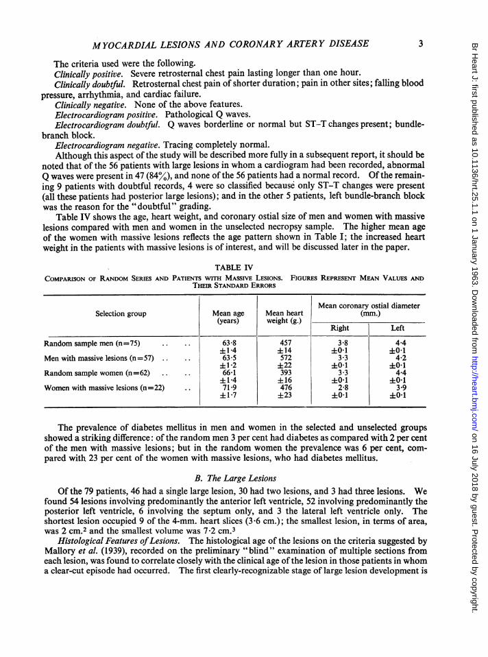
Table IV shows the age, heart weight, and coronary ostial size of men and women with massivelesions compared with men and women in the unselected necropsy sample. The higher mean ageof the women with massive lesions reflects the age pattern shown in Table I; the increased heartweight in the patients with massive lesions is of interest, and will be discussed later in the paper.
TABLE IVCOMPARISON OF RANDOM SERIES AND PATIENTS WITH MASSIVE LESIONS. FIGURES REPRESENT MEAN VALUES AND
THEIR STANDARD ERRORS
Mean coronary ostial diameterSelection group Mean age Mean heart (mm.)
(years) weight (g.)Right Left
Random sample men (n=75) .. .. 63.8 457 3.8 4.4±14 ±14 +O 1 +0O1
Men with massive lesions (n=57) .. .. 63-5 572 3-3 4*2±1*2 ±22 ±01 ±01
Random sample women (n=62) .. .. 66.1 393 3.3 4-4±1*4 ±16 ±O01 +01
Women with massive lesions (n=22) .. 71 9 476 2.8 3.9±1.7 ±23 ±01 +0O1
The prevalence of diabetes mellitus in men and women in the selected and unselected groupsshowed a striking difference: of the random men 3 per cent had diabetes as compared with 2 per centof the men with massive lesions; but in the random women the prevalence was 6 per cent, com-pared with 23 per cent of the women with massive lesions, who had diabetes mellitus.
B. The Large LesionsOf the 79 patients, 46 had a single large lesion, 30 had two lesions, and 3 had three lesions. We
found 54 lesions involving predominantly the anterior left ventricle, 52 involving predominantly theposterior left ventricle, 6 involving the septum only, and 3 the lateral left ventricle only. Theshortest lesion occupied 9 of the 4-mm. heart slices (3 6 cm.); the smallest lesion, in terms of area,was 2 cm.2 and the smallest volume was 7-2 cm.3
Histological Features ofLesions. The histological age of the lesions on the criteria suggested byMallory et al. (1939), recorded on the preliminary "blind" examination of multiple sections fromeach lesion, was found to correlate closely with the clinical age ofthe lesion in those patients in whoma clear-cut episode had occurred. The first clearly-recognizable stage of large lesion development is
3
on 16 July 2018 by guest. Protected by copyright.
http://heart.bmj.com
/B
r Heart J: first published as 10.1136/hrt.25.1.1 on 1 January 1963. D
ownloaded from

MITCHELL AND SCHWARTZ
FIG. 1.-Heart muscle fibres from massive lesion, show- FIG. 2.-Polymorphonuclear leucocyte infiltration ining variable staining. (P.T.A.H.: x 265.) massive lesion. (H. and E.: x 152.)
FIG. 3.-Endocardial aspect of massive lesion,showing zone of surviving muscle fibreson surface and necrotic fibres surroundedby polymorphs below surface layer.(Trichrome: x 90.)
4
on 16 July 2018 by guest. Protected by copyright.
http://heart.bmj.com
/B
r Heart J: first published as 10.1136/hrt.25.1.1 on 1 January 1963. D
ownloaded from

MYOCARDIAL LESIONS AND CORONARY ARTERY DISEASE
FIG. 4.-Healing massive lesion showing lymphocytes. FIG. 5.-Endocardial aspect of healing massive lesionfibroblasts, and young collagen fibres. (H. and E.: showing surviving muscle around small vessel andx 350.) on endocardial surface of lesion (cf. Fig. 3).
(Trichrome: x 68.)
marked variability of muscle fibre staining. This may be the only detectable change in the first 24hours, and can be readily seen on hematoxylin-eosin preparations, some fibres being intenselyeosinophilic. With Mallory's phosphotungstic-acid-hematoxylin stain the fibres no longer stainuniformly, but show a granular appearance (Fig. 1). From the second to the sixth day infiltrationwith neutrophil polymorphonuclear leucocytes is the characteristic feature (Fig. 2), and duringthis phase the affected muscle fibres become necrotic, with the exception of a zone of spared muscleon the endocardial surface of the lesion (Fig. 3). It seems probable that these surviving fibres arebeing nourished by the blood in the ventricular cavity. At the end of the first week, the healing phaseand the removal of necrotic fibres begins, the characteristic cellular pattern during this period beingan infiltration with lymphocytes, some plasma cells, and a fibroblastic proliferation (Fig. 4). Theremoval of dead muscle fibres often produces a striking appearance in the endocardial aspect ofthe lesion, the surface zone and the perivascular zones being spared (Fig. 5). Like Malloryet al. (1939) we have found the presence of pigment-containing macrophages to be a usefulcriterion of the age of the lesion, appearing at the end of the first week, being most conspicuous inthe second and third weeks, and then gradually disappearing. On the other hand, we consider thatthe eosinophil infiltration which they found from the seventh to the fourteenth days is an inconstantfeature. When present, eosinophils are a useful guide to the age of the lesion, but their absence doesnot mean that the lesion is outside the one-to-two-week group. We have not attempted to carry
5
ws.
on 16 July 2018 by guest. Protected by copyright.
http://heart.bmj.com
/B
r Heart J: first published as 10.1136/hrt.25.1.1 on 1 January 1963. D
ownloaded from

6 MITCHELL AND SCHWARTZ
histological ageing beyond the fourth month of development of the lesion. At this stage the necroticmuscle has been removed, neutrophils have disappeared, and the lesion consists of dense collagen witha few lymphocytes, and new blood vessels. Lesions with these features have been grouped togetheras " over 4 months " of age.
C. Coronary Arteries in Patients with Large LesionsUsing the coronary stenosis score as an overall index of the number and degree of stenosing
lesions, Fig. 6 compares male patients with large lesions with the male patients from the unselectednecropsy series without large lesions: Fig. 7 compares female patients in the same way. Consideredas a group, patients with large lesions have higher scores (and therefore more severe coronarynarrowing) than unselected patients of comparable age without large lesions. It should be notedthat there are some patients with large lesions whose coronary scores are conspicuously low, andindeed we found one woman with a large lesion whose coronary arteries showed no narrowing at anypoint.
o- RANDOM: NO LARGE LESIONS
A- RANDOM: LARGE LESIONSA-SELECTED' LARGE LESIONS
KX
80[ °0
a 60
8?E 40z0u2
C
AA
tA
0
0
a8a,
A
±
A
a
aa
a
a80
8§0
00co
8
8
A
ALAAALAAA
LA
A
o AZAAtA
0,
atA8 Ai
O iO A
0a
aa
0
0
a
880
0
AAA
A
AA
A
A
0
OD
35-54 55 - 64 65-74 75-AGE IN YEARS
FIG. 6.-Coronary scores of men with large lesionscompared with men from the random necropsysample without large lesions.
1001
0o 80s
0x 60b0
n 40z00 20
U
o- RANDOM: NO LARGE LESIONSA' RANDOM: LARGE LESIONSA-SELECTED: LARGE LESIONS
1.
I.
0'
0
.0
0oaTO
000
A
ALaa
0
a8
0a0a, A~
AAA
A
LA
a
0
a
00
A
AtA
a
Aa
tA
a
000a
aa000
A
A
35-54 55- 64 65 - 74 75 -
AGE IN YEARSFIG. 7.-Coronary scores of women with large lesions
compared with women from random necropsysample without large lesions.
As the large lesion group consists of patients with different numbers of lesions and with lesions ofdifferent ages and sizes, we have analysed them further. Fig. 8 shows that men with multiplelesions have more severe coronary stenosis than men with one lesion. Fig. 10 compares the coronaryscores of patients who died within two weeks of the development of their first large lesion, with theunselected sample, and shows that this group, especially the younger men, differs less from therandom necropsy sample than does the large lesion group as a whole.
Another reason for lack of homogeneity in the whole group of patients with large lesions is thewide variation in lesion size. Fig. 9 shows that the length of the lesion is closely correlated withthe degree of coronary stenosis. These three factors-number of lesions, age of lesions, and sizeof lesions, must be borne in mind when patients with large lesions are compared with other groups.
)F
on 16 July 2018 by guest. Protected by copyright.
http://heart.bmj.com
/B
r Heart J: first published as 10.1136/hrt.25.1.1 on 1 January 1963. D
ownloaded from

7MYOCARDIAL LESIONS AND CORONARY ARTERY DISEASE
A - I LARGE LESIONA - >1 LARGE LESION
In
6C
4C
20C
A
As
A
A6
A
35- 54 55-64 65 -74AGE IN YEARS
A
AAA
AAtiA
A
A
>1
A
A
9 A
a A
LA
AA
A
I >1
a A
A
a
AIL
a
>1
75 -
FIG. 8.-Coronary scores of men with a single large lesioncompared with men with more than one large lesion.
100
800
LLI0
z
6C
40I
20[
0
o Ro A
0
CB.
C80IL
Mr
la
a
00 A
A
0
00 A
00 A
03
o A
o0 A
88 aaa
Cc A
mu
o- LARGE LESIONS WOMEN*- LARGE LESIONS MEN
loor
0
z0
8C
60[
4C*
20
0
0
0
00
0@
0
I
3*6-6-0 64-80 >-80LESION LENGTH CM
FIG. 9.-Correlation between coronaryscore and length of large lesion asdetermined by the number of trans-verse heart sections on which thelesion was present. The horizontallines indicate the mean coronaryscore of each group.
RANDOM: NO LARGE LESIONS
0- MEN0- WOMEN
SELECTED: SINGLE LARGE LESIONS< 2 WEEKS OF AGE
A- MENA- WOMEN
35- 64 65 -
AGE IN YEARS
FiG. 10.-Coronary scores of men and women with a single large lesion of less than2 weeks' histological age, compared with men and women from the randomnecropsy sample without large lesions.
CKwix
o0Nt OF LARGE
LESIONS _>1
on 16 July 2018 by guest. Protected by copyright.
http://heart.bmj.com
/B
r Heart J: first published as 10.1136/hrt.25.1.1 on 1 January 1963. D
ownloaded from

MITCHELL AND SCHWARTZ
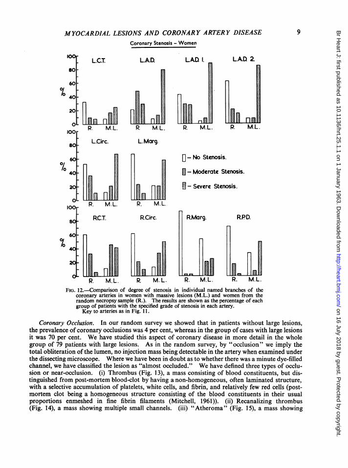
Stenosis of Individual Arteries. So far we have considered the coronary arterial tree as a wholeby using the coronary score as an overall index of severity. Fig. 11 and 12 show the percentageprevalence of the three grades of stenosis in the named arteries in men and women in the randomnecropsy sample and in men and women with large lesions. The patients with large lesions show ahigh prevalence of severe narrowing in every artery, and as in the random group, the left coronarybranches are more severely narrowed than the branches of the right coronary artery. It will alsobe noted that the marked sex difference in the prevalence of severe stenosis seen in the random seriesis abolished in the patients with large lesions, the men and women having a similar prevalence ofsevere narrowing.
Coronary Stenosis - Men.
100LC.CT L.A.D. LAD. 1. LA.D. 2.
60-
40-
100 P. M.L. R. M.L. P. M.L. P. M.L.
8 L.Circ. LMarg.
0/0 60 - No Stenosis.
40 - Moderate Stenosis.
20- Lf Ji J [ Severe Stenosis.
0 R. M.L. R. M.L.
80 R.CT. RCirc. RMarg. R.PD.
60-0/0
-
40
P. M.L. R. M. L. P. M.L. R M.L.
FIG. 1 .-Comparison of degree of stenosis in individual named branches of the coronaryarteries in men with massive lesions (M.L.) and men from the random necropsysample (R.). The results are shown as the percentage of each group of patients withthe specified grade of stenosis in each artery.LCT= Left coronary trunk. LAD= Left anterior descending. LAD I and 2=
2 main branches of LAD. L. circ= left circumflex. L. marg= left marginal.RCT= Right coronary trunk. R.circ= right circumflex. R.marg. = rightmarginal. RPD=right posterior descending.
8
on 16 July 2018 by guest. Protected by copyright.
http://heart.bmj.com
/B
r Heart J: first published as 10.1136/hrt.25.1.1 on 1 January 1963. D
ownloaded from
9MYOCARDIAL LESIONS AND CORONARY ARTERY DISEASECoronary Stenosis - Women
L CT L.A.D. LAD 1. LAD 2.
P. MMM.L.ML M.L. P. M.L.
L.Circ. L. Marg.
0- No Stenosis.
- Moderate Stenosis.
- Severe Stenosis.
P. M.L. R. M.L.
RCT. R.Circ.
fI,L
P. M.L. P. M.L.
R.Marg. R.PD.
R. M.L. R. M.L.
FIG. 12.-Comparison of degree of stenosis in individual named branches of thecoronary arteries in women with massive lesions (M.L.) and women from therandom necropsy sample (R.). The results are shown as the percentage of eachgroup of patients with the specified grade of stenosis in each artery.Key to arteries as in Fig. 11.
Coronary Occlusion. In our random survey we showed that in patients without large lesions,the prevalence of coronary occlusions was 4 per cent, whereas in the group of cases with large lesionsit was 70 per cent. We have studied this aspect of coronary disease in more detail in the wholegroup of 79 patients with large lesions. As in the random survey, by "occlusion" we imply thetotal obliteration of the lumen, no injection mass being detectable in the artery when examined underthe dissecting microscope. Where we have been in doubt as to whether there was a minute dye-filledchannel, we have classified the lesion as "almost occluded." We have defined three types of occlu-sion or near-occlusion. (i) Thrombus (Fig. 13), a mass consisting of blood constituents, but dis-tinguished from post-mortem blood-clot by having a non-homogeneous, often laminated structure,with a selective accumulation of platelets, white cells, and fibrin, and relatively few red cells (post-mortem clot being a homogeneous structure consisting of the blood constituents in their usualproportions enmeshed in fine fibrin filaments (Mitchell, 1961)). (ii) Recanalizing thrombus(Fig. 14), a mass showing multiple small channels. (iii) "Atheroma" (Fig. 15), a mass showing
lOC
80o
6010/0
40.
20
0
Inn'
0/0
80
40
2
0lOOr
80
60[0/0
40
20
0
on 16 July 2018 by guest. Protected by copyright.
http://heart.bmj.com
/B
r Heart J: first published as 10.1136/hrt.25.1.1 on 1 January 1963. D
ownloaded from

MITCHELL AND SCHWARTZ
none of the above characteristics of thrombus or recanalizing thrombus, and usually consisting offibrous tissue, cellular aggregates, and lipid deposits. As emphasized by Duguid (1946) the distinc-tion between these lesions and recanalizing thrombus is often an artificial one.We found 103 completely occluded arterial segments in the 79 patients: no occlusion in 22 patients;
1 occlusion in 27 patients; 2 occlusions in 15 patients; and 3 or more occlusions in 15 patients.Bearing in mind, however, the heterogeneous character of the group of patients with large lesions,we have subdivided them into those with only one lesion and those with multiple lesions, furtherdividing them according to the histological age of the lesions (Tables V, VI, and VII). In thegroup of patients dying after their first large lesion (Table V) the occlusion frequency and type ofoccluding lesion is clearly related to the age of the lesion. Thus, of the 25 patients with singlelesions under 4 months' histological age, 18 had complete occlusions (72%)0 6 were almost occluded(24%)0 and only 1 (40%) had simple narrowing. Of the 41 occluding and near-occluding masses 31(76%) were thrombus, 5 (12%) were recanalizing thrombus, and 5 (12%) were "atheroma." Thisgroup contrasts sharply with the 21 patients with large lesions of more than 4 months' histologicalage, of whom only 10 (48%) had occlusions, 4 (19%) were almost occluded, and 7 (330/a) showednarrowing only. Of the 30 occluding or near-occluding masses, none was composed of thrombus,24 (80%) were recanalizing thrombus, and 6 (20%) were " atheroma." It is clear that any study ofocclusion frequency and nature must take into account the age of the cardiac lesion.
Table VI shows the corresponding figures for patients with more than one lesion. Thesepatients will clearly have some residual arterial lesions from their previous episode, the frequency and
TABLE V
OCCLUSION STATUS AND NATURE OF OCCLUSION IN MEN AND WOMEN WITH SINGLE MASSIVE LESIONS OF VARIOUSHISTOLOGICAL AGES
Number of patients Nature of occlusion or near occlusion-Histological age as number of arteries
of lesionWith Almost Recanalizing
Total occlusions occluded Neither Thrombus thrombus Atheroma
Less than 2 days .. 10 7 3 0 15 0 12-13 days .. .. 1 7 3 1 15 1 12-4 weeks.. 2 2 0 0 1 3 0I-4 months .. 2 2 0 0 0 1 3Over 4 months .. 21 10 4 7 0 24 6
All ages .. 46 28 10 8 31 29 11
TABLE VIOCCLUSION STATUS AND NATURE OF OCCLUSION IN MEN AND WOMEN WITH MORE THAN ONE MASSIVE LESION, ACCORDING
TO HISTOLOGICAL AGE OF YOUNGEST MASSIVE LESION
Number of patients Nature of occlusion or near occlusion-Histological age of as number of arteriesyoungest lesionyueleoWith Almost . RecanalizingTotal occlusions occluded Neither Thrombus thrombus Atheroma
Less than 48 hours 6 5 1 0 9 5 52-13 days .. .. 13 13 0 0 21 8 62-4 weeks.. .. 5 4 1 0 3 6 31-4 months .. 9 7 2 0 1 15 5More than 4 months 0 0 0 0 0 0 0
All ages .. .. 33 29 4 0 34 34 19
10
on 16 July 2018 by guest. Protected by copyright.
http://heart.bmj.com
/B
r Heart J: first published as 10.1136/hrt.25.1.1 on 1 January 1963. D
ownloaded from

MYOCARDIAL LESIONS AND CORONARY ARTERY DISEASE
FIG. 13.-Coronary artery occluded by laminated thrombus. (P.T.A.H.: x 10.)
FIG. 14.-Recanalizing coronary thrombus. (Trichrome: x O00.)
I I
on 16 July 2018 by guest. Protected by copyright.
http://heart.bmj.com
/B
r Heart J: first published as 10.1136/hrt.25.1.1 on 1 January 1963. D
ownloaded from

MITCHELL AND SCHWARTZ
nature of these occlusions depending on the time-lapse since the first lesion. To this will be addedwhatever arterial lesions are related to their second episode. This is reflected in the nature of theocclusions or near-occlusions which show a much higher prevalence of recanalizing thrombus and" atheroma " within four months of the second lesion than was found in patients with one lesion only.Table VII summarizes the findings in the whole group of 79 patients: 26 had experienced retrosternalchest pain lasting for more than an hour, associated with the development of pathological Q wavesin the electrocardiogram, within four weeks of their death. As the findings in such patients are ofconsiderable potential importance in their management we set out their occlusion status inTable VIII.
The anatomical distribution of the complete occlusions is shown in Table IX: 69 per cent of allocclusions occurred in branches of the left coronary artery, and the majority of these were in theanterior descending artery and its branches.
.:A5,.......:
..A2
FIG. 15.-Coronary artery occluded by "atheroma." (Trichrome: x 15.)
TABLE VIIOCCLUSION STATUS AND NATURE OF OCCLUSION IN WHOLE GROUP OF PATIENTS, ACCORDING TO AGE OF MASSIVE
LESION, OR AGE OF YOUNGEST LESION WHEN MORE THAN ONE LESION PRESENT
Number of patients Nature of occlusion or near occlusion-Histological age of as number of arteriesyoungest lesion
TtlWith Almost
Nete hobs RecanalizingAhrmTotal occlusions occluded Neither Thrombus thrombus Atheroma
Less than 2 days .. 16 12 4 0 24 5 62-13 days.. 24 20 3 1 36 9 72-4 weeks ........ .. 7 6 1 0 4 9 31-4 months .. 11 9 2 0 1 16 8Over 4 months .. 21 10 4 7 0 24 6
All ages .. .. 79 57 14 8 65 63 30.__ _ _ _ _
12
on 16 July 2018 by guest. Protected by copyright.
http://heart.bmj.com
/B
r Heart J: first published as 10.1136/hrt.25.1.1 on 1 January 1963. D
ownloaded from

MYOCARDIAL LESIONS AND CORONARY ARTERY DISEASE 13
TABLE VIIIOCCLUSION STATUS OF 26 PATIENTS WHO HAD AN EPISODE OF RETROSTERNAL PAIN LASTING MORE THAN AN HOUR
ASSOCIATED WITH DEVELOPMENT OF PATHOLOGICAL Q WAVES, WITHIN FOUR WEEKS OF DEATH
Number of patients Nature of occlusion or near-occlusion-Histological age as number of arteriesof youngest lesion
With Almost RecanalizingTotal occlusions occluded Neither Thrombus thrombus Atheroma
Less than 2 days 5 4 1 0 6 4 22-13 days 16 13 2 0 25 4 62-4 weeks 5 4 1 0 3 7 3
TABLE IX
SITE OF OCCLUSIONS: NUMBER OF OCCLUSIONS FOUND IN EACH NAMED ARTERY EXPRESSED AS PERCENTAGE OF TOTALNUMBER OF OCCLUSIONS
Occlusion frequency-as percentage of all occlusions
Artery % Artery %
Left coronary trunk 0 Right coronary trunk 11Left anterior descending 23 Right circumflex 14Left anterior descending 1st branch 17 51 Right marginal 3Left anterior descending 2nd branch . I Right posterior descending 3Left circumflex 12Left marginal 4Left posterior descending 2All branches of left coronary artery 69 All branches of right coronary artery 31
TABLE X
PERCENTAGE PREVALENCE OF OTHER CARDIAC LESIONS IN MEN AND WOMEN WITH MASSIVE LESIONS, COMPARED WITHPREVALENCE IN UNSELECTED SERIES
Percentage mean prevalence
Selection group Small Fibrosis
lesionsInterstitial Perivascular Endocardial Papillary
Random sample men (n=75) 31 20 79 45 63Men with massive lesions(n=57) 28 26 81 81 90
Random sample women (n=62) 21 2 1 69 47 60Women with massive lesions(n=22) 18 14 55 73 73
D. Other Cardiac Lesions in Patients with Large LesionsThe mean percentage prevalence of the other types of lesion defined in the random necropsy
survey is shown in Table X.
on 16 July 2018 by guest. Protected by copyright.
http://heart.bmj.com
/B
r Heart J: first published as 10.1136/hrt.25.1.1 on 1 January 1963. D
ownloaded from

MITCHELL AND SCHWARTZ
SECTION LENGTH OF ALL LESIONS0- SMALL LESIONS
L G LARGE LESIONS
2
FIG16-Lnt16alterpaeetlsinon nteuslce n h
~~~~~12~~~~~~~~~~~~~~~12
U.
WC ON LDTh0234 %67 9112 13 467S6 75 1 971092 2 22233425 26 V0 2 4 6 8 K0
LENGTH OF LESIONS CMvFIG. 16.-Length of all the replacement lesions found in the unselected and the
selected necropsy sample.
(1) Small Replacement Lesions. Our survey of the unselected sample suggested that areas ofmuscle necrosis or replacement did not show a continuous size gradation, but segregated into twotypes of lesion, large and small. This view is strongly supported by the additional evidence availablefrom the selected group of patients with large lesions; Fig. 16 shows the lesion length of all the replace-ment lesions found in the two groups and confirms that the size distribution is discontinuous. Therandom survey suggested that the small lesions did not relate to coronary stenosis or occlusions,and that there was no justification for regarding them as ischemic in origin. If they were ischlmic,one would expect them to occur more often in patients with severe coronary disease or occlusion,and they should therefore be more common in the patients with large lesions than in the randomsample. This is not the case (Table X).
(2) Interstitial andPerivascular Fibrosis. This appeared to bear little relation to coronary stenosisin the random population, and this view is reinforced by our failure to find an increased prevalenceof fibrosis in the selected patients with large lesions (Table X).
(3) Endocardial Fibrosis. In our random survey a thick endocardial rind was found to occur inassQciation with large lesions and this is confirmed by the increased prevalence of endocardialfibrosis in patients with large lesions (Table X) in whom it is almost twice as common as in theunselected sample. We consider that the fibrous rind may be the end result of organized muralthrombus. Fig. 17 shows a layer of recent thrombus over a large lesion. (The endocardial zoneof relatively normal muscle is often absent or ill defined where a massive thrombus has formed.)Fig. 18 shows a cleft between papillary muscles in a large lesion in the healing phase: a zone ofspared muscle is present and on the endocardial aspect of it is a layer of organizing thrombus. At alater stage, the mass becomes less cellular, but still contains areas of fibrin (Fig. 19). The finalstage is shown in Fig. 13 of the random survey report, and this thick rind shows a marked elastosis,the inner layers forming a thick, undulating "internal elastic lamina" (Fig. 20).
(4) Papillary Fibrosis. Two types of papillary fibrosis occurred in the random sample; the first
14
on 16 July 2018 by guest. Protected by copyright.
http://heart.bmj.com
/B
r Heart J: first published as 10.1136/hrt.25.1.1 on 1 January 1963. D
ownloaded from

MYOCARDIAL LESIONS AND CORONARY ARTERY DISEASE
FIG. 17.-Endocardial aspect of massive lesion showing ill-defined zone ofsurviving muscle overlaid with recent thrombus. (Trichrome: x 90.)
showed endocardial and perivascular sparing and normal vessels (Fig. 5), whereas the second typesurrounded small vessels (which were often abnormal) and spread to the endocardial surface. Inthe patients with large lesions we find an increased prevalence of papillary fibrosis, but the differenceis less marked than for endocardial fibrosis, as one would expect if only one type of papillary fibrosiswas related to the presence of large lesions. This is confirmed in Table XI which shows that althoughthe marked grade of papillary fibrosis is more than twice as common in the large lesion group as inthe unselected group, the prevalence of small vessel disease is virtually unchanged. We considertherefore that there are two mechanisms by which papillary fibrosis can arise, one being an extensionof a large lesion, and the other being of unknown origin but showing a strong association with smallvessel thickening.
(5) The Subendocardial Plexus. The stereo-radiographs of 6 patients in the random series (4y4)showed a localized increase in vascularity in the ventricular wall, and all these patients had largelesions. Of the selected group of 79 patients with large lesions, 27 (34%/) showed this phenomenon.On the micro-radiographs of the transverse slices the increased vascularity is seen to be due to aparallel mesh of vessels running in a circular fashion around the ventricular wall (Fig. 21) and con-trasting sharply with the radially disposed branching system in the normal heart (Fig. 22). Histo-logical examination of these areas showed that the vessels had a very thin wall, consisting only of asingle layer of flattened cells, and there was no evidence of elastic or muscular tissue on conventionalstaining and microscopy (Fig. 23). The vessels looked like "giant capillaries" and were only seen
15
on 16 July 2018 by guest. Protected by copyright.
http://heart.bmj.com
/B
r Heart J: first published as 10.1136/hrt.25.1.1 on 1 January 1963. D
ownloaded from

MITCHELL AND SCHWARTZ
in healing large lesions. In the early healing phase, the channels were either empty or containedred cells only, and the injection mass did not penetrate to them At a later stage, they filled with theinjection mass, and therefore communicated with the coronary arteries by channels of more than40,u diameter. The youngest lesion in which injected channels were seen was of 4 weeks' histologicalage. It is our impression that these channels are less prominent in large lesions with a clinical age
FIG. 18.-Endocardial aspect of healing massive lesion showing zone ofsurviving muscle separating organizing thrombus from healing lesion.(Trichrome: x 90.)
TABLE XlPERCENTAGE PREVALENCE OF PAPILLARY FIBROSIS AND SMALL VESSEL DISEASE IN MEN AND WOMEN WITH MASSIVE
LESIONS, COMPARED WITH PREVALENCE IN UNSELECTED SERIES OF MEN AND WOMEN
Percentage prevalenceSelection group
Papillary fibrosis Small vessel disease
Absent Present Marked Absent Present Marked
Random sample men and women (n= 137) 38 55 7 50 38 12Men and women with massive lesions (n =79) 15 66 19 57 34 9
16
on 16 July 2018 by guest. Protected by copyright.
http://heart.bmj.com
/B
r Heart J: first published as 10.1136/hrt.25.1.1 on 1 January 1963. D
ownloaded from

MYOCARDIAL LESIONS AND CORONARY ARTERY DISEASE
FIG. 19.-Endocardial aspect ofmassive lesion showing fibrinin organizing mural throm-bus. (P.T.A.H.: x 20.)
FIG. 20.-Endocardial aspect ofmassive lesion showingelastosis in endocardialfibrous rind, separated fromsurviving muscle by denselayer of elastic tissue formingan "internal elastic lamina."(Orcein-elastic: x 45.)
c
17
I
"OW
VC,
on 16 July 2018 by guest. Protected by copyright.
http://heart.bmj.com
/B
r Heart J: first published as 10.1136/hrt.25.1.1 on 1 January 1963. D
ownloaded from

18 MITCHELL AND SCHWARTZ
FIG. 21.-Micro-radiograph of transverse section of injected heart. Histologicalexamination showed a recent antero-lateral large lesion, and a healed postero-septallesion. In this old lesion a mesh of circumferential vessels can be seen forming theso-called "subendocardial plexus."
TABLE XII
PERCENTAGE PREVALENCE OF RADIOLOGICAL CORONARY CALCIFICATION, AND MEAN NUMBER OF CALCIFIED ARTERIESPER PATIENT WITH CALCIFICATION IN MEN AND WOMEN WITH MASSIVE LESIONS. FIGURES IN BRACKETS GIVE VALUES
FouNDiN UNSELECTED SERIES OF MEN AND WOMEN
Percentage prevalence Mean number of calcified arteries per patientaffected
Age (years)Men with massive Women with massive Men with massive Women with massive
lesions lesions lesions lesions
35-54 .. .. 67 1 -4
55-64 .. .. 65 50 26 20
65-74 ..90[58] 63[21] 3
[211]3
[2.3]
75 and over .. 949704[61 2-5] 30[2-5][94] [84] [2.6] [2.2]
on 16 July 2018 by guest. Protected by copyright.
http://heart.bmj.com
/B
r Heart J: first published as 10.1136/hrt.25.1.1 on 1 January 1963. D
ownloaded from

M YOCARDIAL LESIONS AND CORONAR Y ARTER Y DISEASE
FIG. 22.-Micro-radiograph of transverse section of injected normal heart.
of a year or more, but as histological ageing beyond 4 months is unsatisfactory, we have no clearevidence of this. We consider that the channels constituting the subendocardial plexus are possiblyakin to the new vessels that occur in granulation tissue in healing lesions elsewhere in the body.
E. Other ResultsCoronary Calcification. The prevalence of radiological calcification is shown in Table XII and
is compared with that in the random sample. The men with large lesions show a much higherprevalence of calcification in the youngest age-group, but thereafter the difference narrows: thewomen with large lesions differ less from their male counterparts, age for age, than the men andwomen in the random sample. The mean number of calcified arteries in those patients who showedcalcification is higher throughout the large lesion series than in the random sample, and there is onlya slight difference between the sexes.
Dye Mixing. As in the random series we have recorded the frequency with which injection massfrom one coronary artery was detected in branches of the other artery (Table XIII), and it is clearthat cross-filling is more common, and more striking, in patients with large lesions. The increasedprevalence of dye mixing is particularly marked in terms of left-to-right flow: in the random groupwe postulated that the smaller capacity of the normal right system allowed it to fill first and spill overinto the as yet unfilled branches of the left. In the large lesion group, the higher incidence of severedisease in the branches ofthe left coronary artery may have so reduced its capacity as to allow left-to-right flow to occur.
Heart Weight. The mean heart weight of patients with large lesions is higher than that of un-selected patients (Table IV). In Fig. 24 we have plotted heart weight against diastolic blood pressurefor those patients in the random series who did not have large lesions, and for those patients with
19
on 16 July 2018 by guest. Protected by copyright.
http://heart.bmj.com
/B
r Heart J: first published as 10.1136/hrt.25.1.1 on 1 January 1963. D
ownloaded from

MITCHELL AND SCHWARTZ
FIG. 23.-"Subendocardial plexus" showing thin-walled vessels in ahealing large lesion, containing injection mass from bothcoronary arteries. The upper vessels have filled with blue massfrom the right coronary and the lower vessels with white massfrom the left coronary. (Trichrome: x 90.)
TABLE XIIIPERCENTAGE PREVALENCE OF DYE-MIXING IN MEN AND WOMEN WITH MASSIVE LESIONS COMPARED WITH PREVALENCE
IN UNSELECTED SERIES OF MEN AND WOMEN
Percentage prevalence
Selection group Right to left Left to right BothAbsent
Slight Marked Slight Marked ways
Random sample men 18 31 32 5 5 9Men with massive lesions .. 7 7 51 2 21 12Random sample women .. .. .. 24 31 37 4 4 0Women with massive lesions .. 14 14 44 0 19 9
large lesions in whom valid readings were available. The readings accepted for patients with largelesions were (i) those recorded in the 12 months preceding the development of the large lesion, eitherby the patient's general practitioner or during a hospital visit or at life insurance examination;and (ii) those recorded 6 months or more after the development of the large lesion, when the patientwas ambulant: other readings were not used. At each level of pressure, the heart weights are higherin patients with large lesions than in those without. We divided the patients with large lesions intothose with clinical evidence of cardiac failure and those without, and found that this did not account
20
on 16 July 2018 by guest. Protected by copyright.
http://heart.bmj.com
/B
r Heart J: first published as 10.1136/hrt.25.1.1 on 1 January 1963. D
ownloaded from

21MYOCARDIAL LESIONS AND CORONARY ARTERY DISEASE
MEN WOMENRANDOM: NO LARGE
°- LESIONS
U
A a
AAa
IAa
80
wo
IA
o U
A:o A
0 a
o A0A00o
a
A
0
8 Ao ACD0
00
0
U
U
i .-U
<- 90 90-109 110 -
A- SINGLE LARGELESIONS <4 MONTHS
A_ SINGLE LARGE LESIONS>4 MONTHS
MULTIPLE LARGELESIONS
< - 90 90 -109 110-,DIASTOLIC B.P
FIG. 24.-Relation between diastolic blood pressure level (mm. Hg) and heart weight in the random necropsysample and in the selected sample with massive lesions, the latter being subdivided on the basis of thenumber and the age of the lesions.
MEAN HEART WEIGHTS459 534 590
A
a* a
Q ZsnA~a
r
SINGLE SINGLE MULTIPLE
FIG. 25.-Heart weights of patients (men and women) withsingle massive lesions of less than 4 months and those ofover 4 months histological age, and with more than onemassive lesion. The figures above each group show themean heart weight in grams.
< 4 >4 ALL'AGESAGE OF LESION MONTHS
for the discrepancy. It is known that even after recovery from a large lesion the diastolic bloodpressure may remain at a lower level than was found before the lesion, so it is possible that the secondcategory of acceptable pressure readings gives falsely low values and that had we known pre-lesionpressures in all cases, the difference between the patients with and without lesions would havedisappeared. We are reluctant to accept this explanation of the findings because of the remarkablegradation of heart weight seen in the patients with large lesions, when the number and age of lesionsis considered (Fig. 25). Patients dying within four months of their first large lesion have a meanheart weight of 459 g., patients dying more than four months after the development of a single lesionhaving a mean weight of 534 g., and patients dying at any time with more than one lesion having a
' 800
603 600 .
2400
2001
0
A
0aa
1000
800
600
400
200
0
0
0
CB
,
.
aa
AA
a
0
CO
8
8 &CBCO
0
A
80
0
0
0
100O
800
60OI-w
"il-l 400
NO OFI FCuIC
on 16 July 2018 by guest. Protected by copyright.
http://heart.bmj.com
/B
r Heart J: first published as 10.1136/hrt.25.1.1 on 1 January 1963. D
ownloaded from

MITCHELL AND SCHWARTZ
mean weight of 590 g. As our study is of patients who did not survive, the apparent increase inheart weight may reflect an unfavourable prognosis in patients who have large hearts, or it may bethat heart weight does increase after a large lesion has been sustained.
DIsCUSSIONThe group of patients reported in this paper is a selected one, in that in our attempt to study
every patient with a large lesion on whom a necropsy was performed during the period concerned,we collected material from all patients who might conceivably have had such a lesion, processedthem in a standardized way, and then extracted those who showed large areas of necrosis or scarring.One must remember that all these patients died of, or with, large lesions, that they had already beenhighly selected by virtue of their admission to hospital, and that some patients coming to necropsywith large lesions may not have been included because they failed to show any of the selection criterialaid down. Nevertheless, we consider that the group can yield useful information, especially incomparison with the random group, which was equally affected by the factors of hospital admissionand death.
From the random survey we concluded that large lesions were commoner in men than women,that they occurred at an older age in the women, and that they were associated with severe coronarynarrowing and coronary occlusions. This selected group confirms these findings, and in particularemphasizes the strong correlation with coronary occlusion. This is most clearly seen in the group ofpatients with a single lesion, which shows that recent large lesions are related to coronary thrombosis,and can therefore be accurately described as myocardial infarcts. As the lesion ages, the frequencyof occlusion falls and thrombus is replaced by recanalizing thrombus or "atheroma" as the maintype of occluding mass. This is of considerable importance when one realizes that nearly half ourtotal group of patients with large lesions showed more than one lesion, and that their arteries wouldcarry a legacy of their previous episode. One cannot assess then, from the whole group, what thestate of the coronary arteries was before each patient's first lesion. The patients who provide mostinformation on this important point are those with single recent lesions (Fig. 9), and it is significantthat their coronary scores differ less from the random population than do those of the whole largelesion group. The factor that marks out these patients, therefore, is not the severity of coronarystenosis alone but the presence of coronary thrombosis. It is often suggested that wall disease mustbe present for a thrombus to form, but the main evidence for this concept is the rarity with whichthrombosis occurs in a normal vessel. However, if one postulates that stenosis and thrombosis arenot related, but are both age-dependent phenomena, thrombosis would thus be rare in normalarteries, for patients at risk from thrombosis would be unlikely to have normal arteries. There isconsiderable evidence however that thrombus and atheroma are not unrelated, but that organizingand recanalizing thrombus can produce arterial plaques (Duguid, 1946). We consider that ourfindings in lesions of different ages strongly support Duguid's view: the severe degree of stenosisseen in our complete group may in part relate to the presence in the group of patients with severallesions and with old lesions, whose arteries carry the residue of old occlusions.
We have found that the factor that marks out patients with recent large lesions from those with-out such lesions is the presence of occlusion. Previous studies have, however, given conflictingresults on the frequency of occlusion (Davenport, 1928; Barnes and Ball, 1932; Gross and Sternberg,1939; Blumgart, Schlesinger, and Davis, 1940; Master et al., 1941; Master et al., 1944; Yater et al.,1948; Harrison and Wood, 1949; Snow, Morgan Jones, and Daber, 1955, 1956). Branwood andMontgomery (1956) reviewed some of these reports and described their own series of 101 cases, ofwhich 61 had "recent myocardial infarction." Of this latter group, only 21 per cent had occludingthrombi, 38 per cent had recent non-occluding thrombus, and 18 per cent showed atheromatousocclusion. These authors commented that, "the incidence of coronary thrombotic occlusion is adirect function ofthe care and technique employed in the examination." We fully support this view,and consider that differences in technique may account for the discrepancy between our findings and
22
on 16 July 2018 by guest. Protected by copyright.
http://heart.bmj.com
/B
r Heart J: first published as 10.1136/hrt.25.1.1 on 1 January 1963. D
ownloaded from

MYOCARDIAL LESIONS AND CORONARY ARTERY DISEASE
theirs. First, they used 2 mm. thick serial sections of frozen hearts, cut on the bacon slicer to assesscoronary narrowing and occlusion. They comment that by this technique not all the coronarybranches are seen in transverse section. In our experience, this applies to the whole length of theright coronary trunk, and both circumflex arteries, and we have tried to overcome this by usinginjection and radiography to supplement serial sectioning, by clearing the heart slices for examinationunder the dissecting microscope, and by cutting any obliquely-lying artery at right angles to its axiswith a sharp blade before assessing its grade. Second, they do not define the age of their "recentinfarcts," and as we have shown that the frequency of occlusion is strongly related to the histologicalage of the large lesions, different workers' results cannot be compared unless the age range of thelesions studied is known.
There were in our series 8 patients out of 79 whom we did not assign to the occlusion or near-occlusion groups. Of these, 4 had completely healed posterior lesions and in every case there was alocalized area of very severe narrowing in the subtending circumflex artery. Although there wasonly a single channel, it is possible that these plaques represented the end result of a localizedthrombus. Two patients had, in addition to their large lesions, mitral stenosis and sub-acutebacterial endocarditis and may have had coronary emboli. Of the remaining 2 patients who bothhad healed large lesions, one had completely normal coronary arteries, and the other had two tinyplaques that produced barely perceptible narrowing. Neither patient had any source of emboli orhistory of a period of low blood pressure or arrhythmia which could have affected coronary flow.Thus 2 patients of the 79 (3%°) had large lesions for which our examination offers no explanation.
From the results of the random survey we concluded that small replacement lesions were notrelated to coronary stenosis or occlusion, and the present study confirms this view, in that the pre-valence of lesions is no higher in the selected group, whereas severe stenosis and occlusion are morecommon, than in the random group. This is also true of perivascular and interstitial fibrosis and ofthe type of papillary fibrosis associated with small vessel thickening. On the other hand, papillaryfibrosis with perivascular and endocardial sparing, and marked endocardial fibrosis are commonerin patients with large lesions, and we consider that the latter lesion may represent the end stage ofmural thrombosis.
The local increase in radiological small vessel density that was noted by Fulton (1956) and calledthe "subendocardial plexus" was thought to be a compensatory phenomenon which might preventfurther regional infarction. In our view, the vessels forming the plexus are a consequence of thehealing process of a large lesion and are akin to vessels in granulation tissue elsewhere. Theirfunctional significance is unknown, and we have no explanation for their circumferential orientation.
We have found that patients with large lesions have bigger hearts in proportion to their bloodpressure levels than patients without large lesions, thus confirming the findings of Davis and Blumgart(1937) and Morgan (1956). We accept that one possible explanation of this discrepancy could bethat some of the blood pressure readings in the large lesion groups are invalid, but the relation withinthe large lesion group between number and age of lesions and heart weight suggests that other factorsare also ofimportance. Moreover, Barrett (1960) showed that arterial wall thickness, systemic bloodpressure levels, and heart weight were clearly related in a control group of patients. Using arterialwall thickness as an index of blood pressure level, he found that patients with cardiac infarction haddisproportionately large hearts. In the absence of congenital cardiac disease, and valvular disease,cardiac enlargement is often accepted as an index of high blood pressure (Dickinson and Thomson,1960). We consider that until the relation between large lesions and heart weight is clarified,hearts with these lesions, like hearts with congenital and valvular disease, should be excluded fromany study that uses heart weight as an index of blood pressure in life.
SUMMARY
Seventy-nine patients (57 men and 22 women) with large cardiac lesions consisting of necrosis orscarring were studied and compared with an unselected necropsy sample examined in the same way.
23
on 16 July 2018 by guest. Protected by copyright.
http://heart.bmj.com
/B
r Heart J: first published as 10.1136/hrt.25.1.1 on 1 January 1963. D
ownloaded from

MITCHELL AND SCHWARTZ
Large lesions showed a strong association with coronary occlusion and can be accurately describedas myocardial infarcts. The age and number of the large lesions is related to the occlusion frequencyand the type of occluding mass. The significance of this is discussed, with particular reference to thesevere coronary narrowing associated with large lesions. Of the other cardiac lesions defined in therandom survey, two types showed a strong association with large lesions-massive endocardialfibrosis and one type of papillary fibrosis. Small replacement lesions, perivascular and interstitialfibrosis, and the papillary fibrosis associated with small vessel thickening were no more common inpatients with large lesions than in the random sample.
The histological appearance of the so-called "subendocardial plexus" is described, and therelation of this mesh of vessels to healing large lesions is emphasized.
The prevalence of coronary calcification and intercoronary anastomoses is compared with therandom sample, and an association between increased heart weight and the age and number of largelesions is described.
This work was done while J.R.A.M. was a Clinical Research Fellow of the Medical Research Council; C.J.S. wasinitially the C. J. Martin Research Fellow of the National Health and Medical Research Council of Australia and wasthereafter a member of the External Staff of the Medical Research Council.
We are grateful to Sir George Pickering and Dr. A. H. T. Robb-Smith for encouraging us to undertake this study, andto them and Dr. R. G. Macfarlane for invaluable advice and helpful criticism throughout the study. We are indebtedto the staff of the Morbid Anatomy Department, Radcliffe Infirmary, Oxford, for access to the necropsy material andto Dr. J. A. Hamill, Department of Radiology, Radcliffe Infirmary, for devising and carrying out the technique forradiographing the hearts. We are also grateful to the Medical Staff of the United Oxford Hospitals and the generalpractitioners in the area for allowing us to use their records and to question them about the patients studied. Themicroradiographs were taken by Mr. W. Charles of the Nuffield Department of Orthopedic Surgery, by courtesy ofProfessor J. Trueta. Histological preparations were made by Miss Sandra Wheeler, this assistance being provided bya Nuffield Committee grant, and were photographed in collaboration with Dr. T. M. Parry. Mr. L. P. Tugwellphotographed the diagrams and Miss Sheila Briers, Mr. Bruce Abrahams, and Mr. Paul Manners gave invaluabletechnical assistance throughout.
REFERENCESBarnes, A. R., and Ball, R. G. (1932). Amer. J. med. Sci., 183, 215.Barrett, A. M. (1960). Dissertation for the Degree of Doctor of Medicine. University of Cambridge.Blumgart, H. L., Schlesinger, M. J., and Davis, D. (1940). Amer. Heart J., 19, 1.Branwood, A. W., and Montgomery, G. L. (1956). Scot. med. J., 1, 367.Davenport, A. B. (1928). Amer. J. med. Sci., 176, 62.Davis, D., and Blumgart, H. L. (1937). Ann. intern. Med., 11, 1024.Dickinson, C. J., and Thomson, A. D. (1960). Lancet, 2, 342.Duguid, J. B. (1946). J. Path. Bact., 58, 207.Fulton, W. F. M. (1956). Brit. Heart J., 18, 341.Gross, H., and Steinberg, W. H. (1939). Arch. intern. Med., 64, 249.Harrison, C. V., and Wood, P. (1949). Brit. Heart J., 11, 205.Mallory, G. K., White, P. D., and Salcedo-Salgar, J. (1939). Amer. Heart J., 18, 647.Master, A. M., Gubner, R., Dack, S., and Jaffe, H. L. (1941). Arch. intern. Med., 67, 647.
Jaffe, H. L., Dack, S., and Grishman, A. (1944). Amer. Heart J., 27, 803.Mitchell, J. R. A. (1961). In Thrombosis and Anticoagulant Therapy, Proc. Symposium, Dundee, 1960, ed. W. Walker,
p. 9. University of St. Andrews, Livingstone, Edinburgh.Morgan, A. D. (1956). The Pathogenesis of Coronary Occlusion. Blackwell, Oxford.Schwartz, C. J., and Mitchell, J. R. A. (1962). Brit. Heart J., 24, 761.Snow, P. J. D., Morgan Jones, A., and Daber, K. S. (1955). Brit. Heart J., 17, 503.
(1956). Brit. Heart J., 18, 435.Yater, W. M., Traum, A. H., Brown, W. G., Fitzgerald, R. P., Geisler, M. A., and Wilcox, B. B. (1948). Amer. Heart
J., 36, 334 and 683.
24
on 16 July 2018 by guest. Protected by copyright.
http://heart.bmj.com
/B
r Heart J: first published as 10.1136/hrt.25.1.1 on 1 January 1963. D
ownloaded from