The Q-cycle – a personal perspective · 2004-11-20 · Peter Mitchell’s original Q-cycle...
21
Photosynthesis Research 80: 223–243, 2004. © 2004 Kluwer Academic Publishers. Printed in the Netherlands. 223 Minireview The Q-cycle – a personal perspective Antony R. Crofts Department of Biochemistry, 419 Roger Adams Laboratory, 600 South Mathews Avenue, Urbana, IL 61801, USA (e-mail: [email protected]; fax: +1-217-244-6615) Received 30 March 2003, accepted in revised form 8 August 2003 Key words: Jan Berden, Tony Crofts, Simon deVries, Les Dutton, Peter Garland, Peter Mitchell, modified Q-cycle, Bill Slater, Bernie Trumpower, Mårten Wikström Abstract In this Minireview, I provide an overview of the developments over the period 1970 to 1990 that led to the current view of the Q-cycle mechanism of the cytochrome bc 1 complex. The perspective is necessarily personal, and places some emphasis on research on the complex in the photosynthetic bacteria, where the kinetics could be studied in situ and with better time resolution than in mitochondria. Peter Mitchell’s original Q-cycle underwent several early revisions. The version of the Q-cycle currently accepted in most labs owed much to a perceptive critique by Peter Garland, who proposed a modified Q-cycle that allowed the complex to act independently. This was among several variants discussed by Mitchell in a seminal review from 1976. Six years later, despite significant advances in both mitochondrial and bacterial work, discrimination between the half-dozen or so variants that remained in active contention had proved elusive, and the kinetic data from both mitochondrial and photosynthetic systems was refractory. This was the basis of my own opposition to the Q-cycle. While trying to explain this opposition to an undergraduate student in the lab I was led to a re-evaluation of the kinetic data in the light of the substantial advances in our understanding of the biochemistry and thermodynamic properties of the complex. From this it became apparent that one version of the Q-cycle could account with satisfactory economy for the data from the photosynthetic bacteria, and for most results from work with mitochondrial complexes. The resulting model was highly constrained, and, since it incorporated Garland’s suggestions for an independent mechanism, was called the modified Q-cycle. The modified Q-cycle has stood the test of time well, and the recent structural information has both confirmed the general mechanism, and allowed extension to a more detailed understanding of the molecular architecture, and the relation between structure and function. Abbreviations: BAL – British Anti Lewisite, 2,3-dimercaptopropanol; chromatophores – sealed vesicles produced by mechanical disruption of the invaginated cell membranes from photosynthetic bacteria; Cyt – cytochrome; Cyt b H – high-potential heme of cytochrome b (also known as Cyt b K or Cyt b 561 ); Cyt b L – low-potential heme of cytochrome b (also known as Cyt b T ); Cyt bc 1 (also referred simply as bc 1 ) complex – ubiquinol:cytochrome c oxidoreductase (EC 1.10.2.2); DBMIB – 2,5-dibromo-3-methyl-6-isopropyl-p-benzoquinone; E h – ambient redox potential; E m – midpoint redox potential; ISP – (Rieske) iron–sulfur protein; P-phase, N-phase – aqueous phases in which the proton gradient is positive or negative, respectively; P + /P – oxidized and reduced forms of the reaction center primary donor; Q – quinone, oxidized form of ubiquinone; Q i -site – quinone reduc- ing site of bc 1 complex; Q o -site – quinol oxidizing site of bc 1 complex; QH 2 – quinol, reduced form of ubiquinone (ubihydroquinone); Rb. – Rhodobacter; RC – photosynthetic reaction center; Rps. – Rhodopseudo- monas; SQ – semiquinone form of ubiquinone; UHDBT – 5-undecyl-6-hydroxy-4,7-dioxobenzothiazol; UHNQ – 2-undecyl-3-hydroxy-1,4-naphthoquinone Introduction The general concept of Peter Mitchell’s Q-cycle mech- anism was, as revealed by the great man himself, ‘... invented at about 3 A. M. on Tuesday May 20, 1975 ...’ (Mitchell 1990). At that time, Peter Mitchell (1920–1992, Figure 1) was still embroiled in the great controversy about his chemiosmotic mechanism
Transcript of The Q-cycle – a personal perspective · 2004-11-20 · Peter Mitchell’s original Q-cycle...
Photosynthesis Research 80: 223–243, 2004.© 2004 Kluwer Academic Publishers. Printed in the Netherlands.
223
Minireview
The Q-cycle – a personal perspective
Antony R. CroftsDepartment of Biochemistry, 419 Roger Adams Laboratory, 600 South Mathews Avenue, Urbana, IL 61801, USA(e-mail: [email protected]; fax: +1-217-244-6615)
Received 30 March 2003, accepted in revised form 8 August 2003
Key words: Jan Berden, Tony Crofts, Simon deVries, Les Dutton, Peter Garland, Peter Mitchell, modified Q-cycle,Bill Slater, Bernie Trumpower, Mårten Wikström
Abstract
In this Minireview, I provide an overview of the developments over the period 1970 to 1990 that led to the currentview of the Q-cycle mechanism of the cytochrome bc1 complex. The perspective is necessarily personal, and placessome emphasis on research on the complex in the photosynthetic bacteria, where the kinetics could be studied insitu and with better time resolution than in mitochondria. Peter Mitchell’s original Q-cycle underwent severalearly revisions. The version of the Q-cycle currently accepted in most labs owed much to a perceptive critique byPeter Garland, who proposed a modified Q-cycle that allowed the complex to act independently. This was amongseveral variants discussed by Mitchell in a seminal review from 1976. Six years later, despite significant advancesin both mitochondrial and bacterial work, discrimination between the half-dozen or so variants that remained inactive contention had proved elusive, and the kinetic data from both mitochondrial and photosynthetic systemswas refractory. This was the basis of my own opposition to the Q-cycle. While trying to explain this oppositionto an undergraduate student in the lab I was led to a re-evaluation of the kinetic data in the light of the substantialadvances in our understanding of the biochemistry and thermodynamic properties of the complex. From this itbecame apparent that one version of the Q-cycle could account with satisfactory economy for the data from thephotosynthetic bacteria, and for most results from work with mitochondrial complexes. The resulting model washighly constrained, and, since it incorporated Garland’s suggestions for an independent mechanism, was called themodified Q-cycle. The modified Q-cycle has stood the test of time well, and the recent structural information hasboth confirmed the general mechanism, and allowed extension to a more detailed understanding of the moleculararchitecture, and the relation between structure and function.
Abbreviations: BAL – British Anti Lewisite, 2,3-dimercaptopropanol; chromatophores – sealed vesicles producedby mechanical disruption of the invaginated cell membranes from photosynthetic bacteria; Cyt – cytochrome; CytbH – high-potential heme of cytochrome b (also known as Cyt bK or Cyt b561); Cyt bL – low-potential heme ofcytochrome b (also known as Cyt bT); Cyt bc1 (also referred simply as bc1) complex – ubiquinol:cytochromec oxidoreductase (EC 1.10.2.2); DBMIB – 2,5-dibromo-3-methyl-6-isopropyl-p-benzoquinone; Eh – ambientredox potential; Em – midpoint redox potential; ISP – (Rieske) iron–sulfur protein; P-phase, N-phase – aqueousphases in which the proton gradient is positive or negative, respectively; P+/P – oxidized and reduced formsof the reaction center primary donor; Q – quinone, oxidized form of ubiquinone; Qi-site – quinone reduc-ing site of bc1 complex; Qo-site – quinol oxidizing site of bc1 complex; QH2 – quinol, reduced form ofubiquinone (ubihydroquinone); Rb. – Rhodobacter; RC – photosynthetic reaction center; Rps. – Rhodopseudo-monas; SQ – semiquinone form of ubiquinone; UHDBT – 5-undecyl-6-hydroxy-4,7-dioxobenzothiazol; UHNQ –2-undecyl-3-hydroxy-1,4-naphthoquinone
Introduction
The general concept of Peter Mitchell’s Q-cycle mech-anism was, as revealed by the great man himself,
‘. . . invented at about 3 A.M. on Tuesday May 20,1975 . . .’ (Mitchell 1990). At that time, Peter Mitchell(1920–1992, Figure 1) was still embroiled in thegreat controversy about his chemiosmotic mechanism

224
Figure 1. Peter Mitchell c. 1943 (adapted from Mitchell 1981).A young Peter Mitchell in the Department of Biochemistry atCambridge. Left to right: Joan Keilin, Jim Danielli, Peter Mitchelland Mary Danielli. The ideas of David Keilin on the cytochromesand Jim Danielli on the lipid bilayer were seminal in the de-velopment of Mitchell’s views on chemiosmosis and vectorialmetabolism.
(see Harold (2001) for a historical review, and Crofts(1993) for a brief biography). His ideas had receivedwide recognition (cf. Paul Boyer et al. 1977, and theNobel Prize in Chemistry, 1978), and there was somemeasure of agreement, after 14 years of argument, thatthe mechanism of coupling between electron transferand phosphorylation was through the coupled protonpumping activity of the electron transfer chain and theATP synthase, mediated by the electrochemical protongradient. However, as attention turned to mechanism,the central span of the mitochondrial chain was prov-ing particularly troublesome, as discussed in greaterdetail below. With Mitchell’s proposal of the Q-cycleseveral odd experimental observations on the kineticbehavior of the b and c-type cytochrome (Cyt) inter-mediates of the mitochondrial electron transfer chainwere neatly explained. In addition, his mechanismallowed for the energy transduction function of thecentral span of the chain in the context of the proton-motive activity required by the chemiosmotic hypo-thesis (Mitchell 1961, 1966, 1968, and see Jagendorf,2002, for a historical minireview). The energy cou-pling behavior was first shown by the ‘cross-overpoint’ in this span, discussed at length by BrittonChance and Ron Williams (1956). This was the namegiven to a kinetic behavior of Cyt b and Cyt c that wasobserved when the coupled state of the mitochondrialmembrane was changed. Electrons piled up in Cyt b,
and disappeared from Cyt c, as if the coupling mech-anism exerted a back-pressure on the electron transferchain at this point. The cytochrome b observed waslater called Cyt b561, or Cyt bK, where the K recog-nized the rediscovery of the cytochromes by DavidKeilin (Mitchell 1978; Ferguson 2001; see also pa-pers by D.S. Bendall and W.A. Cramer, this issue),who first identified cytochromes a, b and c in cellsand tissues, using a hand spectroscope. In the schemesshown in this review, Cyt bK is referred to as Cyt bH,where the H stands for high redox potential. Chanceand colleagues (Chance et al. 1970) later demonstratedthat two b-cytochrome components, which had previ-ously been identified in redox titrations by Les Duttonand Dave Wilson (1976, and Dutton et al. 1970), par-ticipated in the respiratory chain. They showed thatthe equilibrium constant between the two hemes wasstrongly dependent on the coupled state. The lowerpotential, longer wavelength, component was identi-fied as uniquely involved in energy transduction andcalled Cyt bT (where the T stands for transducing),and is equivalent to Cyt bL (low redox potential) inthe schemes shown in this review. At the time, thisresponse to the coupled state was taken as evidencefor a chemical high-energy intermediate. The otherkinetic effects that the Q-cycle explained were thoserelated to the phenomenon of ‘oxidant-induced reduc-tion of cytochrome b.’ Observations in several labs(nicely reviewed by Bill Slater 1981) had shown thaton addition of oxygen to mitochondrial suspensions,the c-type cytochromes became oxidized, as expectedfrom the linear electron transfer schemes current atthe time, but, unexpectedly, the b-type cytochromesbecame more reduced. This behavior was strongly en-hanced in the presence of antimycin (Wikström andBerden 1972) (see Figures 2 and 3 for group photo-graphs from two meetings some 29 years apart, withsome of the actors in the Q-cycle drama, including BritChance, hardly changed!).
Early variants of the Q-cycle theme
Mårten Wikström and Jan Berden (1972) had sug-gested a neat explanation for the oxidant-inducedreduction effect. They pointed out that oxidation ofubihydroquinone (quinol or QH2) likely occurred viaan intermediate semiquinone, opening the possibilitythat the two electrons released might be transferredto two different acceptor chains. In the scheme theysuggested (similar to that in Scheme 1 of Figure 4),
225
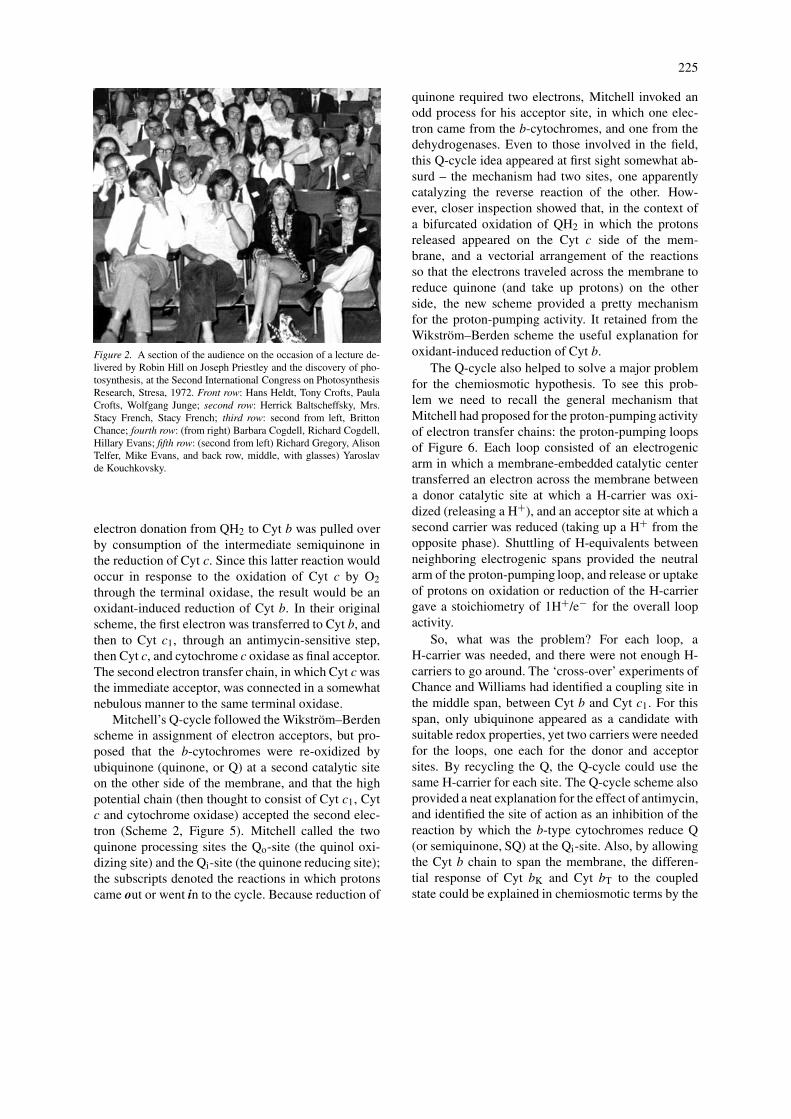
Figure 2. A section of the audience on the occasion of a lecture de-livered by Robin Hill on Joseph Priestley and the discovery of pho-tosynthesis, at the Second International Congress on PhotosynthesisResearch, Stresa, 1972. Front row: Hans Heldt, Tony Crofts, PaulaCrofts, Wolfgang Junge; second row: Herrick Baltscheffsky, Mrs.Stacy French, Stacy French; third row: second from left, BrittonChance; fourth row: (from right) Barbara Cogdell, Richard Cogdell,Hillary Evans; fifth row: (second from left) Richard Gregory, AlisonTelfer, Mike Evans, and back row, middle, with glasses) Yaroslavde Kouchkovsky.
electron donation from QH2 to Cyt b was pulled overby consumption of the intermediate semiquinone inthe reduction of Cyt c. Since this latter reaction wouldoccur in response to the oxidation of Cyt c by O2through the terminal oxidase, the result would be anoxidant-induced reduction of Cyt b. In their originalscheme, the first electron was transferred to Cyt b, andthen to Cyt c1, through an antimycin-sensitive step,then Cyt c, and cytochrome c oxidase as final acceptor.The second electron transfer chain, in which Cyt c wasthe immediate acceptor, was connected in a somewhatnebulous manner to the same terminal oxidase.
Mitchell’s Q-cycle followed the Wikström–Berdenscheme in assignment of electron acceptors, but pro-posed that the b-cytochromes were re-oxidized byubiquinone (quinone, or Q) at a second catalytic siteon the other side of the membrane, and that the highpotential chain (then thought to consist of Cyt c1, Cytc and cytochrome oxidase) accepted the second elec-tron (Scheme 2, Figure 5). Mitchell called the twoquinone processing sites the Qo-site (the quinol oxi-dizing site) and the Qi-site (the quinone reducing site);the subscripts denoted the reactions in which protonscame out or went in to the cycle. Because reduction of
quinone required two electrons, Mitchell invoked anodd process for his acceptor site, in which one elec-tron came from the b-cytochromes, and one from thedehydrogenases. Even to those involved in the field,this Q-cycle idea appeared at first sight somewhat ab-surd – the mechanism had two sites, one apparentlycatalyzing the reverse reaction of the other. How-ever, closer inspection showed that, in the context ofa bifurcated oxidation of QH2 in which the protonsreleased appeared on the Cyt c side of the mem-brane, and a vectorial arrangement of the reactionsso that the electrons traveled across the membrane toreduce quinone (and take up protons) on the otherside, the new scheme provided a pretty mechanismfor the proton-pumping activity. It retained from theWikström–Berden scheme the useful explanation foroxidant-induced reduction of Cyt b.
The Q-cycle also helped to solve a major problemfor the chemiosmotic hypothesis. To see this prob-lem we need to recall the general mechanism thatMitchell had proposed for the proton-pumping activityof electron transfer chains: the proton-pumping loopsof Figure 6. Each loop consisted of an electrogenicarm in which a membrane-embedded catalytic centertransferred an electron across the membrane betweena donor catalytic site at which a H-carrier was oxi-dized (releasing a H+), and an acceptor site at which asecond carrier was reduced (taking up a H+ from theopposite phase). Shuttling of H-equivalents betweenneighboring electrogenic spans provided the neutralarm of the proton-pumping loop, and release or uptakeof protons on oxidation or reduction of the H-carriergave a stoichiometry of 1H+/e− for the overall loopactivity.
So, what was the problem? For each loop, aH-carrier was needed, and there were not enough H-carriers to go around. The ‘cross-over’ experiments ofChance and Williams had identified a coupling site inthe middle span, between Cyt b and Cyt c1. For thisspan, only ubiquinone appeared as a candidate withsuitable redox properties, yet two carriers were neededfor the loops, one each for the donor and acceptorsites. By recycling the Q, the Q-cycle could use thesame H-carrier for each site. The Q-cycle scheme alsoprovided a neat explanation for the effect of antimycin,and identified the site of action as an inhibition of thereaction by which the b-type cytochromes reduce Q(or semiquinone, SQ) at the Qi-site. Also, by allowingthe Cyt b chain to span the membrane, the differen-tial response of Cyt bK and Cyt bT to the coupledstate could be explained in chemiosmotic terms by the
226
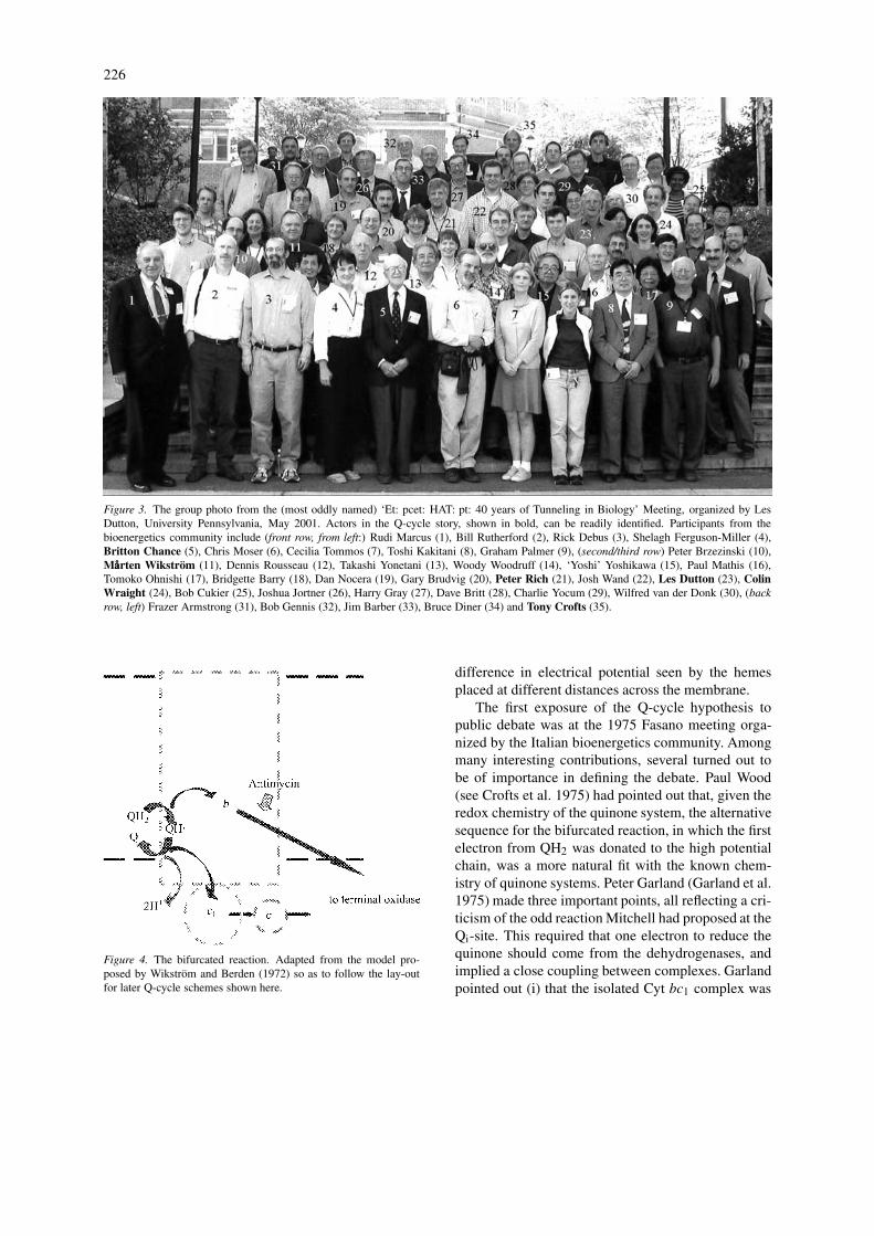
Figure 3. The group photo from the (most oddly named) ‘Et: pcet: HAT: pt: 40 years of Tunneling in Biology’ Meeting, organized by LesDutton, University Pennsylvania, May 2001. Actors in the Q-cycle story, shown in bold, can be readily identified. Participants from thebioenergetics community include (front row, from left:) Rudi Marcus (1), Bill Rutherford (2), Rick Debus (3), Shelagh Ferguson-Miller (4),Britton Chance (5), Chris Moser (6), Cecilia Tommos (7), Toshi Kakitani (8), Graham Palmer (9), (second/third row) Peter Brzezinski (10),Mååårten Wikström (11), Dennis Rousseau (12), Takashi Yonetani (13), Woody Woodruff (14), ‘Yoshi’ Yoshikawa (15), Paul Mathis (16),Tomoko Ohnishi (17), Bridgette Barry (18), Dan Nocera (19), Gary Brudvig (20), Peter Rich (21), Josh Wand (22), Les Dutton (23), ColinWraight (24), Bob Cukier (25), Joshua Jortner (26), Harry Gray (27), Dave Britt (28), Charlie Yocum (29), Wilfred van der Donk (30), (backrow, left) Frazer Armstrong (31), Bob Gennis (32), Jim Barber (33), Bruce Diner (34) and Tony Crofts (35).
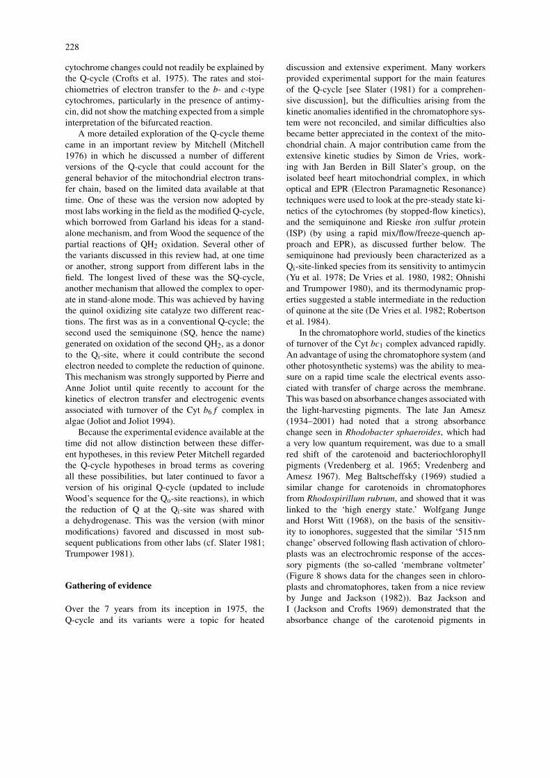
Figure 4. The bifurcated reaction. Adapted from the model pro-posed by Wikström and Berden (1972) so as to follow the lay-outfor later Q-cycle schemes shown here.
difference in electrical potential seen by the hemesplaced at different distances across the membrane.
The first exposure of the Q-cycle hypothesis topublic debate was at the 1975 Fasano meeting orga-nized by the Italian bioenergetics community. Amongmany interesting contributions, several turned out tobe of importance in defining the debate. Paul Wood(see Crofts et al. 1975) had pointed out that, given theredox chemistry of the quinone system, the alternativesequence for the bifurcated reaction, in which the firstelectron from QH2 was donated to the high potentialchain, was a more natural fit with the known chem-istry of quinone systems. Peter Garland (Garland et al.1975) made three important points, all reflecting a cri-ticism of the odd reaction Mitchell had proposed at theQi-site. This required that one electron to reduce thequinone should come from the dehydrogenases, andimplied a close coupling between complexes. Garlandpointed out (i) that the isolated Cyt bc1 complex was

227
Figure 5. Mitchell’s original Q-cycle (Mitchell 1975a). TheQ-cycle later favored by Mitchell and many others was similar, butwith the first electron transfer reaction from QH2 to the high po-tential chain, leaving semiquinone as the reductant for Cyt bL. Thecyclic nature of the reaction can be appreciated by noting that the Qconsumed and the QH2 generated at the Qi-site can be provided by(in the case of Q) or can replace (in the case of QH2) equivalentsinvolved in oxidation of QH2 at the Qo-site, by simple diffusion inthe membrane phase.
Figure 6. Mitchell’s proton pumping loops (Mitchell 1961, 1966).
able to act independently (Leung and Hinkle 1975),(ii) that reversal of the complex did not require suc-cinate dehydrogenase, and (iii) that antimycin didnot inhibit the dehydrogenases. These observationsindicated that the system operated through a self-contained mechanism. Garland suggested that a self-contained Q-cycle could be achieved if the oxidizingsite turned over twice for a complete reaction, deliv-ering two electrons through the b-cytochrome chain to
the Qi-site to generate one QH2, with a net yield of 1QH2 oxidized. He called this a modified Q-cycle.
A third set of observations that were later to fig-ure prominently in discussion related to the mismatchbetween the kinetics of reduction of Cyt b and Cytc. The kinetics, especially in the presence of antimy-cin, appeared to be contrary to the expectations ofa Q-cycle. In a simple interpretation, the bifurcatedreaction provided electrons in equal stoichiometry totwo electron transfer chains. In the isolated complex,Cyt c1 was the terminal acceptor of the high potentialchain. In the presence of antimycin, Cyt bH was theterminal acceptor of the low potential chain. From this,one might have expected that electrons would arriveat these terminal acceptors with similar rates, and inequal stoichiometry. Tsu King (King et al. 1975) re-ported briefly on the pre-steady state kinetics of theisolated complex, in which a faster reduction of Cyt bthan of Cyt c was observed, both in the presence andabsence of antimycin.
Les Dutton (see Figure 7) and Baz Jackson (Duttonand Jackson 1972a, b; Jackson and Dutton 1972),and our lab (Crofts et al. 1972, 1974; Evans andCrofts 1974), had developed protocols for detailedkinetic measurements of the activity of the Cyt bc1complex in situ in ‘chromatophores.’ These were thesealed vesicles produced from the invaginated cellmembrane by mechanical disruption of cells of thephotosynthetic bacteria Rhodopseudomonas (later re-named Rhodobacter) sphaeroides and Rb. capsulatus.In these bacteria, the Cyt bc1 complex was oxid-ized and reduced by the photochemical reaction center(RC), making it possible to activate the enzyme rap-idly with a flash of light. As I discussed at the 1975Fasano meeting, this work showed that the kinetics of
Figure 7. Left to right: Jerry Babcock, Tony Crofts and Les Dutton,on an outing from Helsinki to Tallin, Estonia, after the 7th EuropeanBioenergetics Conference (EBEC) Meeting, 1992.
228
cytochrome changes could not readily be explained bythe Q-cycle (Crofts et al. 1975). The rates and stoi-chiometries of electron transfer to the b- and c-typecytochromes, particularly in the presence of antimy-cin, did not show the matching expected from a simpleinterpretation of the bifurcated reaction.
A more detailed exploration of the Q-cycle themecame in an important review by Mitchell (Mitchell1976) in which he discussed a number of differentversions of the Q-cycle that could account for thegeneral behavior of the mitochondrial electron trans-fer chain, based on the limited data available at thattime. One of these was the version now adopted bymost labs working in the field as the modified Q-cycle,which borrowed from Garland his ideas for a stand-alone mechanism, and from Wood the sequence of thepartial reactions of QH2 oxidation. Several other ofthe variants discussed in this review had, at one timeor another, strong support from different labs in thefield. The longest lived of these was the SQ-cycle,another mechanism that allowed the complex to oper-ate in stand-alone mode. This was achieved by havingthe quinol oxidizing site catalyze two different reac-tions. The first was as in a conventional Q-cycle; thesecond used the semiquinone (SQ, hence the name)generated on oxidation of the second QH2, as a donorto the Qi-site, where it could contribute the secondelectron needed to complete the reduction of quinone.This mechanism was strongly supported by Pierre andAnne Joliot until quite recently to account for thekinetics of electron transfer and electrogenic eventsassociated with turnover of the Cyt b6f complex inalgae (Joliot and Joliot 1994).
Because the experimental evidence available at thetime did not allow distinction between these differ-ent hypotheses, in this review Peter Mitchell regardedthe Q-cycle hypotheses in broad terms as coveringall these possibilities, but later continued to favor aversion of his original Q-cycle (updated to includeWood’s sequence for the Qo-site reactions), in whichthe reduction of Q at the Qi-site was shared witha dehydrogenase. This was the version (with minormodifications) favored and discussed in most sub-sequent publications from other labs (cf. Slater 1981;Trumpower 1981).
Gathering of evidence
Over the 7 years from its inception in 1975, theQ-cycle and its variants were a topic for heated
discussion and extensive experiment. Many workersprovided experimental support for the main featuresof the Q-cycle [see Slater (1981) for a comprehen-sive discussion], but the difficulties arising from thekinetic anomalies identified in the chromatophore sys-tem were not reconciled, and similar difficulties alsobecame better appreciated in the context of the mito-chondrial chain. A major contribution came from theextensive kinetic studies by Simon de Vries, work-ing with Jan Berden in Bill Slater’s group, on theisolated beef heart mitochondrial complex, in whichoptical and EPR (Electron Paramagnetic Resonance)techniques were used to look at the pre-steady state ki-netics of the cytochromes (by stopped-flow kinetics),and the semiquinone and Rieske iron sulfur protein(ISP) (by using a rapid mix/flow/freeze-quench ap-proach and EPR), as discussed further below. Thesemiquinone had previously been characterized as aQi-site-linked species from its sensitivity to antimycin(Yu et al. 1978; De Vries et al. 1980, 1982; Ohnishiand Trumpower 1980), and its thermodynamic prop-erties suggested a stable intermediate in the reductionof quinone at the site (De Vries et al. 1982; Robertsonet al. 1984).
In the chromatophore world, studies of the kineticsof turnover of the Cyt bc1 complex advanced rapidly.An advantage of using the chromatophore system (andother photosynthetic systems) was the ability to mea-sure on a rapid time scale the electrical events asso-ciated with transfer of charge across the membrane.This was based on absorbance changes associated withthe light-harvesting pigments. The late Jan Amesz(1934–2001) had noted that a strong absorbancechange seen in Rhodobacter sphaeroides, which hada very low quantum requirement, was due to a smallred shift of the carotenoid and bacteriochlorophyllpigments (Vredenberg et al. 1965; Vredenberg andAmesz 1967). Meg Baltscheffsky (1969) studied asimilar change for carotenoids in chromatophoresfrom Rhodospirillum rubrum, and showed that it waslinked to the ‘high energy state.’ Wolfgang Jungeand Horst Witt (1968), on the basis of the sensitiv-ity to ionophores, suggested that the similar ‘515 nmchange’ observed following flash activation of chloro-plasts was an electrochromic response of the acces-sory pigments (the so-called ‘membrane voltmeter’(Figure 8 shows data for the changes seen in chloro-plasts and chromatophores, taken from a nice reviewby Junge and Jackson (1982)). Baz Jackson andI (Jackson and Crofts 1969) demonstrated that theabsorbance change of the carotenoid pigments in
229
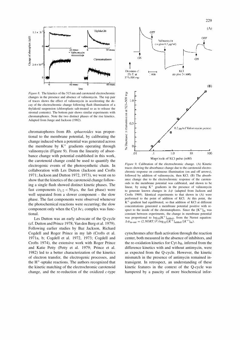
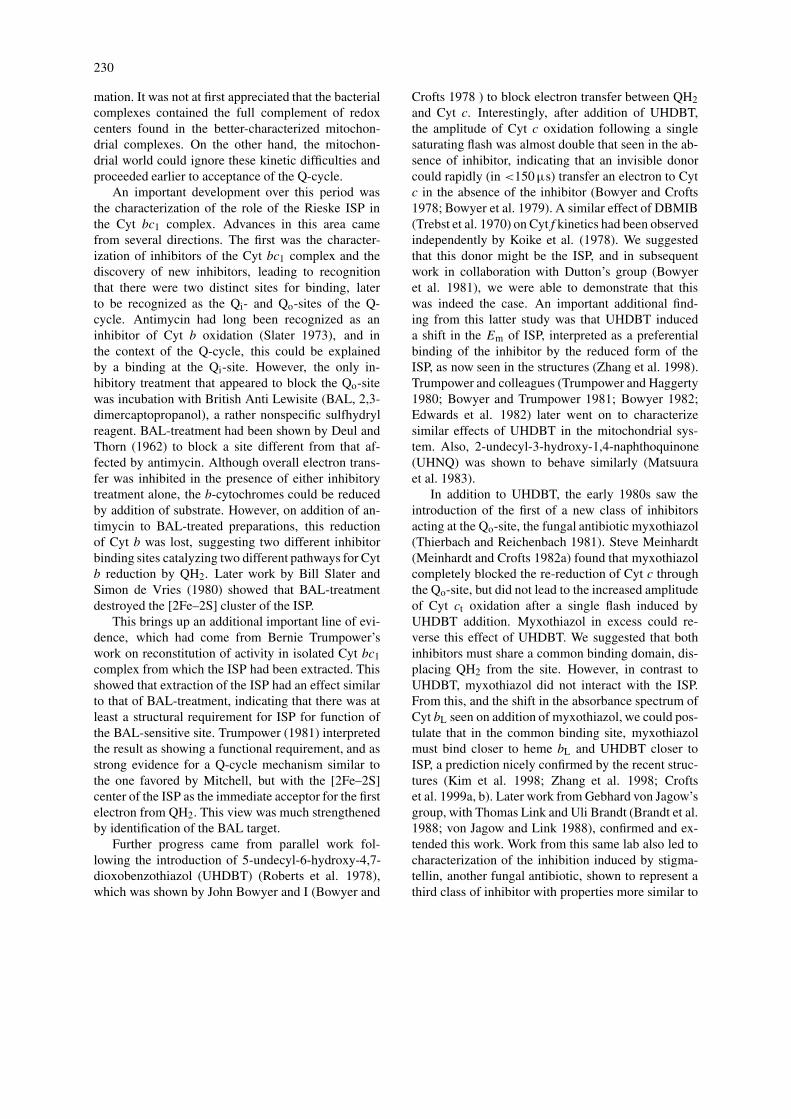
Figure 8. The kinetics of the 515 nm and carotenoid electrochromicchanges in the presence and absence of valinomycin. The top pairof traces shows the effect of valinomycin in accelerating the de-cay of the electrochromic change following flash illumination of athylakoid suspension (chloroplasts salt-treated so as to release thestromal contents). The bottom pair shows similar experiments withchromatophores. Note the two distinct phases of the rise kinetics.Adapted from Junge and Jackson (1982).
chromatophores from Rb. sphaeroides was propor-tional to the membrane potential, by calibrating thechange induced when a potential was generated acrossthe membrane by K+ gradients operating throughvalinomycin (Figure 9). From the linearity of absor-bance change with potential established in this work,the carotenoid change could be used to quantify theelectrogenic events of the photosynthetic chain. Incollaboration with Les Dutton (Jackson and Crofts1971; Jackson and Dutton 1972, 1973), we went on toshow that the kinetics of the carotenoid change follow-ing a single flash showed distinct kinetic phases. Thefast components (t1/2< 50 µs, the fast phase) werewell separated from a slower component – the slowphase. The fast components were observed wheneverthe photochemical reactions were occurring; the slowcomponent only when the Cyt bc1 complex was func-tional.
Les Dutton was an early advocate of the Q-cycle(cf. Dutton and Prince 1978; Van den Berg et al. 1979).Following earlier studies by Baz Jackson, RichardCogdell and Roger Prince in my lab (Crofts et al.1971a, b; Cogdell et al. 1972, 1973; Cogdell andCrofts 1974), the extensive work with Roger Princeand Katie Petty (Petty et al. 1979; Prince et al.1982) led to a better characterization of the kineticsof electron transfer, the electrogenic processes, andthe H+-uptake reactions. The authors recognized thatthe kinetic matching of the electrochromic carotenoidchange, and the re-reduction of the oxidized c-type
Figure 9. Calibration of the electrochromic change. (A) Kinetictraces showing the absorbance change due to the carotenoid electro-chromic response on continuous illumination (on and off arrows),followed by addition of valinomycin, then KCl. (B) The absorb-ance change due to the electrochromic response of the caroten-oids to the membrane potential was calibrated, and shown to belinear, by using K+ gradients in the presence of valinomycinto generate known changes in ψ (adapted from Jackson andCrofts 1969). Identical experiments to that shown in (A) wereperformed to the point of addition of KCl. At this point, theK+-gradient had equilibrated, so that addition of KCl at differentconcentrations generated a membrane potential positive with re-spect to the inside of the chromatophores. Since the [K+]in wasconstant between experiments, the change in membrane potentialwas proportional to log10[K+]added, from the Nernst equation:ψin-out = (2.303RT/F) log10([K+]added/[K+]in).
cytochromes after flash activation through the reactioncenter, both measured in the absence of inhibitors, andthe re-oxidation kinetics for Cyt bH, inferred from thedifference kinetics with and without antimycin, wereas expected from the Q-cycle. However, the kineticmismatch in the presence of antimycin remained in-transigent. In retrospect, an understanding of thesekinetic features in the context of the Q-cycle washampered by a paucity of more biochemical infor-
230
mation. It was not at first appreciated that the bacterialcomplexes contained the full complement of redoxcenters found in the better-characterized mitochon-drial complexes. On the other hand, the mitochon-drial world could ignore these kinetic difficulties andproceeded earlier to acceptance of the Q-cycle.
An important development over this period wasthe characterization of the role of the Rieske ISP inthe Cyt bc1 complex. Advances in this area camefrom several directions. The first was the character-ization of inhibitors of the Cyt bc1 complex and thediscovery of new inhibitors, leading to recognitionthat there were two distinct sites for binding, laterto be recognized as the Qi- and Qo-sites of the Q-cycle. Antimycin had long been recognized as aninhibitor of Cyt b oxidation (Slater 1973), and inthe context of the Q-cycle, this could be explainedby a binding at the Qi-site. However, the only in-hibitory treatment that appeared to block the Qo-sitewas incubation with British Anti Lewisite (BAL, 2,3-dimercaptopropanol), a rather nonspecific sulfhydrylreagent. BAL-treatment had been shown by Deul andThorn (1962) to block a site different from that af-fected by antimycin. Although overall electron trans-fer was inhibited in the presence of either inhibitorytreatment alone, the b-cytochromes could be reducedby addition of substrate. However, on addition of an-timycin to BAL-treated preparations, this reductionof Cyt b was lost, suggesting two different inhibitorbinding sites catalyzing two different pathways for Cytb reduction by QH2. Later work by Bill Slater andSimon de Vries (1980) showed that BAL-treatmentdestroyed the [2Fe–2S] cluster of the ISP.
This brings up an additional important line of evi-dence, which had come from Bernie Trumpower’swork on reconstitution of activity in isolated Cyt bc1complex from which the ISP had been extracted. Thisshowed that extraction of the ISP had an effect similarto that of BAL-treatment, indicating that there was atleast a structural requirement for ISP for function ofthe BAL-sensitive site. Trumpower (1981) interpretedthe result as showing a functional requirement, and asstrong evidence for a Q-cycle mechanism similar tothe one favored by Mitchell, but with the [2Fe–2S]center of the ISP as the immediate acceptor for the firstelectron from QH2. This view was much strengthenedby identification of the BAL target.
Further progress came from parallel work fol-lowing the introduction of 5-undecyl-6-hydroxy-4,7-dioxobenzothiazol (UHDBT) (Roberts et al. 1978),which was shown by John Bowyer and I (Bowyer and
Crofts 1978 ) to block electron transfer between QH2and Cyt c. Interestingly, after addition of UHDBT,the amplitude of Cyt c oxidation following a singlesaturating flash was almost double that seen in the ab-sence of inhibitor, indicating that an invisible donorcould rapidly (in <150 µs) transfer an electron to Cytc in the absence of the inhibitor (Bowyer and Crofts1978; Bowyer et al. 1979). A similar effect of DBMIB(Trebst et al. 1970) on Cyt f kinetics had been observedindependently by Koike et al. (1978). We suggestedthat this donor might be the ISP, and in subsequentwork in collaboration with Dutton’s group (Bowyeret al. 1981), we were able to demonstrate that thiswas indeed the case. An important additional find-ing from this latter study was that UHDBT induceda shift in the Em of ISP, interpreted as a preferentialbinding of the inhibitor by the reduced form of theISP, as now seen in the structures (Zhang et al. 1998).Trumpower and colleagues (Trumpower and Haggerty1980; Bowyer and Trumpower 1981; Bowyer 1982;Edwards et al. 1982) later went on to characterizesimilar effects of UHDBT in the mitochondrial sys-tem. Also, 2-undecyl-3-hydroxy-1,4-naphthoquinone(UHNQ) was shown to behave similarly (Matsuuraet al. 1983).
In addition to UHDBT, the early 1980s saw theintroduction of the first of a new class of inhibitorsacting at the Qo-site, the fungal antibiotic myxothiazol(Thierbach and Reichenbach 1981). Steve Meinhardt(Meinhardt and Crofts 1982a) found that myxothiazolcompletely blocked the re-reduction of Cyt c throughthe Qo-site, but did not lead to the increased amplitudeof Cyt ct oxidation after a single flash induced byUHDBT addition. Myxothiazol in excess could re-verse this effect of UHDBT. We suggested that bothinhibitors must share a common binding domain, dis-placing QH2 from the site. However, in contrast toUHDBT, myxothiazol did not interact with the ISP.From this, and the shift in the absorbance spectrum ofCyt bL seen on addition of myxothiazol, we could pos-tulate that in the common binding site, myxothiazolmust bind closer to heme bL and UHDBT closer toISP, a prediction nicely confirmed by the recent struc-tures (Kim et al. 1998; Zhang et al. 1998; Croftset al. 1999a, b). Later work from Gebhard von Jagow’sgroup, with Thomas Link and Uli Brandt (Brandt et al.1988; von Jagow and Link 1988), confirmed and ex-tended this work. Work from this same lab also led tocharacterization of the inhibition induced by stigma-tellin, another fungal antibiotic, shown to represent athird class of inhibitor with properties more similar to
231
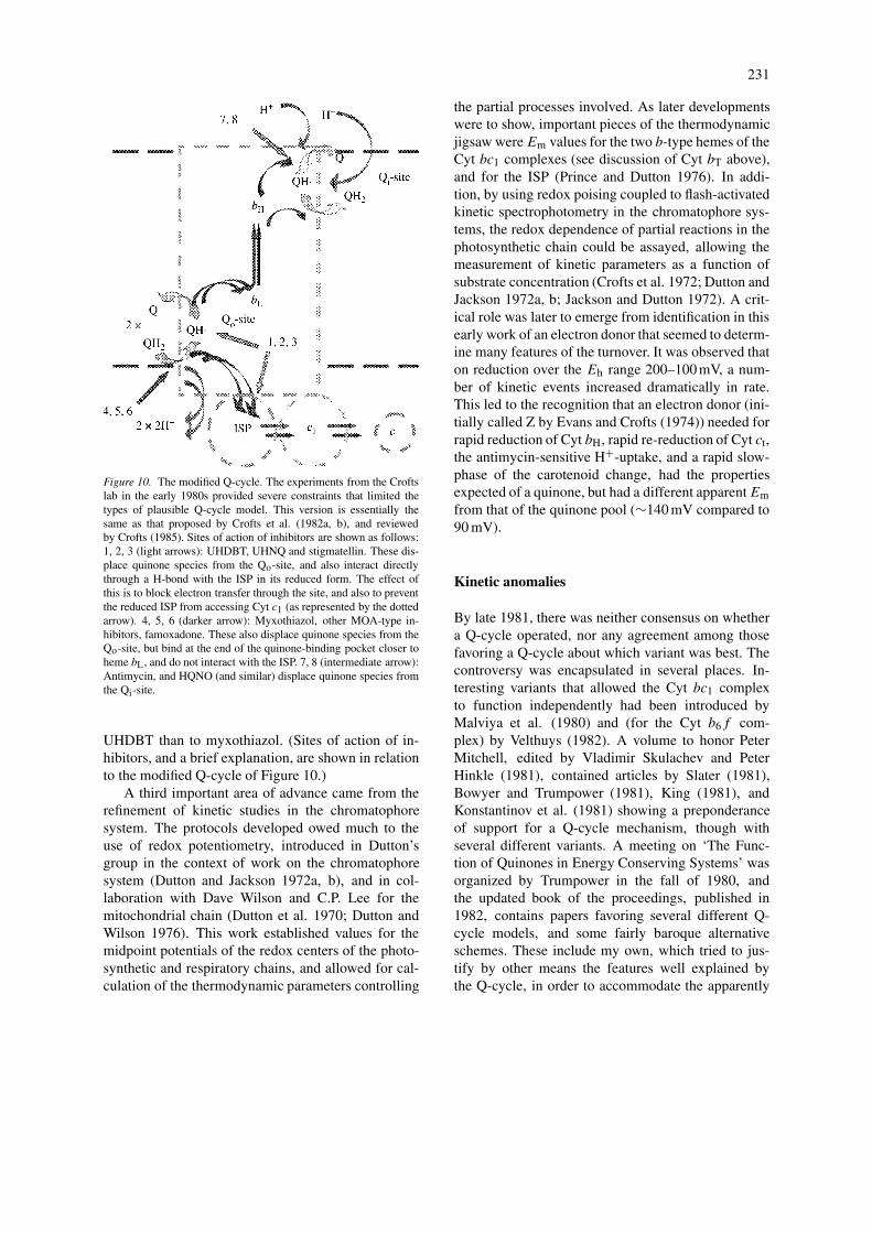
Figure 10. The modified Q-cycle. The experiments from the Croftslab in the early 1980s provided severe constraints that limited thetypes of plausible Q-cycle model. This version is essentially thesame as that proposed by Crofts et al. (1982a, b), and reviewedby Crofts (1985). Sites of action of inhibitors are shown as follows:1, 2, 3 (light arrows): UHDBT, UHNQ and stigmatellin. These dis-place quinone species from the Qo-site, and also interact directlythrough a H-bond with the ISP in its reduced form. The effect ofthis is to block electron transfer through the site, and also to preventthe reduced ISP from accessing Cyt c1 (as represented by the dottedarrow). 4, 5, 6 (darker arrow): Myxothiazol, other MOA-type in-hibitors, famoxadone. These also displace quinone species from theQo-site, but bind at the end of the quinone-binding pocket closer toheme bL, and do not interact with the ISP. 7, 8 (intermediate arrow):Antimycin, and HQNO (and similar) displace quinone species fromthe Qi-site.
UHDBT than to myxothiazol. (Sites of action of in-hibitors, and a brief explanation, are shown in relationto the modified Q-cycle of Figure 10.)
A third important area of advance came from therefinement of kinetic studies in the chromatophoresystem. The protocols developed owed much to theuse of redox potentiometry, introduced in Dutton’sgroup in the context of work on the chromatophoresystem (Dutton and Jackson 1972a, b), and in col-laboration with Dave Wilson and C.P. Lee for themitochondrial chain (Dutton et al. 1970; Dutton andWilson 1976). This work established values for themidpoint potentials of the redox centers of the photo-synthetic and respiratory chains, and allowed for cal-culation of the thermodynamic parameters controlling
the partial processes involved. As later developmentswere to show, important pieces of the thermodynamicjigsaw wereEm values for the two b-type hemes of theCyt bc1 complexes (see discussion of Cyt bT above),and for the ISP (Prince and Dutton 1976). In addi-tion, by using redox poising coupled to flash-activatedkinetic spectrophotometry in the chromatophore sys-tems, the redox dependence of partial reactions in thephotosynthetic chain could be assayed, allowing themeasurement of kinetic parameters as a function ofsubstrate concentration (Crofts et al. 1972; Dutton andJackson 1972a, b; Jackson and Dutton 1972). A crit-ical role was later to emerge from identification in thisearly work of an electron donor that seemed to determ-ine many features of the turnover. It was observed thaton reduction over the Eh range 200–100 mV, a num-ber of kinetic events increased dramatically in rate.This led to the recognition that an electron donor (ini-tially called Z by Evans and Crofts (1974)) needed forrapid reduction of Cyt bH, rapid re-reduction of Cyt ct,the antimycin-sensitive H+-uptake, and a rapid slow-phase of the carotenoid change, had the propertiesexpected of a quinone, but had a different apparentEmfrom that of the quinone pool (∼140 mV compared to90 mV).
Kinetic anomalies
By late 1981, there was neither consensus on whethera Q-cycle operated, nor any agreement among thosefavoring a Q-cycle about which variant was best. Thecontroversy was encapsulated in several places. In-teresting variants that allowed the Cyt bc1 complexto function independently had been introduced byMalviya et al. (1980) and (for the Cyt b6f com-plex) by Velthuys (1982). A volume to honor PeterMitchell, edited by Vladimir Skulachev and PeterHinkle (1981), contained articles by Slater (1981),Bowyer and Trumpower (1981), King (1981), andKonstantinov et al. (1981) showing a preponderanceof support for a Q-cycle mechanism, though withseveral different variants. A meeting on ‘The Func-tion of Quinones in Energy Conserving Systems’ wasorganized by Trumpower in the fall of 1980, andthe updated book of the proceedings, published in1982, contains papers favoring several different Q-cycle models, and some fairly baroque alternativeschemes. These include my own, which tried to jus-tify by other means the features well explained bythe Q-cycle, in order to accommodate the apparently
232
intractable kinetic problems that seemed against it.Also around this time, Colin Wraight wrote a nicereview (1982) in which he referred to the state ofresearch as a Benghazi gallop, and illustrated the con-fusion by schemes for Q-cycle and linear pathwaysthat could have served as choreography for the Whirl-ing Dervishes. In this same volume, William (Bill)Cramer and Crofts (1982) reviewed the photosyntheticarena, and discussed two different Q-cycle variants forthe chloroplast and chromatophore complexes, the lat-ter model being the modified Q-cycle that my lab hadrecently adopted (Crofts and Meinhardt 1982; Croftset al. 1982b). The chloroplast Q-cycle discussed wasan interesting variant suggested by Bruno Velthuys(1982) in which the b-heme chain provided a par-allel pathway for electron transfer between the Qo-and Qi-sites. A somewhat later contribution by PeterRich (1984) provided a serious look at the underlyingphysico-chemical constraints, and careful review ofseveral variants of the Q-cycle still considered to bein serious contention. These included the SQ- and b-cycles that he, and Mårten Wikström (Wikström et al.1981), were enamored of at the time. Although spaceconsiderations preclude detailed discussion of furtherdevelopments in the Cyt b6f field (see G. Hauska,this issue), the early contributions of John Whitmarsh(Selak and Whitmarsh 1982; Jones and Whitmarsh1988), and David Crowther and Geoffery Hind (1982)deserve recognition.
Although the question of the kinetic anomalieslooked hopelessly confused, the groundwork for aresolution in the chromatophore system was alreadylaid. The careful work of John Bowyer in quantifyingthe components of the photosynthetic chain showedthat the stoichiometric ratio of reaction center (RC)to Cyt bc1 complex in Rb. sphaeroides was close to2 (Bowyer et al. 1979, 1981; Crofts et al. 1982a).Bowyer had also demonstrated that Cyt bL was aplayer in the chromatophore chain, and could be rap-idly reduced on a second flash in antimycin inhibitedchromatophores (Bowyer et al. 1981; Crofts et al.1982a). Paul Wood had identified Cyt c1 as a com-ponent of the high potential chain through biochemicalwork (Wood 1980) and we had demonstrated its in-volvement kinetically (Crofts et al. 1982a; Meinhardtand Crofts 1982b). Together with the work showinginvolvement of the ISP, these contributions demon-strated that the bacterial complex had the same redoxcenters as the mitochondrial complex.
Why did the kinetics appear to be in contradictionto the Q-cycle? In steady-state experiments, the ki-
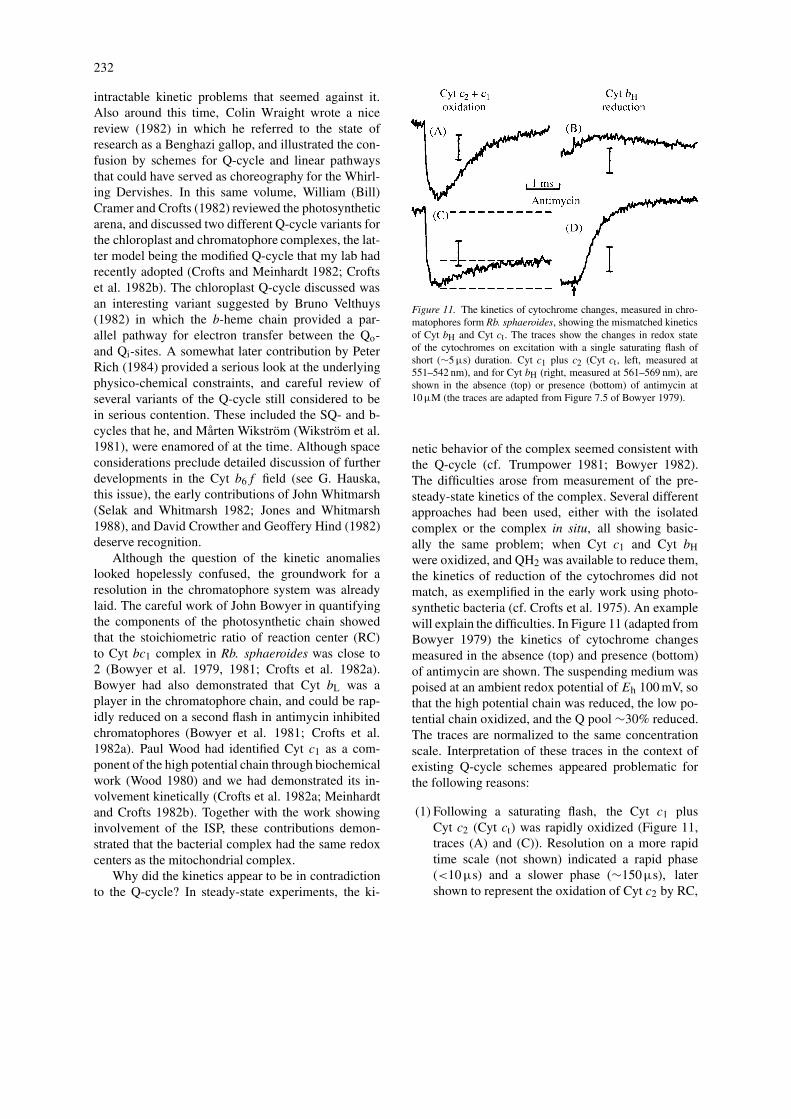
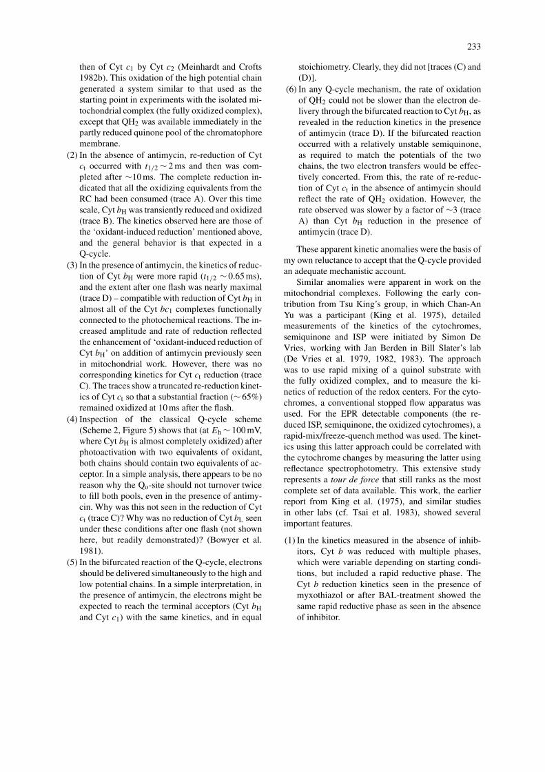
Figure 11. The kinetics of cytochrome changes, measured in chro-matophores form Rb. sphaeroides, showing the mismatched kineticsof Cyt bH and Cyt ct. The traces show the changes in redox stateof the cytochromes on excitation with a single saturating flash ofshort (∼5 µs) duration. Cyt c1 plus c2 (Cyt ct, left, measured at551–542 nm), and for Cyt bH (right, measured at 561–569 nm), areshown in the absence (top) or presence (bottom) of antimycin at10 µM (the traces are adapted from Figure 7.5 of Bowyer 1979).
netic behavior of the complex seemed consistent withthe Q-cycle (cf. Trumpower 1981; Bowyer 1982).The difficulties arose from measurement of the pre-steady-state kinetics of the complex. Several differentapproaches had been used, either with the isolatedcomplex or the complex in situ, all showing basic-ally the same problem; when Cyt c1 and Cyt bHwere oxidized, and QH2 was available to reduce them,the kinetics of reduction of the cytochromes did notmatch, as exemplified in the early work using photo-synthetic bacteria (cf. Crofts et al. 1975). An examplewill explain the difficulties. In Figure 11 (adapted fromBowyer 1979) the kinetics of cytochrome changesmeasured in the absence (top) and presence (bottom)of antimycin are shown. The suspending medium waspoised at an ambient redox potential of Eh 100 mV, sothat the high potential chain was reduced, the low po-tential chain oxidized, and the Q pool ∼30% reduced.The traces are normalized to the same concentrationscale. Interpretation of these traces in the context ofexisting Q-cycle schemes appeared problematic forthe following reasons:
(1) Following a saturating flash, the Cyt c1 plusCyt c2 (Cyt ct) was rapidly oxidized (Figure 11,traces (A) and (C)). Resolution on a more rapidtime scale (not shown) indicated a rapid phase(<10 µs) and a slower phase (∼150µs), latershown to represent the oxidation of Cyt c2 by RC,
233
then of Cyt c1 by Cyt c2 (Meinhardt and Crofts1982b). This oxidation of the high potential chaingenerated a system similar to that used as thestarting point in experiments with the isolated mi-tochondrial complex (the fully oxidized complex),except that QH2 was available immediately in thepartly reduced quinone pool of the chromatophoremembrane.
(2) In the absence of antimycin, re-reduction of Cytct occurred with t1/2 ∼ 2 ms and then was com-pleted after ∼10 ms. The complete reduction in-dicated that all the oxidizing equivalents from theRC had been consumed (trace A). Over this timescale, Cyt bH was transiently reduced and oxidized(trace B). The kinetics observed here are those ofthe ‘oxidant-induced reduction’ mentioned above,and the general behavior is that expected in aQ-cycle.
(3) In the presence of antimycin, the kinetics of reduc-tion of Cyt bH were more rapid (t1/2 ∼ 0.65 ms),and the extent after one flash was nearly maximal(trace D) – compatible with reduction of Cyt bH inalmost all of the Cyt bc1 complexes functionallyconnected to the photochemical reactions. The in-creased amplitude and rate of reduction reflectedthe enhancement of ‘oxidant-induced reduction ofCyt bH’ on addition of antimycin previously seenin mitochondrial work. However, there was nocorresponding kinetics for Cyt ct reduction (traceC). The traces show a truncated re-reduction kinet-ics of Cyt ct so that a substantial fraction (∼ 65%)remained oxidized at 10 ms after the flash.
(4) Inspection of the classical Q-cycle scheme(Scheme 2, Figure 5) shows that (at Eh ∼ 100 mV,where Cyt bH is almost completely oxidized) afterphotoactivation with two equivalents of oxidant,both chains should contain two equivalents of ac-ceptor. In a simple analysis, there appears to be noreason why the Qo-site should not turnover twiceto fill both pools, even in the presence of antimy-cin. Why was this not seen in the reduction of Cytct (trace C)? Why was no reduction of Cyt bL seenunder these conditions after one flash (not shownhere, but readily demonstrated)? (Bowyer et al.1981).
(5) In the bifurcated reaction of the Q-cycle, electronsshould be delivered simultaneously to the high andlow potential chains. In a simple interpretation, inthe presence of antimycin, the electrons might beexpected to reach the terminal acceptors (Cyt bHand Cyt c1) with the same kinetics, and in equal
stoichiometry. Clearly, they did not [traces (C) and(D)].
(6) In any Q-cycle mechanism, the rate of oxidationof QH2 could not be slower than the electron de-livery through the bifurcated reaction to Cyt bH, asrevealed in the reduction kinetics in the presenceof antimycin (trace D). If the bifurcated reactionoccurred with a relatively unstable semiquinone,as required to match the potentials of the twochains, the two electron transfers would be effec-tively concerted. From this, the rate of re-reduc-tion of Cyt ct in the absence of antimycin shouldreflect the rate of QH2 oxidation. However, therate observed was slower by a factor of ∼3 (traceA) than Cyt bH reduction in the presence ofantimycin (trace D).
These apparent kinetic anomalies were the basis ofmy own reluctance to accept that the Q-cycle providedan adequate mechanistic account.
Similar anomalies were apparent in work on themitochondrial complexes. Following the early con-tribution from Tsu King’s group, in which Chan-AnYu was a participant (King et al. 1975), detailedmeasurements of the kinetics of the cytochromes,semiquinone and ISP were initiated by Simon DeVries, working with Jan Berden in Bill Slater’s lab(De Vries et al. 1979, 1982, 1983). The approachwas to use rapid mixing of a quinol substrate withthe fully oxidized complex, and to measure the ki-netics of reduction of the redox centers. For the cyto-chromes, a conventional stopped flow apparatus wasused. For the EPR detectable components (the re-duced ISP, semiquinone, the oxidized cytochromes), arapid-mix/freeze-quench method was used. The kinet-ics using this latter approach could be correlated withthe cytochrome changes by measuring the latter usingreflectance spectrophotometry. This extensive studyrepresents a tour de force that still ranks as the mostcomplete set of data available. This work, the earlierreport from King et al. (1975), and similar studiesin other labs (cf. Tsai et al. 1983), showed severalimportant features.
(1) In the kinetics measured in the absence of inhib-itors, Cyt b was reduced with multiple phases,which were variable depending on starting condi-tions, but included a rapid reductive phase. TheCyt b reduction kinetics seen in the presence ofmyxothiazol or after BAL-treatment showed thesame rapid reductive phase as seen in the absenceof inhibitor.
234
(2) In the presence of antimycin, the rate was slower,and the kinetics simpler. When wavelengths ap-propriate for Cyt bH were used, the kinetics werefairly monotonic.
(3) In the absence or presence of antimycin, the kinet-ics of Cyt c1 reduction were slower than those ofCyt b, and of lower amplitude. In particular, thekinetics in the presence of antimycin were muchslower than the Cyt b reduction in the absence ofantimycin.
(4) In the absence of antimycin, the Qi-site semiqui-none appeared with kinetics similar to those forreduction of Cyt b. Neither the rapid reductionof Cyt b, nor the appearance of semiquinone,was inhibited by BAL-treatment or myxothiazoladdition.
(5) On reduction of the fully oxidized complex, thesignal of the reduced ISP appeared with kineticssimilar to those of Cyt c1 reduction.
Interpretation of these results was again difficult inthe context of the simple Q-cycle scheme. Some ofthe difficulties, in particular the failure to match thekinetics of reduction of Cyt c1 and Cyt bH in the pres-ence of antimycin, were similar to those observed inthe chromatophore system. Other aspects, in particularthe multiphasic kinetics of Cyt b reduction in the ab-sence of inhibitors, were peculiar to the experimentalapproach. For this latter case, the fact that similar rapidkinetics were observed in the presence of myxothiazol,or after BAL-treatment, demonstrated that reductionof Cyt b through the antimycin-sensitive site was morerapid than that through the BAL/myxothiazol-sensitivesite (Slater 1981; De Vries et al. 1983). This antimy-cin sensitivity showed that the most rapid phase of themultiphasic kinetics was a Qi-site function. However,the results left open the question of the rate of oxida-tion of QH2 at the Qo-site in the absence of antimycin.Was the rate equal to or faster than that observedthrough the reduction of Cyt b in the presence of an-timycin? In either case, the slow and/or incompletereduction of Cyt c1 (or Cyt ct in the chromatophoresystem) was clearly contrary to expectations from asimple interpretation of the Q-cycle mechanism.
The complexities were compounded by an ap-parent proliferation of redox species in the Cyt bc1complex. Redox titrations had identified at least threeCyt b species (Dutton and Jackson 1972a, b; Duttonand Wilson 1976). The low potential component, CytbL (also known as Cyt b-566, or Cyt bT) had twopeaks, and some labs reported that these appeared
to behave independently, suggesting a total of fourb-heme components. The ISP also showed severalforms, depending on the state of the quinone pool. DeVries et al. (1983) had proposed an ingenious but elab-orate double Q-cycle to accommodate these differentspecies.
The modified Q-cycle
The process by which I changed my point of view withrespect to the Q-cycle was an epiphany of sorts, butalso somewhat salutary. A bright young undergradu-ate student, Kevin Jones, was doing a research projectin the lab involving measurements of the kinetics ofthe Cyt bc1 complex. Sometime around mid Octo-ber 1981, he asked me if I could once again explainwhy the kinetics we were observing were inconsis-tent with the Q-cycle. With a sigh, I turned to theblackboard, drew up the standard Q-cycle scheme, andstarted saying ‘Well, Kevin – it’s pretty simple. Weexpect the electrons in these two chains to end upin Cyt bH and Cyt ct, . . .’ at which point I suddenlyrealized that things were not that simple. I was reit-erating ideas ingrained from an earlier period, beforewe had all the new information about stoichiometryand thermodynamic properties of components, and theinvolvement of Cyt bL, Cyt c1, and the Rieske ISP. Tomy shame, I had neither thought through the impli-cations in terms of the equilibrium constants involved,nor taken account of the stoichiometric consequences.What stopped me in mid flow was the realization thatthree new factors had to be included:
(1) The stoichiometry determined for our chromato-phores prepared from Rb. sphaeroides Ga strainwas always close to 2RC:1 Cyt c2:1Cyt bc1 (Croftset al. 1982). As a consequence, two oxidizing equi-valents were generated for each Cyt bc1 complexfollowing a saturating flash. The kinetics of Cytct changes showed that all Cyt c oxidized afterthe flash was re-reduced within 10 ms if the Q-pool was initially partly reduced (Figure 10). Toaccount for this in a Q-cycle, the Qo-site wouldhave to turnover twice.
(2) From the Em values of Cyt c1, ISP, Cyt c2, andthe RC donor, P, the equilibrium constants de-termining distribution of electrons and oxidizingequivalents (electron ‘holes’) in the high potentialchain could be calculated. On generation of 2 P+by a flash, the holes would end up in the lower po-tential components. Because Em(ISP) (∼300 mV)

235
was higher than Em(Cyt c1) (∼270 mV), the lat-ter would be preferentially oxidized as holes wereintroduced into the initially reduced high potentialchain. Conversely, ISP would be preferentially re-duced as electrons entered the oxidized chain asthe Qo-site turned over.
(3) The equilibrium constants for the bifurcated reac-tion at the Qo-site could also be calculated. In thepresence of antimycin, it was obvious that the firstquinol oxidized would experience a different valuefor Keq than the second quinol. This is becauseoxidation of the first quinol would consume themost favorable acceptors in each of the two chains– Cyt bH in the low potential chain and ISP in thehigh potential chain – leaving the less favorableacceptors, Cyt bL and Cyt c1, for the second QH2.
How did these affect the discussion? The twoturnovers of the Qo-site raised the question of how theelectrons introduced in the first and second turnoverdistributed themselves – provoking consideration ofthe second and third points above. Because the equi-librium constant favored reduction of ISP over that ofCyt c1, the first electron from QH2 arriving in the highpotential chain (after oxidation by 2 P+) would gomainly to ISP, and therefore would not be seen in theCyt ct kinetics. The kinetics of reduction seen in theabsence of antimycin must therefore reflect predomi-nantly the electron arriving from the second turnoverof the Qo-site. On the other hand, the rate of reductionof Cyt bH in the presence of antimycin must reflectthe first QH2 oxidized. This would explain why thereduction of Cyt bH was faster than the re-reduction ofCyt ct [see simplified reaction Equation (1) below].
(1)
What about the small amplitude of Cyt ct reductionin the presence of antimycin? For the first electron,going to Cyt bH and ISP, the equilibrium constant wasfavorable (Keq ∼ 480) Calculation of Keq for the ox-idation of the second QH2, using Em(bL) ∼ −90 mVand Em (Cyt c1) ∼ 270 mV gave a value of ∼1. Sincethe quinone pool was only 30% reduced at the Eh of100 mV in the experiment of Figure 11, and, after thefirst turnover had reached completion, the ISP was75% reduced, further reduction would be severely lim-ited by the low equilibrium constant. In effect, theQo-site was restricted to one turnover in the presence
of antimycin under these conditions. The fractionalreduction of Cyt ct seen in the presence of antimycinwas therefore just what was expected from the distri-bution of the one electron in the high potential chain[see reaction Equation (2)]!
(2)
A frantic few days of calculation and a refreshercourse on the Q-cycle literature followed, duringwhich it became apparent that the values we had mea-sured were perfectly consistent with a Q-cycle, butonly one in which the mechanism was highly con-strained. The resulting mechanism is shown asScheme 4 (Figure 10), and I outlined the main pointsin a letter to Peter Mitchell (copied to many colleaguesin the field) in early November 1981. The mecha-nism (Crofts and Meinhardt 1982; Crofts et al. 1982b)was similar to the one proposed by Garland et al.(1975), and also to one of the options Mitchell haddiscussed in his 1976 review. The scheme also incor-porated the suggestion of Wood about the sequenceof the semiquinone half-reactions. Since the resultingmechanism was substantially different from the mod-els being considered in other labs, I borrowed fromGarland the name modified Q-cycle. The new mech-anism explained with satisfactory parsimony all thethings that had previously appeared contradictory, andseemed also to account quite well, with modificationof equilibrium constants to take account of differencesin Em values and the different quinol donors used,for the anomalies observed in the kinetic work onmitochondrial complexes.
Over the next few years, we were able to demon-strate the main features of the hypothesis (reviewed inCrofts 1985). Important among these were:
(a) The availability of two acceptors in each chainin the presence of antimycin, and the effect ofthe small equilibrium constant for oxidation of thesecond QH2. These first two features could bedemonstrated by measurement of the dependenceof the extent of Cyt bH and Cyt bL reduction inthe presence of antimycin on flash number andon ambient potential (Meinhardt and Crofts 1983),and the amplitude of Cyt c1 and Cyt c2 oxidationin the presence of myxothiazol and/or UHDBT(or UHNQ) (Meinhardt and Crofts 1982a, b;Meinhardt 1984). We showed that the amplitudes
236
could be well fit by computational modeling of thedistribution of electrons in the high and low po-tential chains to be expected from the equilibriumconstants calculated from the measuredEm values.
(b) The double turnover of the Qo-site, implicit in theCyt ct re-reduction kinetics in the absence of an-timycin, could be confirmed by measurement ofthe kinetics and amplitude of the slow phase ofthe carotenoid change (Crofts et al. 1983; Glaserand Crofts 1984). According to the mechanism, theslow phase must reflect the electrogenic processesaccompanying electron transfer from the Qo-siteacross the membrane to the Qi-site. In the absenceof antimycin, the slow phase should represent theelectric work done moving two charges (from thetwo turnovers of the Qo-site) across the full widthof the membrane. In the presence of antimycin,with the turnover constrained to oxidation of 1QH2, the change should represent the transfer ofone charge, and also the location of heme bH in thelow dielectric phase. Our results, based on com-parison of the kinetics of the slow phase with thoseof Cyt bH, measured under different conditions ofinhibition and redox poise, showed unambiguouslythat the full electrogenic process in the uninhibitedcomplex was due to two successive turnovers ofthe Qo-site. Furthermore, the same rate-limitingstep was observed in the presence or absence ofantimycin.
(c) The successive turnovers raised the question ofthe rates of exchange of Q and QH2 at the Qo-site. This was answered by our recognition of thesecond-order nature of the kinetics of QH2 ox-idation at low [QH2], and the saturation of rateat higher [QH2] – the site behaved as expectedfor an enzyme whose substrate was bound fromthe lipid phase (Baccarini-Melandri et al. 1982;Crofts et al. 1983; Snozzi and Crofts 1984, 1985;Venturoli et al. 1986, 1988). We were able toexplain the characteristics of the component Z interms of the preferential binding of QH2 to formthe enzyme–substrate (ES-) complex (Crofts andWang 1989).
(d) We extended our understanding of the electrogenicprocesses by identifying partial processes associ-ated with reduction of Cyt bH through the Qi-site,and demonstrated that heme bH was located so thatthe electrogenic events associated with reductionand oxidation of this center each contributed abouthalf of the full span (Glaser and Crofts 1984, 1987;Glaser et al. 1984).
(e) We later demonstrated that the rate limiting stepunder conditions of substrate saturation was theoxidation of QH2 bound in the ES-complex. Thispartial reaction was the location of the highestactivation barrier (Crofts and Wang 1989).
(f) The paradox of the multiple b-type cytochromeswas resolved. As previously shown from exten-sive biochemical work, and now emphasized bythe structures (Xia et al. 1997; Zhang et al. 1998),the protein contains only two heme b centers.Full-spectrum redox titration and kinetic resolu-tion of the native complex in Rb. sphaeroides hadshowed that both peaks of Cyt bL behaved thesame, and likely represented a single component(Meinhardt and Crofts 1983). The third compo-nent with Em.7 ∼ 150 mV, seen in redox titrationof complexes from bacteria and mitochondria per-formed in situ, or on the isolated complex, contin-ues to be of interest. The component was lost in thepresence of antimycin, suggesting a link to occu-pancy of the Qi-site (Meinhardt and Crofts 1984).Several different mechanisms have been proposedin which the potential of heme bH is modified bydifferential interaction with different bound spe-cies of the Q/SQ/QH2 system at the Qi-site, butno consensus has been reached as to how this is af-fected (Salerno et al. 1989; Rich et al. 1990; Croftset al. 1995).
(g) Using the electrochromic carotenoid change, andcareful correction of the cytochrome and RC ab-sorbance changes, we measured the changes inpoise of the reactants of the chain as the systemapproached the coupled steady-state on continu-ous illumination or with a series of saturatingflashes (Chen 1989; Chen and Crofts 1990; Croftset al. 1990). The results showed that in the steady-state, the electron transfer reactions were in quasi-equilibrium with the proton gradient, but only ifthe reactions of the modified Q-cycle provided themechanism.
Members of the Crofts group during this period canbe seen in Figure 12.
Recent developments
The development of the modified Q-cycle is an on-going work, now greatly stimulated by the availabil-ity of structures. Insights from the structural studieshave been reviewed extensively elsewhere (Crofts and
237
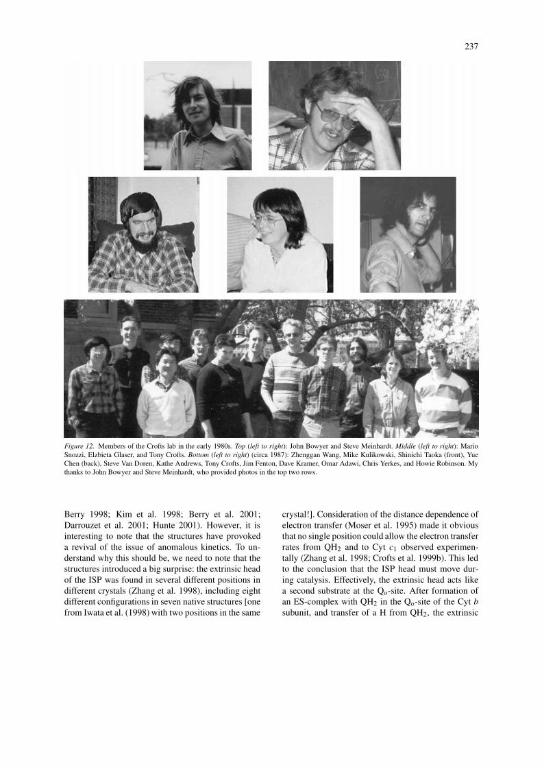
Figure 12. Members of the Crofts lab in the early 1980s. Top (left to right): John Bowyer and Steve Meinhardt. Middle (left to right): MarioSnozzi, Elzbieta Glaser, and Tony Crofts. Bottom (left to right) (circa 1987): Zhenggan Wang, Mike Kulikowski, Shinichi Taoka (front), YueChen (back), Steve Van Doren, Kathe Andrews, Tony Crofts, Jim Fenton, Dave Kramer, Omar Adawi, Chris Yerkes, and Howie Robinson. Mythanks to John Bowyer and Steve Meinhardt, who provided photos in the top two rows.
Berry 1998; Kim et al. 1998; Berry et al. 2001;Darrouzet et al. 2001; Hunte 2001). However, it isinteresting to note that the structures have provokeda revival of the issue of anomalous kinetics. To un-derstand why this should be, we need to note that thestructures introduced a big surprise: the extrinsic headof the ISP was found in several different positions indifferent crystals (Zhang et al. 1998), including eightdifferent configurations in seven native structures [onefrom Iwata et al. (1998) with two positions in the same
crystal!]. Consideration of the distance dependence ofelectron transfer (Moser et al. 1995) made it obviousthat no single position could allow the electron transferrates from QH2 and to Cyt c1 observed experimen-tally (Zhang et al. 1998; Crofts et al. 1999b). This ledto the conclusion that the ISP head must move dur-ing catalysis. Effectively, the extrinsic head acts likea second substrate at the Qo-site. After formation ofan ES-complex with QH2 in the Qo-site of the Cyt bsubunit, and transfer of a H from QH2, the extrinsic
238
head moves ∼23 Å through a rotational displacementto deliver an electron to Cyt c1, and release H+, at aseparate catalytic interface on Cyt c1.
The movement of the ISP as a prerequisite forcatalysis has now been demonstrated through bothbiophysical and mutagenesis approaches from severallabs (reviewed in Darrouzet et al. 2001). This dynamicrole of the ISP introduced questions about how theobserved kinetics might be affected by the movement.In addition to the electron transfer reactions, factorsinvolved are the restricted diffusion imposed by thetethering N-terminal ‘tail,’ and on and off rate con-stants for the different binding processes. This hasraised the possibility of an alternative explanation forthe kinetic disparity between the reduction rates of theb- and c-type cytochromes discussed above – that themovement is limiting (cf. Hansen et al. 2000). Thisreminds me of a remark that Peter Mitchell made tome, apropos the kinetic anomalies, at a Gordon Con-ference around 1981 – ‘Don’t you think the electronmight be getting hung up on the Rieske?’ – a pre-scient observation, though I never knew whether Peterwas thinking of a kinetic or a thermodynamic hang-up.Several labs have recently adopted the idea that the Cytc1 reduction kinetics are slower than those of Cyt bHbecause the movement of the reduced ISP from its siteof reduction close to the Qo-site to its site of oxidationclose to Cyt c1 is the rate limiting step (Hansen et al.2000; Yu et al. 2002).
I believe these authors have failed to take accountof the simpler explanation for the apparent kinetic an-omalies provided by the modified Q-cycle mechanism.In our explication of the modified Q-cycle, the argu-ments were based on electron distribution determinedby the equilibrium constants calculated from the Emvalues. In formulating these arguments, several im-plicit assumptions were made. The kinetic behaviorwas well explained without invoking any specialmodifications to equilibrium constants by interactionsbetween centers, or between different sites. It was as-sumed that equilibration of the ISP and of Cyt bL withtheir reaction partners was much faster than the lim-iting oxidation of QH2. In line with this, our earlierresults had shown that the electron transfer fromthe reduced ISP to Cyt c1 was very rapid (<10 µs),that electron transfer from heme bL to bH was rapid(<100 µs), and that the oxidation of bound QH2 wasrate limiting (∼750µs) (Crofts and Wang 1989). Thislater reaction was also the partial process with thehighest activation barrier. The kinetic anomalies weretherefore accounted for in terms of thermodynamic
distribution, not kinetic limitation. Our own more re-cent experiments have shown unambiguously that inthe chromatophore system the movement of the ISPis not the rate limiting step, either kinetically, or interms of activation barriers (Hong et al. 1999; Croftset al. 2003). These conclusions have been confirmedby work from Frank Millett’s lab, in which the samereactions have been explored on a rapid time scalethrough photoactivation of the isolated enzymes fromboth mitochondria and Rb. sphaeroides using boundruthenium complexes (Engstrom et al. 2002). Fromour results and analysis of the Cyt bc1 complex in situin chromatophores (Crofts et al. 2003), it would seemlikely that the thermodynamic (distribution) factorsalso could provide an adequate explanation for the ki-netic effects seen in the pre-steady state work with mi-tochondrial complexes. A precise accounting is madedifficult by uncertainties in the thermodynamic param-eters, both in the Em values for the redox centers inmitochondria from different species and those arisingfrom use of artificial donor quinones with Em valuesthat differ markedly from the native ubiquinone. Moredetailed studies under well-controlled conditions fromthe labs working on the mitochondrial complexes areclearly needed to answer these questions.
The modified Q-cycle accounts quite neatly for thedata, and continues to provide a simple basis for fur-ther exploration. The structures provide a firm basisfor detailed understanding at a molecular level, andresearch in this direction has been a major theme ofmuch recent work (reviewed in Crofts 2004a, b). Otherdirections that look interesting are questions arisingfrom the dimeric nature of the complex. The modifiedQ-cycle was formulated in the context of a mono-meric mechanism, and independent sites, but thereare some tantalizing indications that things might bemore complicated, and suggestions, and even struc-tural evidence, that some interaction between Qo- andQi-sites might play a role (Iwata et al. 1998; Hunteet al. 2002; Lange and Hunte 2002; Trumpower 2002).It will be interesting to see if, in addition to their con-nection through the b-heme chain (Figure 10), the twoQ-sites communicate across the membrane throughallosteric interaction, or if the two monomers com-municate across the dimer interface. In both cases,complications might be expected from coulombic in-teractions (to modify local potentials), as has beenseen between hemes bH and bL (Shinkarev et al.2001). In addition to simple electron transfer, thereis the possibility of allosteric interactions within orbetween monomers (Trumpower 2002). Distinguishing
239
between the possibilities will present a serious ex-perimental challenge, complicated further by the in-evitable heterogeneity of nanoscale vesicular systems(Crofts et al. 1998).
Finally, the recognition that the Cyt bc1 complexis a major contributor to the production of reactiveoxygen species (ROS) has placed work on the com-plex at center stage in this important medical area(Skulachev 1996; Muller 2000, Chen et al. 2003).The ROS are thought to play a major role in cellu-lar aging through damage to both DNA and protein.The most sensitive target is mitochondrial DNA, forwhich the repair mechanism in animals is less sophist-icated than that in the nucleus. Since the mitochondrialDNA encodes several subunits of the respiratory chain,including Cyt b, mutagenesis may progressively in-crease ROS effects and exacerbate their destructiveeffects over an individual life span. It seems clearthat the semiquinone generated at the Qo-site acts asa donor in production of the 1-electron reduced su-peroxide that is a progenitor of ROS. The rate ofgeneration of superoxide in the presence of antimycin(which maximizes the rate by inhibiting removal of thesemiquinone) is about 80% of that required to accountfor all the ROS production in the cell. However, thephysiological rate is likely considerably less. It is de-termined by the inhibitory effect of the back-pressurefrom the proton gradient, which is itself under meta-bolic control. Nevertheless, production of superoxideat the Qo-site is clearly of importance, and may alsocontribute to the damage in some mitochondrial my-opathies (Fisher and Meunier 2001). Presumably, themechanism of the site must reflect a design by evolu-tion that has minimized this harmful side-reaction, andit will be interesting to see how this has been achieved.Studies of mutant strains to see which residue changeslead to excess production will be a powerful tool forexploration of this problem.
Acknowledgments
I am happy to acknowledge useful discussions withmany colleagues over the years covered by this re-view, but especially Peter Mitchell, Peter Rich, LesDutton, Mårten Wikström, Colin Wraight, Pierre Joliotand Bruno Velthuys. I would also like to acknowledgethe people in the lab who contributed both intellec-tually and experimentally to the research leading toour view of the modified Q-cycle, John Bowyer, SteveMeinhardt, Mario Snozzi, Elzbieta Glaser, Zhenggan
Wang, Yue Chen, who are all shown in Figure 12, andKevin Jones who provoked my epiphany. In particularJohn Bowyer included a detailed discussion of the prosand cons of a Q-cycle explanation for the detailed kin-etic work that went into his PhD thesis, which clearlyshowed that the Q-cycle worked, but still did not get usover the hurdle of the intransigent kinetic anomalies.I would also like to acknowledge the contribution ofDavid Kramer, also shown in Figure 12, who waslater to provide the best kinetic evidence, and analysis,showing that the modified Q-cycle accounted for thebehavior of the Cyt b6f complex (with appropriatemodification of specific physicochemical parameters).Unfortunately, constraints of space preclude a detailedconsideration of these wider aspects. This paper wasedited by John F. Allen and Govindjee.
References
Baccarini-Melandri A, Gabellini N, Melandri BA, Jones KR,Rutherford AW, Crofts AR and Hurt E (1982) Differ-ential extraction and structural specificity of specializedubiquinone molecules in secondary electron transfer in chroma-tophores from Rps. sphaeroides Ga. Arch Biochem Biophys 216:566–580
Baltscheffsky M (1969) Reversed energy transfer in Rhodospirillumrubrum chromatophores. In: Metzner H (ed) Progress in Photo-synthesis Research, Vol III, pp 1306–1312. International Unionof Biological Sciences, Tübingen, Germany
Bendall DS (2004) The unfinished story of cytochrome f. Photo-synth Res 80: 265–276 (this issue)
Bowyer JR (1979) Light-driven electron transfer reactions in photo-synthetic bacteria. PhD thesis. University of Bristol, UK
Bowyer JR (1982) Effects of UHDBT on the reduction–oxidationreactions of the cytochrome b–c1 segment of mammalian mito-chondria. In: Trumpower B (ed) Function of Quinones in EnergyConserving Systems, pp 365–375. Academic Press, New York
Bowyer JR and Crofts AR (1978) Inhibition of electron transport inRps. capsulata by a ubiquinone analogue. In: Dutton PL, LeighJ and Scarpa A (eds) Frontiers of Biological Energetics, Vol I,pp 326–333. Academic Press, New York
Bowyer JR and Crofts AR (1981) On the mechanism of photosyn-thetic electron transfer in Rps. capsulata and Rps. sphaeroides.Biochim Biophys Acta 636: 218–233
Bowyer JR and Trumpower BL (1981) Pathways of electron transferin the cytochrome b–c1 complexes of mitochondria and photo-synthetic bacteria. In: Skulachev VP and Hinkle PC (eds) Chemi-osmotic Proton Circuits in Biological membranes, pp 105–122.Addison-Wesley, Reading, Massachusetts
Bowyer JR, Tierney GV and Crofts AR (1979) Cytochrome c2–reaction centre coupling in chromatophores. FEBS Lett 101:207–212
Bowyer JR, Dutton PL, Prince RC and Crofts AR (1980) The role ofthe Rieske iron–sulfur center as the electron donor to ferricyto-chrome c2 in Rhodopseudomonas sphaeroides. Biochim BiophysActa 592: 445–460
Bowyer JR, Meinhardt SW, Tierney GV and Crofts AR (1981)Resolved difference spectra of redox centers involved in pho-

240
tosynthetic electron flow in Rhodopseudomonas capsulata andRps. sphaeroides. Biochim Biophys Acta 635: 167–186
Boyer PD, Chance B, Ernster L, Mitchell P, Racker E and Slater EC(1977) Oxidative phosphorylation and photophosphorylation.Annu Rev Biochem 46: 955–1026
Brandt U, Schagger H and von Jagow G (1988) Characterizationof binding of the methoxyacrylate inhibitors to mitochondrialcytochrome c reductase. Eur J Biochem 173: 499–506
Chance B (1958) The kinetics and inhibition of cytochromes com-ponents of the succinic oxidase system. J Biol Chem 233:1223–1229
Chance B and Williams GR (1956) The respiratory chain andoxidative phosphorylation. Adv Enzymol 17: 65–134
Chance B, Wilson DF, Dutton PL and Ericinska M (1970) Energycoupling mechanisms in mitochondria: kinetic, spectroscopicand thermodynamic properties of an energy transducing form ofcytochrome b. Proc Natl Acad Sci USA 66: 1175–1184
Chen Y (1989) Kinetic studies of mechanism of ubi-quinol:cytochrome c2 oxidoreductase of R. sphaeroides: asteady state approach. PhD thesis, University of Illinois, Urbana,Illinois
Chen Y and Crofts AR (1990) Operation of UQH2:cyt c2 oxidore-ductase of Rb. sphaeroides in the steady state. In: BaltscheffskyM (ed) Current Research in Photosynthesis, Vol III, pp 287–290.Kluwer Academic Publishers, Dordrecht The Netherlands
Chen Q, Vazquez EJ, Moghaddas S, Hoppel CL and Lesnefsky EJ(2003) Production of reactive oxygen species by mitochondria:central role of complex III. J Biol Chem 278: 36027–36031
Cogdell RJ and Crofts AR (1974) H+-uptake by chromatophoresfrom Rhodopseudomonas sphaeroides. The relation betweenrapid H+-uptake and the H+ pump. Biochim Biophys Acta 247:264–272
Cogdell RJ, Jackson JB and Crofts AR (1972) The effect ofredox potential on the coupling between rapid H+-binding andelectron transport in chromatophores from Rhodopseudomonasspheroides. J Bioenergetics 4: 211–227
Cogdell RJ, Prince RC and Crofts AR (1973) Light inducedH+-uptake catalysed by photochemical reaction centres fromRhodopseudomonas spheroides R26. FEBS Lett 35: 204–208
Cramer WA (2004) Ironies in photosynthetic electron transport: apersonal perspective. Photosynth Res 80: 293–305 (this issue)
Cramer WA and Crofts AR (1982) Electron and proton transfer. In:Govindjee (ed) Photosynthesis: Energy Conversion by Plants andBacteria, Vol I, pp 387–467. Academic Press, New York
Crofts AR (1985) The mechanism of the ubiquinol:cytochromec oxidoreductases of mitochondria and of Rhodopseudomonassphaeroides, In: Martonosi AN (ed) The Enzymes of BiologicalMembranes, Vol 4, pp 347–382. Plenum, New York
Crofts AR (1993) Obituary – Peter Mitchell (1920–1992). Photo-synth Res 35: 1–4
Crofts AR (2004a) The cytochrome bc1 complex – function in thecontext of structure. Annu Rev Physiol (in press)
Crofts AR (2004b) Proton-coupled electron transfer at the Qo-site ofthe bc1 complex controls the rate of ubihydroquinone oxidation.Biochim Biophys Acta (in press)
Crofts AR and Meinhardt SW (1982) A Q-cycle mechanism for thecyclic electron transfer chain of Rps. sphaeroides. Biochem SocTrans 10: 201–203
Crofts AR and Wang Z (1989) How rapid are the internal reactionsof the UQH2:cyt c2 oxidoreductase? Photosynth Res 22: 69–87
Crofts AR and Wraight CA (1983) The electrochemical domain ofphotosynthesis. Biochim Biophys Acta 726: 149–186
Crofts AR, Cogdell RJ and Jackson JB (1971a) The mechanism ofH+-uptake in Rhodopseudomonas spheroides. In: Quagliariello
E, Papa S and Rossi S (eds) Energy Transduction in Respira-tion and Photosynthesis, pp 883–901. Adriatica Editrice, Bari,Italy
Crofts AR, Wraight CA and Fleischman DE (1971b) Energy con-servation in the photochemical reactions of photosynthesis andits relation to delayed fluorescence. FEBS Lett 15: 89–100
Crofts AR, Jackson JB, Evans EH and Cogdell RJ (1972) The high-energy state in chloroplasts and chromatophores. In: Forti G andMelandri A (eds) Proceedings IInd International Congress Pho-tosynthesis Research, Stresa, 1971, pp 873–902. Junk Publishers,The Hague, The Netherlands
Crofts AR, Cogdell RJ, Evans EH and Prince RC (1974) The mech-anism of H+-uptake by chromatophores from photosyntheticbacteria. In: Ernster L and Slater EC (eds) Dynamics of Energy-transducing Membranes, pp 377–387. Elsevier, Amsterdam,The Netherlands
Crofts AR, Crowther D and Tierney GV (1975) Electrogenic elec-tron transport in photosynthetic bacteria. In: Quagliariello E,Papa S, Palmieri F, Slater EC and Siliprandi N (eds) ElectronTransfer Chains and Oxidative Phosphorylation, pp 233–241.North-Holland Publishing, Amsterdam
Crofts AR, Meinhardt SW and Bowyer JR (1982a) The elec-tron transport chain of Rhodopseudomonas sphaeroides. In:Trumpower B (ed) Function of Quinones in Energy ConservingSystems, pp 447–498. Academic Press, New York
Crofts AR, Meinhardt SW, Snozzi M and Jones KR (1982b)A Q-cycle mechanism for cyclic electron transport in Rps.sphaeroides. 2nd EBEC Short Reports, pp 327–328
Crofts AR, Meinhardt SW, Jones KR and Snozzi M (1983) The roleof the quinone pool in the cyclic electron transfer chain of Rps.sphaeroides: a Modified Q-cycle mechanism. Biochim BiophysActa 723: 202–218
Crofts AR, Wang Z, Chen Y, Mahalingham S, Yun C-H andGennis RB (1990) Function, steady state turn-over, and struc-ture of the UQH2:cyt c2 oxidoreductase of Rb. sphaeroides. In:Lenaz G, Barnabei O, Rabbi A and Battino M (eds) Highlightsin Ubiquinone Research, pp 98–103. Taylor & Francis, London
Crofts AR, Barquera B, Bechmann G, Guergova M, Salcedo-Hernandez R, Hacker B, Hong S and Gennis RB (1995) Structureand function in the bc1 complex of Rb. sphaeroides. In: MathisP (ed) Photosynthesis: from Light to Biosphere, Vol II, pp 493–500. Kluwer Academic Publishers, Dordrecht, The Netherlands
Crofts AR, Guergova-Kuras M and Hong S (1998) Chromato-phore heterogeneity explains effects previously attributed tosupercomplexes. Photosynth Res 55: 357–362
Crofts AR, Barquera B, Gennis RB, Kuras R, Guergova-Kuras Mand Berry EA (1999a) The mechanism of ubiquinol oxidationby the bc1 complex: the different domains of the quinol bind-ing pocket, and their role in mechanism, and the binding ofinhibitors. Biochemistry 38: 15807–15826
Crofts AR, Guergova-Kuras M, Huang L-S, Kuras R, Zhang Z andBerry EA (1999b) The mechanism of ubiquinol oxidation by thebc1 complex: the role of the iron sulfur protein, and its mobility.Biochemistry 38: 15791–15806
Crofts AR, Shinkarev VP, Kolling DRJ and Hong S (2003) Themodified Q-cycle explains the apparent mismatch between thekinetics of reduction of cytochromes c1 and bH in the bc1complex. J Biol Chem 278: 39747–39754
Crowther D and Hind G (1982) Cycles and Q-cycles in plant photo-synthesis. In: Trumpower B (ed) Function of Quinones in EnergyConserving Systems, pp 499–510. Academic Press, New York
Darrouzet E, Moser CC, Dutton PL and Daldal F (2001) Large scaledomain movement in cytochrome bc1: a new device for electrontransfer in proteins. TIBS 26: 445–451
241
Deul DH and Thorn MB (1962) Effects of 2,3-dimercaptopropanoland antimycin on absorption spectra of heart muscle prepara-tions. Biochim Biophys Acta 59: 426–436
De Vries S, Albracht SPJ and Leeuwerik FJ (1979) The multiplicityand stoichiometry of the prosthetic groups in QH2:cytochrome coxidoreductase as studied by EPR. Biochim Biophys Acta 546:316–333
De Vries S, Berden JA and Slater EC (1980) Properties of asemiquinone anion located in the QH2:cytochrome c oxidore-ductase segment of the mitochondrial respiratory chain. FEBSLett 122: 143–148
De Vries S, Berden JA and Slater EC (1982) Oxidation–reductionproperties of an antimycin-sensitive semiquinone anion boundto QH2:cytochrome c oxidoreductase. In: Trumpower BL (ed)Function of Quinones in Energy Conserving Systems, pp 235–246. Academic Press, New York
De Vries S, Albracht SPJ, Berden JA, Marres CAM and SlaterEC (1983) The effect of pH, ubiquinone depletion andmyxothiazol on the reduction kinetics of the prosthetic groups ofQH2:cytochrome c oxidoreductase. Biochim Biophys Acta 723:91–103
Dutton PL and Jackson JB (1972a) In situ thermodynamic and ki-netic characterization of components of photosynthetic electrontransport in Rps. spheroides. In: Forti G and Melandri A (eds)Proceedings Second International Congress Photosynthesis Re-search, Stresa, 1971, pp 995–1007. Junk Publishers, The Hague,The Netherlands
Dutton PL and Jackson JB (1972b) Thermodynamic and kinet-ics characterization of electron-transfer components in situ inRps. spheroides and Rhodospirillum rubrum. Eur J Biochem 30:495–510
Dutton PL and Prince RC (1978) Reaction-center-driven cyto-chrome interactions in electron and proton translocation andenergy coupling. In: Clayton RK and Sistrom WR (eds)The Photosynthetic Bacteria, pp 525–570. Plenum Press,New York
Dutton PL and Wilson DM (1976) Redox potentiometry in biolog-ical systems. Meth Enzymol 54: 411–435
Dutton PL, Wilson DF and Lee CP (1970) Oxidation–reductionpotentials of cytochromes in mitochondria. Biochemistry 9:5077–5082
Edwards CA and Bowyer JR and Trumpower BL (1982) Functionof the iron–sulfur protein of the cytochrome b–c1 segment inelectron transfer reactions of the mitochondrial respiratory chain.J Biol Chem 257: 3705–3713
Engstrom G, Xiao K, Yu C-A, Yu L, Durham B and Millett F (2002)Photoinduced electron transfer between the Rieske iron–sulfurprotein and cytochrome c1 in the Rhodobacter sphaeroides cyto-chrome bc1 complex: effects of pH, temperature, and drivingforce. J Biol Chem 277: 31072–31078
Evans EH and Crofts AR (1974) In situ characterization of pho-tosynthetic electron transport in Rhodopseudomonas capsulata.Biochim Biophys Acta 357: 89–102
Ferguson SJ (2001) Keilin’s cytochromes: how bacteria usethem, vary them and make them. Biochem Soc Trans 29:629–640
Fisher N and Meunier B (2001) Effects of mutations in mitochon-drial cytochrome b in yeast and man – deficiency, compensationand disease. Eur J Biochem 268: 1155–1162
Garland PB, Clegg RA, Boxer D, Downie JA and Haddock BA(1975) Proton-translocating nitrate reductase of Escherichia coli.In: Quagliariello E, Papa S, Palmieri F, Slater EC and SiliprandiN (eds) Electron Transfer Chains and Oxidative Phosphorylation,pp 351–358. North-Holland Publishing, Amsterdam
Glaser EG and Crofts AR (1984) A new electrogenic stepin the ubiquinol–cytochrome c2 oxidoreductase complex ofRhodopseudomonas sphaeroides. Biochim Biophys Acta 766:322–333
Glaser EG and Crofts AR (1987) Studies of the electrogenicityof the reduction of cytochrome b561 through the antimycin-sensitive site of the ubiquinol:cytochrome c2 oxidoreductasecomplex of Rhodobacter sphaeroides. In: Papa S, Chance B andErnster L (eds) Cytochrome Systems: Molecular Biology andBioenergetics, pp 625–631. Plenum Publishers, New York
Glaser EG, Meinhardt SW and Crofts AR (1984) Reductionof cytochrome b561 through the antimycin-sensitive site ofthe ubiquinol:cytochrome c2 oxidoreductase complex of Rps.sphaeroides. FEBS Lett 178: 336–342
Hansen KC, Schultz BE, Wang G and Chan SI (2000) Reaction ofEscherichia coli cytochrome bo3 and mitochondrial cytochromebc1 with a photoreleasable decylubiquinol. Biochim BiophysActa 1456: 121–137
Harold FM (2001) Gleanings of a chemiosmotic eye. BioEssays 23:848–855
Hauska G (2004) The isolation of a functional cytochrome b6 f com-plex: from lucky encounter to rewarding experiences. PhotosynthRes 80: 277–291 (this issue)
Hunte C (2001) Insights from the structure of the yeast cytochromebc1 complex: crystallization of membrane proteins with antibodyfragments. FEBS Lett 504: 126–132
Hunte C, Solmaz S and Lange C (2002) Electron transfer betweenyeast cytochrome bc1 complex and cytochrome c: a structuralanalysis. Biochim Biophys Acta 1555: 21–28
Iwata S, Lee JW, Okada K, Lee JK, Iwata M, Rasmussen B, LinkTA, Ramaswamy S and Jap BK (1998) Complete structure ofthe 11-subunit bovine mitochondrial cytochrome bc1 complex.Science 281: 64–71
Jackson JB and Crofts AR (1969) The high energy state in chro-matophores from Rhodopseudomonas spheroides. FEBS Lett 4:185–189
Jackson JB and Crofts AR (1971) The kinetics of light inducedcarotenoid changes in Rhodopseudomonas spheroides and theirrelation to electrical field generation across the chromatophoremembrane. Eur J Biochem 18: 120–130
Jackson JB and Dutton PL (1972) The redox potential dependenceof coupled electron flow in chromatophores from Rhodopseudo-monas spheroides. In: Forti G and Melandri A (eds) Proceed-ings Second Internatonal Congress Photosynthesis Research,Stresa, 1971, pp 1008–1014. Junk Publishers, The Hague,The Netherlands
Jackson JB and Dutton PL (1973) The kinetic and redox poten-tiometric resolution of the carotenoid shifts in Rhodopseudomo-nas spheroides chromatophores: their relationship to electricalfield alterations in electron transport and energy coupling.Biochim Biophys Acta 325: 102–113
Jagendorf AT (1998) Photophosphorylation and the chemiosmoticperspective. Photosynth Res 73: 233–241
Jagendorf AT (2002) Photophosphorylation and the chemiosmoticperspective. Photosynth Res 73: 233–241
Joliot P and Joliot A (1994) Mechanism of electron transfer in thecytochrome b/f complex of algae: evidence for a semiquinonecycle. Proc Natl Acad Sci USA 91: 1034–1038
Jones RW and Whitmarsh J (1988) Inhibition of electron transportand the electrogenic reaction in the cytochrome b/f complex byNQNO and DBMIB. Biochim Biophys Acta 933: 258–268
Junge W and Witt HT (1968) On the ion transport system of pho-tosynthesis – investigations on a molecular level. Z Naturforsch23b: 244–254
242
Kim H, Xia D, Yu CA, Xia JZ, Kachurin AM, Zhang L, Yu L andDeisenhofer J (1998) Inhibitor binding changes domain mobilityin the iron–sulfur protein of the mitochondrial bc1 complex frombovine heart. Proc Natl Acad Sci USA 95: 8026–8033
King TE (1981) Constitution of ‘the b–c1 complex’. In: SkulachevVP and Hinkle PC (eds) Chemiosmotic Proton Circuits in Bio-logical membranes, pp 147–169. Addison-Wesley Publishers,Reading, Massachusetts
King TE, Yu C-A, Yu L and Chiang Y (1975) An examination ofthe components, sequence, mechanisms and their uncertaintiesinvolved in mitochondrial electron transport from succinate tocytochrome c. In: Quagliariello E, Papa S, Palmieri F, SlaterEC and Siliprandi N (eds) Electron Transfer Chains and Oxi-dative Phosphorylation, pp 105–118. North-Holland Publishing,Amsterdam
Koike H, Satoh K and Katoh S (1978) Effects of dibromo-thymoquinone and bathophenanthroline on flash-induced cyto-chrome oxidation in spinach chloroplasts. Plant Cell Physiol 19:1371–1380
Konstantinov A, Kunz WS and Kamensky YA (1981) The Q-cyclein the mitochondrial respiratory chain. In: Skulachev VP andHinkle PC (eds) Chemiosmotic Proton Circuits in Biologicalmembranes, pp 123–146. Addison-Wesley Publishers, Reading,Massachusetts
Lange C and Hunte C (2002) Crystal structure of the yeast cyto-chrome bc1 complex with its bound substrate cytochrome c. ProcNatl Acad Sci USA 99: 2800–2805
Leung KH and Hinkle PC (1975) Reconstitution of ion transport andrespiratory control in vesicles formed from reduced coenzyme Qcytochrome c reductase and phospho lipids. J Biol Chem 250:8467–8471
Malviya AN, Nicholls P and Elliot WB (1980) Observations onthe oxido-reduction of the two cytochromes b in cytochrome c-deficient mitochondria and submitochondrial particles. BiochimBiophys Acta 589: 137–149
Matsuura K, Bowyer JR, Ohnishi T and Dutton PL (1983) Inhibitionof electron transfer by 3-alkyl-2-hydroxy-1,4-naphthoquinonesin the ubiquinol–cytochrome c oxidoreductases of Rhodopseudo-monas sphaeroides and mammalian mitochondria. Interactionwith a ubiquinone-binding site and the Rieske iron–sulfur cluster.J Biol Chem 258: 1571–1579
Meinhardt SW (1984) Electron transfer reactions of the ubi-quinol:cytochrome c2 oxidoreductase of Rhodopseudomonassphaeroides, PhD thesis, University of Illinois at Urbana-Champaign
Meinhardt SW and Crofts AR (1982a) The site and mechanism ofaction of myxothiazol as an inhibitor of electron transport in Rps.sphaeroides. FEBS Lett 149: 217–222
Meinhardt SW and Crofts AR (1982b) Kinetic and thermody-namic resolution of cytochrome c1 and cytochrome c2 from Rps.sphaeroides. FEBS Lett 149: 223–227
Meinhardt SW and Crofts AR (1983) The role of cytochromeb566 in the electron transfer chain of Rps. sphaeroides. BiochimBiophys Acta 723: 219–230
Meinhardt SW and Crofts AR (1984) A new effect of antimycin onthe b-cytochromes of Rps. sphaeroides, In: Sybesma C (ed) Ad-vances in Photosynthesis Research, Vol 1, pp 649–652. MartinusNijhoff/Junk Publishers, The Hague, The Netherlands
Mitchell P (1961) Coupling of phosphorylation to electron and hy-drogen transfer by a chemiosmotic type of mechanism. Nature191: 144–148
Mitchell P (1966) Chemiosmotic Coupling in Oxidative and Photo-synthetic Phosphorylation. Glynn Research, Bodmin, Cornwall,UK
Mitchell P (1968) Chemiosmotic Coupling and Energy Transduc-tion. Glynn Research, Bodmin, Cornwall, UK
Mitchell P (1975a) Proton motive redox mechanism of the cyto-chrome b–c1 complex in the respiratory chain: proton motiveubiquinone cycle. FEBS Lett 56: 1–6
Mitchell P (1975b) Protonmotive function of cytochrome sys-tems. In: Quagliariello E, Papa S, Palmieri F, Slater ECand Siliprandi N (eds) Electron Transfer Chains and Oxida-tive Phosphorylation, pp 305–316. North-Holland Publishing,Amsterdam
Mitchell P (1976) Possible molecular mechanisms of the pro-tonmotive function of cytochrome systems. J Theor Biol 62:327–367
Mitchell P (1978) David Keilin’s respiratory chain concept andits chemiosmotic consequences. Nobel Lectures. Chemistry1971–1980
Mitchell P (1981) Bioenergetic aspects of unity in biochemistry:evolution of the concept of chemical, osmotic, and chemiosmoticmechanisms. In: Semenza G (ed) Of Oxygen, Fuels, and LivingMatter, Part 1. John Wiley & Sons, New York
Mitchell P (1990) The classical mobile carrier function of lipophilicquinones in the osmochemistry of electron-driven proton trans-location. In: Lenaz G, Barnabei O, Rabbi A and Battino M (eds)Highlights in Ubiquinone Research, pp 77–82. Taylor & Francis,London
Moser CC, Page CC, Farid R and Dutton PL (1995) Biologicalelectron transfer. J Bioenerg Biomembr 27: 263–274
Muller F (2000) The nature and mechanism of superoxide produc-tion by the electron transport chain: its relevance to aging. J AmAging Assoc 23: 227–253
Ohnishi T and Trumpower BL (1980) Differential effects of anti-mycin on ubisemiquinone bound in different environments inisolated succinate – cytochrome c reductase complex. J BiolChem 255: 3278–3284
Petty KM, Jackson JB and Dutton PL (1979) Factors controllingthe binding of two protons per electron transferred through theubiquinone and cytochrome b/c2 segment of Rhodopseudomo-nas sphaeroides chromatophores. Biochim Biophys Acta 546:17–42
Prince RC and Dutton PL (1976) Further studies on the Rieske iron–sulfur center in mitochondrial and photosynthetic systems: a pKon the oxidized form. FEBS Lett 65: 117–119
Prince RC, O’Keefe D and Dutton PL (1982) The organization ofthe cyclic electron transfer system in photosynthetic bacterialmembranes: is this the hardware of a chemiosmotic system.In: Barber J (ed) Electron Transport and Photophosphoryla-tion, pp 197–248. Elsevier Biomedical Press, Amsterdam,The Netherlands
Rich PR, Jeal AE, Madgwick SA and Moody AJ (1990) Inhib-itor effects on redox-linked protonations of the b hemes ofthe mitochondrial bc1 complex. Biochim Biophys Acta 1018:29–40
Roberts H, Choo WH, Smith SC, Marzuki S, Linnane AW, PorterTH and Folkers K (1978) The site of inhibition of mitochon-drial electron transfer by coenzyme Q analogues. Arch BiochemBiophys 191: 306–315
Robertson DE, Prince RC, Bowyer JR, Matsuura K, Dutton PL andOhnishi T (1984) Thermodynamic properties of the semiquinoneand its binding site in the ubiquinol:cytochrome c (c2) oxidore-ductase of respiratory and photosynthetic systems. J Biol Chem259: 1758–1763
Salerno JC, Xu Y, Osgood P, Kim CH and King TE (1989) Thermo-dynamic and spectroscopic characterization of the cytochromebc1 complex. J Biol Chem 264: 15398–15403

243
Selak MA and Whitmarsh J (1982) Kinetics of the electrogenic stepand cytochrome b6 and f redox changes in chloroplasts: evidencefor a Q-cycle. FEBS Lett 150: 286–292
Shinkarev VP, Crofts AR and Wraight CA (2001) The electric fieldgenerated by photosynthetic reaction center induces rapid re-versed electron transfer in the bc1 complex. Biochemistry 40:12584–12590
Skulachev VP (1996) Role of uncoupled and non-coupled oxi-dations in maintenance of safely low levels of oxygen and itsone-electron reductants. Q Rev Biophys 29: 169–202
Skulachev VP and Hinkle PC (eds) (1981) Chemiosmotic ProtonCircuits in Biological membranes. Addison-Wesley Publishers,Reading, Massachusetts
Slater EC (1973) The mechanism of action of the respir-atory inhibitor, antimycin. Biochim Biophys Acta 301:129–154
Slater EC (1981) The cytochrome b paradox, the BAL-labile factorand the Q-cycle. In: Skulachev VP and Hinkle PC (eds) Chemi-osmotic Proton Circuits in Biological Membranes, pp 69–104.Addison-Wesley Publishers, Reading, Massachusetts
Slater EC and de Vries S (1980) Identification of the BAL-labilefactor. Nature 288: 717–718
Snozzi M and Crofts AR (1984) Electron transport in chroma-tophores from Rhodopseudomonas sphaeroides Ga fused withliposomes. Biochim Biophys Acta 766: 451–463
Snozzi M and Crofts AR (1985) Kinetics of the c-cytochromes inchromatophores from Rps. sphaeroides as a function of the con-centration of cytochrome c2: influence of this concentration onthe oscillation of the secondary acceptor of the reaction center,QB. Biochim Biophys Acta 809: 260–270
Thierbach G and Reichenbach H (1981) Myxothiazol, a new in-hibitor of the b–c1 segment of the respiratory chain. BiochimBiophys Acta 638: 282–289
Trebst A, Harth E and Draber W (1970) On a new inhibitorof photosynthetic electron transport in isolated chloroplasts. ZNaturforsch 25b: 1157–1159
Trumpower BL (1981) Function of the iron–sulfur protein of thecytochrome b–c1 segment of the respiratory chain. BiochimBiophys Acta 639: 129–155
Trumpower BL (2002) A concerted, alternating sites mechanismof ubiquinol oxidation by the dimeric cytochrome bc1 complex.Biochim Biophys Acta 1555: 166–173
Trumpower BL and Haggerty JG (1980) J Bioenerg Biomembr 12:151–164
Tsai A-L, Olson JS and Palmer G (1983) The oxidation of yeastcomplex III. J Biol Chem 258: 2122–2125
Van den Berg WH, Bonner WD, Prince RC, Bashford CL, TakamiyaK-I, Bonner D and Dutton PL (1979) Electron and proton
transport in the UQ–cyt b–c2 oxidoreductase of Rps.sphaeroides. J Biol Chem 254: 8594–8604
Velthuys B (1982) The function of plastoquinone in electron trans-fer. In: Trumpower B (ed) Function of Quinones in EnergyConserving Systems, pp 401–408. Academic Press, New York
Venturoli G, Fernandez-Velasco JG, Crofts AR and Melandri BA(1986) Demonstration of a collisional interaction of ubiquinonewith the ubiquinol:cytochrome c2 oxidoreductase complex inchromatophores of Rhodobacter sphaeroides. Biochim BiophysActa 851: 340–352
Venturoli G, Fernandez-Velasco JG, Crofts AR and Melandri BA(1988) The effect of the size of the quinone pool on the electro-genic reactions in the UQH2:cyt c2 oxidoreductase of Rhodobac-ter capsulatus. Pool behavior at the quinone reductase site.Biochim Biophys Acta 935: 258–272
von Jagow G and Link T (1988) Use of specific inhibitors on themitochondrial bc1 complex. Meth Enzymol 126: 253–271
Vredenberg WJ and Amesz J (1967) Absorption characteristics ofbacteriochlorophyll types in purple bacteria and efficiency ofenergy transfer between them. Brookhaven Symp Biol 19: 49–60
Vredenberg WJ, Amesz J and Duysens LNM (1965) Light-inducedspectral shifts in bacteriochlorophyll and carotenoid absorptionspectra. Biochem Biophys Res Comm 18: 435–439
Wikström MKF and Berden J (1972) Oxidoreduction of cytochromeb in the presence of antimycin. Biochim Biophys Acta 283:403–420
Wikström MKF, Krab K and Saraste M (1981) Proton translocatingcytochrome complexes. Annu Rev Biochem 50: 623–655
Wood PM (1980) Do photosynthetic bacteria contain cytochromec1? Biochem J 189: 385–391
Wraight CA (1982) Current attitudes in photosynthesis research. In:Govindjee (ed) Photosynthesis: Energy Conversion by Plants andBacteria, Vol I, pp 17–61. Academic Press, New York
Xia D, Yu C-A, Kim H, Xia J-Z, Kachurin AM, Zhang L, Yu Land Deisenhofer J (1997) Crystal Structure of the Cytochromebc1 Complex from Bovine Heart Mitochondria. Science 277:60–66
Yu C-A, Nagoaka S, Yu L and King TE (1978) Evidence for the ex-istence of a ubiquinone protein abd its radical in the cytochromeb and c1 region of the mitochondrial electron transport chain.Biochem Biophys Res Commun 82: 1070–1078
Yu C-A, Wen X, Xiao K, Xia D and Yu L (2002) Inter- andintra-molecular electron transfer in the cytochrome bc1 complex.Biochim Biophys Acta 1555: 65–70
Zhang Z, Huang L-S, Shulmeister VM, Chi Y-I, Kim K-K, HungL-W, Crofts AR, Berry EA and Kim S-H (1998) Electrontransfer by domain movement in cytochrome bc1. Nature 392:677–684