The proximal aspect of the dorsal condylar sagittal ridge ... · The proximal aspect of the dorsal...
167
The proximal aspect of the dorsal condylar sagittal ridge and the adjacent soft tissues in the fetlock joint of the Warmblood horse: Morphology and relationship with cartilage degeneration Stijn Hauspie Dissertation submitted in fulfilment of the requirements for the degree of Doctor of Philosophy (PhD) in Veterinary Sciences 2012 Promoters: Prof. Dr. J. Saunders Prof. Dr. A. Martens Dr. K. Vanderperren Department of Veterinary Medical Imaging and Small Animal Orthopaedics Faculty of Veterinary Medicine Ghent University
Transcript of The proximal aspect of the dorsal condylar sagittal ridge ... · The proximal aspect of the dorsal...
The proximal aspect of the dorsal
condylar sagittal ridge and the adjacent soft
tissues in the fetlock joint of the
Warmblood horse: Morphology and relationship with cartilage degeneration
Stijn Hauspie
Dissertation submitted in fulfilment of the requirements
for the degree of Doctor of Philosophy (PhD) in Veterinary Sciences
2012
Promoters:
Prof. Dr. J. Saunders
Prof. Dr. A. Martens
Dr. K. Vanderperren
Department of Veterinary Medical Imaging and Small Animal Orthopaedics
Faculty of Veterinary Medicine Ghent University

The proximal aspect of the dorsal condylar sagittal ridge and the adjacent soft tissues in the
fetlock joint of the Warmblood horse: Morphology and relationship with cartilage
degeneration.
Stijn Hauspie
Vakgroep Medische Beeldvorming van de Huisdieren en Orthopedie van de Kleine
Huisdieren
Faculteit Diergeneeskunde
Universiteit Gent
ISBN: xxxxxxxxxxxxxxxxx
This PhD thesis was supported by a scientific research grant of the Ghent University Special
Research Fund (BOF 01J04911)

Om innerlijke rust te vinden, moet je afmaken waaraan je begonnen bent
(Boeddha)


Table of Contents
LIST OF ABBREVIATIONS 1
PREFACE 3
CHAPTER 1: The equine metacarpo-/metatarsophalangeal joint 7
CHAPTER 1.1: Anatomy of the equine metacarpo-/metatarsophalangeal joint 9
CHAPTER 1.2: The use of different imaging modalities in a pre-purchase
examination 17
CHAPTER 1.3: The evaluation of the equine metacarpo-/metatarsophalangeal joint
during a pre-purchase examination 35
CHAPTER 2: Scientific Aims 47
CHAPTER 3: Radiographic features of the dorsoproximal aspect of the sagittal ridge of the
third metacarpal and metatarsal bones in young Warmblood stallions 51
CHAPTER 4: The histological appearance of the dorsoproximal aspect of the condylar
sagittal ridge of the third metacarpal and metatarsal bone in young Warmblood horses and the
correlation with detected radiographic variations 65
CHAPTER 5: Radiographic variation of the proximal aspect of the dorsal condylar sagittal
ridge of the metacarpo-/metatarsophalangeal joint in Warmblood horses versus the risk of
cartilage degeneration 81
CHAPTER 6: The position of the dorsal proximal synovial pad during hyperextension of the
equine metacarpo-/metatarsophalangeal joint 95
CHAPTER 7: General Discussion 115

SUMMARY 131
SAMENVATTING 137
CURICULUM VITAE 143
BIBLIOGRAPHY 147
DANKWOORD 153
1
List of abbreviations
CDI Cartilage degeneration index CT Computed tomography
DICOM Digital imaging and communications in medicine
MCIII Third metacarpal bone
MCP Metacarpophalangeal MRI Magnetic resonance imaging
MTIII Third metatarsal bone
MTP Metarsophalangeal P1 Proximal phalanx
PDW Proton density weighted
s.d. Standard deviation
SE Spin echo STIR Short T1-inversion recovery
T Tesla
T1W T1 weighted T2W T2 weighted
TE Echo time
TR Repetition time
US Ultrasonography
Abbreviations of the radiographic projections
D125°Di-PaPrO Dorsal 125° distal-palmaroproximal oblique D35°Di-PaPrO Dorsal 35° distal-palmaroproximal oblique
D45°L-Pa(Pl)MO Dorsal 45° lateral-palmaro(plantaro)medial oblique
D45°M-Pa(Pl)LO Dorsal 45° medial-palmaro(plantaro)lateral oblique
DPa(Pl) Dorsopalmar(plantar) DPr-DDiO Dorsoproximal-dorsodistal oblique
LM Lateromedial


3
Preface


5
Today, a pre-purchase examination is recognised as one of the most important services
offered by an equine practitioner, assessing the risk of buying a horse. This examination is
performed to identify any abnormalities or potential problems that would make the horse
unsuitable for its intended use.
Radiography has become an integral part of this pre-purchase examination to detect
any potential or actual orthopaedic problems. Skeletal lesions can be present during a
radiographic screening even when the horse is clinically sound. Some detected lesions should
not interfere with future performance; others may limit the horse’s ability to work or cause
lameness. Therefore, it is important to identify any abnormal radiographic findings and to try
to predict if they will correlate with future lameness. In case the veterinarian makes a mistake
in interpreting the clinical relevance of the detected lesions, the economic and legal
consequences may be important.
Variation in the radiographic appearance of the proximal aspect of the dorsal condylar
sagittal ridge in the equine MCP/MTP joint is detected in Thoroughbreds. These do not
interfere with their future sports career. However, due to the difference in type and length of
sports career, a simple extrapolation of the conclusions drawn for Thoroughbreds to
Warmbloods is not possible. This makes the assessment of the importance of these variations
challenging when detected during a pre-purchase examination of a Warmblood horse.
Therefore, the need for an improved knowledge of the morphological appearance of
these variations at the level of the proximal aspect of the dorsal condylar sagittal ridge in
Warmbloods, as well as the possible interaction with the surrounding soft tissues and
detrimental effects at the level of the joint is essential.

7
Chapter 1
The equine metacarpo-
/metatarsophalangeal joint


9
Chapter 1.1
Anatomy of the equine metacarpo-
/metatarsophalangeal joint
Adapted from:
Hauspie S., Declercq J., Martens A., Zani D.D., Bergman E.H.J., Saunders J.H. (2011)
Anatomy and imaging of the equine metacarpophalangeal/metatarsophalangeal joint. Vlaams
Diergeneeskundig Tijdschrift 80, 263-270.


Anatomy
11
The anatomic terminology used in italic between brackets herein conforms to that
listed in the Nomina Anatomica Veterinaria (international Committee on Veterinary Gross
Anatomical Nomenclature and General Assembly of the World Associoation of Veterinary
Anatomists, 2012).
The MCP/MTP joint (articulation metacarpophalangea et metatarsophalangea)
comprises of four bones: the MCIII (os metacarpale III) or MTIII (os metatarsale III) bone,
P1 (phalanx proximale) and the paired proximal sesamoid bones (ossa sesamoidea
proximalia) (Fig. 1). The MTIII is longer; stronger and slightly more flattened in a
dorsoplantar direction in its distal third compared to the MCIII. The length of the lateral
cortex of the cannon bone is longer than the length of the medial one, resulting in a slight
oblique orientation of the distal articular surface of the cannon bone compared to the proximal
articular surface. The distal epiphysis of the MCIII/MTIII has two unequal convex condyles
and a sagittal ridge that separates them. The medial condyle is slightly bigger compared to the
lateral one. This distal epiphysis articulates with P1 distally and with the proximal sesamoid
bones palmarly/plantarly (Barone 1986; Alrtib et al. 2012).
Figure 1. Illustration of the bones of the distal front limb: A) Dorsal view of the third metacarpal bone, B) Lateral view of the third metacarpal bone with at its palmar aspect a splint bone, C) dorsal view of 1: the
proximal sesamoid bones, 2: the proximal phalanx, 3: the middle phalanx, 4: the distal phalanx, 5: the distal sesamoid bone or navicular bone (Vakgroep morfologie, faculteit Diergeneeskunde).
An articular capsule (capsula articulares) and multiple ligaments reinforce the
MCP/MTP joint (Fig. 2). The ligaments can be divided into three main groups: the
MCP/MTP ligaments, the sesamoidean ligaments and the palmar/plantar (intersesamoidean)
ligament.

Chapter 1.1
12
The MCP/MTP ligaments are subdivided into the collateral ligaments (ligg.
collateralia), the dorsal reinforcement of the articular capsule and the suspensory ligament
(m. interosseus). Each collateral ligament has a superficial and a deep part. The superficial
part is the longest and strongest, running approximately in a vertical direction. Its origin is
located at the lateral or medial aspect of the MCIII/MTIII, just distal to the distal tip of the
splint bones and it is attaching at the proximal aspect of P1. The deep part is more triangular
shaped, having its origin at the abaxial condylar fossa, running in a distopalmar/plantar
direction and inserts on P1. The deep part of the collateral ligaments is covered by synovium
at its deepest border. The dorsal fibrous reinforcement of the articular capsule has fibres
running in different directions. At the lateral and medial aspect of the MCP/MTP joint it fuses
with the respective collateral ligament. The lateral and medial branch of the suspensory
ligament inserts at the apical and abaxial border of the proximal sesamoid bones. At the level
of their attachment, they form a small “extensor” tendon that runs in a dorsodistal direction
and fuses dorsally with the common (front limb) or long (hind limb) extensor tendon at the
level of P1 (Barone 1989; Weaver et al. 1992; Vanderperren et al. 2008).
Figure 2. Overview of the ligaments and tendons surrounding the metacarpophalangeal joint. 1: the common extensor tendon, 2: the lateral digital extensor tendon, 3: the lateral collateral ligament, 4: the suspensory
ligament, 5: the “extensor” tendon of the suspensory ligament, 6: the superficial digital flexor tendon, 7: the deep digital flexor tendon, 8: the manica flexoria, 9: the oblique sesamoidean ligament (Vakgroep morfologie,
faculteit Diergeneeskunde).
The sesamoidean ligaments are subdivided into the collateral sesamoidean ligaments
(ligg. sesamoidea collateralia) (Fig. 2) and the distal sesamoidean ligaments (Fig. 3). The

Anatomy
13
(lateral and medial) collateral sesamoidean ligaments course from the abaxial surface of the
proximal sesamoid bones to MCIII/MTIII and the tuberosity of P1 (Barone 1989;
Vanderperren et al. 2008). The distal sesamoidean ligaments are organised in multiple layers.
The most superficial located straight sesamoidean ligament (lig. sesamoideum rectum)
originates from the base of the proximal sesamoid bones and the palmar intersesamoidean
ligament and inserts on the second phalanx (phalanx media). The intermediate located oblique
sesamoidean ligament (lig. sesamoidea oblique) originates just dorsal to the straight
sesamoidean ligament at the base of the proximal sesamoid bones (medial and lateral bundle)
and the palmar intersesamoidean ligament (thin sagittal part), running distally and inserting
on the palmar surface of P1. The most deeply located are the short and cruciate sesamoidean
ligaments (ligg.sesamoidea brevia et cruciata). These latter are crossed, originating at the
axial part of the base of the proximal sesamoid bones to the contralateral axial aspect of P1.
The short distal sesamoidean ligaments extend from the dorsal aspect of the base of the
proximal sesamoid bones to the palmar margin of the articular surface of P1. The short and
cruciate distal sesamoidean ligaments form the palmar/plantar wall of the MCP/MTP joint
(Barone 1989; Vanderperren et al. 2008).
Figure 3. Overview of the sesamoidean ligaments and suspensory ligament. 1: the straight sesamoidean ligament, 2: the oblique sesamoidean ligament, 3: the cruciate sesamoidean ligaments (after removal of the
straight sesamoidean ligament), 4: the palmar intersesamoidean ligament, 5: the 2 branches of the suspensory ligament, 6: collateral sesamoidean ligament (Vakgroep morfologie, faculteit Diergeneeskunde).

Chapter 1.1
14
The palmar intersesamoidean ligament (lig. palmaria) (Fig. 3) is thicker and forms at
its dorsal aspect the groove in between both proximal sesamoid bones. At its palmar/plantar
aspect, it covers almost completely the axial margins of both proximal sesamoid bones,
forming the proximal scutum (scutum proximale), over which the flexor tendons slide
(Barone 1989).
The articular capsule is formed by an outer stratum fibrosum, strengthened by the
above-mentioned ligaments, and an inner stratum synoviale, responsible for the homeostasis
of the synovial fluid. The MCP/MTP joint has a small dorsal recess (recessus dorsales) and a
large palmar/plantar recess (recessus palmares/plantares). In the dorsoproximal recess of the
MCP/MTP joint, the synovium and fibrous connective tissue forms a fold (plica) (plica
synovialis), projecting distally from the dorsoproximal attachment of the joint capsule and
tapering to a thin edge. This covers the transition zone between the condylar cartilage and the
attachment of the joint capsule (Fig. 4) (Dabareiner et al. 1996). This synovial plica has a
fibrous structure with a linear arrangement of fibrous connective tissue containing a small
number of blood vessels. The edges are covered by squamous to low-cuboidal cells up to
three cells thick, which represent the synovium (Steyn et al. 1989; White 1990).
Figure 4. Illustration of the synovial plica: A) The synovial plica in situ at the dorsoproximal aspect of the distal metacarpal condyle (arrows), B) Histological detail illustrating the fibrous structure with a linear arrangement of
fibrous connective tissue containing a small number of blood vessels (black oval) and covered by synovium (arrow).
The extensor tendons are located at the dorsal aspect of the MCP/MTP joint (Fig. 2).
In the front limb, both the common and lateral digital extensor tendons (m. extensor digitorum
communis; m. extensor digitorum lateralis) are present, whereas in the hind limb, the long
Anatomy
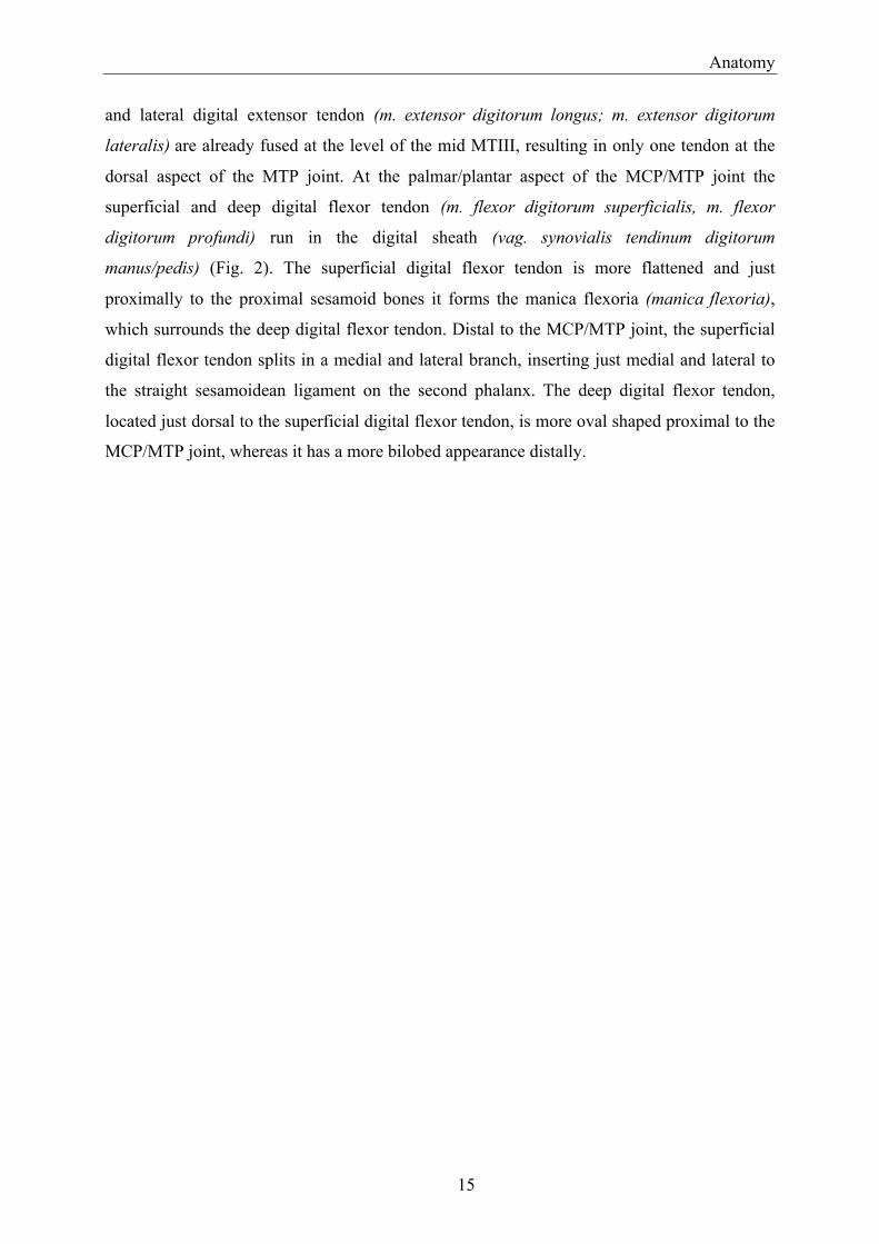
15
and lateral digital extensor tendon (m. extensor digitorum longus; m. extensor digitorum
lateralis) are already fused at the level of the mid MTIII, resulting in only one tendon at the
dorsal aspect of the MTP joint. At the palmar/plantar aspect of the MCP/MTP joint the
superficial and deep digital flexor tendon (m. flexor digitorum superficialis, m. flexor
digitorum profundi) run in the digital sheath (vag. synovialis tendinum digitorum
manus/pedis) (Fig. 2). The superficial digital flexor tendon is more flattened and just
proximally to the proximal sesamoid bones it forms the manica flexoria (manica flexoria),
which surrounds the deep digital flexor tendon. Distal to the MCP/MTP joint, the superficial
digital flexor tendon splits in a medial and lateral branch, inserting just medial and lateral to
the straight sesamoidean ligament on the second phalanx. The deep digital flexor tendon,
located just dorsal to the superficial digital flexor tendon, is more oval shaped proximal to the
MCP/MTP joint, whereas it has a more bilobed appearance distally.


17
Chapter 1.2
The use of different imaging modalities in
a pre-purchase examination
Adapted from:
Hauspie S., Declercq J., Martens A., Zani D.D., Bergman E.H.J., Saunders J.H. (2011)
Anatomy and imaging of the equine metacarpophalangeal/metatarsophalangeal joint. Vlaams
Diergeneeskundig Tijdschrift 80, 263-270.


Imaging modalities in a pre-purchase examination
19
Introduction
The MCP/MTP joint of the horse is prone to injury. It has a relatively small surface
area to transmit the body weight of the horse and it has the highest range of motion of any of
the limb joints, and therefore sustains the greatest forces of acceleration of any of the joints
(Pool and Meagher 1990). Lameness attributable to the MCP/MTP joint is a frequent cause of
early retirement from athletic career in horses and should therefore be detected as early as
possible (Rossdale et al. 1985; Santschi 2008).
Jumpers and dressage horses are often considered as an investment rather than an
avocation and owners have great expectations of the performance and physical condition of
their horse. On the other hand, they have less understanding of the uncertainties and the
limitations of a pre-purchase examination (Marks 1999). To address this, good
communication with the (potential) horse owner is essential and a thorough and standardized
pre-purchase examination is needed (Suslak-Brown 2004; Mitchell 2009). The basis of this
pre-purchase examination is a thorough clinical and physical examination (Marks 1999). This
clinical examination is completed with a radiographic examination for screening purposes.
Additional imaging techniques can be used if abnormalities are detected either during the
clinical or radiographic examination. The decision of what additional imaging technique to
use needs to be faced upon the professional assessment of the horse by the veterinarian and
sound economic considerations (Mitchell 2009).
Radiography has presently become an integral part of the pre-purchase examination
(Suslak-Brown 2004). The radiographic examination can reveal findings inconsistent with the
clinical examination, advocating further radiographic images or other imaging techniques
such as ultrasound, scintigraphy or magnetic resonance imaging. These techniques can be
used to gain more information about the significance of a lesion detected on radiographs (Van
Hoogmoed et al. 2003; Mitchell 2009). Computed tomography is as additional imaging
technique valuable however, due to the need for general anaesthesia, this technique is less
commonly used in a pre-purchase examination.
Chapter 1.2
20
Radiography
Radiography is a relative low cost, widely available technique and very effective for
the evaluation of bony structures. Radiography is based on the principle that x-rays, produced
by accelerating electrons onto a tungsten target, penetrate an object placed in the path of the
x-ray beam. The obtained image is dependent on the total number of x-rays produced, the
distance from the focal spot to the film and the ability of the x-rays to penetrate the tissue
(Butler et al. 2000b).
Over the past decade, digital radiography has largely replaced conventional
radiography. Digital radiography represents on the one hand a higher investment cost than an
analogue system, but on the other hand, it has several distinct advantages: less film waste,
lesser films per examination due to the extra possibilities for image post-processing and
improvement of the image quality due to the almost instantaneous acquisition time (images
can be reviewed on site). For the correct interpretation of digital radiographs, recognition of
the specific artefacts associated with digital radiography is required (Dalla Palma 2000;
Mcknight 2004; Mattoon 2006; Jimenez et al. 2008; Pilsworth and Head 2010).
The regions that are imaged during a pre-purchase examination as well as the obtained
projections of that region are depending on the breed and the purpose of the horse. The
standard projection of the MCP/MTP during a pre-purchase examination of a Warmblood
horse is often limited to a LM projection (Verwilghen et al. 2009). However, up to 4
projections of the MCP/MTP joint can be advocated: LM, D45L-Pa(Pl)MO, D45M-
Pa(Pl)MO, DPa(Pl) (Fig. 5) (Poulos 1992; Richard and Alexander 2007).

Imaging modalities in a pre-purchase examination
21
Figure 5. The normal radiographic appearance of the metacarpo-/metatarsophalangeal joint: A) Conventional LM projection, B) D45L-Pa(Pl)MO projection, C) D45M-Pa(Pl)LO projection, D) DPa(Pl) projection.
The LM projection is performed with the horse weight-bearing and a horizontal x-ray
beam parallel to the heel bulbs. However, if there is some rotation of the distal limb, the
position of the MCP/MTP joint relative to the foot should be evaluated, sometimes
necessitating to angle the line 5° towards palmar/plantar (Edwards 1984; Butler et al. 2000a).
The condyles of the MCIII/MTIII bone and proximal sesamoid bones should be superimposed
on each other and the MCP/MTP joint space should be identifiable (Fig. 6) (Park 2000). The
standard oblique projections are made with the primary beam orientated at an angle of 45°
(medial or lateral) to the sagittal plane. This angle can be adapted depending on the lesion
identified or suspected on the initial LM projection (Edwards 1984). The
dorsomedial/dorsolateral aspect of P1 and the borders of the proximal sesamoid bones,
superimposed on the distal aspect of the MCIII/MTIII bone, should be identifiable on the
oblique projections. Superimposition of the base of the sesamoid bones over the proximal
palmar/plantar process of the P1 should be avoided (Fig. 7) (Park 2000). The DPa/DPl
projection is performed with a horizontal x-ray beam, centred on the joint. The
superimposition of the proximal sesamoid bones over the joint space can be avoided by
angling the x-ray beam proximodistally (approximately 10° for the DPa projection; 15° for
the DPl projection) (Fig. 8) (Butler et al. 2000a).

Chapter 1.2
22
Figure 6. Illustration of: A) A LM projection with superimpostion of the distal third metacarpal condyles on each other, allowing evaluation of the dorsal aspect of the sagittal ridge and B) A slight oblique projection,
prohibiting evaluation of the dorsal aspect of the sagittal ridge.
Figure 7. Illustration of: A) A D45L-PaMO projection with no superimposition of the base of the proximal sesamoid bone on the proximal palmar process of the proximal phalanx, B) A D45L-PaMO projection where
the base of the proximal sesamoid bone is superimposed onto the proximal palmar process of the proximal phalanx.
Imaging modalities in a pre-purchase examination
23
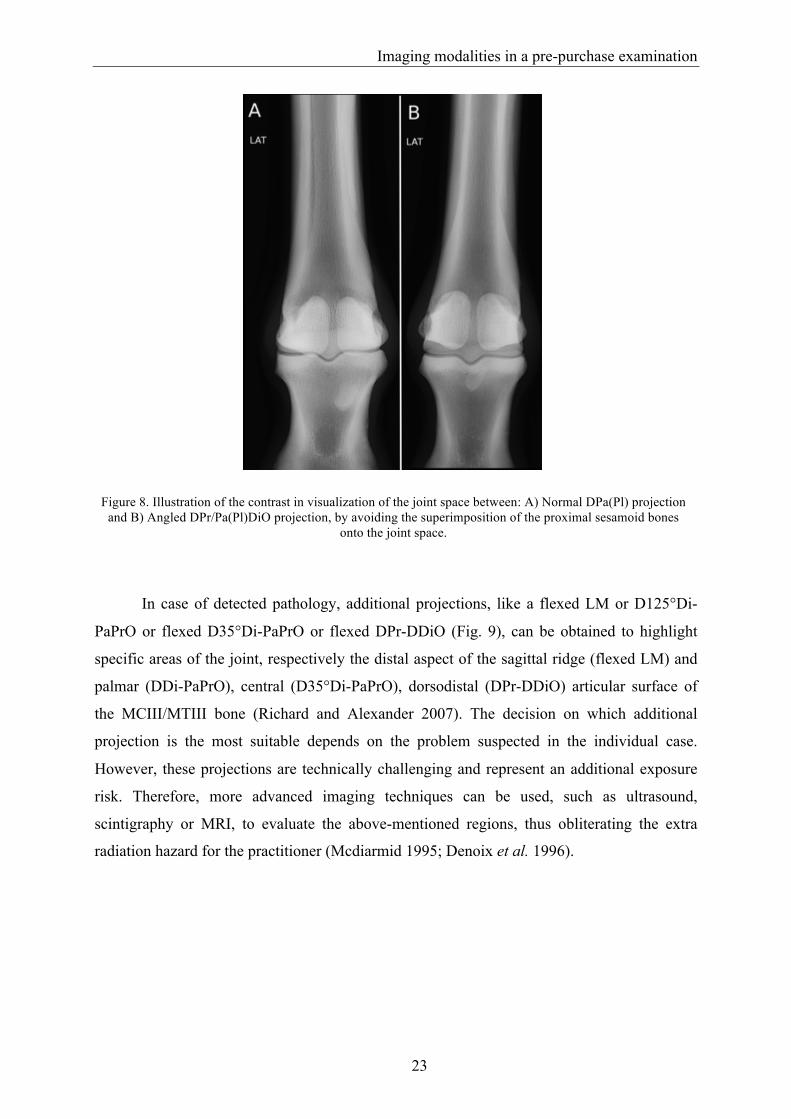
Figure 8. Illustration of the contrast in visualization of the joint space between: A) Normal DPa(Pl) projection and B) Angled DPr/Pa(Pl)DiO projection, by avoiding the superimposition of the proximal sesamoid bones
onto the joint space.
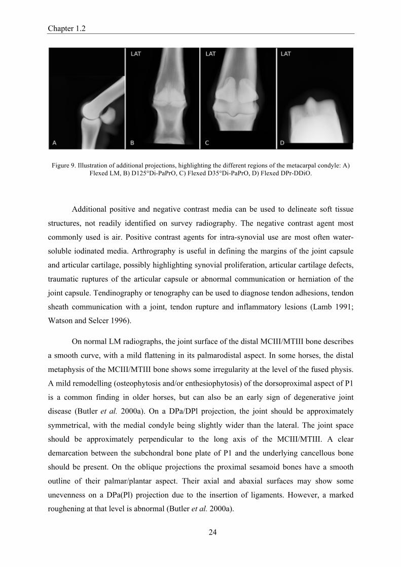
In case of detected pathology, additional projections, like a flexed LM or D125°Di-
PaPrO or flexed D35°Di-PaPrO or flexed DPr-DDiO (Fig. 9), can be obtained to highlight
specific areas of the joint, respectively the distal aspect of the sagittal ridge (flexed LM) and
palmar (DDi-PaPrO), central (D35°Di-PaPrO), dorsodistal (DPr-DDiO) articular surface of
the MCIII/MTIII bone (Richard and Alexander 2007). The decision on which additional
projection is the most suitable depends on the problem suspected in the individual case.
However, these projections are technically challenging and represent an additional exposure
risk. Therefore, more advanced imaging techniques can be used, such as ultrasound,
scintigraphy or MRI, to evaluate the above-mentioned regions, thus obliterating the extra
radiation hazard for the practitioner (Mcdiarmid 1995; Denoix et al. 1996).
Chapter 1.2
24
Figure 9. Illustration of additional projections, highlighting the different regions of the metacarpal condyle: A) Flexed LM, B) D125°Di-PaPrO, C) Flexed D35°Di-PaPrO, D) Flexed DPr-DDiO.
Additional positive and negative contrast media can be used to delineate soft tissue
structures, not readily identified on survey radiography. The negative contrast agent most
commonly used is air. Positive contrast agents for intra-synovial use are most often water-
soluble iodinated media. Arthrography is useful in defining the margins of the joint capsule
and articular cartilage, possibly highlighting synovial proliferation, articular cartilage defects,
traumatic ruptures of the articular capsule or abnormal communication or herniation of the
joint capsule. Tendinography or tenography can be used to diagnose tendon adhesions, tendon
sheath communication with a joint, tendon rupture and inflammatory lesions (Lamb 1991;
Watson and Selcer 1996).
On normal LM radiographs, the joint surface of the distal MCIII/MTIII bone describes
a smooth curve, with a mild flattening in its palmarodistal aspect. In some horses, the distal
metaphysis of the MCIII/MTIII bone shows some irregularity at the level of the fused physis.
A mild remodelling (osteophytosis and/or enthesiophytosis) of the dorsoproximal aspect of P1
is a common finding in older horses, but can also be an early sign of degenerative joint
disease (Butler et al. 2000a). On a DPa/DPl projection, the joint should be approximately
symmetrical, with the medial condyle being slightly wider than the lateral. The joint space
should be approximately perpendicular to the long axis of the MCIII/MTIII. A clear
demarcation between the subchondral bone plate of P1 and the underlying cancellous bone
should be present. On the oblique projections the proximal sesamoid bones have a smooth
outline of their palmar/plantar aspect. Their axial and abaxial surfaces may show some
unevenness on a DPa(Pl) projection due to the insertion of ligaments. However, a marked
roughening at that level is abnormal (Butler et al. 2000a).

Imaging modalities in a pre-purchase examination
25
Opinions differ about the interobserver and intraoberserver agreement of interpretation
of radiographic images. Some state an acceptable to excellent interobserver agreement
(Weller et al. 2001; White et al. 2008), while others state the opposite (Labens et al. 2007). A
good to excellent intraobserver agreement has been mentioned (Labens et al. 2007; White et
al. 2008). However it appears that this agreement is both for intra- as well as for interobserver
agreement depending on the evaluated parameter on the radiograph (Groth et al. 2009).
Ultrasonography
Ultrasonography is a useful imaging modality for the investigation of joint
abnormalities as it enables the evaluation of soft tissue components of the joint and provides
information on the regularity of the bony contours (Redding 2001a; Smith 2008).
This technique uses high frequency waves produced by a transducer. The transducer
converts electrical signals into ultrasound waves, and vice versa for the reflected ultrasound
waves. When placing the transducer on the skin, pulses of ultrasound are sent into the tissues.
Based on the different tissue interfaces, echoes are reflected back to the transducer. These
echoes are processed into an electric signal, which is converted to an image. The time that an
echo needs to return to the transducer determines the distance from the probe. In the resulting
image, this echo is represented by a dot, creating the anatomical echo-generated image. The
brightness of the dot depends on the strength of the echo. The physical interactions of sound
with the tissues determine the appearance of the ultrasound images. At the boundary of 2
materials with different acoustic impedances some of the energy of the ultrasound waves will
be reflected back to the transducer while the remainder of the energy is transmitted through
the second tissue type. At soft tissue to soft tissue interfaces, most of the energy of the
ultrasound wave is transmitted deeper; an interface between bone and soft tissue reflects
approximately 50% of the energy while between soft tissues and air, almost 99.9% of the
energy is reflected. This necessitates the removal of air between the transducer and the patient
(Martin and Ramnarine 2003).
For optimal ultrasonographic examination, the joint should be clipped, cleaned and
coated with conducting gel (Redding 2001a). The MCP/MTP joint can be examined with a
high frequency (7.5-10 Mhz) linear transducer. The use of a standoff pad is helpful to increase
the contact between the probe and the skin and therefore to enlarge the acoustic window as
Chapter 1.2
26
well as to better evaluate the profile of the skin. The MCP/MTP joint can be approached in 6
steps.
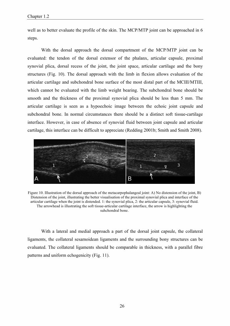
With the dorsal approach the dorsal compartment of the MCP/MTP joint can be
evaluated: the tendon of the dorsal extensor of the phalanx, articular capsule, proximal
synovial plica, dorsal recess of the joint, the joint space, articular cartilage and the bony
structures (Fig. 10). The dorsal approach with the limb in flexion allows evaluation of the
articular cartilage and subchondral bone surface of the most distal part of the MCIII/MTIII,
which cannot be evaluated with the limb weight bearing. The subchondral bone should be
smooth and the thickness of the proximal synovial plica should be less than 5 mm. The
articular cartilage is seen as a hypoechoic image between the echoic joint capsule and
subchondral bone. In normal circumstances there should be a distinct soft tissue-cartilage
interface. However, in case of absence of synovial fluid between joint capsule and articular
cartilage, this interface can be difficult to appreciate (Redding 2001b; Smith and Smith 2008).
Figure 10. Illustration of the dorsal approach of the metacarpophalangeal joint: A) No distension of the joint, B) Distension of the joint, illustrating the better visualisation of the proximal synovial plica and interface of the articular cartilage when the joint is distended. 1: the synovial plica, 2: the articular capsule, 3: synovial fluid.
The arrowhead is illustrating the soft tissue-articular cartilage interface, the arrow is highlighting the suhchondral bone.
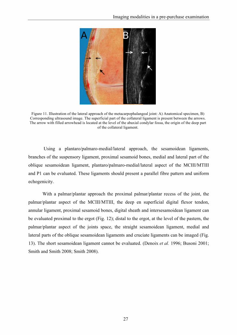
With a lateral and medial approach a part of the dorsal joint capsule, the collateral
ligaments, the collateral sesamoidean ligaments and the surrounding bony structures can be
evaluated. The collateral ligaments should be comparable in thickness, with a parallel fibre
patterns and uniform echogenicity (Fig. 11).
Imaging modalities in a pre-purchase examination
27
Figure 11. Illustration of the lateral approach of the metacarpophalangeal joint: A) Anatomical specimen, B) Corresponding ultrasound image. The superficial part of the collateral ligament is present between the arrows. The arrow with filled arrowhead is located at the level of the abaxial condylar fossa, the origin of the deep part
of the collateral ligament.
Using a plantaro/palmaro-medial/lateral approach, the sesamoidean ligaments,
branches of the suspensory ligament, proximal sesamoid bones, medial and lateral part of the
oblique sesamoidean ligament, plantaro/palmaro-medial/lateral aspect of the MCIII/MTIII
and P1 can be evaluated. These ligaments should present a parallel fibre pattern and uniform
echogenicity.
With a palmar/plantar approach the proximal palmar/plantar recess of the joint, the
palmar/plantar aspect of the MCIII/MTIII, the deep en superficial digital flexor tendon,
annular ligament, proximal sesamoid bones, digital sheath and intersesamoidean ligament can
be evaluated proximal to the ergot (Fig. 12); distal to the ergot, at the level of the pastern, the
palmar/plantar aspect of the joints space, the straight sesamoidean ligament, medial and
lateral parts of the oblique sesamoidean ligaments and cruciate ligaments can be imaged (Fig.
13). The short sesamoidean ligament cannot be evaluated. (Denoix et al. 1996; Busoni 2001;
Smith and Smith 2008; Smith 2008).

Chapter 1.2
28
Figure 12. Illustration of the palmar approach of the metacarpophalangeal joint, just proximal to the proximal sesamoid bones: A) Anatomical specimen, B) Corresponding ultrasound image. 1: the branches of the
suspensory ligament, 2: the deep digital flexor tendon, 3: the superficial digital flexor tendon, 4: the manica flexoria, 5: the palmar cortex of the third metacarpal bone.
Figure 13. Illustration of the palmar approach of the metacarpophalangeal joint, at the mid aspect of the proximal phalanx: A) Anatomical specimen, B) Corresponding ultrasound image. 1: the medial and lateral branch of the
superficial digital flexor tendon, 2: the more bilobed shape of the deep digital flexor tendon, 3: the straight sesamoidean ligament, 4: the medial and lateral part of the oblique sesamoidean ligament. The arrow is
highlighting the palmar cortex of the proximal phalanx.
Dynamic ultrasonographic examination of the MCP/MTP joint allows a better
evaluation of the joint capsule by eliminating hypoechoic relaxation artefacts. Flexion and
extension can also be helpful in demonstrating the mobility of an osteochondral fragment and
in evaluating fluid movement (Modransky et al. 1983; Denoix 1996; Reef 1998; Redding
2001a; Vanderperren et al. 2009). The comparison of the same structure with the contralateral

Imaging modalities in a pre-purchase examination
29
limb improves the sensitivity and specificity of the ultrasonographic diagnosis (Denoix and
Audigie 2001; Redding 2001a).
Ultrasound is known to be a very operator depended technique. It is demonstrated that
significant differences are present for the evaluation of a lesion between operators (Pickersgill
et al. 2001). Therefore, if a difficult problem is expected, it is sometimes better to ask a more
experienced colleague to help (Mitchell 2009).
Ultrasound can be used to complement a pre-purchase examination. However, it is
best to inform the owner of the technical limitations before the exam and have a clear
understanding of the expectations of the owner. Performing ultrasonography during a pre-
purchase examination can pose technical difficulties if the horse is presented with long hair.
Only in fine haired horses, it is possible to perform the examination without clipping. If the
owner is unwilling to allow the horse to be clipped when needed, it is better not to perform
the examination. With an ultrasound examination, suspected joint problems can be evaluated
more completely if suspicious findings occur during the clinical or radiographic examination
(Mitchell 2009).
Scintigraphy
The basic principle of nuclear scintigraphy is the detection of gamma-rays, emitted
during the decay of a radionuclide, by a gamma camera. This radionuclide is attached to a
tracer, together called a radiopharmaceutical, which is most commonly injected IV. Other,
less commonly used methods are subcutaneous injections or inhalation. The most commonly
used radioisotope in equine is technetium 99m. Technetium 99m has a short physical half-life
of approximately 6 hours. This together with the low energy of the gamma rays, results in a
low radiation dose for the patient. On the other hand, the energy value of 140 keV of
technetium 99m allows sufficient gamma-radiation to escape the patient. The choice of tracer
depends on the targeted organ to be examined. For bone imaging, technetium 99m is usually
bound to methylene diphosphonate or hydroxymethylene diphosphonate. These
diphosphonate salts binds to hydroxyapatite in the bone and their accumulation in a specific
area is relative to blood flow to the bone and metabolic activity of the bone. The gamma
camera consists of a lead collimator, a gamma sensitive sodium iodide crystal and a
photomultiplier tube. The collimator allows only those rays moving parallel to its holes to
reach the crystal, which are only a fraction of the radiation leaving the horse. By eliminating

Chapter 1.2
30
the scatter radiation, the origin of the gamma radiation can be determined and positioned
correctly in the resulting image. The gamma rays interact with the sodium iodide crystal and
their energy is converted into light. This emitted light is detected and converted to electrical
pulses by an array of photomultiplier tubes. These electrical pulses are converted into an
image in terms of where in the crystal the light is formed and, indirectly, where the
radiopharmaceutical is located in the patient (Driver 2003; Twardock 2003). Nuclear
medicine can be divided into 3 phases. The vascular phase (or phase I) images are acquired
immediately after injection and highlight the radiopharmaceutical as it courses through the
blood vessels. Pool-phase (or phase II) images are acquired within fifteen to twenty minutes
after injection, while most of the radiopharmaceutical is in the soft tissues. Bone-phase (or
phase III) images are acquired two hours after injection to allow the radiopharmaceutical to
bind to the bone and clear from the soft tissues (Chambers et al. 1995). It is important to
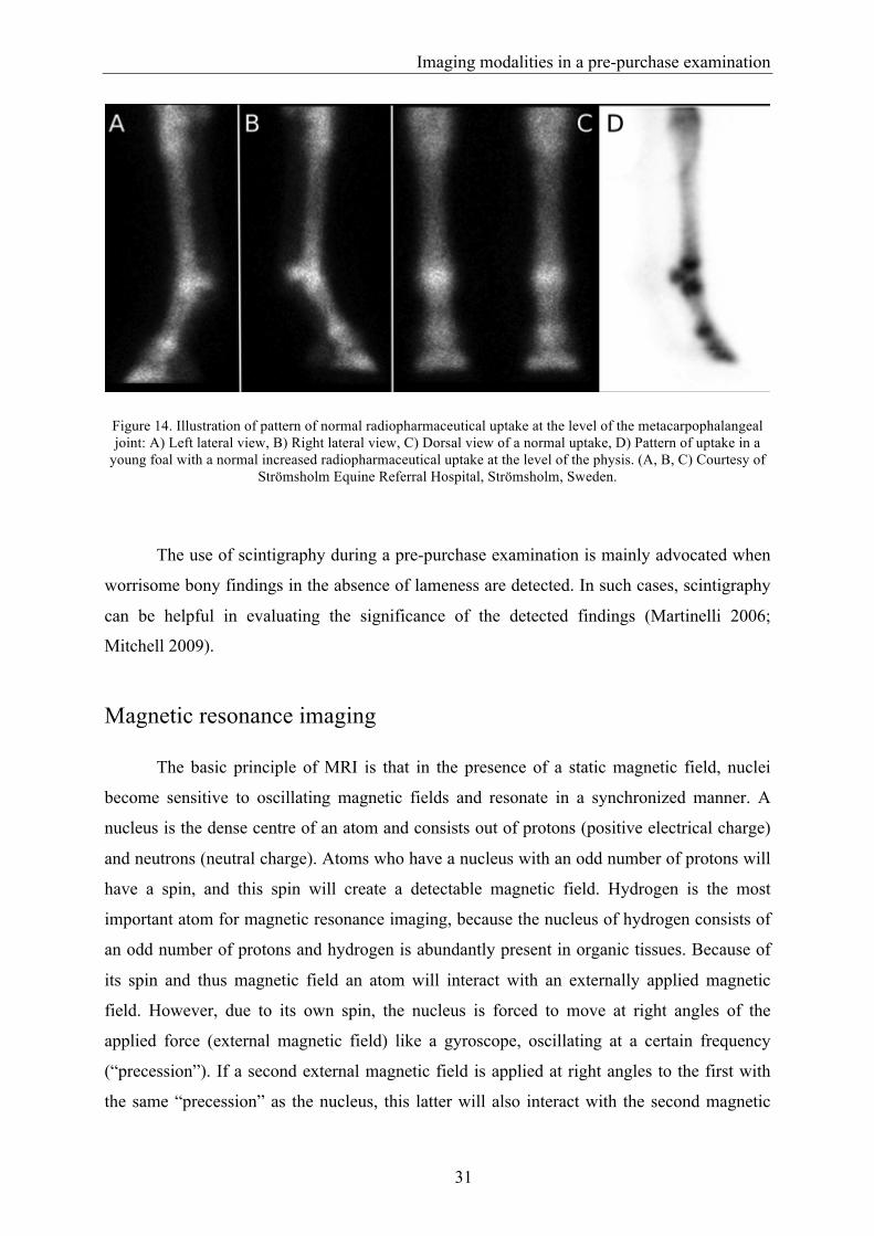
realize that an area of increased radiopharmaceutical uptake, reflecting an area with increased
blood flow or osteoblastic activity, does not necessarily reflect bone pathology, as it can also
represent bone remodelling due to biomechanical loading or development (Fig. 14). A
standard pattern of radiopharmaceutical uptake in the MCP/MTP joint has been established in
non-lame horses without a clear variation over age (Fig. 14) (Weekes et al. 2004) and
significant differences are present compared to lame horses (Biggi et al. 2009). Scintigraphy
is a highly sensitive method to localize a region with a potential problem, and allows to
detected remodelling or lesions before they are radiographically evident (Chambers et al.
1995). However, because of the low specificity, the result must always be interpreted together
with the result of the clinical examination and other imaging modalities in order to avoid
misinterpretation (Weekes et al. 2004). There is an excellent agreement between observers for
assessing relevant increased radiopharmaceutical uptake (Weller et al. 2001).
Imaging modalities in a pre-purchase examination
31
Figure 14. Illustration of pattern of normal radiopharmaceutical uptake at the level of the metacarpophalangeal joint: A) Left lateral view, B) Right lateral view, C) Dorsal view of a normal uptake, D) Pattern of uptake in a
young foal with a normal increased radiopharmaceutical uptake at the level of the physis. (A, B, C) Courtesy of Strömsholm Equine Referral Hospital, Strömsholm, Sweden.
The use of scintigraphy during a pre-purchase examination is mainly advocated when
worrisome bony findings in the absence of lameness are detected. In such cases, scintigraphy
can be helpful in evaluating the significance of the detected findings (Martinelli 2006;
Mitchell 2009).
Magnetic resonance imaging
The basic principle of MRI is that in the presence of a static magnetic field, nuclei
become sensitive to oscillating magnetic fields and resonate in a synchronized manner. A
nucleus is the dense centre of an atom and consists out of protons (positive electrical charge)
and neutrons (neutral charge). Atoms who have a nucleus with an odd number of protons will
have a spin, and this spin will create a detectable magnetic field. Hydrogen is the most
important atom for magnetic resonance imaging, because the nucleus of hydrogen consists of
an odd number of protons and hydrogen is abundantly present in organic tissues. Because of
its spin and thus magnetic field an atom will interact with an externally applied magnetic
field. However, due to its own spin, the nucleus is forced to move at right angles of the
applied force (external magnetic field) like a gyroscope, oscillating at a certain frequency
(“precession”). If a second external magnetic field is applied at right angles to the first with
the same “precession” as the nucleus, this latter will also interact with the second magnetic

Chapter 1.2
32
field, which causes the magnetization to tip over. If this second magnetic field is stopped, the
nucleus will go back to it first state, releasing its excess energy as a radiofrequency signal,
which can be detected and converted into an image (Bolas 2011).
The time constant for the nucleus to go back to its original alignment with the main
external field is called T1, the process is called T1 relaxation or spin-lattice relaxation. During
this relaxation the nuclei will release their excessive energy in the surrounding environment
or lattice. This T1 constant is faster in fat, slower in fluid (water). On a T1 weighted image,
water will appear hypointens (or black); while fat will appear hyperintens (or white) (Fig. 15).
During the application of the second magnetic field, individual nuclei are also “precessing”
together. If the second magnetic field stops, they will gradually lose synchronization. This is
called spin-spin relaxation and makes the main contribution to the relaxation time T2. This T2
relaxation time is much longer in mobile fluids, making that there is an increased T2 time in
tissues with increased water content. On a T2 weighted image water will appear hyperintens
(white) (Fig. 15) (Bolas 2011). Signal intensity varies widely in different tissues, due to
differences in proton density. This determines the tissue’s signal intensity (Kraft and Gavin
2001).
Figure 15. Illustration of a sagittal slice of the metacarpophalangeal joint on MRI illustrating the different appearances of fat and water on a: A) T1 weighted image, B) T2 weighted image. 1: joint fluid, 2: medullary
fat.
The magnetic field strength is measured in Tesla. In equine medicine, several systems
are used, ranging from low field (0,2T) to high field (1,5T). The high field systems enable

Imaging modalities in a pre-purchase examination
33
faster scanning times and have a better image quality (Tucker and Sande 2001). Both low-
field and high-field MRI systems give comparable data about abnormal structures, enabling
the detection of small and subtle lesions without the presence of gross structural changes
(Kraft and Gavin 2001). However, lesions are more detailed with a high field MRI, therefore,
high field MRI is superior in the detection of articular surface abnormalities (Murray et al.
2009). Contrast enhanced MRI with IV gadolinium is possible in the horse and can improve
lesion detection. This contrast enhanced MRI provides additional anatomic and physiologic
assessment of pathologic change (Saveraid and Judy 2012). However this technique is still in
its infancy, but is promising to allow evaluation of articular cartilage pathology (Pease 2012).
Several studies concluded that the inter- and intraobserver agreement is good with
MRI, with a low intra- and interobserver variability (Barrett et al. 2009; De Decker et al.
2011; Wucherer et al. 2012). However, one study mentions only a moderate intra- and
interobserver agreement for evaluation of osteoarthritis status if the equine MCP joint (Olive
et al. 2010).
A standing low field MRI system has been developed for horses to avoid the risk of
general anaesthesia, to ease patient handling and to reduce the operating costs. Its purchase
and maintenance are considerably cheaper, but it provides a poorer magnetic field
homogeneity, which can result in image degradation and artefacts. Due to the lower magnetic
field used in the standing MRI system, longer imaging times are needed. This increases the
risk of movement of the horse and thus the use of motion correction software is necessary
(Tucker and Sande 2001; Mair et al. 2005; Murray and Mair 2005). It generally provides
lower quality images compared to a high field system (Mitchell 2009). On the other hand, at
the level of the MCP/MTP joint, the low field system has enough resolution to detected
pathology at the level of the bone, tendons and ligaments. At the level of the articular
cartilage, a high field system is better to evaluate possible lesions (Murray et al. 2009)
The main advantage of MRI over radiography and diagnostic ultrasound is that it
provides both anatomical and physiological information in multiple planes. Most of the soft
tissues surrounding the MCP/MTP joint can readily be identified even with a low field system
(Martinelli et al. 1997).
Magnetic resonance imaging has its place in a pre-purchase examination to asses a
specific area know to have been previously affected with pathology (Mitchell 2009).
However, often multiple abnormalities or abnormalities on a lame-free limb are detected.

Chapter 1.2
34
During a pre-purchase examination, it is difficult to decide what the significance of these
findings is or what these could mean for the specific horse (Schulze 2010).
Conclusions
A thorough and comprehensive clinical examination remains the basis for every pre-
purchase examination, completed with a radiographic examination for screening purposes.
Some findings during this clinical and radiographic examination may require a further
examination with additional imaging modalities. The decision of which imaging technique to
use, needs to be based on the professional assessment of the horse by the veterinarian and
sound economic considerations.

35
Chapter 1.3
The evaluation of the equine metacarpo-
/metatarsophalangeal joint during a pre-
purchase examination
Adapted from:
Hauspie S., Declercq J., Martens A., Zani D.D., Bergman E.H.J., Saunders J.H. (2011)
Anatomy and imaging of the equine metacarpophalangeal/metatarsophalangeal joint. Vlaams
Diergeneeskundig Tijdschrift 80, 263-270.


Evaluation of the metacarpo-/metatarsophalangeal joint
37
During a pre-purchase examination, several variations can be detected at the level of
the MCP/MTP joint. Some will have an effect on the future performance of the horse, others
not.
In Thoroughbreds and Warmblood horses several radiographic findings are seen at the
level of the MCP/MTP joint (Becht and Park 2000; Kane et al. 2003b; Verwilghen et al.
2009). In Thoroughbreds, flattening of the sagittal ridge, flattening of the distal palmar/plantar
articular surface of the MCIII/MTIII, variations in size and visibility of the transverse ridge,
the medial proximal sesamoid bone being more cuboidal than the lateral one, a separate centre
of ossification at the proximal aspect of the proximal sesamoid bones or the distal border of
the hind proximal sesamoid bones being more flatter than the distal border of the fore
proximal sesamoid bones are considered normal radiographic variations at the level of the
MCP/MTP joint (Becht and Park 2000). Other radiographic findings like palmar
supracondylar lysis, enthesophyte formation on the fore proximal sesamoid bones and
proximal dorsal fragmentation of P1 in the hind MTP joint and enthesophyte formation on the
hind proximal sesamoid bones, have been associated with reduced performance in
Thoroughbred horses (Kane et al. 2003a).
The detected radiographic finding at the level of the MCP/MTP joint in Warmblood
horses includes remodelling of the proximal border of P1, mild surface irregularity or
osteochondral defect of the proximal border of the sagittal ridge of the MCIII/MTIII, dorsal
osteochondral fragments originating from the MCIII/MTIII, plica synovialis or the
dorsoproximal border of P1 to palmar or plantar fragments of P1 (Stock et al. 2005; Declercq
et al. 2008; Declercq et al. 2009; Verwilghen et al. 2009). The clinical relevance of several of
these findings is still unclear (Declercq et al. 2008; Martens et al. 2008).
In Warmbloods and Thoroughbreds variation in radiographic appearance of the
proximal aspect of the dorsal condylar sagittal ridge is detected (Kane et al. 2003b;
Verwilghen et al. 2009). In Warmbloods the described variations were a mild surface
irregularity of the proximal border of the sagittal ridge of the metacarpus/tarsus. In
Thoroughbreds the detected variations at that level were a well defined semicircular notch,
lucency or a fragment/loose body. In Thoroughbreds, these variations were not associated
with reduced performance (Kane et al. 2003a). However, due to the difference in type and
length of the sport career for both breeds (short sports career in Thoroughbreds; long lasting
career in Warmbloods) (Hinchcliff and Hamlin 2004; O'Sullivan and Lumsden 2004), and the

Chapter 1.3
38
difference in described appearance between both breeds of the proximal aspect of the sagittal
ridge, the same conclusion cannot just be extrapolated to Warmblood horses.

References
39
References
Alrtib A.M., Philip C.J., Abdunnabi A.H. and Davies H.M. (2012) Morphometrical study of bony elements of the forelimb fetlock joints in horses. Anatomia Histologia Embryologia, 10.1111/j.1439-0264.2012.01158.x.
Barone R. (1986) Ceinture et membre thoraciques. In: Barone R. (editors), Anatomie
comparée des mammifères domestiques: Tome 1 ostéologie, 3 edn., Vigot freres, Paris. 451-586.
Barone R. (1989) Articulations de la ceinture et du membre thoraciques, Anatomie comparée
des mammifères domestiques: Tome 2 arthrologie et myologie, 3 edn., éditions vigot frères, Paris. 97-222.
Barrett E., Barr F., Owen M. and Bradley K. (2009) A retrospective study of the mri findings
in 18 dogs with stifle injuries. J Small Anim Pract 50, 448-455. Becht J.L. and Park R.D. (2000) A review of selected normal radiographic variations of the
equine fetlock, carpus, tarsus and stifle. In: 46th annual convention of the AAEP, San Antonio, Texas. 362-364.
Biggi M., Dyson S.J. and Murray R.C. (2009) Scintigraphic assessment of the
metacarpophalangeal and metatarsophalangeal joints of horses with joint pain. Veterinary Radiology & Ultrasound 50, 536-544.
Bolas N.M. (2011) Basic mri principles. In: Murray R. (editors), Equine mri, Wiley-
Blackwell, West Sussex. 3-38. Busoni V. (2001) Ultrasonographic examination of the palmar aspect of the fetlock and
pastern: Technique and normal images. Ippologia 12, 5-14. Butler J.A., Colles C.M., Dyson S., Kold S.E. and Poulos P.W. (2000a) Foot, pastern and
fetlock. In: Butler J.A., Colles C.M., Dyson S., Kold S.E. and Poulos P.W. (editors), Clinical radiology of the horse, 2 edn., Blackwell science, Oxford. 27-130.
Butler J.A., Colles C.M., Dyson S., Kold S.E. and Poulos P.W. (2000b) General prinicples.
In: Butler J.A., Colles C.M., Dyson S., Kold S.E. and Poulos P.W. (editors), Clinical radiology of the horse, Blackwell science, Oxford. 1-26.
Chapter 1
40
Chambers M.D., Martinelli M.J., Baker G.J., Kneller S.K. and Twardock A.R. (1995) Nuclear-medicine for diagnosis of lameness in horses. Journal of the American Veterinary Medical Association 206, 792-796.
Dabareiner R.M., White N.A. and Sullins K.E. (1996) Metacarpophalangeal joint synovial
pad fibrotic proliferation in 63 horses. Veterinary Surgery 25, 199-206. Dalla Palma L. (2000) Cost analysis of digital vs analogue radiography. European Radiology
10, S386-S389. De Decker S., Gielen I.M., Duchateau L., Lang J., Dennis R., Corzo-Menendez N., Van Bree
H.J., Van Soens I., Binst D.H., Waelbers T. and Van Ham L.M. (2011) Intraobserver and interobserver agreement for results of low-field magnetic resonance imaging in dogs with and without clinical signs of disk-associated wobbler syndrome. Journal of the American Veterinary Medical Association 238, 74-80.
Declercq J., Martens A., Bogaert L., Boussauw B., Forsyth R. and Boening K.J. (2008)
Osteochondral fragmentation in the synovial pad of the fetlock in warmblood horses. Veterinary Surgery 37, 613-618.
Declercq J., Martens A., Maes D., Boussauw B., Forsyth R. and Boening K.J. (2009)
Dorsoproximal proximal phalanx osteochondral fragmentation in 117 warmblood horses. Veterinary and Comparative Orthopaedics and Traumatology 22, 1-6.
Denoix J.M. (1996) Ultrasonographic examination in the diagnosis of joint disease. In:
McIlwraith C.W. and Trotter G.W. (editors), Joint disease in the horse, W.B. Saunders, Philadelphia. 165-202.
Denoix J.M. and Audigie F. (2001) Ultrasonographic examination of joints in horses. In: 47th
Annual convention of the American Association of Equine Practitioners, San Diego, California, USA. 366-375.
Denoix J.M., Jacot S., Bousseau B. and Perrot P. (1996) Ultrasonographic anatomy of the
dorsal and abaxial aspects of the equine fetlock. Equine Veterinary Journal 28, 54-62. Driver A.J. (2003) Basic principles of equine scintigraphy. In: Dyson S., Pilsworth R.C.,
Twardock A.R. and Martinelli M.J. (editors), Equine scintigraphy, Equine Veterinary Journal, Newmarket. 17-24.
Edwards G.B. (1984) Interpreting radiographs 2: The fetlock joint and pastern. Equine
Veterinary Journal 16, 4-10.
References
41
Groth A.M., May S.A., Weaver M.P. and Weller R. (2009) Intra- and interobserver agreement in the interpretation of navicular bones on radiographs and computed tomography scans. Equine Veterinary Journal 41, 124-129.
Hinchcliff K.W. and Hamlin M. (2004) Veterinary aspects of racing and training standardbred
race horses. In: Hinchcliff K.W., Kaneps A.J. and Geor R.J. (editors), Equine sports medicine and surgery, Saunders, Philadelphia. 1073-1089.
Jimenez D.A., Armbrust L.J., O'Brien R.T. and Biller D.S. (2008) Artifacts in digital
radiography. Veterinary Radiology & Ultrasound 49, 321-332. Kane A.J., McIlwraith C.W., Park R.D., Rantanen N.W., Morehead J.P. and Bramlage L.R.
(2003a) Radiographic changes in thoroughbred yearlings. Part 2: Associations with racing performance. Equine Veterinary Journal 35, 366-374.
Kane A.J., Park R.D., McIlwraith C.W., Rantanen N.W., Morehead J.P. and Bramlage L.R.
(2003b) Radiographic changes in thoroughbred yearlings. Part 1: Prevalence at the time of the yearling sales. Equine Veterinary Journal 35, 354-365.
Kraft S.L. and Gavin P. (2001) Physical principles and technical considerations for equine
computed tomography and magnetic resonance imaging. Veterinary Clinics of North America-Equine Practice 17, 115-130.
Labens R., Innocent G.T. and Voute L.C. (2007) Reliability of a quantitative rating scale for
assessment of horses with distal tarsal osteoarthritis. Veterinary Radiology & Ultrasound 48, 204-211.
Lamb C.R. (1991) Contrast radiography of equine joints, tendon sheaths, and draining tracts.
Veterinary Clinics of North America-Equine Practice 7, 241-257. Mair T.S., Kinns J., Jones R.D. and Bolas N.M. (2005) Magnetic resonance imaging of the
distal limb of the standing horse. Equine Veterinary Education 17, 74-78. Marks D. (1999) Prepurchase examination of jumpers and dressage horses. In: Annual
convention of the american association of equine practitioners, Albuquerque, New Mexico. 4-12.
Martens A., Declercq J. and Vanderperren K. (2008) Osteochondral fragments in equine
joints: Do we have to remove them all? In: European veterinary conference voorjaarsdagen, Amsterdam, Netherlands. 281-282.

Chapter 1
42
Martin K. and Ramnarine K.V. (2003) Physics. In: Hoskins P., Thrush A., Martin K. and Whittingham T.A. (editors), Diagnostic ultrasound, physics and equipment, Greenwich-medical, London. 7-22.
Martinelli M.J. (2006) The value of nuclear scintigraphy in predicting orthopaedic disease in
equine athletes. In: 13th ESVOT congress, Munich, Germany. 181-182. Martinelli M.J., Kuraiashkin I.V., Carragher B.O., Clarkson R.B. and Baker G.J. (1997)
Magnetic resonance imaging of the equine metacarpophalangeal joint: Three-dimensional reconstruction and anatomic analysis. Veterinary Radiology & Ultrasound 38, 193-199.
Mattoon J.S. (2006) Digital radiography. Veterinary and Comparative Orthopaedics and
Traumatology 19, 123-132. Mcdiarmid A. (1995) Ultrasonography of the palmar metacarpus and pastern in the horse. In
practice 17, 368-376. Mcknight A.L. (2004) Digital radiography in equine practice. Clinical techniques in equine
practice 3, 352-360. Mitchell R.D. (2009) Imaging considerations in the purchase examination of the performance
horse. In: 55th Annual convention of the American Association of Equine practitioners, Las Vegas, Nevada, USA. 296-300.
Modransky P.D., Rantanen N.W., Hauser M.L. and Grant B.D. (1983) Diagnostic ultrasound
examination of the dorsal aspect of the equine metacarpophalangeal joint. Journal of Equine Veterinary Science 3, 56-58.
Murray R. and Mair T. (2005) Use of magnetic resonance imaging in lameness diagnosis in
the horse. In practice 27, 138-146. Murray R.C., Mair T.S., Sherlock C.E. and Blunden A.S. (2009) Comparison of high-field
and low-field magnetic resonance images of cadaver limbs of horses. Veterinary Record 165, 281-288.
O'Sullivan C.B. and Lumsden J.M. (2004) Veterinary aspects of racing and training
thoroughbred race horses. In: Hinchcliff K.W., Kaneps A.J. and Geor R.J. (editors), Equine sports medicine and surgery, Saunders, Philadelphia. 1051-1072.

References
43
Olive J., D'anjou M.-A., Alexander K., Laverty S. and Theoret C. (2010) Comparison of magnetic resonance imaging, computed tomography, and radiography for assessment of noncartilaginous changes in equine metacarpophalangeal osteoarthritis. Veterinary Radiology & Ultrasound 51, 267-279.
Park R.D. (2000) Optimal radiographic views for evaluating thoroughbred yearlings - quality
control of the radiographic image. In: 46th Annual convention of the American Association of Equine Practitioners, San Antonio, Texas, USA. 357-358.
Pease A. (2012) Biochemical evaluation of equine articular cartilage through imaging.
Veterinary Clinics of North America-Equine Practice 28, 637-646. Pickersgill C.H., Marr C.M. and Reid S.W. (2001) Repeatability of diagnostic
ultrasonography in the assessment of the equine superficial digital flexor tendon. Equine Veterinary Journal 33, 33-37.
Pilsworth R. and Head M. (2010) Presales radiographic surveys in yearlings 1. Image
interpretation and significance of lesions in the fetlock. In practice 32, 174-183. Pool R.R. and Meagher D.M. (1990) Pathologic findings and pathogenesis of racetrack
injuries. Veterinary Clinics of North America-Equine Practice 6, 1-30. Poulos P.W., Jr. (1992) Radiologic evaluation of the horse relevant to purchase. Veterinary
Clinics of North America-Equine Practice 8, 319-328. Redding W.R. (2001a) Use of ultrasonography in the evaluation of joint disease in horses.
Part 1: Indications, technique and examination of the soft tissues. Equine Veterinary Education 13, 198-204.
Redding W.R. (2001b) Use of ultrasonography in the evaluation of joint disease in horses.
Part 2: Examination of the articular surface. Equine Veterinary Education 13, 275-279.
Reef V.B. (1998) Musculoskeletal ultrasonography. In: Reef V.B. (editors), Equine diagnostic
ultrasound, W.B. Saunders company, Philadelphia. 39-186. Richard E. and Alexander K. (2007) Nonconventional radiographic projections in the equine
orthopaedic examination. Equine Veterinary Education 19, 551-559. Rossdale P.D., Hopes R. and Digby N.J.W. (1985) Epidemiological study of wastage among
racehorses 1982 and 1983. Veterinary Record 116, 66-69.

Chapter 1
44
Santschi E.M. (2008) Articular fetlock injuries in exercising horses. Veterinary Clinics of North America-Equine Practice 24, 117-132.
Saveraid T.C. and Judy C.E. (2012) Use of intravenous gadolinium contrast in equine magnetic resonance imaging. Veterinary Clinics of North America: Equine Practice 28, 617-636.
Schulze T. (2010) Prepurchase mri of horses - definition and clinical implications. In: 15th
ESVOT congress, Bologna, Italy. 247. Smith M. and Smith R. (2008) Diagnostic ultrasound of the limb joints, muscle and bone in
horses. In practice 30, 152-159. Smith R. (2008) Using ultrasound to image joints. In: 10th International congress of World
Equine Veterinary Association, Moscow, Russia. 279-282. Steyn P.F., Schmitz D., Watkins J. and Hoffman J. (1989) The sonographic diagnosis of
chronic proliferative synovitis in the metacarpophalangeal joints of a horse. Veterinary Radiology 30, 125-127.
Stock K.F., Hamann H. and Distl O. (2005) Prevalence of osseous fragments in distal and
proximal interphalangeal, metacarpo- and metatarsophalangeal and tarsocrural joints of hanoverian warmblood horses. Journal of veterinary medicine 52, 388-394.
Suslak-Brown L. (2004) Radiography and the equine prepurchase exam. Clinical techniques
in equine practice 3, 361-364. Tucker R.L. and Sande R.D. (2001) Computed tomography and magnetic resonance imaging
in equine musculoskeletal conditions. Veterinary Clinics of North America-Equine Practice 17, 145-157.
Twardock A.R. (2003) Basic structure and function of the camera. In: Dyson S., Pilsworth
R.C., Twardock A.R. and Martinelli M.J. (editors), Equine scintigraphy, Equine Veterinary Journal, Newmarket. 37-46.
Van Hoogmoed L.M., Snyder J.R., Thomas H.L. and Harmon F.A. (2003) Retrospective
evaluation of equine prepurchase examinations performed 1991-2000. Equine Veterinary Journal 35, 375-381.
Vanderperren K., Ghaye B., Snaps F.R. and Saunders J.H. (2008) Evaluation of computed
tomographic anatomy of the equine metacarpophalangeal joint. American Journal of Veterinary Research 69, 631-638.

References
45
Vanderperren K., Martens A.M., Declercq J., Duchateau L. and Saunders J.H. (2009) Comparison of ultrasonography versus radiography for the diagnosis of dorsal fragmentation of the metacarpophalangeal or metatarsophalangeal joint in horses. Journal of the American Veterinary Medical Association 235, 70-75.
Verwilghen D., Serteyn D., Pille F., Bolen G., Saunders J.H., Grulke S. and Busoni V. (2009)
Prevalence of radiographic findings in candidate sires (2001-2008). Vlaams Diergeneeskundig Tijdschrift 78, 419-428.
Watson E. and Selcer B. (1996) Use of radiographic contrast media in horses. Compendium
on Continuing Education for the Practicing Veterinarian 18, 167-&. Weaver J.C., Stover S.M. and O'Brien T.R. (1992) Radiographic anatomy of soft tissue
attachments in the equine metacarpophalangeal and proximal phalangeal region. Equine Veterinary Journal 24, 310-315.
Weekes J.S., Murray R.C. and Dyson S.J. (2004) Scintigraphic evaluation of
metacarpophalangeal and metatarsophalangeal joints in clinically sound horses. Veterinary Radiology & Ultrasound 45, 85-90.
Weller R., Livesey L., Maierl J., Nuss K., Bowen I.M., Cauvin E.R., Weaver M., Schumacher
J. and May S.A. (2001) Comparison of radiography and scintigraphy in the diagnosis of dental disorders in the horse. Equine Veterinary Journal 33, 49-58.
White J.M., Mellor D.J., Duz M., Lischer C.J. and Voute L.C. (2008) Diagnostic accuracy of
digital photography and image analysis for the measurement of foot conformation in the horse. Equine Veterinary Journal 40, 623-628.
White N.A. (1990) Synovial pad proliferation in the metacarpophalangeal joint. In: White
N.A. and Moore J.N. (editors), Current practice of equine surgery, 1 edn., J. B.Lippincott, Philadelphia. 555-558.
Wucherer K.L., Ober C.P. and Conzemius M.G. (2012) The use of delayed gadolinium
enhanced magnetic resonance imaging of cartilage and t2 mapping to evaluate articular cartilage in the normal canine elbow. Veterinary Radiology & Ultrasound 53, 57-63.


47
Chapter 2
Scientific Aims

Scientific Aims
49
A veterinary pre-purchase examination is an important service offered by the
veterinarian to clients who wish to sell or buy a horse. With this examination, the responsible
veterinarian tries to identify abnormalities or potential problems that could make the horse
unsuitable for the intended use. In a pre-purchase examination, radiography is an important
aid to detect actual or potential orthopaedic problems.
During this pre-purchase examination, radiographic changes are frequently detected at
the level of the MCP/MTP joint. It is up to the veterinarian to decide if these changes will
have an influence on the future sport career of the horse. This decision is best taken on the
basis of scientific data, however this information is often not available, resulting in different
opinions of veterinarians on the same case. In Thoroughbreds, it has been shown that there is
some variation in the radiographic appearance of the proximal aspect of the dorsal condylar
sagittal ridge, but without influence on the sports career of the horse. Extrapolation of the
relevance of these appearances to Warmbloods is however not appropriate because of the
large difference in type and duration of sports career.
The general aim of this research project was therefore to describe the variation in
radiographic appearance of the proximal aspect of the dorsal condylar sagittal ridge in
Warmblood horses, to evaluate their histological basis, as well as to assess the influence of
these variations on the joint cartilage and their interaction with the surrounding soft tissues.
More specific by:
1. Describing the prevalence of variation in the radiographic appearance of the
dorsoproximal aspect of the condylar sagittal ridge in a population of Warmblood
stallions.
2. Evaluating the histological appearance of these variations at the dorsoproximal
aspect of the sagittal ridge.
3. Assessing the possible predisposition of these variations in appearance of the
dorsoproximal aspect of the sagittal ridge to articular cartilage degeneration.
4. Describing the influence of hyperextension of the MCP/MTP joint on the position
of the synovial plica surrounding the proximal aspect of the dorsal condylar sagittal
ridge.


51
Chapter 3
Radiographic features of the
dorsoproximal aspect of the sagittal ridge
of the third metacarpal and metatarsal
bones in young Warmblood stallions
Adapted from:
Hauspie S., Martens A., Declercq J., Busoni V., Vanderperren K., van Bree H., Saunders J.H.
(2010) Radiographic features of the dorsal condylar sagittal ridge of the third metacarpal and
metatarsal bone in young Warmblood horses. Veterinary and Comparative Orthopaedics and
Traumatology 23, 411-416.


Radiographic variation of the sagittal ridge
53
Summary
Radiography is a standard practice during a pre-purchase examination of a horse.
During this examination, several variations can be detected. The objective of this study is to
describe the prevalence of variation in radiographic appearance of the dorsoproximal aspect
of the condylar sagittal ridge of the MCIII/MTIII in young Warmblood stallions.
The LM radiographic projections of the MCP/MTP joints performed on horses as a
part of stallion selection were used. The radiographic appearance of the bone surface at the
dorsoproximal aspect of the condylar sagittal ridge was classified as “smooth”, “irregular”,
“cam”, “indentation” and “lucency”.
The radiographic appearance of the proximal aspect of the sagittal ridge ranged from
“smooth” in 51.5% of the joint, 19.3% was “irregular”, 8.9% presented a “cam”, 8.1% had a
“lucency” and 12.2% had an “indentation”. In 1.2% of the horses a fragment was present at
the level of the dorsoproximal aspect of the sagittal ridge and in 1.7% a fragment was
suspected superimposed on the dorsoproximal aspect of the sagittal ridge.
Radiographic variation is present at the dorsal aspect of the MCP/MTP joint in young
Warmblood stallions. These various aspects should be recognized and described in horses
presented for pre-purchase examination. However, their clinical relevance in the individual
horse is unclear and needs further investigation.
Chapter 3
54
Introduction
The dorsoproximal aspect of the equine MCP/MTP joint is composed of a thick joint
capsule including a synovial plica, a layer of cartilage, subchondral bone, a synovial
membrane and synovial fluid (Dabareiner et al. 1996; Denoix et al. 1996; McIlwraith 2001).
Although the exact function of the synovial plica has not been studied, its location and
structure suggest that it acts as a contact interface or cushion between the proximal dorsal rim
of P1 and the dorsal surface of the distal MCIII/MTIII during full extension of the MCP/MTP
joint (McIlwraith et al. 2005). This anatomical region can be affected by specific disorders
(osteochondrosis, chronic proliferative synovitis in the MCP joint), or it can be involved in a
generalised joint disorder (capsulitis/synovitis, osteoarthritis, infectious or traumatic arthritis)
(Vanderperren and Saunders 2009a, 2009b).
An examination is frequently performed prior to the sale of a horse in order to assess
the suitability of the animal for the purpose for which it is required. Depending on the
intended use and value of the animal, a radiographic examination may be part of the pre-
purchase examination. If radiographs are taken, projections of the MCP/MTP joints will be
included (Van Hoogmoed et al. 2003). Should the veterinarian make a mistake in interpreting
the radiographic images, the economic and legal consequences may be important (Van
Hoogmoed et al. 2003). However, there is a general lack of published information regarding
the clinical significance of many radiographic findings, as well as the common anatomical
variations (McIlwraith et al. 2003). Moreover, in addition to the horse’s function, the owner’s
expectations and intended use for the horse will largely determine their significance (Becht
and Park 2000; Bladon and Main 2003; Kane et al. 2003b). Because of the difference in type
and duration of their sports career, great caution should be exercised when using the
conclusions drawn from Thoroughbred horses to interpret the radiographs of a Warmblood
horse (Kane et al. 2003a; Kane et al. 2003b; McIlwraith et al. 2003; Spike-Pierce and
Bramlage 2003; Van Hoogmoed et al. 2003).
The objective of this study is to describe the prevalence of variation in radiographic
appearance of the dorsoproximal aspect of the condylar sagittal ridge of the MCIII/MTIII in
young Warmblood stallions.

Radiographic variation of the sagittal ridge
55
Materials and methods
The LM radiographic projections of MCP/MTP joints performed on horses presented
at our Institution (Ghent University’s large animal teaching hospital) as a part of stallion
selection between April 2007 and March 2009 were used. Only horses younger than 6 years
were used in this study. A short lameness examination was performed: horses were evaluated
trotting in a straight line and lunging (hard and soft surface). However, due to the nature of
the stallion selection, an in-depth lameness examination, with flexion tests, was not
performed. Using a computed radiography imaging system (Regius model 190, Konica
Minolta, Tokyo, Japan), radiographs were made with a horizontal X-ray beam and with the
horse bearing weight. The radiographs were evaluated using commercially available software
(Osirix, Geneva, Switzerland). If the projection was excessively oblique - defined as
superimposition of the distal condyles of the MCIII/MTIII on the sagittal ridge, preventing a
thorough radiographic interpretation - the radiograph was not used in the study.
Two readers - a Board-certified radiologist (JHS) and a PhD-student (SH) - reviewed
all of the examinations together, and each decision was made consensually. When there was
disagreement, consensus was sought between senior radiologists. Special attention was given
to the dorsoproximal aspect of the condylar sagittal ridge of the MCIII/MTIII (Fig. 1).
Figure 1. LM radiograph of the metacarpo-/metatarsophalangeal joint. The arrow is highlighting the proximal third of the visible dorsal aspect of the sagittal ridge, at the level of the synovial plica.
The dorsoproximal aspect of the condylar sagittal ridge corresponded to the proximal
third of the visible dorsal aspect of the sagittal ridge, at the level of the synovial plica. The

Chapter 3
56
appearance of this area was classified according to these 5 categories: “smooth” (defined as
flat or sharp and smoothly delineated; Fig. 2), “irregular”, small and well-defined bony
prominence or “cam”, irregularly shaped “lucency” or a sharply delineated “indentation” (Fig.
3) (Kane et al. 2003b; Cohen et al. 2006). The presence of a fragment or a suspected fragment
was also noted (a suspected fragment was defined as an ill defined and ill delineated bony
opacity). If possible, the surface area of the fragment was measured directly from the LM
radiographic projection without allowance for magnification. A crude indication of the
surface area was obtained by drawing a closed polygon around the outer edges of the
fragment and then calculating the surface area with commercially available imaging software
(Osirix, Geneva, Switzerland).
Figure 2. LM radiograph of the metacarpo-/metatarsophalangeal joint showing the difference in shape of the proximal aspect of the sagittal ridge in the “smooth” category: A) Flat and smoothly delineated, B) Sharp and
smoothly delineated (arrow).
Figure 3. LM radiograph of the metacarpo-/metatarsophalangeal joint demonstrating the other appearances recorded at the proximal aspect of the sagittal ridge: A) “irregular”, B) “cam”, C) “lucency”, D) “indentation”.
Radiographic variation of the sagittal ridge
57
Statistical analysis was performed with SAS version 9.2 (SAS Institute Inc., Cary,
NC) to determine if there was a difference between left and right MCP/MTP joints and
between the front and hind limbs. Statistical analysis was performed using the chi-square test
(or the Fisher exact test if sample sizes were smaller than 10).
Results
Animals
A total of 1232 radiographs of MCP/MTP joints of 308 Warmblood stallions were
available for this retrospective evaluation. The mean age of the population was 2.22 years
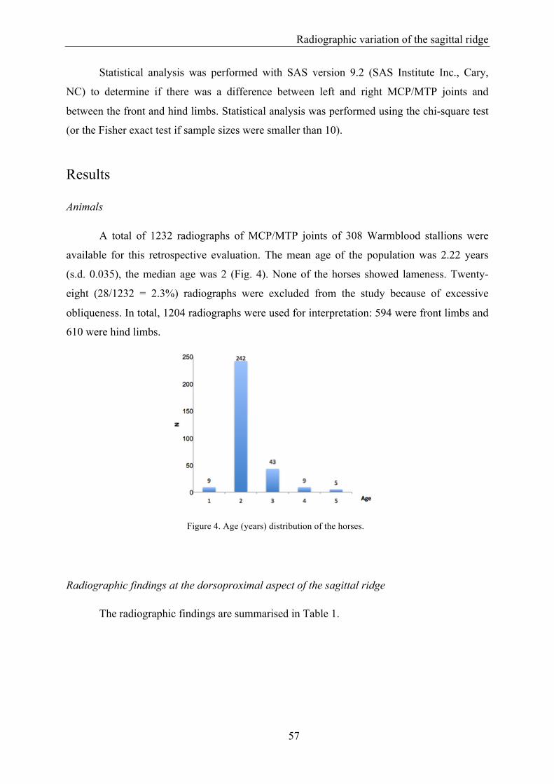
(s.d. 0.035), the median age was 2 (Fig. 4). None of the horses showed lameness. Twenty-
eight (28/1232 = 2.3%) radiographs were excluded from the study because of excessive
obliqueness. In total, 1204 radiographs were used for interpretation: 594 were front limbs and
610 were hind limbs.
Figure 4. Age (years) distribution of the horses.
Radiographic findings at the dorsoproximal aspect of the sagittal ridge
The radiographic findings are summarised in Table 1.

Chapter 3
58
Table 1. Radiographic findings in the metacarpo-/metatarsophalangeal joints in young Warmblood horses.
The radiographic appearance of the dorsoproximal aspect of the condylar sagittal ridge
of the MCIII/MTIII had a “smooth” appearance in 51.5% (620/1204) of the joints, 19.3%
(232/1204) were “irregular”, 8.9% (107/1204) had a “cam”, 8.1% (98/1204) had a “lucent”
area and 12.2% (147/1204) had a well-defined “indentation” (Fig. 3). No significant
differences were found between left and right MCP/MTP joints. A significant difference (P <
0.0001) was found between front limbs and hind limbs: an “indentation” was more present in
the hind limbs than in the front limbs. In 1.2% (15/1204) of the joints, a fragment was present;
always dorsal to, or partially superimposed on the dorsoproximal aspect of the sagittal ridge.
The appearance of the sagittal ridge was evenly distributed over the 5 categories. The shape of
the fragment varied from linear to oval, with a mean surface of 0.093 cm2 (ranging from
0.011 cm2 to 0.472 cm2) (Fig. 5A). In 1.7% (20/1204) of the joints, the presence of a fragment
was suspected; always completely superimposed on the sagittal ridge (Fig. 5B). In this case
the appearance of the sagittal ridge was mostly “normal” to “irregular”. One horse had a
fragment in both front limbs and 1 horse had a fragment in the left front limb and hind limb.

Radiographic variation of the sagittal ridge
59
Figure 5. LM radiograph of the metacarpo-/metatarsophalangeal joint showing: A) The presence or B) the suspected presence of a fragment at the dorsoproximal aspect of the sagittal ridge.
Discussion
In our study, there was a lot of variation in the radiographic appearance of the
dorsoproximal aspect of the sagittal ridge of the MCIII/MTII. Variation in appearance at this
level of the sagittal ridge is also described in Thoroughbreds (notch, lucency and
fragment/loose body) (Kane et al. 2003b) and in an other population of Warmbloods (mild
surface irregularity or well defined osteochondral defect) (Verwilghen et al. 2009). In
Thoroughbreds these variations had no implications for future performance in sport (Kane et
al. 2003a). It is tempting to extrapolate these conclusions. However, in contrast to the
population of Thoroughbred horses used for racing (Kane et al. 2003a), our population
consisted of Warmblood horses used primarily for jumping or dressage. Compressive strains
at the dorsal aspect of the MCII/MTIII have been shown to be more prevalent in racehorses
working at high speed, whereas dressage or jumping horses - which usually work at a lower
level of impact - have a different direction of strain, a longer sports career, and possibly
another conformation as well (Davies et al. 1993; Bennell et al. 1997; Davies and Merritt
2004).
In our study, 55% of the front limbs and 46% of the hind limbs showed a “smooth”
appearance of the dorsoproximal aspect of the sagittal ridge, compared to 72% and 75%,

Chapter 3
60
respectively (categorized as no detected changes) in a previous study of Thoroughbreds (Kane
et al. 2003b). Besides the difference in breed, this difference in percentages can be explained
by the use of additional parameters (“irregular” and “cam”) in the present study. If the
MCP/MTP joints with an “irregular” outlining are considered to be normal, 77% of the front
limbs and 65% of the hind limbs fall into this category, which corresponds more closely to the
previous study (Kane et al. 2003b).
The dorsoproximal aspect of the sagittal ridge was “irregular” outlined in a total of
19,3% of the joints in this study, which is more than the 6.8% described in an other
population of Warmblood stallions (Verwilghen et al. 2009). The presence of a small and
well-defined “cam” was a bilateral finding in 55% of the front limbs and 46% of the hind
limbs. The presence of a small and well-defined “cam” was previously not described as a
possible radiographic appearance of the proximal aspect of the sagittal ridge in
Thoroughbreds, nor in Warmbloods (Kane et al. 2003b; Verwilghen et al. 2009). The
aetiology of this cam or the reason why this was only detected in this population of
Warmblood stallions is unknown. A “lucency” was present in 47 front limbs and 53 hind
limbs and was a bilateral finding in only 2 horses and 5 horses, respectively. This is in
agreement with the study of Kane et al. 2003 in which a “lucency” was most often a unilateral
finding. However, this “lucency” was much more detected in our study: approximately 8% for
each joint, where this only occurred in only 1% of the joints in Thoroughbreds (Kane et al.
2003b). The presence of an “indentation” was a bilateral finding in 17,1% (6 out of 35 horses)
of the front limbs and is in contrast to Thoroughbreds were this was a bilateral finding in 65%
of the front limbs. In 26% (24 out of 92 horses) of the hind limbs the “indentation” was a
bilateral finding, which is more in agreement with the number (27%) found in the latter study
(Kane et al. 2003b). This “indentation” was present in a total of 12.2% of joints, which is
much more than the total of 1% of the joints affected in another Warmblood population
(Verwilghen et al. 2009).
A reason for the difference in occurrence of certain variations at the dorsoproximal
aspect of the sagittal ridge (“irregular” and “indentation”) or the lack of presence of certain
variations (“cam” and “lucency”) between our Warmblood population and the Warmblood
population used in the study of Verwilghen et al. 2009 is not known (Verwilghen et al. 2009).
A bone fragment was more frequently suspected than confirmed. The difficulty of
detecting a fragment can be due to poor fragment opacification, superimposition on other

Radiographic variation of the sagittal ridge
61
bony structures, and/or variation in soft tissues (Widmer and Blevins 1994). In addition, a
fragment without a corresponding contour defect or lucency may also represent (synovial)
calcification, e.g. synovial chondromatosis (Smith et al. 1995). Additional projections can
partially address the problem of detecting these abnormalities, but they are not part of the
standard radiographic protocol for the stallion selection at this institute. Ultrasonography has
been shown to be superior to radiography for detecting and characterising dorsal bone
fragments (Vanderperren et al. 2009). However, performing an ultrasonographic examination
in stallions for screening purposes is unrealistic. Other techniques such as arthroscopy,
computed tomography or MRI may also enable detection of bone fragments (Kawcak et al.
2000; Schneider et al. 2005; Vanderperren et al. 2009). However, due to their cost and
invasiveness, the use of these techniques is even less realistic in equine selection. As
MCP/MTP joint fragments may have an influence on a horse’s future lameness, it is
important to detect them early (Declercq et al. 2009). Radiography remains the diagnostic
method of choice for the practitioner, because of its low cost and high availability compared
to the other imaging modalities (Horstmann et al. 2003).
In conclusion, there is variation in radiographic appearance of the dorsoproximal
aspect of the condylar sagittal ridge of the MCIII/MTIII in young Warmblood stallions. The
histological background and clinical relevance of these variations needs further investigation.

Chapter 3
62
References
Becht J.L. and Park R.D. (2000) A review of selected normal radiographic variations of the equine fetlock, carpus, tarsus and stifle. In: 46th annual convention of the AAEP, San Antonio, Texas. 362-364.
Bennell K.L., Malcolm S.A., Khan K.M., Thomas S.A., Reid S.J., Brukner P.D., Ebeling P.R.
and Wark J.D. (1997) Bone mass and bone turnover in power athletes, endurance athletes, and controls: A 12-month longitudinal study. Bone 20, 477-484.
Bladon B.M. and Main J.P. (2003) Clinical evidence in the evaluation of presale radiography:
Are we in a desert on a horse with no name? Equine Veterinary Journal 35, 341-342. Cohen N.D., Carter G.K., Watkins J.P. and O'Conor M.S. (2006) Association of racing
performance with specific abnormal radiographic findings in thoroughbred yearlings sold in texas. Journal of Equine Veterinary Science 26, 462-474.
Dabareiner R.M., White N.A. and Sullins K.E. (1996) Metacarpophalangeal joint synovial
pad fibrotic proliferation in 63 horses. Veterinary Surgery 25, 199-206. Davies H.M.S., Mccarthy R.N. and Jeffcott L.B. (1993) Surface strain on the dorsal
metacarpus of thoroughbreds at different speeds and gaits. Acta Anatomica 146, 148-153.
Davies H.M.S. and Merritt J.S. (2004) Surface strains around the midshaft of the third
metacarpal bone during turning. Equine Veterinary Journal 36, 689-692. Declercq J., Martens A., Maes D., Boussauw B., Forsyth R. and Boening K.J. (2009)
Dorsoproximal proximal phalanx osteochondral fragmentation in 117 warmblood horses. Veterinary and Comparative Orthopaedics and Traumatology 22, 1-6.
Denoix J.M., Jacot S., Bousseau B. and Perrot P. (1996) Ultrasonographic anatomy of the
dorsal and abaxial aspects of the equine fetlock. Equine Veterinary Journal 28, 54-62. Horstmann W., Gerhards H. and Hatami-Fardi M. (2003) Computed tomography in the
navicular bone and in the distal interphalangeal joint of equine digit specimen compared to conventional radiography. Pferdeheilkunde 19, 511-519.
Kane A.J., McIlwraith C.W., Park R.D., Rantanen N.W., Morehead J.P. and Bramlage L.R.
(2003a) Radiographic changes in thoroughbred yearlings. Part 2: Associations with racing performance. Equine Veterinary Journal 35, 366-374.

Radiographic variation of the sagittal ridge
63
Kane A.J., Park R.D., McIlwraith C.W., Rantanen N.W., Morehead J.P. and Bramlage L.R. (2003b) Radiographic changes in thoroughbred yearlings. Part 1: Prevalence at the time of the yearling sales. Equine Veterinary Journal 35, 354-365.
Kawcak C.E., McIlwraith C.W., Norrdin R.W., Park R.D. and Steyn P.S. (2000) Clinical
effects of exercise on subchondral bone of carpal and metacarpophalangeal joints in horses. American Journal of Veterinary Research 61, 1252-1258.
McIlwraith C.W. (2001) Disease processes of synovial membrane, fibrous capsule, ligaments,
and articular cartilage. In: 47th Annual convention of the AAEP, San Diego, California. 142-156.
McIlwraith C.W., Kane A.J. and Park R.D. (2003) Changes on radiographs at thoroughbred
yearling sales: Prevalence and significance. Clinical techniques in equine practice 2, 329-338.
McIlwraith C.W., Nixon A.J., Wright I.M. and Boening K.J. (2005) Diagnostic and surgical
arthroscopy of the metacarpophalangeal and metatarsophalangeal joints. In: McIlwraith C.W., Nixon A.J., Wright I.M. and Boening K.J. (editors), Diagnostic and surgical arthroscopy in the horse, 3 edn., Elsevier, Philadelphia. 129-196.
Schneider R.K., Sampson S.S. and Gavin P.R. (2005) Magnetic resonance imaging evaluation
of horses with lameness problems. In: 51th annual convention of the American Association of Equine Practitioners, Seattle, Washington, USA.
Smith R.K., Coumbe A. and Schramme M.C. (1995) Bilateral synovial chondromatosis of the
metatarsophalangeal joints in a pony. Equine Veterinary Journal 27, 234-238. Spike-Pierce D.L. and Bramlage L.R. (2003) Correlation of racing performance with
radiographic changes in the proximal sesamoid bones of 487 thoroughbred yearlings. Equine Veterinary Journal 35, 350-353.
Van Hoogmoed L.M., Snyder J.R., Thomas H.L. and Harmon F.A. (2003) Retrospective
evaluation of equine prepurchase examinations performed 1991-2000. Equine Veterinary Journal 35, 375-381.
Vanderperren K., Martens A.M., Declercq J., Duchateau L. and Saunders J.H. (2009)
Comparison of ultrasonography versus radiography for the diagnosis of dorsal fragmentation of the metacarpophalangeal or metatarsophalangeal joint in horses. Journal of the American Veterinary Medical Association 235, 70-75.

Chapter 3
64
Vanderperren K. and Saunders J.H. (2009a) Diagnostic imaging of the equine fetlock region using radiography and ultrasonography. Part 1: Soft tissues. Veterinary Journal 181, 111-122.
Vanderperren K. and Saunders J.H. (2009b) Diagnostic imaging of the equine fetlock region
using radiography and ultrasonography. Part 2: The bony disorders. Veterinary Journal 181, 123-136.
Verwilghen D., Serteyn D., Pille F., Bolen G., Saunders J.H., Grulke S. and Busoni V. (2009)
Prevalence of radiographic findings in candidate sires (2001-2008). Vlaams Diergeneeskundig Tijdschrift 78, 419-428.
Widmer W.R. and Blevins W.E. (1994) Radiographic evaluation of degenerative joint disease
in horses - interpretive principles. Compendium on Continuing Education for the Practicing Veterinarian 16, 907-918.

65
Chapter 4
The histological appearance of the
dorsoproximal aspect of the condylar
sagittal ridge of the third metacarpal and
metatarsal bone in young Warmblood
horses and the correlation with detected
radiographic variations
Adapted from:
Hauspie S., Forsyth R., Vanderperren K., Declercq J., Martens A., Saunders J.H. (2012) The
histological appearance of the proximal aspect of the dorsal condylar sagittal ridge of the third
metacarpal and metatarsal bone in young Warmblood horses: Normal appearance and correlation with
detected radiographic variations. Anatomia, Histologia, Embryologia doi: 10.1111/ahe.12006.

Chapter 4
66
Summary
Is has been shown in previous research that variation in radiographic appearance of the
dorsoproximal aspect of the condylar sagittal ridge is present in Warmblood horses, ranging
from “smooth”, “irregular”, “cam”, “lucency” to “indentation”. However, no information is
available on their histological appearance. The objective of this study was to describe the
histological appearance of the variations detected at the dorsoproximal aspect of the condylar
sagittal ridge of the MCIII/MTIII Warmblood horses.
A total of 25 MCP/MTP joints of 12 Warmblood horses were used. Five samples of
each radiographically described group were selected for histological processing. The
histological appearance of each group and differences in appearance between each group were
described.
Each group showed a bone cortex, covered by hyaline cartilage distally and a
transition to longitudinally aligned collagen fibres proximally, at that level covered by loosely
organized connective tissue. No clear difference in appearance of the cortex was detected
between the “smooth” and “irregular” group. The “cam” group presented an expansion of the
cortex. In the “indentation” and “lucency” group, a depression in the cortex was detected. The
collagen fibres and loosely organized connective tissue were located in the depression in the
“indentation” group whereas their location varied in the “lucency” and “cam” groups.
The detected differences in appearance of the dorsoproximal aspect of the sagittal
ridge on radiography are a representation of anatomical variations resulting from detectable
histological differences. Further research is warranted to determine whether these variations
are developmental or congenital and to evaluate their potential influence on the joint and
surrounding soft tissues during hyperextension.

Histologic appearance of the sagittal ridge
67
Introduction
A radiographic examination of Warmblood horses is an important part of the pre-
purchase examination. The decision of which anatomic regions to be included is mostly based
on the discipline in which the horse is to function. Show jumpers and dressage horses
commonly have front feet, MCP/MTP joint, hock and stifle related lameness and projections
of the latter are therefore frequently included. Veterinarians are asked to assess the
radiographs and to make a judgement on the importance of detected morphological variations
at the level of the MCP/MTP joint (Van Hoogmoed et al. 2003; Mitchell 2009). In
Warmbloods, radiographic variation of the dorsoproximal aspect of the condylar sagittal ridge
of the MCIII/MTIII has been described (“smooth”, “irregular”, “cam”, “lucency” and
“indentation”) (Hauspie et al. 2010).
The origin of these detected variations is not known. These variations can either be
congenital, or be the result of remodelling of the bone in adaptation to sustained workload. In
case of this latter, a reactive process would be expected since joint pattern, bone mass and
turnover are likely to vary, depending on the type and intensity of exercise (Bennell et al.
1997; Davies and Merritt 2004).
The histological background of these radiographically detected variations is until
know not described. The objective of this study is to describe the histological findings of
these consistent and characteristic radiographic features of the dorsoproximal aspect of the
condylar sagittal ridge in young Warmblood horses.
Materials and methods
Collection and preparation of specimen
Thoracic and pelvic limbs of Warmblood horses, aged younger than 6 years were
collected immediately after slaughter. The age and breed of the horses was verified by means
of the chip and passport. The reason for slaughter, level of exercise and possible lameness
was unknown. The limbs were sectioned in the carpal and tarsal joints and after collection the
limbs were stored immediately at -20°C until processing. A LM radiographic projection of the
MCP/MTP joint was obtained post mortem, without the removal of skin and subcutis. The
LM projection was defined as superimposition of the MCIII/MTIII condyles, not prohibiting a
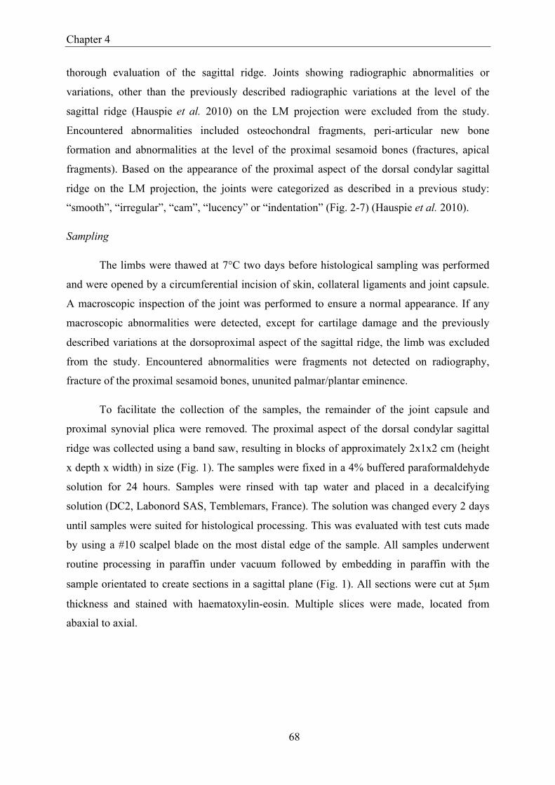
Chapter 4
68
thorough evaluation of the sagittal ridge. Joints showing radiographic abnormalities or
variations, other than the previously described radiographic variations at the level of the
sagittal ridge (Hauspie et al. 2010) on the LM projection were excluded from the study.
Encountered abnormalities included osteochondral fragments, peri-articular new bone
formation and abnormalities at the level of the proximal sesamoid bones (fractures, apical
fragments). Based on the appearance of the proximal aspect of the dorsal condylar sagittal
ridge on the LM projection, the joints were categorized as described in a previous study:
“smooth”, “irregular”, “cam”, “lucency” or “indentation” (Fig. 2-7) (Hauspie et al. 2010).
Sampling
The limbs were thawed at 7°C two days before histological sampling was performed
and were opened by a circumferential incision of skin, collateral ligaments and joint capsule.
A macroscopic inspection of the joint was performed to ensure a normal appearance. If any
macroscopic abnormalities were detected, except for cartilage damage and the previously
described variations at the dorsoproximal aspect of the sagittal ridge, the limb was excluded
from the study. Encountered abnormalities were fragments not detected on radiography,
fracture of the proximal sesamoid bones, ununited palmar/plantar eminence.
To facilitate the collection of the samples, the remainder of the joint capsule and
proximal synovial plica were removed. The proximal aspect of the dorsal condylar sagittal
ridge was collected using a band saw, resulting in blocks of approximately 2x1x2 cm (height
x depth x width) in size (Fig. 1). The samples were fixed in a 4% buffered paraformaldehyde
solution for 24 hours. Samples were rinsed with tap water and placed in a decalcifying
solution (DC2, Labonord SAS, Temblemars, France). The solution was changed every 2 days
until samples were suited for histological processing. This was evaluated with test cuts made
by using a #10 scalpel blade on the most distal edge of the sample. All samples underwent
routine processing in paraffin under vacuum followed by embedding in paraffin with the
sample orientated to create sections in a sagittal plane (Fig. 1). All sections were cut at 5µm
thickness and stained with haematoxylin-eosin. Multiple slices were made, located from
abaxial to axial.

Histologic appearance of the sagittal ridge
69
Figure 1. Diagram illustrating the exact relation of the bone block to the entire bone (square = bone block); the dotted line shows the plane in which the samples were cut: A) Dorsal view, B) Lateral view.
Histological scoring
Descriptive examination was performed by use of a light microscope. The histological
appearance of the cortex, cartilage, the transition zone from cartilage to joint capsule and
loose connective tissue was evaluated. The 5 samples of the group “smooth” radiographic
appearance were considered as the control group, representing the most frequently
encountered variation based on radiography (Hauspie et al. 2010). The amount of overlying
connective tissue in the different groups was evaluated semi-quantitatively and varied from
mild, moderate to pronounced, compared to the amount detected in the “smooth” group, were
it was defined as being moderate. The histological differences observed in the 4 remaining
groups, compared to the control “smooth” group were recorded.
Results
A total of 25 joints (MCP joint = 12; MTP joint = 13) originating from a total of 12
horses (median age of 3 years; max 5 year and min 2 year old), were included in this study.
The control “smooth” group (Fig. 2) showed a smooth contour of the cortex,
consisting of lamellar bone. Distally, the cortex was covered with normal hyaline cartilage.
Proximally, longitudinal aligned collagen fibres were detected (Fig. 3). No clear proximal
border of the cartilage could be observed here. At the level of the transition cartilage to
longitudinal aligned collagen fibres some overlying loosely organised connective tissue was
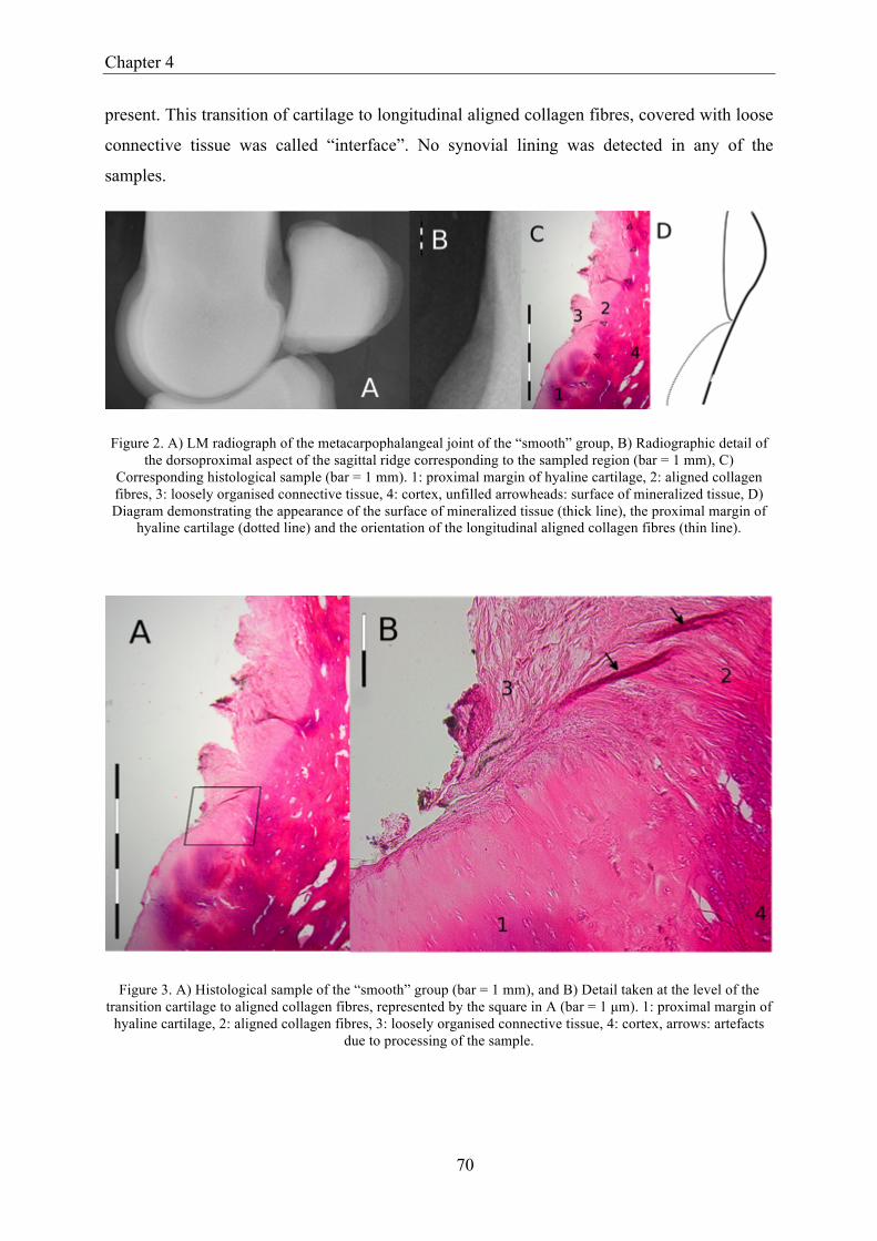
Chapter 4
70
present. This transition of cartilage to longitudinal aligned collagen fibres, covered with loose
connective tissue was called “interface”. No synovial lining was detected in any of the
samples.
Figure 2. A) LM radiograph of the metacarpophalangeal joint of the “smooth” group, B) Radiographic detail of the dorsoproximal aspect of the sagittal ridge corresponding to the sampled region (bar = 1 mm), C)
Corresponding histological sample (bar = 1 mm). 1: proximal margin of hyaline cartilage, 2: aligned collagen fibres, 3: loosely organised connective tissue, 4: cortex, unfilled arrowheads: surface of mineralized tissue, D) Diagram demonstrating the appearance of the surface of mineralized tissue (thick line), the proximal margin of
hyaline cartilage (dotted line) and the orientation of the longitudinal aligned collagen fibres (thin line).
Figure 3. A) Histological sample of the “smooth” group (bar = 1 mm), and B) Detail taken at the level of the transition cartilage to aligned collagen fibres, represented by the square in A (bar = 1 µm). 1: proximal margin of
hyaline cartilage, 2: aligned collagen fibres, 3: loosely organised connective tissue, 4: cortex, arrows: artefacts due to processing of the sample.
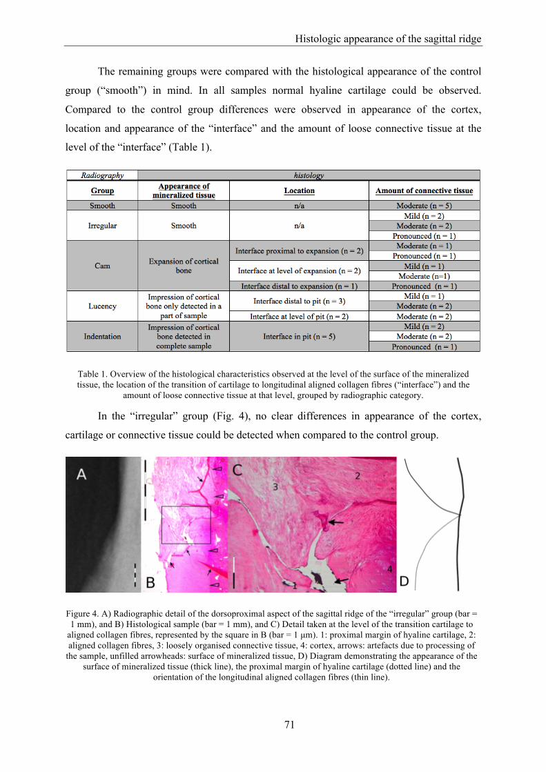
Histologic appearance of the sagittal ridge
71
The remaining groups were compared with the histological appearance of the control
group (“smooth”) in mind. In all samples normal hyaline cartilage could be observed.
Compared to the control group differences were observed in appearance of the cortex,
location and appearance of the “interface” and the amount of loose connective tissue at the
level of the “interface” (Table 1).
Table 1. Overview of the histological characteristics observed at the level of the surface of the mineralized tissue, the location of the transition of cartilage to longitudinal aligned collagen fibres (“interface”) and the
amount of loose connective tissue at that level, grouped by radiographic category.
In the “irregular” group (Fig. 4), no clear differences in appearance of the cortex,
cartilage or connective tissue could be detected when compared to the control group.
Figure 4. A) Radiographic detail of the dorsoproximal aspect of the sagittal ridge of the “irregular” group (bar = 1 mm), and B) Histological sample (bar = 1 mm), and C) Detail taken at the level of the transition cartilage to
aligned collagen fibres, represented by the square in B (bar = 1 µm). 1: proximal margin of hyaline cartilage, 2: aligned collagen fibres, 3: loosely organised connective tissue, 4: cortex, arrows: artefacts due to processing of
the sample, unfilled arrowheads: surface of mineralized tissue, D) Diagram demonstrating the appearance of the surface of mineralized tissue (thick line), the proximal margin of hyaline cartilage (dotted line) and the
orientation of the longitudinal aligned collagen fibres (thin line).

Chapter 4
72
An expansion of cortical bone was observed in the “cam” group (Fig. 5). Depending
on the sample, the “interface” was located distal to, at the level of or more proximal to the
cortical proliferation. The cortical expansion was best identified on the most medial or lateral
sagittal slices rather than on the central sagittal slices.
Figure 5. A) Radiographic detail of the dorsoproximal aspect of the sagittal ridge of the “cam” group (bar = 1mm, white arrow is representing the expansion of cortical bone), and B) Histological sample (bar = 1 mm), and (C, D) Details taken at the level of the transition cartilage to aligned collagen fibres, represented by the squares in B (bar = 1 µm), C) Is representing a detail taken at the level of the full line square, D) Is representing a detail taken at the level of the dotted line square. 1: proximal margin of hyaline cartilage, 2: aligned collagen fibres, 3:
loosely organised connective tissue, 4: cortex, black arrow: artefacts due to processing of the sample, unfilled arrowheads: surface of mineralized tissue, E) Diagram demonstrating the appearance of the surface of
mineralized tissue (thick line), the proximal margin of hyaline cartilage (dotted line) and the orientation of the longitudinal aligned collagen fibres (thin line).
A smooth impression in the cortex was observed in the “lucency” group (Fig. 6). This
impression was present in all samples, but was only detected in a limited amount of slices of
each sample. The remaining part of the sample had a histological appearance comparable with
the control group. The “interface” was located distal to the pit in 3 samples, whereas this was
located at the level of the pit in 2 samples.

Histologic appearance of the sagittal ridge
73
Figure 6. A) Radiographic detail of the dorsoproximal aspect of the sagittal ridge of the “lucency” group (bar = 1mm, white arrow is representing the more lucent area), and B) Histological sample (bar = 1 mm), and C) Detail
taken at the level of the transition cartilage to aligned collagen fibres, represented by the square in B (bar = 1 µm). 1: proximal margin of hyaline cartilage, 2: aligned collagen fibres, 3: loosely organised connective tissue, 4: cortex, black arrows: artefacts due to processing of the sample, unfilled arrowheads: surface of mineralized tissue, D) Diagram demonstrating the appearance of the surface of mineralized tissue (thick line), the proximal
margin of hyaline cartilage (dotted line) and the orientation of the longitudinal aligned collagen fibres (thin line).
A smooth impression in the cortex was also observed in the “indentation” group, (Fig.
7). This pit was present throughout the sample but appeared to be more abruptly compared to
the impression in the “lucency” group. The “interface” was located at the level of this pit in
all 5 samples.
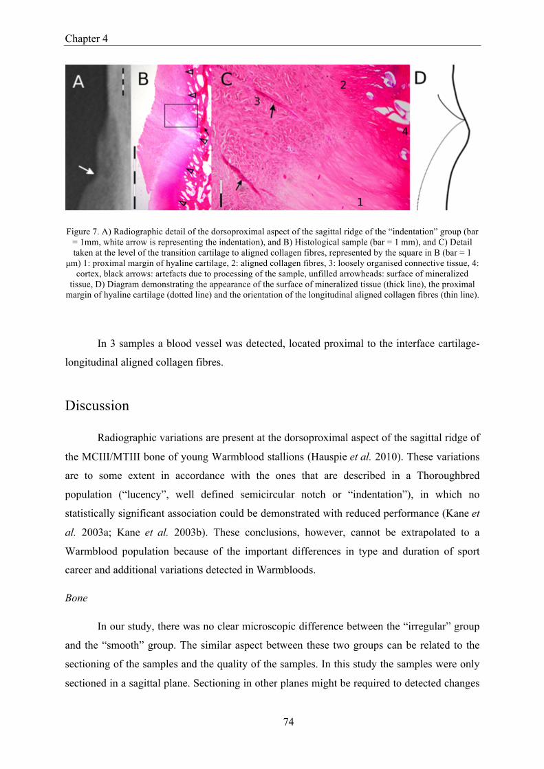
Chapter 4
74
Figure 7. A) Radiographic detail of the dorsoproximal aspect of the sagittal ridge of the “indentation” group (bar = 1mm, white arrow is representing the indentation), and B) Histological sample (bar = 1 mm), and C) Detail taken at the level of the transition cartilage to aligned collagen fibres, represented by the square in B (bar = 1
µm) 1: proximal margin of hyaline cartilage, 2: aligned collagen fibres, 3: loosely organised connective tissue, 4: cortex, black arrows: artefacts due to processing of the sample, unfilled arrowheads: surface of mineralized
tissue, D) Diagram demonstrating the appearance of the surface of mineralized tissue (thick line), the proximal margin of hyaline cartilage (dotted line) and the orientation of the longitudinal aligned collagen fibres (thin line).
In 3 samples a blood vessel was detected, located proximal to the interface cartilage-
longitudinal aligned collagen fibres.
Discussion
Radiographic variations are present at the dorsoproximal aspect of the sagittal ridge of
the MCIII/MTIII bone of young Warmblood stallions (Hauspie et al. 2010). These variations
are to some extent in accordance with the ones that are described in a Thoroughbred
population (“lucency”, well defined semicircular notch or “indentation”), in which no
statistically significant association could be demonstrated with reduced performance (Kane et
al. 2003a; Kane et al. 2003b). These conclusions, however, cannot be extrapolated to a
Warmblood population because of the important differences in type and duration of sport
career and additional variations detected in Warmbloods.
Bone
In our study, there was no clear microscopic difference between the “irregular” group
and the “smooth” group. The similar aspect between these two groups can be related to the
sectioning of the samples and the quality of the samples. In this study the samples were only
sectioned in a sagittal plane. Sectioning in other planes might be required to detected changes

Histologic appearance of the sagittal ridge
75
in the “irregular” group. The suboptimal quality of the samples due to difficulties with
decalcification en subsequent staining might be the reason that subtle alterations in the surface
of the mineralized tissue could have been missed.
Based on histology, a similar type of reaction as observed in the “cam” group is
described at the level of the femoral neck in humans (Keogh and Batt 2008; Emary 2010).
This reaction area appears macroscopically as a circular to oval elevation with a roughened
shiny surface and histology reveals inactive reactive new bone covered with hyaline cartilage
and collagenous tissue (Pitt et al. 1982). The aetiology of this reaction area in the human hip
joint is an impingement or chronic abutment of the acetabular rim on the femoral head-neck
junction or vice versa (Ganz et al. 2003). Macroscopically and microscopically, this type of
lesion shows close resemblance with the “cam” group in our study. In horses, no information
is available on the origin of this “cam”. The equine MCP/MTP joint, however, is a high
motion joint and it has been demonstrated that cartilage degeneration in this joint is most
frequently initiated at the dorsoproximal aspect of P1 in comparison to the more central
regions of the articular surface of P1 (Brommer et al. 2004). It is therefore possible that,
comparable to the human reaction area at the level of the femoral neck, this “cam” is the
result of chronic abutment of the dorsoproximal aspect of P1 against the dorsoproximal aspect
of the distal MCIII/MTIII condyle during hyperextension of the joint. During hyperextension,
the lateral and medial dorsoproximal aspect of P1 will contact the dorsodistal aspect of
MCIII/MTIII before the bones contact in the midline (Brama et al. 2001). This is in
accordance with the more medial or lateral sagittal location of the cortical expansion in this
study.
Other reactions at the femoral neck in the human hip joint may result from “synovial”
herniation. The radiographic appearance of this herniation is an oval to rounded lucent area,
corresponding to a herniation of soft tissue (most likely synovium) through defects of the
cortex (Pitt et al. 1982). In the “indentation” group a cortical impression was detected and the
“interface” or transition of cartilage to longitudinal collagen fibres was located in the
impression in all 5 samples. It is possible that this cortical impression can evolve to a
herniation-like lesion in horse as a herniation pit in humans can increase in size (Pitt et al.
1982). In horses, a possible similar lesion has recently been described with MRI in the medial
condyle of the distal MCIII bone (Gonzalez et al. 2010). A correlation with the “lucency”
group is less plausible because of the variable location of the “interface” and the smooth
impression that is only detected in a limited amount of slices while the remaining part of the

Chapter 4
76
sample has a “normal” appearance. To come to a more definitive conclusion, histological
comparison with a comparable group of older horses is needed.
In 3 samples a blood vessel was detected, resembling with a nutrient channel. This is a
normal finding at the level of the junction of the MCIII/MTIII condyles with the MCIII/MTIII
shaft (Denoix 1996).
The radiographically detected variations at the dorsoproximal aspect of the condylar
sagittal ridge of the MCIII/MTIII are the representation of detectable histological variation of
the cortex. Question is whether the observed histological variation is the result of
remodelling, for example as a reaction on the abutment of the dorsoproximal aspect of P1
against the distal MCIII/MTIII condyle during hyperextension (= developmental joint
adaptation to workload), or whether this variation is already present at birth or shortly after
birth with no major changes during life (congenital). It has been demonstrated that
subchondral bone density, bone mass and bone turnover are likely to vary in structure
depending on the type and intensity of exercise that is undertaken (Bennell et al. 1997;
Kawcak et al. 2000; Davies and Merritt 2004). A reactive process would be expected on
histology if the observed variations were a reaction to the abutment of the dorsoproximal
aspect of P1 against the MCIII/MTIII condyle. This was not the case in this study. However, a
slow ongoing process still remains possible. On the other hand, if the variations are already
present at birth, this could mean that they largely remain unchanged during life. Regardless of
their origin, these variations still can influence the function of the dorsoproximal aspect of the
joint during hyperextension and therefore possibly have an influence on the future sport
career.
Interface/transition bone-soft tissues
The present study focused on the histological appearance of the bony structures and
bone interface. Due to the removal of the articular capsule and proximal synovial plica, the
location of the plica and capsule in relation to the collagen fibres could not be established.
In diarthrodial joints, al non-articular surfaces within the joint cavity are covered with
synovium (Van Der Kraan and Van Den Berg 2007) and fibrocartilage is expected at the
transition of cartilage to joint capsule. In all samples of this study, aligned collagen fibres
were observed proximal to the articular cartilage without the presence of synovial lining.
These parallel collagen fibres could resemble Sharpey’s fibres, described at this level

Histologic appearance of the sagittal ridge
77
(McIlwraith 2002). However based on the strict definition of these fibres, stated as being
accompanied by an arteriole and one ore more nerve fibres (Retzlaff et al. 1982) we were not
able to identify these collagen fibres as true Sharpey’s fibres. The lack of synovium is
possibly explained by the procedure of collecting the samples. However there is a lack of
published data about the normal histologic appearance of the transition cartilage to joint
capsule at this level, making it difficult to formulate a clear conclusion.
The discrete changes in morphology at the interface between articular cartilage and
collagen fibres, and the variation between samples, may indicate an adaptation to local forces
at this level. It has been demonstrated that the dorsoproximal aspect of P1 sustains high
pressure during full extension and is in contact with the distal MCIII/MTIII condyle (Brama
et al. 2001). The longitudinal alignment of the fibres could therefore indicate a damper or
cushion function at this level. However the exact place of contact of P1 on the distal
MCIII/MTIII condyle and interaction of the proximal synovial plica during hyperextension is
unknown.
In conclusion, the difference in radiological appearance of the proximal aspect of the
dorsal condylar sagittal ridge of the third metacarpal and metatarsal bone results from
detectable histological differences. Further research is warranted to determine whether these
variations are developmental or congenital and to evaluate their potential influence on the
function of the joint during hyperextension.

Chapter 4
78
References
Bennell K.L., Malcolm S.A., Khan K.M., Thomas S.A., Reid S.J., Brukner P.D., Ebeling P.R. and Wark J.D. (1997) Bone mass and bone turnover in power athletes, endurance athletes, and controls: A 12-month longitudinal study. Bone 20, 477-484.
Brama P.a.J., Karssenberg D., Barneveld A. and Van Weeren P.R. (2001) Contact areas and
pressure distribution on the proximal articular surface of the proximal phalanx under sagittal plane loading. Equine Veterinary Journal 33, 26-32.
Brommer H., Brama R.a.J., Barneveld A. and Van Weeren P.R. (2004) Differences in the
topographical distribution of articular cartilage degeneration between equine metacarpo- and metatarsophalangeal joints. Equine Veterinary Journal 36, 506-510.
Davies H.M.S. and Merritt J.S. (2004) Surface strains around the midshaft of the third
metacarpal bone during turning. Equine Veterinary Journal 36, 689-692. Denoix J.M. (1996) Ultrasonographic examination in the diagnosis of joint disease. In:
McIlwraith C.W. and Trotter G.W. (editors), Joint disease in the horse, W.B. Saunders, Philadelphia. 165-202.
Emary P. (2010) Femoroacetabular impingement syndrome: A narrative review for the
chiropractor. Journal of the Canadian Chiropractic Association 54, 164-176. Ganz R., Parvizi J., Beck M., Leunig M., Notzli H. and Siebenrock K.A. (2003)
Femoroacetabular impingement: A cause for osteoarthritis of the hip. Clinical Orthopaedics and Related Research 417, 112-120.
Gonzalez L.M., Schramme M.C., Robertson I.D., Thrall D.E. and Redding R.W. (2010) Mri
features of metacarpo(tarso)phalangeal region lameness in 40 horses. Veterinary Radiology & Ultrasound 51, 404-414.
Hauspie S., Martens A., Declercq J., Busoni V., Vanderperren K., Van Bree H. and Saunders
J.H. (2010) Radiographic features of the dorsal condylar sagittal ridge of the third metacarpal and metatarsal bones in young warmblood stallions. Veterinary and Comparative Orthopaedics and Traumatology 23, 411-416.
Kane A.J., McIlwraith C.W., Park R.D., Rantanen N.W., Morehead J.P. and Bramlage L.R.
(2003a) Radiographic changes in thoroughbred yearlings. Part 2: Associations with racing performance. Equine Veterinary Journal 35, 366-374.

Histologic appearance of the sagittal ridge
79
Kane A.J., Park R.D., McIlwraith C.W., Rantanen N.W., Morehead J.P. and Bramlage L.R. (2003b) Radiographic changes in thoroughbred yearlings. Part 1: Prevalence at the time of the yearling sales. Equine Veterinary Journal 35, 354-365.
Kawcak C.E., McIlwraith C.W., Norrdin R.W., Park R.D. and Steyn P.S. (2000) Clinical
effects of exercise on subchondral bone of carpal and metacarpophalangeal joints in horses. American Journal of Veterinary Research 61, 1252-1258.
Keogh M.J. and Batt M.E. (2008) A review of femoroacetabular impingement in athletes.
Sports Medicine 38, 863-878. McIlwraith C.W. (2002) Diseases of joints, tendons, ligaments, and related structures. In:
Stashak T.S. (editors), Adam's lameness in horses, Lippincott Williams & Wilkins, Baltimore. 459-644.
Mitchell R.D. (2009) Imaging considerations in the purchase examination of the performance
horse. In: 55th Annual convention of the American Association of Equine practitioners, Las Vegas, Nevada, USA. 296-300.
Pitt M.J., Graham A.R., Shipman J.H. and Birkby W. (1982) Herniation pit of the femoral
neck. American Journal of Roentgenology 138, 1115-1121. Retzlaff E.W., Mitchell F.L., Upledger J. and Walsh J. (1982) Efficacy of cranial sacral
manipulation: The physiological mechanism of the cranial sutures. Journal of the society of osteopaths 12, 8-12.
Van Der Kraan P.M. and Van Den Berg W.B. (2007) Osteophytes: Relevance and biology.
Osteoarthritis Cartilage 15, 237-244. Van Hoogmoed L.M., Snyder J.R., Thomas H.L. and Harmon F.A. (2003) Retrospective
evaluation of equine prepurchase examinations performed 1991-2000. Equine Veterinary Journal 35, 375-381.


81
Chapter 5
Radiographic variation of the proximal
aspect of the dorsal condylar sagittal ridge
of the metacarpo-/metatarsophalangeal
joint in Warmblood horses versus the risk
of cartilage degeneration
Adapted from:
Hauspie S., Declercq J., Brown B., Duchateau L., Vanderperren K., Saunders J.H.
Radiographic variation of the proximal aspect of the dorsal condylar sagittal ridge of the metacarpo-
/metatarsophalangeal joint in Warmblood horses versus the risk of cartilage degeneration (submitted).

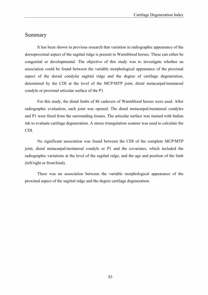
Cartilage Degeneration Index
83
Summary
It has been shown in previous research that variation in radiographic appearance of the
dorsoproximal aspect of the sagittal ridge is present in Warmblood horses. These can either be
congenital or developmental. The objective of this study was to investigate whether an
association could be found between the variable morphological appearance of the proximal
aspect of the dorsal condylar sagittal ridge and the degree of cartilage degeneration,
determined by the CDI at the level of the MCP/MTP joint, distal metacarpal/metatarsal
condyle or proximal articular surface of the P1.
For this study, the distal limbs of 86 cadavers of Warmblood horses were used. After
radiographic evaluation, each joint was opened. The distal metacarpal/metatarsal condyles
and P1 were freed from the surrounding tissues. The articular surface was stained with Indian
ink to evaluate cartilage degeneration. A stereo triangulation scanner was used to calculate the
CDI.
No significant association was found between the CDI of the complete MCP/MTP
joint, distal metacarpal/metatarsal condyle or P1 and the covariates, which included the
radiographic variations at the level of the sagittal ridge, and the age and position of the limb
(left/right or front/hind).
There was no association between the variable morphological appearance of the
proximal aspect of the sagittal ridge and the degree cartilage degeneration.

Chapter 5
84
Introduction
Lameness is an important factor affecting the future of a horse (Rossdale et al. 1985),
therefore, a radiographic survey is frequently performed during the pre-purchase examination
to identify problems or abnormalities that could make the horse unfit for its intended purpose
(Van Hoogmoed et al. 2003). In most cases, the clinical relevance of any radiographic
variations is clear, but not always, which leaves the relevance open to debate and may be
controversial (McIlwraith et al. 2003; Van Hoogmoed et al. 2003).
The MCP/MTP joint is a high motion joint that is frequently injured in athletic horses
(Pool 1996). Radiographic variations have been described at the proximal aspect of the dorsal
condylar sagittal ridge in both Warmblood (Hauspie et al. 2010) and Thoroughbred horses
(Kane et al. 2003b). In Thoroughbreds, they may be seen as a lucency or a well-defined semi-
circular notch at the proximal aspect of the dorsal condylar sagittal ridge. In Warmblood
horses, a sagittal ridge with a smooth or an irregular outline, the presence of a cam, lucency,
or indentation at the level of the ridge may be seen. In Thoroughbreds, no statistically
significant association could be demonstrated with reduced performance in young horses
(Kane et al. 2003a; Kane et al. 2003b). However, in Warmblood horses, who have a different
type and duration of sportive career compared to Thoroughbreds (Hinchcliff and Hamlin
2004; O'Sullivan and Lumsden 2004), the clinical relevance may be different.
A technique was recently described that allows in vitro quantitative assessment of
articular cartilage degeneration of the metacarpal/metatarsal condyle and P1 by objectively
calculating of a CDI (Brommer et al. 2003a; Declercq et al. 2011). The technique is based on
the fact that Indian ink particles have a high affinity for articular cartilage with surface
fibrillation and depletion of proteoglycans, whereas normal intact articular cartilage with a
high proteoglycan-rich matrix will prevent the particles from entering. Depending on the
degree of uptake of Indian ink, the particles will deflect and absorb light, which can be
measured quantitatively to calculate the CDI. This technique can be used to detect
degenerative changes at the level of the cartilage of the MCP/MTP joint at an early stage
(Brommer et al. 2003a).
So far the clinical relevance of the difference in morphological aspect of the proximal
aspect of the dorsal condylar sagittal ridge is unknown. If an influence were present changes
would be expected at the level of the joint, for example degeneration of the articular cartilage.
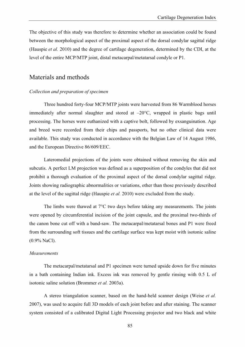
Cartilage Degeneration Index
85
The objective of this study was therefore to determine whether an association could be found
between the morphological aspect of the proximal aspect of the dorsal condylar sagittal ridge
(Hauspie et al. 2010) and the degree of cartilage degeneration, determined by the CDI, at the
level of the entire MCP/MTP joint, distal metacarpal/metatarsal condyle or P1.
Materials and methods
Collection and preparation of specimen
Three hundred forty-four MCP/MTP joints were harvested from 86 Warmblood horses
immediately after normal slaughter and stored at –20°C, wrapped in plastic bags until
processing. The horses were euthanized with a captive bolt, followed by exsanguination. Age
and breed were recorded from their chips and passports, but no other clinical data were
available. This study was conducted in accordance with the Belgian Law of 14 August 1986,
and the European Directive 86/609/EEC.
Lateromedial projections of the joints were obtained without removing the skin and
subcutis. A perfect LM projection was defined as a superposition of the condyles that did not
prohibit a thorough evaluation of the proximal aspect of the dorsal condylar sagittal ridge.
Joints showing radiographic abnormalities or variations, other than those previously described
at the level of the sagittal ridge (Hauspie et al. 2010) were excluded from the study.
The limbs were thawed at 7°C two days before taking any measurements. The joints
were opened by circumferential incision of the joint capsule, and the proximal two-thirds of
the canon bone cut off with a band-saw. The metacarpal/metatarsal bones and P1 were freed
from the surrounding soft tissues and the cartilage surface was kept moist with isotonic saline
(0.9% NaCl).
Measurements
The metacarpal/metatarsal and P1 specimen were turned upside down for five minutes
in a bath containing Indian ink. Excess ink was removed by gentle rinsing with 0.5 L of
isotonic saline solution (Brommer et al. 2003a).
A stereo triangulation scanner, based on the hand-held scanner design (Weise et al.
2007), was used to acquire full 3D models of each joint before and after staining. The scanner
system consisted of a calibrated Digital Light Processing projector and two black and white

Chapter 5
86
cameras that the operator used to reconstruct the shape of the specimen, using principles of
stereo triangulation. A colour camera provided enough colour for the operator to identify the
macroscopically visible cartilage changes on the stored images, but all calculations were
made using the higher quality black and white data (Declercq et al. 2011).
All metacarpal/metatarsal and P1 specimen were clamped and mounted vertically on a
computer-controlled turntable and scanned at 5-degree intervals, making 72 scans that were
automatically assembled using the method described by Brown et al. 2008 (Brown et al.
2008). The scanner was mounted at the angle that allowed the camera to get images of both
the top and sides of the joint so that a complete model of the cartilage surface could be created
without repositioning metacarpal/metatarsal condyles or P1 on the turntable. This model also
eliminated any outlying bright values that resulted from the reflection of light.
Once the 3D models were generated, an operator (the second author) selected the
cartilage region of interest (the articular surface of the distal metacarpal/metatarsal condyle
and proximal articular surface of P1) using a paintbrush metaphor, and this surface area was
measured based on the 3D geometry (Figs. 1 & 2). The CDI (percentage of darkening) was
calculated as the ratio between stained and unstained grey values of the selected cartilage
surface. The ratio was computed for every facet of the model and averaged (Declercq et al.
2011).
Figure 1. A) Proximal phalanx after staining with Indian ink; B) Proximal phalanx: cartilage selection on non-stained colour 3D image.
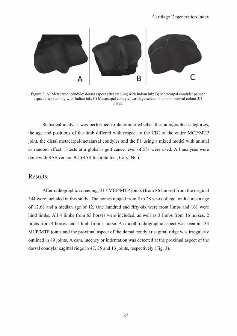
Cartilage Degeneration Index
87
Figure 2. A) Metacarpal condyle: dorsal aspect after staining with Indian ink; B) Metacarpal condyle: palmar aspect after staining with Indian ink; C) Metacarpal condyle: cartilage selection on non-stained colour 3D
image.
Statistical analysis was performed to determine whether the radiographic categories,
the age and positions of the limb differed with respect to the CDI of the entire MCP/MTP
joint, the distal metacarpal/metatarsal condyles and the P1 using a mixed model with animal
as random effect. F-tests at a global significance level of 5% were used. All analyses were
done with SAS version 9.2 (SAS Institute Inc., Cary, NC).
Results
After radiographic screening, 317 MCP/MTP joints (from 86 horses) from the original
344 were included in this study. The horses ranged from 2 to 28 years of age, with a mean age
of 12.68 and a median age of 12. One hundred and fifty-six were front limbs and 161 were
hind limbs. All 4 limbs from 65 horses were included, as well as 3 limbs from 16 horses, 2
limbs from 4 horses and 1 limb from 1 horse. A smooth radiographic aspect was seen in 133
MCP/MTP joints and the proximal aspect of the dorsal condylar sagittal ridge was irregularly
outlined in 89 joints. A cam, lucency or indentation was detected at the proximal aspect of the
dorsal condylar sagittal ridge in 47, 35 and 13 joints, respectively (Fig. 3).
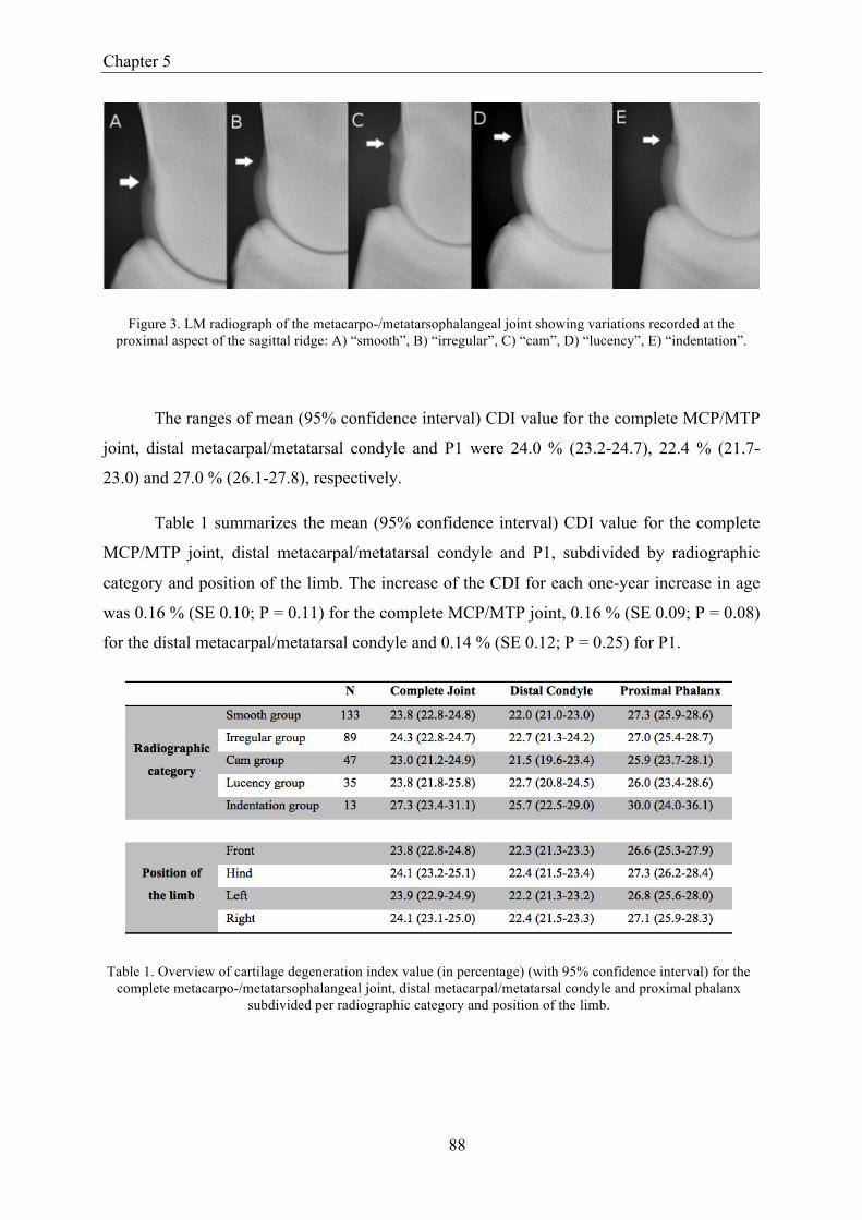
Chapter 5
88
Figure 3. LM radiograph of the metacarpo-/metatarsophalangeal joint showing variations recorded at the proximal aspect of the sagittal ridge: A) “smooth”, B) “irregular”, C) “cam”, D) “lucency”, E) “indentation”.
The ranges of mean (95% confidence interval) CDI value for the complete MCP/MTP
joint, distal metacarpal/metatarsal condyle and P1 were 24.0 % (23.2-24.7), 22.4 % (21.7-
23.0) and 27.0 % (26.1-27.8), respectively.
Table 1 summarizes the mean (95% confidence interval) CDI value for the complete
MCP/MTP joint, distal metacarpal/metatarsal condyle and P1, subdivided by radiographic
category and position of the limb. The increase of the CDI for each one-year increase in age
was 0.16 % (SE 0.10; P = 0.11) for the complete MCP/MTP joint, 0.16 % (SE 0.09; P = 0.08)
for the distal metacarpal/metatarsal condyle and 0.14 % (SE 0.12; P = 0.25) for P1.
Table 1. Overview of cartilage degeneration index value (in percentage) (with 95% confidence interval) for the complete metacarpo-/metatarsophalangeal joint, distal metacarpal/metatarsal condyle and proximal phalanx
subdivided per radiographic category and position of the limb.
Cartilage Degeneration Index
89
There was no significant association between the CDI of the complete MCP/MTP
joint, distal metacarpal/metatarsal condyle and P1 and the different covariates, such as
radiographic categories, the age and the limb (left/right or front/hind).
Discussion
This study examined the relationship between the morphology of the proximal aspect
of the dorsal condylar sagittal ridge, increase in age, the limb, and the presence of
degenerative changes at the level of the articular cartilage in a random Warmblood horse
population.
The Indian ink technique to stain cartilage surfaces and to determine the CDI, is very
valuable, but reflects the cartilage surface damage only (Brommer et al. 2003a). No other
features of osteoarthritis, such as changes in the subchondral bone (Kawcak et al. 2001) or
joint capsule (Palmer and Bertone 1994) are taken into account. The strength of this technique
is to have early detection of cartilage damage and of the whole cartilage surface, but on the
other hand no information is gained on the status of the subchondral bone or the synovium.
For evaluation of the structure, cellularity and tidemark, histological and histochemical
grading as described by Mankin et al. 1971 is recommended for assessment of osteoarthrosis
in horses (Mankin et al. 1971). This has the disadvantage that only the histological sample
taken is evaluated; one sample does not always represents the status of the whole joint
cartilage (McIlwraith et al. 2010).
The CDI technique developed by Brommer et al. 2003, which provides an objective
evaluation of the quality of the articular cartilage over a large articular surface, was used
(Brommer et al. 2003a). However, the technique described by Brommer et al. 2003, a manual
method using digital photography, has been validated only for the proximal articular surface
of the P1 and does not include area calculations on curved regions like the
metacarpal/metatarsal condyles (Brommer et al. 2003a). Therefore a modified technique that
created a complete 3D model of the articular surfaces of the distal metacarpal/metatarsal
condyle and P1 and that allowed area calculations on curved surfaces was used (Declercq et
al. 2011).
No significant differences were found in the mean CDI value of the front and hind
limbs, which agrees with a study published by Riggs et al. 1999 that also found no differences

Chapter 5
90
between the subchondral bone of corresponding regions of the distal metacarpal or metatarsal
bone (Riggs et al. 1999). Changes at the level of the articular cartilage and adjacent
subchondral bone are closely linked and adapt to loading in a coordinated way (Kawcak et al.
2001; Lewis et al. 2005; Lacourt et al. 2012). Therefore, if the subchondral bone architecture
between metacarpal and metatarsal condyles is comparable, the cartilage status is also
expected to be comparable. This was confirmed in our study.
The additional finding that the mean CDI value was not associated with the position of
the limb (left or right) also agrees with the Riggs et al. 1999 study, where no significant
difference was detected between distal metacarpal/metatarsal condyles from either the left or
right limb (Riggs et al. 1999). Another study, however, showed a significantly higher
glucosaminoglycan and water content of the cartilage of the right MCP joint, compared with
the left (Brama et al. 2000). This higher glycosaminoglycan and water content is linked to an
increase in stiffness of the cartilage (Palmer and Bertone 1996). Therefore a difference of the
cartilage in response to loading conditions can be expected, possibly associated with a
difference in the degree of cartilage damage, resulting in a different CDI value. This could
however not be confirmed in our study at the level of the MCP joints.
In Thoroughbreds, no statistical significant association was detected between the
radiographic appearance of the proximal aspect of the dorsal condylar sagittal ridge and
reduced performance, determined as a decreased likelihood of starting a race, average
earnings per start and money earned (Kane et al. 2003a). No significant association was
detected in our study between the radiographic appearance of the proximal aspect of the
dorsal condylar sagittal ridge and changes in the CDI of the MCP/MTP joint in Warmblood
horses. This statement is valid not only for young horses but also older ones, as the age of
horses in our study ranged from 2 to 28 years, with a median age of 12 years. However, the
status of the articular cartilage is not the only factor with an influence on the joint function.
Local CDI values are known to increase from central to dorsal at the proximal articular
surface of P1 (Brommer et al. 2003b) and significant differences have been detected between
local CDI values of different points on the proximal articular surface of P1 (Brommer et al.
2005). A similar trend is likely for the distal metacarpal/metatarsal condyles, although they
were not included in that study. The radiographic variations described in this study are located
at the dorsal condylar sagittal ridge. If these variations influence the joint function, they
probably have most effect at that level. Therefore, dividing the articular joint surfaces into

Cartilage Degeneration Index
91
different regions could identify any differences in the CDI value at the level of the dorsal
aspect of the joint between the different categories.
No significant effect of age was detected, neither on the mean CDI value of the entire
joint, nor on the CDI of the distal metacarpal/metatarsal condyle or the proximal articular
surface of P1. The latter result contrasts with a study published by Brommer et al. 2003 in
which a moderate, but significant, association between the CDI value of the proximal articular
surface of P1 and age was found (Brommer et al. 2003b). A possible explanation is that our
population was larger compared to Brommer’s (317 joints compared with 73), so our results
more accurately represent the population. Finally, our study adjusted for the possible
clustering effect of the limbs of the same horse by adding the horse as a random factor in our
model.
In conclusion, the radiographic appearance of the proximal aspect of the dorsal
condylar sagittal ridge has no significant association with an increase in the CDI at the level
of the joint. This supports the study in Thoroughbreds where no association was detected
between these variations and reduced performance at a young age (Kane et al. 2003a).
Chapter 5
92
References
Brama P.a.J., Tekoppele J.M., Bank R.A., Karssenberg D., Barneveld A. and Van Weeren P.R. (2000) Topographical mapping of biochemical properties of articular cartilage in the equine fetlock joint. Equine Veterinary Journal 32, 19-26.
Brommer H., Laasanen M.S., Brama R.a.J., Van Weeren R.R., Helminen H.J. and Jurvelin
J.S. (2005) Functional consequences of cartilage degeneration in the equine metacarpophalangeal joint: Quantitative assessment of cartilage stiffness. Equine Veterinary Journal 37, 462-467.
Brommer H., Van Weeren P.R. and Brama P.A. (2003a) New approach for quantitative
assessment of articular cartilage degeneration in horses with osteoarthritis. American Journal of Veterinary Research 64, 83-87.
Brommer H., Van Weeren R.R., Brama P.a.J. and Barneveld A. (2003b) Quantification and
age-related distribution of articular cartilage degeneration in the equine fetlock joint. Equine Veterinary Journal 35, 697-701.
Brown B.J., Toler-Franklin C., Nehab D., Burns M., Dobkin D., Vlachopoulos A., Doumas
C., Rusinkiewicz S. and Weyrich T. (2008) A system for high-volume acquisition and matching of fresco fragments: Reassembling theran wall paintings. ACM transactions on graphics 27, 1-9.
Declercq J., Brown B., Oosterlinck M., Van Gool L., Hauspie S., Saunders J. and Martens A.
(2011) Quantitative assesment of articular cartilage degeneration of the metacarpal condyle using a 3d scanning system. In: ECVS Proceedings. 69-69.
Hauspie S., Martens A., Declercq J., Busoni V., Vanderperren K., Van Bree H. and Saunders
J.H. (2010) Radiographic features of the dorsal condylar sagittal ridge of the third metacarpal and metatarsal bones in young warmblood stallions. Veterinary and Comparative Orthopaedics and Traumatology 23, 411-416.
Hinchcliff K.W. and Hamlin M. (2004) Veterinary aspects of racing and training
standardbred race horses. In: Hinchcliff K.W., Kaneps A.J. and Geor R.J. (editors), Equine sports medicine and surgery, Saunders, Philadelphia. 1073-‐1089.
Kane A.J., McIlwraith C.W., Park R.D., Rantanen N.W., Morehead J.P. and Bramlage L.R.
(2003a) Radiographic changes in thoroughbred yearlings. Part 2: Associations with racing performance. Equine Veterinary Journal 35, 366-374.

Cartilage Degeneration Index
93
Kane A.J., Park R.D., McIlwraith C.W., Rantanen N.W., Morehead J.P. and Bramlage L.R. (2003b) Radiographic changes in thoroughbred yearlings. Part 1: Prevalence at the time of the yearling sales. Equine Veterinary Journal 35, 354-365.
Kawcak C.E., McIlwraith C.W., Norrdin R.W., Park R.D. and James S.P. (2001) The role of
subchondral bone in joint disease: A review. Equine Veterinary Journal 33, 120-126. Lacourt M., Gao C., Li A., Girard C., Beauchamp G., Henderson J.E. and Laverty S. (2012)
Relationship between cartilage and subchondral bone lesions in repetitive impact trauma-induced equine osteoarthritis. Osteoarthritis Cartilage 15, 15.
Lewis C.W., Williamson A.K., Chen A.C., Bae W.C., Temple M.M., Wong V.W., Nugent
G.E., James S.P., Wheeler D.L., Sah R.L. and Kawcak C.E. (2005) Evaluation of subchondral bone mineral density associated with articular cartilage structure and integrity in healthy equine joints with different functional demands. American Journal of Veterinary Research 66, 1823-1829.
Mankin H.J., Dorfman H., Lippiell.L and Zarins A. (1971) Biochemical and metabolic
abnormalities in articular cartilage from osteo-arthritic human hips .2. Correlation of morphology with biochemical and metabolic data. Journal of Bone and Joint Surgery-American Volume A 53, 523-537.
McIlwraith C.W., Frisbie D.D., Kawcak C.E., Fuller C.J., Hurtig M. and Cruz A. (2010) The
oarsi histopathology initiative - recommendations for histological assessments of osteoarthritis in the horse. Osteoarthritis Cartilage 18 Suppl 3, S93-105.
McIlwraith C.W., Kane A.J. and Park R.D. (2003) Changes on radiographs at thoroughbred
yearling sales: Prevalence and significance. Clinical techniques in equine practice 2, 329-338.
O'Sullivan C.B. and Lumsden J.M. (2004) Veterinary aspects of racing and training
thoroughbred race horses. In: Hinchcliff K.W., Kaneps A.J. and Geor R.J. (editors), Equine sports medicine and surgery, Saunders, Philadelphia. 1051-‐1072.
Palmer J.L. and Bertone A.L. (1994) Joint structure, biochemistry and biochemical
disequilibrium in synovitis and equine joint disease. Equine Veterinary Journal 26, 263-277.
Palmer J.L. and Bertone A.L. (1996) Joint biomechanics in the pathogenesis of traumatic
arthritis. In: McIlwraith C.W. and Trotter G.W. (editors), Joint disease in the horse, WB Saunders, Philadelphia. 104-119.

Chapter 5
94
Pool R. (1996) Pathologic manifestations of joint disease in the athletic horse. In: McIlwraith C.W. and Trotter G.W. (editors), Joint disease in the horse, WB Saunders, Philadelphia. 87-104.
Riggs C.M., Whitehouse G.H. and Boyde A. (1999) Structural variation of the distal condyles
of the third metacarpal and third metatarsal bones in the horse. Equine Veterinary Journal 31, 130-139.
Rossdale P.D., Hopes R. and Digby N.J.W. (1985) Epidemiological study of wastage among
racehorses 1982 and 1983. Veterinary Record 116, 66-69. Van Hoogmoed L.M., Snyder J.R., Thomas H.L. and Harmon F.A. (2003) Retrospective
evaluation of equine prepurchase examinations performed 1991-2000. Equine Veterinary Journal 35, 375-381.
Weise T., Leibe B., Van Gool L. and Ieee (2007) Fast 3d scanning with automatic motion
compensation, 2007 ieee conference on computer vision and pattern recognition, vols 1-8, IEEE, New York. 2462-2469.
95
Chapter 6
The position of the dorsal proximal
synovial pad during hyperextension of the
equine metacarpo-/metatarsophalangeal
joint
Adapted from:
Hauspie S., Vanderperren K., Gielen I., Pardon B., Kromhout K. Martens A., Saunders J.H.
The position of the dorsal proximal synovial plica during hyperextension of the equine metacarpo-
/metatarsophalangeal joint (submitted).


The position of the proximal synovial pad
97
Summary
A synovial plica is present at the dorsoproximal aspect of the MCP/MTP joint. It is
supposed to function as a cushioning surface or contact interface between P1 and the cannon
bone where these are in contact during full extension of the joint. The objective of this study
was to describe the location of the dorsoproximal synovial plica during hyperextension of the
MCP/MTP joint using MRI, in order to get a better insight in its function.
For this study 20 cadaver limbs from 5 Warmblood horses were used. Of each joint, 2
MRI scans were performed. One with the limb fixed in a normal standing position of the
MCP/MTP joint, a second scan with the joint fixed in a hyperextended position.
Measurements were made of the dorsal and palmar/plantar length and the thickness of the
plica in the normal position. During hyperextension, the position of the plica was described,
the angle of contact between P1 and the condyle was measured, and the dorsal angle of
extension was measured.
The dorsal length differed between front/hind limbs and between the medial/lateral
aspects of the joint. The angle of contact between P1 and condyle differed between front/hind
limbs and between the lateral and medial aspect of the joint and between different positions of
the plica. Four different positions of the plica were observed during hyperextension: (1)
shortened with the tip curved towards palmar/plantar; (2) projecting distally; (3) projecting
towards dorsal and (4) projecting distally with the tip interposed between P1 and the condyle.
During hyperextension, a close relation is present between the proximal synovial plica,
proximal aspect of P1 and the condyle with a variable position of the plica, suggestive for a
contact interface between P1 and the cannon bone. However, due to the different positions of
the plica, it does not seem to act consistently as a cushioning surface.

Chapter 6
98
Introduction
The MCP/MTP joint in the horse comprises of the MCIII/MTIII, P1 and the proximal
sesamoid bones (Barone 1986). An articular capsule and multiple ligaments reinforce this
joint (Barone 1989; Weaver et al. 1992). As a result of this design, the MCP/MTP joint is
limited in its motion mainly to the sagittal plane (Palmer and Bertone 1996; Les et al. 1997;
Butcher and Shley-Ross 2002), resulting in either flexion or extension (Butcher and Shley-
Ross 2002). During hyperextension, the proximal articular surface of P1 articulates with the
dorsal aspect of the condyle of MCIII/MTIII (Palmer and Bertone 1996; Brama et al. 2001).
At the dorsoproximal recess of the MCP/MTP joint, the joint capsule forms a pad or synovial
plica, projecting distally from the dorsoproximal attachment of the joint capsule and tapering
to a thin edge. This plica covers the transition zone between the cartilage and the attachment
of the joint capsule (White 1990; Dabareiner et al. 1996). Several abnormalities are described
at the level of this plica, ranging from synovial plica proliferation to the presence of fragments
(White 1990; Declercq et al. 2008).
Radiographic variations in appearance of the dorsoproximal aspect of the sagittal ridge
are described in the MCP/MTP joint of Warmbloods (Hauspie et al. 2010). Although the
function of the synovial plica is unknown, its position and structure suggests that the plica
acts as a cushioning surface or contact interface between P1 and the cannon bone where these
are in contact during full extension of the joint (White 1990). Due to the location of the
synovial plica and the different variations present at the dorsoproximal aspect of the sagittal
ridge, these are possibly in close relation to each other. Therefore, it is possible that the
variations at the dorsoproximal aspect of the sagittal ridge will interfere with the plica during
hyperextension. However, the exact position of the plica during hyperextension is until know
not described.
Magnetic resonance imaging is commonly being used for investigation of lameness in
horses (Mair et al. 2005; Powell 2012; Smith et al. 2012). The high soft tissue contrast of
MRI makes this modality particularly suited for the evaluation of soft tissues (Kraft and
Gavin 2001). The normal MRI anatomy of the MCP/MTP joint has been described using both
low- and high-field magnets (Park et al. 1987; Martinelli et al. 1996; Martinelli et al. 1997;
Dyson and Murray 2007), showing the ability of outlining the dorsoproximal synovial plica
on different sequences.

The position of the proximal synovial pad
99
The objective of this article was to describe the location of the dorsoproximal synovial
plica during hyperextension of the MCP/MTP joint using MRI, in order to get a better insight
in its function.
Materials and methods
Collection and preparation of the limbs
Fresh cadaver front and hind limbs from Warmblood horses, aged younger than 5
years, were collected for this study. These horses were euthanized for reasons unrelated to this
study and collected at the pathology department of our institute. The limbs, obtained less than
24 hours after euthanasia, were sectioned at the level of the carpometacarpal or tarsocrural
joint. The proximal end of the limbs was covered with a latex glove to prevent blood loss
during handling. The shoe was removed from each digit. After collection each limb was
placed in a plastic bag and frozen at -25 °C. A perfect LM radiograph of each joint, defined as
a superposition of the condyles that did not prohibit a thorough evaluation of the dorsal
condylar sagittal ridge, was obtained without thawing the limb to ensure the normal
appearance of the MCP/MTP joint. If radiographic abnormalities at the level of the joint were
detected, such as bone fragmentation or peri-articular new bone formation, the horse was
excluded from this study and new specimen were collected until a total of 20 normal joints
were collected. Before the MRI examination, limbs were thawed at ambient temperature for
24 hours. It has been proven that this procedure has the least influence on image quality
(Bolen et al. 2011). After thawing, the skin of the limb was clipped and the hoof was cleaned.
MR imaging examinations
Imaging sequences were obtained while the limb was fixed in a custom made loading
frame (Fig. 1). Two scans were performed on each limb, one with the MCP/MTP joint fixed
in normal standing position (dorsal angle between MCIII/MTIII and P1 of approximately
150°) and one with the joint fixed in hyperextension (dorsal angle between the MCIII/MTIII
and P1 of approximately 120°) (Meershoek et al. 2001; Brown et al. 2003). The angle of
extension was measured macroscopically with a protractor at the level of the dorsal aspect of
the distal metacarpus/metatarsus and pastern region. To allow sufficient hyperextension of the
MCP/MTP joint using a custom made wooden loading frame, the suspensory ligament and
accessory ligament of the deep digital flexor tendon had to be sectioned at the proximal aspect

Chapter 6
100
in front limbs, whereas in the hind limbs, the suspensory ligament, accessory ligament of the
deep digital flexor tendon, the deep digital flexor tendon and superficial digital flexor tendon
had to be sectioned at the proximal aspect. Without sectioning of these tendons, a metal
loading frame was needed, making an MRI examination impossible. This methodology was
based on the results of a preliminary study where 20 limbs were hyperextended twice using a
custom made metal loading frame: a first time with the flexor tendons intact; a second time
after selective sectioning of the tendons and/or ligaments as mentioned above. The position of
the bones of the MCP/MTP joint was evaluated by means of radiography (LM, D45°M-
Pa(Pl)LO and D45°L-Pa(Pl)MO projections) and showed that sectioning of these tendons
and/or ligaments did not alter the position of the bones relative to each other during
hyperextension.
Figure 1. Illustration of the custom made loading frame used to facilitate extension of the metacarpo-/metatarsophalangeal joint: A) The limb is fixed in the normal standing position, B) The limb is fixed in a
hyperextended position.
Magnetic resonance imaging was performed using a permanent 0.2 T magnet
(Airismate, Hitachi, Japan). The limb was positioned in right lateral recumbence and placed in
a human knee coil. Sagittal images were made with slice thickness of 3mm without interslice
gap. The image matrix was 256 x 256. In the normal standing position, T1W (TR = 520 ms,
TE = 17 ms); T2W (TR = 4700 ms, TE = 125 ms) and a PDW SE sequence (TR = 4700 ms,
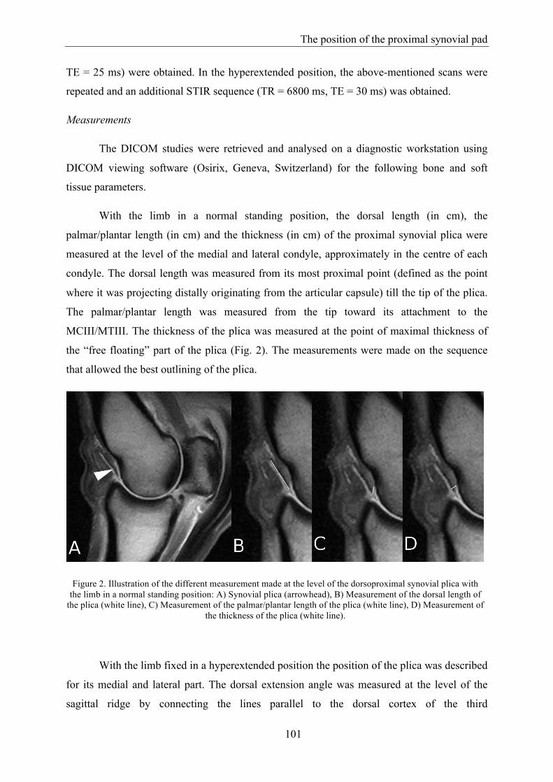
The position of the proximal synovial pad
101
TE = 25 ms) were obtained. In the hyperextended position, the above-mentioned scans were
repeated and an additional STIR sequence (TR = 6800 ms, TE = 30 ms) was obtained.
Measurements
The DICOM studies were retrieved and analysed on a diagnostic workstation using
DICOM viewing software (Osirix, Geneva, Switzerland) for the following bone and soft
tissue parameters.
With the limb in a normal standing position, the dorsal length (in cm), the
palmar/plantar length (in cm) and the thickness (in cm) of the proximal synovial plica were
measured at the level of the medial and lateral condyle, approximately in the centre of each
condyle. The dorsal length was measured from its most proximal point (defined as the point
where it was projecting distally originating from the articular capsule) till the tip of the plica.
The palmar/plantar length was measured from the tip toward its attachment to the
MCIII/MTIII. The thickness of the plica was measured at the point of maximal thickness of
the “free floating” part of the plica (Fig. 2). The measurements were made on the sequence
that allowed the best outlining of the plica.
Figure 2. Illustration of the different measurement made at the level of the dorsoproximal synovial plica with the limb in a normal standing position: A) Synovial plica (arrowhead), B) Measurement of the dorsal length of
the plica (white line), C) Measurement of the palmar/plantar length of the plica (white line), D) Measurement of the thickness of the plica (white line).
With the limb fixed in a hyperextended position the position of the plica was described
for its medial and lateral part. The dorsal extension angle was measured at the level of the
sagittal ridge by connecting the lines parallel to the dorsal cortex of the third

Chapter 6
102
metacarpal/metatarsal bone and P1. At the level of the medial and lateral dorsal eminence of
P1, the place of contact between P1 and the third metacarpal/metatarsal condyle was
determined. Any abnormal signal intensities in the adjacent bone structures at the place of the
contact of P1 and condyles were recorded.
The place of contact between P1 and the third metacarpal/metatarsal condyle was
defined as the place where the most proximal part of the dorsoproximal eminence of P1 was
in contact with the distal third metacarpal/metatarsal condyle (Fig. 3). This place of contact
was determined medial and lateral in the sagittal plane at the point where the most proximal
part of the dorsal eminence of P1 was identified first when scrolling from proximal towards
distal on a transverse reconstruction. It was determined as an angle using a previously
described method (Vanderperren et al. 2009). In summary, the best fitting circle around the
condyle was drawn. Next, a line parallel to the dorsal cortex of the third metacarpal/metatarsal
bone was drawn from the centre of this circle towards P1. The distal crossing of this line with
the circle was the reference point (0°). Next a line was drawn from the most proximal aspect
of the dorsomedial or dorsolateral proximal eminence of P1 towards the centre of the circle.
The angle between these 2 lines was defined as the angle of the place of contact between P1
and the third metacarpal/metatarsal condyle (Fig. 3). All measurements were made in
agreement by two observers (S.H., K.V.). The position of the plica with the limb fixed in a
hyperextended position was defined as either shortened with the tip of the plica bending or
curving towards palmar/plantar (position 1); projecting in a straight line distally parallel with
the third metacarpal/metatarsal condyle (position 2); projecting in a straight line towards
dorsal with the proximal eminence of P1 located underneath it (position 3); projecting distally
with the tip of the plica located between the distal condyle and P1 (position 4) (Fig. 4).

The position of the proximal synovial pad
103
Figure 3. Illustration of the method used to measure the place of contact between the proximal phalanx and the condyle with the limb fixed in a hyperextended position. The long dashed line is representing the line parallel to the dorsal third metacarpal/metatarsal cortex. The short dotted line is representing the angle that was measured
and defined as the angle of the place of contact between the proximal phalanx and the condyle.
Figure 4. Illustration of the different observed positions of the dorsoproximal synovial plica with the limb fixed in a hyperextended position. A) The plica is shortened with the tip of the plica bended towards palmar/plantar (position 1), B) The plica is projecting distally in a straight line (position 2), C) The plica is projecting dorsally with the proximal eminence located underneath it (position 3), D) The plica is projecting distally with the tip
interposed in between the proximal aspect of the proximal phalanx and the condyle (position 4).

Chapter 6
104
Statistical analysis
The unit of analysis was the measurement on a joint within a horse. All continuous
outcome variables were checked for a normal distribution. To determine significant
differences between front or hind limb, left or right limb, medial or lateral side of the joint
and the different positions of the plica for the different outcome variables (dorsal and palmar
length of the plica, thickness of the plica, angle of contact between P1 and condyle) a linear
mixed model (PROC MIXED) was built (maximum likelihood method), with horse included
as a random effect to account for clustering of limbs within a horse. The same model was
used to determine significant differences between front or hind limb, left or right limb and the
different positions of the plica for the outcome variable dorsal angle during hyperextension. In
all models all predictor variables were first tested univariably. All predictors with P < 0.2 in
the univariable model were withheld for the multivariable model, which was built stepwise
backward, gradually excluding non-significant variables. For the final models, pairwise
comparisons for categorical predictors were made, with Scheffé adjustment for multiple
comparisons. All biologically relevant two-way interactions of significant fixed effects were
tested. Significance was set at P < 0.05. Descriptive and statistical analysis was performed in
SAS version 9.3 (SAS Institute Inc., Cary, NC).
Results
Five Warmblood horses (2 geldings, 2 stallions and 1 mare) were used in this study
with a mean and median age of 2.7 years and 3.0 years respectively, ranging from 1.5 to 4
years, The mean ± s.d. angle of the normal and hyperextended position as measured on MRI
was 145.6 ± 6.7° and 124.4 ± 6.1° respectively. The mean ± s.d. dorsal length, palmar length
and thickness of the plica and the mean ± s.d. place of contact between P1 and the third
metacarpal/metatarsal condyle, subdivided between limb and lateral/medial are summarized
in table 1. No abnormal signal intensities were recorded at the place of the contact between P1
and the third metacarpal/metatarsal condyle on the different sequences.
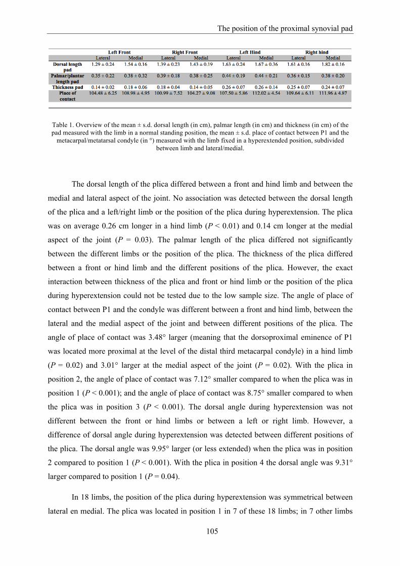
The position of the proximal synovial pad
105
Table 1. Overview of the mean ± s.d. dorsal length (in cm), palmar length (in cm) and thickness (in cm) of the pad measured with the limb in a normal standing position, the mean ± s.d. place of contact between P1 and the
metacarpal/metatarsal condyle (in °) measured with the limb fixed in a hyperextended position, subdivided between limb and lateral/medial.
The dorsal length of the plica differed between a front and hind limb and between the
medial and lateral aspect of the joint. No association was detected between the dorsal length
of the plica and a left/right limb or the position of the plica during hyperextension. The plica
was on average 0.26 cm longer in a hind limb (P < 0.01) and 0.14 cm longer at the medial
aspect of the joint (P = 0.03). The palmar length of the plica differed not significantly
between the different limbs or the position of the plica. The thickness of the plica differed
between a front or hind limb and the different positions of the plica. However, the exact
interaction between thickness of the plica and front or hind limb or the position of the plica
during hyperextension could not be tested due to the low sample size. The angle of place of
contact between P1 and the condyle was different between a front and hind limb, between the
lateral and the medial aspect of the joint and between different positions of the plica. The
angle of place of contact was 3.48° larger (meaning that the dorsoproximal eminence of P1
was located more proximal at the level of the distal third metacarpal condyle) in a hind limb
(P = 0.02) and 3.01° larger at the medial aspect of the joint (P = 0.02). With the plica in
position 2, the angle of place of contact was 7.12° smaller compared to when the plica was in
position 1 (P < 0.001); and the angle of place of contact was 8.75° smaller compared to when
the plica was in position 3 (P < 0.001). The dorsal angle during hyperextension was not
different between the front or hind limbs or between a left or right limb. However, a
difference of dorsal angle during hyperextension was detected between different positions of
the plica. The dorsal angle was 9.95° larger (or less extended) when the plica was in position
2 compared to position 1 (P < 0.001). With the plica in position 4 the dorsal angle was 9.31°
larger compared to position 1 (P = 0.04).
In 18 limbs, the position of the plica during hyperextension was symmetrical between
lateral en medial. The plica was located in position 1 in 7 of these 18 limbs; in 7 other limbs
Chapter 6
106
the plica was located in position 2; in 3 limbs the plica was in position 3 and finally the plica
was located in position 4 in one limb. In the 2 remaining limbs, the medial part of the plica
was in position 1 whereas the lateral part was either in position 2 or 4 (Fig. 4).
Discussion
The overall goal of this study was to describe the exact location of the dorsoproximal
synovial plica during hyperextension of the MCP/MTP joint to get a better insight in its
function. Previously, it has been stated that the position and structure of the plica suggests that
it acts as a cushioning surface or contact interface between P1 and the third metacarpal bone
where these are in contact during full extension of the joint (White 1990). This study
demonstrated that different positions of the synovial plica are possible during hyperextension
of the joint. A close contact with the plica and the dorsoproximal aspect of the distal third
metacarpal/metatarsal condyle was detected, at least with position 1 and 4, suggesting a close
relation between the plica and the dorsoproximal aspect of the sagittal ridge during
hyperextension.
In this study, low-field MRI was performed to describe the exact location of the pad
during hyperextension of the joint. It is generally accepted that MRI is the best imaging
modality to image soft tissue structures (Kraft and Gavin 2001). Although smaller structures
are better identified using high-field MRI (Smith et al. 2011), the overall quality of the
images obtained on our low-field MRI system was considered to be sufficient for the purpose
of this study.
In this study we used T1W, T2W and PDW SE sequences in all limbs in normal and
hyperextended position. A T1W SE sequence gives the best anatomical detail and is
considered to be the standard accepted sequence for baseline information of the
musculoskeletal system (Kleiter et al. 1999; Smith et al. 2011). Additional T2W and PDW SE
sequences were obtained for evaluation of synovial structures, as the hyperintense synovial
fluid creates a better contrast between the synovial plica and the surrounding synovial fluid
(Mair et al. 2005). With the limb hyperextended an additional STIR sequence was obtained to
identify the medullary fluid by eliminating the medullary fat signal of the bones (Mair et al.
2005).

The position of the proximal synovial pad
107
Scans in the dorsal and sagittal planes are generally performed for evaluation of the
articular surface of joints (Kleiter et al. 1999), but for this study only sagittal plane scans were
obtained to evaluate the proximal synovial plica in the MCP/MTP joint (Dyson and Murray
2007).
No abnormal low or high signal intensity was detected on the images at the level of
the dorsoproximal aspect of P1 or the dorsoproximal third metacarpal/metatarsal condyle at
the level where both are in contact during hyperextension. Magnetic resonance imaging has a
high sensitivity for the detection of abnormalities at the level of subchondral bone such as
bone sclerosis or bone bruise (Smith et al. 2012) which means that most likely no
abnormalities were present at the level of the bone at the place of contact during
hyperextension. It is possible that post-mortem changes and freeze-thaw effect could have an
effect on the MR images. It has been reported that there is no subjective difference between
images of the same digits obtained before death and after postmortem freezing and thawing
(Murray et al. 2006). However, it has been demonstrated that freezing-thawing processes can
induce changes of the signal-to-noise ratio in images and may alter the reliability of signal
intensity in ex vivo MRI examinations (Bolen et al. 2011).
To facilitate the extension of the MCP/MTP joint, the suspensory ligament and
accessory ligament of the deep digital flexor tendon were sectioned at the proximal aspect of
the metacarpus and the suspensory ligament, accessory ligament of the deep digital flexor
tendon, the deep digital flexor tendon and superficial digital flexor tendon were sectioned at
the proximal aspect of the metatarsus. Without sectioning of the tendons, up to 12 kN is
needed to obtain the same amount of hyperextension as is detected at the level of the
MCP/MTP joint during jumping (Brama et al. 2001). It was not possible to obtain this amount
of pressure with a non-magnetic loading frame that could be used in a MRI device. Therefore,
sectioning of the flexor tendons was performed to obtain hyperextension with the amount of
pressure that could be obtained using a wooden frame. This procedure was considered to be
acceptable for the purpose of the study as it was shown in a preliminary study that it had no
influence on the position of the bones relative to each other during hyperextension.
In human medicine, plicae are described at the level of the wrist, knee, elbow and hip
joint (Kim et al. 2006; Katz et al. 2010; Al-Hadithy et al. 2011; Ishii et al. 2012). In normal
conditions, these synovial plicae slip to and fro between the articular surfaces without
significant mechanical impingement (Bencardino et al. 2011). However, in each of these
Chapter 6
108
joints, symptomatic plicae are described with concurring histological abnormalities (Kim et
al. 2006; Katz et al. 2010; Al-Hadithy et al. 2011; Ishii et al. 2012). Synovial plica
proliferation is also described in the horse, mainly located at the level of the MCP joint with
concurring histological abnormalities at the level of the plica (Van Veenendaal and Moffatt
1980; Steyn et al. 1989; Kannegieter 1990; White 1990; Dabareiner et al. 1996). The dorsal
length of the medial part of the synovial plica was significantly longer compared to the lateral
part and overall significantly longer in the hind limb. However, the repeatability of these
measurements was not assessed in this study. Most of the cases of synovial plica proliferation
are described at the level of the front limbs without a predilection for medial or lateral (Van
Veenendaal and Moffatt 1980; Steyn et al. 1989; Kannegieter 1990; White 1990; Dabareiner
et al. 1996). A correlation of this condition with possible differences in length of the plica
described in this study seems therefore unlikely. Possibly, the position of the plica during
hyperextension is a predisposing factor for the development of proliferation of the synovial
plica. It is possible that the plica located in position 4 can be bruised during hyperextension. A
correlation between the histogical appearance of the plica and its position during
hyperextension would help to confirm this.
The angle of contact between the proximal aspect of P1 and the condyle was
significantly larger at the level of the medial dorsal eminence of P1 compared to the lateral
dorsal eminence. Fragments at the dorsal aspect of P1 are predominantly detected at the level
of the medial dorsal eminence of P1 (Kawcak and McIlwraith 1994; Declercq et al. 2009).
These fragments probably result from compression of the dorsoproximal portion of P1 against
the distal third metacarpal/metatarsal bone during hyperextension of the joint (Yovich and
McIlwraith 1986). This is in agreement with the larger angle of contact at the level of the
medial dorsal eminence of P1, most likely resulting in higher stress at that level.
In this study a significantly larger angle of contact between P1 and the condyle was
detected at the level of the hind limb. A possible explanation could be the sectioning of all
flexor tendons and ligaments at the level of the hind limb as higher degree of hyperextension
was obtained in the hind limb. However, no significant difference was detected between the
degree of hyperextension in a front or hind limb. Meaning that the higher position of the
dorsal eminences of P1 was not caused by a higher amount of hyperextension of the
MCP/MTP joint. Therefore it is unlikely that sectioning of the tendons is the cause of the
larger angle of contact between P1 and the condyle in the hind limb. This difference in angle
of contact between P1 and the condyle suggests that the dorsal eminences are possible bigger
The position of the proximal synovial pad
109
in a hind limb, compared to the front limb. This could mean that a higher incidence of
dorsoproximal P1 fragments can be present at the level of the hind limb. However, no
predilection for dorsoproximal fragments of P1 at the level of the hind limb is described
(Declercq et al. 2009).
Osteochondral fragmentation at the level of the synovial plica is a condition described
in the MCP/MTP joint (Declercq et al. 2008). The origin of these synovial fragments still
remains unclear. A possible hypothesis is that this osteochondral fragment is a pre-existing
cartilaginous structure that develops a fibrous reactive pattern due to a mechanical stimulus
such as the repetitive contact with P1 during full extension of the joint (Declercq et al. 2008).
This close contact between the plica and the dorsoproximal aspect of P1 has been clearly
demonstrated in this study.
The position of the proximal synovial plica in the MCP/MTP joint was in most limbs
symmetrical between the medial and the lateral part. Only in 2 limbs of 2 different horses a
difference in position of the lateral and medial part was detected. No reason for the difference
in lateral-medial position of the plica in these two limbs was detected. It is possibly that this
medial-lateral asymmetry was coincidental, as the repeatability of the position of the plica
during hyperextension was not assessed in this study
The angle of contact between P1 and the condyle was significantly smaller when the
plica was in position 2 compared to when the plica was in position 1 or 3. In addition, with
the plica in position 2 the dorsal angle during hyperextension was significantly larger
compared to when the plica was in position 1; meaning that there was less extension of the
MCP/MTP joint in the group with the plica in position 2. It is possible that the distal
projection of the plica (position 2) represents an intermediate position that will change to one
of the other three positions when the joint is in full hyperextension (significantly larger angle
of contact P1/condyle for position 1 and 3). To consolidate this hypothesis, further research
on limbs fixed sequentially in different angles of extension should be performed.
Interesting in this study was that the dorsal angle during hyperextension was 9.31°
larger or the joint was less extended when the plica was in position 4 than when the plica was
in position 1. The dorsal angle during hyperextension was not significantly different when the
plica was in position 2 and 4. Changing the angle of the extension on the same limb could
also reveal if this position of the plica (position 4) is an in intermediate position between the

Chapter 6
110
plica projecting distally (position 2) and the position with the tip of the plica bending
palmar/plantar (position 1) or projecting dorsally (position 3).
The mean ± s.d. thickness of the proximal synovial plica was 2.07 ± 0.13 mm in this
study. This is in agreement with the number available in the literature, ranging from 2 to 4
mm (White 1990; Denoix 1996).
This study demonstrated that during hyperextension of the joint, the proximal synovial
plica can have different positions. Either the tip of the plica is projecting distally, dorsally, is
interposed in between P1 and the condyle or the tip of the plica is bending towards
palmar/plantar. During hyperextension, a close relation is present between the proximal
synovial plica, the proximal aspect of P1 and the distal third metacarpal/metatarsal condyle.
The plica therefore most likely acts as a contact interface between P1 and the cannon bone.
However, considering the different positions of the plica during hyperextension the plica does
not seem not to function as a cushioning interface between P1 and the cannon bone during
hyperextension of the joint.

The position of the proximal synovial pad
111
References
Al-Hadithy N., Gikas P., Mahapatra A.M. and Dowd G. (2011) Review article: Plica syndrome of the knee. Journal of orthopeadic surgery (Hong Kong) 19, 354-358.
Barone R. (1986) Ceinture et membre thoraciques. In: Barone R. (editors), Anatomie
comparée des mammifères domestiques: Tome 1 ostéologie, 3 edn., Vigot freres, Paris. 451-586.
Barone R. (1989) Articulations de la ceinture et du membre thoraciques, Anatomie comparée
des mammifères domestiques: Tome 2 arthrologie et myologie, 3 edn., éditions vigot frères, Paris. 97-222.
Bencardino J.T., Kassarjian A., Vieira R.L., Schwartz R., Mellado J.M. and Kocher M. (2011)
Synovial plicae of the hip: Evaluation using mr arthrography in patients with hip pain. Skeletal Radiology 40, 415-421.
Bolen G.E., Haye D., Dondelinger R.E., Massart L. and Busoni V. (2011) Impact of
successive freezing-thawing cycles on 3-t magnetic resonance images of the digits of isolated equine limbs. American Journal of Veterinary Research 72, 780-790.
Brama P.a.J., Karssenberg D., Barneveld A. and Van Weeren P.R. (2001) Contact areas and
pressure distribution on the proximal articular surface of the proximal phalanx under sagittal plane loading. Equine Veterinary Journal 33, 26-32.
Brown N.a.T., Pandy M.G., Buford W.L., Kawcak C.E. and McIlwraith C.W. (2003) Moment
arms about the carpal and metacarpophalangeal joints for flexor and extensor muscles in equine forelimbs. American Journal of Veterinary Research 64, 351-357.
Butcher M.T. and Shley-Ross M.A. (2002) Fetlock joint kinematics differ with age in
thoroughbred [was thoroughbred] racehorses. Journal of Biomechanics 35, 563-571. Dabareiner R.M., White N.A. and Sullins K.E. (1996) Metacarpophalangeal joint synovial
pad fibrotic proliferation in 63 horses. Veterinary Surgery 25, 199-206. Declercq J., Martens A., Bogaert L., Boussauw B., Forsyth R. and Boening K.J. (2008)
Osteochondral fragmentation in the synovial pad of the fetlock in warmblood horses. Veterinary Surgery 37, 613-618.

Chapter 6
112
Declercq J., Martens A., Maes D., Boussauw B., Forsyth R. and Boening K.J. (2009) Dorsoproximal proximal phalanx osteochondral fragmentation in 117 warmblood horses. Veterinary and Comparative Orthopaedics and Traumatology 22, 1-6.
Denoix J.M. (1996) Ultrasonographic examination in the diagnosis of joint disease. In:
McIlwraith C.W. and Trotter G.W. (editors), Joint disease in the horse, W.B. Saunders, Philadelphia. 165-202.
Dyson S. and Murray R. (2007) Magnetic resonance imaging of the equine fetlock. Clinical
techniques in equine practice 6, 62-77. Hauspie S., Martens A., Declercq J., Busoni V., Vanderperren K., Van Bree H. and Saunders
J.H. (2010) Radiographic features of the dorsal condylar sagittal ridge of the third metacarpal and metatarsal bones in young warmblood stallions. Veterinary and Comparative Orthopaedics and Traumatology 23, 411-416.
Ishii H., Asami A. and Sonohata M. (2012) Synovial fringe (plica) of scapho-trapezial joint
following trauma. Hand Surgery 17, 243-245. Kannegieter N.J. (1990) Chronic proliferative synovitis of the equine metacarpophalangeal
joint. Veterinary Record 127, 8-10. Katz L.D., Haims A., Medvecky M. and Mccallum J. (2010) Symptomatic hip plica: Mr
arthrographic and arthroscopic correlation. Skeletal Radiology 39, 1255-1258. Kawcak C.E. and McIlwraith C.W. (1994) Proximodorsal first phalanx osteochondral chip
fragmentation in 336 horses. Equine Veterinary Journal 26, 392-396. Kim D.H., Gambardella R.A., Elattrache N.S., Yocum L.A. and Jobe F.W. (2006)
Arthroscopic treatment of posterolateral elbow impingement from lateral synovial plicae in throwing athletes and golfers. The American Journal of Sports Medicine 34, 438-444.
Kleiter M., Kneissl S., Stanek C., Mayrhofer E., Baulain U. and Deegen E. (1999) Evaluation
of magnetic resonance imaging techniques in the equine digit. Veterinary Radiology & Ultrasound 40, 15-22.
Kraft S.L. and Gavin P. (2001) Physical principles and technical considerations for equine
computed tomography and magnetic resonance imaging. Veterinary Clinics of North America-Equine Practice 17, 115-130.

The position of the proximal synovial pad
113
Les C.M., Stover S.M., Keyak J.H., Taylor K.T. and Willits N.H. (1997) The distribution of material properties in the equine third metacarpal bone serves to enhance sagittal bending. Journal of Biomechanics 30, 355-361.
Mair T.S., Kinns J., Jones R.D. and Bolas N.M. (2005) Magnetic resonance imaging of the
distal limb of the standing horse. Equine Veterinary Education 17, 74-78. Martinelli M.J., Baker G.J., Clarkson R.B., Eurell J.C., Pijanowski G.J., Kuriashkin I.V. and
Carragher B.O. (1996) Correlation between anatomic features and low-field magnetic resonance imaging of the equine metacarpophalangeal joint. American Journal of Veterinary Research 57, 1421-1426.
Martinelli M.J., Kuraiashkin I.V., Carragher B.O., Clarkson R.B. and Baker G.J. (1997)
Magnetic resonance imaging of the equine metacarpophalangeal joint: Three-dimensional reconstruction and anatomic analysis. Veterinary Radiology & Ultrasound 38, 193-199.
Meershoek L.S., Roepstorff L., Schamhardt H.C., Johnston C. and Bobbert M.F. (2001) Joint
moments in the distal forelimbs of jumping horses during landing. Equine Veterinary Journal 33, 410-415.
Murray R.C., Schramme M.C., Dyson S.J., Branch M.V. and Blunden T.S. (2006) Magnetic
resonance imaging characteristics of the foot in horses with palmar foot pain and control horses. Veterinary Radiology & Ultrasound 47, 1-16.
Palmer J.L. and Bertone A.L. (1996) Joint biomechanics in the pathogenesis of traumatic
arthritis. In: McIlwraith C.W. and Trotter G.W. (editors), Joint disease in the horse, WB Saunders, Philadelphia. 104-119.
Park R.D., Nelson T.R. and Hoopes J. (1987) Magnetic-resonance-imaging of the normal
equine digit and metacarpophalangeal joint. Veterinary Radiology 28, 105-116. Powell S.E. (2012) Low-field standing magnetic resonance imaging findings of the
metacarpo/metatarsophalangeal joint of racing thoroughbreds with lameness localised to the region: A retrospective study of 131 horses. Equine Veterinary Journal 44, 169-177.
Smith M.A., Dyson S.J. and Murray R.C. (2011) The appearance of the equine
metacarpophalangeal region on high-field vs. Standing low-field magnetic resonance imaging. Veterinary Radiology & Ultrasound 52, 61-70.
Chapter 6
114
Smith M.A., Dyson S.J. and Murray R.C. (2012) Reliability of high- and low-field magnetic resonance imaging systems for detection of cartilage and bone lesions in the equine cadaver fetlock. Equine Veterinary Journal.
Steyn P.F., Schmitz D., Watkins J. and Hoffman J. (1989) The sonographic diagnosis of
chronic proliferative synovitis in the metacarpophalangeal joints of a horse. Veterinary Radiology 30, 125-127.
Van Veenendaal J.C. and Moffatt R.E. (1980) Soft tissue masses in the fetlock joint of horses.
Australian Veterinary Journal 56, 533-536. Vanderperren K., Martens A., Haers H., Duchateau L. and Saunders J.H. (2009) Arthroscopic
visualisation of the third metacarpal and metatarsal condyles in the horse. Equine Veterinary Journal 41, 526-533.
Weaver J.C., Stover S.M. and O'Brien T.R. (1992) Radiographic anatomy of soft tissue
attachments in the equine metacarpophalangeal and proximal phalangeal region. Equine Veterinary Journal 24, 310-315.
White N.A. (1990) Synovial pad proliferation in the metacarpophalangeal joint. In: White
N.A. and Moore J.N. (editors), Current practice of equine surgery, 1 edn., J. B.Lippincott, Philadelphia. 555-558.
Yovich J.V. and McIlwraith C.W. (1986) Arthroscopic surgery for osteochondral fractures of
the proximal phalanx of the metacarpophalangeal and metatarsophalangeal (fetlock) joints in horses. Journal of the American Veterinary Medical Association 188, 273-279.

115
Chapter 7
General Discussion

General Discussion
117
A veterinary pre-purchase examination is an important professional service offered by
the veterinarian designed to identify any abnormalities or potential problems that can make
the horse unsuitable for its intended use (Bladon and Main 2003; Van Hoogmoed et al. 2003).
During this pre-purchase examination, survey radiographs of the distal limb are often taken in
order to identify these abnormalities or potential problems (Van Hoogmoed et al. 2003). The
predictive value of findings on radiography is controversial but nevertheless, radiography has
become an integral part of a pre-purchase examination (Van Hoogmoed et al. 2003) and the
presence or absence of radiological abnormalities has become an important factor in the horse
business (Stock et al. 2005).
The MCP/MTP joint is a high motion joint with high loading and is therefore
frequently injured (Pool and Meagher 1990). Variations in appearance of the dorsoproximal
aspect of the sagittal ridge have been observed during radiographic examinations of the
MCP/MTP joint in different breeds (Kane et al. 2003b; Wittwer et al. 2006; Verwilghen et al.
2009). However, there is a discrepancy in detected variations between these breeds.
When these variations are identified in a sound horse, the veterinarian will be asked to
evaluate how these will influence the future performance of the horse (Van Hoogmoed et al.
2003). The scientific information on these detected variations of the dorsoproximal aspect of
the sagittal ridge is limited. The presence of these variations at the level of the sagittal ridge
was not associated with loss of performance at young age in Thoroughbreds (Kane et al.
2003a). However, studies performed on Thoroughbred horses cannot be just extrapolated to
Warmbloods. These latter are not being trained until they reach the age of 3 years and will not
be challenged at high speeds as is done in Thoroughbreds at the age of 2 and 3 years.
Warmbloods will only start reaching their toplevel the earliest at 8 years old (Hinchcliff and
Hamlin 2004; O'Sullivan and Lumsden 2004).
In addition, the difference in type of career between these breeds makes a difference in
evaluating the potential influence of these variations. It is easier in Thoroughbreds, running at
high speed, to evaluate limiting performance or subtle lameness. In contrast to a Warmblood
horse, of which the type of training frequently differs from horse to horse, the objective
determination of the cause of not “performing up to the expected level” is very difficult. A
thorough study of the variations at the dorsoproximal aspect of the sagittal ridge is therefore
needed to help in the decision making during a pre-purchase examination.
Chapter 7
118
To evaluate the relevance of these variations, ideally, a longitudinal study would be
needed. However, this is difficult to perform in Warmblood horses. First, a long follow up
period is necessary, because of the long career of a Warmblood horse (up to 15 years).
Secondly, because of the different uses of a Warmblood horse (jumping, dressage, eventing),
a large population is needed. Horses competing in different sports are predisposed for
different injuries (Murray et al. 2006). Last, Warmblood horses are frequently sold and
therefore lost for follow up.
In the present study we started by defining the exact prevalence of the variations at the
dorsoproximal aspect of the sagittal ridge of the MCP/MTP joint in Warmblood horses. After
screening radiographs of 308 stallions, we detected a total of 5 variations. The most
commonly observed variation was a “smooth” appearance of the ridge (51.5%), followed by
an “irregular” outlining (19.3%). The remaining variations detected were a “cam” like lesion
(8.9%), “lucency” (8.1%) and “indentation” (12.2%). Based on our study, a discrepancy was
found in the number of detected variations at the dorsoproximal aspect of the sagittal ridge,
and their frequency of occurrence, compared with an other population of Warmbloods
(Verwilghen et al. 2009) and compared with Thoroughbreds (Kane et al. 2003b). In a
population of Warmbloods, only a mild surface irregularity of the proximal border of the
sagittal ridge was described in 6.8% of the joints and a well defined osteochondral defect at
that level in 1% (Verwilghen et al. 2009). In Thoroughbreds, only a notch in approximately
24% of the joints, lucency (1%) or a fragment/loose body (0.5%) was described (Kane et al.
2003b).
This difference in number and frequency could be expected as different frequencies
are reported between different populations of Thoroughbreds for other abnormalities at the
level of the MCP/MTP joint: for example the reported frequency of osteochondrosis, ranges
from 7.5% to 61% (Howard and Rantanen 1992; Kane et al. 2003b; Furniss et al. 2011). This
difference in number and frequency is most likely explained by the fact that the prevalence of
common radiographic findings in the limbs of young horses are influenced by genetics and
probably are caused by different genes (Stock and Distl 2006).
The origin of these variations in appearance of the dorsoproximal aspect of the sagittal
ridge is unknown. Irregularities and depressions of the subchondral bone at the mid and distal
aspect of the dorsal condylar sagittal ridge are associated with osteochondrosis dissecans in
horses (Yovich et al. 1985; Nixon 1990). Typical histological changes associated with

General Discussion
119
dyschondroplasia are clear alterations in staining of the mineralized matrix of the cartilage
and adjacent subchondral bone or retained cartilage cores surrounded by clusters of
chondrocytes (Henson et al. 1997; Jeffcott and Henson 1998). Histology demonstrated in all
of the samples of the present study a clear demarcated cortex, covered towards distally with
normal appearing hyaline cartilage and towards proximally with longitudinal aligned collagen
fibres. Based on these results, the origin of these variations is not osteochondrotic. However,
since only 25 samples were examined in this study and the quality of the staining was
sometimes suboptimal, more subtle lesions indicating a possible osteochondrotic origin of
these variations, could have been missed.
No information is available if these variations are already detected at birth (congenital)
or if they only develop at later age (joint adaptation to workload). Based on histology a
reactive process is excluded, however a slow ongoing process remains possible. Evolution of
radiographic changes in young horses up to the age of 11 months has been demonstrated (Dik
et al. 1999) and cartilage and its supporting subchondral bone undergo substantial changes at
young age. Both undergo major changes in composition under the age of 5 months and a
slower rate of evolution is detected at a later age. These changes result in differences in
composition of the cartilage and subchondral bone between the dorsoproximal aspect of P1
and the more centrally located regions (Brama et al. 2002a; Brommer et al. 2005). The
maturation of cartilage is also influenced by the type of exercise (Brama et al. 2002b).
However, maturation of cartilage and subchondral bone has up to now only been
demonstrated at the proximal articular surface of P1. A similar evolution is possible at the
level of the proximal aspect of the dorsal condylar sagittal ridge and the condyles but has until
now not been examined. During hyperextension of the MCP/MTP joint the dorsoproximal
aspect of P1 comes into contact with the apposing surface of the distal metacarpal condyle
(Brama et al. 2001). This abutment of P1 against the condyle can result in an adaptation at
young age of the dorsoproximal aspect of the distal metacarpal/metatarsal condyles to
sustained forces in a same way as described at the level of the proximal articular surface of
P1. The period of maturation of cartilage and subchondral bone (Brama et al. 2002a;
Brommer et al. 2005) corresponds with the period of radiographic evolution of osteochondral
abnormalities (Dik et al. 1999).
Based on histology the “cam” group and the “indentation” group in our study showed
a close resemblance with lesions associated with femoroacetabular impingement in the human
hip joint (Pitt et al. 1982). Due to femoral head or acetabular abnormalities, femoroacetabular
Chapter 7
120
impingement occurs as a result of abnormal contact between the proximal femur and
acetabular rim (Keogh and Batt 2008). Based on histology, the osseous excrescence or
reactive area at the level of the femoral head-neck junction (Hong et al. 2010; Bredella et al.
2013) and associated pit herniation are lesions in the human hip joint that show resemblance
with our “cam” and “indentation” group (Pitt et al. 1982). Avulsion of the aceteabular labrum
and cartilage damage are associated lesions with femoroacetabular impingement in the human
hip joint (Hong et al. 2010).
The origin of these lesions at the level of the human hip joint is unclear, but could be
congenital or developmental where abnormalities are formed during adolescence related to an
abnormal femoral head conformation (Bredella et al. 2013). To further elucidate the possible
origin of the different appearances of the dorsoproximal aspect of the sagittal ridge in horses,
a follow up study performed on young Warmblood horses from birth till the age of 1 or 2
years should be performed.
Based on previous research, initial cartilage degeneration in the MCP/MTP joint starts
at the dorsal articular margin of P1 (Brommer et al. 2004a) and early degenerative changes
are frequently encountered at the dorsal aspect of the distal third metacarpal/metatarsal
condyle (Brama et al. 2000). Cartilage degeneration can therefore be initiated by the
variations detected at the dorsoproximal aspect of the sagittal ridge in the equine MCP/MTP
joint. The assessment of articular cartilage abnormalities, however, is difficult. Arthroscopy is
one way to evaluate the articular cartilage surface, although points of discussion remain. First,
the entire cartilage surface of the equine MCP/MTP joint cannot be inspected and evaluated
(Vanderperren et al. 2009). Osteoarthritis is known to be heterogeneous with severely and at
seemingly unaffected sites within the joint (Palmer et al. 1995). Therefore, an evaluation of
the complete surface of the articular cartilage should provide a more complete and correct
assessment of the status of the articular cartilage. Secondly, the visual evaluation of the status
of the articular cartilage is not accurate. Arthroscopy underestimates the severity of the
lesions with mild cartilage damage and overestimates the extent of damage with more
established lesions (Brommer et al. 2004b). In addition, the inter-observer reliability of
arthroscopic grading of cartilage lesions is poor (Spahn et al. 2011). Other techniques
available for a more objective evaluation of the articular cartilage quality, like a high
frequency (40 Mhz) ultrasound biomicroscope (Spriet et al. 2005) or near-infrared-
spectroscopy device (Spahn et al. 2007) are available and are able to detect early changes in
articular cartilage. However, none of these are available for equine arthroscopy. An
General Discussion
121
indentation instrument was evaluated for the more objective assessment of cartilage quality in
equines but was found not to be sensitive enough to detected early changes in cartilage
properties (Brommer et al. 2006).
A second technique available for the assessment of articular cartilage is MRI. The
evaluation of the quality of the articular cartilage of the MCP/MTP joint depends on the
system used (low- or high-field) and is also highly susceptible to partial volume averaging
even when using a 1.5 T MRI- unit (Werpy 2007). Only the most severe lesions are evident
while less severe lesions are frequently not detected using a low-field standing magnet
(Murray et al. 2009; Sherlock et al. 2009; Olive 2010). Audigié et al. 2007 mentioned that the
cartilage is even too thin at the level of the MCP/MTP joint for evaluation with a low-field
MRI (Audigie et al. 2007). In human medicine it is recommended to acquire images using
magnets with a field strength of 1.5 T or greater for morphologic and compositional
assessment of cartilage resolution of articular cartilage (Roemer et al. 2011). Specialized MRI
sequences are available to detect loss of proteoglycans within the cartilage matrix, which is
considered to be one of the first pathophysiologic events in osteoarthritis. Delayed
gadolinium-enhanced MRI of cartilage is able to detect early changes in the proteoglycan
content of articular cartilage (Taylor et al. 2009) and is described for use in equine MRI
studies (Pease 2012; Saveraid and Judy 2012). However, both with high- or low-field MRI,
there is a high likelihood of false positive results for detection of cartilage lesions compared
with histopathology (Smith et al. 2012). As specimen obtained from the slaughterhouse were
used in this thesis, delayed gadolinium-enhanced MRI of cartilage was not possible because
this requires the use of IV injection of gadolinium.
For this ex vivo evaluation of articular cartilage, it was decided to work with the
objective measurement of the “Cartilage Degeneration Index” developed by Brommer et al.
2003 for the evaluation of the proximal articular cartilage surface of P1 (Brommer et al.
2003). This technique uses Indian ink staining of the cartilage surface and is based on the fact
that Indian ink particles have a high affinity for articular cartilage with surface fibrillation and
depletion of proteoglycans, whereas normal intact articular cartilage with a high
proteoglycan-rich matrix will prevent the particles from entering (Meachim 1972; Chang et
al. 1997). It has the advantage over the histopathological scoring systems for articular
cartilage degradation (Mankin et al. 1971; McIlwraith et al. 2010) that it is able to evaluate
the entire surface of the articular surface rather than only small samples obtained for
histology. It has already been used in experimental studies in different species (Roberts et al.
Chapter 7
122
1986; Chang et al. 1997; Cantley et al. 1999) to subjectively evaluate the quality of articular
cartilage. The technique developed by Brommer et al. 2003 is different because they use an
objective evaluation by analysing the grey levels of digitally imaged cartilage surfaces of P1.
This resulted in a calculated CDI that allows the objective evaluation of the degree of
cartilage degeneration (Brommer et al. 2003). The technique develop by Brommer et al. 2003
is however not directly applicable for our study. This technique was only validated using 2D
photographs for the proximal articular surface of P1, considered to be a relatively “flat
surface” and doesn’t include area calculations on curved surfaces like the distal third
metacarpal/metatarsal condyles. Therefore a modified technique that created a complete 3D
model of the articular surfaces of the distal metacarpal/metatarsal condyle and P1 and allowed
area calculations on curved surfaces was used (Declercq et al. 2011). Once the 3D models
were generated, the cartilage regions of interest were selected using a paintbrush metaphor,
and this surface area was measured based on the 3D geometry. The process of selecting the
regions of interest had a good intra-observer repeatability (Declercq et al. 2011). The CDI,
defined as the percentage of darkening, was calculated as the ratio between stained and
unstained grey values of the selected cartilage surface. This procedure was slightly different
compared to Brommer et al. 2003, who first averaged the grey values of each image, then
computed the difference between the average values. However, the results of the
measurements were in the same range in the 2 studies (Brommer et al. 2003; Declercq et al.
2011).
In the present research project, this technique of CDI calculation was applied on
MCP/MTP joints of Warmblood horses collected in the slaughterhouse. The major drawback
of this study design was the lack of clinical data of the horses. Important data as the breed and
age of the horse could be retrieved, but other clinical information such as the presence of
MCP/MTP joint related lameness was not available. Although care was taken not to include
joints presenting variations or abnormalities other than those present at the dorsoproximal
aspect of the sagittal ridge (Hauspie et al. 2010), it cannot be entirely excluded that some of
these joints might have been included. However, a large number of horses were available for
this study (86 horses), possibly compensating for this weakness.
This study revealed no significant association between the different variations at the
dorsoproximal aspect of the sagittal ridge and a higher CDI or increased cartilage damage
neither at the level of the complete MCP/MTP joint, the distal third metacarpal/metatarsal
condyle or the proximal articular surface of P1. This suggests that the presence of these
General Discussion
123
detected variation at the dorsoproximal aspect of the sagittal ridge does not have an influence
on the joint and are therefore anatomic variations.
Although the advantage of the CDI-technique is the evaluation of the complete
articular surface, its major limitation is that only the articular surface is evaluated. First,
degenerative joint disease is a multifactorial problem, not only involving the articular surface,
but also involving the subchondral bone (Kawcak et al. 2001) or joint capsule (Palmer and
Bertone 1994), which were not evaluated with the current study design. Secondly, the
technique of staining of articular cartilage with Indian ink is based on the increased uptake of
these ink particles in the presence of surface fibrillation and proteoglycan depletion (Chang et
al. 1997), but a recent study suggests that the picture of proteoglycan loss early in
osteoarthritis may not be complete and additional factors may also contribute to the process in
early osteoarthritis (Taylor et al. 2009).
Previous research demonstrated that initial cartilage degeneration starts at the dorsal
articular margin of P1 (Brommer et al. 2004a) and early degenerative changes are frequently
encountered at the dorsal aspect of the distal third metacarpal/metatarsal condyle (Brama et
al. 2000). It is possible that the variations in appearance of the dorsoproximal aspect of the
sagittal ridge will have a more localized effect at the dorsal aspect of the joint, since during
hyperextension, the dorsoproximal aspect of P1 comes into contact with the dorsal aspect of
the distal third metacarpal/metatarsal condyle (Brama et al. 2001). Calculation of the CDI
specifically for the dorsal and palmar/plantar aspect of the condyle and articular surface of P1
would possibly further elucidate this more localized effect.
In addition to the detected variations at the dorsoproximal aspect of the sagittal ridge,
a synovial plica is present at that level. This latter is projecting distally from the
dorsoproximal attachment of the joint capsule and tapers to a thin edge. Position and structure
suggests that the pad acts as a cushioning surface or contact interface between the dorsal rim
of P1 and the distal metacarpal/metatarsal condyle during hyperextension (White 1990;
McIlwraith et al. 2005), even though its exact position during hyperextension of the
MCP/MTP joint and its function have not been determined.
In the last study of this research project, different positions of the plica were detected
during hyperextension of the joint, clearly demonstrating a close contact between the
dorsoproximal aspect of P1 and the synovial plica. Based on the different positions of the
plica during hyperextension, a cushioning function of the plica seems less likely. However, it

Chapter 7
124
is not described if this cushioning function is performed by the distal tip of the plica or by the
base of the plica. Based on histology of the bone specimen, longitudinal collagen fibres were
detected just proximal to the articular cartilage. This could suggest some kind of cushioning
function at that level. However, in our study, due to the removal of the soft tissues when
sampling the specimen for histology, the exact relation of these longitudinal collagen fibres
relative to the synovial plica could not be established.
Synovial plica proliferation is a described condition in horses (Van Veenendaal and
Moffatt 1980; Steyn et al. 1989; Dabareiner et al. 1996), and thought to be caused by chronic
trauma to the plica. Histopathology could determine if one of the different positions of the
plica can predispose for this proliferation by determining its content and structure.
A future MRI study in which limbs are hyperextended multiple times immediately
after collection could demonstrate the repeatability of the position of the synovial plica during
hyperextension as well as the place of contact of the dorsoproximal aspect of P1 relative to
the variations at the dorsoproximal aspect of the sagittal ridge. In addition, a subsequent
storage at -20 °C would allow the sampling of the dorsoproximal aspect of P1 with the
surrounding soft tissues still in place. This would provide information about the location of
the plica relative to the variations at the dorsoproximal aspect of the sagittal ridge.
The main conclusion of the present research work is that variations are present at the
dorsoproximal aspect of the sagittal ridge of the MCP/MTP joint (“smooth”, “irregular”,
“cam”, “lucency” and “indentation”). These variations do not demonstrate clear abnormalities
on histology nor are they associated with abnormalities at the level of the articular cartilage.
During hyperextension of the equine MCP/MTP joint, these variations are most likely in close
relation with the dorsoproximal aspect of P1 and the synovial plica.
General Discussion
125
References
Audigie F., Didierlaurent D., Coudry V., Jacquet S. and Denoix J.M. (2007) Interest of magnetic resonance imaging in the diagnosis of equine locomotor disorders. Bulletin De L Academie Veterinaire De France 160, 19-24.
Bladon B.M. and Main J.P. (2003) Clinical evidence in the evaluation of presale radiography:
Are we in a desert on a horse with no name? Equine Veterinary Journal 35, 341-342. Brama P.A., Tekoppele J.M., Bank R.A., Barneveld A. and Van Weeren P.R. (2002a)
Biochemical development of subchondral bone from birth until age eleven months and the influence of physical activity. Equine Veterinary Journal 34, 143-149.
Brama P.a.J., Karssenberg D., Barneveld A. and Van Weeren P.R. (2001) Contact areas and
pressure distribution on the proximal articular surface of the proximal phalanx under sagittal plane loading. Equine Veterinary Journal 33, 26-32.
Brama P.a.J., Tekoppele J.M., Bank R.A., Barneveld A. and Van Weeren P.R. (2002b)
Development of biochemical heterogeneity of articular cartilage: Influences of age and exercise. Equine Veterinary Journal 34, 265-269.
Brama P.a.J., Tekoppele J.M., Bank R.A., Karssenberg D., Barneveld A. and Van Weeren
P.R. (2000) Topographical mapping of biochemical properties of articular cartilage in the equine fetlock joint. Equine Veterinary Journal 32, 19-26.
Bredella M.A., Ulbrich E.J., Stoller D.W. and Anderson S.E. (2013) Femoroacetabular
impingement. Magnetic Resonance Imaging Clinics of North America 21, 45-64. Brommer H., Brama P.a.J., Laasanen M.S., Helminen H.J., Van Weeren R.R. and Jurvelin
J.S. (2005) Functional adaptation of articular cartilage from birth to maturity under the influence of loading: A biomechanical analysis. Equine Veterinary Journal 37, 148-154.
Brommer H., Brama R.a.J., Barneveld A. and Van Weeren P.R. (2004a) Differences in the
topographical distribution of articular cartilage degeneration between equine metacarpo- and metatarsophalangeal joints. Equine Veterinary Journal 36, 506-510.
Brommer H., Laasanen M.S., Brama P.a.J., Van Weeren P.R., Helminen H.J. and Jurvelin J.S.
(2006) In situ and ex vivo evaluation of an arthroscopic indentation instrument to estimate the health status of articular cartilage in the equine metacarpophalangeal joint. Veterinary Surgery 35, 259-266.
Chapter 7
126
Brommer H., Rijkenhuizen A.B.M., Brama P.a.J., Barneveld A. and Van Weeren P.R. (2004b) Accuracy of diagnostic arthroscopy for the assessment of cartilage damage in the equine metacarpophalangeal joint. Equine Veterinary Journal 36, 331-335.
Brommer H., Van Weeren P.R. and Brama P.A. (2003) New approach for quantitative
assessment of articular cartilage degeneration in horses with osteoarthritis. American Journal of Veterinary Research 64, 83-87.
Cantley C.E., Firth E.C., Delahunt J.W., Pfeiffer D.U. and Thompson K.G. (1999) Naturally
occurring osteoarthritis in the metacarpophalangeal joints of wild horses. Equine Veterinary Journal 31, 73-81.
Chang D.G., Iverson E.P., Schinagl R.M., Sonoda M., Amiel D., Coutts R.D. and Sah R.L.
(1997) Quantitation and localization of cartilage degeneration following the induction of osteoarthritis in the rabbit knee. Osteoarthritis Cartilage 5, 357-372.
Dabareiner R.M., White N.A. and Sullins K.E. (1996) Metacarpophalangeal joint synovial
pad fibrotic proliferation in 63 horses. Veterinary Surgery 25, 199-206. Declercq J., Brown B., Oosterlinck M., Van Gool L., Hauspie S., Saunders J. and Martens A.
(2011) Quantitative assesment of articular cartilage degeneration of the metacarpal condyle using a 3d scanning system. In: ECVS Proceedings. 69-69.
Dik K.J., Enzerink E. and Van Weeren P.R. (1999) Radiographic development of
osteochondral abnormalities, in the hock and stifle of dutch warmblood foals, from age 1 to 11 months. Equine veterinary Journal Supplement, 9-15.
Furniss C., Carstens A. and Van Den Berg S.S. (2011) Radiographic changes in thoroughbred
yearlings in south africa. Journal of the South African Veterinary Association 82, 194-204.
Hauspie S., Martens A., Declercq J., Busoni V., Vanderperren K., Van Bree H. and Saunders
J.H. (2010) Radiographic features of the dorsal condylar sagittal ridge of the third metacarpal and metatarsal bones in young warmblood stallions. Veterinary and Comparative Orthopaedics and Traumatology 23, 411-416.
Henson F.M.D., Davies M.E. and Jeffcott L.B. (1997) Equine dyschondroplasia
(osteochondrosis) - histological findings and type vi collagen localization. Veterinary Journal 154, 53-62.
Hinchcliff K.W. and Hamlin M. (2004) Veterinary aspects of racing and training standardbred
race horses. In: Hinchcliff K.W., Kaneps A.J. and Geor R.J. (editors), Equine sports medicine and surgery, Saunders, Philadelphia. 1073-1089.
General Discussion
127
Hong S.J., Shon W.Y., Lee C.Y., Myung J.S., Kang C.H. and Kim B.H. (2010) Imaging findings of femoroacetabular impingement syndrome: Focusing on mixed-type impingement. Clinical Imaging 34, 116-120.
Howard B.A. and Rantanen N.W. (1992) Survey radiographic findings in thoroughbred sales
yearlings. In: 38th annual convention of the AAEP, Orlande, Florida. 397-402. Jeffcott L.B. and Henson F.M.D. (1998) Studies on growth cartilage in the horse and their
application to aetiopathogenesis of dyschondroplasia (osteochondrosis). Veterinary Journal 156, 177-192.
Kane A.J., McIlwraith C.W., Park R.D., Rantanen N.W., Morehead J.P. and Bramlage L.R.
(2003a) Radiographic changes in thoroughbred yearlings. Part 2: Associations with racing performance. Equine Veterinary Journal 35, 366-374.
Kane A.J., Park R.D., McIlwraith C.W., Rantanen N.W., Morehead J.P. and Bramlage L.R.
(2003b) Radiographic changes in thoroughbred yearlings. Part 1: Prevalence at the time of the yearling sales. Equine Veterinary Journal 35, 354-365.
Kawcak C.E., McIlwraith C.W., Norrdin R.W., Park R.D. and James S.P. (2001) The role of
subchondral bone in joint disease: A review. Equine Veterinary Journal 33, 120-126. Keogh M.J. and Batt M.E. (2008) A review of femoroacetabular impingement in athletes.
Sports Medicine 38, 863-878. Mankin H.J., Dorfman H., Lippiell.L and Zarins A. (1971) Biochemical and metabolic
abnormalities in articular cartilage from osteo-arthritic human hips .2. Correlation of morphology with biochemical and metabolic data. Journal of Bone and Joint Surgery-American Volume A 53, 523-537.
McIlwraith C.W., Frisbie D.D., Kawcak C.E., Fuller C.J., Hurtig M. and Cruz A. (2010) The
oarsi histopathology initiative - recommendations for histological assessments of osteoarthritis in the horse. Osteoarthritis Cartilage 18 Suppl 3, S93-105.
McIlwraith C.W., Nixon A.J., Wright I.M. and Boening K.J. (2005) Diagnostic and surgical
arthroscopy of the metacarpophalangeal and metatarsophalangeal joints. In: McIlwraith C.W., Nixon A.J., Wright I.M. and Boening K.J. (editors), Diagnostic and surgical arthroscopy in the horse, 3 edn., Elsevier, Philadelphia. 129-196.
Meachim G. (1972) Light microscopy of indian ink preparations of fibrillated cartilage.
Annals of the Rheumatic Diseases 31, 457-464.
Chapter 7
128
Murray R.C., Dyson S.J., Tranquille C. and Adams V. (2006) Association of type of sport and performance level with anatomical site of orthopaedic injury diagnosis. Equine Veterinary Journal Supplement 38, 411-416.
Murray R.C., Mair T.S., Sherlock C.E. and Blunden A.S. (2009) Comparison of high-field
and low-field magnetic resonance images of cadaver limbs of horses. Veterinary Record 165, 281-288.
Nixon A.J. (1990) Osteochondrosis and osteochondrosis-dissecans of the equine fetlock.
Compendium on Continuing Education for the Practicing Veterinarian 12, 1463-&. O'Sullivan C.B. and Lumsden J.M. (2004) Veterinary aspects of racing and training
thoroughbred race horses. In: Hinchcliff K.W., Kaneps A.J. and Geor R.J. (editors), Equine sports medicine and surgery, Saunders, Philadelphia. 1051-1072.
Olive J. (2010) Distal interphalangeal articular cartilage assessment using low-field magnetic
resonance imaging. Veterinary Radiology & Ultrasound 51, 259-266. Palmer J.L. and Bertone A.L. (1994) Joint structure, biochemistry and biochemical
disequilibrium in synovitis and equine joint disease. Equine Veterinary Journal 26, 263-277.
Palmer J.L., Bertone A.L., Malemud C.J., Carter B.G., Papay R.S. and Mansour J. (1995)
Site-specific proteoglycan characteristics of 3rd carpal articular-cartilage in exercised and nonexercised horses. American Journal of Veterinary Research 56, 1570-1576.
Pease A. (2012) Biochemical evaluation of equine articular cartilage through imaging.
Veterinary Clinics of North America-Equine Practice 28, 637-646. Pitt M.J., Graham A.R., Shipman J.H. and Birkby W. (1982) Herniation pit of the femoral
neck. American Journal of Roentgenology 138, 1115-1121. Pool R.R. and Meagher D.M. (1990) Pathologic findings and pathogenesis of racetrack
injuries. Veterinary Clinics of North America-Equine Practice 6, 1-30. Roberts S., Weightman B., Urban J. and Chappell D. (1986) Mechanical and biochemical-
properties of human articular-cartilage in osteoarthritic femoral heads and in autopsy specimen. Journal of Bone and Joint Surgery-British Volume 68, 278-288.
Roemer F.W., Crema M.D., Trattnig S. and Guermazi A. (2011) Advances in imaging of
osteoarthritis and cartilage. Radiology 260, 332-354.

General Discussion
129
Saveraid T.C. and Judy C.E. (2012) Use of intravenous gadolinium contrast in equine magnetic resonance imaging. Veterinary Clinics of North America-Equine Practice 28, 617-636.
Sherlock C.E., Mair T.S. and Ter Braake F. (2009) Osseous lesions in the
metacarpo(tarso)phalangeal joint diagnosed using low-field magnetic resonance imaging in standing horses. Veterinary Radiology & Ultrasound 50, 13-20.
Smith M.A., Dyson S.J. and Murray R.C. (2012) Reliability of high- and low-field magnetic
resonance imaging systems for detection of cartilage and bone lesions in the equine cadaver fetlock. Equine Veterinary Journal 44, 684-691.
Spahn G., Klinger H.M., Baums M., Pinkepank U. and Hofmann G.O. (2011) Reliability in
arthroscopic grading of cartilage lesions: Results of a prospective blinded study for evaluation of inter-observer reliability. Archives of Orthopaedic Trauma Surgery 131, 377-381.
Spahn G., Plettenberg H., Kahl E., Klinger H.M., Muckley T. and Hofmann G.O. (2007)
Near-infrared (nir) spectroscopy. A new method for arthroscopic evaluation of low grade degenerated cartilage lesions. Results of a pilot study. BMC Musculoskeletal Disorders 8, 47.
Spriet M.P., Girard C.A., Foster S.F., Harasiewicz K., Holdsworth D.W. and Laverty S.
(2005) Validation of a 40 mhz b-scan ultrasound biomicroscope for the evaluation of osteoarthritis lesions in an animal model. Osteoarthritis Cartilage 13, 171-179.
Steyn P.F., Schmitz D., Watkins J. and Hoffman J. (1989) The sonographic diagnosis of
chronic proliferative synovitis in the metacarpophalangeal joints of a horse. Veterinary Radiology 30, 125-127.
Stock K.F. and Distl O. (2006) Genetic correlations between osseous fragments in fetlock and
hock joints, deforming arthropathy in hock joints and pathologic changes in the navicular bones of warmblood riding horses. Livestock Science 105, 35-43.
Stock K.F., Hamann H. and Distl O. (2005) Prevalence of osseous fragments in distal and
proximal interphalangeal, metacarpo- and metatarsophalangeal and tarsocrural joints of hanoverian warmblood horses. Journal of veterinary medicine 52, 388-394.
Taylor C., Carballido-Gamio J., Majumdar S. and Li X. (2009) Comparison of quantitative
imaging of cartilage for osteoarthritis: T2, t1rho, dgemric and contrast-enhanced computed tomography. Magnetic Resonance Imaging 27, 779-784.
Chapter 7
130
Van Hoogmoed L.M., Snyder J.R., Thomas H.L. and Harmon F.A. (2003) Retrospective evaluation of equine prepurchase examinations performed 1991-2000. Equine Veterinary Journal 35, 375-381.
Van Veenendaal J.C. and Moffatt R.E. (1980) Soft tissue masses in the fetlock joint of horses.
Australian Veterinary Journal 56, 533-536. Vanderperren K., Martens A., Haers H., Duchateau L. and Saunders J.H. (2009) Arthroscopic
visualisation of the third metacarpal and metatarsal condyles in the horse. Equine Veterinary Journal 41, 526-533.
Verwilghen D., Serteyn D., Pille F., Bolen G., Saunders J.H., Grulke S. and Busoni V. (2009)
Prevalence of radiographic findings in candidate sires (2001-2008). Vlaams Diergeneeskundig Tijdschrift 78, 419-428.
Werpy N.M. (2007) Magnetic resonance imaging of the equine patient: A comparison of
high- and low-field systems. Clinical techniques in equine practice 6, 37-45. White N.A. (1990) Synovial pad proliferation in the metacarpophalangeal joint. In: White
N.A. and Moore J.N. (editors), Current practice of equine surgery, 1 edn., J. B.Lippincott, Philadelphia. 555-558.
Wittwer C., Hamann H., Rosenberger E. and Distl O. (2006) Prevalence of osteochondrosis in
the limb joints of south german coldblood horses. Journal of veterinary medicine 53, 531-539.
Yovich J.V., McIlwraith C.W. and Stashak T.S. (1985) Osteochondritis dissecans of the
sagittal ridge of the third metacarpal and metatarsal bones in horses. Journal of the American Veterinary Medical Association 186, 1186-1191.

131
Summary


Summary
133
A veterinary pre-purchase examination is an important service offered to the owner of
a horse. Survey radiographs often reveal abnormalities or variations. The predictive value of
these findings is somewhat controversial however has become an important factor in the horse
business. Assessing the future clinical relevance of detected changes can be challenging
especially when clinical signs are absent. Variations in appearance of the dorsoproximal
aspect of the sagittal ridge are described in Thoroughbreds, without any influence on the
performance of the horse. The scientific information on detected variations at the
dorsoproximal aspect of the dorsal condylar sagittal ridge is limited in Warmblood horses.
The present PhD research focus was to describe the prevalence of variations at the
dorsoproximal aspect of the dorsal condylar sagittal ridge in Warmblood horses, to evaluate
their histological basis, assess their influence on the articular joint cartilage and their
interaction with the surrounding soft tissues.
In chapter 1 an overview is provided of the anatomy of the equine metacarpo-
/metatarsophalangeal joint and the diagnostic imaging modalities currently used in an equine
pre-purchase examination.
The scientific aims of this work are presented in chapter 2. The first objective of this
thesis was to describe the prevalence of variation in radiographic appearance of the
dorsoproximal aspect of the condylar sagittal ridge in the equine metacarpo-
/metatarsophalangeal joint in Warmblood horses.
The evaluation of the histological appearance of these detected variations at the
dorsoproximal aspect of the sagittal ridge was the second objective of this thesis.
The third objective of this thesis was to assess the potential association of these
variations in appearance of the dorsoproximal aspect of the sagittal ridge with articular
cartilage degeneration.
The fourth objective of this thesis was to describe the influence of hyperextension of
the metacarpo-/metatarsophalangeal joint on the position of the synovial plica surrounding the
proximal aspect of the dorsal condylar sagittal ridge.
In chapter 3 radiographs of young Warmblood stallions were scrutinized to describe
the prevalence of radiographic variations at the dorsoproximal aspect of the sagittal ridge in
the metacarpo-/metatarsophalangeal joint. Radiographs of a total of 308 Warmblood stallions
were used, giving us a total of 1232 equine metacarpo-/metatarsophalangeal joints available
for this study. The radiographic appearance of the dorsoproximal aspect of the sagittal ridge

Summary
134
was classified into the following categories: “smooth”, “irregular”, “cam”, “indentation” and
“lucency”. A “smooth” appearance was detected in 51.5% of the joints; the ridge was
“irregular” outlined in 19.3%. A “cam” like bony excrescence was present in 8.9%, 12.2% of
the ridges had an “indentation” and in 8.1% a “lucency” was detected. An “indentation” at the
dorsoproximal aspect of the sagittal ridge was significantly more detected at the level of the
hind limbs (P < 0.0001). In conclusion, there is variation in the radiographic appearance of
the proximal aspect of the dorsal condylar sagittal ridge in the metacarpo-
/metatarsophalangeal joint of Warmblood horses. However, their background and possible
influence on the function of the joint needs further investigation.
In chapter 4 the histological origin of the variations in radiographic appearance of the
dorsoproximal of the sagittal ridge of the metacarpo-/metatarsophalangeal joint was
described. A total of 25 joints of 12 young Warmblood horses were selected based on a
radiographic examination to ensure that of each detected variation at the dorsoproximal aspect
of the sagittal ridge 5 specimen were available for histological examination. The “smooth”
variation was used as the reference category because this variation was most detected on
radiography. Each group demonstrated a bone cortex, covered with hyaline cartilage distally
and a transition to longitudinally aligned collagen fibres covered by loosely organized
connective tissue proximally. The “smooth” and “irregular” group showed a smooth bone
cortex. The “cam” like category presented an expansion of the cortex. In the “indentation”
and “lucency” group, a depression in the cortex was detected. The collagen fibres and
connective tissue were located in the depression in the “indentation” category whereas the
location varied in the “lucency” and “cam” group. In conclusion, the detected differences in
appearance of the dorsoproximal aspect of the sagittal ridge on radiography are a
representation of anatomical variations resulting from detectable histological differences.
Further research is warranted to determine whether these variations are developmental or
congenital and to evaluate their potential influence on the joint and surrounding soft tissues
during hyperextension
In chapter 5 the objective was to investigate whether an association could be found
between the variable morphological appearances of the proximal aspect of the dorsal condylar
sagittal ridge and the degree of cartilage degeneration, determined by the “cartilage
degeneration index” at the level of the metacarpo-/metatarsophalangeal joint, distal
metacarpal/metatarsal condyle or proximal articular surface of the proximal phalanx. The
distal limbs of 86 Warmblood horses were used for this study. No significant association was

Summary
135
detected between the “cartilage degeneration index” and the presence of one of the detected
variations, age, front or hind limb and left or right limb. In conclusion, the different
radiographic features at the proximal aspect of the dorsal condylar sagittal ridge have no
influence on the cartilage status of the metacarpo-/metatarsophalangeal joint.
In chapter 6 the location of the dorsoproximal synovial plica was described during
hyperextension of the metacarpo-/metatarsophalangeal joint in order to get a better insight in
its function. The position and structure of the plica suggests that it acts as a cushioning
surface or contact interface between the proximal phalanx and the cannon bone where these
are in contact during full extension of the joint. The distal limbs of 5 Warmblood horses were
collected and magnetic resonance imaging scans were performed. Each joint was scanned
twice: first in a neutral standing position and than in a hyperextended position. During
hyperextension several positions of the plica were detected, ranging from (1) shortened with
the tip of plica bending or angled towards palmar/plantar; (2) projecting distally parallel to the
condyle; (3) projecting dorsally with the proximal margin of the proximal phalanx located
underneath it or (4) the tip of the plica interposed between the proximal aspect of the proximal
phalanx and the condyle. During hyperextension, a close relation is present between the
proximal synovial plica, proximal aspect of the proximal phalanx and the condyle with a
variable position of the plica. This suggests that the plica acts as a contact interface between
the proximal phalanx and the cannon bone. However, due to the different positions of the
plica, the plica consequently seems not to act as a cushioning surface.
The final chapter (chapter 7) presents the general discussion and conclusions.
Variations in appearance of the dorsoproximal aspect of the sagittal ridge are present in the
metacarpo-/metatarsophalangeal joint of Warmblood horses. These variations are not linked
with an increase in articular cartilage damage, nor do they present clear abnormalities on
histology. During hyperextension of the joint, a close contact is present between these
variations at the level of the sagittal ridge, the dorsoproximal aspect of the proximal phalanx
and the synovial plica.


137
Samenvatting

Samenvatting
139
In de “paardenwereld” wordt een aankooponderzoek regelmatig uitgevoerd om na te
gaan of het paard het gewenste prestatieniveau zal aankunnen. Radiografie is hierbij een zeer
belangrijk hulpmiddel om afwijkingen ter hoogte van de ledematen te identificeren. Het
inschatten van het toekomstig belang van waargenomen afwijkingen is een grote uitdaging
indien het paard geen klinische symptomen vertoont op het ogenblik van de keuring.
Vormvariaties ter hoogte van het proximaal aspect van de sagittaalkam zijn zichtbaar bij
volbloeden, zonder invloed op de sportieve prestaties. Tot op heden zijn deze radiografische
vormvariaties nooit eenduidig beschreven bij Warmbloedpaarden, noch hun relatie met
kraakbeendegeneratie of interactie met de omliggende weke delen. Het algemene doel van dit
doctoraatswerk was om hier een beter inzicht in te krijgen.
Het eerste hoofdstuk geeft een overzicht van de anatomie van het kogelgewricht van
het paard en de beeldvormingsmodaliteiten die kunnen gebruikt worden bij een aankoop
onderzoek.
De doelstelling van dit doctoraatswerk worden weergegeven in het tweede hoofdstuk.
De eerste doelstelling was om de prevalentie van vormveranderingen ter hoogte van het
dorsoproximaal aspect van de sagittaalkam van het kogelgewricht te beschrijven in een
populatie van Warmbloedpaarden. De tweede doelstelling bestond erin om het histologisch
uitzicht van deze radiografisch zichtbare vormveranderingen te beschrijven.
De analyse van de potentiële invloed van deze vormveranderingen op de integriteit
van het gewrichtskraakbeen werd de derde doelstelling.
De laatste en vierde doelstelling was een beter inzicht te verwerven in de interactie van
de structuren ter hoogte van het dorsaal aspect van de kogel. Hierbij lag de nadruk op de
positie van de plica synovialis tijdens hyperextensie van het kogelgewricht.
In het derde hoofdstuk werd de prevalentie van deze vormvariaties ter hoogte van het
proximaal aspect van de sagittaalkam beschreven in een populatie Warmbloedpaarden.
Hiervoor werden radiografische opnames van 308 Warmbloedhengsten beoordeeld, wat een
totaal van 1323 radiografische projecties van het kogelgewricht opleverde voor deze studie.
Het röntgenbeeld van het dorsoproximaal aspect van de sagittaalkam werd ingedeeld in
volgende categorieën: vlak en glad afgelijnd (51.5%), onregelmatig afgelijnd (19.3%),
knobbelvormige aanwas (8.9%), aanwezigheid van een indeuking (12.2%) of aanwezigheid
van een lucente zone (8.1%). Een indeuking kwam significant meer voor ter hoogte van het
achterbeen (P < 0.0001). Uit deze studie kon worden besloten dat er radiografisch

140
vormvariaties zichtbaar zijn ter hoogte van het proximaal aspect van de dorsale sagittaalkam
van het kogelgewricht. Hun oorsprong en eventuele invloed op het gewricht vraagt verder
onderzoek.
Het doel van het vierde hoofdstuk was een beter inzicht te verkrijgen in het
histologisch uitzicht van de radiografisch zichtbare vormveranderingen. Hiervoor werd er van
elke radiografische variatie 5 stalen geselecteerd voor verder histologisch onderzoek. Deze
werden verzameld bij Warmbloedpaarden. De vlak en glad afgelijnde categorie diende als
controle groep omdat deze de meest voorkomende radiografische variatie was. Elke categorie
vertoonde een beenderige cortex, distaal afgelijnd met normaal uitziend hyalien kraakbeen,
naar proximaal toe was er een overgang naar longitudinaal gealigneerde collageenvezels die
bedekt was met losmazig georganiseerd bindweefsel. De vlak en glad afgelijnde en
onregelmatig afgelijnde categorie vertoonden geen duidelijke verschillen. Een beenderige
aanwas was zichtbaar in de knobbelvormige aanwas categorie. Een depressie van de cortex
was zichtbaar in de indeuking en lucente zone categorie. De overgang kraakbeen-
collageenvezels had een variabele locatie in de knobbelvormige aanwas en lucente zone
categorie, terwijl deze zich in de indeuking categorie telkens ter hoogte van de depressie van
de cortex bevond. Hieruit kon worden afgeleid dat de radiografische vormvariaties een uiting
waren van duidelijke histologische verschillen ter hoogte van de gemineraliseerde weefsels
zonder duidelijk pathologisch uitzicht. Verder onderzoek is nodig om de invloed van deze
vormverandering op het gewricht en de omgevende weke delen te weten.
De potentiele invloed analyseren van deze vormvariaties van de sagittaalkam op de
integriteit van het kraakbeenoppervlak was het doel van hoofdstuk vijf. Deze integriteit werd
gemeten door een objectieve “Cartilage Degeneration Index”. Been specimens van 86
Warmbloedpaarden werden geanalyseerd in deze studie. De aanwezigheid van een van de
vormvariaties ter hoogte van het proximaal aspect van de sagittaalkam was niet significant
gecorreleerd met verhoogde kraakbeenschade.
Het doel van hoofdstuk zes was een beter inzicht te verwerven in de interactie van de
structuren ter hoogte van het dorsaal aspect van de kogel, met nadruk op de dorsale proximale
plica synovialis. Van deze laatste wordt beschreven dat deze structuur tijdens hyperextensie
van het gewricht, een contactoppervlak en schokdemperfunctie heeft tussen het
dorsoproximaal aspect van het kootbeen en het proximaal aspect van de distale condyl van het
pijpbeen. Hiervoor werden een magnetische resonantie onderzoek uitgevoerd op de been

Samenvatting
141
specimens van 5 Warmbloedpaarden gefixeerd in hyperextensie. Hieruit bleek dat, tijdens
hyperextensie, het dorsoproximaal aspect, de distale condylen van het pijpbeen en de plica
synovialis zich in nauw contact bevonden. De plica zelf bevond zich in verschillende posities:
(1) verkort met de tip van de plica gekruld naar palmair/plantair; (2) distaal geprojecteerd
evenwijdig met de condyl; (3) dorsaal geprojecteerd met de proximale rand van het kootbeen
eronder gelokaliseerd of (4) met de tip van de plica tussen het proximaal aspect van het
kootbeen en de distale metacarpale/metatarsale condyl. Deze studie toonde duidelijk aan dat
er tijdens hyperextensie een nauw contact is van de plica synovialis met het proximaal aspect
van het kootbeen. Dit suggereert dat de plica dient als een contactoppervlak tussen kootbeen
en condyl tijdens hyperextensie. Gelet op de verschillende posities van de plica tijdens
hyperextensie is een schokdemperfunctie tussen kootbeen en pijpbeencondyl minder
waarschijnlijk.
De algemene discussie en conclusies van dit doctoraatswerk zijn vervat in het zevende
hoofdstuk. Vormvariaties zijn zichtbaar ter hoogte van het dorsoproximaal aspect van de
sagittaalkam van het kogelgewricht in het Warmbloedpaard. Deze vormvariaties zijn niet
gelinkt aan verhoogde kraakbeenschade in het gewricht noch hebben ze een duidelijk
pathologisch histologisch uitzicht. Tijdens hyperextensie is er wel een nauw contact aanwezig
van deze vormvariaties met het kootbeen en de plica synovialis.


143
Curriculum Vitae

Curriculum Vitae
145
Stijn Hauspie werd geboren op 1 oktober 1983 te Roeselare. Na het beëindigen van het
secundair onderwijs aan het Sint-Leocollege te Brugge (wetenschappen-wiskunde), startte hij
in 2001 met de studies diergeneeskunde aan de universiteit Gent. Hij behaalde in 2007 het
diploma van Dierenarts (optie paard) met onderscheiding.
Onmiddellijk na het afstuderen, startte hij een internship paard, optie Heelkunde,
Medische Beeldvorming. Daarna trad hij in dienst bij de vakgroep Medische Beeldvorming
van de Huisdieren en Orthopedie van de Kleine Huisdieren.
Geboeid door het wetenschappelijk onderzoek startte hij in 2008 een doctoraatsstudie
bij de vakgroep Medische Beeldvorming van de Huisdieren en Orthopedie van de Kleine
Huisdieren. Deze studie werd gefinancierd door het Bijzonder Onderzoeksfonds van de
universiteit.
Stijn Hauspie is auteur en medeauteur van meerdere wetenschappelijke publicaties en
was spreker op meerdere internationale congressen. Hij won ook de ‘Hippo Zorg Horse
Insurance Award” voor zijn mondelinge presentatie op de voorjaarsdagen 2012.


147
Bibliography

Bibliography
149
Publications
Hauspie S., Martens A., Declercq J., Busoni V., Vanderperren K., van Bree H.,
Saunders J. (2010) Radiographic features of the dorsal condylar sagittal ridge of the third
metacarpal and metatarsal bones in young Warmblood stallions. Veterinary and Comparative
Orthopaedics and Traumatology 23, 411-416.
Claerhoudt S., Pille F., Vanderperren K., Hauspie S., Duchateau L., Van der Vekens
E., Saunders S. (2011) Association between navicular bone fragmentation and shape in
Belgian Warmblood horses. Veterinary and Comparative Orthopaedics and Traumatology
24; 132-136.
Declercq J., Hauspie S., Saunders J., Martens A. (2011) Osteochondral fragments in
the metacarpo- and metatarsophalangeal joint and their clinical importance. Vlaams
Diergeneeskundig Tijdschrift 80, 271-280.
Hauspie S., Declercq J., Martens A., Zani D., Bergmann E., Saunders J. (2011)
Anatomy and imaging of the equine metacarpo-/metatarsophalangeal joint. Vlaams
Diergeneeskundig Tijdschrift 80, 263-270.
Vanderperren K., Gielen I., Van Caelenberg A., Van der Vekens E., Raes E., Hauspie
S, van Bree H., Saunders S. (2012) Ultrasonographic appearance of bony abnormalities at the
dorsal aspect of the fetlock joint in geriatric cadaver horses. The Veterinary Journal 193, 129-
134.
De Vlamynck C., Vlaminck L., Hauspie S., Saunders J., Gasthuys F. (2012)
Ultrasound-guided femoral nerve block as a diagnostic aid in demonstrating quadriceps
involvement in bovine spastic paresis. The Veterinary Journal,
http://dx.doi.org/10.1016/j.tvjl.2012.10.033.
Hauspie S., Forsyth R., Vanderperren K., Declercq J,. Martens A., Saunders J. (2012)
The histological appearance of the third metacarpal and metatarsal bone in young Warmblood
horses: normal appearance and correlation with detected radiographic variations. Anatomia
Histologia Embryologia, DOI: 10.1111/ahe.12006.

Bibliography
150
De Vlamynck C., Pille F., Hauspie S., Saunders J., Van der Stede Y., Gasthuys F.,
Vlaminck L. (2012) Three Ultrasound-guided approaches to inject the femoral nerve in
calves: a cadaver study. American Journal of Veterinary Research. Accepted for publication.
Hauspie S., Declercq J., Brown B., Duchateau L., Vanderperren K., Martens A.,
Saunders J. Radiographic variation of the proximal aspect of the dorsal condylar sagittal ridge
of the metacarpo/metatarspphalangeal joint in Warmblood horses: Does it increase the risk of
cartilage degeneration? Submitted for publication.
Hauspie S., Gielen I., Vanderperren K., Pardon B., Kromhout K., Martens A.,
Saunders J. The position of the dorsal proximal synovial pad during hyperextension of the
equine metacarpo-/metatarsophalangeal joint. Submitted for publication.
Declercq J., Brown B., Oosterlinck M., Van Gool L., Hauspie S., Saunders J. Martens
A. Quantitative assessment of articular cartilage degeneration of the metacarpal condyle using
a 3D scanning system. Submitted for publication.
Declercq J., Hauspie S., Brown B., Maes D., Van Gool L., Saunders J., Martens A.
Quantitative assessment of articular cartilage degeneration in Warmblood horses with and
without dorsoproximal P1 fragments. Submitted for publication.
Presentations:
Declercq J., Brown B., Oosterlinck M., Van Gool L., Hauspie S., Saunders J.,
Martens A. (2011) Quantitative assessment of articular cartilage degeneration of the
metacarpal condyle using a 3D scanning system. ECVS congress, Ghent, Belgium, p69.
(poster presentation).
Hauspie S., Gielen I., De schutter P., Pardon B., Deprez P., Saunders J. (2011)
Contrast enhanced computed tomography in two calves with chronic otitis media. European
congress of bovine health management. 7-8 september, Liege, Belgium. (poster presentation).
Hauspie S., Forsyth R., Declercq J., Martens A., Saunders J. (2011) The proximal
aspect of the dorsal condylar sagittal ridge of the metacarpal/metatarsal bone in young
Warmblood horses: radiographic variation and histological correlation. WEVA congress, 2-5
november, Hyderabad, India. (oral presentation).

Bibliography
151
Oosterlinck M., Verryken K., Hauspie S., Simoens P., Pille F. (2012) Acute instability
of the nuchal ligament following cervical neuromuscular dysfunction in a dressage horse.
Hippos, 10-12 februari, Leuven, Belgium. (poster presentation).
Declercq J., Hauspie S., Brown B., Maes D., Van Gool L., Saunders J., Martens A.
(2012) Quantitative assessment of articular cartilage degeneration in Warmblood horses with
and without dorsoproximal P1 fragments. ECVS congress, Barcelona, Spain. (oral
presentation).
Hauspie S., Declercq J., Vanderperren K., Martens A., Saunders J. (2012) A
comparison between radiography and ultrasonography for detected of fragments at the
dorsoproximal aspect of the proximal phalanx in horses. European Veterinary Conference:
Voorjaarsdagen. 5-7 april, Amsterdam, The Netherlands, p 291-292. (oral presentation,
awarded with the “Hippo Zorg Horse Insurance” award)
De Schutter P., Pardon B., Hauspie S., Saunders J., Deprez P. (2012) Bronchoalveolar
lavage in calves: assessment of sampling place. World Buiatrics congress, 3-8 juni, Lisbon,
Portugal. (poster presentation).


153
Dankwoord


Dankwoord
155
Hier is hij, mijn kindje waar ik de voorbije 4 jaar zorg voor heb gedragen. Na de nodige
zorgen en frustraties ben ik net als elke ouder terecht fier. Zonder de hulp van velen was dit
avontuur nooit tot een goed einde gekomen. Ik besef dat deze woorden hier slechts een kleine
weerspiegeling zijn van hoe dankbaar ik iedereen ben voor hun hulp en vriendschap de voorbije
jaren.
Allereerst wil ik mijn promotoren bedanken. Prof. Saunders, Jimmy. Zonder jou was er
van deze bevalling vandaag geen sprake. Indien je me in februari 2009 niet de vraag gesteld had
hoe ver mijn wetenschappelijke ambities reikten, dan stond ik hier nu niet. Toen er een kleine
kink in de kabel verscheen in het midden van mijn beurs, stond jij direct met een noodplan klaar
om mij op te vangen. Bedankt hiervoor. Prof. Martens, Ann. Jij hebt me de eerste stappen leren
zetten tijdens mijn internship. Tijdens mijn doctoraat bleef je steeds enthousiast, ook al kwam ik
telkens op het laatste moment met mijn zeer “interessante” literatuur af. Je bleef alles kritisch
nalezen. Indien nodig stond je klaar om te helpen met preparaten, een paar extra referenties en
zoveel meer. Dr. Vanderperren, Katrien. Jij gaf me veel tips tijdens het onderzoek, op het laatste
moment stond je klaar om alles nauwkeuring na te lezen. De kleine details zouden zonder jou een
stuk minder strak zijn.
De volgende groep waar ik even bij stilsta zijn de leden van de lees- en examencommissie.
De constructieve opmerkingen van jullie allemaal hebben geholpen om het wetenschappelijke
gehalte van dit werk nog net dat ene niveau hoger te tillen. Prof. Nauwynck. Bedankt om het
voorzitterschap van deze commissie op u te nemen. Dr. Oosterlinck, Maarten. De voorbije jaren
hebben we vaak samengewerkt. Het feit dat jij in mijn commissie zit, is daar een logisch gevolg
van. Bedankt voor je inbreng in mijn doctoraat. Prof. Dik. Elke vrijdag komen we samen in de
wondere wereld van de medische beeldvorming. Telkens weer duidt u mij op dat ene belangrijke
detail op de radiografie. Een gewoonte die u doortrok naar mijn doctoraat. Bedankt om me op die
manier “scherp” te houden. Prof. Casteleyn, Christophe. De revue van 2003, Prof. Lauwers met
zijn “skietskijfcel”, was mijn eerste kennismaking met jou. Toen al had je een duidelijke interesse
voor anatomie en histologie. Zoveel jaren laten ben jij lid van mijn examencommissie. Bedankt
om te zorgen dat ik niets uit het oog verloor. Prof. Van Ryssen. Jij was steeds goedgezind. Toen
het wat moeilijk ging stond jij onmiddellijk klaar om al mijn verbeteringen op een rijtje te zetten.
Dr Busoni, Valeria. Un grand merci de me donner l’honneur de faire partie du jury. Sincères
remerciements pour votre relecture attentive et critique de mon travail. Prof. Libbrecht, Louis.
Eveneens bedankt om deel uit te maken van deze commissie. Ik weet dat dit voor jou niet evident
was gezien het onderwerp. Bedankt voor de hulp bij het plannen van de histologie studie.
Dankwoord
156
Prof. Van Bree, jij toonde een ongebreidelde interesse toen ik schijnbaar doelloos “poten
aan het kleuren” was. U zag er direct het nut van in en kwam zelfs eens persoonlijk kijken in
onderzoeksruimte I van heelkunde. Bedankt voor het “uitlenen” van het computerscherm. Het
heeft zijn taak meer dan volbracht de voorbije weken.
Over de verschillende jaren heen, tijdens mijn internship en de kliniek beeldvorming, heb
ik het genoegen gehad om met veel collega’s samen te werken. Een welgemeende “dank u”!
Maarten, Catharina, Jan en (toen nog) Emilie. Het was een gezellige maand op
verloskunde tijdens mijn internship. Bedankt om me zoveel te leren, rijkelijk aangevuld met de
ontspannende middagpauzes. Met dank aan Maarten weet ik nu perfect hoe het ontwerp van
“Marlies Dekkers” in elkaar zit.
Een kleine stap terug in de gang brengt me naar de dienst inwendige. Ongewild heb ik
daar veel tijd doorgebracht: gaande van de nachtelijke drinks in stal 5 tijdens mijn internship
(Bart, die nacht zal me toch bijblijven: strobaal, koliek, 5u ’s morgens, het klassieke recept) tot de
dagelijkse “resto”-verzameling (ja Nicky en Saar, om jullie op te pikken). Jullie zijn een toffe
bende. Hans die af en toe eens kijkt van “gij zeveraar”; Kirsten die haar energieke zelve is en het
altijd goed voor heeft met iedereen (ik faal op dat punt af en toe); Laurence die altijd wel het
antwoord ergens in haar hoofd heeft; Caroline de volgende keer kom ik wel mee naar de sunset
hoor. Dominique, Sara, Tine, Annelies, Bonnie, Sylvie, Elvin jullie staan altijd klaar voor een
gezellige babbel. Prof. Deprez, wanneer krijg ik dan die lidkaart van de vakgroep? Prof. Van
Loon, Gunther. Een grotere wervelwind ben ik nog niet tegengekomen. Altijd gedreven, strevend
naar dat ultieme echobeeldje.
De dichtstbijzijnde dienst is de dienst heelkunde. Hier is het voor mij begonnen tijdens
mijn internship: een leuk (en af en toe vermoeiend) jaar. Eva, ik ben nog altijd aan het wachten op
die “college”, straks ben ik weg voor ik mijn residency kan vervolledigen. Annelies, de
hoogdagen van de hospitalisatie waren gezellig. We vloekten af en toe, maar verder verzorgden
we de paardjes zo goed mogelijk. Ik wens je alvast proficiat voor jouw opstel en ook veel sterkte.
Lindsey, jij bent al een tijdje weg, maar de operaties waren altijd leuk met jou erbij. Next: Miguel
(and off course Ruben and Alfonso). I don’t know why, but if we meet, it’s always fun (no, the
beer has nothing to do with it). Miguel, good luck with your PhD. The other thing, you’re going to
ace it next time, I’m sure.
Volgende stop, orthopedie. Kelly, ooit waren we samen intern. Vandaag werken we nog
steeds samen. Het zijn leuke momenten als we een “manker” bespreken. Ondertussen ben jij al
bezig aan een nieuw hoofdstuk in je leven. Proficiat met je toekomstige baby, het zal zeker een

Dankwoord
157
topproductje worden (kan ook niet anders als Jan er voor iets tussen zit natuurlijk). Over
topproductjes gesproken, Michèle. Die van jou is er zeker één. De samenwerking is top. Je hebt
nog altijd een “osso bucco” te goed van mij, mag het iets anders zijn? Heidi, jij hebt die laatste al
gehad, maar jij bent zeker nog welkom. Bedankt voor het luisterend oor en de vele koekjes bij jou
op het secretariaat. Binnenkort kom ik nog eens bij jou op “de stoel” zitten. Caroline en Valerie,
jullie staan altijd met een glimlach klaar om te helpen als iets “of the record” kom vragen.
Caroline, en eigenlijk ook Tiphanie. De verhuis was gezellig. Het is een leuk huisje bij
jullie geworden, vooral die “pink room” is prachtig. Tijd om nog eens samen af te spreken.
Misschien nog eens samen tennissen Tiphanie? Vanaf nu heb ik terug wat meer “rust”.
Tot slot, de dienst Medische Beeldvorming (ja ik weet dat de naam officieel nog een stuk
langer is): een gezellige groep. Ik begin bij de persoon die ik al het langste ken/mee samenwerk:
Kim. Het is altijd gezellig, af en toe hebben we inderdaad wel eens onze “momenten”, maar als
“getrouwd koppel” hoort dat zo “zeggen ze”. Het is gewoon leuk om te weten dat je er bent, door
dik en dun. Het zal moeilijk zijn om ooit zonder jou rx’n te trekken. Je hebt me deels de
beeldvormer helpen worden die ik nu ben. Elsje, men favoriete bureaugenoot. Je luistert altijd
naar men frustraties, of omgekeerd. Je komt me altijd helpen in de kliniek, bedankt dat ik altijd op
je kon rekenen ondanks je verstrooide buien. Enkel nog een kleine opmerking: ik weet dat je trots
bent op je nieuwe auto, maar ik hoef hem daarom niet van al te dichtbij te zien.
Laure, Caroline, Anais and Pascaline: you girls rock. Thanks for the mental support the
last couple of weeks. It’s time to have that beer in the centre of Ghent; just have an evening of
fun. Veerle elke avond als je even kwam polsen in de bureau hoe het met me ging werkte
ontspannend deze laatste week. Olga, good luck with the rest of your resicendy.
Yseult en Elke, bedankt om al jullie ervaringen te delen met mij (en om het geduld op te
brengen hiervoor). Jullie hebben me, net als Kim, geleerd hoe een “echte” beeldvormer te worden.
Eva, bedankt om op het laatste moment mijn introductie te “tunen”. Het is altijd leuk je tegen te
komen in de keuken of de gang. Marnix, iedereen heeft jouw inbreng aan dit doctoraat alvast
gezien. Bedankt voor die inspiratie en voor de vele hulp bij de figuren, indien iets niet goed lukte.
Sarah, succes met je doctoraat: niet teveel stressen en altijd rustig blijven. Dan komt alles goed.
Evelien, ondanks dat je het zelf ook druk had, kreeg ik telkens wel een berichtje om te
vragen hoe het met me ging. Dit deed deugd. Ik ga met veel plezier samen wandelen met Max.
Kaatje, jij mag ook altijd mee. Jullie beiden hebben meer dan een keer naar men verzuchtingen
geluisterd, en alles helpen relativeren. Kaatje, zelfs een laatste MRI studie tussen de “soep en de
patatten” (hiermee bedoel ik vooral dat er een strak schema was) was voor jou geen probleem. Dr
Dankwoord
158
Gielen, Ingrid en Annemie, jullie zijn natuurlijk ook bedankt voor de vlotte medewerking. Lynn,
bedankt om me vanaf nu niet meer te vergeten. Tijd dat je nog eens naar de Eluna komt. Als
laatste ook een grote dank je voor de andere collega’s: Yves, Stijn, Walter, Casper, Delphine,
Kathelijn en Eva. Jullie zijn altijd goedgezind, ideaal om zo samen te werken. Het laatste
belangrijkste onderdeel van onze dienst: Claudine, Natasja en Marleen. Bedankt om alle
administratie in de juiste plooi te laten vallen.
Even een kleine overstap maken, Johan en Michiel. Zonder jullie had ik geen studies
kunnen doen. Bedankt om altijd de nodige “stalen” te verzamelen. Officieel toegelaten of niet, dat
deed er niet toe.
Tot slot mag ik de interns niet vergeten (of “het intern” zoals sommigen plachten te
zeggen, een term waar ik zeker niet achter sta). Uit eigen ervaring kan ik zeggen, zonder jullie
draait deze kliniek vierkant, dat mag gezegd worden. Elk jaar is er een nieuwe lichting dus te veel
om iedereen op te noemen. Maar bij deze: Karen, Babette, Evi, Helga, Evelyn, Wendelien, Krisje,
Thomas, Jan, Véronique, jullie waren top, elk op jullie eigen manier… Een kleine aparte
vermelding voor de huidige interns, het komt allemaal goed. Een kleine extra dank u voor 2 in het
bijzonder: Valérie en Steven. Bedankt bij het helpen verzamelen van de laatste beeldjes.
Barbara, eigenlijk hoor jij ook in het intern rijtje thuis, maar ondertussen ben je toch meer
geworden. Op speciaal verzoek krijg je samen met Anneleen hier je eigen alinea (ja mensen, als je
het lief vraagt lukt dat dus bij mij). Jullie staan altijd klaar om te luisteren. Anneleen, het is goed
om te weten dat jouw koelkast altijd gevuld is.
Naast het werk overdag moet er uiteraard ontspanning zijn.
Wim en Yves, jullie deur van de puinstraat staat altijd open. Gelukkig zetten jullie ook al
graag eens een stapje in de wereld. Sill en Delphine, ik wens jullie alvast proficiat. Jullie zullen
zeker goede ouders worden. Frank en Tom. Frank, jij kunt me altijd een lekkere trappist
aanprijzen (de ene al wat lichter dan de andere). Tom, ik heb respect voor je originaliteit (zowel
schrijvend als sprekend), ik had er graag ook een stukje van gehad. Het zou dit schrijven zoveel
makkelijker maken. Seppe en Tine, jullie hostel, dat komt goed. Ik voel het! Wim en Evelien, dat
verbouwproject, ik heb er alle vertrouwen in. Rob en Annelies, jullie zijn net begonnen aan een
volgende etappe in jullie leven, veel geluk samen.
Dan de mensen van de vriendschap. We zijn ondertussen samen een aantal jaren
afgestudeerd. Peter en Siets, jullie staan altijd klaar om me te ontvangen met open armen. Iedere
keer vragen jullie hoe het met me gaat. Het doet deugd om te merken dat jullie altijd oprecht

Dankwoord
159
luisteren. Bart en Alien, hoeveel avonden heb ik niet bij jullie doorgebracht met top gear als
hoofdthema van de avond. Bedankt om er telkens te zijn als het nodig was. Pieter en Alice. Na het
lopen kom ik altijd even “buurten”. Altijd gezellig: een Campari orange om de lente in te zetten;
een kasseisteen uitbreken; een “koffietje” gaan drinken, wat ons uiteindelijk een fles augustijn
opleverde. Peter, we hebben al heel wat watertjes doorzwommen. We draaien veel rond de pot,
maar toch weten we de essentiële dingen van elkaar. Directe communicatie is niet ons sterkste
punt, maar meestal weten we “via via” wel genoeg. Katrien en Joris, jammer genoeg zie ik jullie
niet zo vaak meer. Veel succes met de verdere uitbouw van de praktijk. Herlinde en An, jullie zijn
2 zotte trienen, wel van een soort die ik graag heb. Af en toe een spontaan stapje zetten (“nee, we
gaan niet te lang blijven”), net op dat moment wanneer ik er het meeste nood aan had. Yvan en
Frank, draag zorg voor jullie schatten. Julie, een speciale dank voor jou. Door de jaren heen heb je
altijd zitten vloeken op mij, dat ik een gesloten boek ben. Uiteindelijk denk ik toch dat je een van
de weinigen bent die echt veel van me weet. Bedankt gewoon om er te zijn en altijd te luisteren.
Vincent, monsieur le commissaire, ik ben verdorie toch een beetje jaloers op je paardrijkunsten.
Proficiat voor je prestatie. Bedankt voor je af en toe nuchtere observatietalenten.
Jeroen en Lynn. Jeroen, wat startte als af en toe ‘s middags samen te gaan eten is
ondertussen uitgegroeid tot een goeie vriendschap. Samen hebben we veel meegemaakt, gaande
van elkaars ochtendlijk humeur oppeppen tot 2 maanden in ons “kot” samen te zitten. Nu gaan we
elk afzonderlijk ons verhaal verder uitbouwen. Op naar de toekomst! Lynntje, altijd leuk als jij
erbij bent. Draag goed zorg voor “clerqie” (en omgekeerd natuurlijk ook). Het pensenfestijn komt
eraan.
Het enige wat ik eigenlijk wil zeggen aan jullie allemaal, bedankt om er altijd te zijn.
Zoals een bepaald individu placht te zeggen, “bedankt voor de vriendschap”.
Het laatste deel is natuurlijk voor mijn familie bestemd. Dit is een deel van jezelf dat je
niet kunt kiezen, toch ben ik blij dat jullie er allemaal zijn, stuk voor stuk. Leen, vroeger hebben
we onze meningsverschillen gehad. Tegenwoordig ben ik blij dat we gezellig ergens naartoe
kunnen gaan, nu met Peter erbij: naar het strand om vedett te drinken; een kleine verhuis; en ja,
binnenkort spring ik wel eens binnen. Housewarming? Annelies, ik ben benieuwd om je “project”
in volle glorie te zien, stiekem ben ik wel jaloers op die robot. Louis, ondanks de verspreking van
Saint Germain van mijnentwege, ben je er toch met de juiste persoon geëindigd. Zelfs op deze
speciale dag, heb je me geholpen. Bedankt! Jullie hebben 3 prachtige schatten van kinderen. Als
peter zijnde, is natuurlijk de oudste de tofste, maar samen brengen ze altijd leven in de brouwerij.
Dries en Kim, samen met jullie afspreken zorgt altijd tot ontspanning: van de skivakantie (die ene
waar men vingers afvroren tot diegene waar ik gepromoveerd was tot gps) tot de gezellige concert

Dankwoord
160
avonden (Dries, je doet het goed op podium) en jazz momenten. Het is zeker gezellig. Lieven en
Riitta, You couldn’t make it here today. But the mental connection was sure working. Hopefully
everything will work out fine for you all in the future. I hope we will see each other soon. It
would be nice to visit you more, see how Laura and Lotta grow up into beautiful daughters.
En dan als allerlaatste: de personen zonder wie ik er niet zou zijn vandaag, mijn ouders.
Blijkbaar waren ze zo tevreden met mij dat ze spontaan gestopt zijn (uit angst dat het niet meer
beter kon zeg ik altijd tegen mezelf). Gelukkig hebben ze op mij gewacht. Bedankt om me zoveel
kansen te geven in mijn leven. Ik weet dat ik het soms niet genoeg laat blijken, maar ik ben jullie
echt dankbaar moeke en vake. Als ik het lastig heb, weet ik dat ik altijd mag aanschuiven aan tafel
zonder teveel vragen. Indien nodig staan jullie klaar om mij te steunen op eender welke manier.
Bedankt!! Ik hoop alleen dat ik vanaf nu wat meer zelfstandig ben, maar stiekem geniet ik toch
nog van de huiselijke warmte die ik altijd Thuis kan vinden.
En als laatste, voor diegene die het zich nog afvroeg: Ja, ik denk ook aan je.
Stijn

161
When you're chewing on life's gristle
Don't grumble, give a whistle And this'll help things turn out for the best...
And...Always look on the bright side of life... (Monty Python's Life of Brian)