Antioxidant efficiency of lycopene on oxidative stress - induced ...
Upload
truongthuanCategory
view
216download
1
Biomed Environ Sci, 2011; 24(5): 549‐558 549
# Correspondence should be addressed to XU ZhaoFa, Tel: 86‐24‐23256666‐5395. E‐mail: [email protected] Biographical note of the first author: YANG HaiBo, male, born in 1984, graduate student, majoring in heavy metal
toxicology. Received: November 8, 2010; Accepted: January 18, 2011
Original Article
The Protective Role of Procyanidins and Lycopene Against Mercuric Chloride Renal Damage in Rats
YANG HaiBo, XU ZhaoFa # , LIU Wei, DENG Yu, and XU Bin
Department of Environmental Health, School of Public Health, China Medical University, Shenyang 110001, Liaoning, China
Abstract
Objective This study aims to investigate the protection of procyanidins and lycopene from the renal damage induced by mercuric chloride.
Methods Rats were treated with either procyanidins or lycopene 2h before HgCl2 subcutaneously injection, once daily treatment for 2 successive days.
Results In comparison with HgCl2 group, markers of renal function such as blood urea nitrogen in serum and urinary protein were decreased to (18.45±11.63) mmol/L and (15.93±9.36) mmol/L, (4.54±0.78) g/(g∙Cr) and (4.40±1.12) g/(g∙Cr). N‐acetyl‐beta‐D‐glucosaminidase, lactate dehydrogenase, alkaline phosphatase in urine were depressed to (125.49±11.68) U/(g∙Cr), (103.73±21.79) U/(g∙Cr), (101.99±12.28) U/(g∙Cr), and (113.19±23.74) U/(g∙Cr), (71.14±21.80) U/(g∙Cr), (73.64±21.51) U/(g∙Cr) in procyanidins and lycopene groups. Indicators of oxidative stress, for example, Glutathion was reduced to (45.58±9.89) μmol/(g∙pro) and (45.33±5.90) μmol/(g∙pro), and antioxidant enzymes such as superoxide dismutase, glutathione‐peroxidase were enhanced to (43.07±10.97) U/(mg∙pro) and (39.94±6.04) U/(mg∙pro), (83.85±18.48) U/(mg∙pro), and (85.62±12.68) U/(mg∙pro). Malondialdehyde was lowered to (0.95±0.12) (μmol/g∙pro) and (1.03±0.12) μmol/(g∙pro) in procyanidins and lycopene groups. ROS generation was decreased by 27.63% and 16.40% and apoptosis was also decreased in procyanidins and lycopene groups respectively. Pathological changes were much better as well.
Conclusion Procyanidins and Lycopene play some protective role against mercury kidney damage.
Key words:Mercuric chloride; Procyanidins; Lycopene; Renal damage; Protection
Biomed Environ Sci, 2011; 24(5):549‐558 doi:10.3967/0895‐3988.2011.05.015 ISSN:0895‐3988
www.besjournal.com(full text) CN: 11‐2816/Q Copyright ©2011 by China CDC
INTRODUCTION
t is known that mercury (Hg) and its related compounds are environmental and occupational metals. Mercuric chloride (HgCl2) is a potent
nephrotoxicant that has been widely used in animal models for studying acute renal failure because it causes oxidative stress and renal damage [1‐3] (Sharma et al.; Sener et al.; Mahboob et al.). However, its mechanism has not yet been clarified, and its toxicity data is also not available in literature. El‐Shenawy SM et al. found that oral administration of either
selenium or garlic produced significant protection against kidney damage induced by the HgCl2 injection [4] . Xu et al. reported that GSH, Vit‐C and DMPS pretreatment had antagonistic effects on nephrotoxicity of mercury [5] . Ghosh A et al. demonstrated that the CI protein from Cajanus indicus Spreng, with its potent antioxidant properties, seemed to be a highly promising agent in protecting renal tissues against HgCl2‐induced oxidative stress
[6] . Therefore, it is of great value to develop an effective and natural material to prevent mercury‐caused renal damage in China.
I
550 Biomed Environ Sci, 2011; 24(5): 549‐558
Procyanidins (PC) are one of the most ubiquitous groups of all plant phenols and commonly found in numerous woody and some higher herbaceous plant species, such as grape、hawthorn and gingko. PC has a cholesterol‐lowering effect, cytotoxic effects toward human cancer cells, and cardioprotective properties, and also stimulates angiogenesis in dermal wound healing, without inducing significant toxicological effects [7‐11] .
Lycopene (Lyc), a kind of natural carotene that mainly exists in tomatoes, watermelon and other food, has a variety of biological activity, such as aging prevention, cancer prevention, anti‐inflammatoire, and oxidation. It contains a high level of antioxidant to help prevent different kinds of oxidative damage in tissues and cells. Recently, Lyc has received particular attention as a result of studies, which have demonstrated that it is a highly efficient antioxidant and has the function of singlet‐oxygen and free radical scavenging [12‐15] .
Therefore, the aim of the present study was to prove the possibility to use herbal medication such as PC or Lyc for protection of renal damage induced by HgCl2 in rats. Markers of renal function [alkaline phosphatase (ALP), N‐acetyl‐beta‐D‐glucosaminidase (NAG), lactate dehydrogenase (LDH), Blood urea nitrogen (BUN), and urinary protein], indicators of oxidative stress [lipid peroxidation Malondialdehyde (MDA), Glutathion (GSH), and antioxidant enzymes such as Superoxide Dismutase (SOD), Glutathione‐peroxidase (GSH‐Px)], and the mercury (Hg) concentrations in kidneys and urines were determined to evaluate the protective role of PC and Lyc. ROS and apoptosis were estimated from renal samples. Furthermore, the pathological changes were also observed.
MATERIALS AND METHODS
Reagents
NAG, ALP, BUN, GSH‐Px, and SOD detecting kits were from Nanjing Jiancheng Bio‐Corporation (Nanjing, China); 2',7'‐Dichlorofluorescin‐diacetate (DCFH‐DA) was obtained from Sigma Chemical Company (St. Louis, MO, USA); Annexin V‐FITC‐PI double‐stained apoptosis detection kit was purchased from Nanjing KeyGen biotech. CO. Ltd (Nanjing, China). All other chemicals were of an analytic grade.
Chemicals
HgCl2 was obtained from Beijing Chemical Industry, dissolved in distilled water and injected
subcutaneously at a dose of 2.4 mg/kg body weight. PC was purchased from Changsha Natureway Co,
Ltd. It was suspended in corn oil and administered to animals by gavage at the dose of 450 mg/kg body weight.
Lyc was supplied from Shanxi Sciphar Co, Ltd, and it was dissolved in corn oil and administered to animals by gavage at the dose of 40 mg/kg body weight.
Animals and Treatment
Forty Wistar rats (SPF grade, 170‐190 g) from the Department of Animals of China Medical University were maintained at the controlled temperature (25±2) °C and with 50% to 55% humidity, 12 h alternate light and dark conditions and free access to water and pellet food. The animals were allowed to take 7 days to acclimatize to the local vivarium. And then rats were randomly divided into four groups (5 female and 5 male) and named as Control Group, HgCl2 Group, PC Group, and Lyc Group:
Control Group received injection of NS following treatment with corn oil for 2 h.
HgCl2 Group was injected with HgCl2 2 h after corn oil gavage , which was well documented to induce renal damage in rats.
PC Group received PC pre‐treatment and injected with HgCl2 2 h later.
Lyc Group received Lyc pre‐treatment 2 h prior to administration of HgCl2.
The injected doze and gavage was 5 mL/kg body weight. All of the groups were treated once a day for 2 successive days. At the end of the exposure, all the rats were placed in metabolism cages for collection of 24 h urine samples.
Preparation of Tissue Extract
Rats were anesthetized with ethylether and sacrificed when the last treatment finished. Collect 24 h urine to determine urinary proteins concentrations and LDH, NAG, and ALP activities. Blood of abdominal aorta was withdrawn and centrifuged at 200×g for 5 min to separate their plasma for determination of BUN. Kidney was quickly removed, trimmed of extraneous tissue, washed with ice‐cold physiological saline solution, blotted dry, placed on ice chilled dish, and weighted. The homogenization of tissues was carried out in super‐speed homogenizer (Shanghai Medical Analytical Tool Factory) with a buffer containing physiological saline to obtain 10% (w/v) whole homogenates for measurement of GSH, MDA, SOD,
Biomed Environ Sci, 2011; 24(5): 549‐558 551
GSH‐px. While, a single cell suspension was prepared to determine the levels of ROS and apoptosis(%). Some of the urine and kidney tissues were stored at −80 °C freezer until Hg was analyzed. 4% paraformaldehyde was used to perfuse rats, and left ventricles and pieces of kidney samples were collected for histological studies.
All experimental procedures were conducted in accordance with the guide for care and use of laboratory animals.
Determination of Mercury (Hg) Concentrations
Hg concentrations in urine and renal cortex were determined by cold vapor atomic absorption spectrometry [16] . They were analyzed by F732 Mercury Analyzer (Shanghai Huaguang Instrument and Meter Factory). For Hg determination, 1.0 mL urine was digested with 1.0 mL of concentrated sulfuric acid and 2.0 mL saturated potassium permanganate mixture by being heated in a water bath maintained at 45‐ 50 °C for 2 h. When the said procedure was completed, 20% hydroxylamine hydrochloride was added to reach achromatism. Finally, the volume was made up to 10 mL by addition of distilled water. As for determination of mercury concentrations in renal cortex, renal cortexes were first digested with 2.0 mL of nitric acid for 12 h; then 1:1 sulphuric acid and 3.0 mL saturated potassium permanganate were added; after that, the mixture was heated in boiling water for 1 h and then cooled; finally, drops of hydroxylamine hydrochloride were added to achieve achromatism and the volume was raised up to 10 mL by addition of distilled water. 1.0 mL of all samples was obtained from the tube and 1.0 mL of dehydrated alcohol and 2.0 mL of 20% stannous chloride were added. They were analyzed on Mercury Analyzer against standards for the linear range of concentrations.
Determination of Urinary Lactate Dehydrogenase (LDH) Activity
LDH activity in urine was measured by the method of Hochella and Weinhouse [17] . The activity was represented as U/(g Cr).
The Content of Protein
Protein was measured according to Lowry et al. [18] , by using bovine serum albumin as the standard. It was represented as g/(g Cr).
The Content of Creatinine (Cr) in Urine
Cr was estimated by the alkaline picrate method [19] . To 1.0 mL of urine, 1.0 mL of sulphuric acid, 1.0 mL of sodium tungstate, and 1.0 mL of distilled water were added and mixed thoroughly. The mixture was then centrifuged at 800×g for 5 min. The supernatant was added to a mixture containing 1.0 mL of picric acid and 1.0 mL of sodium hydroxide. The absorbance was recorded at 520 nm after 20 min of incubation.
As endogenous Cr filtrated by the renal glomerular appeared in the urine without any modification by the tubules, the urinary Cr level was related to the glomerular filtration rate. So the enzyme activities and urinary proteins in this study were given as the ratio against urinary creatinine [20] .
N‐acetyl‐beta‐D‐glucosaminidase (NAG) and Akaline Phosphatase (ALP) Activities in Urine and Blood Urea Nitrogen (BUN) in Serum
Estimation of urinary NAG, ALP, and BUN in serum was carried out by using the commercially available NAG, ALP, and BUN assay kit from Nanjing Jiancheng Bio‐Corporation (Nanjing, China).
Level ofMalondialdehyde (MDA) in Kidney
The amount of MDA was quantified by reaction with thiobarbituric acid at 532 nm by the method of Ohkawa et al. [21] . The values were calculated with molar extinction coefficient of chromophore and expressed as μmol/(g pro).
Content of Glutathione (GSH) in Kidney
GSH in tissues was determined by the method of Kum‐Talt and Tan, which used dithionitrobenzoic acid (DTNB) to measure the absorbance at 412 nm [22] . GSH content was expressed as μmol/(mg∙pro).
Activities of Superoxide Dismutase (SOD) and Glutathione‐peroxidase(GSH‐px) in Kidney
Renal SOD,GSH‐px estimation used the commercially available SOD,GSH‐px assay kit from Nanjing Jiancheng Bio‐Corporation (Nanjing, China).
Measurement of Renal Cells Apoptosis
Cells were suspended in 200 μL ice‐cold binding buffer and 10 μL horseradish peroxidase (FITC)‐labled Annexin V and 5 μL propidium iodide (PI) were added. The cell suspension was gently mixed and incubated in dark for 15 min at room
552 Biomed Environ Sci, 2011; 24(5): 549‐558
temperature. Apoptosis was determined by flow cytometry (FCM, FACSCalibur, B‐D Company, USA). In the study, Annexin V positive and PI negative cells were defined as apoptotic cells. Both Annexin V‐FITC and PI negative cells were considered as viable cells, while both Annexin V‐FITC and PI positive cells were regarded as late apoptotic or already dead cells [23] .
Quantification of Reactive Oxygen Species (ROS)
ROS production was detected by using the method of Wang and Joseph [24] . Measurement was based on ROS‐mediated conversion of a non‐fluorescent compound 2, 7‐dichlorofluorescindiacetate (DCFH‐DA) to a highly fluorescent compound (DCF). Briefly, 10 µmol/L of dye and 3mL buffer were added to achieve the desired sample volume and the mixture was incubated at 37 °C for 1 h. After loading, DCF fluorescence was measured at 485 nm excitation and 520 nm emission with a flow cytometry (FCM, FACSCalibur, B‐D Company, USA). Results were expressed as the fluorescence cells (%).
HE Staining
Renal tissues were removed and fixed in 4% paraformaldehyde and later embedded in paraffin. Sections of 5 µm were obtained with a standard microtome and mounted on glass slides. The sections were later stained with haematoxylin and eosin for histological analyses. Slides were examined by observers blind to the treatment, by using AX70+U‐PHOTO (OLYMPUS, JAPAN). Fields covering the deep cortex and upper medulla were examined for each kidney slides.
Electron Microscopy Studies
The tissue of each rat was fixed with a buffered 2.5% glutaraldehyde in 0.1 mL PBS pH (7.2‐7.4). After 1 h of fixation, a 1mm thick slice was cut, and fixation was prolonged overnight in fresh fixative. For electron microscopy, postfixation of slice in 1% OsO4 containing 1.25% potassium ferrocyanide was carried out. Afterwards, tissues were dehydrated in a graded series of acetone and embedded in spurr resin. Finally, blocks were stained with uranylacetate and lead citrate and photographed in a JEM‐1200EX (JEOL, JAPAN) transmission electron microscope equipped with an ultrascan digital camera.
Statistical Analysis
Results were expressed as the x s ± . Statistical
analyses were performed with a one way ANOVA and group means were compared by Fischer’s least significant difference (LSD) procedure. P<0.05 was considered statistically significant.
RESULTS
The General Condition of the Rats after the Mercury Treatment
The rats in HgCl2 Group displayed obvious symptoms of poisoning, for instance, weakness, slow moving, dull eyes, being easily frightened, curling, erect hair, anorexia, increased excretion. Some rats suffered diarrhoea. No rats in HgCl2 Group died.
Mercury Concentrations
Results of the present study revealed that after rats were injected subcutaneously with HgCl2 for 2 successive days, Hg concentrations showed significant increase in kidney and urine by 46.95 fold and 20.50 fold, respectively, as compared with Control Group (Table 1). On the other hand, Hg concentrations in the rats given either PC or Lyc showed non‐significant reduction (Table 1).
Table 1.Mercury Concentrations in Rats (n=6, x s ± )
Group Urine (μg/mol) Renal Cortex (μg/g)
Control 0.04±0.02 1.51±0.26
HgCl2 0.82±0.13 ** 70.89±8.85 **
PC 0.80±0.18 68.57±5.29
Lyc 0.79±0.05 71.49±4.71
Note. ** Significant as compared with Control Group at P<0.01.
Urinary NAG, ALP, LDH Activities, Proteins, and BUN Contents in Rats Pretreated with PC and Lyc
The rats were treated with PC or Lyc respectively before subcutaneous administration of 2.4 mg/kg HgCl2, and urinary NAG, ALP, LDH activities, proteins and BUN contents were shown in Table 2. As shown in the table, urinary NAG, ALP, LDH activities, proteins and BUN contents in HgCl2 Group were significantly higher than those in Control Group. As compared with HgCl2 Group, PC and Lyc pretreatment led to a significant decrease of urinary NAG, ALP, LDH activities, proteins, and BUN contents.
Biomed Environ Sci, 2011; 24(5): 549‐558 553
Table 2. Descriptive Statistics for Markers of Renal Function in Different Treatment Groups (n=6, x s ± )
Group BUN (mmol/L) NAG [U/(g∙Cr)] LDH [U/(g∙Cr)] ALP [U/(g∙Cr)] Protein [g/(g∙Cr)]
Control 6.79±0.28 ▲▲▲ 35.22±12.60 ▲▲ 0.62±0.20 ▲▲ 26.22±5.39 ▲▲▲▲ 0.33±0.18 ▲▲▲
HgCl2 32.55±5.88 **▲▲▲ 211.58±76.75 **▲▲ 135.76±22.10 **▲▲ 187.25±35.45 ** ▲▲ 7.47±1.36 **▲▲
PC 18.45±11.63 *▲▲ 125.49±11.68 **▲▲ 101.99±12.28 **▲▲ 71.14±21.80 **▲▲ 4.54±0.78 **▲▲
Lyc 15.93±9.36 *▲▲ 103.73±21.79 **▲▲ 113.19±23.74 **▲▲ 73.64±21.51 **▲▲ 4.40±1.12 **▲▲
Note. * Significant as compared with Control Group at P<0.05. ** Significant as compared with Control Group at P<0.01. ▲ Significant as compared with HgCl2 Group at P<0.05,
▲▲ Significant as compared with HgCl2 Group at P<0.01.
The Contents of GSH, MDA, and the Activities of GSH‐px, SOD in Kidney Pretreated with PC and Lyc
The significant accumulation of GSH and MDA was observed in HgCl2 Group.(Table 3), while treatment with Hg resulted in reduction of SOD and
GSH‐px activities in kidney (Table 3). In case of PC and Lyc groups, the levels of GSH and MDA were significantly reduced when compared to both control and HgCl2 groups. Significant elevation of activities of GSH‐px and SOD were more marked in PC and Lyc groups when compared to HgCl2 Group.
Table 3. Descriptive Statistics for Oxidative Stress in Different Treatment Groups (n=6, x s ± )
Group GSH (μmol/g∙pro) MDA (μmol/g∙pro) SOD (U/mg∙ pro) GSH‐Px (U/mg∙pro)
Control 33.55±6.14 ▲ ▲ 0.75±0.07 ▲ ▲ 45.41±7.73 ▲ ▲ 123.01±18.01 ▲ ▲
HgCl2 57.90±4.12 **▲▲ 1.23±0.18 **▲▲ 29.41±8.14 ** ▲ 66.51±10.75 **▲
PC 45.58±9.89 **▲▲ 0.95±0.12 *▲▲ 43.07±10.97 ▲▲ 83.85±18.48 **▲
Lyc 45.33±5.90 **▲▲ 1.03±0.12 **▲ 39.94±6.04 ▲▲ 85.62±12.68 **▲
Note. * Significant as compared with Control Group at P<0.05. ** Significant as compared with Control Group at P<0.01. ▲ Significant as compared with HgCl2 Group at P<0.05,
▲▲ Significant as compared with HgCl2 Group at P<0.01.
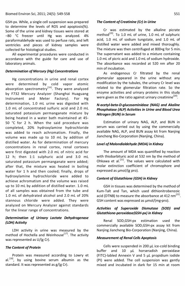
The Rate of Apoptosis in Kidney
Apoptosis was significantly augmented in HgCl2
Group, as compared with Control Group (Figure 1). PC or Lyc groups showed a marked decrease, compared with HgCl2 Group as well as Control Group.
Figure 1. The rate of apoptosis in kidney (n=4). ** Significant as compared with Control Group at P<0.01. ▲▲ Significant as compared with HgCl2 Group at P<0.01.

554 Biomed Environ Sci, 2011; 24(5): 549‐558
The Changes of ROS in Kidney
The data on ROS production were depicted in (Figure 2). As compared with Control Group, the positive rate of staining which indicated generation of the concentrations of intracellular ROS, increased
(63.11%) in HgCl2 treated rats, whereas ROS generation significantly decreased by 27.63% in PC treated rats. Administration with Lyc reduced the generation of the concentrations of intracellular ROS by 16.40% in Lyc Group as compared to HgCl2 Group.
Figure 2. The changes of ROS in kidney (n=4). Note. * Significant as compared with Control Group at P<0.05. ** Significant as compared with Control Group at P<0.01. ▲ Significant as compared with HgCl2 Group at P<0.05. ▲▲ Significant as compared with HgCl2 Group at P<0.01.
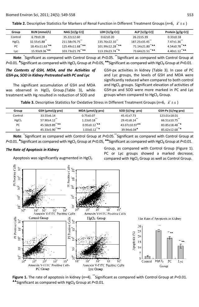
Effect of PC and Lyc on Kidney Histopathology
The protective effect of PC and Lyc on HgCl2 induced renal injury was apparent from histopathological examination. Figure 3 shows photomicrographs of haematoxylin–eosin (H and E) stained kidney tissues. In case of the control group, the kidney showed normal glomeruli, tubules and vessels. In the group that received HgCl2 alone, significant injury was evident. Glomeruli showed hyperemia, widened Bowman’s space. Hg caused cellular damage, desquamation of endotheliocyte and separation with basement membrane, as well as vanishment of nucleus. Cellular continuity was distorted, and cell debris was congested in the lumens of urinary cast. Stroma also showed congestion, vasodilatation. In rats which received pretreatment with PC and Lyc, significant
amelioration against HgCl2 induced injury was observed. Although the treatment did not fully prevent renal damage, there was less oedema and more congestion of stroma.
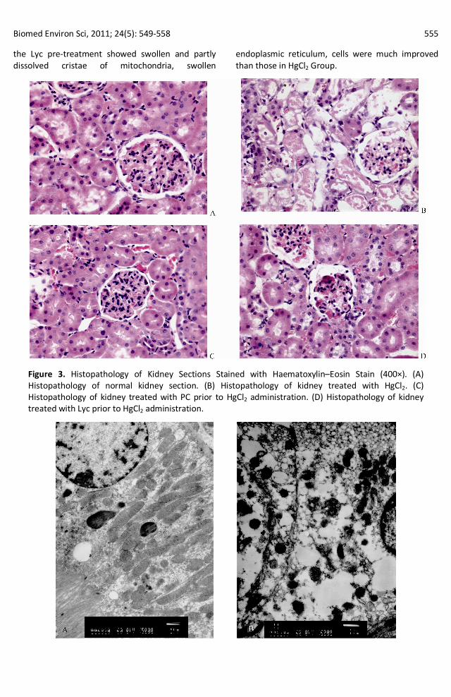
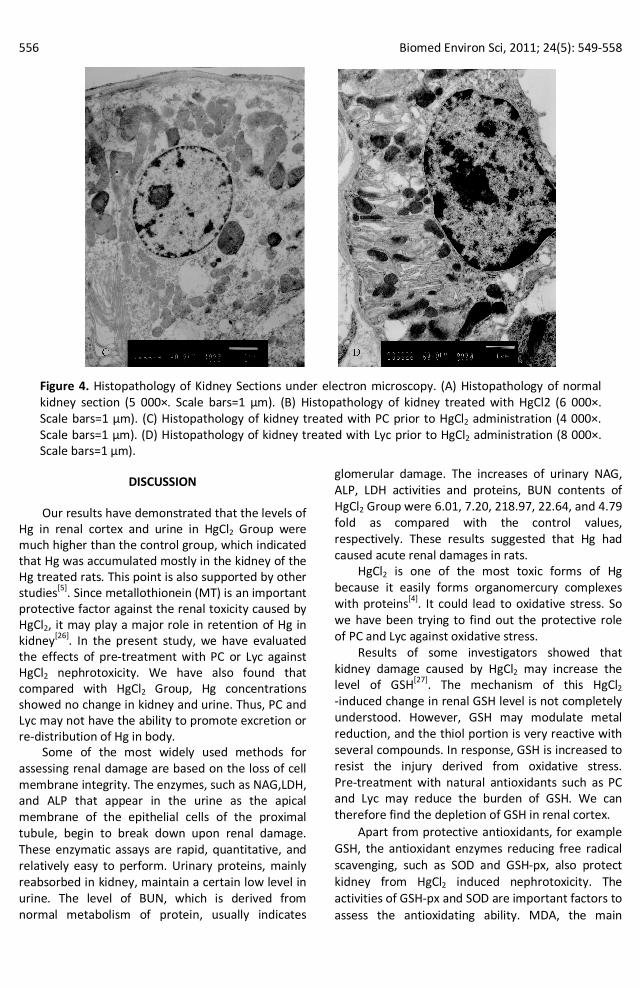
Electron microscopical observation showed that major changes occurred in the proximal convoluted tubules (Figure 4): Fragmentation or even disappearance of the brush border and disruption of cytoplasmic organelles were prominent. The damaged cells often contained swollen or dissolved mitochondria, swollen and degranulated endoplasmic reticulum, as well as lysis of lysosomes.. Electron microscopy showed considerable improvement of renal cells when PC was administered prior to the HgCl2 treatment. We observed better recovery as it almost reversed the changes observed in HgCl2 group, in spite of some dissolved mitochondria that we could still find in cells. While the ultrastructure of
Biomed Environ Sci, 2011; 24(5): 549‐558 555
the Lyc pre‐treatment showed swollen and partly dissolved cristae of mitochondria, swollen
endoplasmic reticulum, cells were much improved than those in HgCl2 Group.
Figure 3. Histopathology of Kidney Sections Stained with Haematoxylin–Eosin Stain (400×). (A) Histopathology of normal kidney section. (B) Histopathology of kidney treated with HgCl2. (C) Histopathology of kidney treated with PC prior to HgCl2 administration. (D) Histopathology of kidney treated with Lyc prior to HgCl2 administration.
556 Biomed Environ Sci, 2011; 24(5): 549‐558
Figure 4. Histopathology of Kidney Sections under electron microscopy. (A) Histopathology of normal kidney section (5 000×. Scale bars=1 µm). (B) Histopathology of kidney treated with HgCl2 (6 000×. Scale bars=1 µm). (C) Histopathology of kidney treated with PC prior to HgCl2 administration (4 000×. Scale bars=1 µm). (D) Histopathology of kidney treated with Lyc prior to HgCl2 administration (8 000×. Scale bars=1 µm).
DISCUSSION
Our results have demonstrated that the levels of Hg in renal cortex and urine in HgCl2 Group were much higher than the control group, which indicated that Hg was accumulated mostly in the kidney of the Hg treated rats. This point is also supported by other studies [5] . Since metallothionein (MT) is an important protective factor against the renal toxicity caused by HgCl2, it may play a major role in retention of Hg in kidney [26] . In the present study, we have evaluated the effects of pre‐treatment with PC or Lyc against HgCl2 nephrotoxicity. We have also found that compared with HgCl2 Group, Hg concentrations showed no change in kidney and urine. Thus, PC and Lyc may not have the ability to promote excretion or re‐distribution of Hg in body.
Some of the most widely used methods for assessing renal damage are based on the loss of cell membrane integrity. The enzymes, such as NAG,LDH, and ALP that appear in the urine as the apical membrane of the epithelial cells of the proximal tubule, begin to break down upon renal damage. These enzymatic assays are rapid, quantitative, and relatively easy to perform. Urinary proteins, mainly reabsorbed in kidney, maintain a certain low level in urine. The level of BUN, which is derived from normal metabolism of protein, usually indicates
glomerular damage. The increases of urinary NAG, ALP, LDH activities and proteins, BUN contents of HgCl2 Group were 6.01, 7.20, 218.97, 22.64, and 4.79 fold as compared with the control values, respectively. These results suggested that Hg had caused acute renal damages in rats.
HgCl2 is one of the most toxic forms of Hg because it easily forms organomercury complexes with proteins [4] . It could lead to oxidative stress. So we have been trying to find out the protective role of PC and Lyc against oxidative stress.
Results of some investigators showed that kidney damage caused by HgCl2 may increase the level of GSH [27] . The mechanism of this HgCl2 ‐induced change in renal GSH level is not completely understood. However, GSH may modulate metal reduction, and the thiol portion is very reactive with several compounds. In response, GSH is increased to resist the injury derived from oxidative stress. Pre‐treatment with natural antioxidants such as PC and Lyc may reduce the burden of GSH. We can therefore find the depletion of GSH in renal cortex.
Apart from protective antioxidants, for example GSH, the antioxidant enzymes reducing free radical scavenging, such as SOD and GSH‐px, also protect kidney from HgCl2 induced nephrotoxicity. The activities of GSH‐px and SOD are important factors to assess the antioxidating ability. MDA, the main
Biomed Environ Sci, 2011; 24(5): 549‐558 557
product of peroxidation in vivo, is the biomarker of the severity of lipid peroxidation injury. We have used these indicators to reflect oxidative stress induced by HgCl2 in kidney and the protective role of PC and Lyc against the stress.
A key issue that we have been trying to address in our ongoing research is to identify primary molecular mechanisms that are involved in the stages of Hg‐induced renal damage. Taken together, the results of the present studies strongly indicate that the accumulation of Hg in kidney can damage renal mitochondria and generate ROS. Oxidative stress refers to excessive generation of ROS. This is in accordance with other studies [3,27] . Excessive ROS and the resulting oxidative stress play a pivotal role in apoptosis and the kidney is therefore injured. Renal damages caused by HgCl2 are inferred from the morphological changes in renal sections. At the same time, HgCl2 imposes a loss of antioxidant potential in terms of SOD and GSH‐px and promotes GSH. In addition, MDA has also been proposed to be occurring in renal tissues following administration of HgCl2. As a result, the oxidation and antioxidation balance breaks down, leading to exaltation of the activities of ALP, LDH, and NAG, excretion of urinary proteins in urine of the rats, and increase of the contents of BUN in serum.
As a safe, novel and highly potent antioxidant, PC provides excellent protection against oxidative stress and free radical‐mediated tissue injury [28] . PC elicits antioxidant action by scavenging ROS, enhancing the cellular antioxidant enzyme SOD, GSH‐px, and reducing the burden of GSH in the cells. The rate of apoptosis is also decreased to resist oxidative damage [29] . The recovery of these enzymes is correlated with the recovery of the renal function. So, oxidative stress elicited by HgCl2 intoxication has been nultified due to the antioxidant activity following pre‐treatment with PC. These changes of biochemical parameters are well correlated with the renal histological results.
As an effective free radical scavenger, Lyc is the most efficient biological carotenoid, being 10‐fold, 47‐fold, and 100‐fold more effective in quenching singlet oxygen than a‐tocopherol, b‐carotene and vitamin E respectively [30‐32] . Lyc removes free radicals produced by Hg in physical and chemical ways, while improving the body's antioxidant enzymes such as SOD, GSH‐px to prevent the oxidative damage caused by mercuric chloride, enhancing the body antioxidant capacity, reducing the levels of lipid peroxidation and maintaining cell membrane permeability. As a result, compared with HgCl2 group,
the urine enzymes such as LDH, ALP, and NAG increased and the levels of urinary proteins and BUN in serum decreased. The rate of apoptosis is furthermore decreased to protect against Hg kidney damage. Tang et al. have shown that Lyc can restrain apoptosis by preventing the upregulation of the expression of p53 and caspase‐3 mRNA in endothelial cells [33] .
In conclusion, HgCl2 induced renal damage involves oxidative stress triggered by ROS generation .The findings from our present study demonstrate that PC and Lyc, with theirs potent antioxidant properties, seem to be a highly promising agent in protecting renal tissues against HgCl2‐induced renal damage.
ACKNOWLEDGEMENTS
The authors gratefully acknowledge ZHAO Ming and YU Miao from China Medical University for their technical assistance and CAO liang from Shenyang for her invaluable suggestions.
REFERENCES
1. Sharma MK, Sharma A, Kumar A, et al. Evaluation of protective efficacy of Spirulina fusiformis against mercury induced nephrotoxicity in Swiss albino mice. Food Chem Toxicol, 2007; 45(6), 879‐87.
2. Sener G, Sehirli O, Tozan A, et al. Ginkgo biloba extract protects against mercury (II)‐induced oxidative tissue damage in rats. Food Chem Toxicol, 2007; 45(4), 543‐50.
3. Mahboob M, Shireen KF, Atkinson A, et al. Lipid peroxidation and antioxidant enzyme activity in different organs of mice exposed to low level of mercury. J Environ Sci Health B, 2001; 36(5), 687‐97.
4. El‐Shenawy SM, Hassan NS. Comparative evaluation of the protective effect of selenium and garlic against liver and kidney damage induced by mercury chloride in the rats. Pharmacological Reports, 2008; 60(2), 199‐208.
5. Xu ZF, Yang JH, Yu JM, et al. Effects of BSO, GSH, Vit‐C and DMPS on the nephrotoxicity of mercury. Toxicology and Industrial Health, 2007; 23(7), 403‐10.
6. Ghosh A, Sil PC. A Protein from Cajanus indicus Spreng Protects Liver and Kidney against Mercuric Chloride‐Induced Oxidative Stress. Pharm Bull, 2008; 31(9), 1651‐8.
7. Preuss HG, Wallerstedt D, Talpur N, et al. Effects of niacinbound chromium and grape seed proanthocyanidin extract on the lipid profile of hypercholesterolemic subjects: a pilot study. J Med, 2000; 31(5‐6), 227‐46.
8. Ye X, Krohn RL, Liu W, et al. The cytotoxic effects of a novel IH636 grape seed proanthocyanidin extract on cultured human cancer cells. Mol Cell Biochem, 1999; 196(1‐2), 99‐108.
9. Sato M, Bagchi D, Tosaki A, et al. Grape seed proanthocyanidin reduces cardiomyocyte apoptosis by inhibiting ischemia/ reperfusion‐induced activation of JNK‐1 and C‐JUN. Free Radic Biol Med, 2001; 31(6), 729‐37.
10.Khanna S, Roy S, Bagchi D, et al. Upregulation of oxidant‐induced VEGF expression in cultured keratinocytes by a grape seed proanthocyanidin extract. Free Radic Biol Med,
558 Biomed Environ Sci, 2011; 24(5): 549‐558
2001; 31(1), 38‐42. 11.Wren AF, Cleary M, Frantz C, et al. 90‐day oral toxicity study of
a grape seed extract (IH636) in rats. J Agric Food Chem, 2002; 50(7), 2180‐92.
12.Velmurugan B, Santhiya ST, Nagini S. Protective effect of S‐allylcysteine and lycopene in combination against N‐methyl‐N ‐nitro‐N‐nitrosoguanidine‐induced genotoxicity. Pol J Pharma, 2004; 56(2), 241‐5
13.Tapiero H, Townsend DM, Tew KD. The role of carotenoids in the prevention of human pathologies. Biomed. Pharmacother, 2004; 58(2), 100‐10.
14.Jonker D, Kuper CF, Frail N, et al. Ninety‐day oral toxicity study of lycopene from Blakeslea trispora in rats. Regul Toxico. Phamacol, 2003; 37(3), 396‐406.
15.Michael McClain R, Bausch I. Summary of safety studies conducted with synthetic lycopene. Regul Toxicol Pharmacol, 2003; 37(2), 274‐85.
16.Adair B M, Cobb G P. Improved preparation of small biological samples for mercury analysis using cold vapor atomic absorption spectroscopy. Chemosphere, 1999; 38(12), 2951‐8.
17.Hochella NJ, Weinhouse S. Automated assay of lactate dehydrogenase in urine. Anal Biochem ,1965; 13(2), 322‐35.
18.Lowry DH, Rosebrough NJ, Farr AL, et al. Protein measurement with the folin‐phenol reagent. Biol Chem, 1951; 193(1), 265‐75.
19.Hare RS. Endogenous creatinine in serum and urine. Proc Soc Exp Bio Med, 1950; 74(1), 148‐51.
20.Toshio H, Toshio Y. A Method for Determining Urinary Enzyme Activities as Nephrotoxic Indicators in Rats. Jpn J Pharmacol, 1990; 54(2), 205‐15.
21.Ohkawa H, Ohishi N, Yagi K. Assay for lipid peroxides in animal tissues by thiobarbituric acid reaction. Anal Biochem, 1979; 95(2), 351‐8.
22.Kum‐Talt L, Tan IK. A new colorimetric method for the determination of glutathione in erythrocytes. Clin Chem Acta, 1974; 53(2), 153‐61.
23.Li CR, Zhou Z, Zhu D, et al. Protective effect of paeoniflorin on irradiation‐induced cell damage involved in modulation of reactive oxygen species and the mitogen‐activated protein kinases. Int J Biochem Cell Bio, 2007; 39(2), 426‐38.
24.Wang H, Joseph JA. Quantifying cellular oxidative stress bydichlorofluor‐escein assay using microplate reader. Free Radic Biol Med, 1999; 27(5‐6), 612‐6.
25.Satoh M, Nishimura N, Kanayama Y, et al. Enhanced renal toxicity by inorganic mercury in metallothionein‐null Mice. J Pharmacol Exp Ther, 1997; 283(3), 1529‐933.
26.Schurz F, Sabater‐Vilar M, Fink‐Gremmels J. Mutagenicity of mercury chloride and mechanisms of cellular defense: the role of metal‐binding proteins. Mutagenesis, 2000; 15(6), 525‐30.
27.Su L, Wang M, Yin ST, et al. The interaction of selenium and mercury in the accumulations and oxidative stress of rat tissues. Ecotoxicol Environ Saf, 2008; 70(3), 483‐9.
28.Bagchi D, Bagchi M, Stohs SJ, et al. Free radicals and grape seed proanthocyanidin extract: importance in human health and disease prevention. Toxicology, 2000; 148(2‐3), 187‐97.
29.Li L. Zhong J. Effect of grape procyanidins on the apoptosis and mitochondrial transmembrane potential of thymus cells. WeiSheng YanJiu, 2004;33(2), 191‐4. (In Chinese)
30.Sandhir R, Mehrotra A, Kamboj SS. Lycopene prevents 3‐nitropropionic acid‐induced mitochondrial oxidative stress and dysfunctions in nervous system. Neurochem Int, 2010; 57(5), 579‐87.
31.Stahl W, Sies H. Carotenoids and protection against solar UV radiation. Skin Pharmacol Appl Skin Physiol, 2002; 15(5), 291‐6.
32.Breinholt V, Lauridsen ST, Daneshvar B, et al. Dose ‐ response effects of lycopene on selected drug ‐ metabolizing and antioxidant enzymes in the rat. Cancer Lett, 2000; 154 (2), 201‐10.
33.Tang X, Yang X, Peng Y, et al. Protective effects of lycopene against H2O2‐induced oxidative injury and apoptosis in human endothelial cells. Cardiovasc Drugs Ther, 2009; 23(6), 439‐48.