
The primary cilium functions as a mechanical and calcium ...
13
RESEARCH Open Access The primary cilium functions as a mechanical and calcium signaling nexus Kristen L Lee 1 , Marie D Guevarra 1 , An M Nguyen 1,2 , Mardonn C Chua 1,3 , Yingxiao Wang 4 and Christopher R Jacobs 1* Abstract Background: The primary cilium is an antenna-like, nonmotile structure that extends from the surface of most mammalian cell types and is critical for chemosensing and mechanosensing in a variety of tissues including cartilage, bone, and kidney. Flow-induced intracellular calcium ion (Ca 2+ ) increases in kidney epithelia depend on primary cilia and primary cilium-localized Ca 2+ -permeable channels polycystin-2 (PC2) and transient receptor potential vanilloid 4 (TRPV4). While primary cilia have been implicated in osteocyte mechanotransduction, the molecular mechanism that mediates this process is not fully understood. We directed a fluorescence resonance energy transfer (FRET)-based Ca 2+ biosensor to the cilium by fusing the biosensor sequence to the sequence of the primary cilium-specific protein Arl13b. Using this tool, we investigated the role of several Ca 2+ -permeable channels that may mediate flow-induced Ca 2+ entry: PC2, TRPV4, and PIEZO1. Results: Here, we report the first measurements of Ca 2+ signaling within osteocyte primary cilia using a FRET-based biosensor fused to ARL13B. We show that fluid flow induces Ca 2+ increases in osteocyte primary cilia which depend on both intracellular Ca 2+ release and extracellular Ca 2+ entry. Using siRNA-mediated knockdowns, we demonstrate that TRPV4, but not PC2 or PIEZO1, mediates flow-induced ciliary Ca 2+ increases and loading-induced Cox-2 mRNA increases, an osteogenic response. Conclusions: In this study, we show that the primary cilium forms a Ca 2+ microdomain dependent on Ca 2+ entry through TRPV4. These results demonstrate that the mechanism of mechanotransduction mediated by primary cilia varies in different tissue contexts. Additionally, we anticipate that this work is a starting point for more studies investigating the role of TRPV4 in mechanotransduction. Keywords: Mechanotransduction, Primary cilium, Calcium signaling, Osteocyte, Kidney epithelia, Biosensor Background Mechanotransduction is a process by which cells sense and convert mechanical signals into biochemical and transcriptional changes. The calcium ion (Ca 2+ ) is a ubi- quitous second messenger that regulates numerous sig- naling pathways. Despite how universal Ca 2+ is, discrete intracellular signaling mechanisms occur because Ca 2+ gradients are spatiotemporal and do not comprise one general pool that changes uniformly. For example, there are discrete microdomain Ca 2+ signals including “Ca 2+ sparks,”“Ca 2+ sparklets,” and “scraps” that modulate constriction and relaxation in vascular smooth muscle cells [1,2]. Mechanical loading generates rapid and tem- poral intracellular Ca 2+ increases in many cell types in- cluding osteocytes, osteoblasts, neurons, and kidney cells. Ca 2+ mobilization is required for flow-induced prostaglandin E2 (PGE 2 ) release and flow-induced osteo- pontin gene regulation in osteocytes, demonstrating that Ca 2+ is upstream of mechanotransduction activities and paracrine signaling [3,4]. The primary cilium is an antenna-like, nonmotile structure that extends from the surface of most mamma- lian cell types into the extracellular space [5,6]. While initially considered a vestigial structure, in the past dec- ade, several labs have demonstrated that primary cilia are critical for chemosensing and mechanosensing in a variety of tissues including cartilage, bone, and kidney * Correspondence: [email protected] 1 Department of Biomedical Engineering, Columbia University, 351 Engineering Terrace, MC 8904, 1210 Amsterdam Ave, New York, NY 10027, USA Full list of author information is available at the end of the article © 2015 Lee et al.; licensee BioMed Central. This is an Open Access article distributed under the terms of the Creative Commons Attribution License (http://creativecommons.org/licenses/by/4.0), which permits unrestricted use, distribution, and reproduction in any medium, provided the original work is properly credited. The Creative Commons Public Domain Dedication waiver (http://creativecommons.org/publicdomain/zero/1.0/) applies to the data made available in this article, unless otherwise stated. Lee et al. Cilia (2015) 4:7 DOI 10.1186/s13630-015-0016-y
Transcript of The primary cilium functions as a mechanical and calcium ...

Lee et al. Cilia (2015) 4:7 DOI 10.1186/s13630-015-0016-y
RESEARCH Open Access
The primary cilium functions as a mechanical andcalcium signaling nexusKristen L Lee1, Marie D Guevarra1, An M Nguyen1,2, Mardonn C Chua1,3, Yingxiao Wang4
and Christopher R Jacobs1*
Abstract
Background: The primary cilium is an antenna-like, nonmotile structure that extends from the surface of mostmammalian cell types and is critical for chemosensing and mechanosensing in a variety of tissues includingcartilage, bone, and kidney. Flow-induced intracellular calcium ion (Ca2+) increases in kidney epithelia dependon primary cilia and primary cilium-localized Ca2+-permeable channels polycystin-2 (PC2) and transient receptorpotential vanilloid 4 (TRPV4). While primary cilia have been implicated in osteocyte mechanotransduction, themolecular mechanism that mediates this process is not fully understood. We directed a fluorescence resonanceenergy transfer (FRET)-based Ca2+ biosensor to the cilium by fusing the biosensor sequence to the sequence ofthe primary cilium-specific protein Arl13b. Using this tool, we investigated the role of several Ca2+-permeablechannels that may mediate flow-induced Ca2+ entry: PC2, TRPV4, and PIEZO1.
Results: Here, we report the first measurements of Ca2+ signaling within osteocyte primary cilia using a FRET-basedbiosensor fused to ARL13B. We show that fluid flow induces Ca2+ increases in osteocyte primary cilia which dependon both intracellular Ca2+ release and extracellular Ca2+ entry. Using siRNA-mediated knockdowns, we demonstratethat TRPV4, but not PC2 or PIEZO1, mediates flow-induced ciliary Ca2+ increases and loading-induced Cox-2 mRNAincreases, an osteogenic response.
Conclusions: In this study, we show that the primary cilium forms a Ca2+ microdomain dependent on Ca2+ entrythrough TRPV4. These results demonstrate that the mechanism of mechanotransduction mediated by primary ciliavaries in different tissue contexts. Additionally, we anticipate that this work is a starting point for more studiesinvestigating the role of TRPV4 in mechanotransduction.
Keywords: Mechanotransduction, Primary cilium, Calcium signaling, Osteocyte, Kidney epithelia, Biosensor
BackgroundMechanotransduction is a process by which cells senseand convert mechanical signals into biochemical andtranscriptional changes. The calcium ion (Ca2+) is a ubi-quitous second messenger that regulates numerous sig-naling pathways. Despite how universal Ca2+ is, discreteintracellular signaling mechanisms occur because Ca2+
gradients are spatiotemporal and do not comprise onegeneral pool that changes uniformly. For example, thereare discrete microdomain Ca2+ signals including “Ca2+
sparks,” “Ca2+ sparklets,” and “scraps” that modulate
* Correspondence: [email protected] of Biomedical Engineering, Columbia University, 351Engineering Terrace, MC 8904, 1210 Amsterdam Ave, New York, NY 10027,USAFull list of author information is available at the end of the article
© 2015 Lee et al.; licensee BioMed Central. ThCommons Attribution License (http://creativecreproduction in any medium, provided the orDedication waiver (http://creativecommons.orunless otherwise stated.
constriction and relaxation in vascular smooth musclecells [1,2]. Mechanical loading generates rapid and tem-poral intracellular Ca2+ increases in many cell types in-cluding osteocytes, osteoblasts, neurons, and kidneycells. Ca2+ mobilization is required for flow-inducedprostaglandin E2 (PGE2) release and flow-induced osteo-pontin gene regulation in osteocytes, demonstrating thatCa2+ is upstream of mechanotransduction activities andparacrine signaling [3,4].The primary cilium is an antenna-like, nonmotile
structure that extends from the surface of most mamma-lian cell types into the extracellular space [5,6]. Whileinitially considered a vestigial structure, in the past dec-ade, several labs have demonstrated that primary ciliaare critical for chemosensing and mechanosensing in avariety of tissues including cartilage, bone, and kidney
is is an Open Access article distributed under the terms of the Creativeommons.org/licenses/by/4.0), which permits unrestricted use, distribution, andiginal work is properly credited. The Creative Commons Public Domaing/publicdomain/zero/1.0/) applies to the data made available in this article,

Lee et al. Cilia (2015) 4:7 Page 2 of 13
[7-13]. The osteocyte primary cilium deflects with mech-anical stimulation and mediates mechanotransduction atthe transcriptional level; however, previous experimentshave not resolved Ca2+ within the primary cilium fromthe cytosol [14].Recent advances in monitoring ciliary Ca2+ mobilization
have improved our understanding of the primary cilium-mediated mechanism of mechanotransduction in kidneyepithelia. In the past, traditional diffusive BAPTA-basedfluorescent indicator dyes were used to measure intracel-lular Ca2+ levels but did not target specific subcellular do-mains. In some pivotal studies, Praetorious and Springand Nauli et al. demonstrated that primary cilia are re-quired for mechanically induced Ca2+ increases in kidneyepithelial cells [12,13,15]. The dependence of flow-inducedCa2+ increases on kidney epithelia primary cilia and thepresence of mechanosensitive Ca2+-permeable channelson the ciliary membrane suggest that mechanical loadingopens stretch-activated ion channels on the primarycilium that mediate Ca2+ entry. In the last couple of years,Delling et al., Su et al., and Jin et al. directed geneticallyencoded single fluorescence Ca2+ biosensors to the pri-mary cilium using a variety of ciliary targeting sequencesin human retina pigmented epithelia and kidney epithelialcells [16-18]. Su et al. and Jin et al. exposed kidney epithe-lial cells to fluid flow, which bent primary cilia and in-creased ciliary and cytosolic Ca2+ levels [17,18]. The Ca2+-permeable channel polycystin-2 (PC2) associates withthe mechanosensitive protein polycystin-1 and localizes tothe primary cilium. Jin et al. reported that flow-inducedCa2+ elevations occur first in the primary cilium and arefollowed by cytosolic Ca2+ mobilization. Both ciliary andcytosolic Ca2+ increases were dependent on PC2 [18]. Fur-thermore, blocking ryanodine receptors inhibited cytosolicCa2+ increases without affecting the flow-induced ciliaryCa2+ response [18,19]. Collectively, these recent flow stud-ies on kidney epithelia primary cilia demonstrate that fluidflow activates PC2 through which extracellular Ca2+ entersand triggers ryanodine receptors in Ca2+-induced Ca2+
release.Current knowledge of the osteocyte primary cilium-
mediated mechanism of mechanotransduction is rela-tively poor compared with recent progress in kidneyepithelia primary cilium mechanotransduction research.Our group previously used the fluorescent dye Fura 2-AMto demonstrate that flow-induced Ca2+ increases in MLO-Y4 osteocyte-like cells are independent of primary cilia andstretch-activated channels, which is different from kidneycells [14]. While these results suggest that the osteocyte pri-mary cilium-regulated mechanism of mechanotransductionis not linked to intracellular Ca2+ levels, it is unknown ifthe local primary cilium Ca2+ environment is distinct fromthe cytosol. We hypothesized that the osteocyte primarycilium mediates mechanotransduction by forming a distinct
Ca2+ microdomain. Therefore, the objective of this studywas to monitor flow-induced ciliary Ca2+ levels and eluci-date the intricate role of the osteocyte primary cilium as abiochemical and mechanical signaling nexus.In this study, we directed a fluorescence resonance en-
ergy transfer (FRET)-based Ca2+ biosensor to the pri-mary cilium by fusing a biosensor sequence to thesequence of the primary cilium-specific protein Arl13b.The modified YC3.6 Ca2+-sensitive FRET-based biosen-sor with the ECFP-YPet donor-acceptor pair contains acalmodulin (CaM) region with four Ca2+-binding do-mains [20]. Binding of Ca2+ results in a conformationalchange that increases FRET signal, which is character-ized by decreased ECFP and increased YPet fluorescence[21]. YPet as an acceptor produces the largest FRETdynamic range in live mammalian cells compared toCitrine, Venus, or cpVenus [20]. Using a targeted versionof a FRET-based Ca2+ biosensor containing ECFP andYPet and the diffusive Ca2+ indicator dye Fura Red, wedetected ciliary and cytosolic Ca2+ increases within indi-vidual MLO-Y4 cells exposed to fluid flow stimulation.Additionally, we examined the role of several Ca2+-per-meable channels on the primary cilium: PC2, transientreceptor potential vanilloid 4 (TRPV4), and PIEZO1.Coste et al. recently characterized mechanically activatedcurrent in neuroblastoma cells and proposed that themultipass transmembrane ion channels PIEZO1 andPIEZO2 mediate mechanically activated cation activity,leading us to explore the role of PIEZO channels in223osteocyte mechanotransduction [22]. Our data demon-strate that TRPV4, but not PC2 or PIEZO1, mediatesflow-induced ciliary Ca2+ increases and a loading-inducedosteogenic response at the transcriptional level. Collect-ively, our study demonstrates that the osteocyte primarycilium microdomain is distinct from the cytosol and thatsources of loading-induced ciliary Ca2+ mobilization aredifferent in kidney epithelia and osteocytes. These are thefirst measurements of Ca2+ signaling within the osteocyteprimary cilium, and we anticipate this work is a startingpoint for more studies investigating the role of TRPV4 inosteocyte mechanotransduction [23].
MethodsPlasmid constructionDrs. Yingxiao Peter Wang and Mingxing Ouyang previ-ously developed a calcium-sensitive FRET-based biosen-sor composed of an ECFP donor, calmodulin region,M13 calmodulin-binding region, and YPet acceptor(CaB). Drs. Kenji Kontani and Kristen Verhey generouslyshared with us the Arl13b gene. We fused Arl13b to theN terminus of CaB using a 15-amino-acid-long flexiblelinker to form Arl13b-L-CaB (ALC)[24]. Deletions ofTrp3 and Phe17 in the M13 region were performed toblock changes in FRET during Ca2+ increases, serving as

Lee et al. Cilia (2015) 4:7 Page 3 of 13
negative controls of CaB and ALC (mutCaB andmutALC) [25].
Cell culture and transfectionMLO-Y4 osteocyte-like cells (a gift from Dr. LyndaBonewald of the University of Missouri-Kansas City)were cultured in MEM alpha (Life Technologies) with5% FBS, 5% CS, and 1% PS at 37°C in 5% CO2. Usingthe BTX 360 with a 300-V, 100-Ω, 1,000-μF pulse, 1.25million cells were transfected with 10 μg plasmid byelectroporation. Cells were co-transfected with the ALCplasmid and 0.5 nmol siRNA. siRNA sequences includedPkd2 (5′-CCUCUUGGCAGUUUCAGCCUGUAAA-3′),Trpv4 (5′-GAUGGACUGCUCUCCUUCUU GUUGA-3′),and Piezo1 (5′-CACCGGCAUCUACGUCAAAUA-3′) [22].Transfected MLO-Y4 cells were seeded onto collagenI-coated glass slides (#1.5 glass, Warner Instruments) ata density of 4,000 cells/cm2 and cultured for 3 days inreduced serum containing 2.5% FBS and 2.5% CS. IMCDcells (purchased from ATCC®, CRL-2123™) were culturedin DMEM (Life Technologies) with 10% FBS and 1% PS.Transfected IMCD cells were seeded onto fibronectin-coated glass slides at a density of 8,000 cells/cm2 and cul-tured for 3 days in 1% FBS. Prior to imaging, cells wereincubated in 20 μM Fura Red-AM (Life Technologies,F-3021) with 0.1% Pluronic® F-127 (20% solution inDMSO) (Life Technologies) for 1 h at room temperatureto label cytosolic Ca2+.
Imaging flow chamberSlides were placed in the RC-30 Confocal ImagingChamber (Warner Instruments) and attached to a syr-inge containing phenol red-free alpha MEM with 1%FBS and 1% CS for MLO-Y4 cells or phenol red-freeDMEM with 1% FBS and 1% PS for IMCD cells. MLO-Y4 cells were stimulated with oscillatory fluid flowresulting in a 10-dyn/cm2 shear stress, and IMCD cellswere stimulated with steady fluid flow resulting in a 1-dyn/cm2 shear stress, both within physiologic range foreach cell type. Thapsigargin was added to the imagingmedia at a final concentration of 10 μM for appropriatesamples for a 20-min rest period and maintained at thesame concentration during flow. A separate syringe with10 μM ionomycin was connected to the chamber to ver-ify cell viability after the initial flow stimulus.
Ca2+ imaging in cilia and cytosolOne fluorescing cell per slide was selected at random forimaging. The selected cilium was focused on, with thecell body captured within the field of view on anOlympus IX71 inverted epifluorescence microscope witha 1.30 N.A. ×40 oil immersion objective. Donor excita-tion was achieved on a xenon lamp using a 430/24-nmfilter while images were collected simultaneously using a
Quad-View system (QV2, Photometrics) with emissionfilters of 470/28, 530/30, and 641/75 nm. Cells were leftto rest for 20 min prior to imaging. For calibration stud-ies, we added ionomycin at a final concentration of5 μM and CaCl2 at a final concentration of 0.1, 0.25, and0.5 mM. Baseline signal was recorded for 30 s followedby 5 min of flow. Images were taken at 4 Hz with a 150-ms exposure.
Image analysisImages of each sample were trimmed to regions of interestthat tightly enclosed the primary cilium or cell duringimaging. Using MATLAB, images were corrected for back-ground and bleedthrough and a pixel-by-pixel basis, result-ing in signal only in the cilium and cytosol. Specifically,using MetaMorph, the same background region for ECFPand YPet images was selected from the untrimmed imagefar from the sample within the field of view, and bleed-through coefficients were calculated prior to performingexperiments [26]. To aid in peak detection, the averageintensity data of a region of interest over time was smooth-ened using a 1D Savitsky-Golay filter with a window widthof 31, and a fourth-degree smoothing polynomial. Oscilla-tion amplitude was identified by finding local maxima withMATLAB. A baseline value for each sample was deter-mined by averaging the baseline signal collected 30 s priorto flow. The maximum baseline oscillation amplitude wasdefined as maximum oscillation amplitude that occurredduring the recording of the baseline signal, 30 s prior toflow. The Ca2+ peak height was defined as the peak ampli-tude that occurred during flow exposure. Multiple Ca2+
peaks may occur during the 5-min period of flow. A sam-ple was considered responsive if a Ca2+ peak height wasequal to or greater than 1.5 times the maximum baselineoscillation amplitude [27,28]. Not all samples were ana-lyzed due to movement of the sample out of plane.
AntibodiesWe used the following primary antibodies: rabbit anti-polycystin-2 (Santa Cruz, sc-25749), rabbit anti-sera toTRPV4 (generously provided by Heller group), rabbit anti-PIEZO1 (Novus NBP1-78537 for immunostaining andNBP2-10504 for Western blot), mouse anti-acetylatedalpha tubulin (Abcam, ab24610), and mouse anti-actin(Abcam, ab11003). We used the following secondary anti-bodies: Alexa Fluor 488 goat anti-rabbit IgG (Life Tech-nologies, A11008), Alexa Fluor 568 goat anti-mouse IgG(Life Technologies, A11031), goat anti-rabbit IgG-HRP(Santa Cruz, sc-2004), and HRP goat anti-mouse Ig (BDBiosciences, 554002).
Immunocytochemistry and confocal microscopyMLO-Y4 and IMCD cells were seeded on 35-mm glass-bottom dishes at approximately 1,000 and 2,000 cells/

Lee et al. Cilia (2015) 4:7 Page 4 of 13
cm2, respectively. Upon reaching 80%–90% confluenceafter 2 days of culture, cells were fixed with 10% forma-lin, permeabilized with 0.1% Triton X-100, and blockedwith 10% goat serum and 1% BSA in PBS. Cells were la-beled with primary antibodies for PC2, TRPV4, PIEZO1,and acetylated alpha tubulin followed by incubation inappropriate Alexa Fluor-labeled secondary antibodies.Nucleic stain was achieved using DAPI (0.5 mg/mL;1:100). Confocal z-stack images were obtained on a LeicaSP5 using a 1.46 N.A. ×100 oil immersion objective.Maximum X-Z projections were constructed with Leicasoftware.
Western blotCells transfected with Pkd2-, Trpv4-, Piezo1-, and MedGC-scrambled siRNA (Life Technologies) were lysed in RIPAbuffer (Santa Cruz, sc-24948) supplemented with sodiumorthovanadate, PMSF, and protease inhibitor cocktail 3 dayspost electroporation. Five micrograms of each protein sam-ple was run through NuPAGE® Novex® 4%–12% Bis-Trisgels (Life Technologies). After electrophoresis, proteinswere transferred to Invitrolon™ PVDF membranes (LifeTechnologies). The membranes were cut in half to separ-ately label actin bands. Membranes for PC2, PIEZO1, andactin were blocked with 5% BSA (Sigma-Aldrich), andmembranes for TRPV4 were blocked with 5% nonfat milk.HRP-conjugated antibodies were detected with chemilu-minescence (Clarity Western ECL Substrate, Bio-Rad) on aFujifilm LAS-4000 biomolecular imager.
Flow chamberTransfected MLO-Y4 cells were seeded onto collagen I-coated glass slides at approximately 5,000 cells/cm2 andcultured for 3 days. Slides were placed in larger, custom-made parallel-plate flow chambers (56 × 24 × 0.28 mm)so that sufficient amounts of RNA are isolated for geneexpression analysis. Chambers with slides were incu-bated for 30 min and then exposed to 5 min of oscilla-tory fluid flow (OFF) at 1 Hz with a peak shear stress of10 dyn/cm2. Slides were removed from chambers after55 min, and RNA was isolated immediately.
Quantitative real-time RT-PCRRNA was extracted from cells using TriReagent(Sigma-Aldrich) and isolated, followed by cDNA syn-thesis using TaqMan reverse transcriptase (AppliedBiosystems). cDNA samples were amplified with Trpv4(Mm00499025_m1), Pkd2 (Mm00435829_m1), Piezo1(Mm01241570_g1), Piezo2 (Mm01262433_m1), Cox-2(Mm00478374_m1), and Gapdh (4352339E) primersand probes (Applied Biosystems) by quantitative real-time RT-PCR using the ABI PRISM 7900 detectionsystem (Applied Biosystems). Samples and standardswere run in triplicate and were normalized to the
endogenous Gapdh expression. Relative gene levels be-tween samples were determined using the relativestandard curve method (ABI Prism 7700 User Bulletin2; Applied Biosystems).
Statistical analysisResults are shown as mean ± SEM. Unpaired t-tests(two-tailed) were used to analyze differences betweentreated and untreated groups. Comparisons of multiplegroups were performed using one-way ANOVA followedby Dunnett’s multiple comparison post hoc test orBonferroni’s multiple comparison post hoc test for Cox-2 mRNA level comparisons. For all tests, p < 0.05 wasconsidered significant.
ResultsArl13b-linker-Ca2+ biosensor detects ciliary Ca2+
For this study, we developed a novel primary cilium-localized, fully ratiometric biosensor using the modifiedYC3.6 Ca2+-sensitive FRET-based biosensor (CaB) con-taining ECFP and YPet fused to ARL13B. First, we estab-lished that ARL13B localizes to primary cilia in MLO-Y4osteocyte-like cells and verified ARL13B localization toIMCD primary cilia (Additional file 1: Figure S1A and B)[29]. Our biosensor design consisted of Arl13b at the Nterminus followed by a 15-amino-acid-long flexiblelinker, ECFP, calmodulin, M13 calmodulin-binding re-gion, and YPet at the C terminus (Figure 1A) [20,24].Addition of Ca2+ leads to increased FRET signal, andtransfecting Arl13b-linker-CaB (ALC) enabled us to de-tect Ca2+ levels within the primary cilium separate fromthe cytosol. The addition of 5 μM ionomycin, a Ca2+
ionophore, in media containing 1.8 mM Ca2+ led to de-tectable increases in FRET (represented by the emissionratio of YPet:ECFP fluorescence intensity) (Figure 1B,Additional file 2: Video S1). Furthermore, the FRETsignal increased at a slower rate and with delay tosmaller concentrations of calcium chloride (CaCl2)added with ionomycin in Ca2+-free media comparedwith higher concentrations of CaCl2 (Figure 1C). As ex-pected with a Kd = 250 nM, the biosensor activity even-tually reaches saturation for all CaCl2 concentrations.Thus, we determined that ALC is sensitive to differentlevels of ciliary Ca2+.
Mechanical stimulation of MLO-Y4 cells leads to ciliaryand cytosolic Ca2+ mobilizationWe preferentially imaged cells with primary cilia havinga vertical component prior to flow which typically bentin the direction of flow (Figure 1D). We loaded the Ca2+
indicator dye Fura Red into cells transfected with ALCto monitor cytosolic Ca2+ levels. Use of a PhotometricsQuad-View beam splitter allowed us to collect imagesthrough several emission filters, and we monitored

Figure 1 (See legend on next page.)
Lee et al. Cilia (2015) 4:7 Page 5 of 13
(See figure on previous page.)Figure 1 ALC localizes to the primary cilium and detects changes in ciliary Ca2+ levels. (A) Schematic of primary cilium-localized biosensor, ALC.(B) ALC FRET signal increases when ionomycin is added to media. FRET signal is reported on a pixel-by-pixel basis. Scale bars, 5 μm. (C) Traces ofbaseline-normalized FRET signal over time with different [CaCl2]. (D) YPet fluorescence images of a vertical primary cilium which bent during flow.Scale bars, 10 μm. (E,F) Individual ciliary and cytosolic Ca2+ measurements during oscillatory fluid flow starting at t = 0 s. (G,H) Percent of viableMLO-Y4 primary cilia and cytosolic domains exhibiting flow-induced Ca2+ increases (n = 15–21). (I,J) Flow-induced Ca2+ increases normalized tobaseline value in the primary cilium and cytosol with untreated or thapsigargin-treated media (untreated (n = 17 or 18), thapsigargin (n = 6)). (K,L)Average peak response over baseline value for cilia and cytosolic domains with thapsigargin treatment for all viable cells (untreated (n = 17 or 18),thapsigargin (n = 6)). Error bars show ±SEM (*p < 0.05).
Lee et al. Cilia (2015) 4:7 Page 6 of 13
ECFP, YPet, and Fura Red signals simultaneously [30].Application of 1 Hz OFF resulting in a 10-dyn/cm2 sur-face shear stress led to ciliary and cytosolic Ca2+ peakswithin approximately 8.2 ± 0.8 and 8.2 ± 0.6 s, respect-ively (Figure 1E, n = 17 or 18). On an individual cellbasis, 57% of Ca2+ peaks occurred in the primary ciliumprior to the cytosol of the same cell. We also stimulatedcells transfected with the inactive biosensor mutALC toverify that the observed peaks during 10 dyn/cm2 OFFwere indeed due to increased Ca2+ levels (Additional file3: Figure S2). Next, cells were treated with thapsigargin,which depletes intracellular Ca2+ stores (n = 6). As ex-pected, we found that Ca2+ release from internal storesis a substantial source of flow-induced Ca2+ mobilizationin the cytosol. Under thapsigargin treatment, there was a45% decrease in responsive samples and a 25% magni-tude reduction averaged from those fewer responsivesamples in response to flow (Figure 1G, H). Additionally,under thapsigargin treatment, the peak mean signal fromall viable samples in the group was significantly reducedin the cytosol but not in the primary cilium (Figure 1K,L). We continued to observe flow-induced ciliary Ca2+
peaks, although there were significant decreases in re-sponsiveness and peak magnitude normalized to baselinevalue (Figure 1G–J). These results imply that Ca2+-per-meable channels on the primary cilium also mediateflow-induced Ca2+ entry.
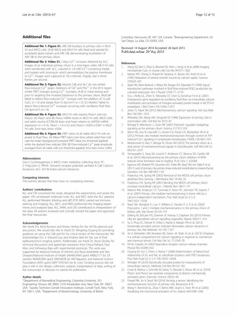
PC2, TRPV4, and PIEZO1 are Ca2+-permeable channels onthe primary ciliumWe examined three Ca2+-permeable channels localizedto the osteocyte primary cilium: PC2, TRPV4, andPIEZO1. PC2 and TRPV4, among other channels,localize to primary cilia and mediate flow-induced Ca2+
increases in kidney epithelia [15,31]. Coste et al. re-ported that mechanically activated ion channels PIEZO1and PIEZO2 are expressed in several murine tissues in-cluding kidney and have relatively lower expression inbrain and heart tissues [22]. We observed Piezo1 mRNAexpression in MLO-Y4 and IMCD cells and Piezo2mRNA expression only in IMCDs (Additional file 4:Figure S3). We immunostained MLO-Y4 cells for PC2,TPRV4, and PIEZO1 and found that all three channelsare present in both the primary cilium and plasma
membrane (Figure 2A–C). siRNA transfection reducedprotein and mRNA levels compared with scrambledsiRNA controls (all groups (n = 4–10) vs. control mRNAlevels (n = 10), p < 0.005)( Figure 2D, E).
TRPV4 mediates flow-induced ciliary Ca2+ increases inosteocytesPC2 mediates mechanically induced ciliary and cytosolicCa2+ signaling in kidney epithelia, and here, our datasuggest that osteocyte mechanotransduction is independ-ent of PC2. Following co-transfection of ALC and eitherPkd2, Trpv4, or Piezo1 siRNA, MLO-Y4 cells were loadedwith Fura Red and exposed to oscillatory flow stimulation(n = 14–16). Upon an initial general analysis of primarycilia from all viable cells (ionomycin-responsive), neitherthapsigargin nor any of the siRNA-mediated knockdownsof Pkd2, Trpv4, or Piezo1 exhibited a significant differencein flow-induced Ca2+ peak amplitude relative to untreatedsamples. Similarly, the only significant difference in peakamplitude relative to untreated samples occurred in thecytosol of cells treated with thapsigargin (Additional file 5:Figure S4). Using the spike threshold of 1.5× baseline,there was no significant difference in the percentage ofcells exhibiting a Ca2+ response in the primary cilium andcytosol with Pkd2,Trpv4, and Piezo1 knockdown. To illus-trate the role of candidate mechanically activated channelsin regulating Ca2+ entry in a different way, we plotted thepercent of viable cells exhibiting flow-induced Ca2+
responses as a function of peak amplitude (a multipleof the sample’s maximum baseline oscillation amplitude)for all treatment groups. Thus, a flow-induced peak wasconsidered a response if its amplitude was 1.5× greaterthan the maximum baseline oscillation amplitude, and wedemonstrated that the percentage of cells responding de-creased as a peak was considered a larger multiple of max-imum baseline oscillation. Out of all the candidate Ca2+-permeable channels, only the loss of Trpv4 clearly im-paired the percent of responding cells (Figure 3A). Trpv4deficiency did not lower the percent of cells exhibitingcytosolic Ca2+ peaks (Figure 3B). Additionally, only thapsi-gargin treatment affected the percent of cells exhibitingflow-induced cytosolic Ca2+ increases (Figure 3B, n = 6).We analyzed the response of Trpv4 siRNA-treated cellsfurther and found that 75% of untreated cells had an

Figure 2 PC2, TRPV4, and PIEZO1 localize to MLO-Y4 primary cilia and plasma membrane. (A–C) Localization of PC2, TRPV4, and PIEZO1 on MLO-Y4primary cilia and plasma membrane. Acetylated alpha-tubulin is in enriched primary cilia. Scale bars, 10 μm. (D) Chemiluminescence-detected Western blotof PC2, TRPV4, PIEZO1, and actin protein in MLO-Y4s treated with scrambled control siRNA and Pkd2, Trpv4, or Piezo1 siRNA. (E) Quantified knockdown ofPkd2 (n= 4), Trpv4 (n= 10), and Piezo1 (n= 4) mRNA expression in MLO-Y4 cells treated with siRNA relative to controls (n= 10). Error bars show ±SEM.
Lee et al. Cilia (2015) 4:7 Page 7 of 13
intense response, a Ca2+ peak height at least five times themaximum baseline oscillation amplitude, compared withonly 30% of Trpv4 siRNA-treated cells that had intenseciliary Ca2+ peaks. Pkd2 and Piezo1 deficiencies did not in-hibit the percent of ciliary or cytosolic Ca2+ peaks. Inter-estingly, Piezo1 knockdown sensitized the cytosolic Ca2+
response to flow, perhaps because of a different Ca2+
permeable channel.As mentioned earlier, there is a delay in peak FRET
signal with lower Ca2+ concentrations relative to higherCa2+ concentrations. Compared with untreated samples,ciliary and cystolic Ca2+ peaks were delayed in MLO-Y4cells treated with thapsigargin or deficient in Trpv4 andPiezo1 (Figure 3C, D). These differences were not statis-tically significant in cells lacking Piezo1. On an individ-ual cell basis, there was no significant change in thepercent of peaks occurring in the primary cilium priorto the cytosol. 57 ± 5% of peaks first occurred in the pri-mary cilium in untreated cells, compared with 75 ± 13%in thapsigargin-treated cells, compared with 54 ± 5% inPkd2 siRNA-treated cells, 43 ± 5% in Trpv4 siRNA-
treated cells, and 55 ± 6% in Piezo1 siRNA-treated cells.Thus, in cells deficient in Trpv4, there was a decrease inthe percent of Ca2+ peaks occurring first in the primarycilium in cells deficient in Trpv4, although this differ-ence was not significant. Collectively, these results sug-gest that fluid flow mechanically activates TRPV4channels on the primary cilium. Separately, fluid flowalso activates intracellular Ca2+ release, which diffusesfrom the cytosol into the primary cilium microdomain.
TRPV4 is required for osteocyte mechanotransductionNext, we were interested in determining if TRPV4 alsomediates osteocyte mechanotransduction at the tran-scriptional level in addition to regulating early ciliaryCa2+ mobilization. The Cox-2 gene encodes an enzymewhich catalyzes formation of PGE2, an osteogenic cyto-kine released by osteoblasts and osteocytes in responseto mechanical stimulation, and Cox-2 expression in-creases correspond to more PGE2 release [14]. After 5min of OFF stimulation (identical to the imaging experi-ments) and a rest period of 55 min, we isolated and

Figure 3 TRPV4 plays a role in osteocyte mechanotransduction. (A,B) Percent of viable cells exhibiting flow-induced Ca2+ response as a function ofpeak amplitude (a multiple of the sample’s maximum baseline oscillation amplitude) (n = 15–21). (C,D) Timing of ciliary and cytosolic Ca2+ peaks oftreated cells relative to untreated controls (untreated (n = 15), thapsigargin (n = 5 or 6)), Pkd2, Trpv4, and Piezo1 (n = 14–16). (E,F) Levels of normalizedCox-2 mRNA expression in scrambled siRNA and Trpv4 or Piezo1 siRNA-treated cells with and without 5 min of oscillatory flow exposure (n = 9–11). Errorbars show ±SEM (*p < 0.05).
Lee et al. Cilia (2015) 4:7 Page 8 of 13
quantified Cox-2 mRNA expression relative to the en-dogenous control Gapdh (n = 9–11). Scrambled siRNA-transfected cells demonstrated a flow-induced increasein Cox-2 mRNA expression levels, and this flow-inducedresponse was blocked in cells lacking Trpv4 (1.3 ± 0.3-fold(control) vs. 1.0 ± 0.1-fold (Trpv4) increase) (Figure 3E).MLO-Y4 cells transfected with scrambled control siRNAand Trpv4 siRNA expressed similar levels of Cox-2 mRNAexpression at baseline. Interestingly, Piezo1 deficiency didnot affect downstream flow-induced changes in Cox-2mRNA. MLO-Y4 cells transfected with Piezo1 siRNAdemonstrated a 1.4 ± 0.2-fold flow-induced increase inCox-2 mRNA expression which was not significantlydifferent from controls, although they exhibited lowerbaseline Cox-2 mRNA expression levels (Figure 3F). Wedid not examine the role of PC2 in flow-induced tran-scriptional changes due to the absence of ciliary and cyto-solic Ca2+ peak amplitude or timing changes with Pkd2
deficiency. Taken together, these data suggest that loss ofTrpv4 alters osteocyte mechanotransduction at the mo-lecular and transcriptional levels.
Primary cilium-regulated mechanisms of mechanotrans-duction are different in kidney epithelia and osteocytesWith the suggestion that the osteocyte primary cilium-mediated mechanism of mechanotransduction dependson TRPV4 and not PC2, we conducted similar flow stud-ies with kidney epithelial cells to verify that this differencewas due to cell type and not the experimental approach.We immunostained IMCD cells for PC2 and found similarlocalization relative to MLO-Y4 cells (Figure 4A). As ex-pected and previously reported, Pkd2 knockdown (11.85± 0.03% relative to IMCD control mRNA levels, p < 0.005(Figure 4B, C, n = 4)) blocked steady flow-induced ciliaryand cytosolic Ca2+ increases in IMCDs normalized to

Lee et al. Cilia (2015) 4:7 Page 9 of 13
baseline value, and these differences were statistically sig-nificant (Figure 4D–G) [18].
DiscussionIn this study, we directed a FRET-based Ca2+ biosensor tothe primary cilium and loaded cells with a nontargetedfluorescent Ca2+ indicator dye to resolve the local Ca2+ en-vironment in the osteocyte primary cilium from the cytosol.We monitored ciliary and cytosolic Ca2+ levels using anepifluorescence microscopy system and observed flow-induced Ca2+ peaks in both domains. Trpv4 deficiency re-duced flow-induced ciliary Ca2+ peaks but did not impairflow-induced cytosolic Ca2+ mobilization, illustrating thatthe primary cilium microdomain is distinct from the cyto-sol. Thapsigargin treatment impaired flow-induced ciliaryand cytosolic Ca2+ peaks, demonstrating that intracellularCa2+ release and separate Ca2+ entry through TRPV4 areboth components of ciliary Ca2+ mobilization. In contrast,knockdown of Pkd2 and Piezo1 did not affect ciliary orcytosolic Ca2+ peaks. Last, we linked the role of TRPV4 inregulating flow-induced ciliary Ca2+ mobilization with adownstream osteogenic response at the transcriptional levelby determining that flow-induced changes in Cox-2 expres-sion depend on TRPV4. Collectively, our study demon-strates that the loading-induced ciliary Ca2+ mechanism isdifferent between kidney epithelia and osteocytes.
Figure 4 PC2 mediates kidney epithelial mechanotransduction. (A) PC2 loc(B) Chemiluminescence-detected Western blot of PC2 and actin protein inQuantified knockdown of Pkd2 mRNA expression in IMCD cells treated withcilia and cytosolic domains exhibiting flow-induced Ca2+ increases (n = 9–1normalized to baseline value in the primary cilium and cytosolic domains ocilium: untreated (n = 6), Pkd2 (n = 4); cytosol: untreated (n = 4), Pkd2 (n = 1)
After observing flow-induced Ca2+ peaks in both theosteocyte primary cilium and cytosol, we were motivated toidentify the source of the ciliary Ca2+ peak. To depleteintracellular Ca2+ stores, we treated cells with thapsigarginand continued to observe ciliary Ca2+ peaks, suggesting thatCa2+-permeable channels on the primary cilium have a rolein mediating flow-induced Ca2+ entry. However, thapsigar-gin treatment did lower flow-induced ciliary and cytosolicCa2+ peak magnitudes and responsiveness compared withuntreated cells, indicating that intracellular Ca2+ release is acomponent of flow-induced ciliary Ca2+ mobilization in os-teocytes. Furthermore, a statistically significant delay inciliary and cytosolic Ca2+ peaks occurred in thapsigargin-treated cells compared with controls. Thus, our data dem-onstrate that intracellular Ca2+ release contributes, in part,to the local primary cilia Ca2+ environment and suggeststhat the primary cilium serves as an important signalintegrator.It is important to note that a general ensemble analysis
of our data did not reveal an effect of PC2, TRPV4, orPIEZO1 on flow-induced ciliary Ca2+ mobilization. In fact,the only significant reduction in peak Ca2+ mobilizationwas demonstrated in the cytosol with thapsigargin treat-ment. On the one hand, although the effect of thapsi-gargin on flow-induced Ca2+ mobilization was clear,using such a potent agent provides limited details. Asexpected, extracting the finer details of the role of Ca2
alizes to IMCD primary cilia and plasma membrane. Scale bar, 10 μm.IMCDs treated with scrambled control siRNA and Pkd2 siRNA. (C)siRNA relative to controls (n = 4). (D,E) Percent of viable IMCD primary0). Error bars show ±SEM (*p < 0.05). (F,G) Flow-induced Ca2+ increasesf responsive cells with Pkd2 siRNA-mediated knockdown (primary).

Lee et al. Cilia (2015) 4:7 Page 10 of 13
channels in targeted Ca2 channel blocking experimentsrequired more selective analysis, which included cat-egorizing cells into responders and nonresponders.While selecting a discriminating threshold can be con-founding to the analysis, we found no significant differ-ence in average peak ciliary Ca2+ mobilization with anytreatment, suggesting that the results of this approachare not sensitive to the magnitude of the threshold(Additional file 5: Figure S4).In this study, we present evidence that fluid flow acti-
vates TRPV4 on the primary cilium membrane and medi-ates Ca2+ influx. Using immunocytochemistry techniques,we determined that the stretch-activated Ca2+-permeablechannel TRPV4 localizes to the primary cilium andplasma membrane. Interestingly, TRPV4 (and PC2 andPIEZO1) appears throughout the cell, which is consistentwith TRPV4 and PC2 immunostaining in the literature[32-35]. While the distribution of the channels appearhigher on the cell membrane relative to the primary ciliumand may mediate a larger Ca2+ flux compared with theflux into the primary cilium, it is likely that the machineryand spatiotemporal molecular pathways unique to theprimary cilium play a role in downstream mechanotrans-duction. Our flow experiments revealed that Trpv4 knock-down lowered flow-induced ciliary Ca2+ peaks but did notimpair cytosolic Ca2+ peaks. Unlike kidney epithelia,where Ca2+ reportedly enters the primary cilium throughPC2 and induces CICR via ryanodine receptor activation[18],Trpv4 deficiency in osteocytes did not affect cytosolicCa2+ mobilization. This result suggests that the TRPV4-mediated ciliary Ca2+ microdomain does not regulateCICR in osteocytes. This is different from astrocytes,where TRPV4-mediated CICR regulates neurovascularcoupling in an IP3R-regulated system [36]. It is also pos-sible that other mechanically activated Ca2+-permeablechannels compensate for the loss of TRPV4 function inthe cytosol, which is consistent with data from this studyshowing that the loss of Piezo1 may sensitize cells to flow.Knockdown of PC2, TRPV4, and PIEZO1 channels didnot impair the flow-induced cytosolic Ca2+ response,which suggests that a different mechanism is in play thatmaintains normal cytosolic Ca2+ levels. Taken together,TRPV4’s location in an area of high membrane strain onthe primary cilium and dependence of the flow-inducedciliary Ca2+ peak on TRPV4 suggest that the primarycilium acts as a Ca2+ and mechanical signaling nexusdependent on TRPV4.Consistent with these results, previously, Malone et al.
blocked flow-induced extracellular Ca2+ entry intoMC3T3 osteoblasts using gadolinium chloride, whichdid not eliminate the flow-induced cytosolic Ca2+ flux[14]. In addition to demonstrating that flow-inducedcytosolic Ca2+ flux is not dependent on extracellular Ca2+ entry through stretch-activated channels, Malone et al.
also reported that the removal of primary cilia did notaffect flow-induced cytosolic Ca2+ flux in osteocytes.The results in this study, here, are consistent with theprevious paper and provide additional insight to the un-derstanding of osteocyte mechanotransduction. Usingadvanced imaging techniques that provide enhancedresolution, our data demonstrate that flow-induced cyto-solic Ca2+ flux is independent of extracellular Ca2+ entrythrough stretch-activated channels; furthermore, flow-induced ciliary Ca2+ flux is dependent on the Ca2+-per-meable, stretch-activated TRPV4 channel. Thus, ourdata suggest that Ca2+ mobilization occurs differently inthe cytosol versus the primary cilium during osteocytemechanotransduction.Our understanding of primary cilium bending me-
chanics and mechanical forces on the plasma membranecovering the primary cilium is essential to elucidatingthe molecular mechanism of mechanotransduction me-diated by the primary cilium. Recently, Young et al.studied the tension force distribution along a primarycilium under flow and suggested that stretch-activatedion channels are likely to be activated and open near thebase of the primary cilium where tension force is thehighest [37]. While primary cilia bending is one potentialphysical event, it is possible that primary cilium deflec-tion is not physiologic and that mechanical loading ofthe primary cilium occurs in other ways. For example, β-1 integrins are localized to MDCK primary cilia, and β-1integrins have been implicated in mediating osteocytemechanotransduction and loading-induced bone forma-tion [38-40]. Thus, increased membrane tension is notlimited to primary cilia bending and may involve pri-mary cilia integrin-extracellular matrix interactions.We anticipate that TRPV4 will be an attractive pharma-
cologic target for treating disuse-induced bone loss due toits role mediating osteocyte mechanotransduction and itssensitivity to existing biochemical agents (agonists: ,GSK1016790A, and RN1747 and antagonist: RN1734) [41-43]. Thus, treatment with TRPV4 agonists and therapiesthat elongate osteocyte primary cilia (lithium chloride,hydrogen sulfide, interleukin-1) to enhance mechanicalstrain levels may amplify osteogenic responses and preventdisuse-induced bone loss in patients restricted to bed rest[8,44,45]. Furthermore, O’Conor et al. have shown thatTRPV4 plays a role as a physical transducer in chondro-cytes, which may provide insight into functional cartilagetissue engineering approaches [46].Other groups have suggested that TRPV4 is a candi-
date therapeutic target for bone loss disease. Mizoguchiet al. and Masuyama et al. have examined TRPV4 defi-ciency in unloading-induced bone formation, and theydetermined that Trpv4 knockout reduces unloading-induced bone loss due to suppressed osteoclast numbersand impaired bone resorption [47,48]. Interestingly,

Lee et al. Cilia (2015) 4:7 Page 11 of 13
Mizoguchi et al. do not exclude the possibility that Trpv4is expressed by osteocytes. Osteocytes are mechanosensingcells in bone that regulate osteoclasts’ resorption activities,and it will be important to understand the role of TRPV4in the population of cells involved in mechanotransduc-tion. The effect of an osteocyte-specific Trpv4 deletion inloading-induced bone formation in vivo would provideevidence indicating whether TRPV4 is involved in themechanotransduction process versus restricted to mediat-ing osteoclast numbers and bone resorption. Additionally,it is important to recognize that primary cilia play import-ant roles in development; however, in this study, we havenot examined how TRPV4 plays a role in developmentalciliary pathways such as Hedgehog and Wnt signalingpathways.While Jin et al. reported that flow-induced Ca2+
mobilization occurs in primary cilia before cytosolicCa2+ increases in kidney epithelial cells, our flow stud-ies do not demonstrate a clear difference in the timingbetween ciliary and cytosolic Ca2+ peaks in osteocytes[18]. While the amount of ciliary Ca2+ peaks occurringbefore cytosolic Ca2+ peaks is numerically higher thanthe number of ciliary Ca2+ peaks occurring after cyto-solic Ca2+ peaks, it is unclear if ciliary Ca2+ triggerscytosolic Ca2+ increases. Unlike the side-dimension im-aging method leveraged by Jin et al. to capture calciumsignaling along the length of the primary cilium, ourimaging method collected signal from only a part of theprimary cilium that was in plane. Thus, we are unableto characterize a relationship between ciliary and cyto-solic Ca2+ peak timing.Several other groups have demonstrated that flow-
induced ciliary Ca2+ mobilization is dependent on PC2in kidney epithelia [15,18,31]. Our data suggest thatflow-induced ciliary Ca2+ mobilization is dependent onTRPV4, and not PC2, in osteocytes. We conductedsimilar flow studies with kidney epithelial cells to verifythat this difference was due to cell type and not the ex-perimental approach. Our studies with IMCD cells alsoshowed that flow-induced ciliary and cytosolic Ca2+ in-creases depend on PC2. The consistency in our resultssuggests that the mechanism of mechanotransductionmediated by primary cilia varies across different tissuecontexts. Another difference between the MLO-Y4 andIMCD flow studies was the type of flow regime, consist-ing of OFF resulting in a 10-dyn/cm2 shear stress forMLO-Y4 cells and steady flow resulting in a 5-dyn/cm2
shear stress for IMCD cells. Previously, Malone et al.determined that flow-induced Ca2+ flux differences inMC3T3-E1 osteoblasts and MDCK kidney cells did notdepend on these specific flow regimes, which suggeststhat primary cilium-mediated mechanosensation in os-teoblasts and kidney cells is indeed different [14]. Thus,the application of different but physiologically relevant
mechanical loads was appropriate for elucidating intri-cacies in the mechanotransduction mechanism inIMCD and MLO-Y4 cells.Interestingly, in addition to being well-established in
kidney epithelial mechanotransduction [15,18,31], PC2has recently been implicated in osteocyte mechano-transduction [49]. The authors found siRNA-mediatedknockdown of PC2 inhibited downstream flow-inducednitric oxide production and inducible nitric oxide syn-thase. While fluid flow-induced shear stress is knownto activate the nitric oxide pathway, this activation oc-curs over a much longer time scale of hours comparedto Ca2+ flux of seconds and minutes. Here, we studiedthe early signaling response to flow occurring withinseconds after exposure to flow. Thus, our finding thatPC2 was independent of mechanically induced ciliaryand cytosolic Ca2+ signaling in osteocytes does notcontradict the findings by Xu et al. It is possible thatPC2 is involved in the later downstream signaling re-sponse to flow. While we found that PC2 may not beinvolved in the early Ca2+ response to flow, it is none-theless an important channel in bone. Pkd2 mutationshave been implicated in skeletal development [50,51],and mutations in Pkd1, encoding the other subunit ofthe polycystin complex, have impaired both skeletal de-velopment and adaptation [52].
ConclusionsIn conclusion, this study highlights the specialization ofprimary cilium mechanisms across different tissue con-texts. Here, we demonstrate that mechanically stimu-lated ciliary Ca2+ mobilization is different betweenkidney epithelia and osteocytes. Strikingly, the osteo-cyte primary cilium forms a distinct microdomain fromthe cytosol during mechanical loading. The primarycilium microdomain may help maintain spatiotemporalorganization within the cell which allows numerousmolecular mechanisms to occur with just a limitednumber of signaling molecules. We expect that the Ca2+-permeable channel TRPV4 will be an attractive thera-peutic target for bone loss disease because of its loca-tion in an area of high membrane strain on the primarycilium, demonstrated role as a physical transducer inosteocytes in this study and chondrocytes in a recentstudy by O’Conor et al., and sensitivity to existing bio-chemical agents [46]. Furthermore, we anticipate thatother flow studies on second messenger-mediated path-ways in the primary cilium microdomain and loading-induced bone formation studies using mice with anosteocyte-targeted deletion of Trpv4 will elucidatehow TRPV4-mediated Ca2+ entry in the primary ciliummicrodomain regulates osteogenic responses to mech-anical loads.
Lee et al. Cilia (2015) 4:7 Page 12 of 13
Additional files
Additional file 1: Figure S1. ARL13B localizes to primary cilia in MLO-Y4 and IMCD cells. (A-B) IMCD and MLO-Y4 cells fixed and stained foracetylated alpha tubulin and ARL13B, demonstrating localization ofARL13B to the primary cilium.
Additional file 2: Video S1. Ciliary Ca2+ increases detected by ALC.Images of an individual primary cilium in a time-lapse video. MLO-Y4 cellswere transfected with ALC, cultured in 1.8 mM Ca2+-containing media,and treated with ionomycin which permeabilizes the plasma membraneto Ca2+. Images were captured at 10-s intervals. Display rate is threeframes per second.
Additional file 3: Figure S2. Mutant CaB and ALC do not exhibitflow-induced Ca2+ peaks. Deletions of Trp3 and Phe17 in the M13 regioninhibit FRET changes during Ca2+ increases. (A-B) In initial testing andprior to targeting the mutated biosensor to the primary cilium, MutCaBfailed to detect flow-induced Ca2+ increase with the addition of 10 μMCaCl2 (n = 3) and steady flow (5 dyn/cm2) (n = 2). (C) MutALC failed todetect flow-induced Ca2+ increases occurring with oscillatory fluid flow(10 dyn/cm2) (n = 4).
Additional file 4: Figure S3. MLO-Y4 cells express Piezo1 and notPiezo2. (A) Piezo1 and (B) Piezo2 mRNA levels in MLO-Y4 cells, IMCD cells,and adult murine (C57BL/6) brain and heart relative to GAPDH mRNAexpression (n = 4–5). Piezo1 = 1.6 ± 0.2 and Piezo2 = 0.003 ± 0.001 in MLO-Y4 cells. Error bars show ±SEM.
Additional file 5: Figure S4. FRET ratios of all viable MLO-Y4 cells ex-posed to fluid flow. (A) Mean FRET ratios over time, where solid lines indi-cate the mean background-corrected FRET ratios (no threshold applied)while the dashed lines indicate SEM. (B) Flow-induced Ca2+ peak amplitudeaveraged from all viable cells (no threshold applied). Error bars show ±SEM.
AbbreviationsCox-2: Cyclooxygenase 2; IMCD: Inner medullary collecting duct; PC-2: Polycystin-2; TRPV4: Transient receptor potential vanilloid 4; CaB: Calciumbiosensor; ALC: Arl13b-linker-calcium biosensor.
Competing interestsThe authors declare that they have no competing interests.
Authors’ contributionsKLL and CRJ conceived the study, designed the experiments, and wrote thepaper. YW contributed molecular tools. KLL and MCC built the ALC plasmid.KLL performed Western blotting and qRT-PCR. MDG carried out immuno-staining and imaging. KLL, MCC, and MDG performed the imaging experi-ments and analyzed data. KLL, AMN, and CRJ contributed to interpretation ofthe data. All authors reviewed and critically revised the paper and approvedthe final manuscript.
AcknowledgementsWe thank Drs. Kenji Kontani and Kristen Verhey for the Arl13b plasmid anddiscussions. We would also like to thank Dr. Mingxing Ouyang for providingguidance on using the CaB and for his critical review of the manuscript. Weacknowledge Drs. X. Edward Guo and Andrew Baik for the use of theirepifluorescence imaging system. Additionally, we thank Dr. Kevin Dooley fortechnical discussions and appreciate assistance from Divya Pathack, PaulMuir, and Aishwarya Raja with experimental protocols. This work wassupported by National Institute of Arthritis and Musculoskeletal and SkinDiseases/National Institute of Health (NIAMS/NIH) grant AR062177 (to CRJacobs), NIAMS/NIH grant AR059038 (to AM Nguyen), and National ScienceFoundation (NSF) grant GRFP 0707425 (to KL Lee). Funding sources did notplay a role in the design, collection, analysis, interpretation of data, writing ofthe manuscript, or decision to submit for publication.
Author details1Department of Biomedical Engineering, Columbia University, 351Engineering Terrace, MC 8904, 1210 Amsterdam Ave, New York, NY 10027,USA. 2Jacobs Technion-Cornell Innovation Institute, Cornell Tech, New York,NY 10011, USA. 3Department of Biotechnology, University of British
Columbia, Vancouver, BC V6T 1Z4, Canada. 4Bioengineering Department, UCSan Diego, La Jolla, CA 92093, USA.
Received: 14 August 2014 Accepted: 28 April 2015
References1. Wang SQ, Wei C, Zhao G, Brochet DX, Shen J, Song LS et al. (2004) Imaging
microdomain Ca2+ in muscle cells. Circ Res 94:1011–10222. Nelson MT, Cheng H, Rubart M, Santana LF, Bonev AD, Knot HJ et al.
(1995) Relaxation of arterial smooth muscle by calcium sparks. Science270:633–637
3. Ajubi NE, Klein-Nulend J, Alblas MJ, Burger EH, Nijweide PJ (1999) Signaltransduction pathways involved in fluid flow-induced PGE2 production bycultured osteocytes. Am J Physiol 276:E171–E178
4. You J, Reilly GC, Zhen X, Yellowley CE, Chen Q, Donahue HJ et al. (2001)Osteopontin gene regulation by oscillatory fluid flow via intracellular calciummobilization and activation of mitogen-activated protein kinase in MC3T3-E1osteoblasts. J Biol Chem 276:13365–13371
5. Jones TJ, Nauli SM (2012) Mechanosensory calcium signaling. Adv Exp MedBiol 740:1001–1015
6. Wheatley DN, Wang AM, Strugnell GE (1996) Expression of primary cilia inmammalian cells. Cell Biol Int 20:73–81
7. Rohatgi R, Milenkovic L, Scott MP (2007) Patched1 regulates hedgehogsignaling at the primary cilium. Science 317:372–376
8. Wann AK, Zuo N, Haycraft CJ, Jensen CG, Poole CA, McGlashan SR et al.(2012) Primary cilia mediate mechanotransduction through control of ATP-induced Ca2+ signaling in compressed chondrocytes. FASEB J 26:1663–1671
9. Muhammad H, Rais Y, Miosge N, Ornan EM (2012) The primary cilium as adual sensor of mechanochemical signals in chondrocytes. Cell Mol Life Sci69:2101–2107
10. Temiyasathit S, Tang WJ, Leucht P, Anderson CT, Monica SD, Castillo ABet al. (2012) Mechanosensing by the primary cilium: deletion of Kif3Areduces bone formation due to loading. PLoS One 7, e33368
11. Egorova AD, Khedoe PP, Goumans MJ, Yoder BK, Nauli SM, ten Dijke P et al.(2011) Lack of primary cilia primes shear-induced endothelial-to-mesenchymaltransition. Circ Res 108:1093–1101
12. Praetorius HA, Spring KR (2002) Removal of the MDCK cell primary ciliumabolishes flow sensing. J Membrane Biol 191:69–76
13. Praetorius HA, Spring KR (2001) Bending the MDCK cell primary ciliumincreases intracellular calcium. J Membr Biol 184:71–79
14. Malone AM, Anderson CT, Tummala P, Kwon RY, Johnston TR, Stearns Tet al. (2007) Primary cilia mediate mechanosensing in bone cells by acalcium-independent mechanism. Proc Natl Acad Sci U S A104:13325–13330
15. Nauli SM, Alenghat FJ, Luo Y, Williams E, Vassilev P, Li X et al. (2003)Polycystins 1 and 2 mediate mechanosensation in the primary cilium ofkidney cells. Nat Genet 33:129–137
16. Delling M, DeCaen PG, Doerner JF, Febvay S, Clapham DE (2013) Primarycilia are specialized calcium signalling organelles. Nature 504:311–314
17. Su S, Phua SC, Derose R, Chiba S, Narita K, Kalugin PN et al. (2013)Genetically encoded calcium indicator illuminates calcium dynamics inprimary cilia. Nat Methods 10:1105–1107
18. Jin X, Mohieldin AM, Muntean BS, Green JA, Shah JV, et al. (2013) Cilioplasmis a cellular compartment for calcium signaling in response to mechanicaland chemical stimuli. Cell Mol Life Sci. 71:2165–78
19. Fill M, Copello JA (2002) Ryanodine receptor calcium release channels.Physiol Rev 82:893–922
20. Ouyang M, Sun J, Chien S, Wang Y (2008) Determination of hierarchicalrelationship of Src and Rac at subcellular locations with FRET biosensors.Proc Natl Acad Sci U S A 105:14353–14358
21. Whitaker M (2010) Genetically encoded probes for measurement ofintracellular calcium. Methods Cell Biol 99:153–182
22. Coste B, Mathur J, Schmidt M, Earley TJ, Ranade S, Petrus MJ et al. (2010)Piezo1 and Piezo2 are essential components of distinct mechanicallyactivated cation channels. Science 330:55–60
23. Prasad RM, Jin X, Nauli SM (2014) Sensing a sensor: identifying themechanosensory function of primary cilia. Biosensors 4:16
24. Wang Y, Botvinick EL, Zhao Y, Berns MW, Usami S, Tsien RY et al. (2005)Visualizing the mechanical activation of Src. Nature 434:1040–1045

Lee et al. Cilia (2015) 4:7 Page 13 of 13
25. Ikura M, Clore GM, Gronenborn AM, Zhu G, Klee CB, Bax A et al. (1992) Solutionstructure of a calmodulin-target peptide complex by multidimensional NMR.Science 256:632–638
26. Palmer AE, Tsien RY (2006) Measuring calcium signaling using geneticallytargetable fluorescent indicators. Nat Protoc 1:1057–1065
27. Hung CT, Pollack SR, Reilly TM, Brighton CT (1995) Real-time calcium responseof cultured bone cells to fluid flow. Clin Orthop Relat Res 313:256-269.
28. Jing D, Baik AD, Lu XL, Zhou B, Lai X, Wang L et al. (2014) In situintracellular calcium oscillations in osteocytes in intact mouse long bonesunder dynamic mechanical loading. FASEB J 28:1582–1592
29. Sharma N, Kosan ZA, Stallworth JE, Berbari NF, Yoder BK (2011) Solublelevels of cytosolic tubulin regulate ciliary length control. Mol Biol Cell22:806–816
30. Baik AD, Qiu J, Hillman EM, Dong C, Guo XE (2013) Simultaneous tracking of3D actin and microtubule strains in individual MLO-Y4 osteocytes underoscillatory flow. Biochem Biophys Res Commun 431:718–723
31. Kottgen M, Buchholz B, Garcia-Gonzalez MA, Kotsis F, Fu X, Doerken M et al.(2008) TRPP2 and TRPV4 form a polymodal sensory channel complex. J Cell Biol182:437–447
32. Cai Y, Maeda Y, Cedzich A, Torres VE, Wu G, Hayashi T et al. (1999)Identification and characterization of polycystin-2, the PKD2 gene product.J Biol Chem 274:28557–28565
33. Geng L, Okuhara D, Yu Z, Tian X, Cai Y, Shibazaki S et al. (2006) Polycystin-2traffics to cilia independently of polycystin-1 by using an N-terminal RVxPmotif. J Cell Sci 119:1383–1395
34. Mendoza SA, Fang J, Gutterman DD, Wilcox DA, Bubolz AH, Li R et al. (2010)TRPV4-mediated endothelial Ca2+ influx and vasodilation in response toshear stress. Am J Physiol Heart Circ Physiol 298:H466–H476
35. Chen WH, Tzen JT, Hsieh CL, Chen YH, Lin TJ, Chen SY et al. (2012)Attenuation of TRPV1 and TRPV4 expression and function in mouseinflammatory pain models using electroacupuncture. Evid BasedComplement Alternat Med 2012:636848
36. Dunn KM, Hill-Eubanks DC, Liedtke WB, Nelson MT (2013) TRPV4 channelsstimulate Ca2+-induced Ca2+ release in astrocytic endfeet and amplifyneurovascular coupling responses. Proc Natl Acad Sci U S A 110:6157–6162
37. Young YN, Downs M, Jacobs CR (2012) Dynamics of the primary cilium inshear flow. Biophys J 103:629–639
38. Praetorius HA, Praetorius J, Nielsen S, Frokiaer J, Spring KR (2004) Beta1-integrinsin the primary cilium of MDCK cells potentiate fibronectin-induced Ca2+signaling. Am J Physiol Renal Physiol 287:F969–F978
39. Litzenberger JB, Kim JB, Tummala P, Jacobs CR (2010) Beta1 integrinsmediate mechanosensitive signaling pathways in osteocytes. Calcif TissueInt 86:325–332
40. Litzenberger JB, Tang WJ, Castillo AB, Jacobs CR (2009) Deletion of beta 1integrins from cortical osteocytes reduces load-induced bone formation.Cell Mol Bioeng 2:416–424
41. Watanabe H, Vriens J, Prenen J, Droogmans G, Voets T, Nilius B et al. (2003)Anandamide and arachidonic acid use epoxyeicosatrienoic acids to activateTRPV4 channels. Nature 424:434–438
42. Thorneloe KS, Sulpizio AC, Lin Z, Figueroa DJ, Clouse AK, McCafferty GP et al.(2008) N-((1S)-1-{[4-((2S)-2-{[(2,4-dichlorophenyl)sulfonyl]amino}-3-hydroxypropa-noyl)-1 -piperazinyl]carbonyl}-3-methylbutyl)-1-benzothiophene-2-carboxamide(GSK1016790A), a novel and potent transient receptor potential vanilloid 4 chan-nel agonist induces urinary bladder contraction and hyperactivity: part I.J Pharmacol Exp Ther 326:432–442
43. Vincent F, Acevedo A, Nguyen MT, Dourado M, DeFalco J, Gustafson A et al.(2009) Identification and characterization of novel TRPV4 modulators.Biochem Biophys Res Commun 389:490–494
44. Miyoshi K, Kasahara K, Miyazaki I, Asanuma M (2011) Factors that influenceprimary cilium length. Acta Med Okayama 65:279–285
45. Han SJ, Kim JI, Park KM (2014) P26 hydrogen sulfide elongates primary ciliain the kidney tubular epithelial cells. Nitric Oxide 39:S24.
46. O’Conor CJ, Leddy HA, Benefield HC, Liedtke WB, Guilak F (2014) TRPV4-mediated mechanotransduction regulates the metabolic response ofchondrocytes to dynamic loading. Proc Natl Acad Sci U S A 111:1316–1321
47. Mizoguchi F, Mizuno A, Hayata T, Nakashima K, Heller S, Ushida T et al. (2008)Transient receptor potential vanilloid 4 deficiency suppresses unloading-induced bone loss. J Cell Physiol 216:47–53
48. Masuyama R, Vriens J, Voets T, Karashima Y, Owsianik G, Vennekens R et al.(2008) TRPV4-mediated calcium influx regulates terminal differentiation ofosteoclasts. Cell Metab 8:257–265
49. Xu H, Guan Y, Wu J, Zhang J, Duan J, An L et al. (2014) Polycystin 2 isinvolved in the nitric oxide production in responding to oscillating fluid shearin MLO-Y4 cells. J Biomech 47:387–391
50. Turco AE, Padovani EM, Chiaffoni GP, Peissel B, Rossetti S, Marcolongo Aet al. (1993) Molecular genetic diagnosis of autosomal dominant polycystickidney disease in a newborn with bilateral cystic kidneys detectedprenatally and multiple skeletal malformations. J Med Genet 30:419–422
51. Khonsari RH, Ohazama A, Raouf R, Kawasaki M, Kawasaki K, Porntaveetus Tet al. (2013) Multiple postnatal craniofacial anomalies are characterized byconditional loss of polycystic kidney disease 2 (Pkd2). Hum Mol Genet22:1873–1885
52. Xiao Z, Dallas M, Qiu N, Nicolella D, Cao L, Johnson M et al. (2011)Conditional deletion of Pkd1 in osteocytes disrupts skeletalmechanosensing in mice. FASEB J 25:2418–2432
Submit your next manuscript to BioMed Centraland take full advantage of:
• Convenient online submission
• Thorough peer review
• No space constraints or color figure charges
• Immediate publication on acceptance
• Inclusion in PubMed, CAS, Scopus and Google Scholar
• Research which is freely available for redistribution
Submit your manuscript at www.biomedcentral.com/submit


