The preference for error-free or error-prone postreplication repair is
13
The Preference for Error-Free or Error-Prone Postreplication Repair in Saccharomyces cerevisiae Exposed to Low-Dose Methyl Methanesulfonate Is Cell Cycle Dependent Dongqing Huang, a Brian D. Piening, b Amanda G. Paulovich a Fred Hutchinson Cancer Research Center, Seattle, Washington, USA a ; Molecular and Cellular Biology Program, University of Washington, Seattle, Washington, USA b Cells employ error-free or error-prone postreplication repair (PRR) processes to tolerate DNA damage. Here, we present a ge- nome-wide screen for sensitivity to 0.001% methyl methanesulfonate (MMS). This relatively low dose is of particular interest because wild-type cells exhibit no discernible phenotypes in response to treatment, yet PRR mutants are unique among repair mutants in their exquisite sensitivity to 0.001% MMS; thus, low-dose MMS treatment provides a distinctive opportunity to study postreplication repair processes. We show that upon exposure to low-dose MMS, a PRR-defective rad18 mutant stalls into a lengthy G 2 arrest associated with the accumulation of single-stranded DNA (ssDNA) gaps. Consistent with previous results fol- lowing UV-induced damage, reactivation of Rad18, even after prolonged G 2 arrest, restores viability and genome integrity. We further show that PRR pathway preference in 0.001% MMS depends on timing and context; cells preferentially employ the error- free pathway in S phase and do not require MEC1-dependent checkpoint activation for survival. However, when PRR is re- stricted to the G 2 phase, cells utilize REV3-dependent translesion synthesis, which requires a MEC1-dependent delay and results in significant hypermutability. T he DNA damage response (DDR) employs a signal transduc- tion network to delay cell cycle progression and promote DNA repair (1). While it is well known that DNA damage checkpoints are critical for maintaining genome integrity, how the cell bal- ances between checkpoint arrest and cell proliferation in the set- ting of constant endogenous and exogenous sources of DNA dam- age (20,000 lesions per day per human cell) remains a critical question (2, 3). For example, cells utilize excision repair and DNA damage tolerance pathways without significant delay of the cell cycle to address low levels of DNA damage (such as spontaneous base lesions), yet when responding to a higher level of DNA dam- age, these processes become tightly integrated with cell cycle delay (1). For genotoxic agents, there is a dose threshold below which checkpoint activation is minimal despite measurable activity of DNA repair pathways (4, 5); this threshold may vary, depending on the damaging agent, organism, and cell type. We (6) and others (4) have described novel cellular phenotypes that manifest only in response to low doses of DNA-damaging agents; however, the field lacks a consistent definition of what constitutes a low dose. To generalize this phenomenon, we propose the definition of a “low dose” of a damaging agent as a treatment condition that does not cause discernible DNA damage sensitivity in treated wild-type cells yet manifests discernible biological effects (such as sensitiv- ity) in mutant genetic backgrounds. While other definitions are equally valid, this definition is not agent specific and thus allows for a comparison of results spanning multiple genotoxic agents. Our use of the terms “low dose” and “high dose” in this study refers to this distinction. DNA-alkylating agents (methyl methanesulfonate [MMS], ethylmethanesulfonate [EMS], melphalan, etc.) are of particular interest at low doses, as this class of genotoxic agents encompasses a number of natural and industrial environmental carcinogens (2). Alkylating agents induce DNA damage by transferring methyl groups to oxygen or nitrogen atoms of DNA bases, resulting in highly mutagenic DNA base lesions, such as O 6 -methylguanine and N 3 -methyladenine (2, 7). Use of such agents at high doses (most prominently the monofunctional agent MMS) have aided in the discovery of novel DDR genes and the elucidation of many biochemical processes underlying the DDR (8–11). While these studies have relied specifically on high doses of MMS, there is reason to believe that the cellular response to exposure to a low dose of MMS is executed differently (5, 6). Recent work has begun to characterize the differences between low- and high-dose DNA damage responses (4, 6). In a recent study chronicling novel, low-dose-specific DDR phenotypes, Hishida et al. continuously exposed yeast cells to low-dose UV light (0.1 J/m 2 /min) over a period of multiple days in order to mimic how yeast might cope with sunlight-induced UV damage in the wild (4). They tested a panel of strains defective for different components of DDR pathways, and the results were striking: only mutants comprising members of postreplicative repair (PRR) pathways exhibited any sensitivity to chronic low-dose UV treat- ment, and despite this, the sensitivity of these mutants was ex- treme. Moreover, they showed that while wild-type cells cycle nor- mally in low-dose UV, a PRR-defective rad18 mutant rapidly synchronizes into prolonged G 2 arrest (4). PRR facilitates the bypass (rather than the repair) of base le- sions through either an error-prone polymerase switch or an er- ror-free template switch mechanism (12–14). The polymerase switch pathway involves a switch to an error-prone translesion synthesis (TLS) polymerase that can catalyze DNA synthesis Received 15 October 2012 Returned for modification 15 November 2012 Accepted 30 January 2013 Published ahead of print 4 February 2013 Address correspondence to Amanda G. Paulovich, [email protected]. Copyright © 2013, American Society for Microbiology. All Rights Reserved. doi:10.1128/MCB.01392-12 April 2013 Volume 33 Number 8 Molecular and Cellular Biology p. 1515–1527 mcb.asm.org 1515 Downloaded from https://journals.asm.org/journal/mcb on 10 February 2022 by 14.9.84.33.
Transcript of The preference for error-free or error-prone postreplication repair is

The Preference for Error-Free or Error-Prone Postreplication Repairin Saccharomyces cerevisiae Exposed to Low-Dose MethylMethanesulfonate Is Cell Cycle Dependent
Dongqing Huang,a Brian D. Piening,b Amanda G. Paulovicha
Fred Hutchinson Cancer Research Center, Seattle, Washington, USAa; Molecular and Cellular Biology Program, University of Washington, Seattle, Washington, USAb
Cells employ error-free or error-prone postreplication repair (PRR) processes to tolerate DNA damage. Here, we present a ge-nome-wide screen for sensitivity to 0.001% methyl methanesulfonate (MMS). This relatively low dose is of particular interestbecause wild-type cells exhibit no discernible phenotypes in response to treatment, yet PRR mutants are unique among repairmutants in their exquisite sensitivity to 0.001% MMS; thus, low-dose MMS treatment provides a distinctive opportunity to studypostreplication repair processes. We show that upon exposure to low-dose MMS, a PRR-defective rad18� mutant stalls into alengthy G2 arrest associated with the accumulation of single-stranded DNA (ssDNA) gaps. Consistent with previous results fol-lowing UV-induced damage, reactivation of Rad18, even after prolonged G2 arrest, restores viability and genome integrity. Wefurther show that PRR pathway preference in 0.001% MMS depends on timing and context; cells preferentially employ the error-free pathway in S phase and do not require MEC1-dependent checkpoint activation for survival. However, when PRR is re-stricted to the G2 phase, cells utilize REV3-dependent translesion synthesis, which requires a MEC1-dependent delay and resultsin significant hypermutability.
The DNA damage response (DDR) employs a signal transduc-tion network to delay cell cycle progression and promote DNA
repair (1). While it is well known that DNA damage checkpointsare critical for maintaining genome integrity, how the cell bal-ances between checkpoint arrest and cell proliferation in the set-ting of constant endogenous and exogenous sources of DNA dam-age (�20,000 lesions per day per human cell) remains a criticalquestion (2, 3). For example, cells utilize excision repair and DNAdamage tolerance pathways without significant delay of the cellcycle to address low levels of DNA damage (such as spontaneousbase lesions), yet when responding to a higher level of DNA dam-age, these processes become tightly integrated with cell cycle delay(1). For genotoxic agents, there is a dose threshold below whichcheckpoint activation is minimal despite measurable activity ofDNA repair pathways (4, 5); this threshold may vary, dependingon the damaging agent, organism, and cell type. We (6) and others(4) have described novel cellular phenotypes that manifest only inresponse to low doses of DNA-damaging agents; however, thefield lacks a consistent definition of what constitutes a low dose.To generalize this phenomenon, we propose the definition of a“low dose” of a damaging agent as a treatment condition that doesnot cause discernible DNA damage sensitivity in treated wild-typecells yet manifests discernible biological effects (such as sensitiv-ity) in mutant genetic backgrounds. While other definitions areequally valid, this definition is not agent specific and thus allowsfor a comparison of results spanning multiple genotoxic agents.Our use of the terms “low dose” and “high dose” in this studyrefers to this distinction.
DNA-alkylating agents (methyl methanesulfonate [MMS],ethylmethanesulfonate [EMS], melphalan, etc.) are of particularinterest at low doses, as this class of genotoxic agents encompassesa number of natural and industrial environmental carcinogens(2). Alkylating agents induce DNA damage by transferring methylgroups to oxygen or nitrogen atoms of DNA bases, resulting inhighly mutagenic DNA base lesions, such as O6-methylguanine
and N3-methyladenine (2, 7). Use of such agents at high doses(most prominently the monofunctional agent MMS) have aidedin the discovery of novel DDR genes and the elucidation of manybiochemical processes underlying the DDR (8–11). While thesestudies have relied specifically on high doses of MMS, there isreason to believe that the cellular response to exposure to a lowdose of MMS is executed differently (5, 6).
Recent work has begun to characterize the differences betweenlow- and high-dose DNA damage responses (4, 6). In a recentstudy chronicling novel, low-dose-specific DDR phenotypes,Hishida et al. continuously exposed yeast cells to low-dose UVlight (0.1 J/m2/min) over a period of multiple days in order tomimic how yeast might cope with sunlight-induced UV damage inthe wild (4). They tested a panel of strains defective for differentcomponents of DDR pathways, and the results were striking: onlymutants comprising members of postreplicative repair (PRR)pathways exhibited any sensitivity to chronic low-dose UV treat-ment, and despite this, the sensitivity of these mutants was ex-treme. Moreover, they showed that while wild-type cells cycle nor-mally in low-dose UV, a PRR-defective rad18� mutant rapidlysynchronizes into prolonged G2 arrest (4).
PRR facilitates the bypass (rather than the repair) of base le-sions through either an error-prone polymerase switch or an er-ror-free template switch mechanism (12–14). The polymeraseswitch pathway involves a switch to an error-prone translesionsynthesis (TLS) polymerase that can catalyze DNA synthesis
Received 15 October 2012 Returned for modification 15 November 2012Accepted 30 January 2013
Published ahead of print 4 February 2013
Address correspondence to Amanda G. Paulovich, [email protected].
Copyright © 2013, American Society for Microbiology. All Rights Reserved.
doi:10.1128/MCB.01392-12
April 2013 Volume 33 Number 8 Molecular and Cellular Biology p. 1515–1527 mcb.asm.org 1515
Dow
nloa
ded
from
http
s://j
ourn
als.
asm
.org
/jour
nal/m
cb o
n 10
Feb
ruar
y 20
22 b
y 14
.9.8
4.33
.
across a damaged template by inserting a noncognate nucleotide(13–15). In contrast, the template switch mechanism is error freeand utilizes the newly synthesized sister chromatid as a templatefor DNA synthesis across the damaged base (13, 14, 16). Bothpathways are initiated by the Rad6/Rad18-mediated ubiquitina-tion of PCNA; the monoubiquitination of PCNA at K164 triggersTLS; however if this site is further polyubiquitinated by Ubc13-Mms2-Rad5, the cell instead employs an error-free templateswitch (12). The conditions that determine PRR pathway choiceare not yet understood.
While the work of Hishida et al. has chronicled the require-ment for PRR for survival under chronic low-dose UV treatmentconditions, significant questions remain. It is unknown whetherthis PRR reliance is low-dose UV specific or if it extends to lowdoses of other DNA-damaging agents. Moreover, Hishida et al.screened a small panel of known DNA repair mutants for low-dose UV sensitivity; it is unknown whether genes outside thispanel of canonical DNA repair genes are also required for survivalunder low-dose conditions. If under low-dose conditions the PRRpathway is predominantly responsible for cell survival, then thisgenotoxic context presents a tremendous opportunity for detailedstudies of PRR mechanisms with minimal competition from re-pair processes and without the need for additional mutations.
In order to address these outstanding questions, we performedthe first genome-wide screen for mutants that cause sensitivity tolow-dose MMS. We show that mutants in PRR pathways are ex-quisitely sensitive to 0.001% MMS, while mutants that function inend resection and homologous recombination (HR)-intermedi-ate processing exhibit only mild sensitivity. We show that in low-dose MMS, loss of PRR function is associated with prolonged G2
arrest that is likely due to unrepaired single-stranded DNA (ss-DNA) gaps occurring during DNA replication. Reactivation ofPRR during this arrest restores cell viability, restarts cell cycle pro-gression, and restores ssDNA to intact chromosomal double-stranded DNA (dsDNA) but results in significant mutagenesis.We show that, unlike PRR during the S phase, which favors theerror-free pathway, delayed PRR activation results in DNA repairpredominantly by error-prone translesion synthesis. Elucidationof these phenotypes was made possible by specifically utilizingcontinuous low-dose MMS treatment, in which S-phase progres-sion is unaffected and wild-type cells rely on tolerance pathways tofacilitate DNA replication.
MATERIALS AND METHODSStrains, medium, and growth conditions. The S. cerevisiae strains used inthis study are listed in Table 1. Strain BY4741 was obtained from OpenBiosystems. All of the other strains used in this study are derived fromBY4741. YPD medium contains 1% yeast extract, 2% peptone, and 2%glucose. YPG medium contains a 2% concentration of galactose to inducethe expression of genes under the control of the pGAL1 promoter. MMSwas purchased from Acros Organics (AC254609). YPD plates containingMMS were prepared approximately 15 h prior to use.
Gene disruptions and integrations. All gene disruptions and integra-tions were achieved by homologous recombination at their respectivechromosomal loci by standard PCR-based methods (17). Briefly, a dele-tion cassette with a 0.5-kb region flanking the target open reading frame(ORF) was amplified by PCR from the corresponding xxx�::KANMXstrain of the deletion array (Open Biosystems) and transformed into thetarget strain for gene knockout. The primers used in the gene disruptionswere designed using 20-bp sequences that are 0.5 kb upstream and down-stream of the target gene (18).
For gene disruptions utilizing the NATMX or HIS3MX cassette, thexxx�::KANMX strain from the deletion array was converted to xxx�::NATMX or xxx�::HIS3MX. The cassette conversion was achieved by am-plifying the NATMX or HIS3MX cassette with primers MX-F (5=-ACATGGAGGCCCAGAATACCCT-3=) and MX-R (5=-CAGTATAGCGACCAGCATTCAC-3=) from plasmids p4339 and pFA6a-His3MX6-pGAL1,respectively (17, 19), and the resulting PCR product was used to transformthe xxx�::KANMX strain (the -MX cassettes each carry an identical 5= TEFpromoter and 3= terminator, which facilitates the KANMX::NATMX orKANMX::HISMX conversion).
In order to integrate the pGAL1 promoter into the �1 position of theRAD18 and RAD57 genes, a region of plasmid pFA6a-His3MX6-pGAL1was amplified by PCR using primers that contain 55 bp of RAD18 orRAD57 gene sequence (�55 to �1 and �1 to �55), followed by 20 bphomologous to pFA6a-His3MX6-pGAL1 (17). The PCR product wasused to transform the indicated target yeast strains and replaced the en-dogenous RAD18 or RAD57 promoter with the pGAL1 promoter andHIS3MX marker. For pGAL-RAD18, the primers used were 5=AAACCATCCGCAAGTGAGCATCACAGCTACTAAGAAAAGGCCATTTTTACTACTCGAATTCGAGCTCGTTTAAAC-3= and 5=-CAGGCTCGGTATTGAAGTAGTCGTGAAGTCGCTTGCAGTGGTTATTTGGTGGTCCATTTGAGATCCGGGTTTT-3=, and for pGAL-RAD57, the primers used were5=-ATGAAAATGATGAACAACCACTGGGAATTCACCATTTTTCAAAGTGTGTAAATTCGAATTCGAGCTCGTTTAAAC-3= and 5=-TTCATCGTAAAGGTCCATATACGTATTGTCAAATTTTATTGATAAGGCCCTAGGCATTTTGAGATCCGGGTTTT-3=.
Genome-wide low-dose sensitivity screen. The deletion array of�4,700 viable yeast single-gene knockout strains (Open Biosystems) wasreplica pinned onto YPD and YPD plus 0.001% MMS plates using a 384-floating-pin replicator (V&P Scientific Inc.). The plates were incubated at
TABLE 1 Saccharomyces cerevisiae strains
Straina Genotype Source
BY4741 MATa his3�1 leu2�0 met15�0 ura3�0 Open BiosystemsyDH125 MATa BY4741 mec1�KANr sml1�NATr This studyyDH143 MATa BY4741 rev3�NATr This studyyDH156 MATa BY4741 rad9�KANr rad57�NATr This studyyDH157 MATa BY4741 mag1�KANr rad57�NATr This studyyDH159 MATa BY4741 mag1�KANr rad9�NATr This studyyDH162 MATa BY4741 pGAL-RAD18::HIS3MX6 This studyyDH179 MATa BY4741 pGAL-RAD18::HIS3MX6 rad57�KANr This studyyDH183 MATa BY4741 sml1�NATr mec1�HIS3MX6 mms2�KANr This studyyDH184 MATa BY4741 sml1�NATr mec1�HIS3MX6 rev3�KANr This studyyDH215 MATa BY4741 rad18�KANr pGAL-RAD57::HIS3MX6 This studyyDH227 MATa BY4741 rad18�KANr This studyyDH231 MATa BY4741 rad18�KANr rad57�NATr This studyyDH237 MATa BY4741 rad9�NATr rad18�KANr This studyyDH240 MATa BY4741 rad18�NATr mag1�KANr This studyyDH253 MATa BY4741 pGAL-RAD18::HIS3MX6 mms2�KANr This studyyDH254 MATa BY4741 pGAL-RAD18::HIS3MX6 rev3�KANr This studyyDH341 MATa BY4741 srs2�NATr rad18�KANr
pGAL-RAD57::HIS3MX6This study
yDH342 MATa BY4741 mms2�KANr rev3�NATr This studyyDH343 MATa BY4741 mec1�KANr sml1�NATr
pGAL-RAD18::HIS3MX6This study
yDH346 MATa BY4741 rad6�KANr This studyyDH347 MATa BY4741 rad57�KANr This studyyDH348 MATa BY4741 rad52�KANr This studyyDH349 MATa BY4741 mag1�KANr This studyyDH350 MATa BY4741 rad9�KANr This studyyDH352 MATa BY4741 top3�KANr This studyyDH353 MATa BY4741 rad5�KANr This studyyDH354 MATa BY4741 mre11�KANr This studyyDH355 MATa BY4741 sgs1�KANr This studyyDH356 MATa BY4741 esc2�KANr This studyyDH357 MATa BY4741 xrs2�KANr This studyyDH358 MATa BY4741 mms22�KANr This studyyDH359 MATa BY4741 rad50�KANr This studyyDH360 MATa BY4741 rtt101�KANr This studyyDH361 MATa BY4741 rmi1�KANr This studyyDH362 MATa BY4741 mms1�KANr This studyyDH363 MATa BY4741 mms2�KANr This studyyDH399 MATa BY4741 exo1�KANr pGAL-RAD18::HIS3MX6 This study
a The wild-type strain is BY4741 (S288C). All the other strains are derived fromBY4741.
Huang et al.
1516 mcb.asm.org Molecular and Cellular Biology
Dow
nloa
ded
from
http
s://j
ourn
als.
asm
.org
/jour
nal/m
cb o
n 10
Feb
ruar
y 20
22 b
y 14
.9.8
4.33
.
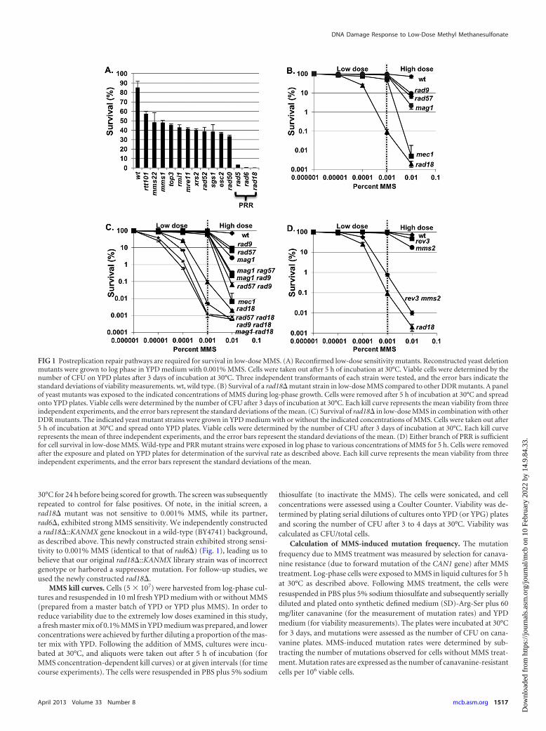
30°C for 24 h before being scored for growth. The screen was subsequentlyrepeated to control for false positives. Of note, in the initial screen, arad18� mutant was not sensitive to 0.001% MMS, while its partner,rad6�, exhibited strong MMS sensitivity. We independently constructeda rad18�::KANMX gene knockout in a wild-type (BY4741) background,as described above. This newly constructed strain exhibited strong sensi-tivity to 0.001% MMS (identical to that of rad6�) (Fig. 1), leading us tobelieve that our original rad18�::KANMX library strain was of incorrectgenotype or harbored a suppressor mutation. For follow-up studies, weused the newly constructed rad18�.
MMS kill curves. Cells (5 � 107) were harvested from log-phase cul-tures and resuspended in 10 ml fresh YPD medium with or without MMS(prepared from a master batch of YPD or YPD plus MMS). In order toreduce variability due to the extremely low doses examined in this study,a fresh master mix of 0.1% MMS in YPD medium was prepared, and lowerconcentrations were achieved by further diluting a proportion of the mas-ter mix with YPD. Following the addition of MMS, cultures were incu-bated at 30°C, and aliquots were taken out after 5 h of incubation (forMMS concentration-dependent kill curves) or at given intervals (for timecourse experiments). The cells were resuspended in PBS plus 5% sodium
thiosulfate (to inactivate the MMS). The cells were sonicated, and cellconcentrations were assessed using a Coulter Counter. Viability was de-termined by plating serial dilutions of cultures onto YPD (or YPG) platesand scoring the number of CFU after 3 to 4 days at 30°C. Viability wascalculated as CFU/total cells.
Calculation of MMS-induced mutation frequency. The mutationfrequency due to MMS treatment was measured by selection for canava-nine resistance (due to forward mutation of the CAN1 gene) after MMStreatment. Log-phase cells were exposed to MMS in liquid cultures for 5 hat 30°C as described above. Following MMS treatment, the cells wereresuspended in PBS plus 5% sodium thiosulfate and subsequently seriallydiluted and plated onto synthetic defined medium (SD)-Arg-Ser plus 60mg/liter canavanine (for the measurement of mutation rates) and YPDmedium (for viability measurements). The plates were incubated at 30°Cfor 3 days, and mutations were assessed as the number of CFU on cana-vanine plates. MMS-induced mutation rates were determined by sub-tracting the number of mutations observed for cells without MMS treat-ment. Mutation rates are expressed as the number of canavanine-resistantcells per 106 viable cells.
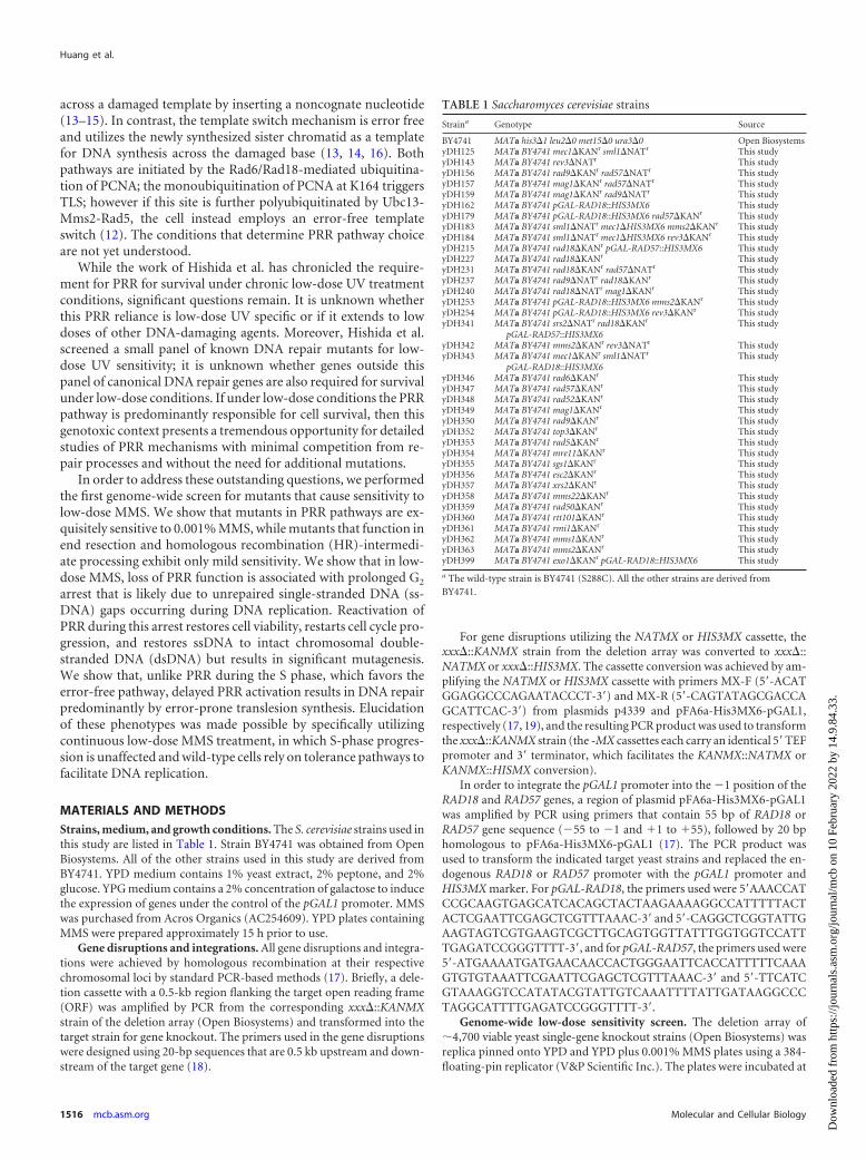
FIG 1 Postreplication repair pathways are required for survival in low-dose MMS. (A) Reconfirmed low-dose sensitivity mutants. Reconstructed yeast deletionmutants were grown to log phase in YPD medium with 0.001% MMS. Cells were taken out after 5 h of incubation at 30°C. Viable cells were determined by thenumber of CFU on YPD plates after 3 days of incubation at 30°C. Three independent transformants of each strain were tested, and the error bars indicate thestandard deviations of viability measurements. wt, wild type. (B) Survival of a rad18� mutant strain in low-dose MMS compared to other DDR mutants. A panelof yeast mutants was exposed to the indicated concentrations of MMS during log-phase growth. Cells were removed after 5 h of incubation at 30°C and spreadonto YPD plates. Viable cells were determined by the number of CFU after 3 days of incubation at 30°C. Each kill curve represents the mean viability from threeindependent experiments, and the error bars represent the standard deviations of the mean. (C) Survival of rad18� in low-dose MMS in combination with otherDDR mutants. The indicated yeast mutant strains were grown in YPD medium with or without the indicated concentrations of MMS. Cells were taken out after5 h of incubation at 30°C and spread onto YPD plates. Viable cells were determined by the number of CFU after 3 days of incubation at 30°C. Each kill curverepresents the mean of three independent experiments, and the error bars represent the standard deviations of the mean. (D) Either branch of PRR is sufficientfor cell survival in low-dose MMS. Wild-type and PRR mutant strains were exposed in log phase to various concentrations of MMS for 5 h. Cells were removedafter the exposure and plated on YPD plates for determination of the survival rate as described above. Each kill curve represents the mean viability from threeindependent experiments, and the error bars represent the standard deviations of the mean.
DNA Damage Response to Low-Dose Methyl Methanesulfonate
April 2013 Volume 33 Number 8 mcb.asm.org 1517
Dow
nloa
ded
from
http
s://j
ourn
als.
asm
.org
/jour
nal/m
cb o
n 10
Feb
ruar
y 20
22 b
y 14
.9.8
4.33
.
Synchronization and cell cycle analysis. Cells were synchronized inthe G1 phase by the addition of �-factor (Zymo Research; catalog numberY1001) at a final concentration of 5 �M to log-phase cultures or culturesreleased from G2 arrest (see below). Cultures were incubated in �-factorfor 2 to 3 h at 30°C to achieve G1 arrest, which was verified microscopicallyand by fluorescence-activated cell sorter (FACS) analysis. To release cellsfrom G1 arrest, cells were harvested and washed once with 1 ml of phos-phate-buffered saline (PBS) and resuspended in 10 ml fresh YPD mediumcontaining 10 �g/ml pronase (Fisher Scientific; catalog number 50-720-3354). For G2/M synchronization, 10 �g/ml of nocodazole (Toronto Re-search Chemicals Inc.; catalog number M330350) was added to log-phasecultures or cultures released from G1 arrest, and the cells were incubatedfor 2 h at 30°C. G2-arrested cells were verified microscopically (as large-budded cells) and by FACS analysis. Cell cycle distributions were deter-mined by flow cytometry (by a method described previously [20]) using aBeckman-Dickson FACSCalibur flow cytometer.
Western blotting. Cell extracts were prepared from log-phase cells, aswell as synchronized cells, using a trichloroacetic acid (TCA) lysis method(21). Proteins were analyzed by SDS-PAGE (22). Rad53p was detectedwith the yC-19 anti-Rad53 antibody (Santa Cruz).
PFGE. To analyze intact yeast chromosomal DNA by pulsed-field gelelectrophoresis (PFGE), DNA plugs were prepared using a CHEF (con-tour-clamped homogeneous electric field) Genomic DNA plug Kit (Bio-Rad; catalog number 170-3591) according to the manufacturer’s instruc-tions. Briefly, cells (�2 � 108) were harvested at different time points andfixed in 70% ethanol. Following ethanol fixation, the cells were resus-pended in 200 �l of suspension buffer (10 mM Tris, pH 7.2, 20 mM NaCl,50 mM EDTA) and mixed with an equal volume of 2% CleanCut low-melting-point agarose at 50°C. The hot mixture was quickly pipetted intothe manufacturer-supplied plug molds and allowed to solidify (each sam-ple produced 3 plugs). In-gel cell lysis was performed by adding lyticase (1mg/ml) for 2 h at 37°C, followed by 1 mg/ml proteinase K treatment for 24h at 37°C. In order to test whether the nondenatured DNA sample con-tained S1-labile ssDNA, a subset of the DNA plugs were digested with 1 Uof S1 nuclease (Sigma; catalog number N5661) for 40 min at 30°C in S1nuclease buffer containing 1 mM phenylmethylsulfonyl fluoride (PMSF)(the undigested set was incubated in the same buffer without S1 nuclease).Both sets of plugs were then loaded on a 1% Megabase agarose gel (Bio-Rad), and genomic DNA was resolved using a Bio-Rad CHEF-DR II sys-tem according to the manufacturer’s instructions. Following electropho-resis, the gels were stained in 1% ethidium bromide for 2 h andphotographed under UV light.
RESULTSPostreplication repair is required for survival in response tocontinuous low-dose MMS exposure. While a genome-widescreen for mutants sensitive to high doses of MMS (0.035%) pre-viously identified 103 sensitive mutant strains (23), we hypothe-sized that a different spectrum of mutants would be sensitive tolow-dose MMS (0.001%). To test this hypothesis, we screened�4,700 unique gene deletion strains representing the yeast hap-loid deletion collection (24) for mutations conferring sensitivityto 0.001% MMS. As a small percentage of the library strains havebeen shown to harbor additional mutations (25, 26), we sought toeliminate any false positives by regenerating all 14 deletion mu-tants that showed low-dose MMS sensitivity in the initial screen(see Materials and Methods). These new deletion mutants werethen retested by quantitative colony-forming assay in response toMMS, and all 14 mutants were confirmed to be sensitive to0.001% MMS (Fig. 1A). Mutations in the PRR genes rad5�,rad6�, and rad18� conferred particularly high (�100-fold) sen-sitivity, while the remaining mutants exhibited a milder 2- to3-fold drop in survival. All of the genes identified as being sensitiveto low-dose MMS had been previously identified as sensitive to
high-dose MMS (23). To reconfirm that PRR mutants are uniqueamong repair genes in their exquisite sensitivity to low-doseMMS, we performed a verification step in which we quantified thesensitivities of a panel of high-dose MMS-sensitive DDR mutantsto a range of MMS exposures to confirm that they do not confersubstantial sensitivity to low-dose MMS. This panel comprisedgenes involved in homologous recombination (HR) (rad57�),base excision repair (BER) (mag1�), and checkpoint activation(rad9� and mec1�) (Fig. 1B). While a rad18� mutant exhibited adrop in viability in MMS in a dose-dependent manner down to0.0001% MMS, none of the other mutants exhibited substantialsensitivity to very low-dose MMS, despite significant sensitivity tohigh-dose (0.01%) treatment. From these results, we concludethat PRR mutants are highly sensitive to low-dose MMS treat-ment, whereas mutations in other DNA repair pathways (HR,BER, etc.) show minimal effect. Although the other pathwaystested (HR, BER, etc.) are far less critical than PRR under low-doseconditions (i.e., �0.001% MMS), combining rad18� with muta-tions in rad57�, rad9�, or mag1� resulted in an additive increasein sensitivity to low-dose MMS versus rad18� alone (Fig. 1C),demonstrating that in the absence of RAD18 these pathways play acompensatory role.
Either PRR subpathway (error free or error prone) is suffi-cient for survival in response to low-dose MMS. Cells employtwo RAD18-dependent PRR mechanisms to tolerate DNA lesions(translesion synthesis and error-free HR-directed bypass). To de-termine whether RAD18-dependent survival in low-dose MMSdepends on one or both of these mechanisms, we examined thelow-dose MMS sensitivities of representative mutants for eachPRR subpathway (REV3, which is required for translesion synthe-sis, and MMS2, which is required for error-free PRR [8, 27]). Asshown in Fig. 1D, a defect in either PRR subpathway alone (rev3�or mms2�) does not affect survival in MMS concentrations of�0.001%, while both exhibit sensitivity to high-dose (0.01%)MMS (with mms2� exhibiting slightly higher sensitivity at thisdose). In contrast, loss of both branches (mms2� rev3�) results insynergistic hypersensitivity to low-dose MMS (Fig. 1D). Thus, weconclude that either PRR subpathway (error free or error prone) issufficient for survival in response to low-dose MMS. Notably, themms2� rev3� double mutant is slightly more MMS resistant thana rad18� strain, suggesting that there is some remaining PRR ac-tivity in mms2� rev3� (possibly through a Rev1-Rad30-depen-dent translesion synthesis mechanism) (27, 28).
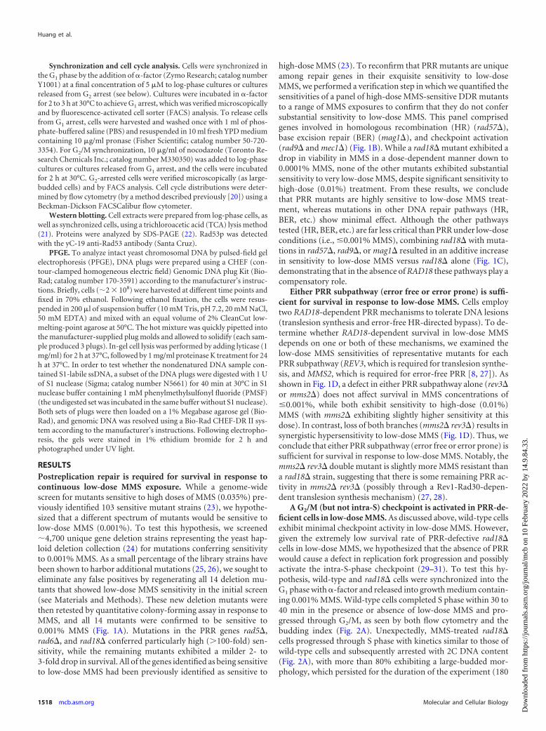
A G2/M (but not intra-S) checkpoint is activated in PRR-de-ficient cells in low-dose MMS. As discussed above, wild-type cellsexhibit minimal checkpoint activity in low-dose MMS. However,given the extremely low survival rate of PRR-defective rad18�cells in low-dose MMS, we hypothesized that the absence of PRRwould cause a defect in replication fork progression and possiblyactivate the intra-S-phase checkpoint (29–31). To test this hy-pothesis, wild-type and rad18� cells were synchronized into theG1 phase with �-factor and released into growth medium contain-ing 0.001% MMS. Wild-type cells completed S phase within 30 to40 min in the presence or absence of low-dose MMS and pro-gressed through G2/M, as seen by both flow cytometry and thebudding index (Fig. 2A). Unexpectedly, MMS-treated rad18�cells progressed through S phase with kinetics similar to those ofwild-type cells and subsequently arrested with 2C DNA content(Fig. 2A), with more than 80% exhibiting a large-budded mor-phology, which persisted for the duration of the experiment (180
Huang et al.
1518 mcb.asm.org Molecular and Cellular Biology
Dow
nloa
ded
from
http
s://j
ourn
als.
asm
.org
/jour
nal/m
cb o
n 10
Feb
ruar
y 20
22 b
y 14
.9.8
4.33
.
min). Thus, rad18� cells arrest in G2 phase after exposure to low-dose MMS but do not experience the significant S-phase delayindicative of intra-S-phase checkpoint activation (20, 31).
To confirm that the G2 arrest of rad18� cells in low-dose MMSwas due to the activation of the G2/M DNA damage checkpoint,we tested for MMS-induced Rad53 phosphorylation (a G2/Mcheckpoint indicator) by Western blotting (21). Indeed, whilewild-type cells exhibited no Rad53 phosphorylation in low-doseMMS (consistent with no MMS-dependent changes in cell cycledistribution by FACS), rad18� cells exhibited Rad53 phosphory-lation beginning at �50 min after the addition of low-dose MMS(Fig. 2B). Of note, Rad53 phosphorylation in rad18� cells wassomewhat delayed after the transition to 2C DNA content byFACS (Fig. 2A and B), suggesting that generating the checkpointactivation signal may require events that occur after the bulk ofreplication is completed.
Low-dose MMS-induced viability loss in rad18� cells re-quires passage through S phase in the presence of MMS. Giventhat the DDR checkpoint is not activated until after the bulk ofreplication has been completed, we hypothesized that the G2/M
arrest and the viability loss in rad18� cells exposed to low-doseMMS are not induced by alkylation lesions directly, but rather, bysecondary lesions (such as ssDNA gaps) resulting from incom-plete postreplication repair (32, 33). One prediction of this hy-pothesis is that rad18� cells exposed to low-dose MMS outside theS phase (i.e., in the G1 or G2 phase) should remain viable, since thiswould preclude the generation of irreparable PRR intermediates.To test this prediction, we induced a mitotic checkpoint arrest inwild-type and rad18� cells with nocodazole treatment and thenreleased the cells into medium containing both 0.001% MMS and�-factor (to restrict MMS exposure to the G1 phase). As expected,when the MMS treatment was confined to the G1 phase (via �-fac-tor treatment), rad18� cells exhibited significantly higher viabilitythan cells allowed to replicate their genomes in the presence ofMMS (no �-factor) (49% viable versus 3.5%) (Fig. 3A). (The�50% drop in viability with �-factor likely reflects a subset ofunrepaired lesions that persist after �-factor is removed; the cellsare plated on YPD afterward, at which point they can cycle nor-mally and must cope with any remaining MMS lesions). In con-clusion, the MMS sensitivity exhibited by a rad18� mutant is due
FIG 2 Low-dose MMS activates the G2/M checkpoint in rad18� cells. (A) Low-dose MMS triggers G2/M arrest in rad18� cells. Wild-type and rad18� cells weresynchronized with �-factor and released into YPD medium 0.001% MMS. Cells were removed at the indicated times and analyzed for cell cycle distributionby FACS, for cell morphology by microscopy, and for viability by colony survival assay. Each flow cytometry graph contains two histograms. The shadedhistograms represent the cell cycle distribution of �-factor-blocked cultures at time zero. The overlaid histograms represent the cell cycle distributions at varioustimes following release from the G1 block. The cell number per milliliter is listed for selected time points. Survival curves for wild-type and rad18� cells at eachtime point are shown below the cell cycle distribution graphs. Each strain was tested in triplicate, and the error bars represent the standard deviations of mean cellviability. (B) Rad53 phosphorylation in response to 0.001% MMS. Wild-type and rad18� cells were treated with �-factor and released into YPD medium with0.001% MMS. Samples were taken out at the indicated times for Western blot analysis with an anti-Rad53 antibody.
DNA Damage Response to Low-Dose Methyl Methanesulfonate
April 2013 Volume 33 Number 8 mcb.asm.org 1519
Dow
nloa
ded
from
http
s://j
ourn
als.
asm
.org
/jour
nal/m
cb o
n 10
Feb
ruar
y 20
22 b
y 14
.9.8
4.33
.

to DNA damage produced during the S phase, likely caused by adefect in DNA damage tolerance during replication.
Previous reports have demonstrated that cells that enter Sphase with irreparable UV lesions generate long stretches ofssDNA, and these ssDNA lesions are later resolved by postreplica-tion repair (32, 33). We hypothesized that low-dose MMS treat-ment during S phase is also associated with the production ofssDNA gaps that are irreparable in a PRR mutant background(and cause the G2 arrest). To test this hypothesis, we employed thesingle-strand-specific S1 endonuclease, which cuts DNA regionscontaining nicks and ssDNA gaps (34, 35), converting them intodouble-strand breaks (DSBs) that can be visualized as fragmenta-
tion by PFGE (36). Wild-type and rad18� cells were synchronizedin the G1 phase with �-factor and released into medium contain-ing low-dose MMS (0.001%) plus nocodazole. (The nocodazoletreatment was used to arrest wild-type cells at the mitotic check-point to minimize the ssDNA component associated with DNAreplication and to make the results comparable to those for theG2-arrested rad18� cells.) After a 60-min low-dose MMS treat-ment, chromosomal DNA was isolated in agarose plugs and sub-jected to S1 nuclease treatment and PFGE (Fig. 3B). For wild-typecells, treatment with low-dose MMS was not associated with de-tectable S1-dependent chromosomal fragmentation (and thus nossDNA gaps). In contrast, rad18� cells exhibited significant
FIG 3 Loss of viability and G2 arrest in rad18� cells in low-dose MMS is associated with S phase. (A) Passage through S phase is required for MMS sensitivity.Wild-type and rad18� cells were synchronized at mitotic checkpoint arrest with nocodazole (Noc) for 2 h and then exposed to 0.001% MMS in the presence orabsence of �-factor for a second 2-h period. MMS was then removed, and the cells were incubated for another 1.5 h, with or without �-factor. Cells werewithdrawn at the indicated time points for the assessment of cell cycle distributions and survival rates. All incubations occurred at 30°C. In the upper right cornerof each graph are shown the duration of treatment, cell number per milliliter, and cell survival percentage at the end of each treatment (each strain was tested intriplicate repeats for survival rates; the mean and standard deviation of the mean survival rate are indicated). The shaded histograms represent the cell cycledistribution of the asynchronous culture before the nocodazole block. The overlaid histograms represent the cell cycle distributions at various times after release.(B) Chromosomes of MMS-treated rad18� cells show S1 nuclease-sensitive components (ssDNA gaps). �-Factor-blocked wild-type and rad18� cells werereleased into YPD medium with 0.001% MMS and 10 �g/ml nocodazole for 60 min. Cells were harvested after �-factor synchronization (�F) and after MMStreatment (MMS). Chromosomal DNA (either S1 treated or mock treated) was analyzed by PFGE.
Huang et al.
1520 mcb.asm.org Molecular and Cellular Biology
Dow
nloa
ded
from
http
s://j
ourn
als.
asm
.org
/jour
nal/m
cb o
n 10
Feb
ruar
y 20
22 b
y 14
.9.8
4.33
.

MMS- and S1-dependent chromosomal fragmentation that is in-dicative of the presence of ssDNA gaps associated with low-doseMMS exposure. From these data, we conclude that the loss of PRRis associated with the production of ssDNA gaps in low-doseMMS, and these lesions are the likely trigger for the prolongedcheckpoint activation exhibited by these cells.
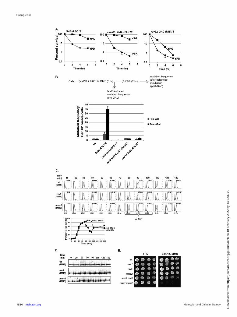
Both viability and chromosome integrity in low-dose MMS-treated rad18-deficient cells can be rescued in a time-limitedfashion by reactivation of RAD18 in G2 phase. Recent work hasshown that for acute high doses of UV and MMS, PRR can bedelayed for a prolonged period past the completion of bulk DNAsynthesis and then reactivated to restore a major proportion of cellviability (28, 37). As sensitivity to low-dose MMS for PRR mutantsis associated with a prolonged G2 checkpoint and ssDNA gaps, wehypothesized that reactivation of PRR in these cells would have a3-fold effect: ssDNA gaps would be eliminated, cells would escapeG2 arrest, and viability would be restored. To test this hypothesis,we constructed a yeast strain harboring a RAD18 gene under thecontrol of a conditional GAL promoter (GAL-RAD18) to controlthe activity of PRR (28). Cells were exposed to low-dose MMSunder RAD18-repressing conditions (i.e., glucose), and cells werewithdrawn at multiple time points for assessment of viability onglucose plates (maintaining RAD18 repression) and galactoseplates (reactivating RAD18) (Fig. 4A). As expected, in glucose,cells harboring the GAL-RAD18 construct exhibited prolongedlow-dose MMS-dependent G2 arrest (data not shown) and loss ofviability, mimicking a rad18� deletion. However, when cells wereremoved and plated on galactose medium (reactivating RAD18)following MMS treatment, there was a marked increase in viability(nearly 100% rescue after up to 4 h in MMS). From these data, weconclude that the loss of viability in PRR-deficient cells in re-sponse to low-dose MMS treatment can be rescued by the reacti-vation of RAD18.
Notably, this rescue was evident even after prolonged MMStreatment but began to steadily decrease after the 4-h time point,and no rescue was observed after 8 h in low-dose MMS. We ini-tially hypothesized that the failure to rescue at later time pointswas a result of new MMS lesions incurred during the prolongedexposure in G2; however, even after removing the MMS after 3 h,GAL-RAD18 cells remained arrested, and the galactose-depen-dent rescue was similarly time limited (data not shown). Thesedata suggest that the loss of RAD18-dependent rescue after 8 h wasnot due to additional MMS lesions but to a secondary mechanism,possibly through further processing of ssDNA to a different lesionirreparable by PRR (32).
To confirm that the RAD18-dependent rescue in low-doseMMS correlates with a decrease in ssDNA gaps (indicative of re-pair by PRR), GAL-RAD18 cells were exposed to low-dose MMSin glucose medium (RAD18-OFF) and then switched to eithergalactose medium (RAD18-ON) or fresh glucose medium in thepresence of nocodazole. DNA samples were prepared in agaroseplugs and subjected to S1 nuclease digestion and PFGE. As previ-ously observed for rad18� (Fig. 3B), low-dose MMS-treated GAL-RAD18 cells in glucose medium exhibited significant S1-depen-dent chromosomal fragmentation (Fig. 4B). However, when cellswere switched to galactose medium (reactivating RAD18), a sig-nificant reduction in S1 nuclease sensitivity was observed, indica-tive of the restoration of ssDNA gaps to dsDNA by PRR. Fromthese data, we conclude that the reactivation of RAD18 after ex-
posure to low-dose MMS is associated with the repair of ssDNAgaps and restoration of cell viability.
The time limit for RAD18-dependent rescue in low-doseMMS is EXO1 dependent. While the reactivation of GAL-RAD18restores cell viability in low-dose MMS even after an extendeddelay (Fig. 4A), we were surprised that the ability to rescue wastime limited. It has been previously reported that Exo1 possesses5=-3= exonuclease activity, and its processing of NER intermedi-ates generates extended ssDNA gaps (38). We hypothesized thatExo1 enlarges ssDNA gaps in low-dose MMS during the pro-longed G2 arrest (either by a direct resection of the S-phase-de-pendent ssDNA or by extending new NER ssDNA intermediatesthat merge with the S-phase-dependent gaps) and that these ex-tended gaps may be irreparable by PRR. This extension of ssDNAgaps into larger PRR-irreparable ssDNA regions could explain theinability to rescue low-dose MMS-treated GAL-RAD18 cells past 8h (Fig. 4A). To test this hypothesis, we deleted EXO1 in a GAL-RAD18 background and examined cell survival after prolongedincubation in low-dose MMS (Fig. 4C). Indeed, while the galac-tose-dependent RAD18 rescue efficiency in an exo1� backgroundwas similar to that of EXO1 at earlier time points, the ability torescue cells by reactivation of RAD18 in exo1� cells persisted wellbeyond 8 h. From this, we conclude that the time limit for RAD18-dependent rescue in low-dose MMS is due to EXO1-dependentresection. Interestingly, we observed a slight drop in rescue in anexo1� background (Fig. 4C), which may reflect the activities ofother nucleases on ssDNA gaps.
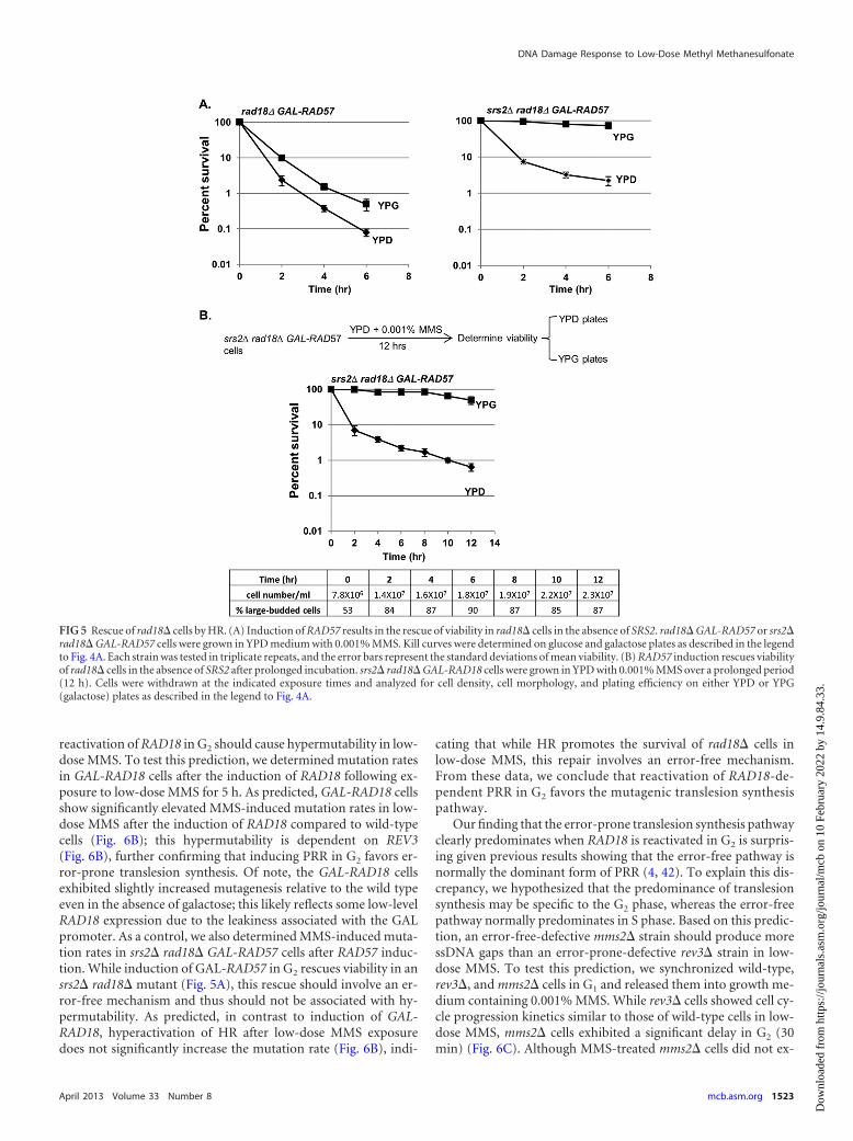
Removal of the SRS2-dependent block to HR results in anefficient postreplicative rescue independent of RAD18. We weresurprised that the extreme loss of viability in a rad18� mutant inlow-dose MMS was due to the presence of ssDNA gaps, as it isunclear why these structures could not be repaired by sister chro-matid recombination in the G2 phase in a PRR-independent man-ner. We hypothesized that while it may be possible for these struc-tures to be physically repaired by recombination, the repression ofrecombination by some unknown factor suppresses the repair.One candidate for this repression is the Srs2 helicase, which hasbeen shown to suppress HR during DNA replication (39). Dele-tion of SRS2 has been shown to rescue a rad18� mutant afterlow-dose UV treatment (40). We asked whether a similar effectmay occur in low-dose MMS, and especially, whether delayed in-duction of HR at various time points during the low-dose MMStreatment may be sufficient to rescue rad18� cells in the absenceof SRS2. In order to test this, we put RAD57 under the control of aconditional GAL promoter and tested the effects of induction ofRAD57 on cell viability in a rad18� background. When rad18�GAL-RAD57 and srs2� rad18� GAL-RAD57 cells were exposed tolow-dose MMS in glucose medium (RAD57-OFF), both strainsexhibited viability loss and synchronization at G2/M (Fig. 5A anddata not shown). However, when MMS-treated srs2� rad18�GAL-RAD57 cells were plated on galactose medium (reactivatingRAD57), we observed a complete rescue of viability, indicatingthat in an srs2� background, HR can compensate for the loss ofRAD18.
We note that even when SRS2 is present, we observed a slightrescue upon reactivation of GAL-RAD57 (Fig. 5A); this is likelydue to RAD57 overexpression, which may partially overcomeSRS2-dependent inhibition (Rad57 can physically block Srs2’sability to disrupt Rad51 nucleoprotein filaments) (41). Interest-ingly, unlike the rescue by GAL-RAD18, the reactivation of GAL-
DNA Damage Response to Low-Dose Methyl Methanesulfonate
April 2013 Volume 33 Number 8 mcb.asm.org 1521
Dow
nloa
ded
from
http
s://j
ourn
als.
asm
.org
/jour
nal/m
cb o
n 10
Feb
ruar
y 20
22 b
y 14
.9.8
4.33
.

RAD57 allows rescue even after 8 h of MMS exposure (Fig. 5B),suggesting that the EXO1-dependent extension of ssDNA can beresolved by HR, but not PRR. From these data, we conclude thatSRS2 represses the HR-dependent repair of ssDNA gaps in PRR-deficient cells.
The preference for error-free and error-prone lesion toler-ance is cell cycle dependent. Since RAD18 is required for the func-tion of either error-free or error-prone PRR, we next askedwhether one pathway is more important for survival in low-doseMMS following the reactivation of GAL-RAD18. To determinethis, we combined the conditional GAL-RAD18 allele with dele-
tions of either rev3� (TLS deficient) or mms2� (error-free-PRRdeficient) and tested the abilities of these mutants to be rescued byinduction of GAL-RAD18 at various times during low-dose MMStreatment. While GAL-RAD18 induction rescued the mms2� mu-tant, the efficiency of rescue by activation of GAL-RAD18 in arev3� background was markedly reduced (Fig. 6A), indicatingthat the postreplicative rescue in low-dose MMS depends on func-tional translesion synthesis. This is consistent with previous ob-servations for UV lesion bypass (28).
Based on the importance of REV3-dependent, error-proneTLS for the rescue of GAL-RAD18 cells, we predicted that the
FIG 4 Reactivation of Rad18 in G2 restores viability and chromosome integrity of low-dose MMS-treated rad18 cells. (A) The viability of PRR-deficient cells canbe restored when RAD18 expression is induced after MMS treatment. Cells harboring a chromosomal RAD18 gene under the control of a GAL promoter(GAL-RAD18) were grown in YPD medium with 0.001% MMS. At the indicated times of exposure, cells were withdrawn and analyzed for plating efficiency oneither glucose or galactose plates. Each kill curve represents the mean viability of three independent experiments, and the error bars represent the standarddeviations of the mean. At all time points after 2 h of exposure to MMS, the cell density remained essentially unchanged, and rad18� cells accumulated with auniform large-budded morphology (bottom). (B) Induction of RAD18 reduces S1 sensitivity in MMS-treated GAL-RAD18 cells. GAL-RAD18 cells were blockedwith �-factor, released into YPD medium with 0.001% MMS for 60 min, and then incubated in MMS-free medium containing glucose or galactose for another2 h in the presence of nocodazole (10 �g/ml). Cells were withdrawn after �-factor synchronization (�F) and MMS exposure and after the subsequent RAD18induction (Glu and Gal). Chromosomal DNA was treated with S1 nuclease or mock treated and subjected to PFGE as described for Fig. 3B. (C) The ability forRAD18-dependent rescue is prolonged in the absence of EXO1. GAL-RAD18 and exo1� GAL-RAD18 cells were grown in YPD with 0.001% MMS for a period of12 h. The survival rates on glucose or galactose plates were determined as for panel A.
Huang et al.
1522 mcb.asm.org Molecular and Cellular Biology
Dow
nloa
ded
from
http
s://j
ourn
als.
asm
.org
/jour
nal/m
cb o
n 10
Feb
ruar
y 20
22 b
y 14
.9.8
4.33
.
reactivation of RAD18 in G2 should cause hypermutability in low-dose MMS. To test this prediction, we determined mutation ratesin GAL-RAD18 cells after the induction of RAD18 following ex-posure to low-dose MMS for 5 h. As predicted, GAL-RAD18 cellsshow significantly elevated MMS-induced mutation rates in low-dose MMS after the induction of RAD18 compared to wild-typecells (Fig. 6B); this hypermutability is dependent on REV3(Fig. 6B), further confirming that inducing PRR in G2 favors er-ror-prone translesion synthesis. Of note, the GAL-RAD18 cellsexhibited slightly increased mutagenesis relative to the wild typeeven in the absence of galactose; this likely reflects some low-levelRAD18 expression due to the leakiness associated with the GALpromoter. As a control, we also determined MMS-induced muta-tion rates in srs2� rad18� GAL-RAD57 cells after RAD57 induc-tion. While induction of GAL-RAD57 in G2 rescues viability in ansrs2� rad18� mutant (Fig. 5A), this rescue should involve an er-ror-free mechanism and thus should not be associated with hy-permutability. As predicted, in contrast to induction of GAL-RAD18, hyperactivation of HR after low-dose MMS exposuredoes not significantly increase the mutation rate (Fig. 6B), indi-
cating that while HR promotes the survival of rad18� cells inlow-dose MMS, this repair involves an error-free mechanism.From these data, we conclude that reactivation of RAD18-de-pendent PRR in G2 favors the mutagenic translesion synthesispathway.
Our finding that the error-prone translesion synthesis pathwayclearly predominates when RAD18 is reactivated in G2 is surpris-ing given previous results showing that the error-free pathway isnormally the dominant form of PRR (4, 42). To explain this dis-crepancy, we hypothesized that the predominance of translesionsynthesis may be specific to the G2 phase, whereas the error-freepathway normally predominates in S phase. Based on this predic-tion, an error-free-defective mms2� strain should produce moressDNA gaps than an error-prone-defective rev3� strain in low-dose MMS. To test this prediction, we synchronized wild-type,rev3�, and mms2� cells in G1 and released them into growth me-dium containing 0.001% MMS. While rev3� cells showed cell cy-cle progression kinetics similar to those of wild-type cells in low-dose MMS, mms2� cells exhibited a significant delay in G2 (30min) (Fig. 6C). Although MMS-treated mms2� cells did not ex-
FIG 5 Rescue of rad18� cells by HR. (A) Induction of RAD57 results in the rescue of viability in rad18� cells in the absence of SRS2. rad18� GAL-RAD57 or srs2�rad18� GAL-RAD57 cells were grown in YPD medium with 0.001% MMS. Kill curves were determined on glucose and galactose plates as described in the legendto Fig. 4A. Each strain was tested in triplicate repeats, and the error bars represent the standard deviations of mean viability. (B) RAD57 induction rescues viabilityof rad18� cells in the absence of SRS2 after prolonged incubation. srs2� rad18� GAL-RAD18 cells were grown in YPD with 0.001% MMS over a prolonged period(12 h). Cells were withdrawn at the indicated exposure times and analyzed for cell density, cell morphology, and plating efficiency on either YPD or YPG(galactose) plates as described in the legend to Fig. 4A.
DNA Damage Response to Low-Dose Methyl Methanesulfonate
April 2013 Volume 33 Number 8 mcb.asm.org 1523
Dow
nloa
ded
from
http
s://j
ourn
als.
asm
.org
/jour
nal/m
cb o
n 10
Feb
ruar
y 20
22 b
y 14
.9.8
4.33
.
Huang et al.
1524 mcb.asm.org Molecular and Cellular Biology
Dow
nloa
ded
from
http
s://j
ourn
als.
asm
.org
/jour
nal/m
cb o
n 10
Feb
ruar
y 20
22 b
y 14
.9.8
4.33
.

hibit substantially increased levels of Rad53 phosphorylation(Fig. 6D), we found that when mms2� is combined with deletionof the checkpoint factor MEC1, cells fail to grow in low-dose MMS(in contrast, a rev3� mec1� strain displays robust growth)(Fig. 6E). From these data, we conclude that loss of the MMS2-dependent error-free pathway is associated with a significant G2
delay and a requirement for MEC1 for survival in low-dose MMS,while loss of the REV3-dependent pathway does not exhibit thesephenotypes.
DISCUSSIONDefinition of “low-dose” treatment. Our motivation for provid-ing a formal definition for what constitutes a “low dose” of aDNA-damaging agent stemmed from difficulties we experiencedin comparing our data with other studies of low-level exposurethat span a variety of dose rates, genotoxic agents, and cells/tis-sues. For example, in previous studies evaluating the role of PRRin response to low-dose UV treatment, Hishida et al. based theirchoice of UV irradiation dose (0.1 J/m2/min) on the natural phe-nomenon (sunlight exposure) that they were attempting to mimic(4, 40); a clear analog for this dose using a chemical carcinogensuch as MMS was not immediately apparent. By providing a phys-iological and agent-agnostic definition of “low-dose” DNA dam-age based upon the sensitivity of wild-type and mutant cells, wewere able to reconcile our MMS results with these previous stud-ies; both 0.1 J/m2/min UV exposure and 0.001% MMS treatmentfail to induce any discernible cellular sensitivity in a wild-type cellpopulation; however, mutant studies reveal an exquisite and pro-found dependence on PRR for survival in response to low-dosedamage (Fig. 1) (4).
PRR mutants are unique in their exquisite sensitivity to low-dose MMS. As the prior Hishida et al. study (4) was UV specificand screened only a small panel of known DDR targets, it re-mained formally possible that PRR reliance was UV specific orthat other, unscreened genes would also be required for survivalunder low-dose conditions. To answer these questions, we per-formed a genome-wide screen in response to low-dose treatmentwith a DNA-alkylating agent (0.001% MMS). Strikingly, we showthat while 0.001% MMS treatment of wild-type yeast cells pro-duces no discernible response (for any assay by which we evaluatethese cells, including survival [Fig. 1], checkpoint activity [Fig. 2],cell growth and division [Fig. 2], and chromosomal integrity/mu-tagenesis [Fig. 3B and 6B]), PRR mutants are exquisitely sensitive,and hence DNA damage tolerance is actively employed and criti-
cal for viability at this dose. The lethal lesions that render PRRmutants extremely MMS sensitive are unreplicated ssDNA gaps(which can be detected as S1-dependent fragmentation in rad18cells by PFGE [Fig. 3B]), and restoration of PRR activity (evenafter significant delay) eliminates these gaps and rescues cell via-bility in low-dose MMS (Fig. 4A to C). Due to the exceptionalrequirement for PRR under low-dose conditions, we were able toglean vital information regarding how PRR functions in the pres-ence (or absence) of cell cycle checkpoints and how timing andcontext play vital roles in determining which PRR pathway (errorfree or error prone) is utilized.
The preference for error-free or error-prone PRR is cell cycledependent. While previous studies have shown a cellular prefer-ence for the error-free pathway (4, 42), recent studies using aninducible PRR system show a clear preference for error-pronetranslesion synthesis (28). In this study, we show that these are notcontradictory conclusions, since our data demonstrate that PRRpathway usage depends on timing and context. Specifically, whileour data show a preference for the error-free pathway when cellsare replicating in low-dose MMS, the error-prone pathway is spe-cifically required for viability when PRR is delayed until the G2
phase (Fig. 6A). These results provide some clarity with regard tohow cells choose error-free versus error-prone repair (in low-doseMMS). During S phase, cells prevent ssDNA gap formation via theMMS2-dependent template switch mechanism (without requir-ing G2 arrest) but, alternatively, can generate and repair an ssDNAgap using a REV3-dependent TLS mechanism (which can be de-layed postreplication but requires checkpoint activation). The cellcycle dependence of error-free versus error-prone repair may re-flect a necessity for template switching to happen within a briefwindow of lesion encounter by replicative polymerases, while TLSmay be less temporally restricted. Alternatively, cells may directlyregulate TLS or error-free factors in a cell cycle-dependent man-ner in order to carry out this programmed response. Consistentwith this hypothesis, REV1 expression peaks during the G2 phaseof the cell cycle; this transcriptional regulation may be one way inwhich cells suppress TLS in the S phase but promote it in G2 (43).
Rescue by reactivation of PRR in MMS has a time limit. Theinducible GAL-RAD18 system also provides information on thestability of ssDNA lesions over a prolonged period. We show thatthere is a time limit for the delayed repair of ssDNA gaps in G2/M;the reactivation of PRR is most effective within �4 h after theinitial G2/M checkpoint activation (Fig. 4A); however, this timelimit can be extended by deletion of the Exo1 exonuclease
FIG 6 TLS is required for the rescue of viability upon RAD18 induction. (A) Reactivation of RAD18 at G2 fails to effectively rescue viability in the absence ofREV3. GAL-RAD18, rev3� GAL-RAD18, and mms2� GAL-RAD18 cells were exposed to 0.001% MMS over a 6-hour period. Cells were withdrawn at theindicated times during the exposure and analyzed for viability by plating efficiency on either glucose or galactose plates as described for Fig. 4A. Each strain wastested in triplicate repeats, and the error bars represent the standard deviations of the mean. (B) Reactivation of RAD18 causes hypermutability. Log-phasewild-type, GAL-RAD18, or GAL-RAD57 cells were grown in YPD with 0.001% MMS for 5 h and then incubated in fresh YPG medium for another 2 h. Cells werewithdrawn before and after low-dose MMS treatment and before and after galactose incubation (Pre-Gal and Post-Gal) and assayed for viability and theinduction of mutation to Canr. Each strain was tested in triplicate repeats, and the error bars represent the standard deviations of mean viability. (C) mms2� cellsshow a G2 delay in low-dose MMS. Wild-type, rev3�, and mms2� cells were synchronized in G1 with �-factor and released into YPD medium with 0.001% MMS.Cells were removed at the indicated times and analyzed for cell cycle distribution by FACS and for cell morphology by microscopy. Each graph contains twohistograms. The shaded histograms represent the cell cycle distribution of �-factor-blocked cultures at time zero. The overlaid histograms represent the cell cycledistribution at the indicated times following release. The cell density in cells per milliliter is listed for selected time points. (D) Detection of Rad53 phosphory-lation in wild-type, mms2�, and rev3� cells in response to 0.001% MMS. Wild-type, rev3�, and mms2� cells were treated with �-factor and released into YPDmedium with 0.001% MMS. Samples were taken out at the indicated times for Western blot analysis with an anti-Rad53 antibody. (E) Interactions between mec1and PRR mutants in low-dose MMS. The wild type and the indicated mutant strains were grown in YPD to saturation overnight at 30°C. Serial 10-fold dilutionswere spotted onto YPD and YPD plus 0.001% MMS plates. (The mec1� strain is sml1� mec1�. The sml1� single mutation does not affect growth or survival atthe tested MMS concentrations [data not shown]).
DNA Damage Response to Low-Dose Methyl Methanesulfonate
April 2013 Volume 33 Number 8 mcb.asm.org 1525
Dow
nloa
ded
from
http
s://j
ourn
als.
asm
.org
/jour
nal/m
cb o
n 10
Feb
ruar
y 20
22 b
y 14
.9.8
4.33
.
(Fig. 4C). From these data, we hypothesize that EXO1-associatedextension of ssDNA gaps leads to large regions of ssDNA that areirreparable by REV3-dependent translesion synthesis. There aremultiple possible mechanisms. For example, Exo1 could performend resection directly on the ssDNA-dsDNA junctions at the endof each gap, similar to its role in resection of NER intermediates(38). Alternatively, this gap extension may be due to Exo1’sknown role in the extension of NER intermediates, and the resec-tion of NER intermediates may encroach upon and merge withnearby ssDNA gaps. While Rev3 is known to be a low-processivitypolymerase (44), it is not yet known on how large an ssDNA re-gion Rev3 can act. One possible scenario is that following reacti-vation of PRR by GAL-RAD18, translesion synthesis must act tofill in ssDNA gaps prior to checkpoint adaptation (45), and furtherEXO1-dependent resection at later time points coupled with alow-processivity Rev3 polymerase may cause adaptation to out-pace translesion synthesis (resulting in the segregation of incom-pletely replicated chromosomes). Thus, the multiple-hour delaybefore rad18-dependent ssDNA gaps become irreparable may bedue to a low processivity rate for Exo1 at ssDNA gaps or mayreflect an inability to maintain repression of Exo1 over prolongedperiods (45, 46).
An additional question to be considered is why the Exo1-me-diated ssDNA gaps are lethal to rad18 cells (which presumablyhave an active HR pathway). In their low-dose UV studies,Hishida et al. discovered that the UV sensitivity of rad18 cells canbe suppressed by deleting the HR inhibitor SRS2 (40). We extendthis to show that the inability to repair ssDNA gaps by HR in rad18cells is due to an SRS2-dependent block; deletion of SRS2 rescuesviability in rad18 cells only when HR is active (via GAL-RAD57)(Fig. 5). Intriguingly, the srs2-dependent repair is not time lim-ited, unlike EXO1-dependent TLS repair of ssDNA gaps (Fig. 4).Thus, the question remains why the Srs2 HR block persists, asssDNA gaps are repairable in its absence. We propose that therepression of HR by Srs2 at ssDNA may be initiated in an irrevers-ible manner at a specific point in the cell cycle (possibly coinci-dental with the passage of the replication fork); hence, followingssDNA gap formation (a precursor to TLS repair), the option toutilize homologous recombination may no longer be available.
The low-dose hypermutability challenges our assumptionsof what is a “safe dose.” In this study, we describe the treatment ofyeast cells with extremely low doses of MMS (from 0.001% to0.0001%), which for normal cells do not induce any discerniblephenotypes and yet are catastrophic for PRR mutants. Despitethis, in the absence of any exogenous damage, a rad18� mutantexhibits growth characteristics that are similar to those of a wild-type cell. Applying this concept to the human condition raisesimportant issues with regard to how we evaluate “safe” levels ofcarcinogenic substances. What might represent a negligible levelfor one person may result in significant unrepaired DNA damagefor a second individual (such as someone who carries one or moremutations in PRR genes). While low-dose X rays have been amainstay of diagnostic medical and dental procedures for decadesand have recently become ubiquitous in airport security measures(47–49), estimations of the cancer risk associated with thesesources are extrapolated from studies involving subjects who haveencountered significantly higher doses (i.e., nuclear accident vic-tims and atomic bomb survivors), largely ignoring individual ge-netic risk (49–54). Underestimating the carcinogenic potential oflow-dose DNA damage is of critical importance, as evidenced by
recent studies showing increased risks for malignancy related toradiation exposure from medical procedures (47, 48, 55, 56). Aninteresting question is whether we could identify at-risk individ-uals based upon their cellular proficiency in the low-dose (i.e.,PRR-dependent) DNA damage response.
ACKNOWLEDGMENTS
We thank Brenda Andrews and Charles Boone for strains and plasmidsused in this study and anonymous reviewers for helpful suggestions.
B.D.P. was supported by a U.S. Department of Defense Breast CancerResearch Program predoctoral fellowship. This work was supported byNIH grant R01 CA 129604.
REFERENCES1. Lazzaro F, Giannattasio M, Puddu F, Granata M, Pellicioli A, Plevani P,
Muzi-Falconi M. 2009. Checkpoint mechanisms at the intersection be-tween DNA damage and repair. DNA Repair 8:1055–1067.
2. Drabløs F, Feyzi E, Aas PA, Vaagbø CB, Kavli B, Bratlie MS, Peña-DiazJ, Otterlei M, Slupphaug G, Krokan HE. 2004. Alkylation damage inDNA and RNA-repair mechanisms and medical significance. DNA Repair3:1389 –1407.
3. Lindahl T. 1993. Instability and decay of the primary structure of DNA.Nature 362:709 –715.
4. Hishida T, Kubota Y, Carr AM, Iwasaki H. 2009. RAD6-RAD18-RAD5-pathway-dependent tolerance to chronic low-dose ultraviolet light. Na-ture 457:612– 615.
5. Tercero JA, Longhese MP, Diffley JF. 2003. A central role for DNAreplication forks in checkpoint activation and response. Mol. Cell 11:1323–1336.
6. Murakami-Sekimata A, Huang D, Piening BD, Bangur C, PaulovichAG. 2010. The Saccharomyces cerevisiae RAD9, RAD17 and RAD24 genesare required for suppression of mutagenic post-replicative repair duringchronic DNA damage. DNA Repair 9:824 – 834.
7. Shulman LN. 1993. The biology of alkylating-agent cellular injury. He-matol. Oncol. Clin. North Am. 7:325–335.
8. Broomfield S, Chow BL, Xiao W. 1998. MMS2, encoding an ubiquitin-conjugating-enzyme-like protein, is a member of the yeast error-freepostreplication repair pathway. Proc. Natl. Acad. Sci. U. S. A. 95:5678 –5683.
9. Prakash L, Prakash S. 1977. Isolation and characterization of MMS-sensitive mutants of Saccharomyces cerevisiae. Genetics 86:33–55.
10. Prakash S, Prakash L. 1977. Increased spontaneous mitotic segregation inMMS-sensitive mutants of Saccharomyces cerevisiae. Genetics 87:229 –236.
11. Tercero JA, Diffley JF. 2001. Regulation of DNA replication fork progres-sion through damaged DNA by the Mec1/Rad53 checkpoint. Nature 412:553–557.
12. Andersen PL, Xu F, Xiao W. 2008. Eukaryotic DNA damage toleranceand translesion synthesis through covalent modifications of PCNA. CellRes. 18:162–173.
13. Ulrich HD, Walden H. 2010. Ubiquitin signaling in DNA replication andrepair. Nat. Rev. Mol. Cell Biol. 11:479 – 489.
14. Ulrich HD. 2011. Timing and spacing of ubiquitin-dependent DNA dam-age bypass. FEBS Lett. 585:2861–2867.
15. Nelson JR, Lawrence CW, Hinkle DC. 1996. Thymine-thymine dimerbypass by yeast DNA polymerase zeta. Science 272:1646 –1649.
16. Blastyák A, Pintér L, Unk I, Prakash L, Prakash S, Haracska L. 2007.Yeast Rad5 protein required for postreplication repair has a DNA helicaseactivity specific for replication fork regression. Mol. Cell 28:167–175.
17. Longtine MS, McKenzie A, III, Demarini DJ, Shah NG, Wach A,Brachat A, Philippsen P, Pringle JR. 1998. Additional modules for ver-satile and economical PCR-based gene deletion and modification in Sac-charomyces cerevisiae. Yeast 14:953–961.
18. Reid RJD, Sunjevaric I, Keddache M, Rothstein R. 2002. EfficientPCR-based gene disruption in Saccharomyces strains using intergenicprimers. Yeast 19:319 –328.
19. Tong AH, Boone C. 2006. Synthetic genetic array analysis in Saccharo-myces cerevisiae. Methods Mol. Biol. 313:171–192.
20. Paulovich AG, Margulies RU, Garvik BM, Hartwell LH. 1997. RAD9,RAD17, and RAD24 are required for S phase regulation in Saccharomycescerevisiae in response to DNA damage. Genetics 145:45– 62.
Huang et al.
1526 mcb.asm.org Molecular and Cellular Biology
Dow
nloa
ded
from
http
s://j
ourn
als.
asm
.org
/jour
nal/m
cb o
n 10
Feb
ruar
y 20
22 b
y 14
.9.8
4.33
.
21. Pellicioli A, Lucca C, Liberi G, Marini F, Lopes M, Plevani P, RomanoA, Di Fiore PP, Foiani M. 1999. Activation of Rad53 kinase in response toDNA damage and its effect in modulating phosphorylation of the laggingstrand DNA polymerase. EMBO J. 18:6561– 6572.
22. Laemmli UK. 1970. Cleavage of structural proteins during the assembly ofthe head of bacteriophage T4. Nature 227:680 – 685.
23. Chang M, Bellaoui M, Boone C, Brown GW. 2002. A genome-widescreen for methyl methanesulfonate-sensitive mutants reveals genes re-quired for S phase progression in the presence of DNA damage. Proc. Natl.Acad. Sci. U. S. A. 99:16934 –16939.
24. Winzeler EA, Shoemaker DD, Astromoff A, Liang H, Anderson K,Andre B, Bangham R, Benito R, Boeke JD, Bussey H, Chu AM,Connelly C, Davis K, Dietrich F, Dow SW, El Bakkoury M, Foury F,Friend SH, Gentalen E, Giaever G, Hegemann JH, Jones T, Laub M,Liao H, Liebundguth N, Lockhart DJ, Lucau-Danila A, Lussier M,M’Rabet N, Menard P, Mittmann M, Pai C, Rebischung C, Revuelta JL,Riles L, Roberts CJ, Ross-MacDonald P, Scherens B, Snyder M,Sookhai-Mahadeo S, Storms RK, Véronneau S, Voet M, Volckaert G,Ward TR, Wysocki R, Yen GS, Yu K, Zimmermann K, Philippsen P,Johnston M, Davis RW. 1999. Functional characterization of the Saccha-romyces cerevisiae genome by gene deletion and parallel analysis. Science285:901–906.
25. Grunenfelder B, Winzeler EA. 2002. Treasures and traps in genome-widedata sets: case examples from yeast. Nat. Rev. Genet. 3:653– 661.
26. Hughes TR, Roberts CJ, Dai H, Jones AR, Meyer MR, Slade D, Bur-chard J, Dow S, Ward TR, Kidd MJ, Friend SH, Marton MJ. 2000.Widespread aneuploidy revealed by DNA microarray expression profil-ing. Nat. Genet. 25:333–337.
27. Barbour L, Ball LG, Zhang K, Xiao W. 2006. DNA damage checkpointsare involved in postreplication repair. Genetics 174:1789 –1800.
28. Daigaku Y, Davies AA, Ulrich HD. 2010. Ubiquitin-dependent DNAdamage bypass is separable from genome replication. Nature 465:951–955.
29. Barbour L, Xiao W. 2003. Regulation of alternative replication bypasspathways at stalled replication forks and its effects on genome stability: ayeast model. Mutat. Res. 532:137–155.
30. Gangavarapu V, Prakash S, Prakash L. 2007. Requirement of RAD52group genes for postreplication repair of UV-damaged DNA in Saccharo-myces cerevisiae. Mol. Cell. Biol. 27:7758 –7764.
31. Paulovich AG, Hartwell LH. 1995. A checkpoint regulates the rate ofprogression through S phase in S. cerevisiae in response to DNA damage.Cell 82:841– 847.
32. Lopes M, Foiani M, Sogo JM. 2006. Multiple mechanisms control chro-mosome integrity after replication fork uncoupling and restart at irrepa-rable UV lesions. Mol. Cell 21:15–27.
33. Prakash L. 1981. Characterization of postreplication repair in Saccharo-myces cerevisiae and effects of rad6, rad18, rev3 and rad52 mutations.Mol. Gen. Genet. 184:471– 478.
34. Geigl EM, Eckardt-Schupp F. 1991. The repair of double-strand breaksand S1 nuclease-sensitive sites can be monitored chromosome-specificallyin Saccharomyces cerevisiae using pulse-field gel electrophoresis. Mol.Microbiol. 5:1615–1620.
35. Ma W, Panduri V, Sterling JF, Van Houten B, Gordenin DA, ResnickMA. 2009. The transition of closely opposed lesions to double-strandbreaks during long-patch base excision repair is prevented by the coordi-nated action of DNA polymerase and Rad27/Fen1. Mol. Cell. Biol. 29:1212–1221.
36. Chu G, Vollrath D, Davis RW. 1986. Separation of large DNA moleculesby contour-clamped homogeneous electric fields. Science 234:1582–1585.
37. Karras GI, Jentsch S. 2010. The RAD6 DNA damage tolerance pathwayoperates uncoupled from the replication fork and is functional beyond Sphase. Cell 141:255–267.
38. Giannattasio M, Follonier C, Tourrière H, Puddu F, Lazzaro F, PaseroP, Lopes M, Plevani P, Muzi-Falconi M. 2010. Exo1 competes with repairsynthesis, converts NER intermediates to long ssDNA gaps, and promotescheckpoint activation. Mol. Cell 40:50 – 62.
39. Pfander B, Moldovan GL, Sacher M, Hoege C, Jentsch S. 2005. SUMO-modified PCNA recruits Srs2 to prevent recombination during S phase.Nature 436:428 – 433.
40. Hishida T, Hirade Y, Haruta N, Kubota Y, Iwasaki H. 2010. Srs2 playsa critical role in reversible G2 arrest upon chronic and low doses of UVirradiation via two distinct homologous recombination-dependent mech-anisms in postreplication repair-deficient cells. Mol. Cell. Biol. 30:4840 –4850.
41. Liu J, Renault L, Veaute X, Fabre F, Stahlberg H, Heyer WD. 2011.Rad51 paralogues Rad55-Rad57 balance the antirecombinase Srs2 inRad51 filament formation. Nature 479:245–248.
42. Zhang H, Lawrence CW. 2005. The error-free component of the RAD6/RAD18 DNA damage tolerance pathway of budding yeast employs sister-strand recombination. Proc. Natl. Acad. Sci. U. S. A. 102:15954 –15959.
43. Waters LS, Walker GC. 2006. The critical mutagenic translesion DNApolymerase Rev1 is highly expressed during G(2)/M phase rather than Sphase. Proc. Natl. Acad. Sci. U. S. A. 103:8971– 8976.
44. Broomfield S, Hryciw T, Xiao W. 2001. DNA postreplication repair andmutagenesis in Saccharomyces cerevisiae. Mutat. Res. 486:167–184.
45. Toczyski DP, Galgoczy DJ, Hartwell LH. 1997. CDC5 and CKII controladaptation to the yeast DNA damage checkpoint. Cell 90:1097–1106.
46. Segurado M, Diffley JF. 2008. Separate roles for the DNA damage check-point protein kinases in stabilizing DNA replication forks. Genes Dev.22:1816 –1827.
47. Claus EB, Calvocoressi L, Bondy ML, Schildkraut JM, Wiemels JL,Wrensch M. 2012. Dental X-rays and risk of meningioma. Cancer 118:4530 – 4537.
48. Longstreth WT, Jr, Phillips LE, Drangsholt M, Koepsell TD, Custer BS,Gehrels JA, van Belle G. 2004. Dental X-rays and the risk of intracranialmeningioma: a population-based case-control study. Cancer 100:1026 –1034.
49. Nguyen PK, Wu JC. 2011. Radiation exposure from imaging tests: is therean increased cancer risk? Expert Rev. Cardiovasc. Ther. 9:177–183.
50. Brenner DJ. 2009. Extrapolating radiation-induced cancer risks from lowdoses to very low doses. Health Phys. 97:505–509.
51. Goodhead DT. 2010. New radiobiological, radiation risk and radiationprotection paradigms. Mutat. Res. 687:13–16.
52. Mullenders L, Atkinson M, Paretzke H, Sabatier L, Bouffler S. 2009.Assessing cancer risks of low-dose radiation. Nat. Rev. Cancer 9:596 – 604.
53. Preston DL, Pierce DA, Shimizu Y, Cullings HM, Fujita S, Funamoto S,Kodama K. 2004. Effect of recent changes in atomic bomb survivor do-simetry on cancer mortality risk estimates. Radiat. Res. 162:377–389.
54. Preston DL, Ron E, Tokuoka S, Funamoto S, Nishi N, Soda M, Mabu-chi K, Kodama K. 2007. Solid cancer incidence in atomic bomb survivors:1958 –1998. Radiat. Res. 168:1– 64.
55. Baerlocher MO, Detsky AS. 2010. Discussing radiation risks associatedwith CT scans with patients. JAMA 304:2170 –2171.
56. Brenner DJ, Hall EJ. 2007. Computed tomography—an increasingsource of radiation exposure. N. Engl. J. Med. 357:2277–2284.
DNA Damage Response to Low-Dose Methyl Methanesulfonate
April 2013 Volume 33 Number 8 mcb.asm.org 1527
Dow
nloa
ded
from
http
s://j
ourn
als.
asm
.org
/jour
nal/m
cb o
n 10
Feb
ruar
y 20
22 b
y 14
.9.8
4.33
.