The phasic dopamine signal maturing: from reward … website/2017 WS... · phasic dopamine signal...
10
The phasic dopamine signal maturing: from reward via behavioural activation to formal economic utility Wolfram Schultz 1 , Wiliam R Stauffer 2 and Armin Lak 3 The phasic dopamine reward prediction error response is a major brain signal underlying learning, approach and decision making. This dopamine response consists of two components that reflect, initially, stimulus detection from physical impact and, subsequenttly, reward valuation; dopamine activations by punishers reflect physical impact rather than aversiveness. The dopamine reward signal is distinct from earlier reported and recently confirmed phasic changes with behavioural activation. Optogenetic activation of dopamine neurones in monkeys causes value learning and biases economic choices. The dopamine reward signal conforms to formal economic utility and thus constitutes a utility prediction error signal. In these combined ways, the dopamine reward prediction error signal constitutes a potential neuronal substrate for the crucial economic decision variable of utility. Addresses 1 Department of Physiology, Development and Neuroscience, University of Cambridge, Cambridge CB2 3DY, UK 2 Department of Neurobiology, Systems Neuroscience Institute, University of Pittsburgh, Pittsburgh, PA 15261, USA 3 Institute of Ophthalmology, University College London, 11-43 Bath Street, London EC1 V9EL, UK Corresponding author: Schultz, Wolfram ([email protected]) Current Opinion in Neurobiology 2017, 43:139–148 This review comes from a themed issue on Neurobiology of learning and plasticity Edited by Leslie Griffith and Tim Vogels http://dx.doi.org/10.1016/j.conb.2017.03.013 0959-4388/ã 2017 Elsevier Ltd. All rights reserved. Introduction Neuronal signals for reward prediction errors (RPE) are reaching adulthood. For more than 21 years now, empiri- cal studies demonstrate activations (increase in activity) in midbrain dopamine neurones with unpredicted reward and depressions with reward omission that conform to the formalism of reward prediction error known from animal learning theory [1–6] (The term ‘error’ is meant to indi- cate a discrepancy between actual and predicted out- come, not wrong behaviour; even ‘unpredicted’ rewards occur on the basis of some, often poorly defined, prediction from the animal’s past experience, and thus can be accomodated into the error concept). During these juvenile years, their robustness has been tested and necessary amendments were made [7–12]. Now the dopa- mine RPE signal should show signs of sophistication and perspective. This text describes four recent develop- ments, namely a two-component signal structure that also addresses relationships to salience and aversiveness, a distinction from recently confirmed phasic changes reflecting general motor and sensory processes leading to behavioural activation, optogenetic dopamine stimula- tion in primates, and formal utility coding that builds the bridge to economic goods. This review is admittedly a short, personal selection, and we apologise to the many authors of recent dopamine studies whose work we will not have the space to discuss. Two-component phasic dopamine responses Similar to neuronal responses in other brain systems, the midbrain dopamine RPE signal consists of several com- ponents. A short-latency, initial activation (increase in neuronal activity) is unselective and detects any event of sufficient intensity; a subsequent activation or depression codes a positive or negative error, respectively, in the prediction of reward or reward-predicting stimulus as soon as the animal has evaluated the event (as shown by behavioural reactions) [13 ]. The transition from unse- lective event detection to value processing is likely to emerge gradually via intermediate steps, including stim- ulus identification and comparison; the transition appears to be more distinct with more demanding stimuli taking longer to identify and evaluate [14]. The initial compo- nent is enhanced by physical stimulus impact, reward context, similarity to reward (generalisation), and novelty [15] (Figure 1). In their capacity to generate stimulus- driven attention, these four factors confer physical, moti- vational and novelty/surprise salience, respectively. How- ever, salience concerns only the first component and transitions rapidly to ultimate reward coding in the sec- ond component. The initial component follows also the formality of temporal prediction error coding [14] and thus forms an integral part of the dopamine RPE signal compatible with temporal difference reinforcement learning [16]. Despite its unselective nature, the initial component may provide advantages; its short latency allows early preparatory neuronal processing for faster reward acquisition while allowing cancellation of behav- iour if the object turns out not to be a reward; the salience would enhance subsequent neuronal processing [17] and facilitate behavioural learning according to attentional Available online at www.sciencedirect.com ScienceDirect www.sciencedirect.com Current Opinion in Neurobiology 2017, 43:139–148
Transcript of The phasic dopamine signal maturing: from reward … website/2017 WS... · phasic dopamine signal...
The phasic dopamine signal maturing: from reward viabehavioural activation to formal economic utilityWolfram Schultz1, Wiliam R Stauffer2 and Armin Lak3
Available online at www.sciencedirect.com
ScienceDirect
The phasic dopamine reward prediction error response is a
major brain signal underlying learning, approach and decision
making. This dopamine response consists of two components
that reflect, initially, stimulus detection from physical impact
and, subsequenttly, reward valuation; dopamine activations by
punishers reflect physical impact rather than aversiveness. The
dopamine reward signal is distinct from earlier reported and
recently confirmed phasic changes with behavioural activation.
Optogenetic activation of dopamine neurones in monkeys
causes value learning and biases economic choices. The
dopamine reward signal conforms to formal economic utility
and thus constitutes a utility prediction error signal. In these
combined ways, the dopamine reward prediction error signal
constitutes a potential neuronal substrate for the crucial
economic decision variable of utility.
Addresses1Department of Physiology, Development and Neuroscience, University
of Cambridge, Cambridge CB2 3DY, UK2Department of Neurobiology, Systems Neuroscience Institute,
University of Pittsburgh, Pittsburgh, PA 15261, USA3 Institute of Ophthalmology, University College London, 11-43 Bath
Street, London EC1 V9EL, UK
Corresponding author: Schultz, Wolfram ([email protected])
Current Opinion in Neurobiology 2017, 43:139–148
This review comes from a themed issue on Neurobiology of learning
and plasticity
Edited by Leslie Griffith and Tim Vogels
http://dx.doi.org/10.1016/j.conb.2017.03.013
0959-4388/ã 2017 Elsevier Ltd. All rights reserved.
IntroductionNeuronal signals for reward prediction errors (RPE) are
reaching adulthood. For more than 21 years now, empiri-
cal studies demonstrate activations (increase in activity)
in midbrain dopamine neurones with unpredicted reward
and depressions with reward omission that conform to the
formalism of reward prediction error known from animal
learning theory [1–6] (The term ‘error’ is meant to indi-
cate a discrepancy between actual and predicted out-
come, not wrong behaviour; even ‘unpredicted’ rewards
occur on the basis of some, often poorly defined,
www.sciencedirect.com
prediction from the animal’s past experience, and thus
can be accomodated into the error concept). During these
juvenile years, their robustness has been tested and
necessary amendments were made [7–12]. Now the dopa-
mine RPE signal should show signs of sophistication and
perspective. This text describes four recent develop-
ments, namely a two-component signal structure that also
addresses relationships to salience and aversiveness, a
distinction from recently confirmed phasic changes
reflecting general motor and sensory processes leading
to behavioural activation, optogenetic dopamine stimula-
tion in primates, and formal utility coding that builds the
bridge to economic goods. This review is admittedly a
short, personal selection, and we apologise to the many
authors of recent dopamine studies whose work we will
not have the space to discuss.
Two-component phasic dopamine responsesSimilar to neuronal responses in other brain systems, the
midbrain dopamine RPE signal consists of several com-
ponents. A short-latency, initial activation (increase in
neuronal activity) is unselective and detects any event of
sufficient intensity; a subsequent activation or depression
codes a positive or negative error, respectively, in the
prediction of reward or reward-predicting stimulus as
soon as the animal has evaluated the event (as shown
by behavioural reactions) [13�]. The transition from unse-
lective event detection to value processing is likely to
emerge gradually via intermediate steps, including stim-
ulus identification and comparison; the transition appears
to be more distinct with more demanding stimuli taking
longer to identify and evaluate [14]. The initial compo-
nent is enhanced by physical stimulus impact, reward
context, similarity to reward (generalisation), and novelty
[15] (Figure 1). In their capacity to generate stimulus-
driven attention, these four factors confer physical, moti-
vational and novelty/surprise salience, respectively. How-
ever, salience concerns only the first component and
transitions rapidly to ultimate reward coding in the sec-
ond component. The initial component follows also the
formality of temporal prediction error coding [14] and
thus forms an integral part of the dopamine RPE signal
compatible with temporal difference reinforcement
learning [16]. Despite its unselective nature, the initial
component may provide advantages; its short latency
allows early preparatory neuronal processing for faster
reward acquisition while allowing cancellation of behav-
iour if the object turns out not to be a reward; the salience
would enhance subsequent neuronal processing [17] and
facilitate behavioural learning according to attentional
Current Opinion in Neurobiology 2017, 43:139–148
140 Neurobiology of learning and plasticity
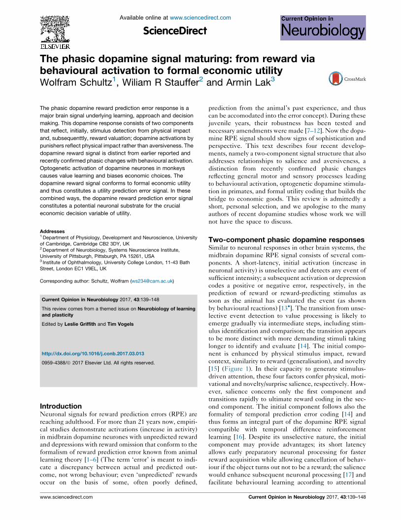
Figure 1
Novelty signal(1st component)
Value signal(2nd component)
p=0.75p=0.5p=0.25
p=0.75
p=0.5
p=0.25
0
0 1 s
30Learning trials
Res
pons
e
Rewardpredictingstimulus
0 30Learning trials
Current Opinion in Neurobiology
Two response components of dopamine neurones. During the learning
of visual stimuli predicting probabilistic rewards, the initial component
reflects stimulus novelty, as it decreases with repetition of the identical
stimulus but does not distinguish between different reward
probabilities (top). The second, subsequent component codes the
reward value of the stimulus (bottom); across successive learning
trials, this component increasingly discriminates between the three
stimuli predicting reward with different probability ( p = 0.25, p = 0.5,
p = 0.75). Not shown is the conclusion from other experiments,
suggesting that the initial activation reflects also physical impact,
similarity to rewarded stimuli, and reward context [13�]. Reprinted with
permission from Ref. [15].
learning theories [18]. The observation that the ultimate
reward elicits a graded RPE suggests that the prediction
from the second, value component lasts well beyond the
behavioural action [14]. Because of the early onset of the
lasting value component, the animal would not be con-
fused and be able to perform the action according to the
signalled value.
The issue of aversive dopamine activationThe two-component structure of the dopamine RPE
response may explain why dopamine research for
>30 years had consistently reported dopamine activations
to aversive stimuli that now turn out to result probably
from the physical impact of the punisher. Starting with
Chiodo et al. [19], followed by ourselves [20,21] and
confirmed by others [22–25], subsets of dopamine
neurones are activated by airpuffs and loud tones. A
recent study used several punishers, various physical
intensities, psychophysically assessment of aversive
values and multiple linear regression analysis to demon-
strate that the ‘aversive’ dopamine activation seems to
Current Opinion in Neurobiology 2017, 43:139–148
concern the initial dopamine response component and
reflect the physical intensity of punishers rather than their
negative aversive value [26�,27]. None of the investigated
dopamine neurones so far show an unanimous true aver-
sive activation in such well controlled conditions.
The physical impact attribution to ‘aversive’ dopamine
activations may affect the interpretation of aversiveness
in dopamine responses. First, dopamine activations by
reward are reduced by an added aversive liquid [27],
which follows an economic model of net benefit being
the sum of reward utility and punisher disutility. Second,
dopamine activation with aversive stimuli may signal
punishment relief, as voltammetric striatal dopamine
decreases with experienced aversive footshock but
increases with successfully avoided footshock [28�].Third, habenula stimulation induces place avoidance
(staying away due to signalled aversiveness) or place
dispreference (reduced preference due to depressed
reward signalling) [29,30,31], either by disynaptically
inhibiting reward-activated dopamine neurones and or
by monosynaptically exciting supposedly punisher-acti-
vated dopamine neurones. An interpretation towards
aversive dopamine activation seen with amazing molec-
ular methods [31] may be constrained by difficulties in
empirically distinguishing place avoidance from place
dispreference, by dopamine activation shown by Fos
possibly arising from rebound excitation of dopamine
neurones inhibited by habenula stimulation [23,30,32–
34], by excitatory postsynaptic currents possibly not
leading to propagated action potentials, and by blunting
from dopamine antagonists not distinguishing between
dopamine excitation and inhibition (although regretta-
bly misstated [13,34], the dopaminergic nature of the
neurones tested with Fos expression and whole-cell
recording were identified by tyrosine hydroxylase immu-
nostaining [31]).
Taken together, the typical dopamine response to pun-
ishers seems to be a depression of activity that reflects the
negative value RPE. Of course, despite all arguments and
reservations, the existence of some truly aversively acti-
vated dopamine neurones can not be completely
excluded, but the potential confound of physical impact
should be addressed experimentally.
Homogeneous RPE responses ofheterogeneous dopamine neuronesTruly aversive activations in subgroups of reward-proces-
sing dopamine neurones would indicate categorical
heterogeneity of dopamine RPE signalling. However,
the attribution of ‘aversive’ activations to the initial,
physical response component [27,35] rather suggests
graded sensitivities to physical impact, conforming to
one single distribution rather than to two statistically
distinct populations. This more nuanced view contrasts
with the notion of categorically different dopamine
www.sciencedirect.com
Phasic dopamine signals Schultz, Stauffer and Lak 141
populations suggested before the two-component nature
became recognised [23]. The notion of rather homoge-
neous RPE signalling is supported by the stereotyped
computational subtraction of received minus predicted
reward with correspondingly scaled sensitivities to posi-
tive and negative RPEs [35,36,37��], and the high noise
correlation and synchrony between dopamine responses
[37��,38]. As a functional consequence, if dopamine
responses vary along a single continuum, each dopamine
neurone contains the full RPE information, and single or
small groups of dopamine neurones effectively convey a
full RPE signal to postsynaptic neurones, without requir-
ing summed population activity through overlapping
projections [37��].
The issue is not whether dopamine neurones are hetero-
geneous or not; like all neurones, they differ in many
aspects including cytology, input source, co-neurotrans-
mitter, presynaptic interaction, reuptake transporter, pro-
jection territory, and presynaptic and postsynaptic recep-
tor location on heterogeneous postsynaptic neurones.
Additional, non-RPE activity changes reflecting more
global phenomena such as behavioural activation (see
below) are also heterogeneous when considering their
time course, polarity, event relationship and proportion of
neurones engaged. By contrast, what seems to be rather
homogeneous and stereotyped is the RPE response,
which varies in a graded rather than categorically distinct
manner across the otherwise heterogeneous dopamine
neurones. The similarity in dopamine RPE responses
contrasts with the classically well separated distributions
of heterogeneous neuronal responses to distinct task
events in prefrontal cortex, striatum, amygdala and most
other brain structures [39–41].
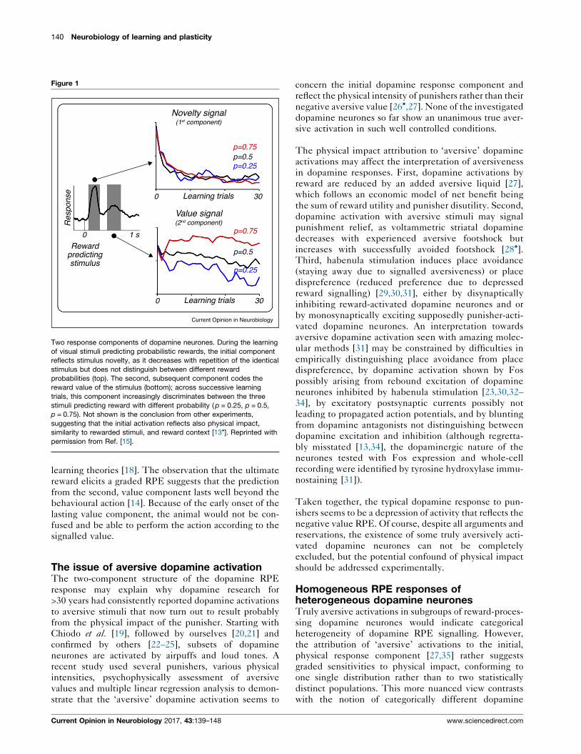
Phasic non-RPE dopamine changesGiven the notion that dopamine neurotransmission acts at
different time courses [42,43] and the renewed interest in
non-RPE signals [44�,45–49], it might be worth consider-
ing the initial studies of non-RPE dopamine signals.
During large arm reaching movements toward food boxes,
which engage a large proportion of arm and eye muscles in
monkeys, select subpopulations of dopamine neurones
are activated (31–44%) or depressed (15–17%) [50,51]
(Figure 2a,b), sometimes distinguishing between contra-
and ipsilateral reaching [50]. Similar activations occur
when these movements occur spontaneously (12%) in a
design that addressed the most impaired movement type
in Parkinsonism [52]. Mouth movements are also accom-
panied by activations (9%) and depressions (1%) [51]
(Figure 2c). Further, some dopamine neurones show slow
activations (5%) or depressions (1%) lasting over whole
trial durations of several seconds [51] (Figure 2d). Besides
the later identified RPE responses, this variety gives rise
to impressive schemes of heterogeneous activities that
likely reflect motor activation, sensory stimulation, and
general behavioural reactivity [50–52] (Figure 2e).
www.sciencedirect.com
However, these activities lack consistency, reproducibil-
ity and coherent functional interpretation. Clear move-
ment relationships are absent in dopamine neurones of
monkeys performing in Pavlovian or simple operant tasks
that separate well sensory from motor events and engage
only limited numbers of muscles; thus, well controlled
reaching to levers, precise elbow flexion-extensions and
ocular saccades consistently fail to activate dopamine
neurones in monkeys [53–55] (Figure 2f–h), as Pavlovian
licking fails to do in mice [12]. Specific analysis identifies
RPE coding as what initially appears to reflect movement
during spatial delayed-alternation and -response [1,55],
cognition during delayed matching-to-sample [56], sub-
jective perception during signal detection [11], and com-
plex behaviour during multistep choices [10]. So far, the
only known slower activation in simple tasks reflects
reward risk [57]. Thus, non-RPE dopamine changes occur
with general behavioural activation processes involving
many muscles and sensory receptors, but less so with
well-controlled movements involving fewer muscles.
Optogenetics is shifting behavioural neurophysiology
towards rodents. Many operant tasks used on rodents
engage large fractions of the body’s musculature and
sensory receptors. Neurophysiological, voltammetric
and optical imaging studies reveal fast and slow changes
in rodent midbrain dopamine neurones and striatal dopa-
mine axons. Global motor activity, stimuli and reward
delivery during T-maze navigation, whole-body turns,
locomotion, wheel running and nose pokes are associated
with dopamine activations [44�,45,47,48], depressions
[49] or both [46] (Figure 2i–k). Compatible with these
tasks engaging major body musculature and somatosen-
sory receptors, some of these activities vary parametrically
with locomotion velocity and acceleration [46,47] and
differ between ipsi- and contralateral movements in some
striatal regions [48]. These studies confirm the dopamine
changes with behavioural activation in monkeys and, in
addition, describe optogenetic stimulation effects on
locomotion speed [46] and willingness for operant perfor-
mance [58] reflecting reward value. Most of these rodent
studies demonstrate also RPE coding, although the many
overlapping sensory and motor components in these
complex behaviours render its precise identification dif-
ficult (as previously in monkeys [50,53]). More simple
Pavlovian or operant tasks comprise well-separated sti-
muli and well-controlled movements engaging limited
numbers of muscles; accompanied by sophisticated data
analysis, such tasks facilitate the assessment of reward
prediction and identification of RPEs [36,37��] and, as in
monkeys [53–55], do not seem to yield much non-RPE
activities.
The question arises whether an absence of dopamine
changes reflecting behavioural activation might provide
an explanation for Parkinsonian akinesia. The notion
would less easily apply to movement-related depression
Current Opinion in Neurobiology 2017, 43:139–148
142 Neurobiology of learning and plasticity
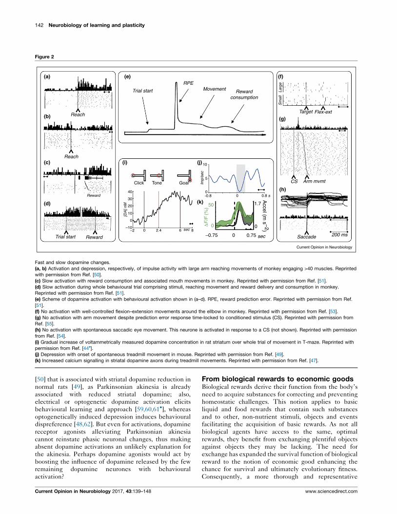
Figure 2
Reach
Reach
Reward
Reward
Trial start
Trial start
RPEMovement Reward
consumption
Target Flex-ext
CS Arm mvmt
Saccade 200 ms
Sm
all
Larg
e
40
Click Tone
[DA
] nM
Imp/
sec
Accel (m
s –2)
Goal
30
20
10
0
–10–2 0 2.4 6 sec 8
10
5
00
0
0
sec
1.7
0ΔF
/F (
%)
50
0.75–0.75
-0.8 0.8 s
(a)
(b)
(c) (i)
(e)
(j)
(k)
(f)
(g)
(h)
(d)
Current Opinion in Neurobiology
Fast and slow dopamine changes.
(a, b) Activation and depression, respectively, of impulse activity with large arm reaching movements of monkey engaging >40 muscles. Reprinted
with permission from Ref. [50].
(c) Slow activation with reward consumption and associated mouth movements in monkey. Reprinted with permission from Ref. [51].
(d) Slow activation during whole behavioural trial comprising stimuli, reaching movement and reward delivery and consumption in monkey.
Reprinted with permission from Ref. [51].
(e) Scheme of dopamine activation with behavioural activation shown in (a–d). RPE, reward prediction error. Reprinted with permission from Ref.
[51].
(f) No activation with well-controlled flexion–extension movements around the ellbow in monkey. Reprinted with permission from Ref. [53].
(g) No activation with arm movement despite prediction error response time-locked to conditioned stimulus (CS). Reprinted with permission from
Ref. [55].
(h) No activation with spontaneous saccadic eye movement. This neurone is activated in response to a CS (not shown). Reprinted with permission
from Ref. [54].
(i) Gradual increase of voltammetrically measured dopamine concentration in rat striatum over whole trial of movement in T-maze. Reprinted with
permission from Ref. [44�].(j) Depression with onset of spontaneous treadmill movement in mouse. Reprinted with permission from Ref. [49].
(k) Increased calcium signalling in striatal dopamine axons during treadmill movements. Reprinted with permission from Ref. [47].
[50] that is associated with striatal dopamine reduction in
normal rats [49], as Parkinsonian akinesia is already
associated with reduced striatal dopamine; also,
electrical or optogenetic dopamine activation elicits
behavioural learning and approach [59,60,61�], whereas
optogenetically induced depression induces behavioural
dispreference [48,62]. But even for activations, dopamine
receptor agonists alleviating Parkinsonian akinesia
cannot reinstate phasic neuronal changes, thus making
absent dopamine activations an unlikely explanation for
the akinesia. Perhaps dopamine agonists would act by
boosting the influence of dopamine released by the few
remaining dopamine neurones with behavioural
activation?
Current Opinion in Neurobiology 2017, 43:139–148
From biological rewards to economic goodsBiological rewards derive their function from the body’s
need to acquire substances for correcting and preventing
homeostatic challenges. This notion applies to basic
liquid and food rewards that contain such substances
and to other, non-nutrient stimuli, objects and events
facilitating the acquisition of basic rewards. As not all
biological agents have access to the same, optimal
rewards, they benefit from exchanging plentiful objects
against objects they may be lacking. The need for
exchange has expanded the survival function of biological
reward to the notion of economic good enhancing the
chance for survival and ultimately evolutionary fitness.
Consequently, a more thorough and representative
www.sciencedirect.com
Phasic dopamine signals Schultz, Stauffer and Lak 143
investigation of neuronal reward function should incor-
porate concepts of economic decision theory.
The survival value of rewards is determined by the
subjective needs of the agent rather than by physical
and chemical factors alone. The value of a third Porter-
house steak is lower than the value of the identical first
one; this is why all-you-can-eat restaurants can stay in
business. Populations of midbrain dopamine neurones,
and voltammetrically assessed striatal dopamine concen-
trations, code reward value on a subjective rather than a
physical basis, as seen with delays (temporal discounting)
[63,64], different reward types [65], risk [65], salt deple-
tion [66��] and effort [64,67,68]. Human striatal voltam-
metric dopamine changes reflect RPEs combined from
obtained and foregone (counterfactual) reward, rather
than from obtained reward alone [69]. Thus, dopamine
RPE signals reflect subjective value in a variety of
scenarios.
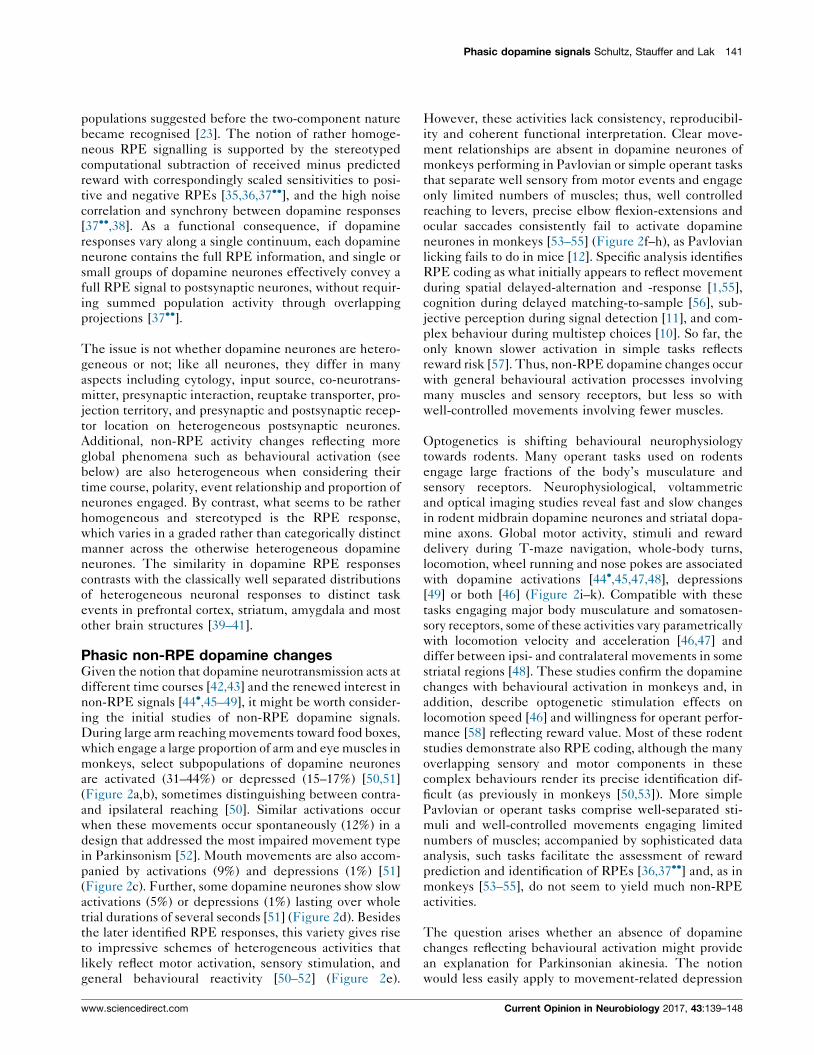
Although temporal delay, satiety, risk and effort contrib-
ute to subjective reward value, only formal economic
utility is normative for the choices individuals undertake
for maximising utility. Utility represents, on an internal
scale (utils), the subjective reward value as a mathemati-
cal function of objective value. Once estimated from
specific choices, a utility function predicts reward-max-
imising choices. As neuronal activity is quasi-continuous
and numeric, a utility function processed by neurones
should have similar properties. Such ‘cardinal’ utility
functions can be estimated under risk [70��], using the
fractile, chaining procedure with a specifically structured
series of psychophysically controlled choices between an
adjustable safe reward and a fixed binary, equiprobable
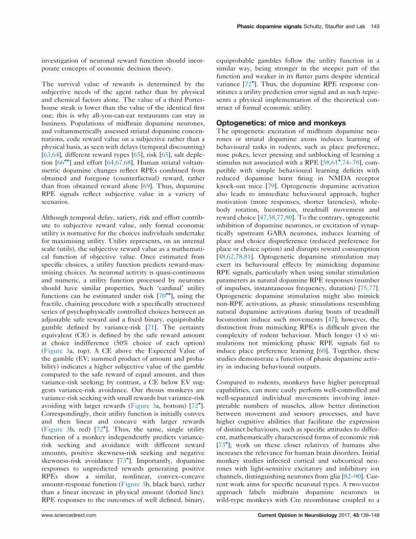
gamble defined by variance-risk [71]. The certainty
equivalent (CE) is defined by the safe reward amount
at choice indifference (50% choice of each option)
(Figure 3a, top). A CE above the Expected Value of
the gamble (EV; summed product of amount and proba-
bility) indicates a higher subjective value of the gamble
compared to the safe reward of equal amount, and thus
variance-risk seeking; by contrast, a CE below EV sug-
gests variance-risk avoidance. Our rhesus monkeys are
variance-risk seeking with small rewards but variance-risk
avoiding with larger rewards (Figure 3a, bottom) [72�].Correspondingly, their utility function is initially convex
and then linear and concave with larger rewards
(Figure 3b, red) [72�]. Thus, the same, single utility
function of a monkey independently predicts variance-
risk seeking and avoidance with different reward
amounts, positive skewness-risk seeking and negative
skewness-risk avoidance [73�]. Importantly, dopamine
responses to unpredicted rewards generating positive
RPEs show a similar, nonlinear, convex–concave
amount-response function (Figure 3b, black bars), rather
than a linear increase in physical amount (dotted line).
RPE responses to the outcomes of well defined, binary,
www.sciencedirect.com
equiprobable gambles follow the utility function in a
similar way, being stronger in the steeper part of the
function and weaker in its flatter parts despite identical
variance [72�]. Thus, the dopamine RPE response con-
stitutes a utility prediction error signal and as such repre-
sents a physical implementation of the theoretical con-
struct of formal economic utility.
Optogenetics: of mice and monkeysThe optogenetic excitation of midbrain dopamine neu-
rones or striatal dopamine axons induces learning of
behavioural tasks in rodents, such as place preference,
nose pokes, lever pressing and unblocking of learning a
stimulus not associated with a RPE [58,61�,74–78], com-
patible with simple behavioural learning deficits with
reduced dopamine burst firing in NMDA receptor
knock-out mice [79]. Optogenetic dopamine activation
also leads to immediate behavioural approach, higher
motivation (more responses, shorter latencies), whole-
body rotation, locomotion, treadmill movement and
reward choice [47,58,77,80]. To the contrary, optogenetic
inhibition of dopamine neurones, or excitation of synap-
tically upstream GABA neurones, induces learning of
place and choice dispreference (reduced preference for
place or choice option) and disrupts reward consumption
[48,62,78,81]. Optogenetic dopamine stimulation may
exert its behavioural effects by mimicking dopamine
RPE signals, particularly when using similar stimulation
parameters as natural dopamine RPE responses (number
of impulses, instantaneous frequency, duration) [75,77].
Optogenetic dopamine stimulation might also mimick
non-RPE activations, as phasic stimulations resembling
natural dopamine activations during bouts of treadmill
locomotion induce such movements [47]; however, the
distinction from mimicking RPEs is difficult given the
complexity of rodent behaviour. Much longer (1 s) sti-
mulations not mimicking phasic RPE signals fail to
induce place preference learning [60]. Together, these
studies demonstrate a function of phasic dopamine activ-
ity in inducing behavioural outputs.
Compared to rodents, monkeys have higher perceptual
capabilities, can more easily perform well-controlled and
well-separated individual movements involving inter-
pretable numbers of muscles, allow better distinction
between movement and sensory processes, and have
higher cognitive abilities that facilitate the expression
of distinct behaviours, such as specific attitudes to differ-
ent, mathematically characterised forms of economic risk
[73�]; work on these closer relatives of humans also
increases the relevance for human brain disorders. Initial
monkey studies infected cortical and subcortical neu-
rones with light-sensitive excitatory and inhibitory ion
channels, distinguishing neurones from glia [82–90]. Cur-
rent work aims for specific neuronal types. A two-vector
approach labels midbrain dopamine neurones in
wild-type monkeys with Cre recombinase coupled to a
Current Opinion in Neurobiology 2017, 43:139–148
144 Neurobiology of learning and plasticity
Figure 3
Choice safe <=> risky
eachp=0.5
0
1
Utility
EV
control
Injected
EV CE CE
100%
%100
50
50
50
0
0
0 trial
0
+
00
1
0.6 0.4 0.8 1.21.2Safe reward (ml)
Utility signal
Unpredicted reward (ml)
Cho
ice
of s
afe
rew
ard
Dop
amin
e ne
uron
e re
spon
ses
(nor
mal
ised
imp/
s)
0Dop
amin
e n
(nor
m
1.2(ml)
0.6ewardd (
50
0Cho
ice
of s
a
0Safe
0re
CS stim CS no stim Choice of stimulated object
CS 0.4 s
(a) (b)
(c) (d)
+
Current Opinion in Neurobiology
Dopamine utility prediction error signal and the effects of optogenetic stimulation.
(a) Top: choices between an adjustable safe reward and a fixed binary, equiprobable gamble, using eye or arm movements. Bottom:
psychophysical assessment of Certainty Equivalent (CE) at low and high reward amounts, respectively. The CE indicates the subjective value of
the gamble in ml of the safe juice reward (choice indifference). The CE > EV indicates risk seeking, CE < EV indicates risk avoidance. EV,
Expected Value of gamble.
(b) Dopamine responses to increasing reward amount follow nonlinear utility function. Red: utility function estimated from CEs in specifically
structured choices under risk. Black: average responses of 16 dopamine neurones to unpredicted reward outside of any behavioural task. (a) and
(b) Reprinted with permission from Ref. [72�].(c) Optogenetic stimulation increasing dopamine response to a conditioned, reward-predicting, visual stimulus (CS). The animal received laser
stimulation of dopamine neurons expressing channelrhodopsin 2 (ChR2) together with unpredicted juice reward (blue stimulus, left), or it receives
the reward alone (red stimulus, right).
(d) Developing oculomotor choice bias toward the blue, optogenetically stimulated CS and away from the red, unstimulated CS (for training
history, see (a)). Traces and dots, blue: choices (overall % and individual selections) of blue vs. red CS with laser stimulation in the ChR2-injected
midbrain (blue); red: laser stimulation in the contralateral, non-ChR2-injected midbrain. (a) and (b) Reprinted with permission from Ref. [91].
tyrosine hydroxylase promotor and inserts channelrho-
dopsin 2 dependent on the expressed Cre-recombinase
[91]. With >35% infection efficacy and >95% specificity,
laser stimulation induces impulses in dopamine neurones,
but not in other neurones in the area, usually in a <1:1
ratio. Laser stimulation simultaneously with unpredicted
juice reward enhances the dopamine responses to the
reward itself, makes an arbitrary visual stimulus acquire
reward-predicting properties and dopamine responses,
and gradually biases the animal’s oculomotor choices
towards the reward-cum-laser option and away from
Current Opinion in Neurobiology 2017, 43:139–148
the reward-only option (Figure 3c,d), thus validating
the notion of dopamine-mediated neuronal and
behavioural reinforcement in monkeys. These optoge-
netic behavioural effects are consistent with those
induced by electrical midbrain stimulation in monkeys
[91,92]. Thus, a first step has been done to transfer
neurone-type specific optogenetic techniques to
monkeys.
The question arises by which output route dopamine
stimulation may exert its long recognised behavioural
www.sciencedirect.com
Phasic dopamine signals Schultz, Stauffer and Lak 145
functions [59]. Among the many projection territories, the
dorsal and ventral striatum receive the densest dopamine
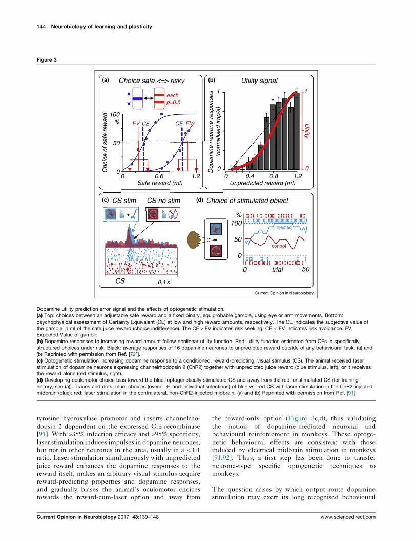
innervation. Schematically, the dopamine RPE signal
(Figure 4a) acts on different medium spiny neurone
classes in dorsal but not ventral striatum [93], via
D1-type receptors (motor-excitatory ‘direct’ pathway to
output) or via D2-type receptors (motor-inhibitory
‘indirect’ pathway). Optogenetic excitation of D1- and
D2-receptor-expressing striatal neurones induces gener-
ally opposite learning and immediate behavioural effects.
The opposing learning effects consist of increases and
decreases of self-stimulation [94] (Figure 4b) and of
changes of bimanual joystick movement speed [95].
The opposing immediate effects consist of biasing nose
poke target choices for reward towards contralateral and
ipsilateral body sides, respectively [96] (Figure 4c), and of
exciting and inhibiting the basal ganglia, thalamus and
cortex [97]. It is interesting to find such straightforward
Figure 4
Use predictionVt
Update prediction
Vt+1 = Vt + α (λt-V t- )
Generateerrorλt –Vt
Dopaminneurone
Receiveoutcome
Outcome =prediction Keep
predictionunchange
Outcome ≠prediction
-> Vt+1 λt
(a)
Effects of dopamine reward prediction error signalling on learning and choic
(a) Block diagramme of error-driven learning. The equation reflects the Resc
reward (reinforcer); t, trial). The red arrows denote the positive and negative
involved in learning and choices. The green-red triangles represent the two-
response).
(b) Learning of self-stimulation via optogenetic activation of mouse striatal n
increased frequency) or D2 receptors (red, decreased frequency). Reprinted
(c) Biasing of nose poke target choices toward contra- and ipsilateral sides
neurones, respectively. Reprinted with permission from Ref. [93].
www.sciencedirect.com
behavioural effects when stimulating striatal neurone
populations with stereotyped protocols that cannot incor-
porate the heterogenous patterns of striatal neurones
activitated with various sensory and motor task
components.
ConclusionsRecent advances in the behavioural neurophysiology of
the phasic dopamine signal seem to follow both a forward
path and a full circle. The forward path concerns the
development from reward response via neuronal reward
prediction error signal to the characterisation of neuronal
reward coding of subjective value and formal economic
utility. Subjective, rather than objective, neuronal value
coding matches intuitively the subjective aspects of
reward. Maybe more importantly, the neuronal coding
of utility signifies the conceptual step from biological
reward, required for survival, to economic good, allowing
es
Striatumneurones
d
Choices
Relative action value
p (le
ft)p
(left)
Learning
% r
espo
nses
D1 dopaminereceptorexpressing
D2 dopaminereceptorexpressing
D1 dopaminereceptorexpressing
contralateral
ipsilateral
D2 dopaminereceptorexpressing
75
50
25
0 Days 30
1
1
1
0
00-1
(c)
(b)
Current Opinion in Neurobiology
es.
orla–Wagner learning rule (V, associative strength or prediction; l,
dopamine prediction error signal and its action on striatal neurones
component dopamine signal (green, initial component; red, value
eurones that differentially express dopamine D1 receptors (blue,
with permission from Ref. [93].
by optogenetic activation of D1-expressing and D2-expressing striatal
Current Opinion in Neurobiology 2017, 43:139–148
146 Neurobiology of learning and plasticity
welfare enhancement by exchange; that exchange value
is based on utility.
The full circle concerns changes of dopamine activity
with large reaching movements of the arm that are,
embarrasingly, not replicated with fine, precise and
well-controlled movements in monkeys. The result of
the well-controlled studies is that the notion of a clear
movement relationship of phasic dopamine activity can-
not be maintained. However, in an apparent full circle
swing, recent work on rodents replicates the dopamine
changes during movements of the whole body or of large
parts of the body; these tasks engage considerable pro-
portions of the body’s muscles and sensory receptors. As a
result, it appears that the dopamine changes during large
movements probably concern general motor activation,
behavioural activation, or sensory stimulation derived
from moving, all of which is difficult to distinguish from
specific motor control. There is still no clear evidence of
phasic dopamine changes with precise, well-controlled
movements of concise numbers of muscles and devoid of
reasonable confounds.
Conflict of interest statementNothing declared.
AcknowledgementsOur work has been supported by the Wellcome Trust (095495, 106101),European Research Council (ERC, 293549), and NIH Conte Center atCaltech (P50MH094258).
References and recommended readingPapers of particular interest, published within the period of review,have been highlighted as:
� of special interest�� of outstanding interest
1. Ljungberg T, Apicella P, Schultz W: Responses of monkeymidbrain dopamine neurons during delayed alternationperformance. Brain Res. 1991, 586:337-341.
2. Schultz W, Apicella P, Ljungberg T: Responses of monkeydopamine neurons to reward and conditioned stimuli duringsuccessive steps of learning a delayed response task. J.Neurosci. 1993, 13:900-913.
3. Mirenowicz J, Schultz W: Importance of unpredictability forreward responses in primate dopamine neurons. J.Neurophysiol. 1994, 72:1024-1027.
4. Montague PR, Dayan P, Sejnowski TJ: A framework formesencephalic dopamine systems based on predictiveHebbian learning. J. Neurosci. 1996, 16:1936-1947.
5. Schultz W, Dayan P, Montague RR: A neural substrate ofprediction and reward. Science 1997, 275:1593-1599.
6. Schultz W: Predictive reward signal of dopamine neurons. J.Neurophysiol. 1998, 80:1-27.
7. Satoh T, Nakai S, Sato T, Kimura M: Correlated coding ofmotivation and outcome of decision by dopamine neurons. J.Neurosci. 2003, 23:9913-9923.
8. Bayer HM, Glimcher PW: Midbrain dopamine neurons encode aquantitative reward prediction error signal. Neuron 2005,47:129-141.
Current Opinion in Neurobiology 2017, 43:139–148
9. Day JJ, Roitman MF, Wightman RM, Carelli RM: Associativelearning mediates dynamic shifts in dopamine signaling in thenucleus accumbens. Nat. Neurosci. 2007, 10:1020-1028.
10. Enomoto K, Matsumoto N, Nakai S, Satoh T, Sato TK, Ueda Y,Inokawa H, Haruno M, Kimura M: Dopamine neurons learn toencode the long-term value of multiple future rewards. Proc.Natl. Acad. Sci. U. S. A. 2011, 108:15462-15467.
11. De Lafuente O, Romo R: Dopamine neurons code subjectivesensory experience and uncertainty of perceptual decisions.Proc. Natl. Acad. Sci. U. S. A. 2011, 108:19767-19771.
12. Cohen JY, Haesler S, Vong L, Lowell BB, Uchida N: Neuron-type-specific signals for reward and punishment in the ventraltegmental area. Nature 2012, 482:85-88.
13.�
Schultz W: Dopamine reward prediction error signalling: a two-component response. Nat. Rev. Neurosci. 2016, 17:183-195.
This review presents the most comprehensive account of the two-com-ponent dopamine response.
14. Nomoto K, Schultz W, Watanabe T, Sakagami M: Temporallyextended dopamine responses to perceptually demandingreward-predictive stimuli. J. Neurosci. 2010, 30:10692-10702.
15. Lak A, Stauffer WR, Schultz W: Dopamine neurons learn relativechosen value from probabilistic rewards. eLife 2016, 5 e18044.
16. Sutton RS, Barto AG: Reinforcement Learning. MIT Press; 1998.
17. Bushnell MC, Goldberg ME, Robinson DL: Behavioralenhancement of visual responses in monkey cerebral cortex. I.Modulation in posterior parietal cortex related to selectivevisual attention. J. Neurophysiol. 1981, 46:755-772.
18. Pearce JM, Hall G: A model for Pavlovian conditioning:variations in the effectiveness of conditioned but not ofunconditioned stimuli. Psychol. Rev. 1980, 87:532-552.
19. Chiodo LA, Antelman SM, Caggiula AR, Lineberry CG: Sensorystimuli alter the discharge rate of dopamine (DA) neurons:evidence for two functional types of DA cells in the substantianigra. Brain Res. 1980, 189:544-549.
20. Schultz W, Romo R: Responses of nigrostriatal dopamineneurons to high intensity somatosensory stimulation in theanesthetized monkey. J. Neurophysiol. 1987, 57:201-217.
21. Mirenowicz J, Schultz W: Preferential activation of midbraindopamine neurons by appetitive rather than aversive stimuli.Nature 1996, 379:449-451.
22. Joshua M, Adler A, Mitelman R, Vaadia E, Bergman H: Midbraindopaminergic neurons and striatal cholinergic interneuronsencode the difference between reward and aversive events atdifferent epochs of probabilistic classical conditioning trials.J. Neurosci. 2008, 28:11673-11684.
23. Matsumoto M, Hikosaka O: Two types of dopamine neurondistinctively convey positive and negative motivationalsignals. Nature 2009, 459:837-841.
24. Brischoux F, Chakraborty S, Brierley DI, Ungless MA: Phasicexcitation of dopamine neurons in ventral VTA by noxiousstimuli. Proc. Natl. Acad. Sci. U. S. A. 2009, 106:4894-4899.
25. Lerner TN, Shilyansky C, Davidson TJ, Luo L, Tomer R,Deisseroth K: Intact-brain analyses reveal distinct informationcarried by SNc dopamine subcircuits. Cell 2015, 162:635-647.
26.�
Fiorillo CD, Song MR, Yun SR: Multiphasic temporal dynamics inresponses of midbrain dopamine neurons to appetitive andaversive stimuli. J. Neurosci. 2013, 33:4710-4725.
This study was the first to distinguish ‘aversive’ dopamine activationsfrom the physical impact of punishers.
27. Fiorillo CD: Two dimensions of value: dopamine neuronsrepresent reward but not aversiveness. Science 2013, 341:546-549.
28.�
Oleson EB, Gentry RN, Chioma VC, Cheer JF: Subseconddopamine release in the nucleus accumbens predictsconditioned punishment and its successful avoidance. J.Neurosci. 2012, 32:14804-14808.
This study was the first to show that dopamine increases may derive from(rewarding) punishment relief.
www.sciencedirect.com
Phasic dopamine signals Schultz, Stauffer and Lak 147
29. Stamatakis AM, Stuber GD: Activation of lateral habenula inputsto the ventral midbrain promotes behavioural avoidance. Nat.Neurosci. 2012, 15:1105-1107.
30. Stopper CM, Tse MTL, Montes DR, Wiedman CR, Floresco SB:Overriding phasic dopamine signals redirects action selectionduring risk/reward decision making. Neuron 2014, 84:177-189.
31. Lammel S, Lim BK, Ran C, Huang KW, Betley MJ, Tye KM,Deisseroth K, Malenka RC: Input-specific control of reward andaversion in the ventral tegmental area. Nature 2012, 491:212-217.
32. Christoph GR, Leonzio RJ, Wilcox KS: Stimulation of the lateralhabenula inhibits dopamine-containing neurons in thesubstantia nigra and ventral tegmental area of the rat. J.Neurosci. 1986, 6:613-619.
33. Ji H, Shepard PD: Lateral habenula stimulation inhibits ratmidbrain dopamine neurons through a GABAA receptor-mediated mechanism. J. Neurosci. 2007, 27:6923-6930.
34. Schultz W: Neuronal reward and decision signals: fromtheories to data. Physiol. Rev. 2015, 95:853-951.
35. Fiorillo CD, Yun SR, Song MR: Diversity and homogeneity inresponses of midbrain dopamine neurons. J. Neurosci. 2013,33:4693-4709.
36. Eshel N, Bukwich M, Rao V, Hemmelder V, Tian J, NaoshigeUchida N: Arithmetic and local circuitry underlying dopamineprediction errors. Nature 2015, 525:243-246.
37.��
Eshel N, Tian J, Bukwich M, Naoshige Uchida N: Dopamineneurons share common response function for rewardprediction error. Nat. Neurosci. 2016, 19:479-486.
Together with Ref. [36], this study quantified the mechanisms of RPEcomputation common to dopamine neurones.
38. Joshua M, Adler A, Prut Y, Vaadia E, Wickens JR, HagaiBergman H: Synchronization of midbrain dopaminergicneurons is enhanced by rewarding events. Neuron 2009,62:695-704.
39. Fuster JM: Unit activity of prefrontal cortex during delayed-response performance: neuronal correlates of transientmemory. J. Neurophysiol. 1973, 36:61-78.
40. Hollerman JR, Tremblay L, Schultz W: Influence of rewardexpectation on behavior-related neuronal activity in primatestriatum. J. Neurophysiol. 1998, 80:947-963.
41. Grabenhorst F, Hernadi I, Schultz W: Prediction of economicchoice by primate amygdala neurons. Proc. Natl. Acad. Sci. U.S. A. 2012, 109:18950-18955.
42. Schultz W: Multiple dopamine functions at different timecourses. Ann. Rev. Neurosci. 2007, 30:259-288.
43. Schultz W: Behavioral dopamine signals. Trends Neurosci. 2007,30:203-210.
44.�
Howe MW, Tierney PL, Sandberg SG, Phillips PEM, Graybiel AM:Prolonged dopamine signalling in striatum signals proximityand value of distant rewards. Nature 2013, 500:575-579.
This study was the first to take up again presumably non-RPE-relateddopamine activity.
45. Totah NKB, Yunbok Kim Y, Moghaddam B: Distinct prestimulusand poststimulus activation of VTA neurons correlates withstimulus detection. J. Neurophysiol. 2013, 110:75-85.
46. Barter JW, Li S, Lu D, Bartholomew RA, Rossi MA, Shoemaker CT,Salas-Meza D, Gaidis E, Yin HH: Beyond reward predictionerrors: the role of dopamine in movement kinematics. Front.Neurosci. 2015, 9:39.
47. Howe MW, Dombeck DA: Rapid signalling in distinctdopaminergic axons during locomotion and reward. Nature2016, 535:505-510.
48. Parker NF, Cameron CM, Taliaferro JP, Lee J, Choi JY,Davidson TJ, Daw ND, Witten IB: Reward and choice encodingin terminals of midbrain dopamine neurons depends onstriatal target. Nat. Neurosci. 2016, 19:845-854.
www.sciencedirect.com
49. Dodson PD, Jakob Dreyer KJK, Jennings KA, Syeda ECJ, Wade-Martins R, Cragg SJ, Bolam JP, Magill PJ: Representation ofspontaneous movement by dopaminergic neurons is cell-typeselective and disrupted in parkinsonism. Proc. Natl. Acad. Sci.U. S. A. 2016, 113:E2180-E2188.
50. Schultz W, Ruffieux A, Aebischer P: The activity of parscompacta neurons of the monkey substantia nigra in relationto motor activation. Exp. Brain Res. 1983, 51:377-387.
51. Schultz W: Responses of midbrain dopamine neurons tobehavioral trigger stimuli in the monkey. J. Neurophysiol. 1986,56:1439-1462.
52. Romo R, Schultz W: Dopamine neurons of the monkeymidbrain: contingencies of responses to active touch duringself-initiated arm movements. J. Neurophysiol. 1990, 63:592-606.
53. DeLong MR, Crutcher MD, Georgopoulos AP: Relations betweenmovement and single cell discharge in the substantia nigra ofthe behaving monkey. J. Neurosci. 1983, 3:1599-1606.
54. Schultz W, Romo R: Dopamine neurons of the monkeymidbrain: contingencies of responses to stimuli elicitingimmediate behavioral reactions. J. Neurophysiol. 1990, 63:607-624.
55. Ljungberg T, Apicella P, Schultz W: Responses of monkeydopamine neurons during learning of behavioral reactions. J.Neurophysiol. 1992, 67:145-163.
56. Matsumoto M, Takada M: Distinct representations of cognitiveand motivational signals in midbrain dopamine neurons.Neuron 2013, 79:1011-1024.
57. Fiorillo CD, Tobler PN, Schultz W: Discrete coding of rewardprobability and uncertainty by dopamine neurons. Science2003, 299:1898-1902.
58. Hamid AA, Pettibone JR, Mabrouk OS, Hetrick VL, Schmidt R,Vander Weele CM, Kennedy RT, Aragona BJ, Berke JD:Mesolimbic dopamine signals the value of work. Nat. Neurosci.2016, 19:117-126.
59. Corbett D, Wise RA: Intracranial self-stimulation in relation tothe ascending dopaminergic systems of the midbrain: amoveable microelectrode study. Brain Res. 1980, 185:1-15.
60. Tsai H-C, Zhang F, Adamantidis A, Stuber GD, Bonci A, deLecea L, Deisseroth K: Phasic firing in dopaminergic neurons issufficient for behavioral conditioning. Science 2009, 324:1080-1084.
61.�
Steinberg EE, Keiflin R, Boivin JR, Witten IB, Deisseroth K,Janak PH: A causal link between prediction errors, dopamineneurons and learning. Nat. Neurosci. 2013, 16:966-973.
The classic and elegant study incorporating the previously describedlearning effects of electrical [59] and optogenetic [60,62] dopaminestimulation into a formal animal learning theory approach (blockingand unblocking).
62. Tan KR, Yvon C, Turiault M, Mirzabekov JJ, Doehner J,Labouebe G, Deisseroth K, Tye KM, Luscher C: GABA neurons ofthe VTA drive conditioned place aversion. Neuron 2012,73:1173-1183.
63. Kobayashi S, Schultz W: Influence of reward delays onresponses of dopamine neurons. J. Neurosci. 2008, 28:7837-7846.
64. Day JJ, Jones JL, Wightman RM, Carelli RM: Phasic nucleusaccumbens dopamine release encodes effort- and delay-related costs. Biol. Psychiatry 2010, 68:306-309.
65. Lak A, Stauffer WR, Schultz W: Dopamine prediction errorresponses integrate subjective value from different rewarddimensions. Proc. Natl. Acad. Sci. U. S. A. 2014, 111:2343-2348.
66.��
Cone JJ, Fortin SM, McHenry JA, Stuber GD, McCutcheon JE,Roitman MF: Physiological state gates acquisition andexpression of mesolimbic reward prediction signals. Proc.Natl. Acad. Sci. U. S. A. 2016, 113:1943-1948.
A particularly elegant study testing the appearance of reward value due tosalt depletion.
Current Opinion in Neurobiology 2017, 43:139–148
148 Neurobiology of learning and plasticity
67. Pasquereau B, Turner RS: Limited encoding of effort bydopamine neurons in a cost-benefit trade-off task. J. Neurosci.2013, 33:8288-8300.
68. Varazzani C, San-Galli A, Gilardeau S, Bouret S: Noradrenalineand dopamine neurons in the reward/effort trade-off: a directelectrophysiological comparison in behaving monkeys. J.Neurosci. 2015, 35:7866-7877.
69. Kishida KT, Saez I, Lohrenz T, Witcher MR, Laxton AW, Tatter SB,White JP, Ellis TL, Phillips PEM, Montague PR: Subseconddopamine fluctuations in human striatum encode superposederror signals about actual and counterfactual reward. Proc.Natl. Acad. Sci. U. S. A. 2016, 113:200-205.
70.��
von Neumann J, Morgenstern O: The Theory of Games andEconomic Behavior. Princeton University Press; 1944.
The classic and genius origin of Expected Utility Theory, and cardinalutility under risk, still valid after all those years with some additions (e.g.Prospect Theory).
71. Caraco T, Martindale S, Whitham TS: An empiricaldemonstration of risk-sensitive foraging preferences. Anim.Behav. 1980, 28:820-830.
72.�
Stauffer WR, Lak A, Schultz W: Dopamine reward predictionerror responses reflect marginal utility. Curr. Biol. 2014,24:2491-2500.
The conceptually important step from objective and subjective rewardvalue coding to formal economic utility signalling.
73.�
Genest W, Stauffer WR, Schultz W: Utility functions predictvariance and skewness risk preferences in monkeys. Proc.Natl. Acad. Sci. U. S. A. 2016, 113:8402-8407.
A behavioural study in monkeys showing the unique capacity of utilityfunctions to predict choices under different forms of risk.
74. Tsai H-C, Zhang F, Adamantidis A, Stuber GD, Bonci A, deLecea L, Deisseroth K: Phasic firing in dopaminergic neurons issufficient for behavioral conditioning. Science 2009, 324:1080-1084.
75. Witten IB, Steinberg EE, Lee SY, Davidson TJ, Zalocusky KA,Brodsky M, Yizhar O, Cho SL, Gong S, Ramakrishnan C,Stuber GD, Tye KM, Janak PH, Deisseroth K: Recombinase-driver rat lines: tools, techniques, and optogenetic applicationto dopamine-mediated reinforcement. Neuron 2011, 72:721-733.
76. Adamantidis AR, Tsai H-C, Boutrel B, Zhang F, Stuber GD,Budygin EA, Tourino C, Bonci A, Deisseroth K, de Lecea L:Optogenetic interrogation of dopaminergic modulation of themultiple phases of reward-seeking behavior. J. Neurosci. 2011,31:10829-10835.
77. Kim KM, Baratta MV, Yang A, Lee D, Boyden ES, Fiorillo CD:Optogenetic mimicry of the transient activation of dopamineneurons by natural reward is sufficient for operantreinforcement. PLoS One 2012, 7:e33612.
78. Ilango A, Kesner AJ, Keller KL, Stuber GD, Bonci A, Ikemoto S:Similar roles of substantia nigra and ventral tegmentaldopamine neurons in reward and aversion. J. Neurosci. 2014,34:817-822.
79. Zweifel LS, Parker JG, Lobb CJ, Rainwater A, Wall VZ, Fadok JP,Darvas M, Kim MJ, Mizumori SJ, Paladini CA, Philipps PEM,Palmiter R: Disruption of NMDAR-dependent burst firing bydopamine neurons provides selective assessment of phasicdopamine-dependent behavior. Proc. Natl. Acad. Sci. U. S. A.2009, 106:7281-7288.
80. Saddoris MP, Sugam JA, Stuber GD, Witten IB, Deisseroth K,Carelli RM: Mesolimbic dopamine dynamically tracks, and is
Current Opinion in Neurobiology 2017, 43:139–148
causally linked to, discrete aspects of value-based decisionmaking. Biol. Psychiatry 2015, 77:903-911.
81. van Zessen R, Phillips JL, Budygin EA, Stuber GD: Activation ofVTA GABA neurons disrupts reward consumption. Neuron2012, 73:1184-1194.
82. Han X, Qian X, Bernstein JG, Zhou HH, Franzesi GT, Stern P,Bronson RT, Graybiel AM, Desimone R, Boyden ES:Millisecondtimescale optical control of neural dynamics in thenonhuman primate brain. Neuron 2009, 62:191-198.
83. Diester I, Kaufman MT, Mogri M, Pashaie R, Goo W, Yizhar O,Ramakrishnan C, Deisseroth K, Shenoy KV: An optogenetictoolbox designed for primates. Nat. Neurosci. 2011, 14:387-397.
84. Cavanaugh J, Monosov IE, McAlonan K, Berman R, Smith MK,Cao V, Wang KH, Boyden ES, Wurtz RH: Optogeneticinactivation modifies monkey visuomotor behavior. Neuron2012, 76:901-907.
85. Galvan A, Hu X, Smith Y, Wichmann T: In vivo optogeneticcontrol of striatal and thalamic neurons in non-humanprimates. PLoS One 2012, 7:e50808.
86. Galvan A, Hu X, Smith Y, Wichmann T: Effects of optogeneticactivation of corticothalamic terminals in the motor thalamusof awake monkeys. J. Neurosci. 2016, 36:3519-3530.
87. Gerits A, Farivar R, Rosen BR, Wald LL, Boyden ES, Vanduffel W:Optogenetically induced behavioral and functional networkchanges in primates. Curr. Biol. 2012, 22:1722-1726.
88. Jazayeri M, Lindbloom-Brown Z, Horwitz GD: Saccadic eyemovements evoked by optogenetic activation of primate V1.Nat. Neurosci. 2012, 15:1368-1370.
89. Ohayon S, Grimaldi P, Schweers N, Tsao DY: Saccademodulation by optical and electrical stimulation in themacaque frontal eye field. J. Neurosci. 2013, 33:16684-16697.
90. Dai J, Brooks DI, Sheinberg DL: Optogenetic and electricalmicrostimulation systematically bias visuospatial choice inprimates. Curr. Biol. 2014, 24:63-69.
91. Stauffer WR, Lak A, Yang A, Borel M, Paulsen O, Boyden E,Schultz W: Dopamine neuron-specific optogenetic stimulationin Rhesus macaques. Cell 2016, 166:1564-1571.
92. Arsenault JT, Rima S, Stemmann H, Vanduffel W: Role of theprimate ventral tegmental area in reinforcement andmotivation. Curr. Biol. 2014, 24:1347-1353.
93. Kupchik YM, Brown RM, Heinsbroek JA, Lobo MK, Schwartz DJ,Kalivas PW: Coding the direct/indirect pathways by D1 and D2receptors is not valid for accumbens projections. Nat.Neurosci. 2015, 18:1230-1232.
94. Kravitz AV, Tye LD, Kreitzer AC: Distinct roles for direct andindirect pathway striatal neurons in reinforcement. Nat.Neurosci. 2012, 15:816-818.
95. Yttri EA, Dudman JT: Opponent and bidirectional control ofmovement velocity in the basal ganglia. Nature 2016, 533:402-406.
96. Tai L-H, Lee AM, Benavidez N, Bonci A, Wilbrecht L: Transientstimulation of distinct subpopulations of striatal neuronsmimics changes in action value. Nat. Neurosci. 2012, 15:1281-1289.
97. Lee HJ, Weitz AJ, Bernal-Casas D, Duffy BA, Choy MK, Kravitz AV,Kreitzer AC, Lee JH: Activation of direct and indirect pathwaymedium spiny neurons drives distinct brain-wide responses.Neuron 2016, 91:412-424.
www.sciencedirect.com