The Pharmaceutical and Chemical Journal, 2018,...
14
The Pharmaceutical and Chemical Journal, 2018, 5(1):211-224 The Pharmaceutical and Chemical Journal 211 Available online www.tpcj.org Research Article ISSN: 2349-7092 CODEN(USA): PCJHBA Bioactive Compounds from Marine Fungus Penicillium citrinum Strain ND7c by Gas Chromatography-Mass Spectrometry Nguyen Tan Binh, Phan Van Ha Lam, Cao Ngoc Diep* Biotechnology R&D Institute, Can Tho University, Viet nam Abstract The objectives of this study were analysis of the secondary metabolite products from extract of marine Penicillium citrinum strain ND7a which isolated from sponges at Ha Tien Sea, Kien Giang province, Vietnam. Sixteen bioactive compounds were identified in the organic solvent hexan-aceton and aceton-methanol. The identification of bioactive chemical compounds is based on the peak area, retention time, molecular weight and molecular formular. GC-MS analysis of Penicillium citrinum strain ND7a revealed the existence of the 9- Hexadecenoic acid, Hexadecane, n-Hexadecanoic acid, Heptacosane, Octadecane, 3-ethyl-5-(2-ethylbutyl), Tributyl acetylcitrate, 17-Pentatriacontene, Hexanedioic acid, bis(2-ethylhexyl) ester, Bis(2-ethylhexyl) phthalate (in organic solvent hexan-aceton) and Oxime-, methoxy-phenyl, 2-Hydroxy-gamma-butyrolactone, 7,9-Di-tert-butyl-1- oxaspiro(4,5)deca-6,9-diene-2,8-dione, Pentadecanoic acid, 14-methyl-, methyl ester, Hexadecanoic acid, 1- (hydroxymethyl)-1,2-ethanediyl ester, Stigmasterol, γ-Sitosterol (in organic solvent aceton-methanol). Keywords bioactive compounds, GC-MS, Ha Tien Sea, Marine sponge, Penicillium citrinum 1. Introduction In the last decade, marine microorganisms, such as bacteria, microalgae and fungi, have become increasingly important as sources for new bioactive natural products [1-5]. Marine microorganisms have been the important study in recent years because of production of novel metabolites which represent various biological properties such as antiviral, antitumor or antimicrobial activities. Fungi have proven to be particularly prolific sources of new compounds when compared to other microbial sources isolated from the sea. The first report of a bioactive natural product from a marine-derived fungus dates back to the 1940s when the fungus Acremonium chrysogenum Gams 1971 was isolated from a sewage outlet in the Mediterranean Sea close to the island of Sardinia. The fungus was the source of cephalosporin C, the parent compound of modern cephalosporin antibiotics that are indispensible for the treatment of numerous bacterial infections [6]. Initially, progress with rigorous evaluation of marine fungal metabolites was slow. This situation changed dramatically in the 1990s when there was a sharp rise in interest in marine microbial metabolites that continues until today. Up to the year 2002, 272 new natural products had been isolated from marine-derived fungi [7]. A dramatic increase in the number of elucidated marine fungal structures began afterwards, illustrated by the fact that between 2002 and 2004, 240 additional compounds were described [8]. Even though the value of marine-derived fungi as a source of new bioactive metabolites is now commonly accepted, there is still much debate on the nature of fungi that are isolated from various marine substrates, such as drifting wood, algae or invertebrates. Due to the fact that numerous, if not most, of these fungi belong to genera already well known from the terrestrial environment, such as Aspergillus, Penicillium, Cladosporium, Phoma, and Fusarium, a true marine origin of these fungal strains is frequently doubted [9-10]. It is possible that several, if not many, marine-derived fungi thus far investigated originated from terrestrial habitats (e.g., soil) from which they were
Transcript of The Pharmaceutical and Chemical Journal, 2018,...

The Pharmaceutical and Chemical Journal, 2018, 5(1):211-224
The Pharmaceutical and Chemical Journal
211
Available online www.tpcj.org
Research Article
ISSN: 2349-7092
CODEN(USA): PCJHBA
Bioactive Compounds from Marine Fungus Penicillium citrinum Strain ND7c by Gas
Chromatography-Mass Spectrometry
Nguyen Tan Binh, Phan Van Ha Lam, Cao Ngoc Diep*
Biotechnology R&D Institute, Can Tho University, Viet nam
Abstract The objectives of this study were analysis of the secondary metabolite products from extract of marine
Penicillium citrinum strain ND7a which isolated from sponges at Ha Tien Sea, Kien Giang province, Vietnam.
Sixteen bioactive compounds were identified in the organic solvent hexan-aceton and aceton-methanol. The
identification of bioactive chemical compounds is based on the peak area, retention time, molecular weight and
molecular formular. GC-MS analysis of Penicillium citrinum strain ND7a revealed the existence of the 9-
Hexadecenoic acid, Hexadecane, n-Hexadecanoic acid, Heptacosane, Octadecane, 3-ethyl-5-(2-ethylbutyl), Tributyl
acetylcitrate, 17-Pentatriacontene, Hexanedioic acid, bis(2-ethylhexyl) ester, Bis(2-ethylhexyl) phthalate (in organic
solvent hexan-aceton) and Oxime-, methoxy-phenyl, 2-Hydroxy-gamma-butyrolactone, 7,9-Di-tert-butyl-1-
oxaspiro(4,5)deca-6,9-diene-2,8-dione, Pentadecanoic acid, 14-methyl-, methyl ester, Hexadecanoic acid, 1-
(hydroxymethyl)-1,2-ethanediyl ester, Stigmasterol, γ-Sitosterol (in organic solvent aceton-methanol).
Keywords bioactive compounds, GC-MS, Ha Tien Sea, Marine sponge, Penicillium citrinum
1. Introduction
In the last decade, marine microorganisms, such as bacteria, microalgae and fungi, have become increasingly
important as sources for new bioactive natural products [1-5]. Marine microorganisms have been the important
study in recent years because of production of novel metabolites which represent various biological properties such
as antiviral, antitumor or antimicrobial activities. Fungi have proven to be particularly prolific sources of new
compounds when compared to other microbial sources isolated from the sea. The first report of a bioactive natural
product from a marine-derived fungus dates back to the 1940s when the fungus Acremonium chrysogenum Gams
1971 was isolated from a sewage outlet in the Mediterranean Sea close to the island of Sardinia. The fungus was the
source of cephalosporin C, the parent compound of modern cephalosporin antibiotics that are indispensible for the
treatment of numerous bacterial infections [6]. Initially, progress with rigorous evaluation of marine fungal
metabolites was slow. This situation changed dramatically in the 1990s when there was a sharp rise in interest in
marine microbial metabolites that continues until today. Up to the year 2002, 272 new natural products had been
isolated from marine-derived fungi [7]. A dramatic increase in the number of elucidated marine fungal structures
began afterwards, illustrated by the fact that between 2002 and 2004, 240 additional compounds were described [8].
Even though the value of marine-derived fungi as a source of new bioactive metabolites is now commonly accepted,
there is still much debate on the nature of fungi that are isolated from various marine substrates, such as drifting
wood, algae or invertebrates. Due to the fact that numerous, if not most, of these fungi belong to genera already well
known from the terrestrial environment, such as Aspergillus, Penicillium, Cladosporium, Phoma, and Fusarium, a
true marine origin of these fungal strains is frequently doubted [9-10]. It is possible that several, if not many,
marine-derived fungi thus far investigated originated from terrestrial habitats (e.g., soil) from which they were

Diep CN et al The Pharmaceutical and Chemical Journal, 2018, 5(1):211-224
The Pharmaceutical and Chemical Journal
212
washed to the sea and survived (as spores) until they were recovered by a marine chemist looking for new
compounds from the sea. On the other hand, in the last few years more and more evidence has accumulated
indicating an adaptation of these ‘‘ubiquitous’’ fungi to the marine environment [11-15]. Nevertheless, the fact
remains that regardless of their true origin, marine-derived fungi have developed into an important source of new
and structurally unprecedented metabolites [8]. Interestingly, sponges continue to be one of the most important
sources for the isolation of metabolite-producing marine-derived fungi [7-9, 16], even though the presence of fungal
mycelia growing in sponges has not yet been proven. Meanwhile, the search of bioactive secondary metabolites
from marine microorganisms is not widely explored in Vietnam [17-18].
In the past few years, Gas chromatography Mass spectrometry (GC-MS) is used as one of the technological platform
for finger print analysis of secondary metabolites in both plant and non-plant species [19]. Taking into consideration
the medicinal importance of this plant, the ethyl acetate root extract of medical plant [20] and/or leaves as Neem
(Azadirachta Indica A. Zuss) [21], flowers Holarrhena antidysentrica Wall [22] were analyzed using GC-MS. This
work will help to identify the bioactive components. GC-MS is the best technique to identify bioactive constituents
of long chain hydrocarbons, alcohols, acids, ester, alkaloids, steroids, amino and nitro compound etc. [20].
In the course of our screening program, the EtOAc extract of a Penicillium citrinum strain ND7a from marine
sponge of Ha Tien Sea, Kien Giang province, Vietnam exhibited an inhibition activity against Salmonella
typhymurium, Echerichia coli, Bacillus cereus and Candida albicans. In this paper, we reported the isolation and
structural elucidation of secondary metabolites from the cultures broth Penicillium citrinum strain ND7a of in two
kinds of organic solvent. The present study was aimed to identify the chemical constituents in ethyl acetate extract
of marine fungus was analyzed by the GC-MS technique.
2. Materials and methods
2.1. Fungus material
The marine sponge was collected in Ha Tien Bay–Kien Giang province in April 2016. The sponge sample (1 g) was
added to the 10 mL of sterile sea water in a conical flask. The flask was agitated for about one hour. The marine
sponge was filtered and the filtrate was serially diluted to obtain 10-1
to 10-7
dilutions using the sterilized sea water.
An aliquot of 100 μL of each dilution was spread on the Glucose Peptone Yeast Extract Agar (GPY) media. The
media containing 50% of sterile sea water were supplemented with ampiciline and streptomycine (100 μg/mL)
(Himedia Mumbai) to inhibit bacterial contamination, respectively. The petriplates were incubated up to 3 weeks at
28°C. The isolated discrete colonies were observed and used for identification.
The obtained fungus strain was identified by using 18S rRNA gene sequencing method. The universal primers
including forward primer, ITS1 and reverse primer, ITS4 were used for amplifying nearly full length of 18S rRNA
gene sequence from 550 to 800 bp [23]. The obtained sequence was analyzed by comparing with bacterial 18S
rRNA sequences in GenBank by BlastN, which showed 99% similarity with Penicillium citrinum strain TDD
(GenBank Accession No. JX192960).
2.2. Fermentation, extraction and isolation
Penicillium citrinum strain ND7a was cultured in 250 ml flasks at 30oC for 24 hours with shaking at 150 rpm.
Fermentation was carried out in 100 L fermenter with 50 L PDA medium and 10% fungal inoculum at 30oC for 52
hours. Neutral pH was maintained automatically by NaOH or HCl 1N. The obtained culture broth (50 L) was
extracted with ethyl acetate (25 L × 3 times). The combined organic solutions were then decanted, filtered and
concentrated under reduced pressure to yield 8.1 g of crude extract which was chromatographed on a silica gel
column using a gradient of 1 - 100% acetone in hexane to afford three fractions F1-9, after that it was continuously
chromatographed on a silica gel column using a gradient of 1 - 100% acetone in methanol to afford three fractions
F10-16. Therefore, sixteen fractions were received from 2 kinds of organic solvent (hexane – acetone and acetone –
methanol).
2.3. GC/MS analysis
The samples were analysed at GC/MS of Chemistry Laboratoty, Department of Chemistry, College of Natural
Science, Can Tho University. GC-MS analysis of the sample was carried out using Shimadzu Thermo with column
Diep CN et al The Pharmaceutical and Chemical Journal, 2018, 5(1):211-224
The Pharmaceutical and Chemical Journal
213
TG-SQC; 15m x 0.25mm x 0.25µm. Helium was used as the carrier gas and the temperature programming was set
as follows:
Speed (oC/min) Temperature (
oC) Keep (min)
Initial 50 1.00
Ramp 1 2.00 70 2.00
Ramp 2 10.00 150 2.00
Ramp 3 10.00 250 10.00
Total time 43 minutes
10 µl sample was injected with split less mode. Mass spectra was recorded over 35-400 amu range with electron
impact ionization energy 70 eV, total running time for a sample was 43 min. Quantitative determination were made
by relating respective peak areas to TIC areas from GC-MS.
3. Results and Discussion
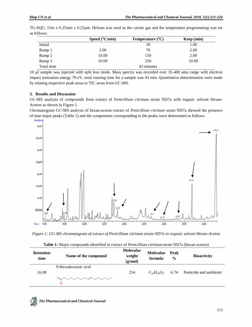
GC-MS analysis of compounds from extract of Penicillium citrinum strain ND7a with organic solvent Hexan-
Aceton as shown in Figure 1.
Chromatogram GC-MS analysis of hexan-aceton extract of Penicillium citrinum strain ND7a showed the presence
of nine major peaks (Table 1) and the components corresponding to the peaks were determined as follows.
Figure 1: GC-MS chromatogram of extract of Penicillium citrinum strain ND7a in organic solvent Hexan-Aceton
Table 1: Major compounds identified in extract of Penicillium citrinum.strain ND7a (hexan-aceton)
Retention
time
Name of the compound
Molecular
weight
(g/mol)
Molecular
formula
Peak
%
Bioactivity
16.08
9-Hexadecenoic acid
254
C16H30O2
6.74
Pesticide and antibiotic

Diep CN et al The Pharmaceutical and Chemical Journal, 2018, 5(1):211-224
The Pharmaceutical and Chemical Journal
214
17.95
Hexadecane
226
C16H34
18.6
Antimicrobial,
antioxidant,
antidiabetic
22.95
n-Hexadecanoic acid
256
C16
H32
O2
44.6
Antibacterial
23.31
Heptacosane
380
C27
H56
7.58
Antimicrobial
26.70
Octadecane, 3-ethyl-5-(2-
ethylbutyl)
366
C26
H54
14.6
Antimicrobial
antifungal
27.73
Tributyl acetylcitrate
402
C20
H34
O8
43.2
Anticancer activity
Antimicrobial activity
29.19
17-Pentatriacontene
490
C35
H70
8.72
Antibacterial, antiviral
30.78
Hexanedioic acid, bis(2-ethylhexyl)
ester
370
C22
H42
O4
56.1
Antioxidant
antimicrobial,
antiproliferative
33.22
Bis(2-ethylhexyl) phthalate
390
C24
H38
O4
38.5
Antibacterial and
antifungal agent

Diep CN et al The Pharmaceutical and Chemical Journal, 2018, 5(1):211-224
The Pharmaceutical and Chemical Journal
215
The first set up peak was determined to be 9-Hexadecenoic acid (Figure 2).
Figure 2: Mass spectrum of 9-Hexadecenoic acid with retention time (RT) = 16.08
The second peak indicated to be Hexadecane (Figure 3) and the third peaks considered to be n-Hexadecanoic acid
(Figure 4).
Figure 3: Mass spectrum of Hexadecane
with RT = 17.95
Figure 4: Mass of spectrum of n-Hexadecanoic acid
with RT = 22.95

Diep CN et al The Pharmaceutical and Chemical Journal, 2018, 5(1):211-224
The Pharmaceutical and Chemical Journal
216
The fourth peak indicated to be Heptacosane (Figure 5) and the fifth peaks considered to be Octadecane, 3-ethyl-5-
(2-ethylbutyl) (Figure 6).
Figure 5: Mass spectrum of Heptacosane with RT = 23.31
Figure 6: Mass spectrum of Octadecane, 3-ethyl-5-(2-
ethylbutyl) with RT = 26.70
Figure 7: Mass spectrum of Tributyl acetylcitrate with
RT = 27.73

Diep CN et al The Pharmaceutical and Chemical Journal, 2018, 5(1):211-224
The Pharmaceutical and Chemical Journal
217
The sixth peak determined to be Tributyl acetylcitrate (Figure 7) and seventh peakconsidered to be 17-
Pentatriacontene (Figure 8)
Figure 8: Mass spectrum of 17-Pentatriacontene with RT = 29.19
The eighth peak indicated to be Hexanedioic acid bis(2-ethylhexyl) ester (Figure 9), and the ninth peak determined
to be Bis(2-ethylhexyl) phthalate (Figure 10).
Figure 9: Mass spectrum of Hexanedioic acid bis(2-
ethylhexyl) ester with RT = 30.78
Figure 10: Mass spectrum of Bis(2-ethylhexyl)
phthalate with RT = 33.22

Diep CN et al The Pharmaceutical and Chemical Journal, 2018, 5(1):211-224
The Pharmaceutical and Chemical Journal
218
Shettima et al. [24] discovered nine compounds from the ethyl acetate root extract of Guiera senegalensis were
identified by Gas-chromatography–Mass spectrometry (GC-MS) analysis. The biological activities of each of the
identified phytocomponents range from antimicrobial, antioxidant and antitumoral activities. The nature of the
identified compounds are mostly organic acids and they also found 9-Hexadecenoic acid, is one in nine compounds
from the root of Guiera senegalensis. Paramanantham and Murugesan [22] analysed the flowers extract show the
presence of 30 phyto compounds and they found Hexadecane and Hexanedioic acid, bis(2-ethylhexyl) ester as
various bioactive compounds justifies the use of the plant flower for various ailments by traditional practitioners.
When using GC-MS analysis of one Ayurvedic medicine Talisapatradi Churnam, this medicine is used to treat
respiratory and digestive disorders. The medicine was subjected to DPPH, FRAP and Hydrogen Peroxide
scavenging assays and it was found that it has good antioxidant potential. The GC MS analysis results indicated the
presence of twenty six bio molecules and n-Hexadecanoic acid and Octadecane, 3-ethyl-5-(2-ethylbutyl) in this
medicine [25]. When study on the phytochemical composition of Adiantum capillus-veneris and to evaluate the
isolates for possible in vitro antifungal and antibacterial activities. The compound obtained were screened by GC-
MS method. While agar-well diffusion method was employed to measure antimicrobial activity against five bacteria
and fourteen fungi and yeast. Thirty-one bioactive phytochemical compounds were identified in the methanolic
extract of Adiantum capillus-veneris. Tributyl acetylcitrate found was one in 31 bioactive phytochemical compounds
[26]. The presence of five secondary bioactive components with 17-pentatriacontene has been identified in
Turbinaria ornata, brown alga, of which four bioactive compounds [27]. Al-Bari et al. [28] isolated Streptomyces
bangladeshensis sp. nov., from soil, which produces bis-(2-ethylhexyl)phthalate, as antimicrobial agent.
GC-MS analysis of compounds from extract of Streptomyces sp. strain ND7a with organic solvent Aceton-Methanol
as shown in Figure 11.
Figure 11: GC-MS chromatogram of extract of Penicillium citrinum strain ND7a in organic solvent Aceton-
Methanol
Chromatogram GC-MS analysis of hexan-aceton extract of Penicillium citrinum strain ND7a showed the presense
of seven major peaks (Table 2) and the components corresponding to the peaks were determined as follows.

Diep CN et al The Pharmaceutical and Chemical Journal, 2018, 5(1):211-224
The Pharmaceutical and Chemical Journal
219
Table 2: Major compounds identified in extract of Penicillium citrinum strain ND7a (aceton-methanol)
Retention
time
Name of the compound Molecular
weight
(g/mol)
Molecular
formula
Peak
%
Bioactivity
6.30 Oxime-, methoxy-phenyl
151 C8H
9NO
2 81.5 Antifungal,
Antibacterial,
Anticancer and
Antitumor
7.82 2-Hydroxy-gamma-butyrolactone
102 C4H
6O
3
47.4 Antioxidant, analgesic,
anti-diabetic,
antibacterial, and
antifungal activity
22.13 7,9-Di-tert-butyl-1-oxaspiro(4,5)deca-
6,9-diene-2,8-dione
276 C17
H24
O3
68.7 Antimicrobial
22.34 Pentadecanoic acid, 14-methyl-, methyl
ester
270 C17
H34
O2
26.6 Antifungal,
Antimicrobial
22.81 Hexadecanoic acid, 1-
(hydroxymethyl)-1,2-ethanediyl ester
568 C35
H68
O5
34.1 Antimicrobial activity
38.36 Stigmasterol
412 C29
H48
O
50.4 Antimicrobial
40.16 γ-Sitosterol
414 C29
H50
O 85.3 Antioxidant,
antibacterial and
prophylactic activities
The first set up peak was determined to be Oxime-,methoxy-phenyl (Figure 12) and the second peak considered to
be 2-Hydroxy-gamma-butyrolactone (Figure 13).

Diep CN et al The Pharmaceutical and Chemical Journal, 2018, 5(1):211-224
The Pharmaceutical and Chemical Journal
220
Figure 12: Mass spectrum of Oxime-,methoxy-phenyl
with RT = 6.30
Figure 13: Mass spectrum of 2-Hydroxy-gamma-
butyrolactone with RT = 7.82
Figure 14: Mass spectrum of 7,9-Di-tert-butyl-1-oxaspiro(4,5)deca-6,9-diene-2,8-dione with RT = 22.13
The third peak determined to be 7,9-Di-tert-butyl-1-oxaspiro(4,5)deca-6,9-diene-2,8-dione (Figure 14), the fourth
peak indicated to be Pentadecanoic acid, 14-methyl-, methyl ester (Figure 15), the fifth peak considered to be
Hexadecanoic acid, 1-(hydroxymethyl)-1,2-ethanediyl ester (Figure 16).

Diep CN et al The Pharmaceutical and Chemical Journal, 2018, 5(1):211-224
The Pharmaceutical and Chemical Journal
221
Figure 15: Mass spectrum of Pentadecanoic acid, 14-
methyl-, methyl ester with RT = 22.34
Figure 16: Mass spectrum of Hexadecanoic acid, 1-
(hydroxymethyl)-1,2-ethanediyl ester with RT = 22.81
The sixth peak indicated to be Stigmasterol (Figure 17) and the seventh peak determined to be γ-Sitosterol (Figure
18).
Figure 17: Mass spectrum of Stigmasterol with RT =
38.36
Figure 18: Mass spectrum of γ-Sitosterol with RT =
40.16

Diep CN et al The Pharmaceutical and Chemical Journal, 2018, 5(1):211-224
The Pharmaceutical and Chemical Journal
222
Based on the data analysis of GC/MS identified 45 bioactive compounds in the n-Hexane extract of Azadirachta
indica leaves out of which 33 have antimicrobial and antifungal activity; Akpuaka et al. [21] found Oxime-,
methoxy-phenyl-, Pentadecanoic acid, 14-methyl-, methyl ester and γ-Sitosterol in Azadirachta indica leaves.
Furthermore, Al-Tamene et al. [29] and Altaee et al. [30] also found Oxime-, methoxy-phenyl-3, Oxime-, methoxy-
phenyl-, Oxime-, methoxy-phenyl-2, in Urtica dioica leaves and Volatile Compounds produced by Pseudomonas
aeruginosa Isolated from UTI Patients, respectively. Altaee et al. [30] showed that Pseudomonas aeruginosa
produce many important secondary metabolites with high biological activities. Park et al. [31] used GC/MS analysis
confirmed the predominant components of garlic extract to be 2-Hydroxy-gamma-butyrolactone in 75% ethanol
extract was the most efficient in terms of the recovery rate and antimicrobial and antioxidant activities. Arora and
Kumar [32] used GC–MS analyses root and stem of Cenchrus setigerus Vahl (Poaceae) showed that majority of
these identified compounds in various crude extracts contain the high percentage of compounds that were identified
in the crude extracts shows chemical and biological importance are bioactive compounds as 7,9-Di-tert-butyl-1-
oxaspiro(4,5)deca-6,9-diene-2,8-dione in methanolic and ethyl acetate extractant which had high antimicrobial.
Hexadecanoic acid, 1-(hydroxymethyl)-1,2-ethanediyl ester was in thirty three bioactive compounds were identified
in the methanolic extract of Vitis vinifera and it was evaluated high antimicrobal [33]. Al-Rubaye [34] used GC-MS
analysis of Malva sylvestris revealed the existence of the bioactive compounds and Stigmasterol was found in these
bioactive compounds. Seven bioactive compounds presented above, we discovered in the organic solvent aceton-
methanol from Penicillium citrinum strain ND7a extract (ethyl acetate). Subramani et al. [35] reported a bioactive
compound identified from the Penicillium sp. FF001 and this fungus was isolated from marine sponge at Fiji island;
Citrinin isolated from sponge associated Penicillium sp. from this study and described antibiotic especially against
Gram positive pathogens. Recently reported that citrinin was also active against Gram positive pathogens Gram
negative pathogens and its antifungal properties [36]. However, we isolated and identified Penicillium citrinum
strain ND7a from marine sponge at Ha Tien Sea, Vietnam but we did not find citrinin as bioactive compound from
Penicillium citrinum strain ND7a.
.
4. Conclusion
In the present study sixteen compounds from the ethyl acetate extract of Penicillium citrinum strain ND7a extract
were identified by Gas-chromatography–Mass spectrometry (GC-MS) analysis in two kinds of organic solvent
(hexan-aceton and aceton-methanol). The biological activities of each of the identified components range from
antimicrobial, antioxidant and antitumoral activities. The nature of the identified compounds are mostly organic
acids. The research findings have shown that the is extensively rich in secondary metabolites and they have been
reported as bioactive compounds and they have been used in the world.
Acknowledgements
The authors would like to acknowledge the technical assistance of Dr. Nguyen Trong Tuan, Department of
Chemistry, College of Natural Science, Can Tho University, Viet nam for the GC-MS analysis and Chemistry BSc.
Students for helpness to us. This study was done by fund from Ministry of Education and Training of Viet nam.
References
1. Ebel, R. (2006). Secondary metabolites from marine-derived fungi. In: (P. Proksch and W.E.G. Muller,
eds) Frontiers in marine biotechnology. Horizon Bioscience, Norfolk. pp. 73–144.
2. Konig, G.M., Kehraus, S., Seibert, S.F., Abdel-Lateff, A. & Muller, D. (2006). Natural products from
marine organisms and their associated microbes. Chem Bio News 7,229–238.
3. Laatsch, H. (2006). Marine bacterial metabolites. In: (P. Proksch and W.E.G. Muller, eds) Frontiers in
marine biotechnology Horizon Bioscience, Norfolk. pp. 225–288.
4. Ramaswamy, A.V., Flatt, P.M., Edwards, D.J., Simmons, T.L., Han, B., & Gerwick, W.H. (2006). The
secondary metabolites and biosynthetic gene clusters of marine cyanobacteria. Applications in

Diep CN et al The Pharmaceutical and Chemical Journal, 2018, 5(1):211-224
The Pharmaceutical and Chemical Journal
223
biotechnology. In: (P. Proksch and W.E.G. Muller, eds) Frontiers in marine biotechnology. Horizon
Bioscience, Norfolk. pp. 175–224.
5. Shimizu, Y. & Li, B. (2006). Microalgae as a source of bioactive molecules: special problems and
methodology. In: (P. Proksch and W.E.G. Muller, eds) Frontiers in marine biotechnology. Horizon
Bioscience, Norfolk. pp. 145–176.
6. Abraham, E.P. and Loder, P.B. (1972). In: (E.H. Flynn, ed.) Cephalosporins and penicillins; chemistry and
biology. Academic Press, New York. pp. 1–26.
7. Bugni, T.S. and Ireland, C.M. (2004). Marine-derived fungi: a chemically and biologically diverse group of
microorganisms. Nat. Prod. Rep. 21, 143–163.
8. Ebel, R. (2006). Secondary metabolites from marine-derived fungi. In: (P. Proksch and W.E.G. Muller,
eds) Frontiers in marine biotechnology. Horizon Bioscience, Norfolk. pp. 73–144.
9. Holler, U., Wright, A.D., Matthee, G.F., Konig, G.M., Draeger, S., Aust, H.J. & Schulz, B. (2000). Fungi
from marine sponges: diversity, biological activity and secondary metabolites. Mycol. Soc. Phytochem.
104, 1354–1365.
10. Kohlmeyer, J. and Volkmann-Kohlmeyer, B. (2003). Fungi from coral reefs: a commentary. Mycol. Res.
107, 386–387.
11. Geiser, D.M., Taylor, J.M., Ritchie, K.B. & Smith, W.G. (1998a). Cause of sea fan death in the West
Indies. Nature. 394, 137–138.
12. Geiser D.M., Frisvad, J.C. & Taylor, J.W. (1998b). Evolutionary relationships in Aspergillus section
Fumigati inferred from partial-tubulin and hydrophobin DNA sequences. Mycologia 90, 831–845.
13. Alker, A.P., Smith, G.W. & Kim, K. (2001). Characterisation of Aspergillus sydowii (Thom et Church), a
fungal pathogen of Caribbean sea fan corals. Hydrobiologia. 460, 105–111.
14. Duncan, R.A. Sullivan, Jr, R., Alderman, S.C., Spatafora, J.W. & White Jr, J.F. (2002). Claviceps purpurea
var. spartinae var. nov.: an ergot adapted to the aquatic environment. Myco-taxonomy. 81, 11–25.
15. Zuccaro, A., Summerbell, R.C, Gams, W., Schroers, H.J. & Mitchell, J.F. (2004). A new Acremonium
species associated with Fucus spp., and its affinity with a phylogenetically distinct marine Emericellopsis
clade. Stud. Mycol. 50, 283–297.
16. Jensen, P.R. and Fenical, W. (2002). Secondary metabolites from marine fungi. In: (K.D. Hyde, ed.) Fungi
in marine environments. Fungal Diversity Research Series 7. Fungal Diversity Press, Hong Kong. pp. 293–
315
17. Chau, V.M., Phan, V.K., Nguyen, V.H., Nguyen, H.N., & Pham, V.C. (2012). Dược liệu biển Việt Nam:
Thực trạng và Cơ hội phát triển, Nxb. Khoa học tự nhiên và Công nghệ, Hà Nội, p.3 (Vietnamese).
18. Do, T.T., Le, D.Q., Quyen, D.T, & Pham, V.C. (2012). Isolation and identification of marine bacteria from
marine mud in Vietnam with antimicrobial activity, Journal of Vietnamese Environment, 3(2),71-75.
19. KellD. B, Brown, M., Davey, H.M, Dunn, W.B., Spasic, I, & Oliver, S.G. (2005). Comprehensive natural
products II. Nat Rev Microbiol. 3,557-565.
20. Muthulakshmi A, Jothibai Margret, R., & Moham, V.R. (2012). GC-MS Analysis of Bioactive components
of Feronia ephantum correa (Rutaceae). Journal of Applied Pharmaceutical Science. 02(02), 69 – 74.
21. Akpuaka, A., Ekwenchi, M.M., Dashak, D.A. & Dildar, A. (2013). Biological Activities of Characterized
Isolates of n-Hexane Extract of Azadirachta Indica A. Juss (Neem) Leaves. Nature and Science. 11(5),
141-147.
22. Paramanantham, M & Murugesan, A. (2014). GC-MS analysis of Holarrhena antidysentrica Wall Flower.
International Journal of Science, Engineering and Technology Research (IJSETR), 3(3),631-639.
23. White, T.J., Bruns, T.D., Lee, S. & Taylor, J. (1990). Analysis of phylogenetic relationships by
amplification and direct sequencing of ribosomal RNA genes. In: (M.A. Innis, D.H. Gelfand, J.J. Sninsky
and T.J. White, eds) PCR protocols: a guide to methods and applications. Academic Press, New York. pp.
315–322.

Diep CN et al The Pharmaceutical and Chemical Journal, 2018, 5(1):211-224
The Pharmaceutical and Chemical Journal
224
24. Shettima, A.Y., Karumi, Y., Sodipo, O.A., Usman, H. & Tijjani, M.A. (2013). Gas Chromatography–Mass
Spectrometry (GC-MS) Analysis of Bioactive Components of Ethyl acetate Root Extract of Guiera
senegalensis J.F. Gmel. Journal of Applied Pharmaceutical Science. 3(03),146-150.
25. Mudiganti, R. K. R; Ravi, A., Narayanan, S., Prabhu, K. Kalaiselvi, V.S., Dinakar, S., Rajan, G,
Kotteeswaran, N. (2016). Antioxidant Study and GC MS Analysis of an Ayurvedic Medicine ‘Talisapatradi
Choornam’. Int. J. Pharm. Sci. Rev. Res., 36(1), 158-166.
26. Hussein, M.H., Hameed, I.H & Ibraheem, O.A. (2016). Antimicrobial Activity and Spectral Chemical
Analysis of Methanolic Leaves Extract of Adiantum Capillus-Veneris Using GC-MS and FT-IR
Spectroscopy. International Journal of Pharmacognosy and Phytochemical Research. 8(3), 369-385.
27. Rajkumar, G. & P. S. Bhavan (2017). Phytochemical characterization of the marine brown alga Turbinaria
ornata. Res. J. Chem. Environ. 21(3), 54-63.
28. Al-Bari, A.M., Bhuiyan, M.S.A., Flores, M.A.. Petrosyan, P., Garcıa-Varela, M. & Islam, M.A.U. (2005).
Streptomyces bangladeshensis sp. nov., isolated from soil, which produces bis-(2-ethylhexyl)phthalate.
International Journal of Systematic and Evolutionary Microbiology. 55,1973–1977.
29. Al-Tameme, H.J., Hadi, M.Y., & Hameed, I.H. (2015) Phytochemical analysis of Urtica dioica leaves by
fourier-transform infrared spectroscopy and gas chromatography-mass spectrometry. Journal of
Pharmacognosy and Phytotherapy. 7(10), 238-250.
30. Altaee, N., Kadhim, M.J. & Hameed, I.H. (2016). Detection of Volatile Compounds Produced by
Pseudomonas aeruginosa Isolated from UTI Patients by Gas Chromatography-Mass Spectrometry.
International Journal of Toxicological and Pharmacological Research. 8(6), 462-470.
31. Park, N-H., Jang, H.R., Lee, S-J., Body, N., & Park, S.C. (2017) Gas chromatographic-mass spectrometric
analysis, antimicrobial and antioxidant effects of ethanolic garlic extract. International Journal of
Phytomedicine. 9, 324-331.
32. Arora, S & Kumar, G. (2017). Gas Chromatography-Mass Spectrometry (GC-MS) determination of
bioactive constituents from the methanolic and ethyl acetate extract of Cenchrus setigerus Vahl (Poaceae).
The Pharma Innovation Journal. 6(11), 635-640.
33. Kadhim, M. J., Al-Rubaye, A. B, & Hameed, I. H. (2017). Determination of Bioactive Compounds of
Methanolic Extract of Vitis vinifera Using GC-MS. International Journal of Toxicological and
Pharmacological Research. 9(2),113-126.
34. Al-Rubaye, A.F., Kaizal, A.F., & Hameed, I.H. (2017) Phytochemical Screening of Methanolic Leaves
Extract of Malva sylvestris. International Journal of Pharmacognosy and Phytochemical Research. 9(4),
537-552.
35. Subramani, R., Kumar, K., Prasad, P., & Aalbersberg, W., (2013). Cytotoxic and antibacterial substances
against multi-drug resistant pathogens from marine sponge symbiont: Citrinin, a secondary metabolite of
Penicillium sp. Asian Pac J Trop Biomed. 3(4), 291-296.
36. Devi P, D’Souza, L., Kamat T., Rodrigues C., & Naik, C.G. (2006). Batch culture fermentation of
Penicillium chrysogenum and a report on the isolation, purification, identification and antibiotic activity of
ctitrinin. Indian J. Mar. Sci. 38, 38-44.