
THE OSTEOPOROSIS SOCIETY OF HONG KONG … · #Guideline for Clinical Management of Postmenopausal...
40
Hong Kong Med J Vol 19 No 2 # Supplement 2 # April 2013 1 OSHK Task Group 3 Preface 4 Executive Summary 5 (A) Epidemiology of osteoporosis 6 (B) Definitions of osteoporosis 7 (C) Diagnosis of osteoporosis 7 (D) Screening for osteoporosis 10 (E) Assessment of fracture risk 11 (F) Clinical assessment of osteoporosis 11 (G) Non-pharmacological management of osteoporosis 12 (H) Pharmacological treatment of osteoporosis 15 (I) Indications for osteoporosis treatment 23 (J) Monitoring of osteoporosis treatment 23 (K) Bisphosphonate-related osteonecrosis of the jaw 25 (L) Atypical femur fractures 26 (M) Duration of bisphosphonate treatment 28 (N) Effect of anti-osteoporosis drugs on fracture healing 28 (O) Cost-effectiveness of osteoporosis treatment 29 (P) Effect of osteoporosis treatment on mortality 29 (Q) Management of osteoporotic fractures 30 (R) Rehabilitation of osteoporotic fractures 31 (S) Conclusions 33 The Osteoporosis Society of Hong Kong (OSHK) is an independent professional medical society composed of specialists from multidisciplinary fields. It is a registered charitable organisation which does not guarantee, warrant, or endorse any commercial product or service. This Guideline is developed by an appointed OSHK Task Group and there are no industrial input / association / sponsorship in relation to the production of this Guideline. Guidelines and recommendations developed and/or endorsed by the OSHK are intended to provide guidance for practice by both primary care physicians and specialists in various fields who are interested in the care of osteoporosis patients, and are subject to periodic revision as warranted by the evolution of medical knowledge, technology, and practice. They are not intended to supplant physician judgement with respect to particular patients or special clinical situations. The OSHK considers adherence to these guidelines to be voluntary, with the ultimate determination regarding their application to be made by the physician in the light of each patient’s individual circumstances. Guidelines and recommendations are intended to promote beneficial or desirable outcomes but cannot guarantee any specific outcome. THE OSTEOPOROSIS SOCIETY OF HONG KONG (OSHK) 2013 OSHK GUIDELINE FOR CLINICAL MANAGEMENT OF POSTMENOPAUSAL OSTEOPOROSIS IN HONG KONG Editor-in-Chief Ignatius TS Yu 余德新 Senior Editors PT Cheung 張璧濤 CB Chow 周鎮邦 Albert KK Chui 徐家強 Michael G Irwin TW Wong 黃大偉 Editors KL Chan 陳廣亮 KS Chan 陳健生 Henry LY Chan 陳力元 David VK Chao 周偉強 TW Chiu 趙多和 Stanley ST Choi 蔡兆堂 LW Chu 朱亮榮 WK Hung 熊維嘉 Bonnie CH Kwan 關清霞 Alvin KH Kwok 郭坤豪 Paul BS Lai 賴寶山 Eric CH Lai 賴俊雄 Stephen TS Lam 林德深 Patrick CP Lau 劉志斌 Arthur CW Lau 劉俊穎 Nelson LS Lee 李禮舜 Danny WH Lee 李偉雄 KY Leung 梁國賢 Danny TN Leung 梁子昂 Thomas WH Leung 梁慧康 WK Leung 梁惠強 Kenneth KW Li 李啟煌 David TL Liu 劉大立 Janice YC Lo 羅懿之 Herbert HF Loong 龍浩鋒 James KH Luk 陸嘉熙 Ronald CW Ma 馬青雲 Ada TW Ma 馬天慧 Henry KF Mak 麥嘉豐 Jacobus KF Ng 吳國夫 Hextan YS Ngan 顏婉嫦 Martin W Pak 白 威 Edward CK So 蘇超駒 PC Tam 談寶雛 William YM Tang 鄧旭明 Martin CS Wong 黃至生 Kenneth KY Wong 黃格元 Patrick CY Woo 胡釗逸 Bryan PY Yan 甄秉言 TK Yau 游子覺 Kelvin KH Yiu 姚啟恒 Advisors on Biostatistics William B Goggins Eddy KF Lam 林國輝 Advisor on Clinical Epidemiology Shelly LA Tse 謝立亞 Volume 19 # Number 2 # APRIL 2013 S U P P L E M E N T 2
Transcript of THE OSTEOPOROSIS SOCIETY OF HONG KONG … · #Guideline for Clinical Management of Postmenopausal...

HongKongMedJVol19No2#Supplement2#April2013 1
OSHK Task Group 3
Preface 4
Executive Summary 5
(A) Epidemiologyofosteoporosis 6(B) Definitionsofosteoporosis 7(C) Diagnosisofosteoporosis 7(D) Screeningforosteoporosis 10(E) Assessmentoffracturerisk 11(F) Clinicalassessmentofosteoporosis 11(G) Non-pharmacologicalmanagementofosteoporosis 12(H) Pharmacologicaltreatmentofosteoporosis 15(I) Indicationsforosteoporosistreatment 23(J) Monitoringofosteoporosistreatment 23(K) Bisphosphonate-relatedosteonecrosisofthejaw 25(L) Atypicalfemurfractures 26(M) Durationofbisphosphonatetreatment 28(N) Effectofanti-osteoporosisdrugsonfracturehealing 28(O) Cost-effectivenessofosteoporosistreatment 29(P) Effectofosteoporosistreatmentonmortality 29(Q) Managementofosteoporoticfractures 30(R) Rehabilitationofosteoporoticfractures 31(S) Conclusions 33
TheOsteoporosisSocietyofHongKong(OSHK)isanindependentprofessionalmedicalsocietycomposedofspecialistsfrommultidisciplinaryfields.Itisaregisteredcharitableorganisationwhichdoesnotguarantee,warrant,orendorseanycommercialproductorservice.ThisGuidelineisdevelopedbyanappointedOSHKTaskGroupandtherearenoindustrialinput/association/sponsorshipinrelationtotheproductionofthisGuideline.
Guidelinesandrecommendationsdevelopedand/orendorsedbytheOSHKareintendedtoprovideguidanceforpracticebybothprimarycarephysiciansandspecialistsinvariousfieldswhoareinterestedinthecareofosteoporosispatients,andaresubjecttoperiodicrevisionaswarrantedbytheevolutionofmedicalknowledge,technology,andpractice.Theyarenotintendedtosupplantphysicianjudgementwithrespecttoparticularpatientsorspecialclinicalsituations.TheOSHKconsidersadherencetotheseguidelinestobevoluntary,withtheultimatedeterminationregardingtheirapplicationtobemadebythephysicianinthelightofeachpatient’sindividualcircumstances.Guidelinesandrecommendationsareintendedtopromotebeneficialordesirableoutcomesbutcannotguaranteeanyspecificoutcome.
THE OSTEOPOROSIS SOCIETY OF HONG KONG (OSHK) 2013 OSHK GuIdElINE FOR ClINICal MaNaGEMENT OF POSTMENOPauSal OSTEOPOROSIS IN HONG KONG
Editor-in-ChiefIgnatius TS Yu 余德新
Senior EditorsPT Cheung 張璧濤CB Chow 周鎮邦
Albert KK Chui 徐家強Michael G Irwin
TW Wong 黃大偉
EditorsKL Chan 陳廣亮KS Chan 陳健生
Henry LY Chan 陳力元David VK Chao 周偉強
TW Chiu 趙多和Stanley ST Choi 蔡兆堂
LW Chu 朱亮榮WK Hung 熊維嘉
Bonnie CH Kwan 關清霞 Alvin KH Kwok 郭坤豪
Paul BS Lai 賴寶山Eric CH Lai 賴俊雄
Stephen TS Lam 林德深Patrick CP Lau 劉志斌Arthur CW Lau 劉俊穎Nelson LS Lee 李禮舜
Danny WH Lee 李偉雄KY Leung 梁國賢
Danny TN Leung 梁子昂Thomas WH Leung 梁慧康
WK Leung 梁惠強Kenneth KW Li 李啟煌
David TL Liu 劉大立Janice YC Lo 羅懿之
Herbert HF Loong 龍浩鋒James KH Luk 陸嘉熙Ronald CW Ma 馬青雲
Ada TW Ma 馬天慧 Henry KF Mak 麥嘉豐
Jacobus KF Ng 吳國夫Hextan YS Ngan 顏婉嫦
Martin W Pak 白 威Edward CK So 蘇超駒
PC Tam 談寶雛William YM Tang 鄧旭明Martin CS Wong 黃至生
Kenneth KY Wong 黃格元Patrick CY Woo 胡釗逸
Bryan PY Yan 甄秉言TK Yau 游子覺
Kelvin KH Yiu 姚啟恒
Advisors on BiostatisticsWilliam B Goggins
Eddy KF Lam 林國輝
Advisor on Clinical Epidemiology Shelly LA Tse 謝立亞
Volume 19 # Number 2 # APRIL 2013
S U P P L E M E N T 2

2 HongKongMedJVol19No2#Supplement2#April2013
Hong Kong Medical Journal is the official peer-reviewed publication of the Hong Kong Academy of Medicine and the Hong Kong Medical Association. It is indexed in MEDLINE / Index Medicus, Science Citation Index Expanded, Current Contents – Clinical Medicine, BIOSIS Previews, Embase / Excerpta Medica, and Index Copernicus, and is published bimonthly by Hong Kong Academy of Medicine Press. ISSN 1024-2708 (Print). ISSN 2226-8707 (Online). Website: <http://www.hkmj.org>. Printed in Hong Kong
The opinions expressed in the Hong Kong Medical Journal are those of the authors and do not necessarily reflect the official policies of the Hong Kong Academy of Medicine, the Hong Kong Medical Association, the institutions to which the authors are affiliated, or those of the publisher.
Copyright © 2013 by the Hong Kong Academy of Medicine. All rights reserved. No part of this publication may be reproduced, stored in a retrieval system, or transmitted in any form or by any means, electronic, mechanical, photocopying, recording, or otherwise, without the prior written permission of the editor or the publisher.
Advertisements Enquiries should be addressed to: Yvonne Kwok, Hong Kong Academy of Medicine Press, Room 901, 99 Wong Chuk Hang Road, Aberdeen, Hong Kong SAR, China; tel: (852) 2871 8822; fax: (852) 2515 9061; email: [email protected].
Subscription Hong Kong Medical Journal is distributed to members of the Hong Kong Academy of Medicine and the Hong Kong Medical Association as part of their membership. It is also available to non-members on subscription. The price of six issues is HK$600 for delivery within Hong Kong or US$160 for airmail delivery outside Hong Kong. Subscription enquiries should be addressed to Hong Kong Academy of Medicine Press, Room 901, 99 Wong Chuk Hang Road, Aberdeen, Hong Kong SAR, China; tel: (852) 2871 8809; fax: (852) 2515 9061; email: [email protected].
Guidelines for AuthorsFull instructions are available from the Hong Kong Medical Journal (HKMJ) website <http://www.hkmj.org>.
HKMJ aims to publish high-quality articles on a wide range of topics pertaining to the art and science of medicine.
Manuscript categoriesOriginal articles—should not exceed 3000 words; number of tables, figures, or both not more than six; and references not more than 40.
Review articles—are, in general, invited papers, but unsolicited reviews may also be considered. The text should not exceed 4000 words; number of tables, figures, or both not more than six; and references not more than 60.
Case reports—will be accepted only if they deal with a clinical problem that is of sufficient interest. The text should not exceed 1500 words; number of tables, figures, or both not more than two; and references not more than 15.
Medical practice—are papers presenting overviews of current medical practice that may include the authors’ own experience. The length restrictions are the same as those for Original articles.
Pictorial medicine—presents unique clinical encounters that are best illustrated by photographs, radiographs, or other figures. These papers are not intended as vehicles for case reports. Two versions have to be submitted: (a) an abridged version which appears in the print copy should have one to two figure(s) only, with a legend/summary of <150 words; authors need to be selective when choosing the figure(s) as the abridged version occupies half a printed page only; and (b) a full version which appears online only should not exceed 500 words; number of tables, figures, or both not more than four; and references not more than five.
Letters to the Editor—Letters discussing a recent article in the HKMJ should be sent within 6 weeks of the article’s publication. Letters that do not refer to a HKMJ article may also be considered. The text should not exceed 250 words, with no more than one figure or table, and five references.
Video clips—The HKMJ invites authors to submit video clips for publication on the Journal’s website, along with their submission of manuscripts. All video clips will be subject to peer review. The Hong Kong Academy of Medicine will hold the copyright on all video clips published on the Journal’s website, unless otherwise indicated. Up to three (3 minutes maximum each) videos per manuscript submission will be accepted. Generally, the video clip is used to support the technique description. Additional data regarding the results of the procedure described should be included with the manuscript. For details on video clip submission, please go to Information for Authors on the journal website <http://www.hkmj.org>.
Submission of manuscripts(1) Submit all elements of the manuscript (refer to Arrangement below) by email attachments to <[email protected]>, together with a separate
covering letter (by fax or email attachment) with the statements indicated in (2) below and signed by ALL the authors. The total file size should not exceed 20 MB.
(2) Authors must declare, and submit copies of, any manuscripts in preparation or submitted elsewhere that are closely related to the manuscript to be considered. The manuscript should be accompanied by the following statements, signed by ALL the authors:
“No work resembling the enclosed article has been published or is being submitted for publication elsewhere. We certify that we have each made a substantial contribution so as to qualify for authorship and that we have approved the contents. We have disclosed all financial support for our work and other potential conflicts of interest.”
ArrangementRefer to a current issue of the Journal for examples of acceptable style and format.
Title page—The first page should contain (1) the title, (2) initials, surnames, and Chinese names (if available) of authors [maximum eight], with their degrees [maximum two] and affiliations, (3) the full address, phone and fax numbers, and email address of the corresponding author, and (4) a short running head of no more than 40 characters. If available, the Chinese names of authors should be provided.
Abstract—The abstract should not exceed 250 words for structured abstracts (Original articles, Review articles) or 150 for unstructured abstracts (Case reports, Medical practice). Provide up to five key words taken from Index Medicus Medical Subject Heading (MeSH) terms. Where possible, a Chinese translation of the abstract should also be provided. For Original Article submissions, authors should also provide one to three points of each of the following: (a) New knowledge added by the study, and (b) Implications for clinical practice or policy.
References—References should follow the Vancouver style and should be numbered in the order they appear. Abbreviate journal names according to Index Medicus. List all authors up to 6; if more than 6, list the first 3 and then ‘et al.’
Tables and Figures—They should be numbered and prepared on separate sheets. Tables require a heading and Figures a legend, also prepared on separate sheets. Figures must be good drawings or original photographs, not photocopies or negatives, and no larger than 21 x 30 cm. Costs of colour printing will be charged to authors.
ConditionsAll manuscripts are subject to editorial review. Those that do not comply with the instructions to authors, or those that are of insufficient priority for publication, are returned. Retained manuscripts are sent for peer review. Reviewers and authors are blinded to each other. Submission of an article for publication implies the transfer of the copyright from the authors to the Hong Kong Academy of Medicine upon acceptance. The final decision of acceptance rests with the Editor-in-Chief. Authors are responsible for all statements made in their papers.

HongKongMedJVol19No2#Supplement2#April2013 3
Chairman
IpTai-Pang SpecialistinEndocrinology,Diabetes&Metabolism
Members
CheungShing-KeeWilliam SpecialistinNuclearMedicine
CheungTak-Cheong SpecialistinRheumatologyandRehabilitation
ChoiPak-TatFrankie SpecialistinNuclearMedicine
ChowSiu-LunEddie SpecialistinGeriatricMedicineandRehabilitation
HoYiu-YanAndrew SpecialistinEndocrinology,Diabetes&Metabolism
KanSik-YauAnita SpecialistinObstetrics&Gynaecology
KungWai-CheeAnnie SpecialistinEndocrinology,Diabetes&Metabolism
LeeKa-Kui SpecialistinEndocrinology,Diabetes&Metabolism
LeungKa-LiFrankie SpecialistinOrthopaedics&Traumatology
LeungYin-YanJenny SpecialistinGeriatricMedicineandEndocrinology,Diabetes&Metabolism
LoSeen-TsingSue DoctorofMedicineandFellowoftheRoyalCollegeofObstetricians&Gynaecologists
SyChung-Tai SpecialistinGeriatricMedicine
WongYat-Wa SpecialistinOrthopaedics&Traumatology
Correspondenceto:Dr Tai-Pang IpDepartmentofMedicine&RehabilitationTungWahEasternHospital19EasternHospitalRoadCausewayBayHongKongTel:(852)21626253Email:[email protected]
OSHK TaSK GROuP FOR THE FORMulaTION OF THE 2013 OSHK GuIdElINE FOR ClINICal MaNaGEMENT OF POSTMENOPauSal OSTEOPOROSIS IN HONG KONG
INTERNATIONAL EDITORIAL ADVISORY BOARD
Sabaratnam Arulkumaran United Kingdom
Robert AtkinsAustralia
Peter CameronAustralia
David ChristianiUnited States
James DickinsonCanada
Adrian DixonUnited Kingdom
Willard Fee, JrUnited States
Robert HoffmanUnited States
Sean HughesUnited Kingdom
Arthur KleinmanUnited States
Xiaoping LuoChina
Jonathan SametUnited States
Rainer SchmelzeisenGermany
Homer YangCanada
EXECUTIVE EDITOR
Cyrus R Kumana
MANAGING EDITOR
Yvonne Kwok 郭佩賢
DEPUTY MANAGING EDITOR
Betty Lau 劉薇薇
ASSISTANT MANAGING EDITOR
Warren Chan 陳俊華

#TheOsteoporosisSocietyofHongKong#
4 HongKongMedJVol19No2#Supplement2#April2013
Similar to many developed countries, Hong Kong is facing the problem of an ageing population and an anticipated high incidence of vertebral and non-vertebral fractures from osteoporosis.
The Osteoporosis Society of Hong Kong has made a tremendous effort to publish this guideline, which covers extensively the epidemiology, diagnosis, screening, and clinical assessment of osteoporosis and fracture risk. The guideline also discusses critically the pharmacological and non-pharmacological management of osteoporosis. The plethora of drugs available in the market to prevent or treat osteoporosis may be confusing to the non-expert. This article gives a fair evaluation of the pros and cons of each modality of treatment based on the evidence available today.
I would highly recommend this article to all health care personnel who are interested in this subject or who have to care for elderly people in Hong Kong.
Rosie Young Honorary Member of the Osteoporosis Society of Hong Kong Emeritus ProfessorLi Ka Shing Faculty of MedicineThe University of Hong KongHong Kong
Preface
#GuidelineforClinicalManagementofPostmenopausalOsteoporosis#
HongKongMedJVol19No2#Supplement2#April2013 5
• Osteoporosis is a common and potentially debilitating disease leading to severe impairment of quality of life and increased mortality.
• Dual energy X-ray absorptiometry (DXA) scan of the spine and proximal femur is the gold standard for diagnosis of osteoporosis before a fracture occurs.
• The decision to treat must not be based on the DXA results alone; clinical risk factors must be taken into consideration.
• For young postmenopausal women below 65 years of age and without a history of hip fracture, selective estrogen receptor modulator may be the preferred treatment. Hormone replacement therapy may also be considered in postmenopausal women in the age range of 50-59 years or less than 10 years post-menopause.
• For postmenopausal women aged 65 years or older, bisphosphonates (alendronate, risedronate or zoledronic acid) or denosumab may be considered the first-line therapy; the choice of agents can be individualised.
• For postmenopausal women with a more severe type of osteoporosis, teriparatide with its bone-forming properties may be considered.
• Strontium ranelate may be considered as an oral alternative for patients with a history of upper gastrointestinal disorders that may be a contra-indication for oral bisphosphonate therapy.
• The decision on the duration of bisphosphonate treatment should be considered on the basis of the risk level of an individual after 5 years of oral or 3 years of intravenous bisphosphonate treatment; treatment should not be stopped for high-risk patients.
Executive Summary

#TheOsteoporosisSocietyofHongKong#
6 HongKongMedJVol19No2#Supplement2#April2013
(A) Epidemiology of Osteoporosis1. General considerations
1.1 Osteoporosis represents a major public health problem worldwide, and this burden is growing with increasing life expectancy and ageing of the world’s population.
1.2 It has been projected that more than half of all hip fractures in the world, amounting to 3250 million cases, would occur in Asia, mostly China, by the year 2050.1
2. Hip fracture incidence in Hong Kong
2.1 Between 1966 and 1985, the age-specific incidence of hip fracture increased by 300% in women and 200% in men aged 50 years or older, concomitant with urbanisation and adoption of a more sedentary lifestyle in Hong Kong.2
2.2 A subsequent territory-wide survey showed that the age-specific incidence of hip fracture had levelled off between 1985 and 1995 (Table 1).3
2.3 Recent data obtained from the Clinical Data Analysis and Reporting System of the Hospital Authority show that the age-specific incidence of hip fracture has demonstrated a downward trend
(almost 50% decline) in subjects aged 50-59 years in both sexes, but remained stable for other age groups between 1995 and 2004; most hip fractures still occurred in men and women aged 80 years and older (Fig 1).4
2.4 The reasons for this improvement are unclear. It has been postulated that the following factors might be involved4:
(i) increase in public awareness of prevention of osteoporosis leading to changes in lifestyle, including higher dietary calcium intake, increased physical activity and sun exposure
(ii) use of hormone replacement therapy (HRT) by peri- and postmenopausal women
(iii) availability of anti-osteoporosis drugs
(iv) better community awareness of fall prevention and promulgation of Tai Chi classes at the district level
(v) secular increase in body weight and height due to better nutrition and medical care
2.5 Despite stabilisation of the age-specific incidence rates, the absolute number of hip fractures is anticipated to continue to grow exponentially.
FIG 1. Age-specific hip fracture rates in Hong Kong from 1995 to 2004 (per 100 000 population): age (a) 50-59 years; (b) 60-69 years; (c) 70-79 years; and (d) 80 years or older4
30
25
20
15
10
5
0
140
120
100
80
60
40
20
0
600
500
400
300
200
100
0
2500
2000
1500
1000
500
0
Inci
denc
e
Inci
denc
e
Inci
denc
e
Inci
denc
e
1995 1996 1997 1998 1999 2000 2001 2002 2003 2004 1995 1996 1997 1998 1999 2000 2001 2002 2003 2004
1995 1996 1997 1998 1999 2000 2001 2002 2003 2004 1995 1996 1997 1998 1999 2000 2001 2002 2003 2004
Year
Year
Year
Year
MenWomen
(a)
(c)
(b)
(d)
Table 1. Age-specific incidence rates for hip fracture in Hong Kong (per 100 000 population)2
Age-group (years) No. of women (per 100 000) No. of men (per 100 000)
1966 1985 1991 1995 1966 1985 1991 199550-59 22 32 26 26 16 28 27 2260-69 54 135 112 108 67 54 73 7170-79 173 501 581 581 224 339 321 308≥80 716 1521 1916 2129 321 1156 1191 1075
50-59 years
70-79 years
60-69 years
≥80 years

#GuidelineforClinicalManagementofPostmenopausalOsteoporosis#
HongKongMedJVol19No2#Supplement2#April2013 7
Assuming no increase in age-specific incidence rates, the total number of hip fractures in the year 2015 are estimated to be 5293 and 2349 in Hong Kong women and men, respectively.5
3. Vertebral fracture incidence in Hong Kong
3.1 Accurate age-adjusted incidence for vertebral fracture is lacking because only about one-third of all vertebral fractures noted on radiographs come to medical attention.
3.2 The local prevalence of vertebral fracture, defined by vertebral height ratio reduction by 3 or more standard deviations (SDs), was 30% in women and 17% in men aged 70-79 years. These rates are much higher than those in Taiwan and Mainland China, and are comparable to those in American Caucasians.6
4. Health impact of osteoporosis
4.1 Osteoporotic fractures have devastating health consequences through their association with increased morbidity and mortality, and they pose a considerable burden to the health care system.
4.2 The most common osteoporotic fractures are fractures of the spine, hip, and distal forearm.
4.3 Up to 20% of hip fracture patients die within 1 year of the event, 40% of the survivors are unable to walk independently, 60% require assistance in at least one essential activity of daily living, and 80% are unable to perform at least one instrumental activity of daily living; 27% require long-term nursing home care.7 A prospective 5-year Australian study identified infections and cardiac diseases to be the main causes of excess mortality during the first 9 months after hip fracture in institutionalised elderly.8
4.4 Vertebral fractures are also associated with excess mortality, which seems to increase progressively after diagnosis of the fracture with an observed 5-year survival of 18-35% lower than expected.9
4.5 Regarding the public health perspective, the vast majority (95%) of the direct cost of osteoporosis is incurred by acute management and rehabilitation of bone fracture and related complications.
4.6 In Hong Kong, the total cost for treatment of hip fractures was US$19 million in 1995. According to the report of the Hospital Authority in 1996, the acute hospital care cost for hip fractures amounted to 1% of the total annual hospital budget, or US$17 million, for a population of 6 million.5
(B) Definitions of Osteoporosis1. Medical definition of osteoporosis
1.1 Osteoporosis was first defined medically in a Consensus Development Conference in 1991 as a
progressive systemic skeletal disease characterised by low bone mass and microarchitectural deterioration of bone tissue, with a consequent increase in bone fragility and susceptibility to fracture.10
1.2 Increased understanding of the disease has changed the concept of osteoporosis such that the National Institutes of Health Consensus Development Panel in 2001 re-defined osteoporosis as a skeletal disorder characterised by compromised bone strength predisposing a person to an increased risk of fracture. Bone strength reflects the integration of two main features: bone density and bone quality. Bone quality refers to other skeletal properties, including bone size, micro-architecture, rate of bone remodelling (turnover), mineralisation, and damage accumulation (microfractures).11
2. Operational definition of osteoporosis
2.1 The 1994 World Health Organization (WHO) diagnostic criteria are currently employed in clinical practice for defining a bone mineral density (BMD)-based diagnosis of osteoporosis. Based on the T-scores derived from BMD measurements at the lumbar spine or proximal femur, the diagnosis is classified as normal, osteopenia, or osteoporosis (Table 2).12,13
3. Clinical definition of osteoporosis
3.1 For practical purposes, if a postmenopausal woman or elderly man has sustained a low-trauma or low-energy fracture, defined as a fracture that occurs from a fall from standing height or lower, a diagnosis of osteoporosis can be clinically established.
(C) Diagnosis of Osteoporosis1. General considerations
1.1 Bone strength is an integration of bone density and bone quality. As methods of measuring bone quality are not available for general clinical use, the diagnosis of osteoporosis has to rely on BMD measurement prior to the development of a fragility fracture.
2. Dual-energy X-ray absorptiometry
2.1 Dual-energy X-ray absorptiometry (DXA) is currently regarded as the gold standard for diagnosis of osteoporosis; it is the only means for diagnostic classification according to the WHO diagnostic criteria.12-15
2.2 DXA measures BMD in gram per cm2, defined as the integral mass of bone mineral per unit projected area.
2.3 Central DXA at the hip and the spine are the recommended sites for a DXA scan. BMD measurements at these regions of interest are the
Table 2. The World Health Organization criteria for diagnosis of osteoporosis13
Diagnostic category Definitions
Normal BMD within 1 SD of the young adult mean (T-score ≥ –1.0)Osteopenia BMD >1 SD below the young adult mean, but <2.5 SD below this value (T-score < –1.0 and > –2.5)Osteoporosis BMD ≥2.5 SD below the young adult mean (T-score ≤ –2.5)Severe (established) osteoporosis BMD ≥2.5 SD below the young adult mean (T-score ≤ –2.5) in the presence of ≥1 fragility fractures
* BMD denotes bone mineral density, and SD standard deviation

#TheOsteoporosisSocietyofHongKong#
8 HongKongMedJVol19No2#Supplement2#April2013
best predictor of fracture risk for the corresponding sites.16
2.4 BMD measurement is expressed in a SD unit called the T-score, which is the difference between the measured BMD and the mean of a young healthy adult (peak bone mass) reference population, matched for gender and ethnicity, and normalised to the SD of that population. The definitions of osteoporosis, osteopenia, and normal BMD based on T-score values are intended to identify patients with high, intermediate and low fracture risks respectively.12,13
T-score =
Measured BMD – young adult mean BMD Young adult population SD
2.5 The Z-score is a similar concept to the T-score, but comparison is made to a healthy age-, gender- and ethnicity-matched population.
Z-score =
Measured BMD – age-matched mean BMD Age-matched population SD
2.6 The Z-score is not used to define osteoporosis. The Z-score is useful for identification of individuals with BMD lower than expected for their age, and for determination of fracture risk compared with their peers. Low Z-scores (< –2.0) should prompt a search for secondary causes of osteoporosis.
2.7 To allow comparison across different populations, the WHO recommended using the third United States (US) National Health and Nutritional Examination Survey (NHANES III) reference database derived from Caucasian women aged 20-29 years as a standardised international hip reference for women and men of all ethnic groups.17 However, in view of the marked difference in body size between Caucasians and Asians, the use of the NHANES III database might produce a distorted T-score in Asian subjects. Asian normative databases for diagnosing osteoporosis in Asian subjects are recommended. Similarly an ethnic male normative database should be used to evaluate Asian men, if available.18
2.8 The indications for BMD testing recommended by the Asia-Pacific Panel Consensus Meeting of the International Society for Clinical Densitometry are18:
(i) women aged 65 years or older
(ii) postmenopausal women younger than 65 years with risk factors for fracture (Table 3)
(iii) peri-menopausal women with clinical risk factors (Table 3) or who are taking medications that predispose them to skeletal risk (Table 4)
(iv) men aged 70 years and older
(v) men younger than 70 years with clinical risk factors for fracture (Table 3)
(vi) adults with a fragility fracture
(vii) adults with a disease or condition associated with low bone mass or bone loss (Table 4)
(viii) adults prescribed medications associated with low bone mass or bone loss (Table 4)
(ix) anyone being considered for pharmacological therapy for fracture prevention
(x) anyone being treated for osteoporosis to monitor treatment effect
(xi) anyone not receiving therapy in whom evidence of bone loss would lead to treatment
2.9 Advantages of DXA: DXA has a very low radiation dose comparable to an average daily background radiation, short scanning time, and good precision.
2.10 Potential sources of error include14:
(i) concomitant osteomalacia
(ii) osteoarthritic changes of the spine and hip
(iii) soft tissue calcification, notably aortic calcification
(iv) overlying metallic objects
(v) contrast media
(vi) prior fracture
(vii) severe scoliosis
(viii) extreme obesity or presence of ascites
(ix) vertebral deformities
(x) inappropriate reference database
(xi) inappropriate measurement technique (calibration, region selection, acquisition mode and positioning)
2.11 Current recommendations for clinical use of DXA are18:
(i) diagnosis of osteoporosis
(ii) assessment of fracture risk
(iii) monitoring of changes in BMD over time
2.12 The WHO cut-off diagnostic criterion of T-score ≤ –2.5 applies only to BMD measurements by DXA, and cannot be indiscriminately applied to other technologies such as quantitative ultrasound (QUS) or computed tomography.
2.13 The T-score obtained from DXA provides a diagnostic threshold but not a treatment threshold, which should take into account the absolute fracture risk (refer to Section E: Assessment of Fracture Risk on page 11).
3. Quantitative ultrasound
3.1 QUS is a non-invasive, portable, inexpensive, and radiation-free technology for measuring bone properties at peripheral skeletal sites.
3.2 QUS does not measure BMD but measures other parameters of bone properties, namely broadband ultrasound attenuation or speed of sound at peripheral skeletal sites.
Table 3. Clinical risk factors for osteoporosis
Risk factor
Female sexIncreasing ageHistory of fragility fractureLow body weight (<45 kg)Family history of osteoporosis or fragility fracturePremature menopause (before age 40 years) or early
menopause (age 40-45 years)Low calcium intake Lack of exercise or sedentary lifestyleSmokingExcessive alcohol intake (≥3 standard drinks per day)Lack of sun exposureProlonged immobilisation

#GuidelineforClinicalManagementofPostmenopausalOsteoporosis#
HongKongMedJVol19No2#Supplement2#April2013 9
3.3 The correlation of QUS parameters with BMD measurements by DXA is relatively poor.
3.4 The T-score measured by QUS is not equivalent to that by DXA measurement and should not be used interchangeably to diagnose osteoporosis according to the WHO diagnostic classification.
3.5 The only validated skeletal site for the clinical use of QUS is the calcaneum. Validated calcaneal QUS devices can have good prediction for fracture risk and can help to identify patients at high or low risk of having osteoporosis.19
3.6 Given the limited precision of QUS and the lack of treatment data on peripheral skeletal sites, QUS should never be recommended for monitoring of bone loss or treatment response.19
3.7 With the wide general availability of DXA in Hong Kong, indiscriminate use of QUS for osteoporosis management is to be discouraged.
4. Quantitative computed tomography
4.1 BMD measured by quantitative computed tomography (QCT) is a true volumetric density in gram per cm3, in contrast to an areal density in gram per cm2 as measured by DXA.
4.2 QCT is available for BMD measurements at the spine, hip, forearm, and tibia. The term peripheral QCT (pQCT) defines the application of QCT to appendicular skeletal sites such as the forearm and tibia.
4.3 QCT is unique in that it can measure trabecular and cortical bone separately. Since trabecular volumes of interest are largely independent of degenerative changes in the spine, QCT may be considered when there are significant degenerative changes and deformities making the assessment of the spine by DXA suboptimal.20
4.4 Trabecular BMD measured by QCT of the spine can predict vertebral fracture in postmenopausal women as good as that measured by DXA, but not at other sites and not in men.19 pQCT of the radius can also predict the risk of fracture at the hip in postmenopausal women, but not at other sites and not in elderly men.20
4.5 As trabecular bone is more responsive than cortical bone to treatment interventions, QCT of the spine can be used for monitoring treatment changes.20
4.6 The use of QCT is limited by its higher precision error, high radiation dose, relatively high cost and, most importantly, limited medical evidence. Definitive advice on its use in clinical practice cannot be provided until more data become available.
5. Other technologies
5.1 Single or dual photon absorptiometry and single X-ray absorptiometry have largely been replaced by DXA.
5.2 Peripheral DXA (pDXA) is specifically designed to measure the BMD of peripheral skeletal sites at the forearm, finger phalanges, and calcaneus using DXA. The advantages of pDXA are that the instruments are smaller and more portable, requiring minimal space to operate, are less expensive, and have an extremely small radiation dose. Since the evidence for pDXA to predict fracture risk is not as substantial as that for central DXA and there is no evidence for it to be used for monitoring purpose, its role in clinical practice remains poorly defined.21
5.3 Plain radiograph of the spine should not be used to assess bone density owing to its low sensitivity, but it is useful for detecting subclinical vertebral fractures. Morphometric vertebral fracture can be easily assessed using the Genant visual
Table 4. Common secondary causes of osteoporosis
Cause Disorder/drug class
Endocrine disorders HyperthyroidismHyperparathyroidismCushing syndromeHypogonadismHyperprolactinaemiaAnorexia nervosa
Disorders of calcium metabolism Vitamin D deficiencyHypercalciuria
Gastrointestinal disorders Primary biliary cirrhosisPancreatic diseasesLow acidity states: gastrectomy, gastric bypass, pernicious anaemia HaemochromatosisMalabsorption syndrome: inflammatory bowel disease, coeliac disease
Medications CorticosteroidsAnticonvulsantsProton pump inhibitorsAromatase inhibitorsThiazolidinediones AnticoagulantsDepo-medroxyprogesteroneChemotherapyImmunosuppressantsExcessive thyroxine replacement
Miscellaneous medical conditions Multiple myelomaThalassaemiasRheumatoid arthritisStroke (hemi-osteoporosis)
#TheOsteoporosisSocietyofHongKong#
10 HongKongMedJVol19No2#Supplement2#April2013
semiquantitative method.22
5.4 An alternative for vertebral fracture assessment is to have a DXA machine capable of forming a high-resolution lateral image of the thoracolumbar spine and measuring vertebral height during concomitant evaluation of BMD measurement. Vertebral fracture assessment is an established low-radiation method for detection of prevalent vertebral fractures.23
6. Bone turnover markers
6.1 Bone turnover markers (BTMs) are biochemical by-products of bone remodelling. Biochemical markers of bone formation and resorption provide information on the rates of bone turnover. Higher levels are associated with faster, and possibly greater, bone loss.
6.2 Bone resorption markers are mostly fragments of type 1 collagen released during osteoclastic bone resorption and are measured in the serum or urine; they include N-telopeptide (NTX), C-telopeptide (CTX), deoxypyridinoline (DPD), pyridinoline (PYD) and tartrate-resistant acid phosphatases (TRAP). Serum CTX and urine NTX are currently considered the best indices for assessment of bone resorption.24
6.3 Bone formation markers (BFMs) are proteins secreted by osteoblasts or by-products of type 1 collagen deposition that are produced during the bone formation process, and are measured in the serum; BFMs include bone-specific alkaline phosphatase (bs-ALP), procollagen type 1 N-propeptide (P1NP), procollagen type 1 C-propeptide (P1CP) and osteocalcin. Serum P1NP appears to be the most sensitive marker of bone formation.25
6.4 Measurement of BTMs is subject to variability that
can be classified as biological (pre-analytical) and analytical.
6.4.1 Biological variability include age, gender, menopausal status, recent fractures, pregnancy, lactation, co-morbidities (thyroid disease, diabetes mellitus, impaired renal function, liver disease), drugs (glucocorticoids, anticonvulsants, heparin, gonadotropin hormone releasing hormone agonists), immobility, circadian variability, fasting status, and exercise.24,25
6.4.2 Analytical variability is affected by processing of the specimen (collection, handling, and storage) as some BTMs are sensitive to temperature, ultraviolet light exposure and freeze/thaw cycles, and by the type of BTM and assay used.25,26
6.5 Currently, there is no role for these markers in the diagnosis of osteoporosis as there is substantial overlap in values for healthy and osteoporosis subjects.25,27 However, unexpected high levels of markers should raise the suspicion for other disorders associated with high bone turnover such as hyperthyroidism, hyperparathyroidism, Paget’s disease, and osseous metastases.
(D) Screening for Osteoporosis1.1 Osteoporosis is regarded as an ‘asymptomatic’
disease and the diagnosis is usually made after a fracture.
1.2 There is no convincing evidence for the benefit of a population-based screening strategy applicable to the local situation.28
1.3 A case-finding approach by increasing physician awareness of the clinical risk factors for osteoporosis (Table 3) and, hence, deciding who should have further BMD assessment is more practicable than population-based screening. In general, there seems to be an additive effect of risk factors, in that the presence of more risk factors means a higher risk of osteoporosis.
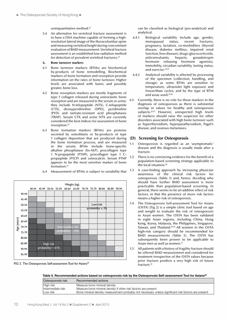
1.4 The Osteoporosis Self-assessment Tool for Asians (OSTA) [Fig 2] is a simple clinic tool based on age and weight to evaluate the risk of osteoporosis in Asian women. The OSTA has been validated in eight Asian regions, including China, Hong Kong, Korea, Malaysia, the Philippines, Singapore, Taiwan, and Thailand.29,30 All women in the OSTA high-risk category should be recommended for BMD measurements (Table 5). The OSTA has subsequently been proven to be applicable to Asian men as well as women.31
1.5 All patients with a history of fragility fracture should be offered BMD measurement and considered for treatment irrespective of the OSTA values because prior fracture predicts a very high risk of future fracture.32
Table 5. Recommended actions based on osteoporosis risk by the Osteoporosis Self-assessment Tool for Asians29
Osteoporosis risk Recommended actions
High risk Measure bone mineral densityIntermediate risk Measure bone mineral density if other risk factors are presentLow risk Bone mineral density measurement probably not necessary unless significant risk factors are present
FIG 2. The Osteoporosis Self-assessment Tool for Asians29
40-44
45-49
50-54
55-59
60-64
65-69
70-74
75-79
80-84
85-89
90-94
95-99
40-44 45-49 50-54 55-59 60-64 65-69 70-74 75-79 80-84 85-89 90-94
Age
(ye
ars)
Weight (kg)
High riskprobability ~ 61%
Intermediate riskprobability ~ 15%
Low riskprobability ≤ 3%
#GuidelineforClinicalManagementofPostmenopausalOsteoporosis#
HongKongMedJVol19No2#Supplement2#April2013 11
(E) Assessment of Fracture Risk1. Clinical use of bone mineral density to assess fracture
risk
1.1 Conventionally, BMD evaluation has been the primary focus for risk assessment.
1.2 BMD measurements with DXA at the lumbar spine and proximal femur give the best relative risk prediction for future fracture at the corresponding sites. In general, the relative risk of fracture increases by 1.5-3.0 times for each SD decrease in BMD.12,16
1.3 However, prospective population cohort studies have shown that most hip fractures (>50%) occurred in subjects without a BMD diagnosis of osteoporosis at baseline,33,34 such that BMD has a low sensitivity for fracture prediction.
1.4 Development of a new risk assessment tool, combining BMD and clinical risk factors, should enable more sensitive fracture risk prediction (refer to Section E3: The WHO Fracture Risk Assessment Tool on this page).
2. Clinical use of bone turnover markers to assess fracture risk
2.1 Population studies have shown that higher levels of bone formation and resorption markers were associated with significantly faster and greater bone loss. These markers may have the potential to help clinicians to identify fast bone losers for prompt intervention.26,27
2.2 Large prospective studies have shown that increases in biochemical markers of bone resorption, but not BFMs, are consistently associated with increases in risk of vertebral and non-vertebral fractures independent of BMD.26,27
2.3 Combining the measurements of BMD and markers of bone resorption may further refine the assessment of fracture risk.35
3. The WHO Fracture Risk Assessment Tool (FRAX®)
3.1 Facture risk is age-, gender- and country-specific, and is dependent on BMD, body mass index, and other clinical risk factors.
3.2 Those clinical risk factors, which have been identified to be independent fracture risk predictors, include age, low body weight, prior fragility fracture, a parental history of hip fracture, smoking, use of systemic corticosteroids, excess alcohol consumption, and rheumatoid arthritis.36
3.3 In 2008, the WHO successfully launched the WHO Fracture Risk Assessment Tool (FRAX®), which is a simple ethnic-specific web-based tool that integrates clinical information in a quantitative manner to predict a 10-year probability of major osteoporotic fracture and hip fracture for both women and men (http://www.shef.ac.uk/FRAX/).37
3.4 FRAX is a practical tool derived from a series of meta-analyses using the primary data from population-based cohorts that have identified a number of clinical risk factors for fracture. The performance characteristics of clinical risk factors have been validated in independent, population-based, prospectively studied cohorts with over 1 million person-years of observation.38
3.5 Computation of the 10-year fracture probability would help to guide individual treatment decisions. The level of absolute fracture probability above which pharmacological treatment is indicated depends on the availability of health care resources and priority of the health care system for treatment of osteoporosis.
3.6 The US National Osteoporosis Foundation (NOF) recommends treatment if the 10-year probability of a major osteoporotic fracture is ≥20% or the 10-year probability of a hip fracture is ≥3% based on the US-adapted WHO algorithm.39 This recommended treatment threshold has been proven to be cost-effective in a US economic model.40
3.7 A Hong Kong population–specific FRAX algorithm has become available, which is based on a prospective follow-up study of 1435 treatment-naïve community-dwelling, postmenopausal, southern Chinese women for incident osteoporotic fracture, and the 10-year risk of osteoporotic fracture was predicted from the risk factor assessment and BMD measurement.41
3.8 It must be cautioned that FRAX only applies to postmenopausal women or men aged 50 years or older who have not been treated. FRAX does not apply to pre-menopausal women, younger adults, or children. FRAX has not accommodated other known clinical risk factors such as fall and biochemical markers. Overall, FRAX does not replace clinical judgement. The decision to treat must still be made on an individual case-by-case basis.
(F) Clinical Assessment of Osteoporosis1.1 A comprehensive approach to all patients with
osteoporosis is recommended.
1.2 A detailed history and physical examination should be obtained. Central DXA BMD assessment should preferably be performed as a baseline. Vertebral fracture assessment may be obtained at the time of DXA measurements, if available. Otherwise, X-rays of the thoracic and lumbar spine should be performed if vertebral fracture is suspected.
1.3 The Hong Kong population–specific FRAX 10-year fracture probability is useful to establish an individual patient’s fracture risk, especially for those with BMD in the osteopenic range.
1.4 One of the main objectives of a detailed assessment is to exclude underlying secondary causes of osteoporosis (Table 4). This is particularly relevant for male subjects because a Caucasian study suggested that >50% of men presenting with symptomatic vertebral fracture had an underlying secondary cause.42
1.5 Basic laboratory investigations should include complete blood count, erythrocyte sedimentation rate, liver and renal function tests (including alkaline phosphatase, serum calcium and phosphate), thyroid function, and a 24-hour urine test for calcium excretion. Testosterone level should be considered in men.
1.6 Additional special tests such as serum protein electrophoresis, parathyroid hormone (PTH), 25-hydroxyvitamin D (25OHD) and cortisol level
#TheOsteoporosisSocietyofHongKong#
12 HongKongMedJVol19No2#Supplement2#April2013
may be considered if the history and initial workup suggest a related disorder. Specific bone marker tests may be considered, if available.
(G) Non-pharmacological Management of Osteoporosis
1. Lifestyle measures
1.1 Lifestyle measures remain the basic universal recommendation to the general population for prevention and non-pharmacological management of osteoporosis. A ‘population approach’ targeting adolescents before their accretion of peak bone mass is especially important to reduce the burden of the disease in the community.
1.2 Lifestyle measures include consumption of a healthy balanced diet rich in calcium and vitamin D, regular weight-bearing and muscle-strengthening exercises, avoidance of smoking and excessive alcohol intake, and adequate sunlight exposure.
2. Calcium
2.1 Importance of calcium
2.1.1 Adequate calcium intake is important to optimise bone health.
2.1.2 A meta-analysis of 23 randomised trials involving 41 419 adults aged 50 years or older confirmed that adequate calcium supplementation, with or without vitamin D, was associated with significantly reduced rates of bone loss of 0.54% at the hip and 1.19% in the spine.43
2.1.3 The same article also reported another meta-analysis of 17 randomised trials involving 52 625 adults aged 50 years or older with fracture as an outcome measure showed that adequate calcium supplementation, with or without vitamin D, was associated with a significant 12% risk reduction of fractures of all types. The treatment effects were greatest with calcium doses of ≥1200 mg and with vitamin D doses of ≥800 IU daily.43
2.1.4 After a comprehensive review of all the available evidence, the Institute of Medicine (IOM) of the US National Academy of Sciences stated in its 2011 report that the recommended dietary allowance (RDA) for calcium was 1000 mg daily for adults of both sexes and a higher 1200 mg daily for women older than 50 years and men older than 70 years.44 The RDA reflects the estimated requirement for 97.5% of the general healthy population.
2.1.5 According to early reports from the 1980s and 1990s, a traditional Chinese diet contained a much lower calcium content of around 400 mg daily.45,46 Publications from the 2000s showed an increase in daily dietary calcium content to around 500-600 mg.47,48
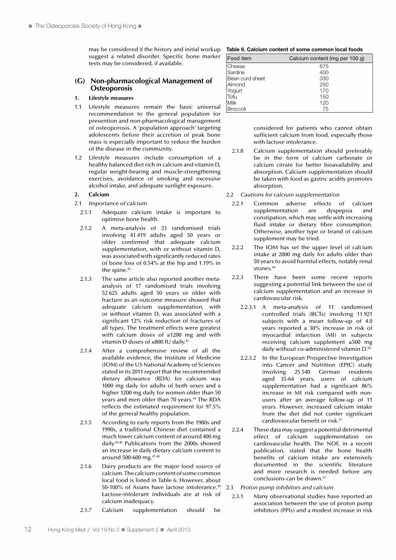
2.1.6 Dairy products are the major food source of calcium. The calcium content of some common local food is listed in Table 6. However, about 50-100% of Asians have lactose intolerance.49 Lactose-intolerant individuals are at risk of calcium inadequacy.
2.1.7 Calcium supplementation should be
considered for patients who cannot obtain sufficient calcium from food, especially those with lactose intolerance.
2.1.8 Calcium supplementation should preferably be in the form of calcium carbonate or calcium citrate for better bioavailability and absorption. Calcium supplementation should be taken with food as gastric acidity promotes absorption.
2.2 Cautions for calcium supplementation
2.2.1 Common adverse effects of calcium supplementation are dyspepsia and constipation, which may settle with increasing fluid intake or dietary fibre consumption. Otherwise, another type or brand of calcium supplement may be tried.
2.2.2 The IOM has set the upper level of calcium intake at 2000 mg daily for adults older than 50 years to avoid harmful effects, notably renal stones.44
2.2.3 There have been some recent reports suggesting a potential link between the use of calcium supplementation and an increase in cardiovascular risk.
2.2.3.1 A meta-analysis of 11 randomised controlled trials (RCTs) involving 11 921 subjects with a mean follow-up of 4.0 years reported a 30% increase in risk of myocardial infarction (MI) in subjects receiving calcium supplement ≥500 mg daily without co-administered vitamin D.50
2.2.3.2 In the European Prospective Investigation into Cancer and Nutrition (EPIC) study involving 25 540 German residents aged 35-64 years, users of calcium supplementation had a significant 86% increase in MI risk compared with non-users after an average follow-up of 11 years. However, increased calcium intake from the diet did not confer significant cardiovascular benefit or risk.51
2.2.4 These data may suggest a potential detrimental effect of calcium supplementation on cardiovascular health. The NOF, in a recent publication, stated that the bone health benefits of calcium intake are extensively documented in the scientific literature and more research is needed before any conclusions can be drawn.52
2.3 Proton pump inhibitors and calcium
2.3.1 Many observational studies have reported an association between the use of proton pump inhibitors (PPIs) and a modest increase in risk
Table 6. Calcium content of some common local foods
Food item Calcium content (mg per 100 g)
Cheese 675Sardine 400Bean curd sheet 330Almond 250Yogurt 170Tofu 150Milk 120Broccoli 75

#GuidelineforClinicalManagementofPostmenopausalOsteoporosis#
HongKongMedJVol19No2#Supplement2#April2013 13
of fractures, possibly mediated through an effect of inhibition of gastric acid secretion on calcium absorption and BMD.53
2.3.2 Two recent meta-analyses have suggested that the risk of fracture was increased by 10-40% above baseline in subjects receiving PPIs, especially when they were used at high doses and over long durations (>1 year), but not in patients taking histamine 2-receptor antagonists.54,55
2.3.3 Patients who require continuous PPI therapy should be strongly encouraged to ensure that they receive the recommended daily intake of calcium and vitamin D.
2.4 Recommendations for calcium intake
2.4.1 A daily elemental calcium intake of 1000-1200 mg should be recommended for osteoporosis patients if there are no contra-indications; the total amount of calcium should not exceed 2000 mg daily.
2.4.2 Individuals should preferably meet their daily calcium requirement from dietary sources. Calcium supplementation is indicated for those with a low dietary calcium intake and who are unable to achieve the recommended level through dietary sources.
2.4.3 Patients who require continuous PPI therapy should be strongly encouraged to ensure that they receive the recommended daily intake of calcium and vitamin D.
3. Vitamin D
3.1 Importance of vitamin D
3.1.1 Vitamin D is essential for promoting calcium absorption in the gut and maintaining adequate serum calcium and phosphate concentrations to enable normal mineralisation of bone. In addition, vitamin D plays an important role in neuromuscular function.
3.1.2 Vitamin D inadequacy results in increased PTH secretion (secondary hyperparathyroidism), which in turn accelerates bone resorption, notably from cortical sites.
3.1.3 Optimal serum 25OHD concentration is considered to be the level that is associated with maximal PTH suppression. Estimates of that threshold level have been found to be clustered around 68-75 nmol/L (27.2-30.0 ng/mL).56
3.1.4 The conversion factor for vitamin D is 1 ng/mL = 2.5 nmol/L.
3.1.5 Vitamin D deficiency occurs when the serum level of 25OHD falls below 25 nmol/L (10 ng/mL) resulting in rickets in children or osteomalacia in adults. However, there have been ongoing debates regarding the levels of 25OHD that are considered to be ‘optimal’ for bone health.
3.1.6 Previous studies and meta-analyses have been inconsistent in accurately defining the efficacy of vitamin D supplementation in fracture prevention. The discordant findings may be explained, in part, by the differences in the criteria for including trials in the analyses, with
respect to blinding, vitamin D formulation (oral vs injectable) or accommodations for non-adherence.
3.1.7 A recent meta-analysis employing pooled participant-level data (ie according to the actual intake of each participant) from 11 double-blind, RCTs of oral vitamin D supplementation (daily, weekly, or every 4 months), with or without calcium, compared with placebo or calcium alone in 31 022 subjects aged 65 years or older showed that reduction in fracture risk was only evident at the highest quartile of actual vitamin D intake (792-2000 IU daily) with a significant 30% reduction in the risk of hip fracture and a significant 14% reduction in the risk of any non-vertebral fracture. Benefits at the highest level of vitamin D intake were fairly consistent across subgroups defined by age-group, type of dwelling, baseline 25OHD level, and additional calcium intake.57
3.1.8 Regarding the effect of vitamin D on fall prevention, a meta-analysis of eight randomised trials involving 2426 elderly subjects with a mean age of 65 years or older showed that high-dose supplemental vitamin D (700-1000 IU daily) significantly reduced fall risk by 19%, whereas achieved serum 25OHD concentrations of ≥60 nmol/L (≥24 ng/mL) resulted in a significant 23% fall reduction. Falls were not notably reduced by low-dose supplemental vitamin D (200-600 IU daily) or by achieved serum 25OHD concentrations of <60 nmol/L.58
3.1.9 The potential non-skeletal health benefits of vitamin D59 are beyond the scope of the current guideline.
3.1.10 The 2010 IOM report recommended that the RDA for vitamin D was 600 IU daily for adults and a higher 800 IU daily for elderly (>70 years), corresponding to a serum 25OHD level of ≥50 nmol/L.44 These recommendations are intended to meet the requirement of 97.5% of the healthy general population.
3.1.11 The NOF, the International Osteoporosis Foundation (IOF), and the US Endocrine Society all recommended a higher 25OHD level of 75 nmol/L (30 ng/mL) to be the desired target for fracture and fall prevention.39,60,61 The estimated mean vitamin D requirement to reach this target 25OHD level is 800-1000 IU/day. A considerably higher dose (up to 2000 IU/day) may be required for individuals who are obese, and for those with osteoporosis, limited sun exposure (institutionalised, homebound) or malabsorption, and for certain ethnic populations known to be at high risk for vitamin D deficiency (those from the Middle East and South Asia).60
3.1.12 Synthesis of cholecalciferol (vitamin D3) from its precursors in the skin under the effect of ultraviolet light of wavelength 290-315 nm constitutes a major source of vitamin D. Around 10-15 minutes of sunlight exposure every day to the exposed areas over the face, hands, and arms is considered adequate

#TheOsteoporosisSocietyofHongKong#
14 HongKongMedJVol19No2#Supplement2#April2013
to meet the daily requirement of vitamin D in young adults. The effect of ageing can decrease the ability of the skin to synthesise vitamin D by more than 2-fold in elderly people.62
3.2 Issue of vitamin D inadequacy
3.2.1 Vitamin D inadequacy is more prevalent in the elderly population due to less efficient synthesis of vitamin D3 in the skin, decreased renal production of 1,25-dihydroxyvitamin D (1,25OHD), less efficient absorption of calcium in the gastro-intestinal tract and re-absorption in the kidney tubules.
3.2.2 Using a cut-off value of 75 nmol/L, the prevalence of vitamin D inadequacy has been reported to be >50% in community-dwelling postmenopausal women across all geographical regions of the world, with Asia having a prevalence of 71.4%.63
3.2.3 A local study also showed that vitamin D inadequacy was present in >60% of community-dwelling adults older than 50 years.64 Lack of outdoor activities and preference for avoiding sunlight exposure have been demonstrated among local middle-aged and elderly women.65
3.2.4 Local studies have confirmed genuine vitamin D inadequacy in high-risk groups, notably hospitalised patients with hip fracture.66,67
3.3 Vitamin D supplementation
3.3.1 Vitamin D is relatively scarce in food. The chief dietary sources of vitamin D are saltwater fish (ie salmon, tuna, and mackerel), fish liver oil, liver, egg yolks, and vitamin D-fortified milk or cereal products.
3.3.2 Vitamin D supplements are available as ergocalciferol (vitamin D2) or cholecalciferol (vitamin D3) in strengths up to 50 000 IU per tablet.
3.3.3 With daily dosing, vitamin D2 appears to be as effective as vitamin D3 in maintaining circulating concentrations of 25OHD,68 but with intermittent (weekly or monthly) dosing, vitamin D3 appears to be about 3 times more potent than vitamin D2.69 On average, each 100 IU of added vitamin D3 will increase the serum 25OHD level by about 2.5 nmol/L or 1.0 ng/mL.70
3.3.4 The margin of safety for prescription of a vitamin D supplement is considerably wide. A serum 25OHD concentration consistently >500 nmol/L is considered toxic. Studies have shown that a daily vitamin D dose of 10 000 IU could only achieve serum 25OHD concentrations of <140 nmol/L. Daily vitamin D intake of >40 000 IU is required to achieve a toxic level of >500 nmol/L.71
3.3.5 The 2010 IOM report has set the upper level of vitamin D intake to be 4000 IU daily.44
3.3.6 Patients in all the landmark clinical trials of anti-osteoporosis medications had received calcium, with or without vitamin D, supplementation. There is evidence that
calcium and vitamin D could enhance the antiresorptive and anti-fracture efficacy of bisphosphonates.72,73 A recent observational study showed that a mean 25OHD level of ≥33 ng/mL (≥82.5 mmol/L) was needed to maintain a favourable bisphosphonate response.74
3.3.7 A recent RCT involving 2256 community-dwelling elderly women aged 70 or older showed that annual oral administration of high-dose (500 000 IU) vitamin D3 resulted in increased risk of falls and fracture, especially during the first 3 months after dosing.75 It was proposed that the high oral dose may have triggered a short-term ‘protective’ upregulation of the enzyme responsible for degrading 1,25OHD, resulting in decreased blood and tissue levels of 1,25OHD, leading to falls.76
3.4 Recommendations for vitamin D supplementation
3.4.1 Vitamin D supplementation should be given whenever anti-osteoporosis medications are started unless there are contra-indications.
3.4.2 An average vitamin D intake of ≥800 IU daily is recommended; a higher dose is required for certain subgroups of patients who are at risk of vitamin D inadequacy. There is ample evidence that a high vitamin D intake is necessary to enhance the anti-fracture efficacy of most anti-osteoporosis medications, especially the bisphosphonates.
3.4.3 The upper level of vitamin D intake is set at 4000 IU daily.
3.4.4 Vitamin D supplementation can be given as a daily, weekly, or monthly oral dose of either vitamin D2 or D3, but should not be given as an annual oral high dose.
3.4.5 Contra-indications include recurrent urolithiasis and untreated diseases of bone and mineral metabolism such as hyperparathyroidism and metastatic bone diseases.
4. Active vitamin D analogues
4.1 Active vitamin D analogues are hydroxylated metabolites of vitamin D, and they increase intestinal calcium absorption pharmacologically. Examples include 1α-hydroxyvitamin D3 (alphacalcidol) and 1,25-dihydroxyvitamin D3 (calcitriol).
4.2 In a meta-analysis involving two RCTs, active vitamin D analogues reduced the risk of fall by 22%.77 These analogues probably reduced vertebral fracture, whereas their effect on reducing non-vertebral fracture was uncertain.77
4.3 A meta-analysis showed that hypercalcaemia was significantly increased by 4.4-fold in patients receiving calcitriol.78
4.4 These analogues have a relatively low margin of safety, with potential risks for hypercalcaemia and hypercalciuria. Generally, they are not recommended for use as vitamin D supplementation in otherwise healthy postmenopausal women.
5. Exercise
5.1 Exercise plays an important role in achieving peak bone mass and building and maintaining bone
#GuidelineforClinicalManagementofPostmenopausalOsteoporosis#
HongKongMedJVol19No2#Supplement2#April2013 15
strength. Exercise also modestly helps to reduce bone loss in elderly people.79
5.2 In addition to its effect on bone strength, exercise also helps to build muscle mass and maintain muscle strength.
5.3 Weight-bearing, muscle strengthening, and balance-training exercises are particularly useful to improve agility, strength, posture, coordination and balance, and reduce the risk of fall.
5.4 Immobilisation or inactivity, on the other hand, accelerates bone loss and should be avoided for elderly people as far as possible. Worries about falling should not be a reason of physical inactivity.
5.5 Weight-bearing exercises include brisk walking, jogging, Tai Chi, dancing, and stair-climbing. High-impact weight-bearing exercises are not recommended for elderly people as many of them have concomitant osteoarthritis.
5.6 Tai Chi is a form of low-impact weight-bearing exercise, and has been shown in the western literature to improve balance and reduce the incidence of fall and fall-related injuries.80,81 A more detailed discussion of Tai Chi is available in Section G6 (Tai Chi exercise on this page).
5.7 Muscle-strengthening exercises include weight training and other isometric resistance exercises.
5.8 Structured exercise programme for osteoporotic subjects should specifically target on posture, balance, gait, coordination, and hip and trunk stabilisation.
5.9 Exercise should be recommended for all age-groups not only for osteoporosis prevention and treatment but also for overall health benefits.
6. Tai Chi exercise
6.1 Tai Chi Chuan is a traditional Chinese martial art, with a history of over 300 years from the late Ming Dynasty.
6.2 Tai Chi is a unique form of low-impact exercise that requires high neuromuscular coordination and specific training in low-velocity muscle contraction.
6.3 Local small-scale studies have shown that short-term training with Tai Chi exercise improved
cardiopulmonary fitness,82 trunk flexibility,82 lower limb muscle strength,82,83 muscle endurance,83 coordination,84,85 and balance85,86 among the elderly.
6.4 A prospective, RCT in the US showed that 15 weeks’ training with Tai Chi resulted in a significant 47.5% reduction in the risk of multiple falls among 200 community-dwelling elderly aged 70 years or older, with significant concomitant improvements in measures of fear of falling.81
6.5 The effect of Tai Chi on BMD was demonstrated in a RCT in which Tai Chi training (45 minutes a day, 5 days a week for 12 months) in 132 postmenopausal women with a mean age of 54 years significantly retarded the rate of both cortical and trabecular bone loss in the weight-bearing bones as measured by pQCT.87
6.6 A meta-analysis involving 47 studies showed that benefits of Tai Chi were reported in balance and strength, cardiovascular and respiratory function, flexibility, the immune system, symptoms of arthritis, muscular strength, and psychological effects.80
6.7 Tai Chi is strongly recommended as an appropriate and safe exercise for older adults for general health and fall prevention.
(H) Pharmacological Treatment of Osteoporosis
1. General considerations
1.1 The ultimate goal of pharmacological treatment is to reduce fracture risk, increase survival, and improve quality of life.
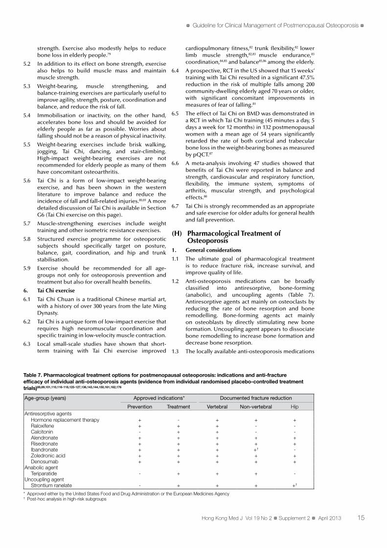
1.2 Anti-osteoporosis medications can be broadly classified into antiresorptive, bone-forming (anabolic), and uncoupling agents (Table 7). Antiresorptive agents act mainly on osteoclasts by reducing the rate of bone resorption and bone remodelling. Bone-forming agents act mainly on osteoblasts by directly stimulating new bone formation. Uncoupling agent appears to dissociate bone remodelling to increase bone formation and decrease bone resorption.
1.3 The locally available anti-osteoporosis medications
* Approved either by the United States Food and Drug Administration or the European Medicines Agency † Post-hoc analysis in high-risk subgroups
Table 7. Pharmacological treatment options for postmenopausal osteoporosis: indications and anti-fracture efficacy of individual anti-osteoporosis agents (evidence from individual randomised placebo-controlled treatment trials)88,89,101,110,116-118,125-127,136,143,144,150,161,162,176
Age-group (years) Approved indications* Documented fracture reduction
Prevention Treatment Vertebral Non-vertebral HipAntiresorptive agents
Hormone replacement therapy + - + + +Raloxifene + + + - -Calcitonin - + + - -Alendronate + + + + +Risedronate + + + + +Ibandronate + + + +† -Zoledronic acid + + + + +Denosumab + + + + +
Anabolic agentTeriparatide - + + + -
Uncoupling agentStrontium ranelate - + + + +†

#TheOsteoporosisSocietyofHongKong#
16 HongKongMedJVol19No2#Supplement2#April2013
that have been approved by the US Food and Drug Administration (FDA) for treatment and/or prevention of postmenopausal osteoporosis include HRT, calcitonin, bisphosphonates (including alendronate, risedronate, ibandronate, and zoledronic acid), raloxifene, teriparatide, and denosumab. In addition, the European Medicines Agency (EMA) has also approved the use of strontium ranelate.
1.4 Phytoestrogens and tibolone are other agents that have aroused interest in this field, but there is insufficient clinical evidence for phytoestrogens and there are potential safety concerns with tibolone, so these drugs have not been approved for treatment of postmenopausal osteoporosis.
1.5 Since there is a lack of direct head-to-head comparison studies with fracture as the primary endpoint, the most effective treatment for osteoporosis has yet to be determined.
2. Hormone replacement therapy
2.1 Estrogen suppresses osteoclastic bone resorption, reduces bone turnover to the premenopausal state, and maintains a positive calcium balance through its effect on the intestine and kidneys.
2.2 The beneficial effects of estrogen on bone mass and fracture risk reduction have clearly been demonstrated in large prospective, double-blind, RCTs. The Women’s Health Initiative (WHI) study, involving 27 347 postmenopausal women with a mean age of 63 years, confirmed that estrogen-only therapy (ET) or combined estrogen-progestogen therapy (EPT) reduced the risk of hip fracture by 33-39% and the risk of any fracture by 24%.88,89
2.3 However, the significant increase in incidences of breast cancer, stroke, heart attack, and venous thromboembolism (VTE) in the EPT arm and the significant increase in the incidence of stroke in the ET arm outweighed the benefit of fracture risk reduction in this group of relatively older postmenopausal women.88,89
2.4 Secondary analysis of the WHI data, however, supports the initiation of HRT around the time of menopause. ET was shown to have a reduced coronary artery disease risk (coronary revascularisation, MI, and coronary death) when initiated in younger and more recently postmenopausal hysterectomised women.90
2.5 ET was also demonstrated to offer no increase in risk of breast cancer after an average of 7.1 years of use regardless of the age at initiation of therapy.91
2.6 The latest 2012 North American Menopause Society (NAMS) Position Statement supported the initiation of HRT around the time of menopause to treat menopause-related symptoms and to prevent osteoporosis in women at high risk for fracture. The report stated that the benefit-risk ratio is especially favourable for ET in hysterectomised women in whom the duration of use can be flexibly extended up to 7 years, whereas the earlier appearance of increased breast cancer risk for EPT precludes a recommendation for its use beyond 3-5 years.92
2.7 The US Endocrine Society Scientific Statement also supported the start of HRT in the subgroups of women aged between 50 and 59 years or those less
than 10 years after onset of menopause because congruent trends suggested additional benefits, including reduction of overall mortality and coronary artery disease.93
2.8 The safety of HRT in recently postmenopausal women was supported by two recently published RCTs.
2.8.1 The Danish Osteoporosis Prevention Study, involving 1006 women aged 45-58 years with a mean of 7 months postmenopause, showed that after 10 years of randomised treatment, women receiving HRT early after menopause had a significantly reduced risk of mortality, MI or heart failure, without any apparent increase in risk of cancer, VTE or stroke.94
2.8.2 The Kronos Early Estrogen Prevention Study (KEEPS), involving 727 US women aged 42-58 years within 3 years of menopause, showed that after 4 years of randomised treatment, there were no significant differences in adverse events (breast cancer, endometrial cancer, MI, transient ischaemic attack, stroke, or VTE) between the HRT and placebo groups.95
2.9 Recommendations: Current evidence supports the use of HRT (notably ET for hysterectomised women) as an option for young postmenopausal women (age 50-59 years or <10 years postmenopause) for prevention and treatment of osteoporosis, especially for those with climacteric symptoms. Patients need to be adequately counselled on the risks and benefits of long-term use, beyond 3-5 years for EPT and beyond 7 years for ET. Contra-indications include a history of VTE or breast cancer.
3. Phytoestrogens
3.1 Phytoestrogens are natural chemicals found in plants. Animal studies have demonstrated that phytoestrogens have a protective effect against estrogen-related bone loss.
3.2 The two main classes of phytoestrogens that are of medical interest are isoflavones and lignans. Isoflavones are found in beans and soya products, eg soya milk and tofu. Lignans are found in ryes, berries, fruits, vegetables, and whole grains.
3.3 Extracted phytoestrogens have been marketed as dietary supplements.
3.4 Local cross-sectional and prospective studies have shown that phytoestrogens had positive effects on BMD and bone markers in postmenopausal women.96-98
3.5 There is no clinical up-to-date evidence that phytoestrogens in any form reduce the risk of any osteoporotic fracture in prospective clinical trials.99
4. Tibolone
4.1 Tibolone is a synthetic steroid, and its metabolites have estrogenic, progestogenic, and androgenic activities.
4.2 Tibolone has been shown to prevent bone loss both in animal and in human studies.
4.3 Tibolone has been licensed in more than 90 countries for treatment of climacteric symptoms.
4.4 In the Long-term Intervention on Fractures
#GuidelineforClinicalManagementofPostmenopausalOsteoporosis#
HongKongMedJVol19No2#Supplement2#April2013 17
with Tibolone study (LIFT), tibolone given to postmenopausal women at a dose of 1.25 mg daily significantly reduced the risks of vertebral fracture by 45% and of non-vertebral fracture by 26% over 34 months. However, a significant 2.2-fold increase in the risk of stroke necessitated premature termination of the study.100
4.5 Despite its anti-fracture efficacy, tibolone has not been approved by the FDA for long-term treatment of osteoporosis, and it definitely should not be recommended for elderly women or for women with risk factors for stroke.
5. Selective estrogen receptor modulators
5.1 Selective estrogen receptor modulators (SERMs) are non-hormonal agents that bind with high affinity to the estrogen receptors, but with differential effects at different target tissues. They exhibit estrogen-agonistic effects on bone and estrogen-antagonistic effects on the endometrium and breast.
5.2 Raloxifene is the SERM currently available for prevention and treatment of postmenopausal osteoporosis in Hong Kong.
5.3 Efficacy
5.3.1 In the Multiple Outcomes of Raloxifene Evaluation (MORE) study, treatment with raloxifene at a dose of 60 mg daily for 36 months increased spinal BMD by 2.6% and femoral neck BMD by 2.1% over placebo, and reduced bone turnover to premenopausal levels. The risk of vertebral fracture was significantly reduced by 30%, but the reduction in risk of non-vertebral fracture was insignificant.101
5.3.2 An Asian study has also confirmed the efficacy of raloxifene in increasing BMD and suppressing biochemical BTMs in healthy Asian postmenopausal women.102
5.3.3 The increase in BMD with raloxifene treatment is very modest, and only explains 4% of the vertebral fracture risk reduction associated with raloxifene treatment.103
5.4 Extra-skeletal benefits
5.4.1 Raloxifene reduced total cholesterol and low-density lipoprotein cholesterol by about 6% and 10%, respectively.104
5.4.2 Raloxifene reduced the risk of invasive breast cancer by approximately 70%.105,106
5.4.3 Raloxifene has been approved by the FDA for chemoprevention of invasive breast cancer in high-risk women; its efficacy was reported in the 5-year Study of Tamoxifen and Raloxifene (STAR) trial to be comparable to tamoxifen, but with a lower risk of endometrial cancer.107
5.4.4 An updated analysis with an 81-month median follow-up of the STAR trial participants confirmed the long-term efficacy of raloxifene in prevention of invasive breast cancer, but with much less toxicity than tamoxifen.108
5.5 Adverse effects: common minor adverse effects include hot flushes and leg cramps. Major complications include VTE.
5.6 Long-term data: 8-year long-term follow-up data showed that raloxifene was associated with a 1.7-
fold increase in incidence of VTE. There was no increase in the incidence of MI, stroke, uterine cancer, endometrial hyperplasia, ovarian cancer, or postmenopausal bleeding.109
5.7 Preparation: raloxifene is prescribed as a 60-mg tablet to be taken daily without regard to the timing of meals.
5.8 Recommendations: With the paucity of evidence for non-vertebral fracture risk reduction, raloxifene is recommended more preferably for use in younger postmenopausal women when the risk of hip fracture is not particularly high. Raloxifene may be safely administered in the long term, but a switch to more potent agents may be needed when the risk of hip fracture becomes higher as patients age.
6. Calcitonin
6.1 Calcitonin was approved by the FDA in 1995 for treatment of postmenopausal osteoporosis.
6.2 Calcitonin is a peptide hormone with antiresorptive properties on the osteoclasts. Calcitonin can be administered either by subcutaneous injection or as a nasal spray.
6.3 Efficacy
6.3.1 In the 5-year Prevent Recurrence of Osteoporotic Fractures (PROOF) study, a significant 36% reduction in vertebral fracture risk was shown only in the group treated with intranasal calcitonin 200 IU daily. Risk reduction in non-vertebral fracture was not significant at any of the doses tested.110
6.3.2 There are no data on the use of calcitonin for prevention of osteoporosis.
6.3.3 Both subcutaneous and intranasal calcitonin have been shown to reduce pain and hasten remobilisation of patients after acute vertebral compression fractures.111,112
6.4 Adverse effects
6.4.1 Common adverse effects include rhinitis, irritation of the nasal mucosa, epistaxis, and anaphylaxis reactions.
6.4.2 After complete review of the benefits and risks of calcitonin-containing medications, the EMA, in July 2012, concluded that there was evidence of a small increase in risk of cancer of various types with its long-term use (2.4% for the intranasal formulation). The benefits of calcitonin did not outweigh the risks, so calcitonin should no longer be used for treatment of osteoporosis and the nasal spray formulation should be withdrawn from the market. Parenteral formulations of calcitonin should be limited to use at the smallest effective dose for the shortest possible time for three conditions: Paget’s disease, acute bone loss due to sudden immobilisation, and hypercalcaemia caused by cancer.113
6.5 Recommendations: Due to its inconsistent anti-fracture efficacy, calcitonin is seldom used for long-term treatment of osteoporosis. With the recent EMA warnings on the potential risk of cancer, calcitonin should no longer be used for treatment of osteoporosis.

#TheOsteoporosisSocietyofHongKong#
18 HongKongMedJVol19No2#Supplement2#April2013
7. Bisphosphonates as a class
7.1 Bisphosphonates are currently considered to be the mainstay of pharmacological therapy for the prevention and treatment of osteoporosis in both postmenopausal women and elderly men.
7.2 Bisphosphonates bind tightly to the mineralised bone surface, where they are ingested by osteoclasts leading to apoptosis and loss of function of the osteoclasts. The rate of bone turnover is reduced and secondary mineralisation is enhanced.
7.3 Etidronate was the first-generation bisphosphonate, but continuous exposure to etidronate could cause mineralisation defects, so it is currently not recommended for osteoporosis management.
7.4 Currently approved bisphosphonates include alendronate, ibandronate, risedronate and zoledronic acid, all of which are nitrogen-containing derivatives that are 100-1000 times more potent than etidronate, and are effective in inhibiting bone resorption without causing mineralisation defects.
7.5 The potency and long duration of action of the currently approved bisphosphonates favour their intermittent use, and these compounds can be administered orally as a daily (alendronate, risedronate), weekly (alendronate, risedronate), or monthly (ibandronate, risedronate) dose, or intravenously at intervals of 3 months (ibandronate) or 12 months (zoledronic acid).
7.6 Infrequent oral dosing and intravenous preparations may help to improve adherence and acceptance of treatment.
7.7 Bisphosphonates are poorly absorbed from the gastro-intestinal tract (<1%) and oral preparations must be given with special instructions to ensure absorption and reduce adverse effects:
(i) bisphosphonates must be taken after a prolonged fast (usually first thing in the morning) and should not be taken with meals or calcium tablets, and preferably not with other medications
(ii) bisphosphonates must be taken with a full glass of water (≥250 mL)
(iii) patients should remain upright (either sitting or standing) and take nothing else by mouth for ≥30 minutes
7.8 Common adverse effects include upper gastro-intestinal effects and musculoskeletal pain. Upper gastro-intestinal adverse effects are a result of direct chemical irritation of the mucosa by the bisphosphonate and include epigastric discomfort, pain, flatulence, regurgitation, heartburn and, on rare occasions, oesophagitis, oesophageal erosion and ulceration. Musculoskeletal pain may be disabling and can occur during initial use of bisphosphonates, or even weeks or months afterwards.
7.9 The Cochrane Database of Systematic Reviews on 18 RCTs involving more than 26 000 patients reassured that the incidence of upper gastro-intestinal adverse effects were similar between the treatment groups and placebo114,115 if the instructions for drug ingestion were followed correctly.
7.10 Contra-indications to oral bisphosphonates
include hypersensitivity to bisphosphonates, hypocalcaemia, active peptic diseases, and oesophageal abnormalities such as reflux oesophagitis, oesophageal stricture or achalasia. Bisphosphonates should be used with caution in patients with renal impairment and are contra-indicated in patients with creatinine clearance of <30 mL/min/1.73 m2.
8. Alendronate
8.1 Alendronate was the first bisphosphonate approved by the FDA in 1995 for prevention and treatment of osteoporosis in postmenopausal women.
8.2 Efficacy
8.2.1 Large-scale prospective RCTs have shown that alendronate increased BMD at the spine by 6-9% and at the hip by 3-6% at 36 months.116-118
8.2.2 Local studies have shown a comparable increase in BMD of 5-6% at the lumbar spine and 3-4% at the hip after 1 year of treatment with alendronate in postmenopausal osteoporotic Chinese women.119,120
8.2.3 The 2008 Cochrane Database of Systematic Reviews on 11 RCTs involving 12 068 postmenopausal women, showed that alendronate significantly reduced the relative risk of vertebral fracture by 45% both for primary and secondary prevention, reduced non-vertebral fracture by 23% for secondary prevention, and reduced hip fracture by 53% for secondary prevention.114
8.2.4 Alendronate has also been shown to be effective in increasing BMD and prevention of vertebral fracture in men121 and in subjects with glucocorticoid-induced osteoporosis.122
8.3 Long-term data: Long-term follow-up data have confirmed that the anti-fracture efficacy of alendronate was maintained for up to 10 years with a good tolerability and safety profile.123
8.4 Preparations: Alendronate is available either as an oral daily dose of 10 mg or a weekly dose of 70 mg.124 Weekly combination tablets with vitamin D3 2800 or 5600 IU are available.
8.5 Recommendations: With its proven efficacy in reduction of vertebral, non-vertebral and hip fractures, alendronate is recommended as one of the first-line drugs for treatment of postmenopausal osteoporosis unless specifically contra-indicated (refer to Section H7.10 on this page).
9. Risedronate
9.1 Risedronate was the second bisphosphonate approved by the FDA in 2000 for prevention and treatment of osteoporosis in postmenopausal women.
9.2 Efficacy
9.2.1 The Vertebral Efficacy with Risedronate Therapy (VERT) studies125,126 and the Hip Intervention Program (HIP) study127 showed that risedronate significantly increased BMD at the spine by 5-6% and at the hip by 2-5% at 36 months.
9.2.2 A local study showed a comparable increase in BMD of 6.6% at the lumbar spine and
#GuidelineforClinicalManagementofPostmenopausalOsteoporosis#
HongKongMedJVol19No2#Supplement2#April2013 19
2.7% at the total hip after 1-year treatment of risedronate in postmenopausal osteoporotic Chinese women.128
9.2.3 The 2008 Cochrane Database of Systematic Reviews on seven RCTs involving 14 049 postmenopausal women, showed that risedronate significantly reduced the relative risk of vertebral fracture by 39%, that of non-vertebral fracture by 20% and that of hip fracture by 26%, all for secondary prevention.115
9.2.4 Pooled analyses revealed that risedronate significantly reduced clinical vertebral and non-vertebral fractures as early as 6 months after treatment.129,130
9.2.5 A subgroup analysis among very elderly osteoporotic women (age ≥80 years) showed a significant 81% risk reduction of vertebral fracture at 1 year and a 44% risk reduction at 3 years; the numbers needed to treat (NNTs) were 12 and 16, respectively. However, the risk reduction of non-vertebral fracture was not significant.131
9.2.6 A recent report confirmed the risk reduction of both vertebral and non-vertebral fractures in male osteoporotic subjects treated with risedronate for 2 years.132
9.2.7 Risedronate has also been shown to improve BMD and reduce the incidence of vertebral fracture in glucocorticoid-treated subjects.133
9.3 Long-term data: Long-term follow-up data have confirmed that the anti-fracture efficacy of risedronate was maintained for up to 7 years with a good tolerability and safety profile.134
9.4 Preparations: Risedronate is available either as an oral daily dose of 5 mg or a weekly dose of 35 mg. A monthly oral dose of 150 mg has recently become available, which has demonstrated similar efficacy and safety to the standard daily dose.135
9.5 Recommendations: With its proven efficacy in reduction of vertebral, non-vertebral and hip fractures, risedronate is recommended as one of the first-line drugs for treatment of postmenopausal osteoporosis unless specifically contra-indicated (refer to Section H7.10 on page 18).
10. Ibandronate
10.1 Ibandronate is a potent, nitrogen-containing bisphosphonate with antiresorptive potency 1000- to 10 000-fold that of etidronate.
10.2 This potency, combined with favourable tolerability and bone-binding characteristics, allows ibandronate to be effectively administered less frequently than weekly.
10.3 Efficacy
10.3.1 The Oral Ibandronate Osteoporosis Vertebral Fracture Trial in North America and Europe (BONE) showed that treatment with oral ibandronate at a daily dose of 2.5 mg for 3 years in postmenopausal women significantly reduced the risk of morphometric vertebral fracture by 62%, concomitant with a significant 6.5% improvement in spinal BMD and a 3.4%
improvement in total hip BMD. However, a significant 69% reduction in risk of non-vertebral fracture was only seen in a post-hoc analysis of a high-risk subgroup of subjects with a femoral neck BMD T-score of < –3.01.136
10.3.2 The subsequent 2-year Monthly Oral Ibandronate In Ladies (MOBILE) study demonstrated that once-monthly oral ibandronate at a dose of 150 mg is at least as effective as the daily regimen in terms of efficacy in BMD improvement and safety profile.137
10.3.3 In the head-to-head comparison Monthly Oral Therapy with Ibandronate for Osteoporosis Intervention (MOTION) study, once-monthly ibandronate 150 mg was shown to be clinically comparable with weekly alendronate 70 mg at increasing BMDs in both the lumbar spine and total hip after 12 months.138
10.3.4 The Dosing Intravenous Administration (DIVA) study confirmed the non-inferiority of intermittent intravenous dosing regimens of ibandronate (either 2 mg every 2 months or 3 mg every 3 months) in terms of BMD improvement and tolerability over 2 years of treatment.139
10.3.5 Intermittent intravenous ibandronate has also been shown to improve BMD and reduce the incidence of vertebral fracture in glucocorticoid-treated subjects.140
10.4 Preparations: Ibandronate is available as an oral 150-mg tablet given monthly or as a 3-mg intravenous injection administered every 3 months.
10.5 Adverse effects: Up to 10% of subjects receiving the oral or intravenous regimens reported mild-to-moderate influenza-like symptoms within 3 days of drug administration, usually during the initial administrations.137,139
10.6 Short-term 6-month cross-over studies demonstrated stronger patient preference for monthly ibandronate over weekly alendronate, which might imply a higher level of long-term adherence and compliance with therapy with ibandronate treatment.141,142
10.7 Recommendations: The lack of data on risk reduction for hip fracture and the fact that significant non-vertebral fracture risk reduction was only demonstrated in a post-hoc analysis of a high-risk subgroup would make ibandronate a less favourable bisphosphonate option for treatment of postmenopausal osteoporosis. However, with its more convenient intermittent monthly oral or 3-monthly intravenous dosing regimens, ibandronate may have an advantage for certain patient subgroups for whom adherence to therapy poses a significant consideration.
11. Zoledronic acid
11.1 Zoledronic acid is the most potent bisphosphonate currently available, and was approved by the FDA in 2007 for treatment of postmenopausal osteoporosis.
11.2 Efficacy

#TheOsteoporosisSocietyofHongKong#
20 HongKongMedJVol19No2#Supplement2#April2013
11.2.1 In the Health Outcomes and Reduced Incidence with Zoledronic Acid Once Yearly (HORIZON) Pivotal Fracture Trial (PFT), zoledronic acid at a dose of 5 mg given as an annual intravenous infusion significantly reduced the incidence of morphometric vertebral fracture by 70%, non-vertebral fracture by 25%, and hip fracture by 41% in postmenopausal osteoporotic women. There was a concomitant significant 6.7% BMD improvement at the spine and 6.0% at the total hip at 36 months.143
11.2.2 Zoledronic acid is the first anti-osteoporosis medication which has demonstrated survival benefit in an RCT. In the HORIZON Recurrent Fracture Trial (RFT), zoledronic acid infusion administered within 90 days after hip fracture surgery was associated with a 28% reduction in all-cause mortality in a group of 1065 men and women followed up for a median of 1.9 years.144 Exploratory analysis showed that zoledronic acid–treated subjects were less likely to die from pneumonia and arrhythmias than placebo-treated subjects.145 The exact underlying mechanism responsible for the improved survival is yet to be elucidated.
11.2.3 A RCT involving 1199 men with primary or hypogonadism-associated osteoporosis also showed a significant 67% reduction in vertebral fracture after 24 months’ treatment.146
11.2.4 A recent report also showed that zoledronic acid was non-inferior to, and possibly more effective than, oral risedronate for prevention and treatment of glucocorticoid-induced osteoporosis.147
11.3 Long-term data: In the 3-year HORIZON-PFT Extension Study, subjects who were randomised to receive a total of 6 years’ zoledronic acid therapy had a lower risk of new morphometric vertebral fracture than those who were randomised to placebo after an initial 3-year active treatment period in the core study (odds ratio [OR]=0.51; P=0.035), whereas other types of fractures were not different.148 There were no cases of atypical femur fracture and there was one case of osteonecrosis of the jaw (ONJ), which resolved with appropriate treatment.148
11.4 Preparation: Zoledronic acid is prescribed as a 5-mg slow intravenous infusion over a minimum of 15 minutes once yearly. A longer infusion time is preferred for patients with pre-existing renal impairment. Zoledronic acid is contra-indicated in patients with creatinine clearance of <35 mL/min/1.73 m2.
11.5 Adverse effects
11.5.1 The most frequent side-effect is a post-dose flu-like syndrome with pyrexia, myalgia, and bone and musculoskeletal pain, which may last for a few days after the first infusion. Prophylactic paracetamol may be useful to decrease the severity of the syndrome. Post-dose syndrome usually becomes less severe with repeated dosing.
11.5.2 No adverse effects on fracture healing were noted in the HORIZON-RFT, in which
zoledronic acid was administered within 90 days post-hip fracture surgery.144
11.5.3 An initial report of a significant increase in the risk of serious atrial fibrillation (1.3% in the treated group vs 0.5% in the placebo group)143 was not confirmed in subsequent studies.144,146,147
11.5.4 Renal impairment and renal failure have been observed (18 cases per 100 000 patient-years) following administration of intravenous zoledronic acid. Risk factors include pre-existing renal impairment, advanced age, concomitant nephrotoxic medications, concomitant diuretic therapy or dehydration. Appropriate hydration must be ensured and monitoring of renal function should be considered for at-risk patients.149
11.6 Recommendations
11.6.1 With its proven efficacy in reduction of vertebral, non-vertebral and hip fractures, zoledronic acid is recommended as one of the first-line drugs for treatment of postmenopausal osteoporosis. Zoledronic acid is especially indicated for patients with recent hip fractures, contra-indications for oral bisphosphonate therapy, existing polypharmacy or poor compliance with oral medications.
12. Teriparatide (recombinant-human parathyroid hormone 1-34)
12.1 Teriparatide was approved by the FDA in 2002 as the only bone-forming agent for treatment of postmenopausal osteoporosis.
12.2 Teriparatide is structurally the 1-34 amino-terminal fragment of the human parathyroid hormone (PTH). It is the first pure bone-forming agent for treatment of osteoporosis. Intermittent PTH administration increases cortical and trabecular thickness and bone diameter, and improves bone strength and architecture.
12.3 Efficacy
12.3.1 In the Fracture Prevention Trial (FPT), subcutaneous injection of teriparatide at a dose of 20 μg daily reduced the relative risk of vertebral fracture by 65% and that of non-vertebral fracture by 53% after a median treatment duration of 21 months in postmenopausal osteoporotic women. There was a concomitant significant 9% increase in BMD at the spine and 3% increase at the hip.150
12.3.2 Histomorphometry and microcomputed tomography of 51 paired iliac crest biopsy specimens from women treated with teriparatide in the FPT revealed significant increases in cancellous bone volume, cancellous bone connectivity density, cancellous bone plate-like structure and cortical thickness, and a reduction in marrow star volume.151
12.3.3 Improvement of BMD has also been reported in men with osteoporosis152 and in subjects with glucocorticoid-induced osteoporosis, in which the BMD gain was better with teriparatide than with alendronate.153

#GuidelineforClinicalManagementofPostmenopausalOsteoporosis#
HongKongMedJVol19No2#Supplement2#April2013 21
12.3.4 There is no therapeutic synergy when combining teriparatide with antiresorptive drugs. In fact, bisphosphonates would attenuate PTH-induced stimulation of bone formation.154,155
12.3.5 Treatment with teriparatide can achieve significant improvement in BMD regardless of previous long-term exposure to antiresorptive therapies, although prior antiresorptive treatment would modestly blunt the BMD response to teriparatide.156,157
12.3.6 Bone loss occurs after stopping teriparatide, and it is recommended that teriparatide treatment should be followed by bisphosphonates or SERM to maintain the gain in BMD.158,159
12.3.7 A meta-analysis of five clinical trials showed that teriparatide-treated subjects had 40-60% lower incidence of new or worsening back pain than placebo-, alendronate-, or HRT-controlled subjects.160
12.4 Preparations: Teriparatide is prescribed as a daily 20 μg subcutaneous injection and is approved for a treatment duration of up to 24 months.
12.5 Adverse effects: Adverse effects are mild and transient. They include nausea, headache, and orthostatic hypotension. Transient asymptomatic hypercalcaemia has been observed in about 5% of patients, but it is not clinically necessary to monitor calcium levels during therapy.
12.6 Contra-indications: Contra-indications include a history of skeletal malignancy or irradiation involving the skeleton, Paget’s disease, unexplained elevated bs-ALP and any form of untreated or unresolved hyperparathyroidism.
12.7 Recommendations: Due to its bone-forming action, significant improvement of bone microarchitecture, and proven efficacy in reduction of vertebral and non-vertebral fractures, teriparatide is recommended as one of the first-line drugs for treatment of postmenopausal osteoporosis. Teriparatide is especially indicated in patients with established osteoporosis (≥1 osteoporotic fractures), with very low BMD (T-score < –3), who experience fracture while taking antiresorptive therapies, or with continuing bone loss despite appropriate antiresorptive therapies after exclusion of non-compliance and secondary causes. The main limitations to the widespread use of teriparatide are its relatively high cost and the need for daily subcutaneous injection.
13. Strontium ranelate
13.1 Strontium ranelate is an anti-osteoporosis drug with a novel mechanism of action that is different from that of the conventional antiresorptive or bone-forming agents. Although the mechanism of its molecular mode of action has not been fully elucidated, strontium ranelate appears to dissociate bone remodelling to increase bone formation and decrease bone resorption.
13.2 In-vitro strontium ranelate stimulates proliferation of pre-osteoblasts and their differentiation into mature osteoblasts, while inhibiting osteoclastogenesis and the bone resorption activity
of mature osteoclasts.161
13.3 Efficacy
13.3.1 The Spinal Osteoporosis Therapeutic Intervention (SOTI) and Treatment of Peripheral Osteoporosis (TROPOS) studies confirmed that treatment with oral strontium ranelate at a dose of 2 g daily in postmenopausal women for 3 years reduced the risk of new vertebral and non-vertebral fractures by 41% and 16%, respectively, with a concomitant uncorrected increase of BMD of 14% at the spine and 8-10% at the hip.162,163
13.3.2 A post-hoc analysis showed a significant 36% reduction in hip fracture in a high-risk subgroup of women aged 74 years or older and with femoral neck BMD of ≤ –3.163
13.3.3 The anti-fracture efficacies at the spine and non-vertebral sites, including the hip, were maintained in an extension study, with randomisation maintained for up to 5 years.164
13.3.4 In a pre-planned pooled subgroup analysis of 1488 very elderly women, aged 80-100 years, treated with strontium ranelate for 3 years, the risks of vertebral, non-vertebral, and clinical fractures were significantly reduced by 32%, 31%, and 22%, respectively. A significant anti-fracture effect was already evident within 1 year, with a risk reduction for vertebral, non-vertebral, and clinical (symptomatic) fractures of 59%, 41%, and 37%, respectively.165
13.3.5 In another subgroup analysis involving 353 young postmenopausal women, aged 50-65 years, with vertebral fracture at baseline, strontium ranelate treatment for 4 years significantly reduced the risk of vertebral fracture by 35%.166
13.3.6 In women with lumbar spine osteopenia, strontium ranelate treatment for 3 years significantly reduced the risk of vertebral fracture by 41% and, in women with osteopenia at both the lumbar spine and femoral neck, treatment significantly reduced the risk of vertebral fracture by 52%.167
13.3.7 A study of Asian women has shown a significant 3-5% increase in BMD over 1 year in the lumbar spine, femoral neck, and total hip BMD in the strontium ranelate–treated group compared with the placebo group, and its effect on BMD and safety profile were consistent with those reported from Caucasian studies.168
13.3.8 Data on male osteoporosis are emerging. Two short-term studies showed that strontium ranelate produced significantly greater increases in lumbar spine and femoral neck BMD in male osteoporotic subjects at 12 months, with a magnitude comparable to those previously observed in postmenopausal women.169,170
13.3.9 At present, there are no data on the combined use of strontium ranelate with other anti-osteoporosis agents, and neither are there any data on glucocorticoid-induced osteoporosis.
13.4 Long-term data: Five-year extension follow-up data showed a continuous improvement in lumbar
#TheOsteoporosisSocietyofHongKong#
22 HongKongMedJVol19No2#Supplement2#April2013
BMD over a 10-year period and a continuous improvement in total hip and femoral neck BMD for up to 7 years; the cumulative incidences of new vertebral and non-vertebral fractures in the 5-year extension study was statistically similar to those in the first 5 years, despite a theoretical increase in fracture risk with ageing.171
13.5 Extra-skeletal benefits: A post-hoc analysis of pooled data from the SOTI and TROPOS trials involving 1105 women with concomitant baseline radiological spinal osteoarthritis showed that the proportion of patients with worsening overall spinal osteoarthritis score in the lumbar radiographs was significantly reduced by 42% in the strontium ranelate group and significantly more patients in the treatment group experienced improvement in back pain after 3 years compared with the placebo group, suggesting that strontium ranelate could reduce progression of the radiographic features of spinal osteoarthritis and back pain in women with osteoporosis and prevalent spinal osteoarthritis.172
13.6 Preparation: Strontium ranelate is prescribed as a powder suspension to be taken orally at night at a dose of 2 g daily. Food should be avoided for at least 2 hours before and after its ingestion and it must not be taken with calcium tablets.
13.7 Adverse effects
13.7.1 Common adverse effects include headache, nausea, diarrhoea, and skin rash.
13.7.2 In a recent safety review by the EMA in March 2012, it was noted that strontium ranelate was associated with an increase in risk of VTE in patients with a history of VTE, those who are temporarily or permanently immobilised and in elderly patients older than 80 years, so the EMA has recommended that strontium ranelate should no longer be given to immobilised patients or patients with VTE, and its use in elderly patients older than 80 years should be re-evaluated.173
13.7.3 The EMA also issued an update to warnings regarding potentially fatal serious skin reactions such as drug rash eosinophilia and systemic symptoms (DRESS) syndrome, Stevens-Johnson syndrome, and toxic epidermal necrolysis (TEN) to alert health care professionals and patients to the likely signs and symptoms of these conditions for immediate drug discontinuation.173
13.8 Contra-indications: Strontium ranelate is contra-indicated in patients with a history of or current VTE or in immobilised patients.173
13.9 Recommendations: With its proven efficacy in reduction in risk of vertebral and non-vertebral fractures in RCTs and reduction in hip fracture in post-hoc analysis of a high-risk group, strontium ranelate can be considered as an oral alternative for patients with a history of upper gastrointestinal disorders that may be a contra-indication for oral bisphosphonate therapy, but the benefit-risk ratio in elderly patients older than 80 years needs to be carefully evaluated. A special alert to the development of severe skin reactions needs to be emphasised.
14. Denosumab
14.1 Denosumab was approved by the FDA in June 2010 as an injectable treatment for postmenopausal osteoporosis.
14.2 Denosumab is a fully human monoclonal antibody that specifically binds to the receptor activator of nuclear factor-kappa B ligand (RANKL), a cytokine that is essential for the differentiation, activity, and survival of osteoclasts. By binding to RANKL, denosumab prevents the interaction of RANKL with its receptor, RANK, on osteoclasts and osteoclast precursors and reversibly inhibits osteoclast-mediated bone resorption.174
14.3 Following a single subcutaneous dose, serum denosumab levels decline over a period of 4-5 months, with a mean half-life of approximately 26 days. Clearance of denosumab occurs through the reticuloendothelial system, and is thus independent of renal clearance so that no dosage adjustment is necessary for patients with renal impairment.175
14.4 Efficacy
14.4.1 In the Fracture Reduction Evaluation of Denosumab in Osteoporosis Every 6 Months (FREEDOM) trial, denosumab at a dose of 60 mg given as a subcutaneous injection every 6 months for 36 months significantly reduced the risk of new radiographic vertebral fracture by 68%, risk of hip fracture by 40%, and risk of non-vertebral fracture by 20% in postmenopausal women, with a concomitant increase in BMD of 9.2% at the spine and 6% at the total hip.176 The reduction in risk of new radiographic vertebral fracture was similar during each year of the study at 61%, 78% and 65% in the first, second and third years of treatment respectively.176
14.4.2 In a post-hoc analysis of the FREEDOM study, the treatment efficacy and incidence of adverse events were shown to be similar between patient subgroups stratified by the level of renal function from stage 1 (estimated glomerular filtration rate [eGFR] ≥90 mL/min) to stage 4 (eGFR 15-29 mL/min) chronic kidney disease.177
14.4.3 An off-treatment study demonstrated that the treatment effects of denosumab on both BMD and BTMs were reversible. BMD declined back to baseline at all sites after 24 months and BTMs rapidly increased above baseline within 3-6 months of discontinuation and returned to baseline by 48 months. This observation of reversibility is consistent with the current knowledge that denosumab does not persist in bone tissues.178
14.4.4 When compared with oral alendronate in a 12-month comparison trial, denosumab showed significantly larger gains in BMD at all measured skeletal sites and achieved a significantly greater reduction of BTMs.179
14.4.5 Transition to denosumab for patients previously taking alendronate produced greater increases in BMD at all measured skeletal sites and a greater reduction in bone turnover than did continued alendronate over

#GuidelineforClinicalManagementofPostmenopausalOsteoporosis#
HongKongMedJVol19No2#Supplement2#April2013 23
a 12-month period.180
14.4.6 The Preference and Satisfaction Questionnaire (PSQ) study showed that patients had a greater preference for, and were more satisfied with, a 6-month injection regimen than a weekly oral regimen for osteoporosis treatment.181
14.4.7 In the Denosumab Adherence Preference Satisfaction (DAPS) study, a 24-month, randomised, open-label, crossover comparison with alendronate in 250 postmenopausal osteoporotic women, subjects were more adherent, compliant, and persistent with subcutaneous denosumab injections every 6 months than with once-weekly alendronate tablets, and they reported increased treatment preference (92.4%) and satisfaction with injectable denosumab over oral alendronate.182
14.4.8 Male treatment data with fracture as an endpoint are also available. In a RCT involving 1468 men who had received androgen-deprivation therapy for non-metastatic prostate cancer, denosumab was associated with increased BMD at all sites, and a significant 62% reduction in the incidence of new vertebral fracture at 24 months.183
14.4.9 Another RCT of 242 men with low bone mass also showed significant improvement in BMD at all skeletal sites and a significant reduction in bone markers levels after 1 year of denosumab treatment.184
14.5 Adverse effects: In the initial 3-year FREEDOM trial, there was a higher incidence of eczema and serious cellulitis in the denosumab-treated group; otherwise, there were no significant differences in the risk of cancer, infection, cardiovascular disease, delayed fracture healing or hypocalcaemia, and there were no cases of ONJ or atypical femur fracture.176
14.6 Long-term data
14.6.1 The 2-year FREEDOM Extension Study showed that reduction in BTMs were maintained and BMD increased further, resulting in total 5-year BMD gains of 13.7% and 7.0% in the lumbar spine and total hip, respectively, and the annual fracture incidence was below the rate observed in the FREEDOM placebo group.185
14.6.2 The incidence of eczema and cellulitis was not increased in the Extension Study.185
14.6.3 ONJ developed in two patients who crossed over from the placebo group to receive denosumab in the extension phase, and had healed completely within 137 and 227 days. There were no cases of atypical femur fracture.185
14.6.4 Reports of ONJ have also recently been reported in patients treated with denosumab for cancer and skeletal metastases.186-188
14.6.5 Given its relative short post-marketing period, vigilance regarding potential serious adverse effects such as ONJ and atypical femur fracture is advised.
14.7 Preparation: Denosumab is given at a dose of 60 mg by subcutaneous injection every 6 months.
14.8 Recommendations: With its proven efficacy in reduction of vertebral, non-vertebral and hip fractures, denosumab is recommended as one of the first-line drugs for treatment of postmenopausal osteoporosis. Denosumab is especially indicated in patients with polypharmacy, poor compliance to oral drugs, contra-indications to oral bisphosphonate therapy, who cannot tolerate oral or intravenous bisphosphonate therapies, or who have suboptimal BMD responses to other antiresorptive therapies. Denosumab may also serve as an option for switching from bisphosphonates for high-risk patients who have been taking prolonged bisphosphonate treatment. There is no contra-indication for its use in patients with renal impairment up to stage 4 chronic kidney disease.
(I) Indications for Osteoporosis Treatment1. Patients at high risk for fracture should be treated
with specific anti-osteoporosis drugs. In general, the treatment guidelines recommended by the US NOF are widely adopted,39 as follows:
(i) prior low-energy hip or vertebral fractures
(ii) BMD T-score of ≤ –2.5 at the lumbar spine or proximal femur on DXA scan
(iii) low bone mass (T-score between -1 and -2.5) and one of following:
(a) 10-year probability of any major osteoporotic fracture of ≥20% computed by the US-adapted WHO FRAX algorithm
(b) 10-year probability of hip fracture of ≥3% computed by the US-adapted WHO FRAX algorithm
2. These US criteria are for clinical guidance only. All treatment decisions require clinical judgement and consideration of individual patient factors, including patient preferences, co-morbidities, quality of life, life expectancy, and other risk factors not captured in the FRAX model such as frailty and falls. The decision to treat must be made on a case-by-case basis.
(J) Monitoring of Osteoporosis Treatment1. General considerations
1.1 Osteoporosis is a chronic disease and patients receiving treatment should be monitored for:
(i) efficacy of treatment
(ii) adequacy of calcium and vitamin D intake (from diet or supplementation)
(iii) side-effects of medications
(iv) adherence to therapy
(v) new co-morbidities or medications that might alter the expected treatment effect
1.2 Monitoring for treatment efficacy is limited by the absence of clinical tools that directly measure bone strength so that it is necessary to monitor therapy with surrogate markers such as BMD and BTMs.
2. Role of bone mineral density in monitoring

#TheOsteoporosisSocietyofHongKong#
24 HongKongMedJVol19No2#Supplement2#April2013
2.1 Measurement of BMD by DXA as a surrogate marker of treatment efficacy has been widely used in clinical trials, but the value of serial densitometry for monitoring osteoporosis treatment is still a subject of controversy.189
2.2 Accurate detection of BMD changes during treatment requires that the change is greater than the precision error of the measurements. From a strictly statistical viewpoint, to detect the least significant change (LSC) at the 95% confidence level, the BMD changes have to be at least 2.8 times the precision error.190 The precision error, in turn, depends on equipment error, technician variability, differences in patient positioning, and other technical artefacts. To minimise precision error, serial BMD measurements should ideally be performed with the same instrument and preferably by the same technologist.
2.3 Quantitative comparison of BMD measured with different instruments made by the same manufacturer or by different manufacturers cannot be made because of errors associated with possible differences in software, method of dual-energy production, calibration, bone edge detection and regions of interest unless cross-calibration has been done.191
2.4 Long-term precisions of 1.12%, 1.32%, and 2.21% have been obtained for the lumbar spine, total hip, and femoral neck BMD, respectively.192 Therefore, a repeat densitometry study cannot be interpreted unless the variation is at least 3% at the spine and 4-6% at the femoral neck or total hip respectively. Since the mean gain in BMD at the spine after 1 year of most antiresorptive treatments is likely to be within the precision error, an interval of at least 2 years is needed in most cases to identify responding patients.
2.5 Change in BMD, however, is not a sensitive surrogate marker to predict fracture risk reduction with antiresorptive treatment. Current evidence shows that increases in BMD can only explain 7-18% of the fracture risk reduction associated with bisphosphonate treatment193-195 and 4% of the vertebral fracture risk reduction associated with raloxifene treatment.103 This evidence suggests that factors other than BMD are important for fracture risk reduction.
2.6 On the other hand, the gain in BMD during treatment with a bone-forming agent accounts for a more significant proportion of the observed clinical fracture risk reduction. Teriparatide-mediated increases in spine BMD accounted for 30-41% of the reduction in vertebral fracture risk.196
2.7 The highest prediction was seen in strontium ranelate–treated patients, in whom the 3-year changes in femoral neck and total proximal femur BMD explained 76% and 74%, respectively, of the reduction in vertebral fractures at 3 years. Each percentage point increase in femoral neck and total proximal femur BMD was calculated to be associated with a 3% and 2% reduction in risk of a new vertebral fracture, respectively.197
2.8 Despite controversy about the use of BMD for monitoring, both the NOF and the International Society for Clinical Densitometry (ISCD)
recommend BMD measurements for routine monitoring of treatment.39,198 The NOF advocates that repeat measurements should normally be made every 2 years,39 whereas the ISCD advocates that the first follow-up measurement should be performed 1 year after initiation or change of therapy with “longer intervals once the therapeutic effect is established.”198
2.9 Recommendations for BMD monitoring:
(i) a repeat BMD measurement should be performed 1-2 years after initiation of antiresorptive treatment and preferably 1 year after treatment with agents with bone-forming properties
(ii) BMD monitoring can then be performed at 2- to 3-year intervals after the therapeutic effect is established
(iii) taking into account the concept of LSC, a BMD loss of 0-4% for most DXA facilities in an individual patient should be interpreted as stable or no significant loss of BMD
(iv) patients who have a significant decrease in BMD (≥4%) despite treatment should be evaluated for adherence to treatment, inadequate calcium and/or vitamin D intake, poor drug absorption, or presence of previously unrecognised secondary causes of osteoporosis
2.10 Feedback of BMD results to patients may improve treatment adherence; however, the exact impact of BMD monitoring on adherence needs to be assessed.
3. Role of bone turnover markers in monitoring
3.1 Changes in BTMs are much more rapid than the changes in BMD.
3.2 With most effective antiresorptive therapies, BTMs decrease rapidly and reach a drug- and dose-dependent plateau within a few months.199 Short-term reduction in BTMs have also been shown to correlate with the longer-term BMD response to therapy199 and reduction in fracture risk.199-202
3.3 For the bone-forming agent teriparatide, an early increase in levels of bone formation markers is predictive of a subsequent increase in BMD,203 whereas the predictive value of changes in levels of bone formation markers for strontium ranelate has not been reported.
3.4 Although there is an absence of authoritative clinical practice guidelines for the use of BTMs, there is emerging support for their use in monitoring treatment response, especially within the first 3-6 months of initiation of antiresorptive therapy when BMD changes are too small to be detected clinically.
3.5 The clinical value of BTMs for monitoring is currently limited by inadequate appreciation of the sources of measurement variability, by limited data for comparison of treatments using the same BTM and by inadequate quality control.
3.6 The IOF and the International Federation of Clinical Chemistry and Laboratory Medicine (IFCC), in their recent joint position paper, recommended one bone formation marker (serum P1NP) and one

#GuidelineforClinicalManagementofPostmenopausalOsteoporosis#
HongKongMedJVol19No2#Supplement2#April2013 25
bone resorption marker (serum C-telopeptide) to be used as reference markers, and measured by standardised assays in clinical studies in order to compare the performance of alternative markers and to enlarge the international experience of the application of markers to clinical medicine.204
4. Monitoring adherence to therapy
4.1 Treatment of osteoporosis will not be effective unless the patient is taking the prescribed anti-osteoporosis medication regularly and correctly, together with adoption of a healthy lifestyle.
4.2 A systematic review showed that patients taking weekly bisphosphonate preparations had better compliance and a higher persistence rate than those taking daily preparations, but the overall 1-year persistence rate with bisphosphonate therapy was still suboptimal at around 17.9-78.0%.205
4.3 The reasons for low persistence may include lack of immediate patient-recognised benefits, real or perceived adverse effects, costs, and inconvenient dosing.
4.4 Less frequent dosing appears to be associated with improved persistence and compliance. Analyses of data from different prescription databases consistently reported that once-monthly ibandronate was associated with significantly improved treatment persistence and compliance relative to weekly bisphosphonates at 1 year.206-209
4.5 Intravenous bisphosphonates given at longer dosing intervals have also been reported to have a higher preference rate (>75%) by patients over oral bisphosphonates in short-term studies of 12 months’ duration.210,211
4.6 Preference for 6-monthly denosumab injection over weekly oral alendronate has also been demonstrated in two studies.181,182
4.7 Patients with better compliance and higher persistence have a better clinical outcome. Poor compliance was associated with suboptimal increases in spinal BMD.212 All reported studies consistently demonstrated that good compliance and high persistence significantly reduced fracture rates.212,213 A recent meta-analysis of six studies involving 171 063 patients followed up for 1.0-2.5 years provided a pooled 46% increase in fracture risk in non-bisphosphonate–compliant patients versus bisphosphonate-compliant patients.213
4.8 In a randomised study comparing the adherence rate of raloxifene at 1 year among three groups of patients receiving nurse interview monitoring every 12 weeks, BTM monitoring every 12 weeks and no monitoring, the monitored groups were found to have an increase in cumulative adherence to therapy by 57% compared with the no-monitoring group, and there was no difference in cumulative adherence between the nurse-monitored and BTM-monitored groups.214 BTM monitoring, in addition to regular contact by nurses, may further improve persistence with therapy.
4.9 It is recommended that follow-up contact with a health care professional within several months of starting treatment should be considered. This serves as an opportunity to determine whether the patient:
(i) takes the medication regularly and correctly
(ii) develops any side-effects or concerns about possible side-effects
(iii) has an adequate intake of calcium and vitamin D
(iv) has adopted a healthy lifestyle such as avoidance of smoking and excess alcohol, regular physical activities and exposure to sunlight
(K) Bisphosphonate-Related Osteonecrosis of the Jaw
1. Bisphosphonate-related osteonecrosis of the jaw (BRONJ) is defined as an area of uncovered bone in the maxillofacial region that does not heal within 8 weeks after identification by a health care provider in a patient who is receiving or has been exposed to bisphosphonate therapy, without previous radiation therapy to the craniofacial region after exclusion of malignancy.215
2. BRONJ is not an uncommon problem when bisphosphonates are given at high accumulated doses for treatment of malignant bone diseases or Paget’s disease.216 The risk of BRONJ in cancer patients treated with high doses of intravenous bisphosphonates is estimated to be in the range of 1-10 per 100 patients.217
3. Reports of BRONJ in patients treated with oral bisphosphonates at a low dose for osteoporosis are notably few. Four cases have been reported from Hong Kong.218,219 Cases are commonly related to tooth extraction, occasionally to other dental trauma and even occur spontaneously.
4. The reported population incidences of BRONJ are very low, ranging from 0.00038% in Germany220 to 0.07% in Korea,221 and the risk of BRONJ associated with oral bisphosphonate therapy for osteoporosis has been estimated to be between 1 in 10 000 and <1 in 100 000 treatment years.217
5. In the largest Asian series reported from Korea, of the 95 cases of ONJ identified over a 3-year period in a university maxillofacial centre, less than one-third (24 cases) were found to be related to oral bisphosphonate treatment for osteoporosis.221
6. The two most important risk factors for ONJ are the presence of malignancy and prior chemotherapy. Studies in cancer patients suggest other risk factors are involved, including high potency of the bisphosphonate, long treatment duration, preceding dentoalveolar surgery, concomitant oral diseases such as periodontal abscess, older age, smoking, alcohol abuse, and presence of co-morbid medical diseases, notably diabetes, corticosteroid therapy, renal dialysis, and anaemia.215,217
7. Since the background incidence rate of ONJ in the general population unexposed to bisphosphonates is not known, the current level of evidence does not support a direct cause-and-effect relationship between oral bisphosphonates and ONJ. The current data do not suggest a significant impact on the risk-benefit ratio for the use of bisphosphonates in osteoporosis treatment. However, physicians who prescribe bisphosphonates must be aware

#TheOsteoporosisSocietyofHongKong#
26 HongKongMedJVol19No2#Supplement2#April2013
of this very rare potential side-effect, and patients receiving bisphosphonate treatment should be informed of the need to maintain adequate levels of dental hygiene with routine dental care.
8. According to recommendations from the Task Force of the American Society for Bone and Mineral Research (ASBMR), patients with periodontal disease taking oral bisphosphonate treatment should receive appropriate non-surgical therapy. Endodontic treatment is preferable to extraction or periapical surgery when possible. There are no data to suggest that stopping the bisphosphonate will improve dental outcomes given the long retention of bisphosphonates in the skeleton.217
9. For intravenous zoledronic acid, in the HORIZON-PFT Core and Extension studies, two cases of ONJ were reported in the active treatment group, one during the core study and the other during the extension phase, while one case was reported in the placebo group during the core study.148 At present, there have been no findings to suggest a difference in the risk of ONJ associated with the intravenous route of administration at the doses approved for osteoporosis compared with oral bisphosphonate therapy for management of osteoporosis.
(L) Atypical Femur Fractures1. General considerations
1.1 Fractures located in the subtrochanteric/diaphyseal regions account for 7-10% of all fractures of the femur.222 Epidemiological studies show that fractures at these regions follow an age and sex distribution similar to typical osteoporotic hip fractures.
1.2 There are recently published case reports and case series on the occurrence of atypical femur (subtrochanteric/diaphyseal) fractures in patients taking oral bisphosphonates for osteoporosis, including two reports from Hong Kong.223-227
1.3 Atypical femur fracture refers to a special type of fracture occurring in the subtrochanteric/diaphyseal region with characteristics of ‘atypia’. To standardise comparison, a unified case definition of atypical femur fracture with major and minor atypia features was developed by a special Task Force appointed by the ASMBR in 2010 (Table 8228). All major features need to be present in order to designate a fracture as ‘atypical’. Minor features that have been described in some reports may or
may not be present in individual patients.228
1.4 Atypical femur fractures can occur in patients who have not been treated with bisphosphonates, and their true incidences in treated and untreated patients are unknown.
1.5 Radiologically, atypical femur fractures bear striking similarities to stress/insufficiency fractures, but the exact pathogenetic mechanisms have not been fully defined. Postulated mechanisms include:
(i) excessive suppression of bone remodelling by bisphosphonates resulting in accumulation of microdamage223,225
(ii) alterations to the normal pattern of collagen maturity and cross-linking in the organic bone matrix, and bone mineralisation density distribution by bisphosphonates228
(iii) reduction in bone vascularity and angiogenesis by bisphosphonates229
1.6 Bone biopsies from patients with atypical femur fractures do not universally support the theory of excessive suppression of bone turnover.230
1.7 The association between bisphosphonate use and atypical femur fracture is discussed in detail in the following sections.
2. Evidence from epidemiology of femur fractures
2.1 Despite the widespread clinical use of bisphosphonates since 1995, the number of admissions for subtrochanteric/diaphyseal femur fractures in the US remained unchanged from 1996 to 2006 both in men and women with an annual incidence in women of <30 per 100 000 person years.222 The number of admissions for femoral shaft fracture was also unchanged from 1998 to 2004 in Sweden.231
2.2 However, the annual incidence of conventional hip fractures (femoral neck, and trochanteric and intertrochanteric regions) in women decreased from about 600 per 100 000 to 400 per 100 000 person-years during the decade after 1996.222
2.3 These findings confirmed that conventional hip fracture incidence has significantly declined since bisphosphonates were approved for clinical use without a concomitant increase in subtrochanteric/diaphyseal femur fractures. However, a stable incidence with no decrease in the number of subtrochanteric/diaphyseal fractures does not exclude the possibility of a shift from typical osteoporotic to atypical fractures at these sites.
Table 8. Atypical femur fracture: major and minor features228
Major features Location anywhere along the femur from the lesser trochanter to the proximal supracondylar flareAssociated with no or minimal traumaTransverse or short oblique configurationNon-comminutedComplete fractures extend through both cortices and may be associated with a medial spike; incomplete fractures involve only the lateral cortex
Minor features Localised periosteal reaction of the lateral cortexGeneralised increase in cortical thickness of the diaphysisProdromal symptoms such as dull or aching pain in the groin or thighBilateral fractures and symptomsDelayed healingCo-morbid conditions: vitamin D deficiency, rheumatoid arthritis, hypophosphatasiaUse of pharmaceutical agents: bisphosphonates, glucocorticoids, proton pump inhibitors

#GuidelineforClinicalManagementofPostmenopausalOsteoporosis#
HongKongMedJVol19No2#Supplement2#April2013 27
3. Evidence from randomised clinical trials
3.1 In a recent secondary analysis involving 14 195 women from three large randomised clinical trials (Fracture Intervention Trial [FIT], FIT Long-term Extension [FLEX], and HORIZON-PFT), 12 subtrochanteric/diaphyseal femur fractures were identified in 10 patients, three of whom were in the placebo groups.232
3.2 The occurrence of subtrochanteric/diaphyseal femur fractures was found to be rare, at a rate of 2.3 per 10 000 patient years, even among women treated with alendronate for as long as 10 years, and there was no significant increase in risk associated with bisphosphonate use when compared with placebo, albeit with wide confidence intervals.232
3.3 The limitations of this analysis232 were that only a minority of patients received >4 years of alendronate treatment, some received a lower dose of alendronate (5 mg), radiographs were not available for evaluation of definite atypia and statistical power was extremely low because of the rarity of these fractures.
4. Evidence from cohort studies
4.1 Most of the information regarding the association between bisphosphonate use and atypical femur fractures are derived from cohort studies.
4.2 The reported duration of bisphosphonate use in patients with atypical femur fractures ranged from 6 months to 17 years,227,228,233 with a median duration of 7.5 years.233 Several case-control cohort studies showed that the risk of atypical femur fractures correlated with the duration of bisphosphonate use.234-236
4.3 In the largest case-control cohort study published to date, 716 Canadian women with subtrochanteric/diaphyseal femur fractures (ASBMR atypia criteria were not ascertained) were compared with 9723 women with typical site osteoporotic hip fractures while taking bisphosphonate therapy. Bisphosphonate treatment for ≥5 years was significantly associated with an increased risk of subtrochanteric/diaphyseal femur fracture (adjusted OR=2.74).235
4.4 In the same Canadian cohort, extended bisphosphonate treatment for ≥5 years was also associated with a significantly reduced risk of typical site osteoporotic fractures (adjusted OR=0.76).235
4.5 In a Swedish National Registry in 2008 involving 1 521 131 women aged 55 years or older, 12 777 had fractures of the femur. Fifty-nine (0.4%) femur fractures were identified as atypical after review of the radiographs. Although the age-adjusted relative risk of atypical fracture with bisphosphonate use was high (relative risk=47.3), the increase in absolute risk was small (5 cases per 10 000 patient-years), corresponding to an average number needed to harm of 2000 per year of use.236
5. Benefit-risk assessment of bisphosphonate use
5.1 Bisphosphonates are highly effective at reducing the risk of osteoporotic vertebral and non-vertebral fractures in at risk subjects.
5.2 The ASBMR, IOF, and FDA all stated that according to the current available evidence, there was no clear direct cause-and-effect relationship between
the use of oral bisphosphonates and the risk of atypical femur fractures.228,233,237
5.3 The IOF and the European Society for Clinical and Economic Aspects of Osteoporosis and Osteoarthritis (ESCEO) jointly published a Position Paper in 2011 on atypical femur fracture.233 To assess the benefit-risk ratio of bisphosphonate use, the IOF and ESCEO have made the following evaluations. Assuming the average population risk of hip fracture to be 1% per year in postmenopausal women, 300 hip fractures are expected for every 10 000 high-risk patients per year. If these patients were treated, and assuming an effectiveness of bisphosphonate therapy of 36% relative risk reduction, 108 hip fractures could be averted by treatment (and approximately 750 fractures at other sites). On the debit side, three subtrochanteric fractures (both typical and atypical) can be expected, which might increase to six if bisphosphonate therapy doubled the risk for all subtrochanteric fractures. The IOF and ESCEO concluded that the overall benefit-risk ratio remained favourable for bisphosphonate use.233
6. Clinical approach to atypical femur fractures
6.1 Clinicians must be vigilant to patients taking bisphosphonates especially when patients have symptoms of aching pain over the thigh and groin.
6.2 Conventional plain radiographs of the femur in anteroposterior and lateral projections may show characteristic findings of complete or incomplete fracture, including transverse/oblique fracture lines, focal (beaking) or diffuse cortical thickening, especially of the lateral cortex, and medial spike in complete fracture.
6.3 Subtle or non-diagnostic findings may require more sophisticated imaging such as technetium bone scan, magnetic resonance imaging (MRI) or computed tomography to detect early stages of stress or insufficiency fractures.
6.4 For complete fracture, since bisphosphonates inhibit osteoclastic remodelling, endochondral fracture repair with an intramedullary reconstruction nail is the preferred method of treatment.228
6.5 For incomplete fracture, prophylactic reconstruction nail fixation is recommended if the fracture is accompanied by pain. If there is minimal pain, a trial of conservative therapy with limited weight bearing may be considered for 2-3 months. Prophylactic nail fixation still needs to be considered if there are no symptomatic and radiographic improvements. If there is no associated pain, weight bearing may be continued, but vigorous activity must be avoided. Reduced activity should be continued until there is no bone oedema on MRI.228
6.6 Bilaterality has been shown to be present in around 30% of subjects so the contralateral femur must always be evaluated radiographically.228
6.7 For medical treatment, potent antiresorptive agents should be discontinued. Adequate calcium and vitamin D status should be ensured. Teriparatide or strontium ranelate therapy may improve or hasten healing of these fractures according to anecdotal reports.238,239
#TheOsteoporosisSocietyofHongKong#
28 HongKongMedJVol19No2#Supplement2#April2013
(M) Duration of Bisphosphonate Treatment1. Oral bisphosphonates
1.1 There is currently no consensus for the optimal duration of treatment with bisphosphonates.
1.2 In the randomised FLEX study, during the 5 years following discontinuation of alendronate, women showed a moderate decline in BMD and a gradual rise in biochemical markers, but there were no significant differences in the cumulative risks of both morphometric vertebral and non-vertebral fractures compared with those who continued alendronate for a total of 10 years.240 However, there was a significant 55% reduction in risk of clinical vertebral fracture in women who continued alendronate.240
1.3 A post-hoc analysis of the FLEX study showed that, in women without vertebral fracture at baseline, continuation of alendronate for 10 years significantly reduced non-vertebral fracture by 50% in those whose femoral neck T-score remained ≤ –2.5 after 5 years of alendronate therapy.241
1.4 Using data from the FLEX study to estimate the NNT for 5 additional years to prevent one clinical vertebral fracture in subgroups defined by BMD at the femoral neck and by prevalent vertebral fracture status (after 5 years of initial alendronate treatment) showed that the risk of vertebral fracture was highest and the NNTs were lowest for patients with a femoral neck T score of ≤ –2.5 (NNT=17 and 24 for those with prevalent and no prevalent vertebral fracture respectively). On the other hand, the NNT is much higher (NNT=102) for those with no pre-existing fracture and who achieved a femoral neck T-score of > –2.0 after an initial treatment period of 5 years.242 These data suggested that the former group of patients may reasonably be expected to benefit by continuing bisphosphonate therapy, whereas the latter group can discontinue treatment with a relatively low risk of subsequent vertebral fracture.
1.5 Recommendations: It is reasonable to reassess the need for continuing treatment after an initial treatment duration of 5 years. Decisions to continue treatment must be based on individual assessment of risks and benefits and on patient preference. Physicians should re-evaluate in the context of the indications for treatment, progress while receiving therapy, current BMD measurements, and current bone marker levels (if available), and risk factors for fracture. For those patients with high fracture risk and for those with an achieved femoral neck T-score of ≤ –2.5 after 5 years of treatment, it seems reasonable to continue treatment since the benefit-risk ratio is well on the side of continued treatment. Patients should not stop treatment because of the worry of atypical femur fracture, which is a rare occurrence. Patients who decide to stop treatment should be monitored for recurrence of bone loss with biochemical BTMs and/or serial BMDs for decision of resumption of therapy.
2. Intravenous bisphosphonate
2.1 In the HORIZEN-PFT 3-year Extension Study, women who were randomised to continue zoledronic acid treatment for 3 additional years had stable BMDs at
all sites, whereas women who were randomised to stop treatment after 3 years showed a slight drop in BMDs at all sites, but theses values remained above pretreatment levels. Biochemical markers remained constant in the former group, but rose slightly in the latter group, remaining well below pretreatment levels in both groups.148
2.2 Risk of new morphometric vertebral fracture was a significant 49% lower in the 6-year treatment group, while risks of other fractures were not different.148
2.3 A post-hoc analysis showed that the predictors for risk of new vertebral fracture were low achieved femoral neck or total hip BMD T-score (≤ –2.5) at the Extension Study baseline (OR=3.3 and 4.01, respectively) and incident vertebral fracture during the core study (OR=4.74).148
2.4 Recommendations: The benefit of continued vertebral fracture risk reduction suggests that women at high fracture risk, particularly for vertebral fracture, may benefit from continued treatment beyond 3 years, especially if the hip BMD T-score remains ≤ –2.5 after 3 years of zoledronic acid therapy or if they have an incident vertebral fracture.
(N) Effect of Anti-osteoporosis Drugs on Fracture Healing
1. Regeneration of bone after a fragility fracture occurs in three stages: inflammatory, reparative, and remodelling phases.
2. The initial inflammatory phase and subsequent bone formation during the reparative phase are largely osteoclast independent whereas the remodelling phase depends on osteoclast activity.
3. Antiresorptive medications would be expected to affect the remodelling phase, whereas anabolic agents would be expected to stimulate osteoblastic activity in the reparative phase.
4. Bisphosphonates
4.1 Experimental animal models have demonstrated that there is no evidence for a negative effect on callus formation. Bisphosphonate treatment is associated with increased callus size, and mineralisation, reduced callus remodelling and improved mechanical strength.243
4.2 The most reassuring clinical evidence came from the HORIZON-RFT in which zoledronic acid administered within 90 days after hip fracture surgery did not impair fracture healing. There was no association between the timing of bisphosphonate administration and the risk for delayed fracture repair.144
4.3 Preliminary clinical data suggested that bisphosphonates improved osseo-integration to facilitate implant fixation.244
5. Denosumab
5.1 Experimental animal models have shown that denosumab, similar to bisphosphonates, delayed callus remodelling, with improved callus strength and stiffness.245
5.2 In the subset of 199 patients in the FREEDOM trial with incident non-vertebral fracture, the continued use of denosumab was not associated with delayed

#GuidelineforClinicalManagementofPostmenopausalOsteoporosis#
HongKongMedJVol19No2#Supplement2#April2013 29
healing or with any complications following fracture or surgical management.246
6. Teriparatide
6.1 Experimental animal models demonstrated that teriparatide enhances fracture healing by increasing mineralisation, BMD, and mechanical strength throughout the remodelling phase of fracture healing.247
6.2 Evidence from small RCTs showed a consistent positive impact of teriparatide at a daily dose of 20 μg on clinical fracture healing and non-union.243,247
7. There have been no clinical randomised studies on the effect of raloxifene or strontium ranelate on fracture healing. Only isolated case reports supporting a positive impact of strontium ranelate and fracture healing and non-union are available.243
8. A Consensus Workshop of the International Society for Fracture Repair (ISFR) in 2009 recommended that the evidence base for secondary prevention should overwhelm the non-evidence–based concerns about the potential adverse consequences of pharmaceutical treatment of osteoporosis on fracture healing so that anti-osteoporosis therapy should not be withheld after a fracture occurs. Secondary prevention treatment should be implemented as soon as practical before discharge from the acute fracture ward.248
(O) Cost-effectiveness of Osteoporosis Treatment
1. A detail analysis of the health economics of each type of osteoporosis treatment is beyond the scope of the current guideline.
2. Firm conclusions on the relative effectiveness of different anti-osteoporosis therapies on fracture risk reduction cannot be confidently and accurately made without direct head-to-head comparison studies using fracture incidence as the endpoint.
3. In a summary of meta-analyses of therapies for postmenopausal osteoporosis, the NNTs in the high-risk population to prevent a vertebral fracture over a period of 2 years were 72, 96, and 99 for alendronate, risedronate and raloxifene, respectively. The corresponding NNTs for prevention of a non-vertebral fracture were 24 and 43 for alendronate and risedronate respectively.249 The NNT would be expected to be lower for patients at higher risk or for treatment over longer periods of time.
4. Reports on cost-effectiveness analyses have also suggested that raloxifene was cost-effective for treatment of postmenopausal women at an increased risk of vertebral fracture,250 and strontium ranelate was cost-effective for treatment of postmenopausal women with low BMD.251 Denosumab was also cost-effective when compared with no therapy or oral treatment, particularly for women at high risk of fracture252,253 and low expected adherence to oral treatment252 or who are intolerant and/or have contra-indications for oral bisphosphonates.253
5. Studies on the cost-effectiveness of osteoporosis treatment in the local population are pending.
(P) Effect of Osteoporosis Treatment on Mortality
1. Excess mortality has been reported to be associated with both vertebral and non-vertebral fractures.8,9
2. A recent meta-analysis showed that older adults have a 5- to 8-fold increased risk for all-cause mortality during the first 3 months after hip fracture. Excess annual mortality persisted over time such that white women who had a hip fracture at the age of 80 years had excess annual mortality compared with white women of the same age without a fracture of 8%, 11%, 18% and 22% at 1, 2, 5 and 10 years, respectively. Men were noted to have a higher mortality than women at any given age.254
3. The excess mortality was attributed to infection (hazard ratio [HR]=6.66) for women and cardiac diseases (HR=2.68) both for men and women in a prospective cohort study involving 229 institutionalised elderly people with hip fracture matched to 229 controls.8
4. Survival benefit with osteoporosis treatment was first demonstrated in the HORIZON-RFT in which zoledronic acid infusion administered within 90 days after hip fracture surgery was associated with a 28% reduction in all-cause mortality in a group of 1065 men and women followed up for a median of 1.9 years.144
5. Although most previous clinical trials were not powered to detect mortality differences, a meta-analysis of >1400 deaths in >40 000 patients in 10 clinical studies of five agents (alendronate, risedronate, strontium ranelate, zoledronic acid, and denosumab) showed that treatment of osteoporosis was associated with a significant 10% reduction in mortality. The mortality reduction was mainly observed in studies of older, frailer individuals at high risk of fracture.255
6. Cohort studies also provide evidence of improved survival with osteoporosis treatment. In the prospective Australian Dubbo Osteoporosis Epidemiology Study, the use of oral bisphosphonate therapy was associated with an observed reduction in mortality up to 70% in elderly women and 50% in elderly men when compared with the general population. However, treatment with hormone therapy or calcium with or without vitamin D did not improve survival.256
7. Pooled analyses also showed that all-cause mortality was 10% lower among postmenopausal women receiving raloxifene compared with placebo, due primarily to reduction in non-cardiovascular and non-cancer deaths.257
8. The mechanism by which deaths are prevented with osteoporosis treatment is not clear. Prevention of fracture seems to explain only a small proportion (2-8%) of the observed mortality reduction.256,257
9. The reduction in mortality with effective osteoporosis treatment, when added to the established reduction in morbidity from such treatment, provides another important reason for vigorous intervention for frail elderly patients at high risk of osteoporotic fracture.
#TheOsteoporosisSocietyofHongKong#
30 HongKongMedJVol19No2#Supplement2#April2013
(Q) Management of Osteoporotic Fractures1. Management of osteoporotic vertebral compression
fractures
1.1 Osteoporotic vertebral compression fractures are the most common osteoporotic fractures and may be asymptomatic at initial presentation.
1.2 A common presentation is sudden onset of back pain immediately or shortly after relatively mild trauma. The most frequent location is at the thoracolumbar junction. Referred pain to the lower back is also common. The presence of local tenderness at the thoracolumbar junction may help to locate the source of pain.
1.3 Clinical consequences may include chronic disabling pain, spinal deformity with round kyphosis at the thoracolumbar spine, pulmonary dysfunction, loss of mobility, gross impairment in activities of daily living, psychosocial disturbance, notably depression, and excess long-term mortality. Most patients, however, recover without significant sequelae.
1.4 The diagnosis can usually be established by plain radiographic examination, as follows:
(i) for acute fracture, there may be well-demarcated fracture lines or cortical breakage
(ii) for old healed fracture, sclerosis, dense cortical margin, or osteophytes may be seen
(iii) presence of pedicle spreading suggests burst fracture secondary to more severe trauma
(iv) presence of intravertebral clefts is associated with gross instability. Patients may be symptomatic and the possibility of non-union is higher
1.5 MRI examination may reveal impending osteoporotic collapses before X-ray changes are visible. MRI may also be useful to exclude neoplasm or infection and assist with preoperative planning if patients have significant neurological deficits.
1.6 Conservative management results in improvement of symptoms over 6-8 weeks in the majority of patients:
(i) a short period of bed rest for no more than a few days
(ii) adequate analgesics for pain relief; morphine or other potent analgesics may occasionally be required
(iii) encouragement for gradual mobilisation after an initial period of acute pain
(iv) thoraco-lumbo-sacral orthosis may further help pain control and mobilisation
1.7 Patients who fail conservative treatment may be considered for vertebral body augmentation procedures (refer to Section Q2: Vertebral body augmentation: vertebroplasty and kyphoplasty on this page) or open surgery.
1.8 Open surgical intervention is usually indicated for patients with significant neurological deficits, progressive and severe kyphotic deformity or intractable pain (despite vertebral augmentation). The neurological deficit is either a result of spinal instability or nerve compression by bony fragments.
1.9 The objectives of surgery are stabilisation and
decompression. Surgeons should carefully balance the risks and benefits of surgery.
1.10 Common surgical procedures include reduction osteotomy, combined anterior and posterior column reconstruction through the posterior approach and posterior instrumentation coupled with cement vertebral augmentation. Combined anterior and posterior spinal column reconstruction gives the strongest stability that decreases the possibility of instrumentation failure in osteoporotic bone. To reinforce the posterior anchors, surgeons can combine pedicle screw fixation with kyphoplasty or vertebroplasty. In addition, surgeons can use a combination of hooks, pedicle screws, and sublaminar wires to increase the strength of the construct.
2. Vertebral body augmentation: vertebroplasty and kyphoplasty
2.1 Either vertebroplasty or kyphoplasty offers an alternative minimally invasive procedure for treatment of painful osteoporotic compression fractures that do not respond to conservative treatment.
2.2 Vertebroplasty involves percutaneous injection of 4-6 mL of acrylic bone cement, (polymethylmethacrylate) directly into the fractured vertebral body under imaging guidance; fracture reduction is also partially achieved through intra-operative positioning. A vast majority of patients (70-90%) with osteoporotic fractures report significant and durable pain relief at the treated levels, with improvement of function and mobility.258
2.3 Kyphoplasty is similar to vertebroplasty, but there is an additional procedure of insertion of a balloon tamp. When inflated with radiocontrast medium, the balloon compacts the cancellous bone and re-expands the collapsed vertebral body so that some correction of the kyphotic deformity can be achieved. This will be followed by injection of bone cement to provide structural support. Early reports have demonstrated a 90% symptomatic and functional improvement rate. Kyphoplasty may offer an additional advantage of realigning the sagittal balance of the vertebral column and restoring the height of the vertebral body.258
2.4 A meta-analysis involving 74 vertebroplasty and 35 kyphoplasty studies, none of which were double-blinded RCTs, showed that vertebral augmentation resulted in superior pain control within the first 2 weeks of intervention compared with optimal medical management. Improvement of physical disability, general health, and pain relief were also better 3 months after intervention both for vertebroplasty and for kyphoplasty.259
2.5 Three open-label RCTs have shown either vertebroplasty or kyphoplasty achieved superior short-term pain improvement within 1-2 months, and improvement of quality of life and function measures of up to 12 months in patients with acute painful vertebral fractures compared with non-surgical management.260-262
2.6 However, two double-blind RCTs with both control groups receiving a sham procedure showed that improvements in pain and pain-related disability in

#GuidelineforClinicalManagementofPostmenopausalOsteoporosis#
HongKongMedJVol19No2#Supplement2#April2013 31
patients treated with vertebroplasty were similar to the control groups at all time points from 1 week to 6 months in the first study and at 1 month in the second study.263,264
2.7 These two double-blind RCTs have been heavily criticised because of significant patient selection bias, in that those patients in greatest pain were excluded, which served to decrease the measured treatment effect and efficacy of vertebroplasty.265
2.8 While awaiting results of studies with more appropriate patient selection, vertebroplasty or kyphoplasty should remain a treatment option for highly selected patients with recent fractures and severe intractable pain that cannot be relieved by a reasonable period of conservative treatment.265
2.9 The most common complication is asymptomatic cement leakage, which occurs in 30-67% of vertebroplasties and 9% of kyphoplasties, but neurological complications such as radiculopathy and spinal cord compression are rare (0.6% for vertebroplasty and 0.03% for kyphoplasty). Pulmonary embolism is also rare (0.6% for vertebroplasty and 0.01% for kyphoplasty). Other complications such as epidural haematoma and infection are not common.258,266
3. Management of hip fractures
3.1 Despite advances in medical and surgical management, hip fracture is associated with high mortality and morbidity.7 This is partially related to the high prevalence of concurrent medical co-morbidities and complications.
3.2 The vast majority of hip fractures in elderly patients should be treated by surgery because the results for surgery are much better than those for non-operative treatment in terms of functional recovery, pain relief, and avoidance of complications.267
3.3 Non-operative management with prolonged bed rest and traction remains an option only for truly non-ambulatory, demented patients, or those with life-threatening co-morbidities.268
3.4 Fracture reduction and internal fixation is usually chosen for undisplaced femoral neck fractures in relatively young patients (<70 years).269 Both sliding hip screw and multiple cancellous screw fixation are effective methods.269 Joint replacement or arthroplasty is usually chosen for displaced femoral neck fractures in older patients (>70 years).267
3.5 Sliding hip screw or intramedullary nailing is the treatment of choice for intertrochanteric fractures.270
3.6 Operations should be performed within 24-48 hours of admission, after essential preoperative workup.271,272 Pre-existing medical conditions such as electrolyte imbalances, significant anaemia, and bleeding tendency should be corrected before surgery. However, since hip fracture surgery is not an elective procedure, it is not necessary to perform extensive medical workup or to pursue unrealistic medical goals that may lead to unnecessary delay.273
3.7 The presence of any surgical complications (eg failure of fixation, infection) invariably leads to poor outcome.269,270,274 Hence, hip fracture surgery should be performed by experienced surgeons or under their direct supervision.
3.8 Nutritional assessment for hip fracture patients, with appropriate dietetic interventions such as supplementation with high-energy protein preparations containing minerals and vitamins, should be considered.
3.9 Pressure sore prevention is an important element of hip fracture rehabilitation. Patients should receive risk assessment with appropriate prevention and management strategies implemented.
3.10 The implementation of a geriatric hip fracture clinical pathway with early and active multidisciplinary involvement may improve the clinical outcomes of geriatric hip fracture.275
4. Management of wrist fractures
4.1 Wrist fracture commonly occurs as a result of a fall with an outstretched hand in an osteoporotic patient.
4.2 The usual treatment for these fractures is non-operative, with manipulation and cast application for 4-6 weeks,276 despite the fact that most of these fractures will heal with significant shortening and malalignment.277 The consensus that the compromise in function will not be too significant for patients with low functional demand is generally accepted.278
4.3 With the introduction of new locking screws and fixed-angle metal implants, good alignment can often be achieved and maintained by placing a fixed-angle plate in the volar side of the wrist.279 This treatment method permits early wrist rehabilitation without the need for additional splinting, which makes it easier for elderly patients to resume activities of daily living earlier than they would be able to after prolonged immobilisation.
4.4 As more elderly people are enjoying an active lifestyle, the physical and psychological demands of a patient must also be taken into consideration when deciding whether early operative intervention should be performed.280
(R) Rehabilitation of Osteoporotic Fractures1. General principles of rehabilitation
1.1 Osteoporosis rehabilitation involves prevention of fractures in osteoporotic patients and rehabilitation management after occurrence of fractures. The role of exercise cannot be overemphasised.
1.2 Physical activities and exercise modestly help to reduce bone loss in elderly people.79 In addition to their effect on bone mass, physical activities and exercise benefit the whole person by enhancing cardiovascular fitness, improving balance and posture, decreasing the risk of fall, increasing flexibility of soft tissues, decreasing depression, and generally providing a better quality of life.
1.3 Fall appears to precede the majority of fractures, notably hip and wrist fractures. Public awareness and prevention of fall among elderly people is an important element of osteoporosis management.
1.4 Physical activity and an active lifestyle should be encouraged in the community, including for patients with osteoporosis. Inactivity purely due to fear of a fall should be discouraged.
1.5 Rehabilitation of osteoporotic fractures aims to

#TheOsteoporosisSocietyofHongKong#
32 HongKongMedJVol19No2#Supplement2#April2013
maximise individual activity, participation (social position and roles) and quality of life, and minimise distress to caregivers.
1.6 Adequate pain control, minimisation of bed rest or inactivity with early mobilisation, early multidisciplinary rehabilitation with attention to a patient’s needs and environmental contextual factors are common keys to success in osteoporotic fracture rehabilitation.
2. Physical activities and exercise
2.1 The importance of exercise in prevention of osteoporosis has been outlined in Section G5 (Exercise) on page 14.281-284
2.2 Weight-bearing, strength-training, and balance-training exercises are important elements of an osteoporosis treatment programme.
2.3 Specific exercises that strengthen back extensor muscles and those improving strength around the shoulder blades, flexibility, balance, and posture may be helpful.
2.4 Certain types of exercise are contra-indicated for osteoporotic patients to avoid excessive stresses on the weaker bones. These include exercises with explosive movements or high-impact loading or those causing excessive twisting, flexion or compression of the spine, eg sit-ups, stomach crunches, and toe touches.
2.5 Maximal muscle strength training may not be appropriate for patients with severe osteoporosis. During treadmill walking, patients with significant vertebral fractures may have balance problems due to a forward shift of their centre of gravity. Alternative training with cycle ergometry may be more appropriate.
2.6 Non-weight–bearing or low-impact exercises, despite their uncertain effects on bone mass, may help to improve balance and coordination and maintain muscle mass, which can help to prevent falls.
2.7 Immobilisation and bed rest must be discouraged. Older adults, including those with chronic illnesses and disabilities, are encouraged to remain active as far as their health condition allows. They should adjust their physical activity levels appropriate to their fitness condition. They should consult rehabilitation specialists or health care professionals who can prescribe exercise programmes appropriate to their illnesses and abilities, and help them increase physical activity and function in a safe manner.
2.8 For those patients who cannot engage in regular physical activities due to disability, specially designed individualised rehabilitation and exercise programmes aiming at increasing activity and function are important.
2.9 Osteoporotic patients should be educated on the correct mode of performing their activities of daily living, eg bending at the trunk to pick up objects must be avoided to prevent vertebral compression fractures.
3. Fall prevention
3.1 Fall appears to precede the majority of fractures, notably hip and wrist fractures. While it may not be
possible to prevent falls completely, interventions may help older people fall less frequently. An active approach against fall prevention should receive at least as much attention as drug therapy for osteoporosis.
3.2 A report from the Elderly Health Services of the Department of Health published in 2003 revealed that 18% of elderly people aged 70 years or older reported a history of falls, 6% of which resulted in fracture.285 Female sex, history of repeated falls, musculoskeletal problems, urinary incontinence, depressive mood, and poor financial state were identified as the most important risk factors for fall.285
3.3 Fall occurs as a result of the interplay between a patient and environmental factors. It is logical that fall prevention and intervention measures should be targeted at both dimensions.
3.4 Key strategies for prevention and management of elderly falls, as recommended by local or international authorities, are outlined below.286-291
3.4.1 Comprehensive multi-factorial fall risk assessment and intervention can reduce falls.
3.4.2 Both acute and chronic medical risk factors predisposing elderly people to fall should be identified and treated early.
3.4.3 Effective exercise programmes incorporating training in balance, gait, coordination, and muscle strengthening should be offered to elderly people with high fall risk. Flexibility and endurance training should also be offered. Multiple-component group exercise, Tai Chi, or individually prescribed multiple-component home-based exercises are effective in preventing falls.
3.4.4 Assessment of the sensory systems, especially visual and vestibular functions, proprioception and hearing are important. Cataract is a common correctable cause of impairment of vision in elderly people. A recent observational study reported that elderly cataract patients who had cataract surgery had lower odds of hip fracture within 1 year after surgery.292
3.4.5 Neuromusculoskeletal conditions such as Parkinson’s disease, stroke, and arthritis need to be evaluated and treated with appropriate rehabilitation input. Assessment of parameters such as muscle strength, lower extremity, peripheral nerves, reflexes and tests of cortical, extra-pyramidal and cerebellar functions, gait and balance, and lower extremity joint function should be included.
3.4.6 Assessment of mental status and cognitive function, psychological status, confidence and fear of fall, and risky behavioural issues are essential.
3.4.7 Cardiovascular assessment is important for syncopal falls, for which further investigations such as the tilt-table test may be considered, as appropriate.
3.4.8 Foot conditions, including footwear and mobility aids, should be assessed.
3.4.9 Medications need to be reviewed regularly

#GuidelineforClinicalManagementofPostmenopausalOsteoporosis#
HongKongMedJVol19No2#Supplement2#April2013 33
to minimise side-effects that may increase the risk of fall. Withdrawal or minimisation of psychoactive medications is especially important. Postural hypotension due to over-energetic treatment of hypertension should be avoided.
3.4.10 Continence problems should be assessed and managed accordingly. Diuretics should be kept to the minimum doses in patients with urge incontinence.
3.4.11 Elderly people with balance problems should be prescribed appropriate walking aids, and balance and gait-training exercises. Proper walking-aid use with good compliance should not be neglected.
3.4.12 Population-based multi-strategy, multi-focused programmes could help to prevent fall-related injury and may serve as the basis of public health practice.
3.4.13 Supplemental vitamin D intake in adequate dosages or an adequate serum vitamin D concentration is associated with fewer falls.58
3.4.14 Elderly people with risk factors for, or known history of, fall should be assessed by rehabilitation physicians, geriatricians, appropriate professionals or multi-disciplinary centres with expertise in fall management for proper training and rehabilitation. The service may be provided in the context of a fall clinic.
3.4.15 When there is a history of fall, the fall circumstances, including time, place and activity at the time of the fall, triggering causes and consequences, should be comprehensively evaluated. Proxy interviews may help if the patient cannot give a reliable history.
3.4.16 Hip protectors are useful for hip fracture risk reduction for institutionalised elderly people. The effectiveness of hip protectors for community dwelling elderly people is less clear.293 No important adverse effects of hip protectors have been found, but low compliance, particularly in the long term, is a major problem. Reasons for not wearing hip protectors include discomfort, extra
time needed to put on the device, urinary incontinence, physical difficulty, or illness. A local study involving 654 institutionalised elderly people showed that compliance with a specially designed hip protector adapted to the Chinese body build varied from 55-70% at 1 year and hip protectors achieved an 82% reduction in the risk of hip fracture compared with matched controls.294
3.4.17 A safe home and community benefits all. Despite difficulties in establishing its effectiveness in fall prevention, home and environmental safety should be emphasised, especially for those at higher risk for fall. Adverse environmental hazards such as poor lighting, and slippery or irregular floor surfaces should be corrected. Special attention should be paid to risk factors that may not be obvious at initial assessment, eg slippery footwear, misplaced toys, and wet floors.
3.4.18 Fall prevention education programmes may be useful.
3.4.19 Caregivers should be aware of the risk of fall and educated on the correct techniques for transfer of elderly people to avoid fall and fall-related injuries.
(S) Conclusions1. There has been a vast quantity of information in the
field of osteoporosis published in the literature in the past decade.
2. This Guideline aims to summarise the current understanding of the disease, with special emphasis on aspects relating to the anti-osteoporosis medications currently available, and potential adverse effects and existing controversies so that the Guideline can serve as an updated reference for local specialists and primary care practitioners for management of patients with postmenopausal osteoporosis in their clinical practice.
3. The choice of anti-osteoporosis medication may be influenced by the age of a patient, co-morbidities, anti-fracture efficacy, potential adverse effects and affordability of individual medications and, most importantly, the preference of individual patients.
1. Cooper C, Campion G, Melton LJ 3rd. Hip fractures in the elderly: a world-wide projection. Osteoporos Int 1992;2:285-9.
2. Lau EM, Cooper C. The epidemiology of osteoporosis. The oriental perspective in a world context. Clin Orthop Relat Res 1996;(323):65-74.
3. Lau EM, Cooper C, Fung H, Lam D, Tsang KK. Hip fracture in Hong Kong over the last decade—a comparison with the UK. J Public Health Med 1999;21:249-50.
4. Kung AW, Yates S, Wong V. Changing epidemiology of osteoporotic hip fracture rates in Hong Kong. Arch Osteoporos 2007;2:53-8.
5. International Osteoporosis Foundation. The Asian Audit. Epidemiology, costs and burden of osteoporosis in Asia 2009 – China, Hong Kong SAR. Available from: http://www.iofbonehealth.org/download/osteofound/filemanager/ publications/pdf/Asian-audit-09/2-2009-Asian_Audit-Hong_
Kong.pdf. 6. Lau EM, Chan HH, Woo J, et al. Normal ranges for vertebral
height ratios and prevalence of vertebral fracture in Hong Kong Chinese: a comparison with American Caucasians. J Bone Miner Res 1996;11:1364-8.
7. Cooper C. The crippling consequences of fractures and their impact on quality of life. Am J Med 1997;103:12S-17S.
8. Cameron ID, Chen JS, March LM, et al. Hip fracture causes excess mortality owing to cardiovascular and infectious disease in institutionalized older people: a prospective 5-year study. J Bone Miner Res 2010;25:866-72.
9. Cooper C, Atkinson EJ, Jacobsen SJ, O'Fallon WM, Melton LJ 3rd. Population-based study of survival after osteoporotic fractures. Am J Epidemiol 1993;137:1001-5.
10. Consensus development conference: prophylaxis and treatment of osteoporosis. Osteoporos Int 1991;1:114-7.
References
#TheOsteoporosisSocietyofHongKong#
34 HongKongMedJVol19No2#Supplement2#April2013
11. NIH Consensus Development Panel on Osteoporosis Prevention, Diagnosis, and Therapy. Osteoporosis prevention, diagnosis, and therapy. JAMA 2001;285:785-95.
12. WHO. Assessment of fracture risk and its application to screening for postmenopausal osteoporosis. Report of a WHO Study Group. Geneva: WHO; 1994. Technical Report Series No.: 843.
13. Kanis JA, Melton LJ 3rd, Christiansen C, Johnston CC, Khaltaev N. The diagnosis of osteoporosis. J Bone Miner Res 1994;9:1137-41.
14. Kanis JA, Delmas P, Burckhardt P, Cooper C, Torgerson D. Guidelines for diagnosis and management of osteoporosis. The European Foundation for Osteoporosis and Bone Disease. Osteoporos Int 1997;7:390-406.
15. Kanis JA, Glüer CC. An update on the diagnosis and assessment of osteoporosis with densitometry. Committee of Scientific Advisors, International Osteoporosis Foundation. Osteoporos Int 2000;11:192-202.
16. Marshall D, Johnell O, Wedel H. Meta-analysis of how well measures of bone mineral density predict occurrence of osteoporotic fractures. BMJ 1996;312:1254-9.
17. Looker AC, Wahner HW, Dunn WL, et al. Updated data on proximal femur bone mineral levels of US adults. Osteoporos Int 1998;8:468-89.
18. Kung AW, Wu CH, Itabashi A, et al. International Society for Clinical Densitometry official positions: Asia-Pacific Region consensus. J Clin Densitom 2010;13:346-51.
19. Krieg MA, Barkmann R, Gonnelli S, et al. Quantitative ultrasound in the management of osteoporosis: the 2007 ISCD Official Positions. J Clin Densitom 2008;11:163-87.
20. Engelke K, Adams JE, Armbrecht G, et al. Clinical use of quantitative computed tomography and peripheral quantitative computed tomography in the management of osteoporosis in adults: the 2007 ISCD Official Positions. J Clin Densitom 2008;11:123-62.
21. Hans DB, Shepherd JA, Schwartz EN, et al. Peripheral dual-energy X-ray absorptiometry in the management of osteoporosis: the 2007 ISCD Official Positions. J Clin Densitom 2008;11:188-206.
22. Genant HK, Wu CY, van Kuijk C, Nevitt MC. Vertebral fracture assessment using a semiquantitative technique. J Bone Miner Res 1993;8:1137-48.
23. Schousboe JT, Vokes T, Broy SB, et al. Vertebral fracture assessment: the 2007 ISCD Official Positions. J Clin Densitom 2008;11:92-108.
24. Seibel MJ. Biochemical markers of bone turnover: part I: biochemistry and variability. Clin Biochem Rev 2005;26:97-122.
25. Delmas PD, Eastell R, Garnero P, Seibel MJ, Stepan J; Committee of Scientific Advisors of the International Osteoporosis Foundation. The use of biochemical markers of bone turnover in osteoporosis. Osteoporos Int 2000;11(Suppl 6):S2-17.
26. Seibel MJ, Lang M, Geilenkeuser WJ. Interlaboratory variation of biochemical markers of bone turnover. Clin Chem 2001;47:1443-50.
27. Garnero P. Markers of bone turnover for the prediction of fracture risk. Osteoporos Int 2000;11(Suppl 6):S55-65.
28. Hui Y. Osteoporosis: should there be a screening programme in Hong Kong? Hong Kong Med J 2002;8:270-7.
29. Koh LK, Sedrine WB, Torralba TP, et al. A simple tool to identify Asian women at increased risk of osteoporosis. Osteoporos Int 2001;12:699-705.
30. Kung AW, Ho AY, Sedrine WB, Reginster JY, Ross PD. Comparison of a simple clinical risk index and quantitative bone ultrasound for identifying women at increased risk of osteoporosis. Osteoporos Int 2003;14:716-21.
31. Kung AW, Ho AY, Ross PD, Reginster JY. Development of a clinical assessment tool in identifying Asian men with low bone mineral density and comparison of its usefulness to quantitative bone ultrasound. Osteoporos Int 2005;16:849-55.
32. Klotzbuecher CM, Ross PD, Landsman PB, Abbot TA 3rd, Berger M. Patients with prior fractures have an increased risk of future
fractures: a summary of the literature and statistical synthesis. J Bone Miner Res 2000;15:721-39.
33. Siris ES, Chen YT, Abbott TA, et al. Bone mineral density thresholds for pharmacological intervention to prevent fractures. Arch Intern Med 2004;164:1108-12.
34. Schuit SC, van der Klift M, Weel AE, et al. Risk factors for incident vertebral fractures in men and women: the Rotterdam Study. Bone 2004; 34(1):195-202. [AQ14: an article with a different title but with the same authors and source is found: Fracture incidence and association with bone mineral density in elderly men and women: the Rotterdam Study. Bone 2004;34:195-202. Pls confirm accuracy of the cited info]
35. Garnero P, Hausherr E, Chapuy MC, et al. Markers of bone resorption predict hip fracture in elderly women: the EPIDOS Prospective Study. J Bone Miner Res 1996;11:1531-8.
36. Kanis JA, Borgstrom F, De Laet C, et al. Assessment of fracture risk. Osteoporos Int 2005;16:581-9.
37. FRAX. WHO fracture risk assessment tool. Available from: http://www.shef.ac.uk/FRAX.
38. Kanis JA, Oden A, Johnell O, et al. The use of clinical risk factors enhances the performance of BMD in the prediction of hip and osteoporotic fractures in men and women. Osteoporos Int 2007;18:1033-46.
39. National Osteoporosis Foundation. Clinician’s guide to prevention and treatment of osteoporosis. (Revised Ed) Jan 2010. Available from: http://www.nof.org/sites/default/ files/pdfs/NOF_ClinicianGuide2009_v7.pdf.
40. Tosteson AN, Melton LJ 3rd, Dawson-Hughes B, et al. Cost-effective osteoporosis treatment thresholds: the United States perspective. Osteoporos Int 2008;19:437-47.
41. Kung AW, Lee KK, Ho AY, Tang G, Luk KD. Ten-year risk of osteoporotic fractures in postmenopausal Chinese women according to clinical risk factors and BMD T-scores: a prospective study. J Bone Miner Res 2007;22:1080-7.
42. Baillie SP, Davison CE, Johnson FJ, Francis RM. Pathogenesis of vertebral crush fractures in men. Age Ageing 1992;21:139-41.
43. Tang BM, Eslick GD, Nowson C, Smith C, Bensoussan A. Use of calcium or calcium in combination with vitamin D supplementation to prevent fractures and bone loss in people aged 50 years and older: a meta-analysis. Lancet 2007;370:657-66.
44. Institute of Medicine of the National Academies. Dietary reference intakes for calcium and vitamin D. Washington: National Academies Press; 2010.
45. Pun KK, Chan LW, Chung V. The problem of calcium deficiency in Hong Kong. Hong Kong Practitioner 1989;11:287-94.
46. Haines CJ, Chung TK, Leung PC, Leung DH, Wong MY, Lam LL. Dietary calcium intake in postmenopausal Chinese women. Eur J Clin Nutr 1994;48:591-4.
47. Ho SC, Chen YM, Woo JL, Lam SS. High habitual calcium intake attenuates bone loss in early postmenopausal Chinese women: an 18-month follow-up study. J Clin Endocrinol Metab 2004;89:2166-70.
48. Chan RS, Woo J, Chan DC, Cheung CS, Lo DH. Estimated net endogenous acid production and intake of bone health-related nutrients in Hong Kong Chinese adolescents. Eur J Clin Nutr 2009;63:505-12.
49. Vesa TH, Marteau P, Korpela R. Lactose intolerance. J Am Coll Nutr 2000;19(2 Suppl):165S-175S
50. Bolland MJ, Avenell A, Baron JA, et al. Effect of calcium supplements on risk of myocardial infarction and cardiovascular events: meta-analysis. BMJ 2010;341:c3691.
51. Li K, Kaaks R, Linseisen J, Rohrmann S. Associations of dietary calcium intake and calcium supplementation with myocardial infarction and stroke risk and overall cardiovascular mortality in the Heidelberg cohort of the European Prospective Investigation into Cancer and Nutrition study (EPIC-Heidelberg). Heart 2012;98:920-5.
52. National Osteoporosis Foundation. Bone Basics Alert. Calcium and the heart. 2012. Available from: http://www.nof.org/sites/default/files/pdfs/BoneBasicsAlert_Calcium Basics.pdf.
53. Fournier MR, Targownik LE, Leslie WD. Proton pump inhibitors,

#GuidelineforClinicalManagementofPostmenopausalOsteoporosis#
HongKongMedJVol19No2#Supplement2#April2013 35
osteoporosis, and osteoporosis-related fractures. Maturitas 2009;64:9-13.
54. Eom CS, Park SM, Myung SK, Yun JM, Ahn JS. Use of acid-suppressive drugs and risk of fracture: a meta-analysis of observational studies. Ann Fam Med 2011;9:257-67.
55. Kwok CS, Yeong JK, Loke YK. Meta-analysis: risk of fractures with acid-suppressing medication. Bone 2011;48:768-76.
56. Durazo-Arvizu RA, Dawson-Hughes B, Sempos CT, et al. Three-phase model harmonizes estimates of the maximal suppression of parathyroid hormone by 25- hydroxyvitamin D in persons 65 years of age and older. J Nutr 2010;140:595-9.
57. Bischoff-Ferrari HA, Willett WC, Orav EJ, et al. A pooled analysis of vitamin D dose requirements for fracture prevention. N Engl J Med 2012;367:40-9.
58. Bischoff-Ferrari HA, Dawson-Hughes B, Staehelin HB, et al. Fall prevention with supplemental and active forms of vitamin D: a meta-analysis of randomised controlled trials. Br Med J 2009;339:b3692.
59. Holick MF. Vitamin D: extraskeletal health. Rheum Dis Clin North Am 2012;38:141-60.
60. Dawson-Hughes B, Mithal A, Bonjour JP, et al. IOF position statement: vitamin D recommendations for older adults. Osteoporos Int 2010;21:1151-4.
61. Holick MF, Binkley NC, Bischoff-Ferrari HA, et al. Evaluation, treatment, and prevention of vitamin D deficiency: an Endocrine Society clinical practice guideline. J Clin Endocrinol Metab 2011;96:1911-30.
62. MacLaughlin J, Holick MF. Aging decreases the capacity of human skin to produce vitamin D3. J Clin Invest 1985;76:1536-8.
63. Lips P, Hosking D, Lippuner K, et al. The prevalence of vitamin D inadequacy amongst women with osteoporosis: an international epidemiological investigation. J Intern Med 2006;260:245-54.
64. Wat WZ, Leung JY, Tam S, Kung AW. Prevalence and impact of vitamin D insufficiency in southern Chinese adults. Ann Nutr Metab 2007;51:59-64.
65. Kung AW, Lee KK. Knowledge of vitamin D and perceptions and attitudes toward sunlight among Chinese middle-aged and elderly women: a population survey in Hong Kong. BMC Public Health 2006;6:226.
66. Pun KK, Wong FH, Wang C, et al. Vitamin D status among patients with fractured neck of femur in Hong Kong. Bone 1990;11:365-8.
67. Lau EM, Woo J, Swaminathan R, MacDonald D, Donnan SP. Plasma 25-hydroxyvitamin D concentration in patients with hip fracture in Hong Kong. Gerontology 1989;35:198-204.
68. Holick MF, Biancuzzo RM, Chen TC, Klein EK, Young A, Bibuld D, et al. Vitamin D2 is as effective as vitamin D3 in maintaining circulating concentrations of 25-hydroxyvitamin D. J Clin Endocrinol Metab 2008;93:677-81.
69. Armas LA, Hollis BW, Heaney RP. Vitamin D2 is much less effective than vitamin D3 in humans. J Clin Endocrinol Metab 2004;89:5387-91.
70. Heaney RP, Davies KM, Chen TC, Holick MF, Barger-Lux MJ. Human serum 25-hydroxycholecalciferol response to extended oral dosing with cholecalciferol. Am J Clin Nutr 2003;77:204-10.
71. Vieth R. Vitamin D supplementation, 25-hydroxyvitamin D concentrations, and safety. Am J Clin Nutr 1999;69:842-56.
72. Adami S, Isaia G, Luisetto G, et al. Fracture incidence and characterization in patients on osteoporosis treatment: the ICARO study. J Bone Miner Res 2006;21:1565-70.
73. Bonnick S, Broy S, Kaiser F, Teutsch C, Rosenberg E, DeLucca P, Melton M. Treatment with alendronate plus calcium, alendronate alone, or calcium alone for postmenopausal low bone mineral density. Curr Med Res Opin 2007;23:1341-9.
74. Carmel AS, Shieh A, Bang H, Bockman RS. The 25(OH)D level needed to maintain a favorable bisphosphonate response is ≥33 ng/ml. Osteoporos Int 2012;23:2479-87.
75. Sanders KM, Stuart AL, Williamson EJ, et al. Annual high-dose oral vitamin D and falls and fractures in older women: a randomized controlled trial. JAMA 2010;303:1815-22.
76. Dawson-Hughes B, Harris SS. High-dose vitamin D supplementation: too much of a good thing? JAMA 2010;303:1861-2.
77. Papadimitropoulos E, Wells G, Shea B, Gillespie W, Weaver B, Zytaruk N, et al. Meta-analyses of therapies for postmenopausal osteoporosis. VIII: Meta-analysis of the efficacy of vitamin D treatment in preventing osteoporosis in postmenopausal women. Endocr Rev 2002:560-9.
78. Avenell A, Gillespie WJ, Gillespie LD, O'Connell D. Vitamin D and vitamin D analogues for preventing fractures associated with involutional and postmenopausal osteoporosis. Cochrane Database Syst Rev 2009;(2):CD000227.
79. Lau EM, Woo J, Leung PC, Swaminathan R, Leung D. The effects of calcium supplementation and exercise on bone density in elderly Chinese women. Osteoporos Int 1992;2:168-73.
80. Wang C, Collet JP, Lau J. The effect of Tai Chi on health outcomes in patients with chronic conditions: a systematic review. Arch Intern Med 2004;164:493-501.
81. Wolf SL, Barnhart HX, Kutner NG, McNeely E, Coogler C, Xu T. Reducing frailty and falls in older persons: an investigation of Tai Chi and computerized balance training. Atlanta FICSIT Group. Frailty and injuries: cooperative studies of intervention techniques. J Am Geriatr Soc 1996;44:489-97.
82. Lan C, Lai JS, Chen SY, Wong MK. 12-month Tai Chi training in the elderly: its effect on health fitness. Med Sci Sports Exerc 1998;30:345-51.
83. Lan C, Lai JS, Chen SY, Wong MK. Tai Chi Chuan to improve muscular strength and endurance in elderly individuals: a pilot study. Arch Phys Med Rehabil 2000;81:604-7.
84. Wong AM, Lin YC, Chou SW, Tang FT, Wong PY. Coordination exercise and postural stability in elderly people: effect of Tai Chi Chuan. Arch Phys Med Rehabil 2001;82:608-12.
85. Tse SK, Bailey DM. Tai chi and postural control in the well elderly. Am J Occup Ther 1992;46:295-300.
86. Tsang WW, Hui-Chan CW. Effect of 4- and 8-wk intensive Tai Chi Training on balance control in the elderly. Med Sci Sports Exerc 2004;36:648-57.
87. Chan K, Qin L, Lau M, et al. A randomized, prospective study of the effects of Tai Chi Chun exercise on bone mineral density in postmenopausal women. Arch Phys Med Rehabil 2004;85:717-22.
88. Rossouw JE, Anderson GL, Prentice RL, et al. Risks and benefits of estrogen plus progestin in healthy postmenopausal women: principal results from the Women's Health Initiative randomized controlled trial. JAMA 2002;288:321-33.
89. Anderson GL, Limacher M, Assaf AR, et al. Effects of conjugated equine estrogen in postmenopausal women with hysterectomy: the Women's Health Initiative randomized controlled trial. JAMA 2004;291:1701-12.
90. Rossouw JE, Prentice RL, Manson JE, et al. Postmenopausal hormone therapy and risk of cardiovascular disease by age and years since menopause. JAMA 2007;297:1465-77.
91. Stefanick ML, Anderson GL, Margolis KL, et al. Effects of conjugated equine estrogens on breast cancer and mammography screening in postmenopausal women with hysterectomy. JAMA 2006;295:1647-57.
92. The 2012 hormone therapy position statement of The North American Menopause Society. Menopause 2012;19:257-71.
93. Santen RJ, Allred DC, Ardoin SP, et al. Postmenopausal hormone therapy: an Endocrine Society scientific statement. J Clin Endocrinol Metab 2010;95(7 Suppl 1):s1-s66.
94. Schierbeck LL, Rejnmark L, Tofteng CL, Stilgren L, Eiken P, Mosekilde L, et al. Effect of hormone replacement therapy on cardiovascular events in recently postmenopausal women: randomised trial. BMJ 2012;345:e6409.
95. Kronos Longevity Research Institute. Hormone therapy has many favorable effects in newly menopausal women: initial findings of the Kronos Early Estrogen Prevention Study (KEEPS) [news release]. Available from: http://www.keepstudy.org/news/pr_100312_a.cfm.
96. Mei J, Yeung SS, Kung AW. High dietary phytoestrogen intake is associated with higher bone mineral density in postmenopausal

#TheOsteoporosisSocietyofHongKong#
36 HongKongMedJVol19No2#Supplement2#April2013
but not premenopausal women. J Clin Endocrinol Metab 2001;86:5217-21.
97. Chen YM, Ho SC, Lam SS, Ho SS, Woo JL. Soy isoflavones have a favorable effect on bone loss in Chinese postmenopausal women with lower bone mass: a double-blind, randomized, controlled trial. J Clin Endocrinol Metab 2003;88:4740-7.
98. Ho SC, Woo J, Lam S, Chen Y, Sham A, Lau J. Soy protein consumption and bone mass in early postmenopausal Chinese women. Osteoporos Int 2003;14:835-42.
99. Alexandersen P, Toussaint A, Christiansen C, et al. Ipriflavone in the treatment of postmenopausal osteoporosis: a randomized controlled trial. JAMA 2001;285:1482-8.
100. Cummings SR, Ettinger B, Delmas PD, et al. The effects of tibolone in older postmenopausal women. N Engl J Med 2008;359:697-708.
101. Ettinger B, Black DM, Mitlak BH, et al. Reduction of vertebral fracture risk in postmenopausal women with osteoporosis treated with raloxifene: results from a 3-year randomized clinical trial. Multiple Outcomes of Raloxifene Evaluation (MORE) Investigators. JAMA 1999;282:637-45.
102. Kung AW, Chao HT, Huang KE, et al. Efficacy and safety of raloxifene 60 milligrams/day in postmenopausal Asian women. J Clin Endocrinol Metab 2003;88:3130-6.
103. Sarkar S, Mitlak BH, Wong M, Stock JL, Black DM, Harper KD. Relationships between bone mineral density and incident vertebral fracture risk with raloxifene therapy. J Bone Miner Res 2002;17:1-10.
104. Delmas PD, Bjarnason NH, Mitlak BH, et al. Effects of raloxifene on bone mineral density, serum cholesterol concentrations, and uterine endometrium in postmenopausal women. N Engl J Med 1997;337:1641-7.
105. Cauley JA, Norton L, Lippman ME, et al. Continued breast cancer risk reduction in postmenopausal women treated with raloxifene: 4-year results from the MORE trial. Multiple outcomes of raloxifene evaluation. Breast Cancer Res Treat 2001;65:125-34.
106. Martino S, Cauley JA, Barrett-Connor E, et al. Continuing outcomes relevant to Evista: breast cancer incidence in postmenopausal osteoporotic women in a randomized trial of raloxifene. J Natl Cancer Inst 2004;96:1751-61.
107. Vogel VG, Costantino JP, Wickerham DL, et al. Effects of tamoxifen vs raloxifene on the risk of developing invasive breast cancer and other disease outcomes: the NSABP Study of Tamoxifen and Raloxifene (STAR) P-2 trial. JAMA 2006;295:2727-41.
108. Vogel VG, Costantino JP, Wickerham DL, et al. Update of the National Surgical Adjuvant Breast and Bowel Project Study of Tamoxifen and Raloxifene (STAR) P-2 trial: preventing breast cancer. Cancer Prev Res (Phila) 2010;3:696-706.
109. Martino S, Disch D, Dowsett SA, Keech CA, Mershon JL. Safety assessment of raloxifene over eight years in a clinical trial setting. Curr Med Res Opin 2005;21:1441-52.
110. Chesnut CH 3rd, Silverman S, Andriano K, et al. A randomized trial of nasal spray salmon calcitonin in postmenopausal women with established osteoporosis: the prevent recurrence of osteoporotic fractures study. PROOF Study Group. Am J Med 2000;109:267-76.
111. Pun KK, Chan LW. Analgesic effect of intranasal salmon calcitonin in the treatment of osteoporotic vertebral fractures. Clin Ther 1989;11:205-9.
112. Silverman SL, Azria M. The analgesic role of calcitonin following osteoporotic fracture. Osteoporos Int 2002;13:858-67.
113. European Medicines Agency. Questions and answers on the review of calcitonin-containing medicines. Available from: http://www.ema.europa.eu/docs/en_GB/document_library/Referrals_document/Calcitonin_31/WC500130149.pdf.
114. Wells GA, Cranney A, Peterson J, et al. Alendronate for the primary and secondary prevention of osteoporotic fractures in postmenopausal women. Cochrane Database Syst Rev 2008;(1):CD001155.
115. Wells G, Cranney A, Peterson J, et al. Risedronate for the primary and secondary prevention of osteoporotic fractures in postmenopausal women. Cochrane Database Syst Rev
2008;(1):CD004523.116. Liberman UA, Weiss SR, Bröll J, et al. Effect of oral
alendronate on bone mineral density and the incidence of fractures in postmenopausal osteoporosis. The Alendronate Phase III Osteoporosis Treatment Study Group. N Engl J Med 1995;333:1437-43.
117. Black DM, Cummings SR, Karpf DB, et al. Randomised trial of effect of alendronate on risk of fracture in women with existing vertebral fractures. Fracture Intervention Trial Research Group. Lancet 1996;348:1535-41.
118. Cummings SR, Black DM, Thompson DE, et al. Effect of alendronate on risk of fracture in women with low bone density but without vertebral fractures: results from the Fracture Intervention Trial. JAMA 1998;280:2077-82.
119. Kung AW, Yeung SS, Chu LW. The efficacy and tolerability of alendronate in postmenopausal osteoporotic Chinese women: a randomized placebo-controlled study. Calcif Tissue Int 2000;67:286-90.
120. Lau EM, Woo J, Chan YH, Griffith J. Alendronate prevents bone loss in Chinese women with osteoporosis. Bone 2000;27:677-80.
121. Orwoll E, Ettinger M, Weiss S, et al. Alendronate for the treatment of osteoporosis in men. N Engl J Med 2000;343:604-10.
122. Saag KG, Emkey R, Schnitzer TJ, et al. Alendronate for the prevention and treatment of glucocorticoid-induced osteoporosis. Glucocorticoid-Induced Osteoporosis Intervention Study Group. N Engl J Med 1998;339:292-9.
123. Bone HG, Hosking D, Devogelaer JP, et al. Ten years' experience with alendronate for osteoporosis in postmenopausal women. N Engl J Med 2004;350:1189-99.
124. Schnitzer T, Bone HG, Crepaldi G, et al. Therapeutic equivalence of alendronate 70 mg once-weekly and alendronate 10 mg daily in the treatment of osteoporosis. Alendronate Once-Weekly Study Group. Aging (Milano) 2000;12:1-12.
125. Harris ST, Watts NB, Genant HK, et al. Effects of risedronate treatment on vertebral and nonvertebral fractures in women with postmenopausal osteoporosis: a randomized controlled trial. Vertebral Efficacy With Risedronate Therapy (VERT) Study Group. JAMA 1999;282:1344-52.
126. Reginster J, Minne HW, Sorensen OH, et al. Randomized trial of the effects of risedronate on vertebral fractures in women with established postmenopausal osteoporosis. Vertebral Efficacy with Risedronate Therapy (VERT) Study Group. Osteoporos Int 2000;11:83-91.
127. McClung MR, Geusens P, Miller PD, et al. Effect of risedronate on the risk of hip fracture in elderly women. Hip Intervention Program Study Group. N Engl J Med 2001;344:333-40.
128. Leung JY, Ho AY, Ip TP, Lee G, Kung AW. The efficacy and tolerability of risedronate on bone mineral density and bone turnover markers in osteoporotic Chinese women: a randomized placebo-controlled study. Bone 2005;36:358-64.
129. Roux C, Seeman E, Eastell R, et al. Efficacy of risedronate on clinical vertebral fractures within six months. Curr Med Res Opin 2004;20:433-9.
130. Harrington JT, Ste-Marie LG, Brandi ML, et al. Risedronate rapidly reduces the risk for nonvertebral fractures in women with postmenopausal osteoporosis. Calcif Tissue Int 2004;74:129-35.
131. Boonen S, McClung MR, Eastell R, El-Hajj Fuleihan G, Barton IP, Delmas P. Safety and efficacy of risedronate in reducing fracture risk in osteoporotic women aged 80 and older: implications for the use of antiresorptive agents in the old and oldest old. J Am Geriatr Soc 2004;52:1832-9.
132. Ringe JD, Farahmand P, Faber H, Dorst A. Sustained efficacy of risedronate in men with primary and secondary osteoporosis: results of a 2-year study. Rheumatol Int 2009;29:311-5.
133. Reid DM, Hughes RA, Laan RF, et al. Efficacy and safety of daily risedronate in the treatment of corticosteroid-induced osteoporosis in men and women: a randomized trial. European Corticosteroid-Induced Osteoporosis Treatment Study. J Bone Miner Res 2000;(6):1006-13.
134. Mellström DD, Sörensen OH, Goemaere S, Roux C, Johnson TD, Chines AA. Seven years of treatment with risedronate in

#GuidelineforClinicalManagementofPostmenopausalOsteoporosis#
HongKongMedJVol19No2#Supplement2#April2013 37
women with postmenopausal osteoporosis. Calcif Tissue Int 2004;75:462-8.
135. Delmas PD, McClung MR, Zanchetta JR, et al. Efficacy and safety of risedronate 150 mg once a month in the treatment of postmenopausal osteoporosis. Bone 2008;42:36-42.
136. Chesnut III CH, Skag A, Christiansen C, et al. Effects of oral ibandronate administered daily or intermittently on fracture risk in postmenopausal osteoporosis. J Bone Miner Res 2004;19:1241-9.
137. Reginster JY, Adami S, Lakatos P, et al. Efficacy and tolerability of once-monthly oral ibandronate in postmenopausal osteoporosis: 2 year results from the MOBILE study. Ann Rheum Dis 2006;65:654-61.
138. Miller PD, Epstein S, Sedarati F, Reginster JY. Once-monthly oral ibandronate compared with weekly oral alendronate in postmenopausal osteoporosis: results from the head-to-head MOTION study. Curr Med Res Opin 2008;24:207-13.
139. Eisman JA, Civitelli R, Adami S, et al. Efficacy and tolerability of intravenous ibandronate injections in postmenopausal osteoporosis: 2-year results from the DIVA study. J Rheumatol 2008;35:488-97.
140. Ringe JD, Dorst A, Faber H, Ibach K, Sorenson F. Intermittent intravenous ibandronate injections reduce vertebral fracture risk in corticosteroid-induced osteoporosis: results from a long-term comparative study. Osteoporos Int 2003;14:801-7.
141. Emkey R, Koltun W, Beusterien K, Seidman L, Kivitz A, Devas V, Masanauskaite D. Patient preference for once-monthly ibandronate versus once-weekly alendronate in a randomized, open-label, cross-over trial: the Boniva Alendronate Trial in Osteoporosis (BALTO). Curr Med Res Opin 2005;21:1895-903.
142. Hadji P, Minne H, Pfeifer M, et al. Treatment preference for monthly oral ibandronate and weekly oral alendronate in women with postmenopausal osteoporosis: a randomized, crossover study (BALTO II). Joint Bone Spine 2008;75:303-10.
143. Black DM, Delmas PD, Eastell R, et al. Once-yearly zoledronic acid for treatment of postmenopausal osteoporosis. N Engl J Med 2007;356:1809-22.
144. Lyles KW, Colón-Emeric CS, Magaziner JS, et al. Zoledronic acid and clinical fracture and mortality after hip fracture. N Engl J Med 2007;357:1799-809.
145. Colón-Emeric CS, Mesenbrink P, Lyles KW, et al. Potential mediators of the mortality reduction with zoledronic acid after hip fracture. J Bone Miner Res 2010;25:91-7.
146. Boonen S, Reginster JY, Kaufman JM, et al. Fracture risk and zoledronic acid therapy in men with osteoporosis. N Engl J Med 2012;367:1714-23.
147. Reid DM, Devogelaer JP, Saag K, et al. Zoledronic acid and risedronate in the prevention and treatment of glucocorticoid-induced osteoporosis (HORIZON): a multicentre, double-blind, double-dummy, randomised controlled trial. Lancet 2009;373:1253-63.
148. Black DM, Reid IR, Boonen S, et al. The effect of 3 versus 6 years of zoledronic acid treatment of osteoporosis: a randomized extension to the HORIZON-Pivotal Fracture Trial (PFT). J Bone Miner Res 2012;27:243-54.
149. Medicines and Healthcare Products Regulatory Agency. Drug safety update. Available from: http://www.mhra.gov.uk/Publications/ Safetyguidance/DrugSafetyUpdate/CON076501.
150. Neer RM, Arnaud CD, Zanchetta JR, et al. Effect of parathyroid hormone (1-34) on fractures and bone mineral density in postmenopausal women with osteoporosis. N Engl J Med 2001;344:1434-41.
151. Jiang Y, Zhao JJ, Mitlak BH, Wang O, Genant HK, Eriksen EF. Recombinant human parathyroid hormone (1-34) [teriparatide] improves both cortical and cancellous bone structure. J Bone Miner Res 2003;18:1932-41.
152. Orwoll ES, Scheele WH, Paul S, et al. The effect of teriparatide [human parathyroid hormone (1-34)] therapy on bone density in men with osteoporosis. J Bone Miner Res 2003;18:9-17.
153. Saag KG, Shane E, Boonen S, et al. Teriparatide or alendronate in glucocorticoid-induced osteoporosis. N Engl J Med 2007;357:2028-39.
154. Black DM, Greenspan SL, Ensrud KE, et al. The effects of parathyroid hormone and alendronate alone or in combination in postmenopausal osteoporosis. N Engl J Med 2003;349:1207-15.
155. Finkelstein JS, Hayes A, Hunzelman JL, Wyland JJ, Lee H, Neer RM. The effects of parathyroid hormone, alendronate, or both in men with osteoporosis. N Engl J Med 2003;349:1216-26.
156. Obermayer-Pietsch BM, Marin F, McCloskey EV, et al. Effects of two years of daily teriparatide treatment on BMD in postmenopausal women with severe osteoporosis with and without prior antiresorptive treatment. J Bone Miner Res 2008;23:1591-600.
157. Boonen S, Marin F, Obermayer-Pietsch B, et al. Effects of previous antiresorptive therapy on the bone mineral density response to two years of teriparatide treatment in postmenopausal women with osteoporosis. J Clin Endocrinol Metab 2008;93:852-60.
158. Black DM, Bilezikian JP, Ensrud KE, et al. One year of alendronate after one year of parathyroid hormone (1-84) for osteoporosis. N Engl J Med 2005;353:555-65.
159. Eastell R, Nickelsen T, Marin F, et al. Sequential treatment of severe postmenopausal osteoporosis after teriparatide: final results of the randomized, controlled European Study of Forsteo (EUROFORS). J Bone Miner Res 2009;24:726-36.
160. Nevitt MC, Chen P, Dore RK, et al. Reduced risk of back pain following teriparatide treatment: a meta-analysis. Osteoporos Int 2006;17:273-80.
161. Reginster JY, Malaise O, Neuprez A, Bruyere O. Strontium ranelate in the prevention of osteoporotic fractures. Int J Clin Pract 2007;61:324-8.
162. Meunier PJ, Roux C, Seeman E, et al. The effects of strontium ranelate on the risk of vertebral fracture in women with postmenopausal osteoporosis. N Engl J Med 2004;350:459-68.
163. Reginster JY, Seeman E, De Vernejoul MC, et al. Strontium ranelate reduces the risk of nonvertebral fractures in postmenopausal women with osteoporosis: Treatment of Peripheral Osteoporosis (TROPOS) Study. J Clin Endocrinol Metab 2005;90:2816-22.
164. Reginster JY, Felsenberg D, Boonen S, et al. Effects of long-term strontium ranelate treatment on the risk of nonvertebral and vertebral fractures in postmenopausal osteoporosis: results of a five-year, randomized, placebo-controlled trial. Arthritis Rheum 2008;58:1687-95.
165. Seeman E, Vellas B, Benhamou C, et al. Strontium ranelate reduces the risk of vertebral and nonvertebral fractures in women eighty years of age and older. J Bone Miner Res 2006;21:1113-20.
166. Roux C, Fechtenbaum J, Kolta S, Isaia G, Andia JB, Devogelaer JP. Strontium ranelate reduces the risk of vertebral fracture in young postmenopausal women with severe osteoporosis. Ann Rheum Dis 2008;67:1736-8.
167. Seeman E, Devogelaer JP, Lorenc R, et al. Strontium ranelate reduces the risk of vertebral fractures in patients with osteopenia. J Bone Miner Res 2008;23:433-8.
168. Liu JM, Wai-Chee Kung A, Pheng CS, et al. Efficacy and safety of 2 g/day of strontium ranelate in Asian women with postmenopausal osteoporosis. Bone 2009;45:460-5.
169. Kaufman JM, Audran M, Bianchi G, et al. Strontium ranelate in the treatment of male osteoporosis. One-year results of a placebo-controlled study [abstract]. Arthritis Rheum 2011;63:1104.
170. Ringe JD, Dorst A, Farahmand P. Efficacy of strontium ranelate on bone mineral density in men with osteoporosis. Arzneimittelforschung 2010;60:267-72.
171. Reginster JY, Kaufman JM, Goemaere S, et al. Maintenance of antifracture efficacy over 10 years with strontium ranelate in postmenopausal osteoporosis. Osteoporos Int 2012;23:1115-22.
172. Bruyere O, Delferriere D, Roux C, et al. Effects of strontium ranelate on spinal osteoarthritis progression. Ann Rheum Dis 2008;67:335-9.
173. European Medicines Agency. European Medicines Agency confirms positive benefit-risk balance of Protelos/Osseor, but recommends new contraindications and revised warnings [press release]. Available from: http://www.ema.europa.eu/docs/en_
#TheOsteoporosisSocietyofHongKong#
38 HongKongMedJVol19No2#Supplement2#April2013
GB/document_library/Press_release/2012/03/WC500124206.pdf.
174. Delmas PD. Clinical potential of RANKL inhibition for the management of postmenopausal osteoporosis and other metabolic bone diseases. J Clin Densitom 2008;11:325-38.
175. Bekker PJ, Holloway DL, Rasmussen AS, et al. A single-dose placebo-controlled study of AMG 162, a fully human monoclonal antibody to RANKL, in postmenopausal women. J Bone Miner Res 2004;19:1059-66.
176. Cummings SR, San Martin J, McClung MR, et al. Denosumab for prevention of fractures in postmenopausal women with osteoporosis. N Engl J Med 2009;361:756-65.
177. Jamal SA, Ljunggren O, Stehman-Breen C, et al. Effects of denosumab on fracture and bone mineral density by level of kidney function. J Bone Miner Res 2011;26:1829-35.
178. Bone HG, Bolognese MA, Yuen CK, et al. Effects of denosumab treatment and discontinuation on bone mineral density and bone turnover markers in postmenopausal women with low bone mass. J Clin Endocrinol Metab 2011;96:972-80.
179. Brown JP, Prince RL, Deal C, et al. Comparison of the effect of denosumab and alendronate on BMD and biochemical markers of bone turnover in postmenopausal women with low bone mass: a randomized, blinded, phase 3 trial. J Bone Miner Res 2009;24:153-61.
180. Kendler DL, Roux C, Benhamou CL, et al. Effects of denosumab on bone mineral density and bone turnover in postmenopausal women transitioning from alendronate therapy. J Bone Miner Res 2010;25:72-81.
181. Kendler DL, Bessette L, Hill CD, et al. Preference and satisfaction with a 6-month subcutaneous injection versus a weekly tablet for treatment of low bone mass. Osteoporos Int 2010;21:837-46.
182. Freemantle N, Satram-Hoang S, Tang ET, et al. Final results of the DAPS (Denosumab Adherence Preference Satisfaction) study: a 24-month, randomized, crossover comparison with alendronate in postmenopausal women. Osteoporos Int 2012;23:317-26.
183. Smith MR, Egerdie B, Hernández Toriz N, et al. Denosumab in men receiving androgen-deprivation therapy for prostate cancer. N Engl J Med 2009;361:745-55.
184. Orwoll E, Teglbjærg CS, Langdahl BL, et al. A randomized, placebo-controlled study of the effects of denosumab for the treatment of men with low bone mineral density. J Clin Endocrinol Metab 2012;97:3161-9.
185. Papapoulos S, Chapurlat R, Libanati C, et al. Five years of denosumab exposure in women with postmenopausal osteoporosis: results from the first two years of the FREEDOM extension. J Bone Miner Res 2012;27:694-701.
186. Diz P, López-Cedrún JL, Arenaz J, Scully C. Denosumab-related osteonecrosis of the jaw. J Am Dent Assoc 2012;143:981-4.
187. Pichardo SE, Kuypers SC, van Merkesteyn JP. Denosumab osteonecrosis of the mandible: a new entity? a case report. J Craniomaxillofac Surg 2012 Dec 19. [Epub ahead of print]
188. Malan J, Ettinger K, Naumann E, Beirne OR. The relationship of denosumab pharmacology and osteonecrosis of the jaws. Oral Surg Oral Med Oral Pathol Oral Radiol 2012;114:671-6.
189. Compston J. Monitoring osteoporosis treatment. Best Pract Res Clin Rheumatol 2009;23:781-8.
190. Glüer CC, Blake G, Lu Y, Blunt BA, Jergas M, Genant HK. Accurate assessment of precision errors: how to measure the reproducibility of bone densitometry techniques. Osteoporos Int 1995;5:262-70.
191. Shepherd JA, Lu Y, Wilson K, Fuerst T, et al. Cross-calibration and minimum precision standards for dual-energy X-ray absorptiometry: the 2005 ISCD Official Positions. J Clin Densitom 2006;9:31-6.
192. Patel R, Blake GM, Rymer J, Fogelman I. Long-term precision of DXA scanning assessed over seven years in forty postmenopausal women. Osteoporos Int 2000;11:68-75.
193. Cummings SR, Karpf DB, Harris F, et al. Improvement in spine bone density and reduction in risk of vertebral fractures during treatment with antiresorptive drugs. Am J Med 2002;112:281-9.
194. Watts NB, Cooper C, Lindsay R, et al. Relationship between
changes in bone mineral density and vertebral fracture risk associated with risedronate: greater increases in bone mineral density do not relate to greater decreases in fracture risk. J Clin Densitom 2004;7:255-61.
195. Watts NB, Geusens P, Barton IP, Felsenberg D. Relationship between changes in BMD and nonvertebral fracture incidence associated with risedronate: reduction in risk of nonvertebral fracture is not related to change in BMD. J Bone Miner Res 2005;20:2097-104.
196. Chen P, Miller PD, Delmas PD, Misurski DA, Krege JH. Change in lumbar spine BMD and vertebral fracture risk reduction in teriparatide treated postmenopausal women with osteoporosis. J Bone Miner Res 2006;21:1785-90.
197. Bruyere O, Roux C, Detilleux J, et al. Relationship between bone mineral density changes and fracture risk reduction in patients treated with strontium ranelate. J Clin Endocrinol Metab 2007;92:3076-81.
198. ISCD. 2007 Official Positions of the International Society for Clinical Densitometry. Available from: http://www.iscd.org/Visitors/positions/OfficialPositionsText.cfm.
199. Delmas PD. Markers of bone turnover for monitoring treatment of osteoporosis with antiresorptive drugs. Osteoporos Int 2000;11(Suppl 6):S66-76.
200. Eastell R, Barton I, Hannon RA, Chines A, Garnero P, Delmas PD. Relationship of early changes in bone resorption to the reduction in fracture risk with risedronate. J Bone Miner Res 2003;18:1051-6.
201. Bauer DC, Black DM, Garnero P, et al. Change in bone turnover and hip, non-spine, and vertebral fracture in alendronate-treated women: the fracture intervention trial. J Bone Miner Res 2004;19:1250-8.
202. Sarkar S, Reginster JY, Crans GG, Diez-Perez A, Pinette KV, Delmas PD. Relationship between changes in biochemical markers of bone turnover and BMD to predict vertebral fracture risk. J Bone Miner Res 2004;19:394-401.
203. Chen P, Satterwhite JH, Licata AA, et al. Early changes in biochemical markers of bone formation predict BMD response to teriparatide in postmenopausal women with osteoporosis. J Bone Miner Res 2005;20:962-70.
204. Vasikaran S, Eastell R, Bruyère O, et al. Markers of bone turnover for the prediction of fracture risk and monitoring of osteoporosis treatment: a need for international reference standards. Osteoporos Int 2011;22:391-420.
205. Cramer JA, Gold DT, Silverman SL, Lewiecki EM. A systematic review of persistence and compliance with bisphosphonates for osteoporosis. Osteoporos Int 2007;18:1023-31.
206. Silverman SL, Cramer JA, Sunyecz JA, et al. Women are more persistent with monthly bisphosphonate therapy compared to weekly bisphosphonates: 12 month results from two retrospective databases. J Bone Miner Res 2007;22(Suppl 1):S454.
207. Cotté FE, Fardellone P, Mercier F, Gaudin AF, Roux C. Adherence to monthly and weekly oral bisphosphonates in women with osteoporosis. Osteoporos Int 2010;21:145-55.
208. Devine J, Trice S, Finney Z, et al. A retrospective analysis of extended-interval dosing and the impact on bisphosphonate compliance in the US Military Health System. Osteoporos Int 2012;23:1415-24.
209. Lewiecki EM, Babbitt AM, Piziak VK, Ozturk ZE, Bone HG. Adherence to and gastrointestinal tolerability of monthly oral or quarterly intravenous ibandronate therapy in women with previous intolerance to oral bisphosphonates: a 12-month, open-label, prospective evaluation. Clin Ther 2008;30:605-21.
210. McClung M, Recker R, Miller P, et al. Intravenous zoledronic acid 5 mg in the treatment of postmenopausal women with low bone density previously treated with alendronate. Bone 2007;41:122-8.
211. Yood RA, Emani S, Reed JI, Lewis BE, Charpentier M, Lydick E. Compliance with pharmacologic therapy for osteoporosis. Osteoporos Int 2003;14:965-8.
212. Siris ES, Selby PL, Saag KG, Borgström F, Herings RM, Silverman SL. Impact of osteoporosis treatment adherence on fracture

#GuidelineforClinicalManagementofPostmenopausalOsteoporosis#
HongKongMedJVol19No2#Supplement2#April2013 39
rates in North America and Europe. Am J Med 2009;122(2 Suppl):S3-13.
213. Imaz I, Zegarra P, González-Enríquez J, Rubio B, Alcazar R, Amate JM. Poor bisphosphonate adherence for treatment of osteoporosis increases fracture risk: systematic review and meta-analysis. Osteoporos Int 2010;21:1943-51.
214. Clowes JA, Peel NF, Eastell R. The impact of monitoring on adherence and persistence with antiresorptive treatment for postmenopausal osteoporosis: a randomized controlled trial. J Clin Endocrinol Metab 2004;89:1117-23.
215. Ruggiero SL, Dodson TB, Assael LA, Landesberg R, Marx RE, Mehrotra B. American Association of Oral and Maxillofacial Surgeons position paper on bisphosphonate-related osteonecrosis of the jaws—2009 update. J Oral Maxillofac Surg 2009;67(Suppl 1):2-12.
216. Ruggiero SL, Mehrotra B, Rosenberg TJ, Engroff SL. Osteonecrosis of the jaws associated with the use of bisphosphonates: a review of 63 cases. J Oral Maxillofac Surg 2004;62:527-34.
217. Khosla S, Burr D, Cauley J, et al. Bisphosphonate-associated osteonecrosis of the jaw: report of a task force of the American Society for Bone and Mineral Research. J Bone Miner Res 2007;22:1479-91.
218. Wong YK, Cheng JC. Bisphosphonate-related osteonecrosis of the jaws—report of 2 cases and strategies on prevention and management. Quintessence Int 2008;39:195-201.
219. Tong CK, Ho ST, Wong SL. Osteonecrosis of the jaw after oral bisphosphonate for osteoporosis. Hong Kong Med J 2010;16:145-8.
220. Felsenberg D, Hoffmeister B. Necrosis of the jaw after high-dose bisphosphonate therapy. Deatsches Arzteblatt 2006;103:3078.
221. Hong JW, Nam W, Cha IH, et al. Oral bisphosphonate-related osteonecrosis of the jaw: the first report in Asia. Osteoporos Int 2010;21:847-53.
222. Nieves JW, Bilezikian JP, Lane JM, et al. Fragility fractures of the hip and femur: incidence and patient characteristics. Osteoporos Int 2010;21:399-408.
223. Odvina CV, Zerwekh JE, Rao DS, Maalouf N, Gottschalk FA, Pak CY. Severely suppressed bone turnover: a potential complication of alendronate therapy. J Clin Endocrinol Metab 2005;90:1294-301.
224. Lenart BA, Lorich DG, Lane JM. Atypical fractures of the femoral diaphysis in postmenopausal women taking alendronate. N Engl J Med 2008;358:1304-6.
225. Neviaser AS, Lane JM, Lenart BA, Edobor-Osula F, Lorich DG. Low-energy femoral shaft fractures associated with alendronate use. J Orthop Trauma 2008;22:346-50.
226. Cheung RK, Leung KK, Lee KC, Chow TC. Sequential non-traumatic femoral shaft fractures in a patient on long-term alendronate. Hong Kong Med J 2007;13:485-9.
227. Leung F, Lau TW, To M, Luk KD, Kung AW. Atypical femoral diaphyseal and subtrochanteric fractures and their association with bisphosphonates. BMJ Case Rep 2009. [Epub 2009 Mar 5]
228. Shane E, Burr D, Ebeling PR, et al. Atypical subtrochanteric and diaphyseal femoral fractures: report of a task force of the American Society for Bone and Mineral Research. J Bone Miner Res 2010;25:2267-94.
229. Deckers MM, Van Beek ER, Van Der Pluijm G, et al. Dissociation of angiogenesis and osteoclastogenesis during endochondral bone formation in neonatal mice. J Bone Miner Res 2002;17:998-1007.
230. Jamal SA, Dion N, Ste-Marie LG. Atypical femoral fractures and bone turnover. N Engl J Med 2011;365:1261-2.
231. Weiss RJ, Montgomery SM, Al Dabbagh Z, Jansson KA. National data of 6409 Swedish inpatients with femoral shaft fractures: stable incidence between 1998 and 2004. Injury 2009;40:304-8.
232. Black DM, Kelly MP, Genant HK, et al. Bisphosphonates and fractures of the subtrochanteric or diaphyseal femur. N Engl J Med 2010;362:1761-71.
233. Rizzoli R, Akesson K, Bouxsein M, et al. Subtrochanteric fractures after long-term treatment with bisphosphonates: a European Society on Clinical and Economic Aspects of Osteoporosis
and Osteoarthritis, and International Osteoporosis Foundation Working Group Report. Osteoporos Int 2011;22:373-90.
234. Lenart BA, Neviaser AS, Lyman S, et al. Association of low-energy femoral fractures with prolonged bisphosphonate use: a case control study. Osteoporos Int 2009;20:1353-62.
235. Park-Wyllie LY, Mamdani MM, Juurlink DN, et al. Bisphosphonate use and the risk of subtrochanteric or femoral shaft fractures in older women. JAMA 2011;305:783-9.
236. Schilcher J, Michaëlsson K, Aspenberg P. Bisphosphonate use and atypical fractures of the femoral shaft. N Engl J Med 2011;364:1728-37.
237. FDA Advisory Committee. September 9, 2011: Joint Meeting of the Reproductive Health Drugs Advisory Committee and the Drug Safety and Risk Management Advisory Committee Meeting Announcement. Available from: http://www.fda.gov/ downloads/AdvisoryCommittees/CommitteesMeetingMaterials/Drugs/ReproductiveHealthDrugsAdvisoryCommittee/UCM271911.pdf.
238. Gomberg SJ, Wustrack RL, Napoli N, Arnaud CD, Black DM. Teriparatide, vitamin D, and calcium healed bilateral subtrochanteric stress fractures in a postmenopausal woman with a 13-year history of continuous alendronate therapy. J Clin Endocrinol Metab 2011;96:1627-32.
239. Carvalho NN, Voss LA, Almeida MO, Salgado CL, Bandeira F. Atypical femoral fractures during prolonged use of bisphosphonates: short-term responses to strontium ranelate and teriparatide. J Clin Endocrinol Metab 2011;96:2675-80.
240. Black DM, Schwartz AV, Ensrud KE, et al. Effects of continuing or stopping alendronate after 5 years of treatment: the Fracture Intervention Trial Long-term Extension (FLEX): a randomized trial. JAMA 2006;296:2927-38.
241. Schwartz AV, Bauer DC, Cummings SR, et al. Efficacy of continued alendronate for fractures in women with and without prevalent vertebral fracture: the FLEX trial. J Bone Miner Res 2010;25:976-82.
242. Black DM, Bauer DC, Schwartz AV, Cummings SR, Rosen CJ. Continuing bisphosphonate treatment for osteoporosis—for whom and for how long? N Engl J Med 2012;366:2051-3.
243. Goldhahn J, Féron JM, Kanis J, et al. Implications for fracture healing of current and new osteoporosis treatments: an ESCEO consensus paper. Calcif Tissue Int 2012;90:343-53.
244. Aspenberg P. Bisphosphonates and implants: an overview. Acta Orthop 2009;80:119-23.
245. Gerstenfeld LC, Sacks DJ, Pelis M, et al. Comparison of effects of the bisphosphonate alendronate versus the RANKL inhibitor denosumab on murine fracture healing. J Bone Miner Res 2009;24:196-208.
246. Adami S, Libanati C, Adachi J, et al. Denosumab administration is not associated with fracture healing complications in postmenopausal women with osteoporosis: results from the FREEDOM trial. J Bone Miner Res 2010;25(Suppl 1):MO0405.
247. Bukata SV, Puzas JE. Orthopedic uses of teriparatide. Curr Osteoporos Rep 2010;8:28-33.
248. Goldhahn J, Little D, Mitchell P, et al. Evidence for anti-osteoporosis therapy in acute fracture situations—recommendations of a multidisciplinary workshop of the International Society for Fracture Repair. Bone 2010;46:267-71.
249. Cranney A, Guyatt G, Griffith L, et al. Meta-analyses of therapies for postmenopausal osteoporosis. IX: summary of meta-analyses of therapies for postmenopausal osteoporosis. Endocr Rev 2002;23:570-8.
250. Kanis JA, Borgström F, Johnell O, Oden A, Sykes D, Jönsson B. Cost-effectiveness of raloxifene in the UK: an economic evaluation based on the MORE study. Osteoporos Int 2005;16:15-25.
251. Borgström F, Jönsson B, Ström O, Kanis JA. An economic evaluation of strontium ranelate in the treatment of osteoporosis in a Swedish setting: based on the results of the SOTI and TROPOS trials. Osteoporos Int 2006;17:1781-93.
252. Jönsson B, Ström O, Eisman JA, et al. Cost-effectiveness of denosumab for the treatment of postmenopausal osteoporosis. Osteoporos Int 2011;22:967-82.
#TheOsteoporosisSocietyofHongKong#
40 HongKongMedJVol19No2#Supplement2#April2013
253. Chau D, Becker DL, Coombes ME, Ioannidis G, Adachi JD, Goeree R. Cost-effectiveness of denosumab in the treatment of postmenopausal osteoporosis in Canada. J Med Econ 2012;15(Suppl 1):3-14.
254. Haentjens P, Magaziner J, Colón-Emeric CS, et al. Meta-analysis: excess mortality after hip fracture among older women and men. Ann Intern Med 2010;152:380-90.
255. Bolland MJ, Grey AB, Gamble GD, Reid IR. Effect of osteoporosis treatment on mortality: a meta-analysis. J Clin Endocrinol Metab 2010;95:1174-81.
256. Center JR, Bliuc D, Nguyen ND, Nguyen TV, Eisman JA. Osteoporosis medication and reduced mortality risk in elderly women and men. J Clin Endocrinol Metab 2011;96:1006-14.
257. Grady D, Cauley JA, Stock JL, et al. Effect of raloxifene on all-cause mortality. Am J Med 2010;123:469.e1-7.
258. Garfin SR, Yuan HA, Reiley MA. New technologies in spine: kyphoplasty and vertebroplasty for the treatment of painful osteoporotic compression fractures. Spine 2001;26:1511-5.
259. McGirt MJ, Parker SL, Wolinsky JP, Witham TF, Bydon A, Gokaslan ZL. Vertebroplasty and kyphoplasty for the treatment of vertebral compression fractures: an evidenced-based review of the literature. Spine J 2009;9:501-8.
260. Wardlaw D, Cummings S, Van Meirhaeghe J, et al. Efficacy and safety of balloon kyphoplasty compared with non-surgical care for vertebral compression fracture (FREE): a randomised controlled trial. Lancet 2009;373:1016-24.
261. Klazen CA, Lohle PN, de Vries J, et al. Vertebroplasty versus conservative treatment in acute osteoporotic vertebral compression fractures (Vertos II): an open-label randomised trial. Lancet 2010;376:1085-92.
262. Blasco J, Martinez-Ferrer A, Macho J, et al. Effect of vertebroplasty on pain relief, quality of life, and the incidence of new vertebral fractures: a 12-month randomized follow-up, controlled trial. J Bone Miner Res 2012;27:1159-66.
263. Buchbinder R, Osborne RH, Ebeling PR, et al. A randomized trial of vertebroplasty for painful osteoporotic vertebral fractures. N Engl J Med 2009;361:557-68.
264. Kallmes DF, Comstock BA, Heagerty PJ, et al. A randomized trial of vertebroplasty for osteoporotic spinal fractures. N Engl J Med 2009;361:569-79.
265. Munk PL, Liu DM, Murphy KP, Baerlocher MO. Effectiveness of vertebroplasty: a recent controversy. Can Assoc Radiol J 2009;60:170-1.
266. Hulme PA, Krebs J, Ferguson SJ, Berlemann U. Vertebroplasty and kyphoplasty: a systematic review of 69 clinical studies. Spine (Phila Pa 1976) 2006;31:1983-2001.
267. Bhandari M, Devereaux PJ, Tornetta P 3rd, et al. Operative management of displaced femoral neck fractures in elderly patients. An international survey. J Bone Joint Surg Am 2005;87:2122-30.
268. Winter WG. Nonoperative treatment of proximal femoral fractures in the demented, nonambulatory patient. Clin Orthop Relat Res 1987;(218):97-103.
269. Bhandari M, Tornetta P 3rd, Hanson B, Swiontkowski MF. Optimal internal fixation for femoral neck fractures: multiple screws or sliding hip screws? J Orthop Trauma 2009;23:403-7.
270. Bhandari M, Schemitsch E, Jönsson A, Zlowodzki M, Haidukewych GJ. Gamma nails revisited: gamma nails versus compression hip screws in the management of intertrochanteric fractures of the hip: a meta-analysis. J Orthop Trauma 2009;23:460-4.
271. Gdalevich M, Cohen D, Yosef D, Tauber C. Morbidity and mortality after hip fracture: the impact of operative delay. Arch Orthop Trauma Surg 2004;124:334-40.
272. Leung F, Lau TW, Kwan K, Chow SP, Kung AW. Does timing of surgery matter in fragility hip fractures? Osteoporos Int 2010;21(Suppl 4):S529-34.
273. Wong GT, Sun NC. Providing perioperative care for patients with hip fractures. Osteoporos Int 2010;21(Suppl 4):S547-53.
274. Madsen JE, Naess L, Aune AK, Alho A, Ekeland A, Strømsøe K.
Dynamic hip screw with trochanteric stabilizing plate in the treatment of unstable proximal femoral fractures: a comparative study with the Gamma nail and compression hip screw. J Orthop Trauma 1998;12:241-8.
275. Lau TW, Leung F, Siu D, Wong G, Luk KD. Geriatric hip fracture clinical pathway: the Hong Kong experience. Osteoporos Int 2010;21(Suppl 4):S627-36.
276. Chung KC, Shauver MJ, Birkmeyer JD. Trends in the United States in the treatment of distal radial fractures in the elderly. J Bone Joint Surg Am 2009;91:1868-73.
277. Mackenney PJ, McQueen MM, Elton R. Prediction of instability in distal radial fractures. J Bone Joint Surg Am 2006;88:1944-51.
278. Grewal R, MacDermid JC. The risk of adverse outcomes in extra-articular distal radius fractures is increased with malalignment in patients of all ages but mitigated in older patients. J Hand Surg Am 2007;32:962-70.
279. Leung F, Zhu L, Ho H, Lu WW, Chow SP. Palmar plate fixation of AO type C2 fracture of distal radius using a locking compression plate—a biomechanical study in a cadaveric model. J Hand Surg Br 2003;28:263-6.
280. Leung F. Surgery for wrist fractures in the elderly: for better form or better function? J Orthop Surg (Hong Kong) 2005;13:221-2.
281. American Academy of Physical Medicine and Rehabilitation. How PM&R physicians use exercise to prevent and treat osteoporosis. Available from: http://www.aapmr.org/ condtreat/other/osteotreat.htm.
282. American College of Sports Medicine. ACSM’s guidelines for exercise testing and prescription. 8th ed. Philadelphia: Wolters Kluwer Health/ Lippincott Williams & Wilkins; 2010.
283. American College of Sports Medicine, Chodzko-Zajko WJ, Proctor DN, et al. American College of Sports Medicine position stand. Exercise and physical activity for older adults. Med Sci Sports Exerc 2009;41:1510-30.
284. Nelson ME, Rejeski WJ, Blair SN, et al. Physical activity and public health in older adults: recommendation from the American College of Sports Medicine and the American Heart Association. Med Sci Sports Exerc 2007;39:1435-45.
285. Ho KS, Chan WM. Falls in elderly—a “clinical syndrome” and a public health issue. Public Health Epidemiol Bul 2003;12:13-7.
286. Co-ordinating Committee in Internal Medicine, Geriatrics Subcommittee. Guideline for prevention and management of elderly falls. Hong Kong: Hospital Authority; revised 2004 Dec 13.
287. The American Geriatrics Society. AGS/BGS clinical practice guideline: prevention of falls in older persons 2010. Available from: http://www.americangeriatrics.org/ files/documents/health_care_pros/Falls.Summary.Guide.pdf.
288. McClure RJ, Turner C, Peel N, Spinks A, Eakin E, Hughes K. Population-based interventions for the prevention of fall-related injuries in older people. Cochrane Database Syst Rev 2005;(1):CD004441.
289. Gillespie LD, Robertson MC, Gillespie WJ, et al. Interventions for preventing falls in older people living in the community. Cochrane Database Syst Rev 2009;(2):CD007146.
290. Howe TE, Rochester L, Neil F, Skelton DA, Ballinger C. Exercise for improving balance in older people. Cochrane Database Syst Rev 2011;(11):CD004963.
291. Lin JT, Lane JM. Nonpharmacologic management of osteoporosis to minimize fracture risk. Nat Clin Pract Rheumatol 2008;4:20-5.
292. Tseng VL, Yu F, Lum F, Coleman AL. Risk of fractures following cataract surgery in medicare beneficiaries. JAMA 2012;308:493-501.
293. Parker MJ, Gillespie WJ, Gillespie LD. Hip protectors for preventing hip fractures in older people. Cochrane Database Syst Rev 2005;(3):CD001255.
294. Woo J, Sum C, Yiu HH, Ip K, Chung L, Ho L. Efficacy of a specially designed hip protector for hip fracture prevention and compliance with use in elderly Hong Kong Chinese. Clin Rehabil 2003;17:203-5.


