The Origin and Developmerit of the Spermatophoric Mass ofa
6
The Origin and Developmerit of the Spermatophoric Mass of a Nephropsid Lobster, Enoplometopus occidentalis (Randall) 1 ' DONALD C. MATTHEWS 2 INTRODUCTION THE PURPOSE OF THIS PAPER on the origin and development of the spermatophoric mass of Enoplometopus oeeidentalis (Randall) is three- (1) to increase our knowledge of the blOlogy of this little-known species, (2) to show evidence in suppott of external fertiliza- tion, an? (3) extend the list of mechanically sperm-ltberatmg families in Hawaii to include auet., Nephrop- s1dae fide Holthuis, 1946). With the possible exception of the Cape crayfish (jasus lalandii, von Bonde, 1936), all recorded observations indicate that the pali- nurids, the astacids, and the homarids produce continuous, highly convoluted spermato- phoric tubes embedded in putty-like m1.trices (Panulirus interruptus-Allen, 1916; Fasten, 1917; Wilson, 1949; P, argus-Crawford and De Smidt, 1923; P. penicillatus-Matthews, 1951; Potamobius trowbridgii-Andrews, 1931; Homarus amerieanus-Herrick, 1895). Although the process has not observed in every case, female peteiopodal gouging of these spermatophores during oviposition probably allows the liberation of their sper- matozoa and the subsequent external fertil- ization. Among macruran spermatophores studied, mechanical liberation of sperm reaches its highest development in the scyllarid Parri- baeus antaretieus (Lund). The spermatophoric mass. resembles that of the palinurids, the astaClds, and the homarids, but certain of the pereiopods areprovided with special structures 1 Contribution number 43, Hawaii Marine Lab- oratory. Manuscript received May 28, 1953. of Zoology and Entomology, Uni- versity of Hawau, Honolulu, Hawaii. for the mechanical liberation of the sperma- tozoa (Andrews, 1912; Matthews, 1953). Here, likewise, fertilization is probably external. Although once obtainable from Honolulu markets (vide Rathbun, 1906: 900), E. oeci- -.dentalis is now seldom seen here except at the Honolulu Aquarium, although it is occasion- ally taken on the reefs and at depths of a few fathoms (vide Edmondson, 1946: 257). Yet large numbers of Enoplometopus postlarvae found in the stomachs of the yellowfin tuna Neothunnus maeropterus (Reintjes, unpubl. ms.) attest its prevalence in deeper water. Although only males were obtainable for dissection a single female in the collection of the Bernice P. Bishop Museum was available for observa- tion. specimen was devoid of sperma- tophonc mass. The spermatophoric mass of E. oecidentalis in so far as can be ascertained has not been investigated. . .. . The wishes to thank Mr. Spencer Tmker, d!rector of the Honolulu Aquarium, who furn1shed the living specimens. METHODS AND TECHNIQUES Mature male specimens of E. oeeidentalis obtained from the Honolulu Aquarium November, 1952, were used in this study. The reproductive organs were removed fixed in either Bouin's or Zenker's fluid, 'dehy- drated and cleared in dioxane, embedded in Tissuemat (54-56°C.) , and sectioned at 10 microns. The mounted sections were stained' with standard alumhaematoxylin and coun- terstained with eosin. Certain vasa efferentia were placed in 0.1 N KOH. Others were dissected, the sperma- tophoric mass smeared on small glass plates and these immersed in sea water. ' 115
Transcript of The Origin and Developmerit of the Spermatophoric Mass ofa

The Origin and Developmerit of the SpermatophoricMass of a Nephropsid Lobster, Enoplometopus
occidentalis (Randall) 1 '
DONALD C. MATTHEWS2
INTRODUCTION
THE PURPOSE OF THIS PAPER on the originand development of the spermatophoric massof Enoplometopus oeeidentalis (Randall) is threef~ld: (1) to increase our knowledge of theblOlogy of this little-known species, (2) toshow evidence in suppott of external fertilization, an? (3) ~o extend the list of mechanicallysperm-ltberatmg families in Hawaii to includet~eNephropsidae(Homaridaeauet., Nephrops1dae fide Holthuis, 1946).
With the possible exception of the Capecrayfish (jasus lalandii, von Bonde, 1936), allrecorded observations indicate that the palinurids, the astacids, and the homarids producecontinuous, highly convoluted spermatophoric tubes embedded in putty-like m1.trices(Panulirus interruptus-Allen, 1916; Fasten,1917; Wilson, 1949; P, argus-Crawford andDe Smidt, 1923; P. penicillatus-Matthews,1951; Potamobius trowbridgii-Andrews, 1931;Homarus amerieanus-Herrick, 1895).
Although the process has not bee~observedin every case, female peteiopodal gouging ofthese spermatophores during ovipositionprobably allows the liberation of their spermatozoa and the subsequent external fertilization.
Among macruran spermatophores studied,mechanical liberation of sperm reaches itshighest development in the scyllarid Parribaeus antaretieus (Lund). The spermatophoricmass. resembles that of the palinurids, theastaClds, and the homarids, but certain of thepereiopods are provided with special structures
1 Contribution number 43, Hawaii Marine Laboratory. Manuscript received May 28, 1953.
2.Departmen~. of Zoology and Entomology, University of Hawau, Honolulu, Hawaii.
for the mechanical liberation of the spermatozoa (Andrews, 1912; Matthews, 1953). Here,likewise, fertilization is probably external.
Although once obtainable from Honolulumarkets (vide Rathbun, 1906: 900), E. oeci-
-.dentalis is now seldom seen here except at theHonolulu Aquarium, although it is occasionally taken on the reefs and at depths of a fewfathoms (vide Edmondson, 1946: 257). Yetlarge numbers of Enoplometopus postlarvaefound in the stomachs of the yellowfin tunaNeothunnus maeropterus (Reintjes, unpubl. ms.)attest its prevalence in deeper water. Althoughonly males were obtainable for dissection asingle female in the collection of the BerniceP. Bishop Museum was available for observation. ~his specimen was devoid of spermatophonc mass. The spermatophoric mass ofE. oecidentalis in so far as can be ascertainedhas not been investigated. ... .The w~iter wishes to thank Mr. SpencerTmker, d!rector of the Honolulu Aquarium,who furn1shed the living specimens.
METHODS AND TECHNIQUES
Mature male specimens of E. oeeidentalisobtained from the Honolulu Aquarium i~November, 1952, were used in this study.The reproductive organs were removed fixedin either Bouin's or Zenker's fluid, 'dehydrated and cleared in dioxane, embedded inTissuemat (54-56°C.) , and sectioned at 10microns. The mounted sections were stained'with standard alumhaematoxylin and counterstained with eosin.
Certain vasa efferentia were placed in 0.1N KOH. Others were dissected, the spermatophoric mass smeared on small glass platesand these immersed in sea water. '
115
116
----I:~'I__-----a
--11---1~--M ---C
s----=~-----d
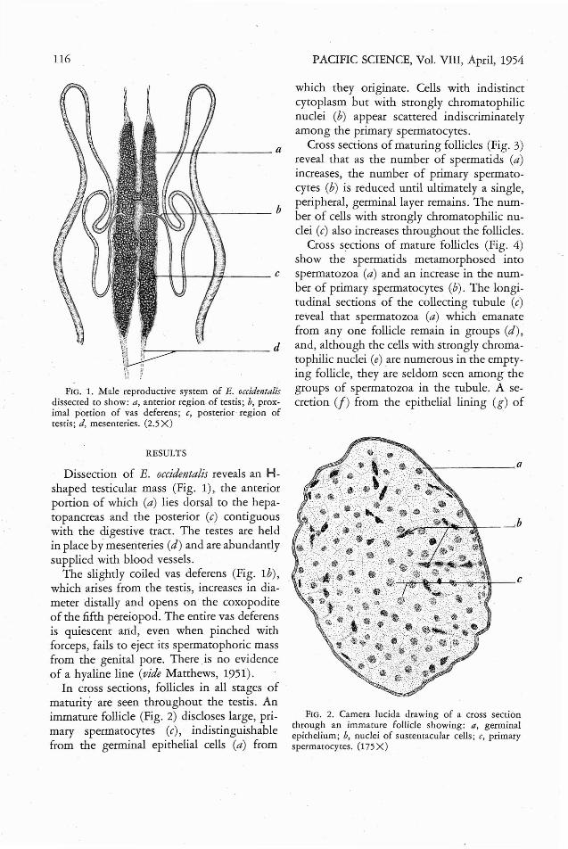
FIG. 1. Male reproductive sysrem of E. occidentalisdissected to show: a, anterior region of testis; b, proxiIDal p'ortion of vas deferens; c, posterior region oftestis; d, mesenteries. (2.5 X)
RESULTS
Dissection of E. occidentalis reveals an Hshaped testicular mass (Fig. 1), the anteriorportion of which (a) lies dorsal to the hepatopancreas and the posterior (c) contiguouswith the digestive tract. The testes are heldin place by mesenteries (d) and are abundantlysupplied with blood vessels.
The slightly coiled vas deferens (Fig. 1b),which arises from the testis, increases in diameter distally and opens on the coxopoditeof the fifth pereiopod. The entire vas deferensis quiescent arid, even when pinched withforceps, fails to eject its spermatophoric massfrom the genital pore. There .is no evidenceof a hyaline line (vide Matthews, 1951).
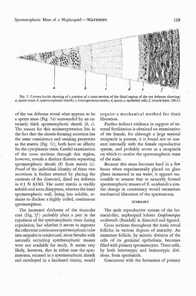
In cross sections, follicles in all stages ofmaturity are seen throughout the testis. Animmature follicle (Fig. 2) discloses large, primary spermatocytes (c), indistinguishablefrom the germinal epithelial cells (a) from
PACIFIC SCIENCE, Vol. VIII, April, 1954
which they originate. Cells with indistinctcytoplasm bur with strongly chromatophilicnuclei (b) appear scattered indiscriminatelyamong the primary spermatocytes.
Cross sections of maturing follicles (Fig. 3)reveal that as the number of spermatids (a)increases, the number of primary spermatocytes (b) is reduced until ultimately a single,peripheral, germinal layer remains. The number of cells with strongly chromatophilic nuclei (c) also increases throughout the follicles.
Cross sections of mature follicles (Fig. 4)show the spermatids metamorphosed intospermatozoa (a) and an increase in the number of primary spermatocytes (b). The longitudinal sections of the collecting tubule (c)reveal that spermatozoa (a) which emanatefrom anyone follicle remain in groups (d),and, although the cells with strongly chromatophilic nuclei (e) are numerous in the emptying follicle, they are seldom seen among thegroups of spermatozoa in the tubule. A secretion (j) from the epithelial lining (g) of
FIG. 2. Camera lucida drawing of a cross sectionthrough an immature follicle showing: a, germinalepithelium; b, nuclei of sustentacular cells; c, primaryspermarocytes. (175 X)

Spermatophoric Mass of a Nephropsid-MAlTHEWS 117
i
I
I \- a
';1"----- b
r--- d
FIG. 3. Camera lucida drawing of a cross sectionthrough a follicle in the early stages of maturity showing: a, spermatids; b, primary spermatocytes; c, nucleiof sustentacular cells; d, meiotic figures of spermatidformation. (175 X)
the tubule surrounds each group of spermatozoa.
Cross sections of the minute, proximal portion of the vas deferens (Fig. 5) reveal a tubein which the sperm mass (a) is surroundedby columnar epithelium (b) and a muscular
'layer (c). Although the columnar epithelialcells appear active, there is no evidence, inthis region of the vas deferens, that a sheathis being formed around the sperm mass.
A portion of a cross section of the vasdeferens (Fig. 6) (distad to the portion illustrated in Fig. 5) shows the sperm mass (a)enveloped by a spermatophoric sheath (b) andthat this sheath is the product of the secretionfrom the epithelial cells (c). The thickness ofthe muscular layer (d) is only slightly increased over that illustrated in Figure 5.
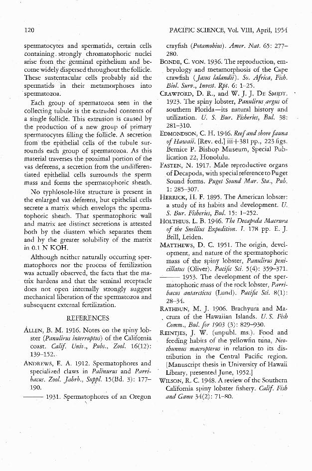
Figure 7 illustrates a portion of a crosssection through the distal region of the vasdeferens. The sperm mass (a) is not onlyenveloped by a spermatophoric sheath (b),but a homogeneous matrix (c) is added bythe epithelial cells (e). The matrix (c) is oftenseparated from the epithelial cells (e) by aspace (d). The muscular layer (f) is increased
in thickness and consists of longitudinal, diagonal, and circular fibers.
DISCUSSION
By mitotic division of the germinal epithelial cells (Fig. 2a), follicles fill with primaryspermatocytes (c). During this process, certain cells with strongly chromatophilic nuclei(b) apparently arise from the germinal epithelial cells and occupy positions between theprimary spermatocytes (c). Although mitoticfigures are associated with the increase of primary spermatocytes, no mitotic figures accompany the increase of the strongly chromatophilic cells. Their method of multiplication remains obscure.
Concurrently, the primary spermatocytes(Fig. 3b) by two successive divisions (d) give
FIG. 4. Camera lucida drawing of cross sec.tionthrough mature follicles discharging their contents intothe collecting tubule showing: a, spermatozoa; b, primary spermatocytes; c, longitudinal section of collecting tubule; d, groups of spermatozoa ejected fromdifferent follicles; e, contents of a single follicle beingejected into collecting tubule; f, secretion separatingsperm groups; g, epithelial cells. (150X)

118 PACIFIC SCIENCE, Vol. VIII, April, 1954
FIG. 6. Camera lucida drawing of a portion of a crosssection of the vas deferens (distad ro the portion illus- ,trated ill Fig. 5) showing: a, sperm mass; b, spermatophoric sheath; c, epirhelial cells; d, muscle layer. (soX)
tione,d, this secretion mixes freely betweenthe groups of spermatozoa.
Cross sections of the minute, proximal portion of the vas deferens (Fig. 5) reveal thatthe sperm mass (a) has not yet acquired itssheath. However, the columnar epithelial cells(b) are extremely active, and their secretionsmay be identical with this structure (Fig. 6b)seen in more distal sections.
Less than one third of the vas deferens istraversed by the descending sperm mass (Fig.Sa) before it acquires its spermatophoricsheath (Fig. 6b). This sheath is the productof the secretion from epithelial cells (c) which,although shorrer than those previously encountered, show little specialized arrangement. There are no deep folds or crypts (videMatthews, 1951: 363, fig. 7). The epithelialcells bounding the lumen are arranged in asingle cell layer, and their secretion envelopesthe sperm mass forming the spermatophoricsheath.
Cross sections through the distal portion
dcba
FIG. 5. Camera lucida drawing of a cross sectionthrough the minute, proximal portion of the vas deferens showing: a, sperm mass; b, columnar epithelium;c, muscle layer. (90X)
rise to spermatids (it) which later cluster aboutthe cells with sttongly chromatophilic nuclei(c). These are probably sustentacular cells (c)which serve the spermatids in their metamorphosis to spermatozoa.
Although most cross sections of the testisshow mature follicles, few reveal the actualopenings of these follicles into the collectingtubule; yet on these sections alone dependsthe understanding of the rhythmical natureof follicular activity. Concurrently with themetamorphosing of sp,ermatids to spermatozoa (Fig. 4a), the germinal epithelium, bymitotic division, again produces primary spermatocytes (b) which fill the follicles and expe~
the spermatozoa (a). The sustentacular cells,once numerous among the metamorphosingspermatids (e), are difficult to observe in theextruded follicular contents. It is quite evident, however, that the· many groups of spermatozoa (d) seen descending the collectingtubule (c) are each the entire extruded contentof one single follicle' and not, as. previouslyreported for Panulirus penicillatus (Matthews,1951)· the result of the activity of sustentacularcells. These groups of spermatozoa are furtherseparated by a secretion (j) from the epitheliallining (g) of the tubule. As previously men-
Spermatophoric Mass of a Nephropsid-MATTHEWS 119
FIG. 7. Camera lucida drawing of a portion of a cross section of the distal region of the vas deferens showing;a, sperm mass; b, spermatophoric sheath; c, homogeneous matrix; d, space; e, epithelial cells;/, muscle layer. (90X)
of the vas deferens reveal what appears to bea sperm mass (Fig. 7a) surrounded by an extremely thick spermarophoric sheath (b, c).The reason for this misinterpretation lies inthe fact that the sheath-forming secretion hasthe same consistency and staining propertiesas the matrix (Fig. 7c); both have an affinityfor the cytoplasmic stain. Careful examinationof the cross sections through this region,however, reveals a distinct diastem separatingspermatophoric sheath (b) from matrix (c).Proof of the individual identity of these twosecretions is further attested by placing thecontents of the dissected, distal vas deferensin 0.1 N KOH. The outer matrix is readilysoluble and soon disappears, whereas the innerspermatophoric wall, being less soluble, remains to disclose a highly coiled, continuousspermatophore.
The increased thickness of the muscularcoat (Fig. 7f) probably plays a part in theexpulsion of the spermatophoric mass duringcopulation; but whether it serves to segmentthe otherwise continuous spermatophoric tubeinto ampules is conjectural, since females withnaturally. occurring spermatophoric masseswere not available for study. It seems verylikely, however, that in either case the spermatozoa, encased in a spermatophoric sheathand enveloped in a hardened matrix, would
require a mechanical method for theirliberation.
Further indirect evidence in support of external fertilization is obtained on examinationof the female, for although a large seminalreceptacle is present, it is found not to connect internally with the female reproductivesystem, and probably serves as a receptacleon which w receive the spermatophoric massof the male.
Because this mass becomes hard in a fewhours when experimentally placed on glassplates immersed in sea water, it appears reasonable to assume that in naturally formedspermatophoric masses of E. occidentalis a similar change in consistency would necessitatemechanical liberation of the spermatozoa.
SUMMARY
The male reproductive system of the homarid-like, nephropsid lobster Enoplometopusoccidentalis (Randall) is dissected and figured.
Cross sections throughout the testis revealfollicles in various degrees of maturity. Animmature follicle, by mitotic division of thecells of its germinal epithelium, becomesfilled with primary spermatocytes. These cells,by both heterotypic and homeotypic divisions, form spermatids.
Concurrent with the formation of primary
120
spermatocytes and spermatids, certain cellscontaining. strongly chromatophoric nucleiarise from the germinal epithelium and become widely dispersed throughout the follicle.These sustentacular cells probably aid thespermatids in their metamorphoses intospermatozoa.
Each group of spermatozoa seen in thecollecting tubule is the extruded contents ofa single follicle. This extrusion is <caused bythe production of a new group of primaryspermatocytes filling the follicle. A secretionfrom the epithelial cells of the tubule surrounds each group of spermatozoa. As thismaterial traverses the proximal portion of thevas deferens, a secretion from the undifferentiated epithelial cells surrounds the spermmass and forms the spermatophoric sheath.
No typhlosole-like structure is present inthe enlarged vas deferens, but epithelial cellssecrete a matrix which envelops the spermatophoric sheath. That spermatophoric walland matrix are distinct secretions is attestedboth by the diastem which separates themand by the greater solubility of the matrixin 0.1 N KOH.
Although neither naturally occurring spermatophores nor the process of fertilizationwas actually observed, the facts that the matrix hardens and that the seminal receptacledoes not open internally strongly suggestmechanical liberation of the spermatozoa andsubsequent external fertilization.
REFERENCES
ALLEN, B. M. 1916. Notes on the spiny lobster (Panulirus interruptus) of the Californiacoast. Calif Univ., Pubs., Zool. 16(12):139-152..
ANDREWS, E. A. 1912. Spermatophores andspecialized claws in Palinurus and Parribacus. Zool. Jahrb., Suppl. 15(Bd. 3): 177190.
---·1931. Spermatophores of an Oregon,
PACIFIC SCIENCE, Vol. VIII, April, 1954
crayfish (Potamobius). Amer. Nat. 65: 277280.
BONDE, C. VON. 1936. The reproduction, embryology and metamorphosis of the Capecrawfish (jasus lalandii). So. Africa, Fish.BioI. Surv., Invest. Rpt. 6: 1-25.
CRAWFORD, D. R., and W. J. J. DE SMIDT.1923. The spiny lobster, Panulirusargus ofsouthern Florida-its natural history andutilization. U. S. Bur. Fisheries, Bul. 38:281-310.
EDMONDSON, C. H. 1946. Reefand shore faunaof Hawaii. [Rev. ed.] iii+ 381 pp., 223 figs.Bernice P. Bishop Museum, Special Publication 22, Honolulu.
FASTEN, N. 1917. Male reproductive organsof Decapoda, with specialreferencetoPugetSound forms. Puget Sound Mar. Sta., Pub.1: 285-307.
HERRICK, H. F. 1895. The American lobster:a study of its habits and development. U.S. Bur. Fisheries, Bul. 15: 1-252.
HOLTHIUS.1. B. 1946. The Decapoda Macruraof the Snellius Expedition. 1. 178 pp. E. J.Brill, Leiden.
MATTHEWS, D. C. 1951. The origin, development, and nature of the spermatophoricmass of the spiny lobster, Panulirus penicillatus (Oliver). Pacific Sci. 5(4): 359-371.
--- 1953. The development of the spermatophoric mass of the rock lobster, Parribacus antarcticus (Lund). Pacific Sci. 8(1):28-34.
RATHBUN, M. J. 1906. Brachyura and Ma-o crura of the Hawaiian Islands. U. S. Fish
Comm., Bul. for 1903 (3): 829-930.REINTJES, J. W. (unpub\. ms.). Food and
feeding habits of the yellowfin nina, Neothunnus macropterus in relation to its distribution in the Central Pacific region.[Manuscript thesis in University of HawaiiLibrary, presented June, 1952.]
WILSON, R. C. 1948. A review of the SouthernCalifornia spiny lobster fishery. Calif Fishand Game 34(2): 71-RO.