The Origin and Development of the Internal Musculature of ...
13
THE ORIGIN AND DEVELOPMENT OF THE INTERNAL MUSCULATURE OP THE FROG LUNG (RANA PIPIENS) F. A. WATERMAN Department of Zoology, The Ohio State University, Columbus, Ohio INTRODUCTION The general literature regarding the histological structure of the frog lung is quite ambiguous on certain points. There is much disagreement concerning the distribution of the arteries and the veins and their relationships to one another. Whether or not cilia are present in the frog lung is also controversial. Neither is there any agreement about the component parts of the alveolar septa; nor is any information concerning their development available. Smooth muscle is known to be present in the lung, but there is no accurate information concerning its distribution and morphology. Therefore, the writer undertook a histological and embryological investigation of these points. LITERATURE The veins and the arteries are distributed as companions to one another, according to the sketches of Renaut (1897), and copied by Oppel (1905), Winterstein (1921), and Holmes (1927). This is in complete disagreement with Kuttner (1874), Wieder- sheim (1882), and Gaupp (1904), who found the pulmonary artery on the periphery of the lung coursing its way from the root to the apex, giving off numerous lateral branches along the way and a main pulmonary vein on the extreme interior, collecting the blood from the intervening capillary network and returning it toward the root of the lung. Renaut's diagram of a section through the lung is misleading with respect to the distribution of cilia. He shows cilia present only in the tracheal tube at the root of the lung. On the other hand, Kuttner, mentioned above, as well as Hoffman (1878), Haslam (1889), and Konigstein (1903), find cilia not only in the tracheal tubes, but also on the margins of the numerous longi- tudinal and transverse septa on the interior of the lungs. In regard to the internal septal partitions of the lung, Bourne (1909) has stated that they are found less developed in 97
Transcript of The Origin and Development of the Internal Musculature of ...

THE ORIGIN AND DEVELOPMENT OF THEINTERNAL MUSCULATURE OP THE FROG
LUNG (RANA PIPIENS)
F. A. WATERMANDepartment of Zoology, The Ohio State University,
Columbus, Ohio
INTRODUCTION
The general literature regarding the histological structureof the frog lung is quite ambiguous on certain points. There ismuch disagreement concerning the distribution of the arteriesand the veins and their relationships to one another. Whetheror not cilia are present in the frog lung is also controversial.Neither is there any agreement about the component parts ofthe alveolar septa; nor is any information concerning theirdevelopment available. Smooth muscle is known to be presentin the lung, but there is no accurate information concerning itsdistribution and morphology. Therefore, the writer undertooka histological and embryological investigation of these points.
LITERATURE
The veins and the arteries are distributed as companions toone another, according to the sketches of Renaut (1897), andcopied by Oppel (1905), Winterstein (1921), and Holmes (1927).This is in complete disagreement with Kuttner (1874), Wieder-sheim (1882), and Gaupp (1904), who found the pulmonaryartery on the periphery of the lung coursing its way from theroot to the apex, giving off numerous lateral branches alongthe way and a main pulmonary vein on the extreme interior,collecting the blood from the intervening capillary networkand returning it toward the root of the lung.
Renaut's diagram of a section through the lung is misleadingwith respect to the distribution of cilia. He shows cilia presentonly in the tracheal tube at the root of the lung. On the otherhand, Kuttner, mentioned above, as well as Hoffman (1878),Haslam (1889), and Konigstein (1903), find cilia not only in thetracheal tubes, but also on the margins of the numerous longi-tudinal and transverse septa on the interior of the lungs.
In regard to the internal septal partitions of the lung,Bourne (1909) has stated that they are found less developed in
97

"98 F. A. WATERMAN Vol. XXXIX
the lower portion of the lung wall and are completely absent inthe apex. This is contrary to the diagrams of Miller (1893),Renaut, and Oppel, who find them as completely formed in theapex and lower portion as in the region near the root.
Of special interest were the accounts of various writers con-cerning the smooth musculature of the frog lung. Those will bediscussed in chronological order.
Arnold (1863) describes the distribution of the elastic andsmooth muscle fibers in the alveolar and septal walls of the frogand of the human lung. He speaks of the smooth muscle fibersas being the essential structural constituent of the frog lung.
Kiittner (1874) mentions that the smooth muscle forms inthe lung of the frog, "a scaffolding for the support of thearteries and nerves, and for the veins a protective cap locatedin the crown or inner margin of the numerous septa." Heascribes to this musculature the function of a tension regulator,presumably in reference to the intrapulmonic pressure.
Weidersheim and Haslam state that the lung musculature isarranged in large bands which form a network on the interiorfrom which pass finer processes of muscle tissue in the septalwalls toward the periphery where they join a thin and incom-plete muscular layer in the external wall of the lung.
Thomson (1899) briefly describes the frog lung as a trans-parent oval sac with muscle fibers in its walls.
Konigstein states that a histological section through theamphibian lung convinces one that the greater part of the com-pact lung substance consists of smooth muscular tissue. Heassigns to this musculature the function of diminishing the sizeof the lung cavity, so that when the glottis is closed, the air inthis cavity will be forced out into the peripherally arrangedalveoli.
Gaupp (1904) states that the big veins of the lungs courseonly on the interior in the crowns (Kuppen) of the septa under-neath muscle ridges (Randmuskelbalken).
Oppel (1905) has shown by diagram the distribution ofsmooth musculature in reference to a lung section and to anindividual septal partition. In these his emphasis is entirelyupon the distribution of the muscle bundles with completedisregard of the veins which are associated with them, in spiteof the fact that he includes blood cells in his sketches. Regardingthe blood vessels of the lungs, he relies upon the statements of•other authors.

No. 2 MUSCULATURE OF FROG LUNG 99
Brown (1909) registered graphically small pressure changesin the lung sac, after inhalation, with the glottis closed andattributed them to the contraction and relaxation of the smoothmusculature of the lung.
Carlson and Luckhardt (1920) describe the amphibian lungas a paired muscular sac in which the smooth musculaturecovers the entire wall and extends into the smallest septa of theinterior. The arrangement of this musculature is such that itscontraction will reduce the size of the lung cavity and raise theintrapulmonic pressure if the glottis is closed. In their studiesthey report the discovery of a "persistent peripheral motorautomatism" which is normally repressed or controlled byinhibitory impulses from the central nervous system. Betweenbreathing movements, (inhalation and exhalation) when thepause is of sufficient duration, they find an initial rise in theintrapulmonic pressure as did Brown, and, like the latter, con-clude that it is due to the contraction of the lung musculaturerather than to any other possible external or internal factor.
HISTOLOGICAL STUDIES
1. METHODS
Histological preparations were made from fifteen frog lungs (Ranapipiens). Each was obtained immediately after pithing the brain andcord. The lungs were then severed at the roots and at once submergedin Bouin's fixative solution. Some of the lungs were prepared in thecollapsed, others, in the inflated state. Each specimen was imbeddedin rubberized paraffin for sectioning. Some of the series were stained inDelafield's hematoxylin and eosin; others, in orcein, a specific stain forelastic tissue; still others, in Castroviejo's triple stain, which is partic-ularly good for showing muscular tissue distribution and for showingthe presence of erythrocytes in the blood vessels. The blood vessels inmost of these lungs were injected with India ink from the heart beforebeing severed from the animal. This procedure very effectively aidedthe study of the distribution of the blood vessels (arteries, capillaries,and veins) throughout the entire lung structure.
2. DISCUSSION AND RESULTS
ArteriesAccording to Renaut, Oppel, and Winterstein, the arteries and the
veins are distributed as companions to one another, whereas Kiittner,Weidersheim, and Gaupp found a main pulmonary artery coursing itsway from the root to the apex, giving off numerous lateral branchesalong the way. While a principal artery is present in some cases, thisdistribution is by no means invariable. Serial sections of four lungpreparations show that the pulmonary artery divides into two branches.

100 F. A. WATERMAN Vol. XXXIX
One of these again subdivides, forming a total of three vessels of uniformsize near the root of the lung.
CiliaRenaut found cilia to be present only in the tracheal tubes. In
agreement with Kuttner, Hoffman, Haslam, and Konigstein, the writerfinds cilia not only in the tracheal tubes but also on the margins of thenumerous longitudinal and transverse septa throughout the interior ofthe lungs.
Fig. 1. Longitudinal section near the external wall, showing the peripherallyarranged septa: (a) lung cavity, (b) internal septa, (c) small internal vein, (d) inter-nal muscle bundle. 10 X
SeptaBourne found the internal septal partitions less developed in the
apex of the lung than in the root. This is contrary to the diagrams ofMiller, Renaut, and Oppel and to my own observations. They areas completely formed in the apex and lower portion of the lung as inthe region near the root. Fig. 1.
Musculature and Venous ReturnIt is apparent from an examination of the literature that the great
amount of disagreement concerns the distribution of the smooth musclefibers and the venous system of the lungs. A great deal of this is, nodoubt, due to the fact that the muscles and veins are structurally soclosely associated that many writers investigating the one have failedto observe the other. Because of this close association, it will benecessary to discuss them together.
No. 2 MUSCULATURE OF FROG LUNG 101
Most of the authors who have considered the muscular tissuedeclare it to be a structure independent of the adjacent veins; some ofthem even failed to observe the closely associated veins. A modifica-tion of Castroviejo's triple stain has been particularly helpful in makingpossible the interpretation of the histological structure of the internalseptal marginal veins. Through this stain the muscle tissue appears ingreen; the white fibrous tissue, in blue; the erythrocytes, in yellow; andthe nuclei, in red. With such a stain, that which all the precedingauthors have mentioned as an independent muscular network on theinterior of the amphibian lung appeared to the writer at first to be aconstituent portion of these internal veins. These veins appear todiffer from the typical vein in having the smooth muscle tissue, exceptrarely for a few scattered fibers, assembled asymetrically in a singlebundle on their free border. White fibrous connective tissue, whichtypically is found abundant in both the tunica intima and tunicaexterna of veins, is, in the case of these septal veins, merged into asingle layer for two-thirds of their lumenal circumference. For theremaining third, these layers separate and include the muscle bundle.Moreover, this bundle lies immediately adjacent to the tunica intimaof these veins and also possesses numerous interwoven elastic tissuefibers, such as is found in the tunica media of veins. This gives it theappearance of being the tunica media, which has been concentratedin one side.
One cannot escape wondering why there is this peculiar assemblingof the muscular tissue of these veins into this single bundle along theirinternal wall. If the view of the foregoing investigators as to the functionof these muscle bundles is accepted, viz., that they act along with theother muscular tissue of the amphibian lung to reduce the size of thelung cavity and thereby raise the intrapulmonic pressure, it is easy tosee that from a mechanical standpoint this would be an advantage. Asingle bundle of muscle fibers located on the free internal border of thesepta would act with greater facility and with less impediment inreducing the size of the lung cavity than the same amount of muscletissue scattered throughout the entire wall of these veins. In a sense thisnetwork of muscle bundles is really a complex internal lattice-likesphincter of inter-crossing, longitudinal and transverse, circularly-arranged strands, among which are interspersed elastic connectivetissue fibers.
In fact, if the above hypothesis is correct, one may assign twofunctions to the muscular tissue of these veins, namely: that its con-traction will bring about a rise in intrapulmonic pressure and, at thesame time, be effective in increasing the blood flow through the pul-monary veins from the lungs. This action would increase the oxygenabsorption (Gegenbaur, 1901) of the air in the lung cavity and force thefreshly oxygenated blood back to the heart. Both of these functionshave their counterpart among mammals in the action of the diaphragmand other inspiratory muscles. Thus, this vascular sphincter would notonly perform its normal function of influencing circulation, but alsoassume a new function of assisting in breathing as well.

102 F. A. WATERMAN Vol. XXXIX
Development of the Lung MusculatureThe study of the development of the Anuran lung has not extended
in the past beyond the very young tadpole. Greil (1905) studied thegross development in Rana temporaria, Bufo vulgaris, and Bombinatorigneus. His plates show wax reconstructions of the trachea and lung-budanlagen, with the veins in blue and the arteries in red. The largesttadpole used was 8.4 millimeters.
Fig. 2. A. Drawing of the left lung, showing exit of the pulmonary vein.B. Diagram of the dissected lung at portion indicated by dotted line in A. Pulm.,pulmonary vein; Epith., epithelium; Muse, muscle; Plu., pleura; Ct., connectivetissue.
Fedrow (1910) made a comparative study of the development ofthe lung veins in various vertebrates. The forms and sizes used wereas follows:
Rana temporaria 4.4-11.7 mm.Triton taeniatus 8.7-11.0 mm.Lacerta agilis 32-38 somitesGallus domesticus 3 daysCavia cobaya 21 days
His object was to show in cross section the similarities of the locationand structure of the developing lung sacs and their accompanying bloodvessels. In all of his stages, the lung buds were only mere sacs of endo-derm with no indication of developing internal septa and smooth muscles.

No. 2 MUSCULATURE OF FROG LUNG 103
It seems illogical to think that the muscle network is tunica mediabecause a vein of such construction is, to the writer's best knowledge,not found anywhere else in the animal kingdom. It would seem thatthe musculature must arise independently of the veins, and the latterbecome very closely associated with it so as to give a false appearanceof being tunica media.
This is borne out by the following facts: The main pulmonary veinoriginates at the apex of the interior of the lung and courses anteriorlyalong its meso-lateral border for about three-fourths of the length ofthe lung. It then passes through the lung wall and proceeds onward tothe left auricle. When this vein is on the inside of the lung, it has thesame structure as the other veins associated with the musculature. Ifthis muscle bundle is the tunica media of the vein, serial sections of thelung, reading from the posterior to the anterior, should show a transi-tion from the asymmetrical to the symmetrical condition at the pointwhere the vein takes its departure toward the heart. This, however, isnot the case. When this point is reached, the muscle bundle continuesin association with a tributary vein, while the main vein with a tunicaadventitia passes on towards the left auricle. Fig. 2.
Since this problem could not be solved through adult anatomicalstudy, the only alternative left was to study the frog lung from anembryological point of view.
Materials and MethodsThe eggs of Rana pipiens were collected early in April, and at the
time of collection, they were in the neural groove stage. Samples weretaken each twenty-four hours for the first week. For the next twoweeks, the samples were taken every forty-eight hours and from thenon, twice a week until the animals emerged. When the tadpoles reachedthe stage where hind legs were beginning to develop, the lung buds werecarefully dissected out, fixed, and sectioned. The slides were nextstained by a dioxan modification of Castroviejo's (1932) method,Waterman (1937).
The critical stages in the development of the frog lung are as follows:1. Late operculum 10.5 mm.2 13.5 mm.3 16.0 mm.4 30.0 mm.5 45.0 mm.6 60.0 mm.7. Four legs and long tail 58.0 mm.8. Four legs 56.0 mm.9. Short tail 39.0 mm.
10. Very short tail 30.0 mm.11. Tail practically resorbed 26.5 mm.12. Small mature frog13. Mature frog, medium size14. Normal mature frog
The sections showing the development of the lung and its mus-culature were then drawn.

104 F. A. WATERMAN Vol. XXXIX
DiscussionThe lung develops as an endodermal evagination of the floor of the
pharynx. At the late operculum stage, this evagination consists of asingle layer of somewhat cuboidal cells. The laryngo-tracheal grooveis surrounded by loose mesodermal cells and a few strands of developing
B
e/>/tt
•muse rmusc
F
Fig. 3. Camera lucida drawings showing the formation of the alveolar septaand internal musculature of the anuran lung.
A. Lung bud of a 10.5-mm. tadpole of the late operculum stage, epith., epithe-lium; ct., connective tissue; plu., pleura. 200 X.
B. Lung bud of a 13.5-mm. tadpole, showing earliest appearances of smoothmuscle cells, muse, muscle cells; epith., epithelium; plu., pleura. 400 X.
C. Cross section of the lung of a 40-mm. tadpole, showing condensed musclesheet, muse, muscle; plu., pleura; ct., connective tissue; epith., epithelium.
D. Cross section of the lung of a 46-mm. tadpole showing the formation of therudimentary alveolar septa, pv., pulmonary vein; epith., epithelium; muse,muscle sheet; ct., loose connective tissue. 400 X.
E. Cross section through the lung of a tadpole with hind legs, showing primitivealveolar septa. Length of tadpole, 56.0-mm. epith., epithelium; muse,muscle fibers; pv., pulmonary vein. 100 X.
F. Cross section of the lung of a 60-mm. tadpole, taken near the distal end,showing the advanced condition of the musculature, pv., pulmonary vein;muse, muscle bundle; epith., epithelium; ct., loose connective tissue; plu.,pleura.

No. 2 MUSCULATURE OF FROG LUNG 105
muscle fibers. The anlage of the trachea has the same general structurebut more posteriorly becomes somewhat flattened.
The lung buds consist of a single layer of very much flattened andsomewhat spindle-shaped cells and are surrounded by very loose con-nective tissue. No signs of muscle cells are present. Fig. 3A.
By the time the tadpole has reached the length of 13.5 millimeters,muscle cells can be seen developing just under the epithelium. Theyform a single chain of spindle-shaped cells, which extend around theentire lung. The cells of the epithelium are still flattened, and a fewscattered strands of connective tissue can be seen between the pleuraand muscle cells. Fig. 3B.
One can best visualize this condition by imagining the lung as beingcomposed of two sacs, exclusive of the plura, one fitting very closely
Fig. 4. Diagram showing the formation of the alveolar septa and internalmusclature of the Anuran Lung.
A. Lattice-like venous plexus of pulmonary vein with the epithelium and mus-cular sheets growing through the interstices.
B. Perforations in muscle sheet, exposing the epithelial sheet.C. Perforations in muscle sheet increase as the muscle thickens over the veins.D. The muscle is completely condensed over the veins. The epithelium is grown
together, forming alveolar septa.Pv., pulmonary vein; Mus., muscle tissue; Ep., epithelium.
106 F. A. WATERMAN Vol. X X X I X
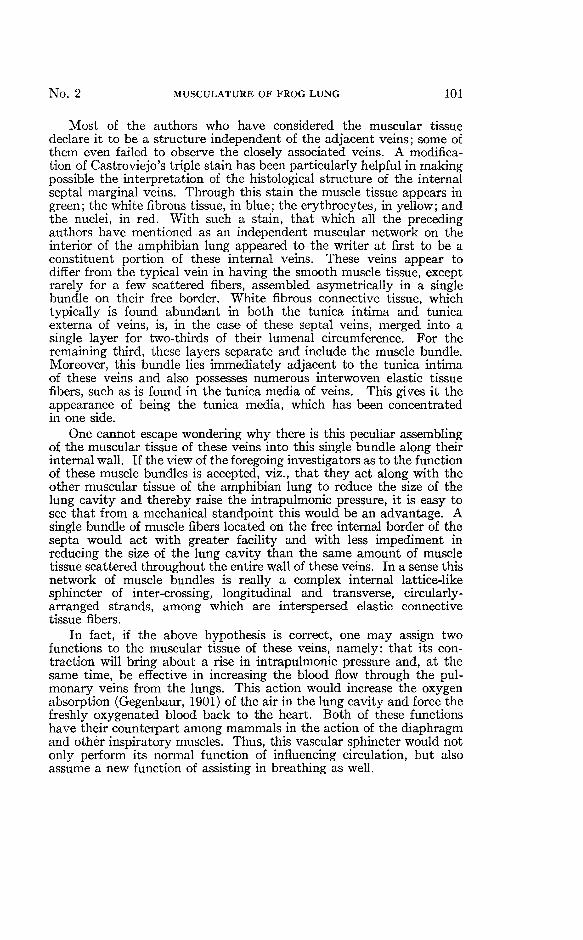
around the other. The inner sac is composed of evaginated endoderm,consisting of very much flattened cells. Around this sac, a second sacfits very closely and is composed of smooth muscle cells, which areloosely joined together.
Very little change can be noticed in the musculature of the lunguntil the tadpole has reached the length of 40.1 millimeters. At thisstage, the muscle fibers have become condensed and extend throughoutthe interior of the lung just under the epithelium. The cells of theepithelium have become somewhat cuboidal. Fig. 3C.
IBM%
1 — - — ^
Fig. 5. Highly magnified view of a pulmonary vein and its associated struc-tures: (a) cilia, (b) pseudo-stratified ciliated columnar epithelium, (c) loose whitefibrous tissue, (d) muscle bundle, (e) lumen of vein containing India Ink and cor-puscles. 800 X.
The veins can also be seen forming just under the muscle, their wallsconsisting of a single layer of endothelial cells. The connective tissueunderlying the muscle strands is very loose. These veins form a rudi-mentary lattice-like plexus. The epithelium and muscle strands thengrow into the openings of this lattice work. This leaves the veins sur-mounted by thickening muscle strands and epithelium extending asprocesses into the lumen of the lung cavity. This marks the firstappearance of internal septa. Fig. 3D.
With the appearance of the hind legs, the muscular sac becomesthickened over the internal margins of the venous system. The mus-cular sac now ceases to be a sac, becomes perforated, and leaves alattice-like muscular framework that remains permanently associatedwith the internal border of the venous plexus. Fig. 3E and Fig. 4.
No. 2 MUSCULATURE OF FROG LUNG 107
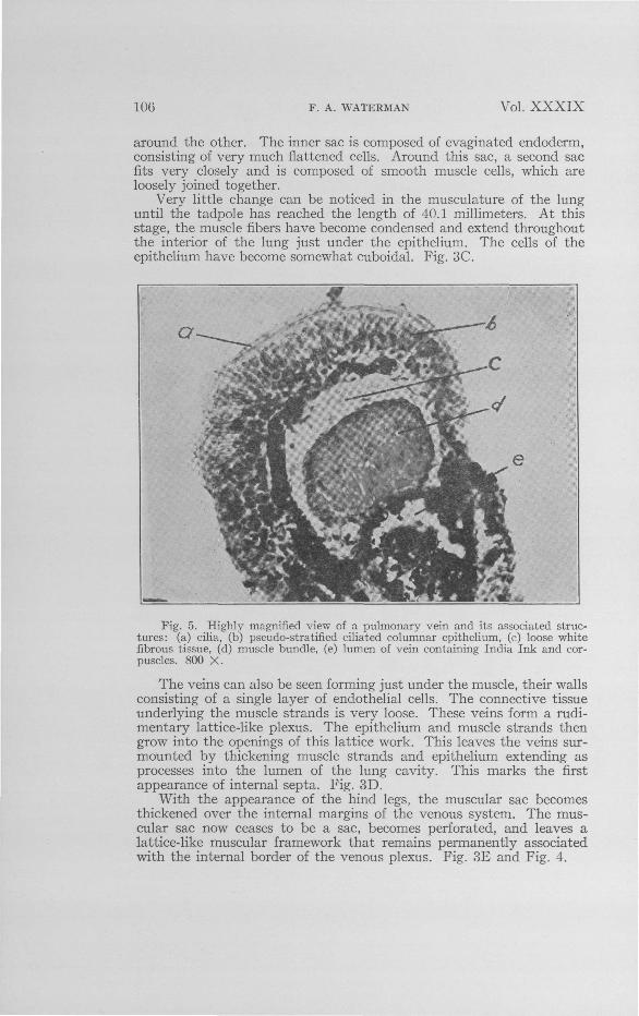
It is interesting to notice that the sections, reading serially from theanterior to the posterior, show that the lungs become more developedtoward the apex. This, at first glance, may appear quite contradictoryto the well-established fact that an animal develops more rapidly at theanterior end than at the posterior end. However, it is to be remem-bered that the metabolic gradient, while higher at the anterior end ofan organism than the posterior end, is also higher at the ends of theappendages. Thus it would seem logical for the apex of the lung todevelop more rapidly than the root, since the lungs arise as appendagesof the fore gut, and unless the anterior end remained in a more general-ized condition, backward growth would be impossible.
Fig. 6. Cross section through the pulmonary vein of the interior of the froglung. Muse, muscle bundle; P. V., lumen of pulmonary vein; Ct., connective tissue;Epith., ciliated columnar epithelium.
Towards the apex of the same lung, as shown in Fig. 3E, the mus-culature, as well as the veins, has become very well developed. Theepithelium is now approaching the columnar type, but it is still com-posed of a single layer of nonciliated cells which are not of the truecolumnar type as found in the adult organism. Fig. 3F.
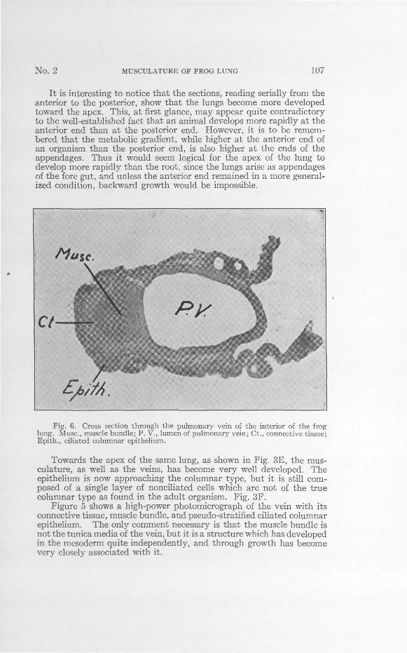
Figure 5 shows a high-power photomicrograph of the vein with itsconnective tissue, muscle bundle, and pseudo-stratified ciliated columnarepithelium. The only comment necessary is that the muscle bundle isnot the tunica media of the vein, but it is a structure which has developedin the mesoderm quite independently, and through growth has becomevery closely associated with it.

108 F. A. WATERMAN Vol. XXXIX
The main pulmonary vein, as shown diagrammatically in Fig. 2,when inside of the adult frog lung has the same structure as the otherveins. A cross section taken through it shows that the vein and itsassociated muscles have developed in place and the epithelium hasgrown around them, leaving a raphae which is visible on the externalsurface of the lung when examined under high power with reflectedlight. Fig. 6 shows a photomicrograph through this vein in cross section.
SUMMARY
1. Cilia are found not only in the tracheal sacs at the rootof the frog lung, but also on the free internal borders of all thenumerous transverse and longitudinal septa.
2. The arteries and veins are not distributed as companionsto one another, but independently, the arteries are restricted tothe periphery and the veins are located on the extreme interiorof the lungs, with the capillaries connecting these relativelywidely separated vessels.
3. It is possible for the musculature to affect simultaneouslyboth the intrapulmonic pressure and the venous return of theblood from the lung to the left auricle.
4. Embryonically the frog lung increases in complexityfrom the root to the apex.
5. The muscle bundle located beneath the epithelium of thealveolar septa is not a part of the tunica media, but is anindependent structure, having developed as such, but in closeassociation with the veins.
6. The alveolar septa are formed as follows:(a) A sac of endodermal epithelium, which has evaginated from
the floor of the pharynx, becomes surrounded by a secondclosely fitting sac, composed of smooth muscle cells.
(b) Around this second sac, a network of blood tubes developsand condenses into a lattice-like plexus of veins.
(c) The epithelial and muscle sheets grow into the openings ofthis lattice work.
(d) The muscle sheet thins and becomes perforated in the centerof the lattice squares and thickens on the lung-cavity sideof the veins.
LITERATURE CITEDArnold, Julius. Vorlaufige Mitteilung uber das Epithel der Lungenalveolen. Vir-
chow's Arch., 1863, Bd. 27, S. 396.Zur Histologie der Lunge. Virchow's Arch., 1863, Bd. 27, S. 433.
Bourne, G. C. Comparative anatomy of animals. 1909, Vol. I, p. 42. London.Brown, T. G. Die Atembewegungen des Frosches und ihre Beeinflussung durch die
nervosen Zentren und durch das Labyrinth. Arch. f. d. gesamte Physiol.1909, Bd. 130, S. 193.

No. 2 MUSCULATURE OF FROG LUNG 109
Carlson, A. J. and A. B. Luckhardt. Lung automatism and lung reflexes in the frog.I. Amer. Journ. Physiol., 1920, liv, p. 55.
Cardiac and yasomotor reflexes induced by visceral stimulation in Amphibiaand Reptilia. V. Amer. Journ. Physiol., 1921, lv, p. 31.
Castroveijo, R. Modifications of differential stains with special reference to thetrichromatic stain of Cajol. American Journal of Clinical Pathology, 1932,ii, No.. 2.
Fedrow, V. U"eber die Entwickelung der Lungenvene. Anatomische Hefte, 1910,xl, S. 533-603.
Gaupp, E. Zur Lehre von dem Athmungsmechanismus beim Frosch. Arch. f.Anat. (u. Physiol.), 1896, S. 329.
Histologischer Bau der Lungenwandung. Ecker u. Wiedersheim's, Anatomie desFrosches, 1904, Abt. 3. Braunschweig. S. 196.
Gegenbaur, Carl. Vergleichende Anatomie der Wirbelthiere, 1901, Bd. 2, S. 300,Leipzig.
Greil, Alfred. Tiber die Anlage des Lungen, Sowie der Ultimobranchialen (Post-branchialen, Supraperikardialen) Korper bei Anuren Amphibien. Anato-mische Hefte, 1905, Band 29, Hefte 3, S. 447-506.
Haslam, G. Ecker's, Anatomy of the frog. 1889. p. 317. Oxford.Hoffman, C. K. Bronn's, Klassen und Ordnungen des Thierreichs. 1878. S. 514.
Leipsig U. Heidelberg.Holmes, S. J. Biology of the frog. 1927. 4th ed., p. 165. New York. The MacMillan
Co.Konigstein, Hans. Die Function der Muskelatur in der Amphibienlunge. Arch.
f. d. gesamte Physiol., 1903, Bd. 95, S. 616.Kiittner, Dr. Beitrag zu den Kreislaufsverhaltnissen der Froschlunge. Virchow's
Arch., 1874, Bd. 61, S. 21.Luckhardt, A. B., and A. J. Carlson. Lung automatism and lung reflexes in the
salamanders. II. Amer. Journ. Physiol., 1920, liv., p. 122.Lung automatism and lung reflexes in Cryptobranchus with further notes on the
physiology of the lung of Necturus. VI. Amer. Journ. Physiol., 1921, lv, p. 212.On the presence of yasomotor fibers in the vagus nerve to the pulmonary vessels
of the Amphibian and the Reptilian lung. VIII. Amer. Journ. Physiol.,lvi, p. 72.
Miller, W. S. The structure of the lung. Journ. Morph., 1893, viii, p. 165.Oppel, Albert. Die Lunge. Lehrb. d. vergl. mikrosk. Anat. d. Wirbd. 1905, Bd. 6,
S. 265, Jena.Renaut, J. Traite d'histologie pratique. 1897. T. 2, Fasc. 1. p. 488, Paris.Thomson, J. A. Outline of zoology. 1899. 3rd ed., p. 546. New York.Waterman, F. A. A dioxan method for triple staining. Stain Technology, 1937,
xii, 21-23.Wiedersheim, R. Die Lungen. A. Ecker's, Anatomie des Frosches, 1882. Abt. 3,
S. 33. Braunschweig.Winterstein, Hans. Haut- und Lungenatmung. Handb. d. vergl. Physiol., 1921,
Bd. 1, Hft. 2, S. 191. Jena.