
THE OCCURRENCE OF BACILLUS INFLUENZÆ IN THROATS ...
12
THE OCCURRENCE OF BACILLUS INFLUENZ:E IN THROATS AND SALIVA. BY IDA W. PR.ITCHETT AND ERNEST G. STILLMAN, M.D. (From the Hospital of The Rockefeller .Institute for Medical Research.) PLATES 10 TO 13. (Received for publication, January 15, 1919.) In the present paper are reported the facts obtained during an in- vestigation of the bacteriology of influenza which included (1) the use of oleate hemoglobin agar, (2) the incidence of Bacillus influenza in uncomplicated and complicated cases of influenza and the associated types of pneumococcl, (3) the occurrence of BacilMs influenza in convalescents, and (4) the occurrence of Bacillus influen~.m in the mouth secretions or throats of normal people. The medium used in this work was Avery's oleate hemoglobin agar. ~ Agar of an average hydrogen ion concentration of 7.2 was used. As the domestic Sodium oleate which was tested from time to time gave less consistent results, Kahlbaum's sodium oleate was used. This medium is especially favorable to the growth of Gram-negative organisms in influenza work. On these plates pneumococci do not grow and streptococci rarely grow, although some staphylococci show a scant growth (Figs. 1, 2, and 6). Mter 36 to 48 hours incu- bation several types of Bacillus influem~e colonies are encountered on this medium. The characteristic colony of Bacillus influen~.ae was clear, round, slightly glistening, convex, and discrete. The size var- ied from pin-point colonies, easily confused with diphtheroids or catarrhalis colonies, to large round colonies of 1 to 3 ram. in diameter. The larger characteristic colonies had a distinct nucleus which was much darker than the rest of the colony (Figs. 3, 4, and 7). The colonies were very sticky in consistency and would streak across the agar and follow the loop in long threads. The medium beneath the colony appears discolored after a colony is fished. 1 Avery, O. T., Y. Am. Med. Assn., 1918, Ixxi, 2050. 259 on February 13, 2018 jem.rupress.org Downloaded from
Transcript of THE OCCURRENCE OF BACILLUS INFLUENZÆ IN THROATS ...
THE OCCURRENCE OF BACILLUS INFLUENZ:E IN THROATS AND SALIVA.
BY IDA W. PR.ITCHETT AND ERNEST G. STILLMAN, M.D.
(From the Hospital of The Rockefeller .Institute for Medical Research.)
PLATES 10 TO 13.
(Received for publication, January 15, 1919.)
In the present paper are reported the facts obtained during an in- vestigation of the bacteriology of influenza which included (1) the use of oleate hemoglobin agar, (2) the incidence of Bacillus influenza in uncomplicated and complicated cases of influenza and the associated types of pneumococcl, (3) the occurrence of BacilMs influenza in convalescents, and (4) the occurrence of Bacillus influen~.m in the mouth secretions or throats of normal people.
The medium used in this work was Avery's oleate hemoglobin agar. ~ Agar of an average hydrogen ion concentration of 7.2 was used. As the domestic Sodium oleate which was tested from time to time gave less consistent results, Kahlbaum's sodium oleate was used. This medium is especially favorable to the growth of Gram-negative organisms in influenza work. On these plates pneumococci do not grow and streptococci rarely grow, although some staphylococci show a scant growth (Figs. 1, 2, and 6). Mter 36 to 48 hours incu- bation several types of Bacillus influem~e colonies are encountered on this medium. The characteristic colony of Bacillus influen~.ae was clear, round, slightly glistening, convex, and discrete. The size var- ied from pin-point colonies, easily confused with diphtheroids or catarrhalis colonies, to large round colonies of 1 to 3 ram. in diameter. The larger characteristic colonies had a distinct nucleus which was much darker than the rest of the colony (Figs. 3, 4, and 7). The colonies were very sticky in consistency and would streak across the agar and follow the loop in long threads. The medium beneath the colony appears discolored after a colony is fished.
1 Avery, O. T., Y. Am. Med. Assn., 1918, Ixxi, 2050.
259
on February 13, 2018
jem.rupress.org
Dow
nloaded from
260 B. INFLUENZ3E IN Tlff-ROATS AND SALIVA
The colonies most readily confused on inspection with those of Bacillus influenzce were those of Micrococcus catarrhalis, meningo- coccus, diphtheroids, and especially an unidentified organism called "Bacillus X." This organism was encountered in about two dozen individuals. The colony varied between 1 and 2 ram. in diameter, was clear, convex, and mucoid. The nucleus so common in the in- fluenza colony was absent, but the colony left a slight discoloration on the agar. This bacillus is Gram-negative, takes the counterstain deeply, and when taken from agar surface cultures appears as long tangled threads, somewhat similar to the threads often seen in pure cultures of Bacillus influenzte. In blood broth Cultures it appears as a small fat bacillus without chain formation. I t does not grow on plain sheep serum, or glucose agar. On blood agar the abundant growth appears as a clear, somewhat granular, fairly heavy film, with marked hemolysis of the red cells. This hemolysis is as marked as that pro- duced by hemolytic streptococci (Fig. 5). Complete hemolysis is produced in blood broth after 24 hours incubation. After several days both blood broth and blood agar cultures turned brown and then blackish from the formation of methemoglobin. After 7 days incubation, litmus milk, to which a little blood had been added, showed an alkaline reaction with peptonization. No pathogenicity could be demonstrated for mice, rats, or rabbits3
In all the cultures from convalescents and normal persons, and in many from the hospital patients, the organism was isolated in pure culture and was demonstrated to possess the typical morphology and shown to be strictly hemoglobinophilic. All evidence based simply
The other organisms commonly found in cultures from the throat on this medium may be briefly described as follows in the frequency of their occurrence. (1) Gram-negative cocci of the Micrococcus catarrhalls group. The colonies are round, sharply defined, with a ridged or lined surface. Some colonies are so scaly that they lift off the medium as a pellicle; other colonies of this group will slide across the surface of the agar without breaking. (2) Staphylococci. Minute, pin-point colonies. (3) Diphtheroids. Minute, pin-point colonies. (4) Meningo- cocci. The colonies are clear, round, and discrete. (5) Friedl~nder's bacillus. The colonies are white, mucoid, and occasionally confluent.
Occasionally in some plates a few Gram-positive streptococci may grow. These colonies are minute, dense, nucleated, and sharply defined. Pneumococci were never found and "spreaders" rarely grow.
on February 13, 2018
jem.rupress.org
Dow
nloaded from
IDA W. PRITCI-YETT AND ERNEST G. STILLMAI~ 261
on the presence of slender Gram-negative bacilli in films" was dis- carded as of no value. The wide variations in the morphology of Bacillus influenzce noted by Wollstein 3 were observed in this series. This study was made during the widespread epidemic of influenza which occurred in New York. The epidemic began about the middle of September, reached its height about the middle of October, .and then gradually fell, though cases are still present in the city.
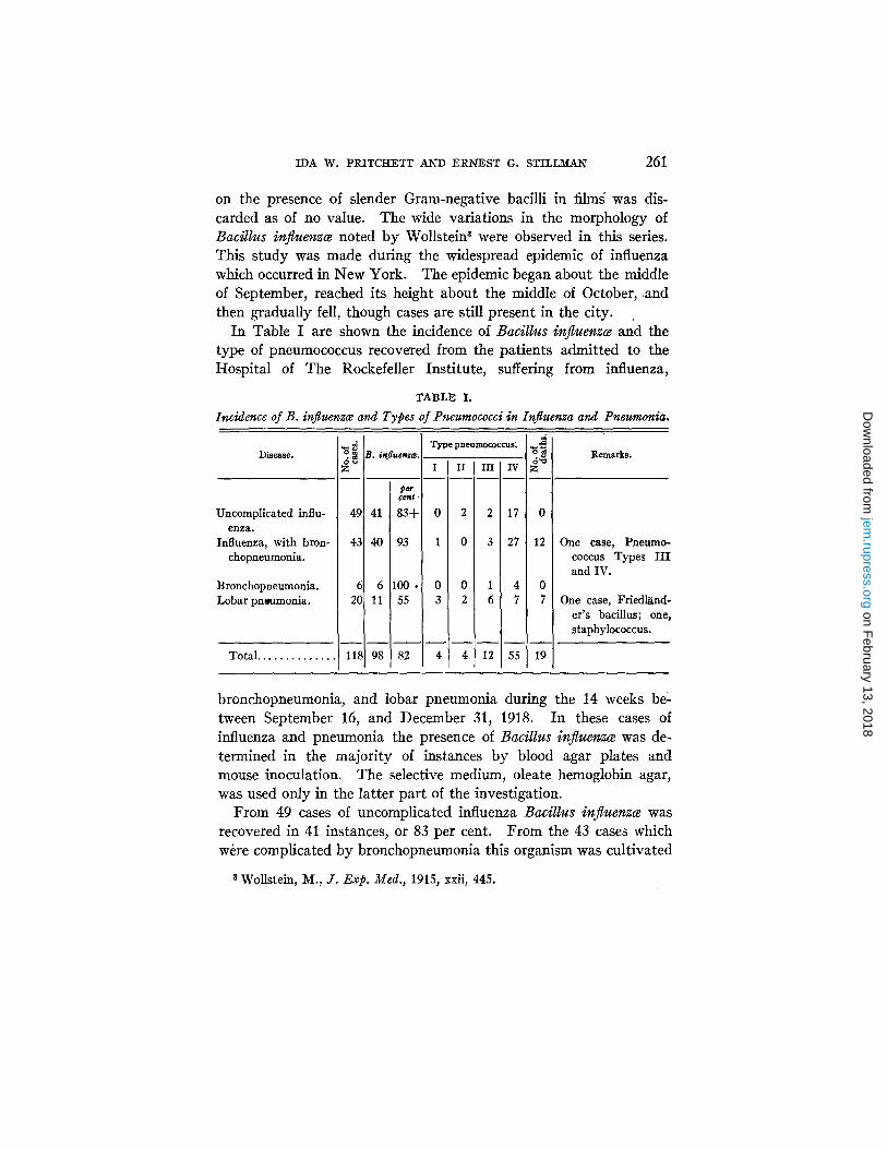
In Table I are shown the incidence of Bacillus influenzce and the type of pneumococcus recovered from the patients admitted to the Hospital of The Rockefeller Institute, suffering from influenza,
TABLE I.
Incidence of B. influen~ce and Types of Pneumococci in Influenza and Pneumonia.
Disease.
Uncomplicated influ- enza.
Influenza, with bron- chopneumonia.
B ronchopneumonia. Lobar pn~amonia.
Total . . . . . . . . . . . . . .
6~ B. inj
49 41
43 40
6 6 20 11
1181 98
Type pneumococcus: en~
I I I [ I I I IV F
pe 6~f,
I
B3 0 2 ] 2 17
P3 1 0 3 27
DO 0 0 1 4 0 55 3 2 6 7 7
82 4 4 12 55 19
0
12
Remarks.
One case, Pneumo- coccus Types I I I and IV.
One case, FriedhXnd- er's bacillus; one, staphylococcus.
bronchopneumonia, and lobar pneumonia during the 14 weeks be- tween September 16, and December 31, 1918. In these cases of influenza and pneumonia the presence of Bacillus influenz¢e was de- termined in the majority of instances by blood agar plates and mouse inoculation. The selective medium, oleate hemoglobin agar, was used only in the latter part of the investigation.
From 49 cases of uncomplicated influenza Bacillus influenzce was recovered in 41 instances, or 83 per cent. From the 43 cases which wire complicated by bronchopneumonia this organism was cultivated
s Wollstein, M., J. Exp. Med., 1915, xxii, 445.
on February 13, 2018
jem.rupress.org
Dow
nloaded from
262 B. INFLUEI~Z/E II~ THI~OATS AND SALIVA
in 40, or 93 per cent. The 6 cases of bronchopneumonia, which were probably late cases of influenza, all showed influenza bacilli. The incidence of Bacillus influemce is much lower in the 20 cases of lobar pneumonia, as only 11 cases, or 55 per cent, were positive.
The distribution of the types of pneumococci in these cases is especially interesting. Whereas normally pneumococci of Types I and II are found associated with over 60 per cent of the cases of lobar pneumonia, in the cases of bronchopneumonia complicating influenza they are rare in comparison with Type I I I and IV pneumococci, which are normally and commonly found in the mouths of healthy persons.
In order to determine how many normal persons and those con- valescent from influenza harbor influenza bacilli, a study was made of the personnel of the Laboratories and the Hospital of The Rocke- feller Institute and the War Demonstration Hospital. From each individual a throat culture was taken with a sterile swab, which was then rubbed across a corner of the oleate hemoglobin plate. A small amount of mouth saliva was also collected in a sterile dish from each case. A loopful of saliva was later placed on a side of the plate. Each plate was streaked with a loop according to the Sunburst method 4 (Figs. 8 and 9). The plates were examined for Bacillus in- fluem6e after an incubation period of from 36 to 48 hours at 37°C.
From Table II it is seen that of 54 convalescent cases, 25, or 46 per cent, harbored Bacillus influengce. The length of time that had elapsed between recovering from influenza and the date of the cul- ture varied from 1 week to 4 months. The shortest period in which a convalescent had apparently ceased to be a carrier of Bacillus influem~ was 1 week. The longest period during which a conva- lescent was known to carry Bacillus influen~.~ was 3 months. The average carrying period, as fixed by the time of taking the cultures, was about 6 weeks. Of 177 persons who gave no history of having had influenza, 74, or 42 per cent, carried Bacillus influemaz. The total incidence of influenza carriers among the 231 normals and late convalescent individuals was 99, or 43 per cent.
4Standard technique for meningococcus carrier detection; adopted by the Medical Department of the U. S. Army, U. S. Navy, and U. S. Public Health Service.
on February 13, 2018
jem.rupress.org
Dow
nloaded from

IDA W. PRITCHETT AI~D ERNEST G. STILLM_A_W 263
In the above cases both throat and saliva cultures were made on oleate plates.
In all the 231 cases here represented, 73 showed a positive culture of Bacillus influemce from a throat swab (Table III). In each in- stance only a single throat culture or sputum culture was taken. 55
TABLE I I .
Incidence of B. influenzce in Convalescents and in Normal Individuals.
Individuals. No. Positive cases.
per cent
Convalescents . . . . . . . . . . . . . . . . . . . . . . . . . . . . 54 25 46
Normals . . . . . . . . . . . . . . . . . . . . . . . . . . . . . . . . 177 74 42
To ta l . . . . . . . . . . . . . . . . . . . . . . . . . . . . . . . . . 231 99 43
TABLE I I I .
Comparison of Throat and Saliva Cultures.
IndividuMs.
Mnvalescents . . . . Iormals.
To ta l . .
Throat +; saliva --.
14 30
44
Throat -- I Throat +; saliva + . saliva + . _ _
6 5 20 24
26 29
Throat --; saliva - - . T o ~ l .
29 54
103 177
132 231
TABLE IV.
Incidence of Positive Carriers among Vaccinated and Non-Vaccinated Normal Individuals.
Vaccinated. Non-vaccinated. Vaccinated. Non-vaccinated.
Negat ive . Pos i t ive .
30 16
Negative. Positive. Total. Positive. Total. Positive.
73 58 46 35 131 44
positive cultures of Bacillus ir~fluenz¢e were obtained from saliva. Thi~ would seem to indicate that throat cultures are on the whole preferable to saliva cultures as a means of isolating Bacillus influenz6e from normal individuals or late convalescents. I t must, however, be pointed out that in 26 instances Bacillus influenzce was recovered from the saliva only, the throat cultures being negative.
on February 13, 2018
jem.rupress.org
Dow
nloaded from
264 B. INFLUENZzE IN THROATS AND SALIVA
Of the 177 normal people studied, 46 had received influenza vaccine. Of these, 35 per cent were positive carriers as compared with an inci- dence of 44 per cent among the 131 non-vaccinated individuals (Table IV). These observations were based on single cultures in each case. Thus it is apparent that there is no striking relation between vaccina- tion and the carrier state. The importance of our observations is greatly lessened by the fact that many of those vaccinated had received their vaccine elsewhere and were as a rule unable to give any accurate information as to the dosage used.
DISCUSSION.
As a result of this s tudy it is evident that during the influenzal epidemic period Bacillus influenzce could be easily recovered from throats and saliva when a selective medium was used. The chief difficulty of isolating influenza bacilli from throats is analogous to that encountered in isolating meningococci from the nasopharynx; namely, that other organisms tend to overgrow Bacillus influen~.ce just as they overgrow the delicate meningococci. The frequent oc- currence of other Gram-negative bacilli, which morphologically may be easily confused with Bacillus influenzte, renders the diagnosis of this organism from films alone valueless.
The high incidence of cultivation of Bacillus influenzce from the upper respiratory tract of cases of influenza and bronchopneumonia during the epidemic is a point in favor of the view that this organism may be of significance in the disease in question. The fact that the incidence was high during this period in the cases of lobar pneu- monia is not necessarily opposed to this view. Convalescent patients and normal individuals show about the same per cent of positive find- ings. I t is noticeable that during the latter part of December, coin- cident with the ebb of the epidemic, the number of positive cultures decreased. The types of pneumococci found associated with influ- enza cases are of interest. Whereas normally 60 per cent of the cases of lobar pneumonia are associated with the presence of Type I and Type II pneumococci, these types are infrequently encountered in the pulmonary complications of influenza. On the contrary, the types which occur frequently in normal mouths, Types III and IV, are
on February 13, 2018
jem.rupress.org
Dow
nloaded from
IDA W. PRITCIlETT AND EldEST G. STILI~AN 265
usually recovered from the cases of bronchopneumonia which develop following influenza. At the same time a considerable number of the cases of lobar pneumonia occurring during this period were associated with the presence of pneumococci of Types I and II.
From this study it is clear that a diagnosis of Bacillus influen~.ce is valueless if based only on the evidence obtained from direct films of saliva, throats, or cultures. I t is also apparent that Bacillus in- fluemce may be recovered in pure culture from a large percentage of acute cases of influenza, from convalescents, and from normal per- sons, if a suitable differential medium is used.
CONCLUSION.
1. Oleate hemoglobin agar is a good selective culture medium for Bacillus influem~e.
2. Bacillus influemce has been cultivated from the mouth of 93 per cent of cases of influenza and bronchopneumonia.
3. Bacillus influemce was present at the time of this study in the mouths of 43 per cent of normal individuals.
4. The types of pneumococci found associated with the compli- caring bronchopneumonias of influenza are the types which are usually found in normal mouths.
EXPLANATION OF PLATES.
PLATE I0.
FIG. I. Plain blood agar plate. The lateral sectors show growth of pneumo- coccus and Streptococcus heemolyticus. The lower sector shows B. Cnfluen~; growth is present, but not visible in the photograph.
FIG. 2. Oleate hemoglobin agar plate. Corresponding sectors are planted with the same organisms as in Fig. I and show enhanced growth of B. influenzce in the lower sector and complete inhibition of growth of pneumococcus and Streptococcus hcemolyt¢cus.
PLATE 11.
lrxo. 3. Pure culture of B. influenza~ on oleate hemoglobin agar showing large nucleated colonies after 36 hours incubation.
FIG. 4. Throat culture from a case of lobar pneumonia on oleate hemoglobin agar showing large nucleated colonies of B. influenza~ after 36 hours incubation.
FIG. 5. Blood agar plate showing hemolysis produced by hemolytic strepto- coccus (top) and that produced by "Bacillus X" (bottom).
on February 13, 2018
jem.rupress.org
Dow
nloaded from

266 B. I~FLUENZ~ II~ THROATS AND SALIVA
PLATE 12.
FIG. 6. Growth of B. influenzce on oleate hemoglobin agar; 15 hours growth at 37°C.
FIG. 7. Throat culture from a normal person on oleate hemoglobin agar showing large colonies of B. influenzce; 36 hours growth at 37°C.
PLATE 13.
FIG. 8. Throat culture from a case of lobar pneumonia on oleate hemoglobin agar showing large colonies of B. influen~; 36 hours incubation at 37°C.
FIG. 9. Throat culture from a normal person on oleate hemoglobin agar show- ing many small colonies of B. influen~ce; 36 hours incubation at 37°C.
on February 13, 2018
jem.rupress.org
Dow
nloaded from

THE JOURNAL OF EXPERIMENTAL MEDICINE VOL. XX lX . PLATE 10.
FIG. 1.
F io . 2.
(Pritchett and Stillman: B. influensce in throats and saliva.)
on February 13, 2018
jem.rupress.org
Dow
nloaded from

THE JOURNAL OF EXPERIMENTAL MEDICINE VOL. XXIX. PLATE 11.
FIG. 3. FIG. 4.
FIG. 5.
(Prltchett and Stillman: B. infi, u e n ~ in throats a~d saliva.)
on February 13, 2018
jem.rupress.org
Dow
nloaded from

THE JOURNAL OF EXPERIMENTAL MEDICINE VOL. XXIX . PLATE 12.
FIG. 6.
FIG. 7.
(Pr~tchett and Stillman: B. i n f l u e ~ in throats and saliva.)
on February 13, 2018
jem.rupress.org
Dow
nloaded from

THE JOURNAL OF EXPERIMENTAL MEDICINE VOL. XXIX, PLATE 13.
F I o . 8.
FIG. 9,
Pritchett and Stillman: B. inJluenzg in throats and saliva.)
on February 13, 2018
jem.rupress.org
Dow
nloaded from