The Nucleocytoplasmic Rabies Virus P Protein Counteracts...
9
JOURNAL OF VIROLOGY, Apr. 2007, p. 4255–4263 Vol. 81, No. 8 0022-538X/07/$08.000 doi:10.1128/JVI.01930-06 Copyright © 2007, American Society for Microbiology. All Rights Reserved. The Nucleocytoplasmic Rabies Virus P Protein Counteracts Interferon Signaling by Inhibiting both Nuclear Accumulation and DNA Binding of STAT1 Aurore Vidy, 1 † Jamila El Bougrini, 2 † Mounira K. Chelbi-Alix, 2 and Danielle Blondel 1 * CNRS, UMR2472, IFR 115, and INRA, UMR1157, Virologie Mole ´culaire et Structurale, 91198 Gif sur Yvette, France, 1 and FRE 2944 CNRS, Institut Lwoff, 7 rue Guy Moquet, 94801 Villejuif, France 2 Received 5 September 2006/Accepted 29 January 2007 Rabies virus P protein inhibits alpha interferon (IFN-)- and IFN--stimulated Jak-STAT signaling by retaining phosphorylated STAT1 in the cytoplasm. Here, we show that P also blocks an intranuclear step that is the STAT1 binding to the DNA promoter of IFN-responsive genes. As P is a nucleocytoplasmic shuttling protein, we first investigated the effect of the cellular distribution of P on the localization of STAT1 and consequently on IFN signaling. We show that the localization of STAT1 is correlated with the localization of P: in cells expressing a nuclear form of P (the short P3 isoform or the complete P in the presence of the export inhibitor leptomycin B), STAT1 is nuclear, whereas in cells expressing a cytoplas- mic form of P, STAT1 is cytoplasmic. However, the expression of nuclear forms of P inhibits the signaling of both IFN- and IFN-, demonstrating that the retention of STAT1 in the cytoplasm is not the only mechanism involved in the inhibition of IFN signaling. Electrophoretic mobility shift analysis indicates that P expression in the cell extracts of infected cells or in stable cell lines prevents IFN-induced DNA binding of STAT1. The loss of the DNA binding of STAT1 and ISGF3 was also observed when purified recombinant P or P3 was added to the extracts of IFN-- or IFN--treated cells, indicating that P directly affects the DNA binding activity of STAT1. Then products of the rabies virus P gene are able to counteract IFN signaling by creating both cytoplasmic and nuclear blocks for STAT1. The interferon (IFN) response is one of the host response system’s primary defense mechanisms against viral infection. Type I IFN (alpha/beta interferon [IFN-/]) is produced by most cells as a direct response to viral infection, while type II IFN (IFN-) is synthesized almost exclusively by activated NK cells and activated T cells in response to virus-infected cells. Both type I and II IFNs achieve antiviral effects by binding to their respective receptors (IFNAR for IFN-/ or IFNGR for IFN-), resulting in the activation of a distinct but related “Janus” tyrosine kinase/signal transducer and activator of tran- scription (Jak/STAT) pathway (12). Briefly, the interaction of IFN-/ with IFNAR leads to the activation of the Jak protein tyrosine kinases (Tyk 2 and Jak1) that phosphorylate STAT1 and STAT2. The phosphorylated STATs heterodimerize and bind to a DNA binding protein, IFN regulatory factor 9 (IRF9), to form a complex, IFN-stimulated growth factor 3 (ISGF3). ISGF3 translocates into the nucleus and binds to an IFN-stimulated response element (ISRE) to induce IFN-stim- ulated genes (ISGs). The binding of IFN- to its receptor, IFNGR, results in the phosphorylation of STAT1 by Jak1 and Jak2. STAT1 homodimers form, migrate to the nucleus, and bind to a DNA element termed GAS (for gamma-activated sequence) to induce specifically the transcription of IFN- target genes (12). All the IFN-induced biological responses are believed to be mediated by ISG products that have been shown to display intrinsic antiviral activities (8, 24). Viruses that require cellular machinery for their replication have evolved different strategies to counteract IFN action, particularly by altering IFN induction, IFN signaling, and IFN- induced mediators (1, 9, 15). Several viral proteins acting as IFN antagonists have been identified in Mononegavirales, such as members of the Paramyxoviridae families (10, 16). Very recently, interference with IFN production and signaling was described for rabies virus of the Lyssavirus genus that belongs to the Rhabdoviridae family (4, 5, 6, 28). Rabies virus has a linear, nonsegmented, single-strand RNA genome of negative polarity. The ribonucleoprotein contains the RNA genome tightly encapsidated by the viral nucleopro- tein (N) and the RNA polymerase complex, which consists of the large protein (L) and its cofactor, the phosphoprotein (P). Both L and P are involved in transcription and replication. A positive-stranded leader RNA and five mRNAs are synthe- sized during transcription. The replication process yields nu- cleocapsids containing full-length antisense genome RNA, which in turn serves as a template for the synthesis of sense genome RNA. The rabies virus P protein is a noncatalytic cofactor and a regulatory protein that plays a role in viral transcription and replication: it stabilizes the RNA polymerase L to the N-RNA template and binds to the soluble N, preventing its aggregation and keeping it in a suitable form for specific encapsidation of viral RNA. P protein has other specific functions in the host cells (6). Interestingly, rabies virus P protein interacts directly with two proteins, STAT1 and promyelocytic leukemia protein * Corresponding author. Mailing address: Unite ´ Mixte de Virologie Mole ´culaire et Structurale, UMR 2472, CNRS, 91198 Gif sur Yvette Cedex, France. Phone: 33-1-69 82 38 37. Fax: 33-1-69 82 43 08. E-mail: [email protected]. † These authors contributed equally to this work. Published ahead of print on 7 February 2007. 4255 on June 19, 2018 by guest http://jvi.asm.org/ Downloaded from
Transcript of The Nucleocytoplasmic Rabies Virus P Protein Counteracts...

JOURNAL OF VIROLOGY, Apr. 2007, p. 4255–4263 Vol. 81, No. 80022-538X/07/$08.00�0 doi:10.1128/JVI.01930-06Copyright © 2007, American Society for Microbiology. All Rights Reserved.
The Nucleocytoplasmic Rabies Virus P Protein Counteracts InterferonSignaling by Inhibiting both Nuclear Accumulation and DNA
Binding of STAT1�
Aurore Vidy,1† Jamila El Bougrini,2† Mounira K. Chelbi-Alix,2 and Danielle Blondel1*CNRS, UMR2472, IFR 115, and INRA, UMR1157, Virologie Moleculaire et Structurale, 91198 Gif sur Yvette, France,1 and
FRE 2944 CNRS, Institut Lwoff, 7 rue Guy Moquet, 94801 Villejuif, France2
Received 5 September 2006/Accepted 29 January 2007
Rabies virus P protein inhibits alpha interferon (IFN-�)- and IFN-�-stimulated Jak-STAT signaling byretaining phosphorylated STAT1 in the cytoplasm. Here, we show that P also blocks an intranuclear stepthat is the STAT1 binding to the DNA promoter of IFN-responsive genes. As P is a nucleocytoplasmicshuttling protein, we first investigated the effect of the cellular distribution of P on the localization ofSTAT1 and consequently on IFN signaling. We show that the localization of STAT1 is correlated with thelocalization of P: in cells expressing a nuclear form of P (the short P3 isoform or the complete P in thepresence of the export inhibitor leptomycin B), STAT1 is nuclear, whereas in cells expressing a cytoplas-mic form of P, STAT1 is cytoplasmic. However, the expression of nuclear forms of P inhibits the signalingof both IFN-� and IFN-�, demonstrating that the retention of STAT1 in the cytoplasm is not the onlymechanism involved in the inhibition of IFN signaling. Electrophoretic mobility shift analysis indicatesthat P expression in the cell extracts of infected cells or in stable cell lines prevents IFN-induced DNAbinding of STAT1. The loss of the DNA binding of STAT1 and ISGF3 was also observed when purifiedrecombinant P or P3 was added to the extracts of IFN-�- or IFN-�-treated cells, indicating that P directlyaffects the DNA binding activity of STAT1. Then products of the rabies virus P gene are able to counteractIFN signaling by creating both cytoplasmic and nuclear blocks for STAT1.
The interferon (IFN) response is one of the host responsesystem’s primary defense mechanisms against viral infection.Type I IFN (alpha/beta interferon [IFN-�/�]) is produced bymost cells as a direct response to viral infection, while type IIIFN (IFN-�) is synthesized almost exclusively by activated NKcells and activated T cells in response to virus-infected cells.Both type I and II IFNs achieve antiviral effects by binding totheir respective receptors (IFNAR for IFN-�/� or IFNGR forIFN-�), resulting in the activation of a distinct but related“Janus” tyrosine kinase/signal transducer and activator of tran-scription (Jak/STAT) pathway (12). Briefly, the interaction ofIFN-�/� with IFNAR leads to the activation of the Jak proteintyrosine kinases (Tyk 2 and Jak1) that phosphorylate STAT1and STAT2. The phosphorylated STATs heterodimerizeand bind to a DNA binding protein, IFN regulatory factor 9(IRF9), to form a complex, IFN-stimulated growth factor 3(ISGF3). ISGF3 translocates into the nucleus and binds to anIFN-stimulated response element (ISRE) to induce IFN-stim-ulated genes (ISGs). The binding of IFN-� to its receptor,IFNGR, results in the phosphorylation of STAT1 by Jak1 andJak2. STAT1 homodimers form, migrate to the nucleus, andbind to a DNA element termed GAS (for gamma-activatedsequence) to induce specifically the transcription of IFN-�target genes (12).
All the IFN-induced biological responses are believed to bemediated by ISG products that have been shown to displayintrinsic antiviral activities (8, 24).
Viruses that require cellular machinery for their replicationhave evolved different strategies to counteract IFN action,particularly by altering IFN induction, IFN signaling, and IFN-induced mediators (1, 9, 15). Several viral proteins acting asIFN antagonists have been identified in Mononegavirales, suchas members of the Paramyxoviridae families (10, 16). Veryrecently, interference with IFN production and signaling wasdescribed for rabies virus of the Lyssavirus genus that belongsto the Rhabdoviridae family (4, 5, 6, 28).
Rabies virus has a linear, nonsegmented, single-strand RNAgenome of negative polarity. The ribonucleoprotein containsthe RNA genome tightly encapsidated by the viral nucleopro-tein (N) and the RNA polymerase complex, which consists ofthe large protein (L) and its cofactor, the phosphoprotein (P).Both L and P are involved in transcription and replication. Apositive-stranded leader RNA and five mRNAs are synthe-sized during transcription. The replication process yields nu-cleocapsids containing full-length antisense genome RNA,which in turn serves as a template for the synthesis of sensegenome RNA.
The rabies virus P protein is a noncatalytic cofactor and aregulatory protein that plays a role in viral transcription andreplication: it stabilizes the RNA polymerase L to the N-RNAtemplate and binds to the soluble N, preventing its aggregationand keeping it in a suitable form for specific encapsidation ofviral RNA. P protein has other specific functions in the hostcells (6). Interestingly, rabies virus P protein interacts directlywith two proteins, STAT1 and promyelocytic leukemia protein
* Corresponding author. Mailing address: Unite Mixte de VirologieMoleculaire et Structurale, UMR 2472, CNRS, 91198 Gif sur YvetteCedex, France. Phone: 33-1-69 82 38 37. Fax: 33-1-69 82 43 08. E-mail:[email protected].
† These authors contributed equally to this work.� Published ahead of print on 7 February 2007.
4255
on June 19, 2018 by guesthttp://jvi.asm
.org/D
ownloaded from

(PML) (3, 28), playing an important role in the IFN-inducedantiviral response. In addition, P protein impairs IRF-3 phos-phorylation, leading to the inhibition of IFN production (4).This multifunctionality of P may be linked to the high poly-morphism of protein expression. It is phosphorylated by twokinases, rabies virus protein kinase and protein kinase C, lead-ing to the formation of different phosphorylated forms of the Pprotein (14). In addition, the P gene encodes not only P butalso additional shorter P products (P2, P3, P4, and P5) whosetranslation is initiated from downstream and in-frame AUGcodons by a leaky scanning mechanism (7). These small ver-sions of P have different intracellular distributions. The nuclearlocalizations of P3, P4, and P5 are due to the presence of anuclear localization signal (NLS) located in the C-terminalpart of the protein, whereas the cytoplasmic distributions of Pand P2 are the result of a CRM1 nuclear export signal (NES)located in the N-terminal part of the protein (20).
We and others have previously shown that rabies virus Pprotein inhibits signaling by blocking the nuclear accumulationof STAT1 (5, 28). By analyzing the molecular mechanismsleading to the inhibition of IFN signaling by rabies virus Pprotein, we have shown that P protein and the nuclear P3isoform inhibit an additional step that occurs in the nucleus:the binding of STAT1 or ISGF3 to the DNA promoters (i.e., toGAS and ISRE) of IFN-�- or IFN-�-responsive genes, respec-tively.
MATERIALS AND METHODS
Cells and viruses. All experiments were performed with human glioblastomaastrocytoma cells (U373-MG). Cells were grown in Dulbecco’s modified Eaglemedium supplemented with 10% fetal calf serum.
The CVS strain of rabies virus was grown in BSR cells cloned from BHK21(baby hamster kidney) cells.
Stably transfected U373-MG cells. Stable P-expressing cell lines were pro-duced by transfecting U373-MG cells with plasmid pCDNA3.1-Hygro (Invitro-gen) (encoding the wild-type P protein described below) by the calcium phos-phate coprecipitation procedure. After 48 h, the transfection medium wasreplaced by Dulbecco’s modified Eagle’s medium containing 500 �g/ml hygro-mycin B (Invitrogen). Surviving cells were transferred and expanded in thepresence of hygromycin B. Control U373-MG cells were generated the same waywith pCDNA3.1-Hygro.
Interferons, antibodies, and leptomycin B (LMB) treatment. Human IFN-�(hIFN-�-100) with a specific activity of 5 � 106 U/ml was from StrathmannBiotec, and hIFN-� with a specific activity of 2 � 107 U/mg was from RousselUclaf (Romainville, France).
The mouse polyclonal anti-P antibody has been described previously (22).Rabbit anti-STAT1 (catalog no. sc-346), anti-STAT1 phosphotyrosine 701 (cat-alog no. sc-7988), anti-PML (catalog no. H-238), anti-PKR (catalog no. sc-707),and anti-IRF1 (catalog no. sc-497) antibodies were obtained from Santa CruzBiotechnology, Inc. Rabbit anti-STAT2 (catalog no. 06-502) and anti-STAT2phosphotyrosine 689 (catalog no. 07-224) antibodies were obtained from UpstateBiotechnology.
Monoclonal anti-tubulin antibody from Amersham (N356) was used. LMB(Sigma) was added to culture medium to a final concentration of 20 nM for 1.5 hbefore IFN treatment.
Plasmid constructions. The constructs p-P-GFP, p-P�N52-GFP, and pP�N44-GFP have been described previously (20). The plasmids pLex-P and pLex-P�N52have been described previously by Raux et al. (23), and the plasmids pET22-P-hisand pET22-P�N52 (P3-his) have been described previously by Gigant et al. (11).
The plasmid pLex-P�N44 differed from pLex-P by a deletion of 162 bp at the5� end terminus of the P gene. The deletion was introduced by PCR amplificationof the wild-type P gene using the forward oligonucleotide GCCGAATTCGAAGTGGACAACCTCCT with an EcoRI site (underlined) and the backward oli-gonucleotide GCCGTCGACTTATATTCCTGAAGATCG (complementary tothe 3� end of the P mRNA) with a SalI site (underlined). The amplified double-stranded cDNA was digested by EcoRI and SalI and inserted in frame with
LexA-BD into the corresponding cloning sites of pLex10 as described previously(28).
The construct pCDNA3.1-P was obtained by inserting the P gene intopCDNA3.1-Hygro (Invitrogen). The P gene was amplified by PCR using aforward oligonucleotide (GCCGCTAGCATGAGCAAGATCTTTGTT) con-taining an NheI site (underlined) and a backward oligonucleotide (GCCTCTAGATTAGCAGGATGTATAGCG) which was complementary to the 3� end ofthe P mRNA. The XbaI site of the backward oligonucleotide is underlined. Theamplified double-stranded cDNA was digested by NheI and XbaI and insertedinto the corresponding cloning sites of pCDNA3.1-Hygro.
Cell infection and transient transfections. Monolayers of U373-MG cells weregrown to 80% confluence in 6-cm dishes and infected with 5 PFU/cell of rabiesvirus (CVS strain). Cells were used for experiments at 24 h postinfection.
Monolayers of U373-MG cells were grown in 12-well plates or on a sterileglass coverslip in 6-well plates (from 50 to 80% confluence) and were trans-fected by the calcium phosphate coprecipitation procedure with 2.5 �g or 5�g of plasmid DNA.
Luciferase assays. Cells in 12-well plates were transfected with 2.5 �g ofplasmid encoding P-green fluorescent protein (GFP), P3-GFP, or P�N44-GFP;0.75 �g of pRL-TK; and 2.5 �g of pISREluc (or pGASluc). At 48 h posttrans-fection, cells were untreated or treated with 2,000 U/ml of human recombinantIFN-� (hIFN-�) or hIFN-�. Cells were harvested at 6 h after IFN treatment andassayed for firefly and Renilla luciferase activities as described by the manufac-turer (dual-luciferase reporter assay system; Promega). Relative expression lev-els were calculated by dividing the values for firefly luciferase by those for Renillaluciferase. In some cases, P-expressing cells were transfected with pRL-TK andpISREluc (or pGASluc) and treated as described above.
P expression and purification. Recombinant His-tagged P and P3 proteinswere produced in Escherichia coli and purified as described previously by Gigantet al. (11).
EMSA. Uninfected or infected cells were not treated or treated with 2,000U/ml of hIFN-� for 30 min. Cells were harvested, and total cell extracts wereprepared. Briefly, 3 � 107 cells were washed with cold phosphate-buffered salineand lysed in 800 �l of cold freshly prepared lysis buffer (0.5% NP-40, 50 mM Tris[pH 8.0], 10% glycerol, 0.1 mM EDTA, 200 mM NaCl, 1 mM dithiothreitol[DTT]) with a mixture of proteases inhibitors (Complete protease inhibitorcocktail; Boehringer Mannheim). Proteins were examined by electrophoreticmobility shift assays (EMSA) as described elsewhere (21) with a 32P-labeledGAS probe. The probe was generated with the duplex oligonucleotide 5�-TACAACAGCCTGATTTCCCCGAAATGACGC-3� (the respective antisense oli-gonucleotide is not shown; the GAS-like site is underlined). The presence ofspecific gamma-activated factor (GAF) complexes was confirmed with specificanti-STAT1 antibody.
EMSAs were also performed with cell extracts from IFN-�- or IFN-�-treatedcells in the presence of recombinant His-tagged P, His-tagged P3, or His-taggedGp17 protein (protein of the phage Spp1 provided by I. Petitpas, LVMS, CNRS,Gif sur Yvette, France). In the case of IFN-� treatment, the GAS probe was usedas described above. In the case of IFN-� treatment, EMSA was performed withnuclear cell extracts and an ISRE probe. Briefly, 5 � 105 cells were lysed in 125�l of cold freshly prepared buffer A (0.5% NP-40, 10 mM HEPES [pH 7.9], 10mM KCl, 0.1 mM EDTA, 1 mM DTT, 1 mM phenylmethylsulfonyl fluoride).Nuclear extracts were incubated in 50 �l of cold freshly prepared buffer N (20mM HEPES [pH 7.9], 400 mM NaCl, 0.1 mM EDTA, 1 mM DTT, 1 mMphenylmethylsulfonyl fluoride) completed with protease inhibitors. Proteins wereexamined by EMSA with a 32P-labeled ISRE probe. The probe was generatedwith the duplex oligonucleotide 5�-AAAGGGAAAGTGAAACTAGAAAGTGAAAGA-3� (ISRE element). The binding of ISGF3 to the probe was confirmedwith specific anti-STAT2 antibody.
Cells extracts and immunoblotting. In some experiments, cells were lysed inhot Laemmli sample buffer for 5 min and proteins were analyzed by 12% sodiumdodecyl sulfate-polyacrylamide gel electrophoresis (PAGE) and transferred ontoa nitrocellulose membrane as described previously (28).
Immunofluorescence staining and confocal microscopy. Cells were fixed andpermeabilized for 5 min with methanol at 20°C. They were then prepared fordouble-immunofluorescence staining and analyzed by confocal microscopy. Theintracellular distribution of STAT1 or phosphorylated STAT1 (pSTAT1) wasanalyzed by using rabbit anti-STAT1 or anti-pSTAT1 antibodies at a dilution of1/100 or 1/50, respectively, and the corresponding anti-rabbit immunoglobulin G(IgG) antibody conjugated to Alexa Fluor 568 (Molecular Probes). The viral Pprotein was stained by using mouse polyclonal anti-P antibody at a dilution of1/1,000 and the corresponding anti-mouse IgG antibody conjugated to AlexaFluor 488 (Molecular Probes). The cells were mounted in mounting mediumcontaining 4,6-diamidino-2-phenylindole (DAPI) to stain nuclei.
4256 VIDY ET AL. J. VIROL.
on June 19, 2018 by guesthttp://jvi.asm
.org/D
ownloaded from

Confocal laser microscopy was performed with a Leica SP2 microscope (63�oil immersion objective) using ultraviolet excitation at 351 nm (DAPI), blue laserexcitation at 488 nm (Alexa Fluor 488), and green laser excitation at 545 nm(Alexa Fluor 568) in sequential recording mode.
RESULTS
The localization of STAT1 depends on the localization of P:STAT1 colocalizes with P in the cytoplasm and with P3 in thenucleus. In order to study the mechanism involved in thecytoplasmic retention of STAT1 in the presence of P, we firstinvestigated whether the localization of STAT1 is correlatedwith the localization of P. We took advantage of our previousdata (20) demonstrating that P protein is a nucleocytoplasmicshuttling protein that contains an NLS in the C-terminal do-main and a CRM1-dependent NES in the N-terminal domain(Fig. 1A). These signals determine the localization of the N-terminally truncated P proteins (P2, P3, P4, and P5) synthe-sized from the P mRNA: P and P2 are excluded from thenucleus due to the NES, and P3 to P5 are nuclear because theyhave only the NLS (20).
In order to analyze the effect of P localization on IFN-induced STAT1 nuclear accumulation, we used first LMB toinhibit the CRM1-dependent nuclear export of P (Fig. 2A and3A) and second deleted P mutants (Fig. 2B). Indirect immu-nofluorescence was performed to analyze the subcellular dis-tribution of STAT1 after stimulation with IFN.
Control or P-expressing U373-MG cell lines were stainedwith anti-P antibody and anti-pSTAT1 (Fig. 2A). In controlcells, pSTAT1 was rapidly redistributed into the nucleus fol-lowing IFN-� or IFN-� stimulation (Fig. 2A, upper panel). As
expected, P displayed cytoplasmic localization and its expres-sion prevented the nuclear accumulation of STAT1 in re-sponse to IFN-� or IFN-�, resulting in the cytoplasmic local-ization of pSTAT1 (Fig. 2A, middle panel) (28). Accordinglyto our previous results, similar cytoplasmic localization of totalSTAT1 in response to IFN was observed in the presence ofrabies virus P (Fig. 3A) (28). Although CRM1-dependent NESelements have been identified on STAT1, it has been reportedthat the addition of LMB for 1 or 2 h before IFN treatment (30min) influenced neither STAT1 cytoplasmic localization in theresting state nor its nuclear accumulation upon activation, in-dicating the existence of additional export mechanism (2). Byusing this condition, we observed the same insensitivity ofSTAT1 to the drug (Fig. 3B). In contrast, the localization of Pwas sensitive to LMB treatment, resulting in the nuclear re-tention of P as previously described (Fig. 2A, lower panel, and3A) (28). In addition, the nuclear P localization appeared to becorrelated with the nuclear accumulation of pSTAT1 (Fig. 2A,lower panel) or total STAT1 (Fig. 3A) upon IFN-� and IFN-�treatment.
U373-MG cells were also transfected with plasmids encod-ing complete P and truncated P proteins in fusion with GFP.As previously shown, the amino-terminally truncated P�N52-GFP, also named P3-GFP, was nuclear because it contains onlythe NLS (Fig. 1A and 2B); in contrast, P�N44-GFP that con-tains more residues than P3-GFP does to reconstitute the NESwas cytoplasmic (Fig. 1A and 2B). Both mutants contain theSTAT1 binding domain that is located in the C-terminal do-main of P and interacted with STAT1 (Fig. 1B) (28). In cellsexpressing P3-GFP, pSTAT1 displayed a nuclear localization(Fig. 2B, medium panel), whereas in cells expressing P�N44-GFP, pSTAT1 was cytoplasmic (Fig. 2B, lower panel). Theseresults indicate that the localization of STAT1 in response toIFN is correlated with the localization of P.
The inhibition of IFN signaling is not correlated to theretention of STAT1 in the cytoplasm. We analyzed the effect ofthe localization of P or STAT1 on the IFN transcriptionalresponses. IFN-�/� and IFN-� luciferase reporter gene assayswere conducted with transiently and stably transfected U373-MGcells (Fig. 4).
As expected, cells receiving IFN-� treatment resulted in theinduction of the luciferase reporter gene activity compared tothat for untreated cells (Fig. 4A). Expression of the cytoplas-mic P protein in transfected cells (Fig. 4A) inhibited IFN-�-responsive transcription, as did the cytoplasmic P�N44-GFPprotein (Fig. 4A). IFN-� signaling inhibition was also observedin the presence of the nuclear P3 protein (Fig. 4A). Similarresults were obtained after IFN-� treatment (data not shown).These data indicate that the IFN evasion activity does notdepend on the localization of P and suggest that the nuclear P3product interferes with an intranuclear step of IFN signaling.To confirm these data, we studied the effect of P on IFN-� andIFN-� responses in cell lines stably expressing P. Experimentsthat induced the nuclear localization of P were performed inthe absence or presence of LMB, as shown in Fig. 2A. Similarinhibition of IFN signaling by P protein was observed in theabsence or presence of LMB (Fig. 4B and C), demonstratingthat the retention of STAT1 in the cytoplasm is not the onlymechanism involved in this inhibition. In addition, P proteinexpressed in a stable cell line was able to impair the synthesis
FIG. 1. Rabies virus P protein is a nucleocytoplasmic protein. (A) Pcontains a CRM1-dependent NES between the residues 48 and 59(underlined) and a conformational NLS in the carboxy-terminal partof P containing a short lysine-rich stretch located in close proximity toarginine 260 (211KKYK214-R260) (20). P3 (residues 53 to 297) is anoriginal product translated from the P gene and is present in infectedcells (7); the first methionine (bold) of P3 is located inside the NES.The protein P�N44 contains nine more residues than does P3 and alsocontains the NES. (B) Interaction of P3 and P�N44 with STAT1 by atwo-hybrid system. L40 yeast cells expressing the indicated bait andprey pairs were streaked onto plates lacking tryptophan and leucine.The induction of the lacZ reporter gene was assayed by the appearanceof blue colonies as previously described by Vidy et al. (28).
VOL. 81, 2007 INHIBITION OF IFN SIGNALING BY RABIES VIRUS P PROTEIN 4257
on June 19, 2018 by guesthttp://jvi.asm
.org/D
ownloaded from

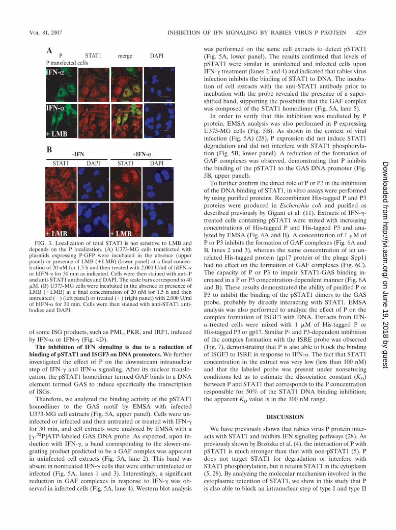
FIG. 2. Localization of pSTAT1 is correlated with the localization of P. (A) U373-MG control (upper panel) or P-expressing cell lines wereincubated in the absence (middle panel) or presence of LMB (�LMB) at a final concentration of 20 nM for 1.5 h (lower panel). Cells wereunstimulated or stimulated with 2,000 U/ml of hIFN-� or IFN-� for 30 min as indicated, and they were fixed, permeabilized, and then stained withanti-P and anti-pSTAT1 antibodies and DAPI. (B) U373-MG cells were transfected with plasmids expressing P-GFP, P3-GFP (P�N52-GFP), orP�N44-GFP. Forty-eight hours after transfection, cells were treated with 2,000 U/ml of hIFN-� or hIFN-� for 30 min as indicated. Cells were thenstained with anti-P and anti-pSTAT1 antibodies and DAPI.
4258 VIDY ET AL. J. VIROL.
on June 19, 2018 by guesthttp://jvi.asm
.org/D
ownloaded from
of some ISG products, such as PML, PKR, and IRF1, inducedby IFN-� or IFN-� (Fig. 4D).
The inhibition of IFN signaling is due to a reduction ofbinding of pSTAT1 and ISGF3 on DNA promoters. We furtherinvestigated the effect of P on the downstream intranuclearstep of IFN-� and IFN-� signaling. After its nuclear translo-cation, the pSTAT1 homodimer termed GAF binds to a DNAelement termed GAS to induce specifically the transcriptionof ISGs.
Therefore, we analyzed the binding activity of the pSTAT1homodimer to the GAS motif by EMSA with infectedU373-MG cell extracts (Fig. 5A, upper panel). Cells were un-infected or infected and then untreated or treated with IFN-�for 30 min, and cell extracts were analyzed by EMSA with a[�-32P]ATP-labeled GAS DNA probe. As expected, upon in-duction with IFN-�, a band corresponding to the slower-mi-grating product predicted to be a GAF complex was apparentin uninfected cell extracts (Fig. 5A, lane 2). This band wasabsent in nontreated IFN-� cells that were either uninfected orinfected (Fig. 5A, lanes 1 and 3). Interestingly, a significantreduction in GAF complexes in response to IFN-� was ob-served in infected cells (Fig. 5A, lane 4). Western blot analysis
was performed on the same cell extracts to detect pSTAT1(Fig. 5A, lower panel). The results confirmed that levels ofpSTAT1 were similar in uninfected and infected cells uponIFN-� treatment (lanes 2 and 4) and indicated that rabies virusinfection inhibits the binding of STAT1 to DNA. The incuba-tion of cell extracts with the anti-STAT1 antibody prior toincubation with the probe revealed the presence of a super-shifted band, supporting the possibility that the GAF complexwas composed of the STAT1 homodimer (Fig. 5A, lane 5).
In order to verify that this inhibition was mediated by Pprotein, EMSA analysis was also performed in P-expressingU373-MG cells (Fig. 5B). As shown in the context of viralinfection (Fig. 5A) (28), P expression did not induce STAT1degradation and did not interfere with STAT1 phosphoryla-tion (Fig. 5B, lower panel). A reduction of the formation ofGAF complexes was observed, demonstrating that P inhibitsthe binding of the pSTAT1 to the GAS DNA promoter (Fig.5B, upper panel).
To further confirm the direct role of P or P3 in the inhibitionof the DNA binding of STAT1, in vitro assays were performedby using purified proteins. Recombinant His-tagged P and P3proteins were produced in Escherichia coli and purified asdescribed previously by Gigant et al. (11). Extracts of IFN-�-treated cells containing pSTAT1 were mixed with increasingconcentrations of His-tagged P and His-tagged P3 and ana-lyzed by EMSA (Fig. 6A and B). A concentration of 1 �M ofP or P3 inhibits the formation of GAF complexes (Fig. 6A andB, lanes 2 and 3), whereas the same concentration of an un-related His-tagged protein (gp17 protein of the phage Spp1)had no effect on the formation of GAF complexes (Fig. 6C).The capacity of P or P3 to impair STAT1-GAS binding in-creased in a P or P3 concentration-dependent manner (Fig. 6Aand B). These results demonstrated the ability of purified P orP3 to inhibit the binding of the pSTAT1 dimers to the GASprobe, probably by directly interacting with STAT1. EMSAanalysis was also performed to analyze the effect of P on thecomplex formation of ISGF3 with DNA. Extracts from IFN-�-treated cells were mixed with 1 �M of His-tagged P orHis-tagged P3 or gp17. Similar P- and P3-dependent inhibitionof the complex formation with the ISRE probe was observed(Fig. 7), demonstrating that P is also able to block the bindingof ISGF3 to ISRE in response to IFN-�. The fact that STAT1concentration in the extract was very low (less than 100 nM)and that the labeled probe was present under nonsaturingconditions led us to estimate the dissociation constant (KD)between P and STAT1 that corresponds to the P concentrationresponsible for 50% of the STAT1 DNA binding inhibition;the apparent KD value is in the 100 nM range.
DISCUSSION
We have previously shown that rabies virus P protein inter-acts with STAT1 and inhibits IFN signaling pathways (28). Aspreviously shown by Brzózka et al. (4), the interaction of P withpSTAT1 is much stronger than that with non-pSTAT1 (5). Pdoes not target STAT1 for degradation or interfere withSTAT1 phosphorylation, but it retains STAT1 in the cytoplasm(5, 28). By analyzing the molecular mechanism involved in thecytoplasmic retention of STAT1, we show in this study that Pis also able to block an intranuclear step of type I and type II
FIG. 3. Localization of total STAT1 is not sensitive to LMB anddepends on the P localization. (A) U373-MG cells transfected withplasmids expressing P-GFP were incubated in the absence (upperpanel) or presence of LMB (�LMB) (lower panel) at a final concen-tration of 20 nM for 1.5 h and then treated with 2,000 U/ml of hIFN-�or hIFN-� for 30 min as indicated. Cells were then stained with anti-Pand anti-STAT1 antibodies and DAPI. The scale bars correspond to 40�M. (B) U373-MG cells were incubated in the absence or presence ofLMB (�LMB) at a final concentration of 20 nM for 1.5 h and thenuntreated () (left panel) or treated (�) (right panel) with 2,000 U/mlof hIFN-� for 30 min. Cells were then stained with anti-STAT1 anti-bodies and DAPI.
VOL. 81, 2007 INHIBITION OF IFN SIGNALING BY RABIES VIRUS P PROTEIN 4259
on June 19, 2018 by guesthttp://jvi.asm
.org/D
ownloaded from

IFN signaling: the binding of STAT1 and ISGF3 to the DNApromoters.
Previous data have shown that (i) P is a nucleocytoplamicprotein that shuttles between the cytoplasm and the nucleus
(20), (ii) the N-terminally truncated P3 is nuclear (20), and (iii)the STAT1 binding site is located in the carboxyl-terminaldomain of P (28). We confirm here that P3 shares the STAT1binding site with P.
FIG. 4. Expression of the nuclear forms of P inhibits IFN-� and IFN-� signaling. (A) U373-MG cells were transfected with an ISRE-fireflyluciferase reporter plasmid (pISRE-f. luc) and a Renilla luciferase expression vector (pTK-r.luc) and either an empty vector or a plasmid expressingP-GFP, P3-GFP, or P�44-GFP as indicated. At 48 hours after transfection, cells were untreated () or treated (�) with 2,000 U/ml of hIFN-�for 6 h prior to lysis and luciferase assays. (B) U373-MG cell lines expressing P or a control plasmid were transfected with pISRE-f.luc andpTK-r.luc. At 48 hours after transfection, cells were incubated in the absence or presence of LMB at a final concentration of 20 nM for 1 h. Cellswere then unstimulated () or stimulated (�) with 2,000 U/ml of hIFN-� for 6 h prior to lysis and luciferase assays. (C) Same as described forpanel B, but an IFN-�-responsive GAS luciferase reporter was used instead of ISRE luciferase (pGAS-luc) and hIFN-� was used instead ofhIFN-�. All bars represent average values of firefly luciferase from triplicate samples, normalized to the expression of Renilla luciferase andexpressed as percentages of IFN-stimulated controls; error bars indicate standard deviations. (D) U373-MG control or P-expressing cell lines wereuntreated () or treated with 2,000 U/ml of hIFN-� (��) or hIFN-� (��) for 24 h. The expression of PML, PKR, and IRF1 was studied byWestern blot analysis with specific antibodies.
4260 VIDY ET AL. J. VIROL.
on June 19, 2018 by guesthttp://jvi.asm
.org/D
ownloaded from

We first show that following IFN activation, the localizationof STAT1 is correlated with the localization of P. In cells stablyor transiently expressing a nuclear form of P (P in the presenceof LMB or P3), STAT1 is nuclear, whereas in cells expressinga cytoplasmic form of P (P�N44 or P), STAT1 is cytoplasmic.It should be noted that in the absence of IFN treatment,STAT1 does not relocalize to the nucleus in the presence ofP3, indicating that P or P3 interacts more efficiently with thephosphorylated form of STAT1 as previously shown byBrzózka et al. (4). Surprisingly, the nuclear forms of P are ableto inhibit IFN signaling as tested by luciferase activity, dem-onstrating that this inhibition is not due to the retention ofSTAT1 in the cytoplasm. Therefore, we examined the follow-
ing nuclear step that is the DNA binding activity of STAT1.We show by EMSA of cell extracts from infected cells or cellsstably expressing P that the capacity of IFN-� to induce DNAbinding of STAT1 was inhibited. Interestingly, the addition ofpurified recombinant P or P3 to extracts from IFN-�- or IFN-�-treated cells prevents the binding of pSTAT1 to the GAS orof ISGF3 to the ISRE, demonstrating that P interacts directlywith STAT1, leading to the inhibition of type I and type II IFNresponses.
It is unclear at present how P protein inhibits the bindingactivity of pSTAT1 to the DNA. As described previously, ra-bies virus P protein interacts with the coiled-coil or DNAbinding domains of STAT1 (28); therefore, the direct interac-tion of P with the DNA binding domain of STAT1 couldinterfere with the DNA binding activity of STAT1.
The fact that viral P and P3 proteins share the STAT1binding domain and localize to different compartments of thecell provides the virus a dual strategy for blocking both cyto-plasmic and nuclear forms of STAT1. This is also the case withNipah virus V and W proteins that inhibit STAT1 activationfrom the cytoplasm and the nucleus, respectively (25, 26, 27).
P has been also shown to impair nuclear accumulation ofSTAT1 (5, 28), suggesting that P may inhibit IFN signaling attwo different and independent steps. However, we cannot ex-clude the possibility that both steps are related and the inhi-bition of nuclear accumulation of STAT1 is due to a reduction
FIG. 5. Effect of P expressed in infected cells and in P-expressingcell lines on the formation of GAF complexes. (A) U373-MG cellswere not infected () (lanes 1 and 2) or infected (�) (lanes 3 and 4).Twenty-four hours after infection, cells were not treated () (lanes 1and 3) or treated (�) (lanes 2 and 4) with 2,000 U/ml of hIFN-� for 30min. Total cell lysates were analyzed by EMSA using a GAS �-32P-labeled probe and native PAGE (upper panel). The same cell extractswere analyzed by immunoblotting with anti-pSTAT1 and anti-tubulinantibodies (lower panel). In addition, extracts of noninfected and IFN-�-treated cells were incubated with anti-STAT1 antibodies prior toincubation with GAS �-32P-labeled probe (lane 5). The GAF complexand the supershifted GAF complex-IgG are indicated. (B) Control andP-expressing cell lines were not treated () (lanes 1 and 3) or treated(�) (lanes 2 and 4) with 2,000 U/ml of hIFN-� for 30 min. Total celllysates were analyzed by EMSA using a GAS �-32P-labeled probe andnative PAGE. The same cell extracts were analyzed by immunoblottingwith anti-pSTAT1 and anti-tubulin antibodies (lower panel).
FIG. 6. Effect of recombinant P and P3 proteins on the formationof GAF complexes. Total cell extracts of hIFN-�-treated U373-MGcells were mixed with increasing concentrations of His-tagged P (A) orHis-tagged P3 (B) protein or unrelated His-tagged Gp17 protein(C) and submitted to EMSA analysis. (D) The same cell extracts wereanalyzed by immunoblotting with anti-pSTAT1 and anti-tubulin anti-bodies.
VOL. 81, 2007 INHIBITION OF IFN SIGNALING BY RABIES VIRUS P PROTEIN 4261
on June 19, 2018 by guesthttp://jvi.asm
.org/D
ownloaded from

of the DNA binding activity. Indeed, it has been proposed thatDNA binding controls the nuclear accumulation of STAT1:DNA binding protects STAT1 from dephosphorylation, andthe DNA-bound STAT1 is thus retained in the nucleus (17, 18,19). In this model, the loss of DNA binding is associated withthe cytoplasmic accumulation of STAT1. In our case, the lossof DNA binding is necessary but not sufficient to explain thedifferent localization of STAT1 in the presence of P or P3; inaddition, the presence of a strong export signal in the N-terminal part of P may be involved in the nuclear export ofSTAT1, as suggested by the results obtained with the P�N44mutant.
Viral inhibition of the Jak-STAT pathway has been shown inother negative-strand RNA viruses, and among members ofthe Paramyxoviridae family, there is a great diversity in theevasion STAT signaling. Viral proteins can target STAT1 andSTAT2 for degradation and inhibit phosphorylation anddimerization or nuclear accumulation of STAT1 (6, 15, 16).Very few cases of inhibition of the DNA binding activity ofSTAT1 have been reported, and this inhibition is not direct butdescribed as a consequence of the impairment of one of the
upstream steps. To our knowledge, only one report has shownthat Sendai C protein directly inhibits the binding of theSTAT1 homodimer on DNA (13).
It is interesting that rabies virus P protein, in addition toinhibiting IFN type I synthesis, acts at three different levels ofthe IFN signaling: it inhibits the nuclear accumulation ofSTAT1, the binding of STAT1 to the DNA, and the functionof ISG products such as PML. Quite frequently, viruses usemore than one strategy to evade the IFN system at one or morelevels, and this may reflect how difficult it is to completely shutdown this host antiviral response. In that sense, rabies virus Pcan be termed a multifunctional IFN antagonist. This gives arabies virus with limited coding capacity the ability to inhibitmultiple arms of the host’s innate immune response.
ACKNOWLEDGMENTS
We are grateful to Yves Gaudin for helpful discussions and carefulreading of the manuscript. We acknowledge Spencer Brown and Sus-anne Bolt for help and assistance with confocal microscopy.
The microscopy was performed on the “Plate-forme Imagerie etBiologie Cellulaire” of the CNRS campus supported by the InstitutFederatif de Recherche 87 “La plante et son environnement” and theprogram ASTRE of the Conseil general de l’Essonne. The work in theteam of M. Chelbi-Alix is supported by the “Association pour la Re-cherche sur le Cancer.”
REFERENCES
1. Basler, C. F., and A. Garcia-Sastre. 2002. Viruses and the type I interferonantiviral system: induction and evasion. Int. Rev. Immunol. 21:305–337.
2. Begitt, A., T. Meyer, M. van Rossum, and U. Vinkemeier. 2000. Nucleocy-toplasmic translocation of Stat1 is regulated by a leucine-rich export signal inthe coiled-coil domain. Proc. Natl. Acad. Sci. USA 97:10418–10423.
3. Blondel, D., T. Regad, N. Poisson, B. Pavie, H. Harper, P. P. Pandolfi, H. DeThe, and M. K. Chelbi-Alix. 2002. Rabies virus P and small P productsinteract directly with PML and reorganize PML nuclear bodies. Oncogene21:7957–7970.
4. Brzozka, K., S. Finke, and K. K. Conzelmann. 2005. Identification of therabies virus alpha/beta interferon antagonist: phosphoprotein P interfereswith phosphorylation of interferon regulatory factor 3. J. Virol. 79:7673–7681.
5. Brzozka, K., S. Finke, and K. K. Conzelmann. 2006. Inhibition of interferonsignalling by rabies virus phosphoprotein P: activation-dependent binding ofSTAT1 and STAT2. J. Virol. 80:2675–2683.
6. Chelbi-Alix, M. K., A. Vidy, J. El. Bougrini, and D. Blondel. 2006. Rabiesviral mechanisms to escape the IFN system: the viral protein P interfereswith IRF-3, Stat1, and PML nuclear bodies. J. Interferon Cytokine Res.26:271–280.
7. Chenik, M., K. Chebli, and D. Blondel. 1995. Translation initiation atalternate in-frame AUG codons in the rabies virus phosphoproteinmRNA is mediated by a ribosomal leaky scanning mechanism. J. Virol.69:707–712.
8. Darnell, J. E., Jr., I. M. Kerr, and G. R. Stark. 1994. Jak-STAT pathways andtranscriptional activation in response to IFNs and extracellular signallingproteins. Science 264:1415–1421.
9. Garcıa-Sastre, A. 2002. Mechanisms of inhibition of the host interferonalpha/beta-mediated antiviral responses by viruses. Microbes Infect. 4:647–655.
10. Garcin, D., J. Curran, and D. Kolakofsky. 2000. Sendai virus C proteins mustinteract directly with cellular components to interfere with interferon action.J. Virol. 74:8823–8830.
11. Gigant, B., F. Iseni, Y. Gaudin, M. Knossow, and D. Blondel. 2000. Neitherphosphorylation nor the amino-terminal part of rabies virus phosphoproteinis required for its oligomerization. J. Gen. Virol. 81:1757–1761.
12. Goodbourn, S., L. Didcock, and R. E. Randall. 2000. Interferons: cell sig-naling, immune modulation, antiviral response and virus countermeasures.J. Gen. Virol. 81:2341–2364.
13. Gotoh, B., T. Komatsu, K. Takeuchi, and J. Yokoo. 2003. The C-terminalhalf-fragment of the Sendai virus C protein prevents the gamma-activatedfactor from binding to a gamma-activated sequence site. Virology 316:29–40.
14. Gupta, A. K., D. Blondel, S. Ghoudhary, and A. K. Banerjee. 2000. Thephosphoprotein of rabies virus is phosphorylated by a unique cellular proteinkinase and specific isomers of protein kinase C. J. Virol. 74:91–98.
15. Haller, O., G. Kochs, and F. Weber. 2006. The interferon response circuit:induction and suppression by pathogenic viruses. Virology 344:119–130.
FIG. 7. Effect of recombinant P and P3 proteins on the formationof ISGF3 complexes to the ISRE promoter. (A) U373-MG cells werenot treated () (lane 1) or treated (�) (lanes 2 to 7) with 2,000 U/mlof hIFN-� for 30 min. Nuclear cell extracts were mixed with 1 �M ofHis-tagged P (lane 5), His-tagged P3 (lane 6), or His-tagged Gp17(lane 7) proteins and submitted to EMSA analysis using a ISRE �-32P-labeled probe and native PAGE gel. In addition, extracts of IFN-�-treated cells were incubated with anti-STAT2 antibodies prior toincubation with �-32P-labeled probe (lane 3, *). (B) The same cellextracts were analyzed by immunoblotting with anti-pSTAT2 and anti-tubulin antibodies.
4262 VIDY ET AL. J. VIROL.
on June 19, 2018 by guesthttp://jvi.asm
.org/D
ownloaded from

16. Horvath, C. M. 2004. Silencing STATs: lessons from paramyxovirus in-terferon evasion. Cytokine Growth Factor Rev. 15:117–127. (Erratum,15:477.)
17. Meyer, T., A. Begitt, L. Lodige, M. Van Rossum, and U. Vinkemeier. 2002.Constitutive and IFN-gamma-induced nuclear import of STAT1 proceedthrough independent pathways. EMBO J. 21:344–354.
18. Meyer, T., A. Marg, P. Lemke, B. Wiesner, and U. Vinkemeier. 2003. DNAbinding controls inactivation and nuclear accumulation of the transcriptionfactor Stat1. Genes Dev. 17:1992–2005.
19. Meyer, T., L. Hendry, A. Begitt, S. John, and U. Vinkemeier. 2004. Asingle residue modulates tyrosine dephosphorylation, oligomerization,and nuclear accumulation of STAT transcription factors. J. Biol. Chem.279:18998–19007.
20. Pasdeloup, D., N. Poisson, H. Raux, Y. Gaudin, R. W. Ruigrok, and D.Blondel. 2005. Nucleocytoplasmic shuttling of the rabies virus P proteinrequires a nuclear localization signal and a CRM1-dependent nuclear exportsignal. Virology 334:284–931.
21. Pelicano, L., F. Li, C., Schindler, and M. K. Chelbi-Alix. 1997. Retinoic acidenhances the expression of interferon-induced proteins: evidence for multi-ple mechanisms of action. Oncogene 15:2349–2359.
22. Raux, H., F. Iseni, F. Lafay, and D. Blondel. 1997. Mapping of monoclonalantibody epitopes of the rabies virus P protein. J. Gen. Virol. 78:119–124.
23. Raux, H., A. Flamand, and D. Blondel. 2000. Interaction of the rabies virusP protein with the LC8 dynein light chain. J. Virol. 74:10212–10216.
24. Regad, T., and M. K. Chelbi-Alix. 2001. Role and fate of PML nuclear bodiesin response to interferon and viral infections. Oncogene 20:7274–7286.
25. Rodriguez, J. J., C. D. Cruz, and C. M. Horvath. 2004. Identification of thenuclear export signal and STAT-binding domains of the Nipah virus Vprotein reveals mechanisms underlying interferon evasion. J. Virol. 78:5358–5367.
26. Shaw, M. L., A. Garcia-Sastre, P. Palese, and C. F. Basler. 2004. Nipah virusV and W proteins have a common STAT1-binding domain yet inhibit STAT1activation from the cytoplasmic and nuclear compartments, respectively.J. Virol. 78:5633–5641.
27. Shaw, M. L., W. B. Cardenas, D. Zamarin, P. Palese, and C. F. Basler. 2005.Nuclear localization of the Nipah virus W protein allows for inhibition ofboth virus- and Toll-like receptor 3-triggered signaling pathways. J. Virol.79:6078–6088.
28. Vidy, A., M. Chelbi-Alix, and D. Blondel. 2005. Rabies virus P proteininteracts with STAT1 and inhibits interferon signal transduction pathways.J. Virol. 79:14411–14420.
VOL. 81, 2007 INHIBITION OF IFN SIGNALING BY RABIES VIRUS P PROTEIN 4263
on June 19, 2018 by guesthttp://jvi.asm
.org/D
ownloaded from