THE NUCLEIC ACIDS AND PROTEIN SYNTHESIS. Friedrich Miescher in 1869 isolated what he called nuclein...
29
THE NUCLEIC ACIDS AND PROTEIN SYNTHESIS
-
Upload
everett-warren -
Category
Documents
-
view
219 -
download
1
Transcript of THE NUCLEIC ACIDS AND PROTEIN SYNTHESIS. Friedrich Miescher in 1869 isolated what he called nuclein...

THE NUCLEIC ACIDS AND PROTEIN SYNTHESIS

Friedrich Miescher in 1869
isolated what he called nuclein from the nuclei of pus cells
Nuclein was shown to have acidic properties, hence it became called nucleic acid

Two types of nucleic acid are found
Deoxyribonucleic acid (DNA) Ribonucleic acid (RNA)

The distribution of nucleic acids in the eukaryotic cell DNA is found in the nucleus
with small amounts in mitochondria and chloroplasts
RNA is found throughout the cell

DNA as genetic material: The circumstantial evidence1. Present in all cells and virtually restricted to the
nucleus
2. The amount of DNA in somatic cells (body cells) of any given species is constant (like the number of chromosomes)
3. The DNA content of gametes (sex cells) is half that of somatic cells.

NUCLEIC ACID STRUCTURE
Nucleic acids are polynucleotides Their building blocks are nucleotides

NUCLEOTIDE STRUCTURE
PHOSPATE SUGAR
Ribose or Deoxyribose
NUCLEOTIDE
BASEPURINES PYRIMIDINES
Adenine (A)Guanine(G)
Cytocine (C)Thymine (T)Uracil (U)

Ribose is a pentose
C1
C5
C4
C3 C2
O

RIBOSE DEOXYRIBOSE
CH2OH
H
OH
C
C
OH OH
C
O
H HH
C
CH2OH
H
OH
C
C
OH H
C
O
H HH
C
Spot the difference

THE SUGAR-PHOSPHATE BACKBONE The nucleotides are all
orientated in the same direction The phosphate group joins the
3rd Carbon of one sugar to the 5th Carbon of the next in line by a phosphodiester bond.
P
P
P
P
P
P

ADDING IN THE BASES
The bases are attached to the 1st Carbon
Their order is important
It determines the genetic information of the molecule
P
P
P
P
P
P
G
C
C
A
T
T

DNA IS MADE OF TWO STRANDS OF POLYNUCLEOTIDE
P
P
P
P
P
P
C
G
G
T
A
A
P
P
P
P
P
P
G
C
C
A
T
T
Hydrogen bonds
© 2007 Paul Billiet ODWS
DNA IS MADE OF TWO STRANDS OF POLYNUCLEOTIDE The sister strands of the DNA molecule run in opposite
directions (antiparallel) They are joined by the bases Each base is paired with a specific partner:A is always paired with T G is always paired with CPurine with Pyrimidine This the sister strands are complementary but not
identical The bases are joined by hydrogen bonds, individually
weak but collectively strong

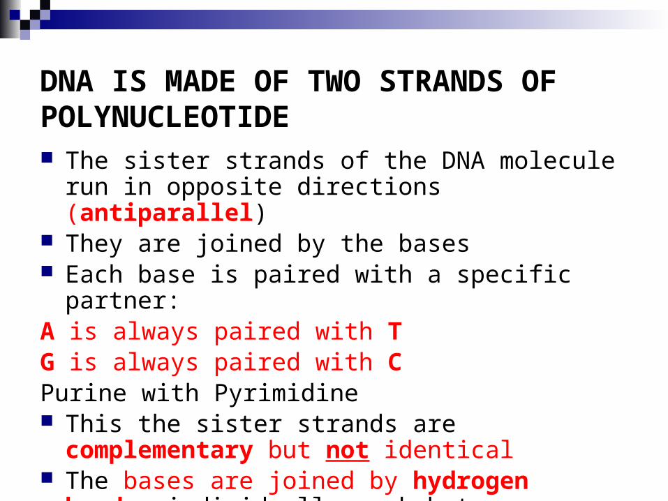
Purines & Pyrimidines
Adenine
CytosineGuanine
Thymine
Watson & Crick Base pairing

The Double Helix (1953)

A, B and Z DNA A form – favored by RNA B form – Standard DNA
double helix under physiological conditions
Z form – laboratory anomaly, Left Handed Requires Alt. GC High Salt/ Charge
neutralization
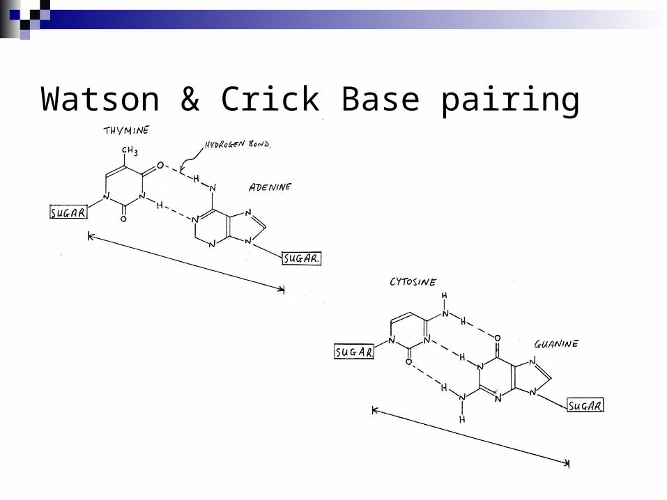
DNA Replication When a eukaryotic cell divides, the process is called mitosis
- the cell splits into two identical daughter cells
- the DNA must be replicated so that each daughter cell has a copy
DNA replication involves several processes:
- first, the DNA must be unwound, separating the two strands
- the single strands then act as templates for synthesis of the new strands, which are
complimentary in sequence
- bases are added one at a time until two new DNA strands that exactly duplicate the
original DNA are produced
The process is called semi-conservative replication because one strand of each
daughter DNA comes from the parent DNA and one strand is new
The energy for the synthesis comes from hydrolysis of phosphate groups as the
phosphodiester bonds form between the bases
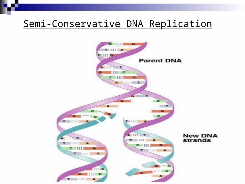
Semi-Conservative DNA Replication

Protein Synthesis The two main processes involved in protein synthesis are
- the formation of mRNA from DNA (transcription)- the conversion by tRNA to protein at the ribosome (translation)
Transcription takes place in the nucleus, while translation takes place in the cytoplasm
Genetic information is transcribed to form mRNA much the same way it is replicated during cell division

Transcription
Several steps occur during transcription:
- a section of DNA containing the gene unwinds
- one strand of DNA is copied starting at the initiation point,
which has the sequence TATAAA
- an mRNA is synthesized using complementary base pairing
with uracil (U) replacing thymine (T)
- the newly formed mRNA moves out of the nucleus to
ribosomes in the cytoplasm and the DNA re-winds

RNA Polymerase During transcription, RNA polymerase moves along the DNA
template in the 3’-5’direction to synthesize the corresponding mRNA
The mRNA is released at the termination point

Processing of mRNA
Genes in the DNA of eukaryotes contain exons that code for proteins along with introns that do not
Because the initial mRNA, called a pre-RNA, includes the noncoding introns, it must be processed before it can be read by the tRNA
While the mRNA is still in the nucleus, the introns are removed from the pre-RNA
The exons that remain are joined to form the mRNA that leaves the nucleus with the information for the synthesis of protein

Removing Introns from mRNA

mRNA Codons and Associated Amino Acids
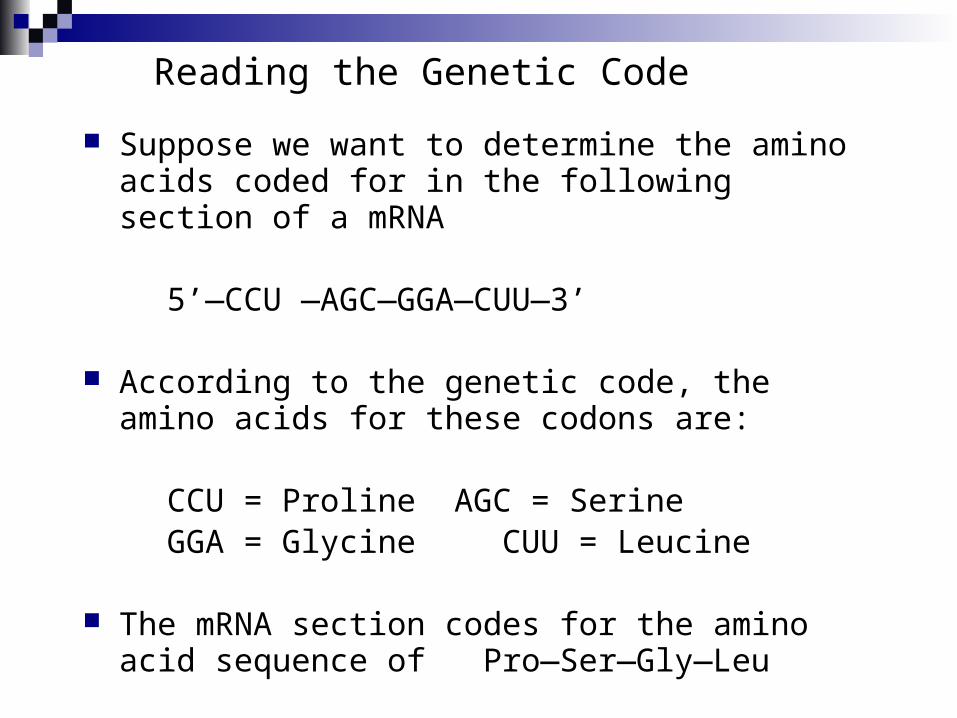
Reading the Genetic Code
Suppose we want to determine the amino acids coded for in the following section of a mRNA
5’—CCU —AGC—GGA—CUU—3’
According to the genetic code, the amino acids for these codons are:
CCU = Proline AGC = Serine GGA = Glycine CUU = Leucine
The mRNA section codes for the amino acid sequence of Pro—Ser—Gly—Leu

Translation and tRNA Activation
Once the DNA has been transcribed to
mRNA, the codons must be tranlated
to the amino acid sequence of the
protein
The first step in translation is
activation of the tRNA
Each tRNA has a triplet called an
anticodon that complements a codon
on mRNA
A synthetase uses ATP hydrolysis to
attach an amino acid to a specific
tRNA

Initiation and Translocation Initiation of protein synthesis occurs when a mRNA attaches to
a ribosome On the mRNA, the start codon (AUG) binds to a tRNA with
methionine The second codon attaches to a tRNA with the next amino acid A peptide bond forms between the adjacent amino acids at the
first and second codons The first tRNA detaches from the ribosome and the ribosome
shifts to the adjacent codon on the mRNA (this process is called translocation)
A third codon can now attach where the second one was before translocation

Termination
After a polypeptide with all the amino acids for a protein is
synthesized, the ribosome reaches the the “stop” codon:
UGA, UAA, or UAG
There is no tRNA with an anticodon for the “stop” codons
Therefore, protein synthesis ends (termination)
The polypeptide is released from the ribosome and the
protein can take on it’s 3-D structure
(some proteins begin folding while still being synthesized,
while others do not fold up until after being released from
the ribosome)