The nuclear pore complex and chromatin boundaries...THREAD/DIAP1 are required to inhibit apoptosis...
4
RING-finger-dependent manner [12]. Such discrepancies might be explained through experimental differences and will require further clarification. Nevertheless, the contribution of ubiquitylation to the realm of apoptosis remains intriguing. With respect to individual proteins, further assessment of the causal or consequential nature of their ubiquitylation is essential. References 1 Shi, Y. (2002) Mechanisms of caspase activation and inhibition during apoptosis. Mol. Cell 9, 459–470 2 Salvesen, G.S. and Duckett, C.S. (2002) Apoptosis. IAP proteins: blocking the road to death’s door. Nat. Rev. Mol. Cell Biol. 3, 401–410 3 Rodriguez, A. et al. (2002) Unrestrained caspase-dependent cell death caused by loss of Diap1 function requires the Drosophila Apaf-1 homolog, Dark. EMBO J. 21, 2189–2197 4 Wang, S.L. et al. (1999) The Drosophila caspase inhibitor DIAP1 is essential for cell survival and is negatively regulated by HID. Cell 98, 453–463 5 Goyal, L. et al. (2000) Induction of apoptosis by Drosophila reaper, hid and grim through inhibition of IAP function. EMBO J. 19, 589–597 6 Lisi, S. et al. (2000) Diverse domains of THREAD/DIAP1 are required to inhibit apoptosis induced by REAPER and HID in Drosophila. Genetics 154, 669–678 7 Crook, N.E. et al. (1993) An apoptosis-inhibiting baculovirus gene with a zinc finger-like motif. J. Virol. 67, 2168–2174 8 Bergmann, A. et al. (1998) Mechanisms and control of programmed cell death in invertebrates. Oncogene 17, 3215–3223 9 Yoo, S.J. et al. (2002) Apoptosis inducers Hid, Rpr and Grim negatively regulate levels of the caspase inhibitor DIAP1 by distinct mechanisms. Nat. Cell Biol. 4, 416–424 10 Hays, R. et al. (2002) Morgue mediates apoptosis in the Drosophila retina by promoting degradation of DIAP1. Nat. Cell Biol. 4, 425–431 11 Wing, J.P. et al. (2002) Drosophila morgue is a novel F box/ubiquitin conjugase domain protein important in grim-reaper mediated programmed cell death. Nat. Cell Biol. 4, 451–456 12 Ryoo, H.D. et al. (2002) Regulation of Drosophila IAP1 degradation and apoptosis by reaper and ubcD1. Nat. Cell Biol. 4, 432–438 13 Holley, C.L. et al. (2002) Reaper eliminates IAP protein through stimulated IAP degradation and generalized translational inhibition. Nat. Cell Biol. 4, 439–444 14 Wilson, R. et al. (2002) The DIAP1 RING finger mediates ubiquitination of Dronc and is indispensable for regulating apoptosis. Nat. Cell Biol. 4, 445–450 15 Deng, L. et al. (2000) Activation of the IkappaB kinase complex by TRAF6 requires a dimeric ubiquitin-conjugating enzyme complex and a unique polyubiquitin chain. Cell 103, 351–361 16 Yang, Y. et al. (2000) Ubiquitin protein ligase activity of IAPs and their degradation in proteasomes in response to apoptotic stimuli. Science 288, 874–877 17 Suzuki, Y. et al. (2001) X-linked inhibitor of apoptosis protein (XIAP) inhibits caspase-3 and -7 in distinct modes. J. Biol. Chem. 276, 27058–27063 18 Huang, H. et al. (2000) The inhibitor of apoptosis, cIAP2, functions as a ubiquitin-protein ligase and promotes in vitro monoubiquitination of caspases 3 and 7. J. Biol. Chem. 275, 26661–26664 19 Miller, L.K. (1999) An exegesis of IAPs: salvation and surprises from BIR motifs. Trends Cell Biol. 9, 323–328 20 MacFarlane, M. et al. Proteasome-mediated degradation of Smac during apoptosis: XIAP promotes Smac ubiquitination in vitro. J. Biol. Chem. (in press) Mark Ditzel Pascal Meier* The Breakthrough Toby Robins Breast Cancer Research Centre, Institute of Cancer Research, Chester Beatty Laboratories, Fulham Road, London UK SW3 6JB. *e-mail: [email protected] TRENDS in Cell Biology Vol.12 No.10 October 2002 http://tcb.trends.com 0962-8924/02/$ – see front matter © 2002 Elsevier Science Ltd. All rights reserved. PII: S0962-8924(02)02367-X 452 Research Update The nuclear pore complex and chromatin boundaries Chi-Yun Pai and Victor G. Corces The organization of the chromatin fiber within specific nuclear compartments and its arrangement into domains of higher-order structure are aspects of the regulation of gene expression that remain largely undefined. Boundary and insulator elements are likely to play important roles in establishing and maintaining the nuclear architecture required by eukaryotic cells to orchestrate the intricate array of events such as DNA replication, transcription and RNA processing. Recent results suggest a link between chromatin organization and nuclear transport and offer a glimpse of how nuclear organization might facilitate the integration of complex nuclear processes. Published online: 3 September 2002 Chromatin insulators and boundary elements are a class of DNA sequences defined operationally by two properties: they prevent enhancer–promoter interactions and they buffer transgenes from chromosomal position effects arising from genomic sequences adjacent to the transgene insertion site. Boundaries and insulators must play an important and conserved function in nuclear biology as they have now been found in a variety of organisms ranging from yeasts to human [1,2]. The observed effects of boundaries and insulators on transcription are merely the readout of the experimental approaches used to identify and define these sequences, and their effects on gene expression are likely a manifestation of their normal role in the biology of the cell. The two types of transcriptional properties characteristic of insulators – inhibition of enhancer–promoter interaction and buffering of chromosomal position effects – suggest very different alternative functions for these sequences in nuclear physiology. The fact that insulators can affect enhancer–promoter interactions can be viewed in the context of a role for these sequences in modulating enhancer function. In this view, insulators would be another regulatory sequence, in the same category as enhancers, silencers and promoter elements, whose primary function is the regulation of transcription. On the other hand, the fact that insulators can shield a transgene from position effects suggests that these sequences might separate euchromatic from heterochromatic regions and, in the process, set up chromatin domains that are permissive for transcription. In this view, the primary role of boundaries and insulators would be to compartmentalize the genome and organize the chromatin fiber within the nucleus. Such a role for insulators would bring these sequences into the same realm as matrix- and scaffold-attachment region (MAR/SAR) elements, sequences that are thought to be located at the base of chromatin loops and serve structural roles in chromatin organization [3]. Boundary elements could have an additional functional role in setting up chromatin domains permissive for transcription, and regulation of this function during cell differentiation could
Transcript of The nuclear pore complex and chromatin boundaries...THREAD/DIAP1 are required to inhibit apoptosis...
RING-finger-dependent manner [12].Such discrepancies might be explainedthrough experimental differences and will require further clarification.Nevertheless, the contribution ofubiquitylation to the realm of apoptosisremains intriguing. With respect toindividual proteins, further assessment of the causal or consequential nature oftheir ubiquitylation is essential.
References
1 Shi, Y. (2002) Mechanisms of caspase activationand inhibition during apoptosis. Mol. Cell 9,459–470
2 Salvesen, G.S. and Duckett, C.S. (2002)Apoptosis. IAP proteins: blocking the road todeath’s door. Nat. Rev. Mol. Cell Biol. 3, 401–410
3 Rodriguez, A. et al. (2002) Unrestrained caspase-dependent cell death caused by loss ofDiap1 function requires the DrosophilaApaf-1homolog, Dark. EMBO J. 21, 2189–2197
4 Wang, S.L. et al. (1999) The Drosophila caspaseinhibitor DIAP1 is essential for cell survival and is negatively regulated by HID. Cell 98,453–463
5 Goyal, L. et al. (2000) Induction of apoptosis byDrosophila reaper, hid and grim throughinhibition of IAP function. EMBO J. 19, 589–597
6 Lisi, S. et al. (2000) Diverse domains ofTHREAD/DIAP1 are required to inhibit apoptosis
induced by REAPER and HID in Drosophila.Genetics 154, 669–678
7 Crook, N.E. et al. (1993) An apoptosis-inhibitingbaculovirus gene with a zinc finger-like motif.J. Virol. 67, 2168–2174
8 Bergmann, A. et al. (1998) Mechanisms andcontrol of programmed cell death in invertebrates.Oncogene 17, 3215–3223
9 Yoo, S.J. et al. (2002) Apoptosis inducers Hid, Rprand Grim negatively regulate levels of the caspaseinhibitor DIAP1 by distinct mechanisms.Nat. Cell Biol. 4, 416–424
10 Hays, R. et al. (2002) Morgue mediates apoptosisin the Drosophila retina by promotingdegradation of DIAP1. Nat. Cell Biol. 4, 425–431
11 Wing, J.P. et al. (2002) Drosophila morgue is anovel F box/ubiquitin conjugase domain proteinimportant in grim-reaper mediated programmedcell death. Nat. Cell Biol. 4, 451–456
12 Ryoo, H.D. et al. (2002) Regulation of DrosophilaIAP1 degradation and apoptosis by reaper andubcD1. Nat. Cell Biol. 4, 432–438
13 Holley, C.L. et al. (2002) Reaper eliminates IAPprotein through stimulated IAP degradation andgeneralized translational inhibition. Nat. CellBiol. 4, 439–444
14 Wilson, R. et al. (2002) The DIAP1 RING fingermediates ubiquitination of Dronc and isindispensable for regulating apoptosis. Nat. CellBiol. 4, 445–450
15 Deng, L. et al. (2000) Activation of theIkappaB kinase complex by TRAF6 requires a dimeric ubiquitin-conjugating enzyme
complex and a unique polyubiquitin chain. Cell103, 351–361
16 Yang, Y. et al. (2000) Ubiquitin protein ligaseactivity of IAPs and their degradation inproteasomes in response to apoptotic stimuli.Science 288, 874–877
17 Suzuki, Y. et al. (2001) X-linked inhibitor ofapoptosis protein (XIAP) inhibits caspase-3 and -7in distinct modes. J. Biol. Chem. 276,27058–27063
18 Huang, H. et al. (2000) The inhibitor of apoptosis, cIAP2, functions as a ubiquitin-proteinligase and promotes in vitro monoubiquitinationof caspases 3 and 7. J. Biol. Chem. 275,26661–26664
19 Miller, L.K. (1999) An exegesis of IAPs: salvationand surprises from BIR motifs. Trends Cell Biol. 9,323–328
20 MacFarlane, M. et al. Proteasome-mediateddegradation of Smac during apoptosis: XIAPpromotes Smac ubiquitination in vitro. J. Biol.Chem. (in press)
Mark Ditzel
Pascal Meier*
The Breakthrough Toby Robins Breast Cancer Research Centre, Institute of Cancer Research, Chester Beatty Laboratories, Fulham Road, London UK SW3 6JB.*e-mail: [email protected]
TRENDS in Cell Biology Vol.12 No.10 October 2002
http://tcb.trends.com 0962-8924/02/$ – see front matter © 2002 Elsevier Science Ltd. All rights reserved. PII: S0962-8924(02)02367-X
452 Research Update
The nuclear pore complex and chromatin boundaries
Chi-Yun Pai and Victor G. Corces
The organization of the chromatin fiber
within specific nuclear compartments and
its arrangement into domains of higher-order
structure are aspects of the regulation of
gene expression that remain largely
undefined. Boundary and insulator elements
are likely to play important roles in
establishing and maintaining the nuclear
architecture required by eukaryotic cells to
orchestrate the intricate array of events
such as DNA replication, transcription and
RNA processing. Recent results suggest a
link between chromatin organization and
nuclear transport and offer a glimpse of how
nuclear organization might facilitate the
integration of complex nuclear processes.
Published online: 3 September 2002
Chromatin insulators and boundaryelements are a class of DNA sequencesdefined operationally by two properties:they prevent enhancer–promoterinteractions and they buffer transgenesfrom chromosomal position effects arising
from genomic sequences adjacent to thetransgene insertion site. Boundariesand insulators must play an importantand conserved function in nuclear biologyas they have now been found in a varietyof organisms ranging from yeasts tohuman [1,2].
The observed effects of boundaries andinsulators on transcription are merely thereadout of the experimental approachesused to identify and define these sequences,and their effects on gene expression arelikely a manifestation of their normal role in the biology of the cell. The two types of transcriptional propertiescharacteristic of insulators – inhibition ofenhancer–promoter interaction andbuffering of chromosomal position effects –suggest very different alternativefunctions for these sequences in nuclearphysiology. The fact that insulators canaffect enhancer–promoter interactions canbe viewed in the context of a role for thesesequences in modulating enhancer function.In this view, insulators would be another
regulatory sequence, in the same categoryas enhancers, silencers and promoterelements, whose primary function is theregulation of transcription. On the otherhand, the fact that insulators can shield atransgene from position effects suggeststhat these sequences might separateeuchromatic from heterochromatic regionsand, in the process, set up chromatindomains that are permissive fortranscription. In this view, the primary roleof boundaries and insulators would be tocompartmentalize the genome and organizethe chromatin fiber within the nucleus.Such a role for insulators would bring thesesequences into the same realm as matrix-and scaffold-attachment region (MAR/SAR)elements, sequences that are thought tobe located at the base of chromatin loopsand serve structural roles in chromatinorganization [3]. Boundary elements couldhave an additional functional role insetting up chromatin domains permissivefor transcription, and regulation of thisfunction during cell differentiation could
account for broad aspects of gene regulationduring development.
A connection between insulators andnuclear organization is reinforced byobservations on the gypsy insulator ofDrosophila. This insulator, originallyidentified in the gypsy retrotransposon, isalso present at several-hundred sites in thefly genome. These sites come together atapproximately 25 locations in the nuclearperiphery, organizing the chromatin fiberinto rosette structures. These rosettes areformed by chromatin loops, eachpresumably representing independentdomains of chromatin organization andgene expression [4]. Recent results gofurther in showing a clear connectionbetween the function of boundary andinsulator elements and their ability toestablish separate chromatin domains.
More interestingly, these results suggestthat the boundary activity of a DNAsequence depends on its interaction withprotein components of the nuclear porecomplex (NPC). This nuclear structure,whose role in nuclear physiology has beenthought to be mostly limited to the transportof proteins and RNAs between the nucleusand cytoplasm, might thus also play animportant role in the regulation of geneexpression.
The nuclear pore complex and boundary
activity
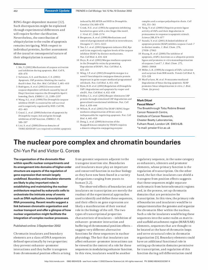
In a recent paper, Ishii et al. [5] describeresults from a clever boundary element-trapscreen designed to unbiasly identify newproteins with boundary function. In thisassay, two reporter genes, ADE2 and URA3,were inserted between the E and I silencers
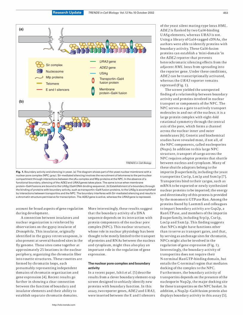
of the yeast silent mating-type locus HML.ADE2 is flanked by two Gal4-bindingUASg elements, whereas URA3 is not.Using a library of Gal4-tagged cDNAs, theauthors were able to identify proteins withboundary activity. These Gal4-fusionproteins can establish a ‘mini-domain’ inthe ADE2 reporter that preventsheterochromatic silencing effects from theadjacent HML locus from spreading intothe reporter gene. Under these conditions,ADE2 can be transcriptionally activated,whereas the URA3 reporter remainsrepressed (Fig. 1).
The screen yielded the unexpectedfinding of a relationship between boundaryactivity and proteins involved in nucleartransport or components of the NPC. TheNPC serves as a gate to actively transportmolecules in and out of the nucleus; it is alarge protein complex with eight-foldrotational symmetry through the centralaxis of the pore, which forms a channelacross the nuclear inner and outermembranes [6]. Genetic and biochemicalstudies have revealed most, if not all, ofthe NPC components, called nucleoporins(Nups). In addition to this large NPCstructure, transport of cargo across theNPC requires adaptor proteins that shuttlebetween nucleus and cytoplasm. Many ofthese shuttle adaptors belong to theimportin β superfamily, including the yeasttransportins Cse1p, Los1p and Sxm1p [7].Transportins bind to their cargos, such asmRNA to be exported or newly synthesizednuclear proteins to be imported; the energyand directionality of this process is providedby the monomeric GTPase Ran. Among theproteins found by Laemmli and colleaguesto impart boundary activity are Gsp2p, aRanGTPase, and members of the importinβ superfamily, including Srp1p, Cse1p,Los1p and Sxm1p. This finding suggeststhat NPCs might have functions otherthan to serve as transport gates, and that,by serving as anchorage sites for chromatin,NPCs might also be involved in theregulation of gene expression (Fig. 1).Interestingly, the boundary activity oftransportins does not require theirN-terminal RanGTP-binding domain, butentails the C-terminal region that mediatesdocking of the complex to the NPC.Furthermore, the boundary activity oftransportins depends on the presence of thenucleoporin Nup2p, the major docking sitefor these transportins on the NPC basket. Inaddition, a Nup2p–Gal4 fusion protein alsodisplays boundary activity in this assay [5].
TRENDS in Cell Biology Vol.12 No.10 October 2002
http://tcb.trends.com
453Research Update
TRENDS in Cell Biology
Sir complex
Nucleosome
Mlp proteins
Telomere
E and I silencers
URA3 gene
ADE2 gene
USAg
Transportin–Gal4fusion protein
Membraneprotein–Gal4 fusion
(a)
(b)
Fig. 1. Boundary activity and silencing in yeast. (a) The diagram shows part of the yeast nuclear membrane with anuclear pore complex (NPC; gray). Sir-mediated silencing involves the recruitment of telomeres to the perinuclearcompartment through interactions between the yKu complex and Mlp proteins at the NPC. In the absence of afunctional boundary, silencing of the ADE2 and URA3 genes takes place. The same is true when membraneprotein–Gal4 fusions are bound to the UASg (Gal4 DNA-binding sequence). (b) Establishment of a boundary throughthe binding of proteins with boundary activity, such as transportin–Gal4 fusion proteins, to the UASg is accomplishedby interactions between transportins and the NPC. The boundary interferes with Sir-mediated silencing and results ina chromatin structure permissive for transcription. The ADE2 gene is active, whereas the URA3 gene is repressed.
Boundaries, silencing and nuclear
compartments
There is some precedence for links betweenthe NPC and chromatin regulation in yeast.Repression of transgenes adjacent to yeasttelomeres and HM loci is accompanied byanchoring at perinuclear sites mediatedby the yeast yKu70–yKu80 complex [8,9],which is a protein heterodimer involved innon-homologous DNA end-joining reactionsas well as maintenance of proper telomerestructure. This repression is caused byhigh local concentration of the Sirheterochromatic silencing proteins boundto telomeric sites. A search for yKu-bindingproteins involved in nuclear envelopetethering resulted in the identification ofMlp1p and Mlp2p, which are extensionsof the NPC [10]. Furthermore, tethering ofa reporter gene to the nuclear envelopethrough a membrane-spanning proteinresults in silencing of the gene owingto the high local concentration ofSir proteins [11].
In the experiments carried out byLaemmli and coworkers, the same silencingmechanism is probably responsible forrepression of the ADE2 and URA3 reportergenes in the absence of a functionalboundary, and tethering to the NPC mightspecifically interfere with this silencingprocess. What is the mechanism wherebythe boundary activity interferes with thepropagation of the silencing activity fromthe adjacent HML E and I silencers? SinceNPCs are located in the nuclear envelope,one might ask whether tethering to thenuclear periphery is sufficient forestablishing boundary activity or whetherthe NPC plays a specific role in the process.Surprisingly, tethering of transgenesthrough Yif1–Gal4 and Yip1–Gal4 fusionproteins, which are proteins involved inER-to-Golgi membrane transport thataccumulate in the ER and the nuclearmembrane and should target the reporterto the nuclear membrane [11], does notresult in any boundary activity (Fig. 1) [5].It thus appears as if only tethering to theNPC establishes a functional chromatinboundary and protects the reporter genefrom heterochromatic repression.
Does tethering to the NPC always resultin boundary activity? It has previously beenshown that tethering of yeast telomeres tothe nuclear envelope is mediated bybinding of the telomeric chromatin to theNPC components Mlp1 and Mlp2, whichin turn bind to Nup60 and Nup145 [12].This NPC-mediated tethering, however,
results in gene silencing instead ofboundary activity, although in this caseonly one tethering site is present adjacentto the reporter gene. In addition, insulatoractivity mediated by transportins isMlp1/Mlp2-independent [5], suggestingthat simply tethering a gene to the NPCdoes not result in boundary activity andthat NPC-mediated silencing follows adifferent biochemical path than thatrequired to establish a boundary. Thereason for this different behavior is unclear.One common feature of the transportinsthat establish boundary activity is thatthe interaction with their substrates istransient and highly dynamic. This featureis also a property of the nucleoporinNup2p, which is the only nucleoporinthat has a RanGTP-binding domain.Similarly to transportins, Nup2p shuttlesback and forth between the nucleus andthe cytoplasm to transport theSrp1/importin α in a RanGTP-dependentmanner [13].
How does the NPC establish a boundary?
Previous results from Drosophila [4], aswell as the new results from Laemmli andcolleagues, suggest that tethering to aperinuclear compartment is required forthe establishment of boundary activity.The yeast results actually suggest that thetethering site is quite specific as targetingto the NPC, but not the nuclear envelope,is required for the formation of a boundary.One possible explanation for theinvolvement of the NPC in boundaryactivity is that factors that suppressSir-mediated silencing being imported intothe nucleus through the NPC create anenvironment that antagonizes repressionand allows transcription. This explanationis, however, at odds with the observationthat Mlp-mediated NPC tethering does notresult in boundary activity [5]. In addition,it is difficult to conceive that only factorsthat antagonize Sir-mediated silencingare preferentially transported oraccumulate at the NPC. Finally, in theexperimental set-up described by Ishii et al.,the local pools of these putativechromatin-opening factors should be verysimilar around the insulated ADE2 andthe repressed adjacent URA3 genes.
An alternative explanation is that theinteraction of the Gal4–transportin fusionproteins with the NPC creates a domaintopologically isolated from the adjacentsilenced chromatin. It has been shownpreviously that silencing at the HML locus
causes the chromatin to assume a distincttopology resulting from an increase innegative supercoiling of the DNA [14]. It is possible that the establishment of aboundary through the interactions oftransportins with the NPC interfereswith the propagation of these changes inDNA topology. If this is the case, thenature of the interaction between proteinswith boundary activity and the NPC musthave special characteristics that allowthe establishment of the boundary asMlp1-mediated targeting does not resultin this activity. Intuitively, one wouldexpect that a very strong interactionwould be required to interfere with thepropagation of Sir-induced changes inDNA topology. Surprisingly, in this contextis the observation that the proteinsidentified as conferring boundaryactivity, by the nature of their normalfunction in nuclear transport, onlyinteract transiently with the NPC.Perhaps it is the very transient nature ofthis interaction that, by mechanisms thatwe do not yet understand, is at the heart ofboundary function.
Beyond yeast: mechanisms of insulator
function in higher eukaryotes
An important question elicited by thework of Laemmli and colleagues iswhether the mechanism of boundary andinsulator function involves tethering toNPCs in Drosophila and vertebrates too.A general role for the NPC in establishingchromatin boundaries would haveinteresting consequences as it wouldensure that transcriptionally active geneswould be located close to nuclear sites thatfacilitate export of the newly transcribedRNAs to the cytoplasm. WhileNPC-tethering represents a possible geneinsulating mechanism, other mechanismsobviously coexist – as the insulator activitymediated by Gal4 fused to BEAF-32, aDrosophila insulator binding protein, isNup2p independent [5]. Unlike yeast, theinternal side of the nuclear envelope ofhigher-eukaryotic cells is surrounded by afilamentous meshwork that forms thenuclear lamina. This structure consists of nuclear lamins and a number of integraland peripheral membrane proteins, suchas the lamin B receptor (LBR),lamina-associated polypeptide 1 (LAP1),LAP2, Tpr, emerin and barrier toautointegration factor (BAF). The nuclearlamina is part of the framework referredto as the nuclear matrix, which might
TRENDS in Cell Biology Vol.12 No.10 October 2002
http://tcb.trends.com
454 Research Update
serve as a foundation on which manynuclear activities, such as RNAtranscription, DNA replication and theestablishment of higher-order chromatinstructure, are organized. Some nuclearlamina proteins, such as lamin B, LAP2 andBAF, have been shown to bind to DNA [15],making this structure a potential dockingsite for chromatin and a good candidate tomediate silencing and boundary activities.In fact, MARs/SARs, which have beenshown to mediate the anchoring of thechromatin fiber to the nuclear scaffold ornuclear matrix, also play roles intranscription and, in some cases, containinsulators or boundary elements [16].These results, together with theobservation of gypsy insulator sites thataggregate into multi-insulator clusters atthe nuclear periphery of Drosophila cells,suggest a role for the nuclear lamina inboundary function in higher eukaryotes,although a role for the NPC in this processis also possible.
Concluding remarks
The results of Laemmli and colleagues givecredence to the idea of an involvement of theperinuclear compartment, and specificallythe NPC, in the establishment of chromatinboundaries and nuclear organization. Themodel is attractive because it integratesmechanisms of gene regulation with nucleargeography, making chromatin regionsprimed for transcription easily accessibleto factors being imported into the nucleusas well as ensuring the efficient transport
to the cytoplasm of newly synthesizedRNAs. The attractiveness of this modelshould be nevertheless tempered by therealization that, although components ofthe NPC complex might display boundaryactivity in an artificial assay, it does notnecessarily follow that NPC proteinsnormally play this role in nuclear function.The results are nevertheless enticing andshould stimulate the field to further testthis possibility and determine whetherNPC components are also important forboundary function in other organisms.
Acknowledgements
We thank Fabien Mongelard andMariano Labrador for ideas and discussionson the manuscript. Our work is supportedby U.S. Public Health Service AwardGM35463 from the NIH.
References
1 Gerasimova, T.I. and Corces, V.G. (2001)Chromatin insulators and boundaries: effects ontranscription and nuclear organization. Annu.Rev. Genet. 35, 193–208
2 West, A.G. et al. (2002) Insulators: many functions, many mechanisms. Genes Dev. 16,271–288
3 Hart, C.M. and Laemmli, U.K. (1998) Facilitationof chromatin dynamics by SARs. Curr. Opin.Genet. Dev. 8, 519–525
4 Gerasimova, T.I. et al. (2000) A chromatininsulator determines the nuclear localization ofDNA. Mol. Cell 6, 1025–1035
5 Ishii, K. et al. (2002) Chromatin boundaries inbudding yeast: the nuclear pore connection. Cell109, 551–562
6 Gorlich, D. and Kutay, U. (1999) Transportbetween the cell nucleus and the cytoplasm.Annu. Rev. Cell Dev. Biol. 15, 607–660
7 Chook, Y.M. and Blobel, G. (2001) Karyopherinsand nuclear import. Curr. Opin. Struct. Biol. 11,703–715
8 Hediger, F. and Gasser, S.M. (2002) Nuclearorganization and silencing: putting things in theirplace. Nat. Cell Biol. 4, E53–55
9 Cockell, M. and Gasser, S.M. (1999) Nuclearcompartments and gene regulation. Curr. Opin.Genet. Dev. 9, 199–205
10 Galy, V. et al. (2000) Nuclear pore complexes in theorganization of silent telomeric chromatin.Nature 403, 108–112
11 Andrulis, E.D. et al. (1998) Perinuclear localizationof chromatin facilitates transcriptional silencing.Nature 394, 592–595
12 Feuerbach, F. et al. (2002) Nuclear architecture and spatial positioning help establishtranscriptional states of telomeres in yeast.Nat.Cell Biol.4, 214–221
13 Solsbacher, J. et al. (2000) Nup2p, a yeastnucleoporin, functions in bidirectional transport of importin alpha. Mol. Cell. Biol. 20,8468–8479
14 Bi, X. and Broach, J.R. (1997) DNA intranscriptionally silent chromatin assumes adistinct topology that is sensitive to cell cycleprogression. Mol. Cell. Biol. 17, 7077–7087
15 Vlcek, S. et al. (2001) Nuclear envelope andnuclear matrix: interactions and dynamics.Cell. Mol. Life Sci. 58, 1758–1765
16 Kalos, M. and Fournier, R.E. (1995)Position-independent transgene expressionmediated by boundary elements from theapolipoprotein B chromatin domain. Mol. Cell.Biol. 15, 198–207
Chi-Yun Pai
Victor G. Corces*
Dept of Biology, The Johns HopkinsUniversity, 3400 N. Charles St, Baltimore,MD 21218, USA.*e-mail: [email protected]
TRENDS in Cell Biology Vol.12 No.10 October 2002
http://tcb.trends.com 0962-8924/02/$ – see front matter © 2002 Elsevier Science Ltd. All rights reserved. PII: S0962-8924(02)02359-0
455Research Update
Meeting Report
Making new connections
Shelley Halpain
A Keystone symposium held in Taos,
New Mexico, 21–26 March 2002 provided
the setting for a pioneering gathering of cell
biologists and neuroscientists. Under the
guidance of organizers Tom Pollard,
James Sabry and Carla Schatz, the meeting,
entitled ‘Cellular Motility and Signaling in
the Wiring and Plasticity of Nervous
Systems’, brought together two groups of
researchers that ordinarily rarely connect at
scientific conferences. The goal of this new
collegial interchange was to fuse expertise
on cytoskeletal dynamics with emerging
ideas in neuronal development.
At the meeting, many neuroscientistsreceived their first in-depth exposure torecent advances in the area of actinfilament regulation. T. Pollard (New Haven,USA) set the scene by describing thecentral role of the Arp2/3 complex in cellmotility [1] and explained how newfindings on the structure of these proteinsare yielding insights into the mechanismsfor formation of actin filament branches atthe leading edge of motile cells. Regulationof the Arp2/3 complex by WASP-familyproteins is a key step, and M. Rosen(Dallas, USA) continued the structural
theme with NMR-based studies of WASPcomplexed with the GTPase Cdc42 thatprovide novel insights into WASPsignaling.
A new angle on branching
Actin filaments at the leading edge arebranched at rather precise 70° angles, andG. Borisy (Chicago, USA) suggested thatthis angle might provide the most efficientprotrusive force for leading edge advance.He presented a ‘Darwinian’model ofprotrusive motility, wherein a self-organizing assembly of actin filaments is