The New Toxicology of Sophisticated Materials: Nanotoxicology and
21
TOXICOLOGICAL SCIENCES 120(S1), S109–S129 (2011) doi:10.1093/toxsci/kfq372 Advance Access publication December 22, 2010 The New Toxicology of Sophisticated Materials: Nanotoxicology and Beyond Andrew D. Maynard,* David B. Warheit,† and Martin A. Philbert‡ ,1 *Risk Science Center, University of Michigan School of Public Health, Ann Arbor Michigan 48019; †DuPont Haskell Laboratory for Health and Environmental Sciences, Newark, Delaware 19714-0050; and ‡Toxicology Program, University of Michigan School of Public Health, Ann Arbor, Michigan 48019 1 To whom correspondence should be addressed at Toxicology Program, University of Michigan School of Public Health, 1415 Washington Heights, Ann Arbor, MI 48019. Fax: (734) 763-8095. E-mail: [email protected]. Received October 4, 2010; accepted December 1, 2010 It has long been recognized that the physical form of materials can mediate their toxicity—the health impacts of asbestiform materials, industrial aerosols, and ambient particulate matter are prime examples. Yet over the past 20 years, toxicology research has suggested complex and previously unrecognized associations between material physicochemistry at the nanoscale and biological interactions. With the rapid rise of the field of nanotechnology and the design and production of increasingly complex nanoscale materials, it has become ever more important to understand how the physical form and chemical composition of these materials interact synergistically to determine toxicity. As a result, a new field of research has emerged—nanotoxicology. Research within this field is highlighting the importance of material physicochemical properties in how dose is understood, how materials are characterized in a manner that enables quantitative data inter- pretation and comparison, and how materials move within, interact with, and are transformed by biological systems. Yet many of the substances that are the focus of current nanotoxicology studies are relatively simple materials that are at the vanguard of a new era of complex materials. Over the next 50 years, there will be a need to understand the toxicology of increasingly sophisticated materials that exhibit novel, dynamic and multifaceted functionality. If the toxicology community is to meet the challenge of ensuring the safe use of this new generation of substances, it will need to move beyond ‘‘nano’’ toxicology and toward a new toxicology of sophisticated materials. Here, we present a brief overview of the current state of the science on the toxicology of nanoscale materials and focus on three emerging toxicology-based challenges presented by sophisti- cated materials that will become increasingly important over the next 50 years: identifying relevant materials for study, physico- chemical characterization, and biointeractions. Key Words: nanotechnology; nanotoxicology; engineered nanomaterials; biokinetics; biointeractions; dose; physicochemical characterization. In 1990, two consecutive articles appeared in the Journal of Aerosol Science asking whether inhaled particles smaller than 100 nm in diameter elicit a greater than expected pulmonary response (Ferin et al., 1990; Oberdo ¨ rster et al., 1990). On a mass for mass basis, nanometer-scale particles of TiO 2 and Al 2 O 3 elicited a significantly greater inflammatory response in the lungs of rats compared with larger particles with the same chemical composition. The two studies were at the vanguard of research challenging long-held assumptions that response to particulate exposure can be understood in terms of chemical composition and suggested unusual biological activity associ- ated with nanometer-scale materials. Fourteen years later, this growing field of research would be formalized as the field of ‘‘nanotoxicology’’ (Donaldson et al., 2004). The size-specific effects observed by Oberdo ¨rster, Ferin and colleagues were attributed to an increased rate of interstitializa- tion of nanometer-scale particles in the lungs. Oberdo ¨rster et al. concluded, ‘‘Phagocytosis of particles in the alveoli counteracts the translocation of particles into the interstitial space. Alveolar macrophage death or dysfunction promotes translocation from alveoli inter interstitium. Particles of about 0.02–0.03 lm in diameter penetrate more easily than particles of ~0.2–0.5 lm. Small particles usually form aggregates. Their aerodynamic size determines the deposition in the airways. After deposition, they may deagglomerate. If the primary particle size is ~0.02–0.03 lm, deagglomeration may affect the translocation of the particles more than for aggregates consisting of larger particles’’ (Oberdo ¨rster et al., 1990). This simple statement outlined two emerging aspects of materials that potentially mediated their toxicology: particle size and dynamic behavior. In follow-up studies, further associa- tions between material physicochemistry and effects were uncovered—most notably the role of particle surface area in mediating pulmonary toxicity. Using TiO 2 samples comprising Ó The Author 2010. Published by Oxford University Press on behalf of the Society of Toxicology. All rights reserved. For permissions, please email: [email protected] Downloaded from https://academic.oup.com/toxsci/article/120/suppl_1/S109/1626018 by guest on 22 January 2022
Transcript of The New Toxicology of Sophisticated Materials: Nanotoxicology and

TOXICOLOGICAL SCIENCES 120(S1), S109–S129 (2011)
doi:10.1093/toxsci/kfq372
Advance Access publication December 22, 2010
The New Toxicology of Sophisticated Materials:Nanotoxicology and Beyond
Andrew D. Maynard,* David B. Warheit,† and Martin A. Philbert‡,1
*Risk Science Center, University of Michigan School of Public Health, Ann Arbor Michigan 48019; †DuPont Haskell Laboratory for Health and Environmental
Sciences, Newark, Delaware 19714-0050; and ‡Toxicology Program, University of Michigan School of Public Health, Ann Arbor, Michigan 48019
1To whom correspondence should be addressed at Toxicology Program, University of Michigan School of Public Health, 1415 Washington Heights, Ann Arbor,
MI 48019. Fax: (734) 763-8095. E-mail: [email protected].
Received October 4, 2010; accepted December 1, 2010
It has long been recognized that the physical form of materials
can mediate their toxicity—the health impacts of asbestiform
materials, industrial aerosols, and ambient particulate matter are
prime examples. Yet over the past 20 years, toxicology research
has suggested complex and previously unrecognized associations
between material physicochemistry at the nanoscale and biological
interactions. With the rapid rise of the field of nanotechnology and
the design and production of increasingly complex nanoscale
materials, it has become ever more important to understand how
the physical form and chemical composition of these materials
interact synergistically to determine toxicity. As a result, a new field
of research has emerged—nanotoxicology. Research within this
field is highlighting the importance of material physicochemical
properties in how dose is understood, how materials are
characterized in a manner that enables quantitative data inter-
pretation and comparison, and how materials move within, interact
with, and are transformed by biological systems. Yet many of the
substances that are the focus of current nanotoxicology studies are
relatively simple materials that are at the vanguard of a new era of
complex materials. Over the next 50 years, there will be a need to
understand the toxicology of increasingly sophisticated materials
that exhibit novel, dynamic and multifaceted functionality. If the
toxicology community is to meet the challenge of ensuring the safe
use of this new generation of substances, it will need to move beyond
‘‘nano’’ toxicology and toward a new toxicology of sophisticated
materials. Here, we present a brief overview of the current state of
the science on the toxicology of nanoscale materials and focus on
three emerging toxicology-based challenges presented by sophisti-
cated materials that will become increasingly important over the
next 50 years: identifying relevant materials for study, physico-
chemical characterization, and biointeractions.
Key Words: nanotechnology; nanotoxicology; engineered
nanomaterials; biokinetics; biointeractions; dose; physicochemical
characterization.
In 1990, two consecutive articles appeared in the Journal ofAerosol Science asking whether inhaled particles smaller than
100 nm in diameter elicit a greater than expected pulmonary
response (Ferin et al., 1990; Oberdorster et al., 1990). On
a mass for mass basis, nanometer-scale particles of TiO2 and
Al2O3 elicited a significantly greater inflammatory response in
the lungs of rats compared with larger particles with the same
chemical composition. The two studies were at the vanguard of
research challenging long-held assumptions that response to
particulate exposure can be understood in terms of chemical
composition and suggested unusual biological activity associ-
ated with nanometer-scale materials. Fourteen years later, this
growing field of research would be formalized as the field of
‘‘nanotoxicology’’ (Donaldson et al., 2004).
The size-specific effects observed by Oberdorster, Ferin and
colleagues were attributed to an increased rate of interstitializa-
tion of nanometer-scale particles in the lungs. Oberdorster et al.concluded, ‘‘Phagocytosis of particles in the alveoli counteracts
the translocation of particles into the interstitial space. Alveolar
macrophage death or dysfunction promotes translocation from
alveoli inter interstitium. Particles of about 0.02–0.03 lm in
diameter penetrate more easily than particles of ~0.2–0.5 lm.
Small particles usually form aggregates. Their aerodynamic size
determines the deposition in the airways. After deposition, they
may deagglomerate. If the primary particle size is ~0.02–0.03 lm,
deagglomeration may affect the translocation of the particles
more than for aggregates consisting of larger particles’’
(Oberdorster et al., 1990).
This simple statement outlined two emerging aspects of
materials that potentially mediated their toxicology: particle size
and dynamic behavior. In follow-up studies, further associa-
tions between material physicochemistry and effects were
uncovered—most notably the role of particle surface area in
mediating pulmonary toxicity. Using TiO2 samples comprising
� The Author 2010. Published by Oxford University Press on behalf of the Society of Toxicology. All rights reserved.For permissions, please email: [email protected]
Dow
nloaded from https://academ
ic.oup.com/toxsci/article/120/suppl_1/S109/1626018 by guest on 22 January 2022

of two distinct sizes of primary particles, Oberdorster et al.showed that, while inflammatory responses following inhala-
tion in rats depended on particle size, normalizing by surface
area led to a size-invariant dose-response function (Oberdorster,
2000). With surface area as the dosemetric instead of the more
conventional mass concentration, Maynard and Kuempel
(2005) and others showed that a range of insoluble materials
typically classified as ‘‘nuisance dusts’’ followed a similar dose-
response curve for pulmonary inflammation in rats. However,
more chemically active materials such as crystalline quartz
demonstrated a markedly different dose-response (Maynard and
Kuempel, 2005).
This early research was driven by occupational aerosol
exposures and concerns that the hazards associated with fine
dusts ranging from welding fume to metal and metal aerosol
powders were not predictable from the chemical composition of
these materials alone. What began to emerge was an un-
derstanding that the physicochemical nature of inhaled particles
was more relevant than previously thought in eliciting a re-
sponse and that materials with a nanometer-scale biologically
accessible structure (whether they were discrete nanometer-
scale particles or had a nanometer-scale surface structure, as in
the case of aggregates of nanoparticles) had the potential to
show previously unrecognized biological behavior. That this
new research on what were termed ‘‘ultrafine aerosols’’ came
out of occupational toxicology is perhaps not surprising, given
the field’s long history of addressing hazards associated with
exposure to aerosol particles with varying sizes, shapes, and
compositions (Maynard, 2007a).
Although research into occupational exposure to ultrafine
aerosols was developing in the 1990’s, environmental epidemi-
ology studies were beginning to uncover associations between
ambient aerosol particle size and morbidity and mortality.
Starting with the six-cities study (Dockery et al., 1993),
evidence emerged for ambient particles approximately smaller
than 2.5 lm (PM 2.5) having an elevated impact on human
health (Pope, 1996; Schwartz and Morris, 1995; Schwartz et al.,1996). As small particles were implicated in eliciting more
pronounced pulmonary and cardiovascular effects following
inhalation exposure (Seaton et al., 1995), researchers began to
correlate impacts with exposure to ultrafine particles (Brown
et al., 2002; Chalupa et al., 2004; Pekkanen et al., 2002;
Wichmann and Peters, 2000). Although clear associations
between ultrafine particle exposure and health impacts remained
uncertain, this research was suggestive of a link between
aerosol inhalation and health impacts that was mediated by
particle size as well as chemistry, with smaller particles
exhibiting a higher degree of potency. In this respect,
epidemiological studies began to complement contemporary
toxicology studies on inhalation exposure to fine particles.
These two streams of research began to coincide in the late
1990’s. But it was the formal advent of the field of
nanotechnology toward the end of the 1990’s that galvanized
action toward developing a more complete understanding of
how material physicochemical characteristics impact on
material hazard and how nanoscale materials might lead to
previously unanticipated health impacts.
In the 1990’s, federal research agencies in the United States
began looking to identify and nurture a new focus for science,
engineering, and technology that would stimulate research
funding and lead to economic growth. At the time, advances
across the physical sciences were leading to breakthroughs in
understanding of how material structure at the near-atomic
scale influenced functionality and how this nanoscale structure
might be intentionally manipulated. Recognizing the potential
cross-disciplinary and cross-agency significance of these
breakthroughs, an Interagency Working Group on Nanotech-
nology was established to promote the science and technology
of understanding and manipulating matter at the nanometer
scale (IWGN, 1999).
Although not fully realized until late in the 20th century (the
first documented coining of the term ‘‘nanotechnology’’ is often
credited to N. Taniguchi [Taniguchi, 1974]), the field of
nanotechnology had its roots in 20th century advances in
materials science and high-resolution imaging and analytical
techniques. As techniques such as X-ray diffraction and
transmission electron microscopy began to illuminate the
structure of materials at the atomic scale—and how this
structure influenced functionality—interest grew in improving
materials through manipulating this structure. The fields of
materials science and synthetic chemistry began to explore how
small changes in structure at the atomic and molecular level
could alter behavior at the macroscale. But it was perhaps the
physicist Richard Feynman who first articulated a grander vision
of nanoscale engineering. In a 1959 lecture at Caltech titled
‘‘There’s plenty of room at the bottom,’’ Feynman speculated on
the revolutionary advances that could be made if scientists and
engineers developed increasingly sophisticated control over
how substances were built up at the nanoscale (Feynman,
1960)—a level of control which at the time remained largely out
of reach. Despite Feynman’s lecture often being considered the
foundation of modern nanotechnology, there is little evidence
that it had much impact at the time (Toumey, 2008, 2010).
However, the advent of Scanning Probe Microscopy in 1982
(Binnig et al., 1982), together with advances throughout the
physical and biological sciences in imaging and understanding
the nature of matter at the nanometer scale, began to open up the
possibility of altering the functionality of a wide range of
materials through nanoscale engineering.
Some of the more extreme and speculative possibilities of
building materials and even devices molecule by molecule
were captured in the popular book ‘‘Engines of Creation’’ by
Eric Drexler, inspired by shrinking human-scale materials
engineering down to the nanoscale (Drexler, 1986). Although
many of the ideas put forward by Drexler were treated with
caution and occasionally skepticism by the scientific commu-
nity, there was a ground swell of excitement through the 1980’s
and 1990’s over the possibilities that emerging techniques were
S110 MAYNARD, WARHEIT, AND PHILBERT
Dow
nloaded from https://academ
ic.oup.com/toxsci/article/120/suppl_1/S109/1626018 by guest on 22 January 2022

opening up to systematically manipulating matter at the
nanoscale, allowing nanoscale structure-mediated functionality
to be exploited at the macroscale. This excitement was buoyed
up by the discovery of carbon nanotubes (Iijima, 1991)—a new
and functionally unique allotrope of carbon—and the demon-
stration of single-atom manipulation using scanning probe
microscopy (Eigler and Schweizer, 1990). Working at this scale,
new opportunities were arising for enhancing the structure of
materials, for engineering materials tailored to exhibit specific
physical, chemical, and biological behavior, for exploiting
novel electron behavior in materials that begins to dominate at
nanometer length scales, and for building increasingly sophis-
ticated materials that could demonstrate multiple and context-
specific functionality. The door was being opened to a new era
of enhancing existing materials and products and creating
innovative new ones by intentionally manipulating the compo-
sition and physical form of substances at the nanoscale.
Riding the wave of this cross-disciplinary ‘‘revolution’’ in
science, engineering, and technology, President Clinton
announced a new U.S. initiative to explore and exploit the
science and technology of the nanoscale on January 21, 2000
(Clinton, 2000). In an address at Caltech on science and
technology, he asked his audience to imagine ‘‘materials with
10 times the strength of steel and only a fraction of the weight;
shrinking all the information at the Library of Congress into
a device the size of a sugar cube; detecting cancerous tumors
that are only a few cells in size,’’ and laid the foundation for the
U.S. National Nanotechnology Initiative (NNI). Since then, the
NNI has set the pace for national and international research and
development in nanoscale science and engineering and has led
the world in generating and using new knowledge in the field
of nanotechnology.
As nanotechnology began to gain ground, it did not take
long for concerns to be raised over the potential health and
environmental implications of nanotechnology. In 2000, the
cofounder of Sun Microsystems Bill Joy wrote an influential
essay for Wired Magazine titled ‘‘Why the Future Doesn’t
Need Us’’ in which he raised concerns about the impacts of
nanotechnology (Joy, 2000). This was followed by calls for
a moratorium on research until more was known about the
possible adverse impacts by one Civil Society group (ETC
Group, 2003). More scientifically, sound concerns were raised
by the reinsurance company Swiss Re in 2004 (Hett, 2004),
and later that year, the UK Royal Society and Royal Academy
of Engineering launched a highly influential report on the
opportunities and uncertainties of nanotechnology (RS/RAE,
2004). At the center of the Royal Society and Royal Academy
of Engineering report were concerns that engineered nanoscale
materials with unique functionality may lead to unexpected
exposure routes, may have access to unanticipated biological
compartments, and may exhibit unconventional biological
behavior associated with their size. In particular, concern was
expressed over materials intentionally engineered to have
nanoscale structure—nanomaterials—and particles and fibers
with nanometer-scale dimensions—nanoparticles and nano-
fibers.
The Royal Society and Royal Academy of Engineering
report marked a move toward a more integrated approach to the
potential risks associated with nanotechnology. As global
investment in nanotechnology research and development has
grown (it has been estimated that global research and
development investment in nanotechnologies exceeded $18
billion in 2008 and that the value of products utilizing these
technologies in some way has been projected to exceed $3
trillion by 2015 [Lux Research, 2009]), so has interest in
identifying, understanding, and addressing potential risks to
human health and the environment (Chemical Industry Vision
2020 technology Partnership and SRC, 2005; ICON, 2008a;
Luther, 2004; Maynard, 2006; Maynard et al., 2006; NNI,
2008; Oberdorster et al., 2005; PCAST, 2010; RCEP, 2008;
SCENIHR, 2005, 2009). This interest has been stimulated by
concerns that novel materials have the potential to lead to novel
hazards and risks. But fueling it has been the research noted
earlier on the role of particle size, physical form, and chemistry
in mediating biological interactions and responses. With the
advent of nanotechnology and the production of increasingly
sophisticated engineered nanomaterials, research strands de-
veloping an understanding of the potential human health
impacts of fine particles were thrust into the mainstream and
became the basis of new thinking about how potential risks
associated with new materials can be addressed.
As research began to focus on the potential hazards presented
by engineered nanomaterials, the term ‘‘nanotoxicology’’ began
to be used informally to describe this growing area of study.
This was formalized in an editorial in Occupational andEnvironmental Medicine by Donaldson et al. (2004). Writing
about the human health challenges presented by the emerging
field of nanotechnology, Donaldson et al. noted that:
‘‘NP [nanoparticles] have greater potential to travel through
the organism than other materials or larger particles. The various
interactions of NP with fluids, cells, and tissues need to be
considered, starting at the portal of entry and then via a range of
possible pathways towards target organs. The potential for
significant biological response at each of these sites requires
investigation. In addition, at the site of final retention in the
target organ(s), NP may trigger mediators which then may
activate inflammatory or immunological responses. Importantly
NP may also enter the blood or the central nervous system,
where they have the potential to directly affect cardiac and
cerebral functions. We therefore propose that a new subcategory
of toxicology—namely nanotoxicology—be defined to address
gaps in knowledge and to specifically address the special
problems likely to be caused by nanoparticles.’’
The new field was consolidated in 2005 with a highly cited
paper by Oberdorster et al. titled ‘‘Nanotoxicology: an
emerging discipline evolving from studies of ultrafine
particles’’ (Oberdorster et al., 2005), and the launch of the
journal Nanotoxicology in 2007.
NANOTOXICOLOGY AND BEYOND S111
Dow
nloaded from https://academ
ic.oup.com/toxsci/article/120/suppl_1/S109/1626018 by guest on 22 January 2022

Since the early 2000’s, research into the potential impacts
of nanomaterials and nanoparticles in particular has increased
substantially. In the United States, the combined investment
across federal agencies in research and development addressing
environmental health and safety implications of nanotechnol-
ogy was $34.8 million in 2005 (NSET, 2006). In 2011, this
figure is estimated to rise to $116.9 million (NSET, 2010).
Global publications addressing human health and environmen-
tal impacts of engineered nanomaterials have similarly in-
creased. In 2005, there were an estimated 179 articles
published on the potential environmental health and safety
implications of engineered nanomaterials. By 2009, that
number had risen to 791 publications (PCAST, 2010). Of
these, the majority address the potential hazards of engineered
nanomaterials. A search for publications with the key terms
‘‘nano*’’ and ‘‘toxic*’’ between 2000 and 2010 shows a rapidly
increasing peer review literature in this area (Fig. 1)
Yet for all this activity, the field of nanotoxicology is
suffering from something of an identity crisis. There is a strong
sense that emerging, novel and complex materials that have been
engineered at the nanoscale may exhibit unusual or unantici-
pated toxicity from a conventional perspective and that research
is needed to understand and address how these designed
materials might cause harm in ways that are not readily
understood at present. This concern is supported by a growing
body of research which indicates that some nanometer-scale
materials do demonstrate biological behavior that is mediated by
physical form as well as chemical composition (Donaldson
et al., 2010; Nel et al., 2006; Oberdorster, 2010). Yet a clear
identification and formulation of the problems being faced
remain elusive. For example, what is meant by the ‘‘nanoscale’’
is far from clear, meaning that there is considerable ambiguity
over which materials are embraced by ‘‘nanotoxicology.’’
Widely accepted definitions of nanotechnology refer to a size
range of approximately 1–100 nm ‘‘where unique phenomena
enable novel applications’’ (NSET, 2010). Yet these are largely
definitions of convenience, not of science. And while the
definitions defining the field of nanotechnology have been
important in driving new science and technology innovation, it
is not clear how they apply to a new material’s propensity to
cause harm in unexpected ways.
Within generally accepted definitions of nanotechnology,
there is considerable ambiguity over the terms ‘‘uniqueness’’
and ‘‘novelty’’—and how these attributes might lead to new
materials that raise new health concerns. To a degree, ‘‘nano-
toxicology’’ has been underpinned by an assumption that
materials engineered to utilize unique properties associated
with the nanoscale must, by definition, exhibit nanoscale-
specific toxicity. Yet this assumption is far from secure. Indeed,
a body of research has suggested that the toxicity of many
nanomaterials is scalable—and thus predictable—from non-
nanoscale materials (Oberdorster et al., 2007), questioning the
uniqueness of the nanoscale. This does not of course negate the
importance of studying nanomaterial toxicology—it simply
brings into questions some of the blanket assumptions that direct
this research. One of these assumptions is that the toxicity of
nanomaterials is dominated by ‘‘quantum effects’’—an assump-
tion that is currently not supported in simple terms by the
literature.
There is also uncertainty over the relationship between
emerging nanoscale materials and established nanomaterials,
including natural nanomaterials that have been present
throughout human evolutionary history and anthropogenic
nanomaterials (whether engineered or produced as a by-
product) that have been part of human exposure for decades
and even centuries. Although the argument is often made that
engineered nanomaterials are unique by nature of their
intentionally designed functionality and their precise physico-
chemical form, the boundaries between engineered nanoscale
materials and other nanoscale materials in the real world can
become blurred very rapidly. For example, humans have
developed as a species in the presence of airborne carbona-
ceous nanoparticles from combustion, and our bodies have
evolved to handle exposure to such materials. Since before the
industrial revolution, people have been exposed to airborne
metal and metal oxide nanoscale particles from hot processes
(Maynard and Kuempel, 2005), and while these materials are
rarely innocuous, we have an understanding of how they
impact on human health. Even some forms of intentionally
engineered nanomaterials have been used for many deca-
des—the product Aerosil from Evonik (formally Degussa), for
instance, is a fumed silica intentionally engineered to have
a primary structure of the order of a few nanometers in scale.
Aerosil has been used commercially for over 60 years.
FIG. 1. .Publications related to nanotoxicology, 2000–2010. Source: ISI
Science Citation Index (Expanded). These data include research related to
environmental and human health impacts, as well as toxicology-related research
on nanoscale therapeutics, and thus provide an indicative rather than
quantitative perspective on publications addressing nanomaterial toxicity in
humans. ‘‘†’’ Denotes data for 2010 were collected on September 19 and were
pro rata’d for the full year to allow comparison with previous years. Actual
2010 data: ‘‘nano* AND toxic*’’: 1364 publications; ‘‘nanotoxicology’’: 64
publications.
S112 MAYNARD, WARHEIT, AND PHILBERT
Dow
nloaded from https://academ
ic.oup.com/toxsci/article/120/suppl_1/S109/1626018 by guest on 22 January 2022

This context does not detract from the emerging challenges
presented by increasingly sophisticated new materials. But it
does demand that careful thought is given to the toxicity of these
materials and whether they are genuinely an unknown quantity
or whether we have a body of evidence and understanding from
which to address them. And it does require a distinction to be
made between the language and terminology that drives a new
field of technology innovation such as nanotechnology and that
which drives research into understanding potential health
impacts. History suggests that not every new technology leads
to new hazards and not every new hazard is associated with
a new technology.
Nevertheless, there is an array of increasingly sophisticated
materials that are emerging from advances in science, technol-
ogy, and engineering that do demand careful consideration of the
new risks they might pose. In this respect, a differential approach
to toxicology studies is required—one which helps identify
where emerging materials and products deviate from established
ones in their potential to cause harm and focuses research on
narrowing the resulting knowledge gap. Undoubtedly, materials
intentionally designed and engineered to behave in specific ways
because of their fine structure are at the forefront of the new
challenges being faced in toxicology. These materials in-
creasingly demonstrate biological behavior that results from
a synergistic interaction between chemical composition and
physical form. But whether these new challenges can be
confined to a narrow size scale implied by ‘‘nanotoxicology’’
is debatable. Rather, we would argue that a broader perspective
is needed on the challenges presented by novel and functional
materials that capture the idea of ‘‘sophisticated materials.’’
These are substances that arise at the intersection of scientific
disciplines and technology platforms and demonstrate novel and
even time and context-dependent functionality based on their
engineered and increasingly complex physicochemical struc-
ture. Although many of these materials will depend on nanoscale
engineering, decoupling the materials from the underlying
technology—or technologies—is helpful in formulating sci-
ence-based questions regarding their toxicity. In this respect, the
toxicology challenge presented by sophisticated materials is to
understand and address the hazards presented by materials that
have the ability to enter the body, interact with it, and elicit an
adverse response in ways that are not adequately understood
through a conventional and chemical composition–dominated
perspective on toxicology.
In this review, we present a brief overview of the current
state of the science on the toxicology of nanoscale materials
and focus on three areas of emerging toxicology-based
challenges presented by sophisticated materials: identifying
relevant materials for study, physicochemical characterization,
and biointeractions. Given the rapidly increasing breadth of
research on the potential hazards and risks presented by
engineered nanomaterials, a comprehensive evaluation of the
field is beyond the scope of this review. It is also somewhat
redundant, given the large number of excellent previously
published reviews and analyses (Aitken et al., 2009; Balbus
et al., 2007; ICON, 2008b; Maynard and Kuempel, 2005;
Maynard et al., 2006; Oberdorster, 2010; Oberdorster et al.,2005, 2007; SCENIHR, 2005, 2009; Warheit et al., 2007).
Rather, here we consider aspects of nanoscale materials that set
them apart from more conventional materials and build on
these to explore the emerging challenges of understanding the
toxicology of sophisticated materials.
THE TOXICOLOGY OF NANOSCALE MATERIALS
In 2005, the European Commission Scientific Committee on
Emerging and Newly Identified Health Risks (SCENIHR)
published a comprehensive assessment of the state of the
science regarding potential risks associated with ‘‘engineered
and adventitious products of nanotechnologies’’ (SCENIHR,
2005). It was one of the first in a long series of assessments and
reviews of the toxicology of nanoscale materials that have
helped identify emerging issues surrounding the potential
health impacts of these materials, and although the state of the
science has moved on since its publication, the overarching
issues identified by the committee remain contemporary.
The SCENIHR committee was tasked with addressing three
questions: Are existing methodologies appropriate to assess
potential and plausible risks associated with different kinds of
nanotechnologies and processes associated with nanosized
materials as well as the engineered and adventitious products of
nanotechnologies? If existing methodologies are not appropri-
ate to assess the hypothetical and potential risks associated with
certain kinds of nanotechnologies and their engineered and
adventitious products, how should existing methodologies be
adapted and/or completed? And in general terms, what are the
major gaps in knowledge necessary to underpin risk assessment
in the areas of concern?
In common with most other reviews addressing the toxicity of
nanomaterials, SCENIHR focused on materials that are
physically able to enter the body via inhalation, ingestion, and
potentially dermal penetration, leading to an emphasis on the
particulate form of nanomaterials—and nanometer-scale par-
ticles (nanoparticles) in particular. In reviewing the literature,
the committee identified three nanoscale mediators of toxicity:
particle size, shape, and chemical composition. Drawing on
evidence of material toxicity that was influenced by physical
form as well as chemical composition, SCENIHR explored how
these three mediators potentially affect bioavailability and
biointeractions and influence exposure and dose. Specific
mechanisms of toxicity highlighted included epithelial tissue
injury, inflammation, oxidative stress, and allergy. Concluding
that ‘‘there is insufficient data available to identify any generic
rules governing the likely toxicology and ecotoxicology of
nanoparticles in general,’’ the committee identified a number
of major knowledge gaps that prevented a complete risk
assessment of engineered nanomaterials. These included
NANOTOXICOLOGY AND BEYOND S113
Dow
nloaded from https://academ
ic.oup.com/toxsci/article/120/suppl_1/S109/1626018 by guest on 22 January 2022

understanding the mechanisms and kinetics governing nano-
material release, quantifying the range of potential exposures,
developing an understanding of the extent to which data from
non-nanosized materials can be extrapolated to the nanoscale,
generating toxicokinetic data associated with various portals of
entry to the body, and addressing worker health.
Although the SCENIHR report was published nearly 6 years
ago, it outlined issues associated with synergistic interaction
between the chemical composition and physical form of
nanoscale materials and their biological interactions that
continue to be relevant. Although the state of the science has
moved on since the report’s publication, the key themes that
the committee laid out remain central to understanding and
addressing the toxicology of nanoscale materials.
These themes are reflected and expanded on in one of the
more recent reviews of the field by Oberdorster (Oberdorster,
2010). Although there is a growing literature on the toxicology
of nanoscale materials and many reviews of the potential risks
presented by such materials (Aitken et al., 2009; Balbus et al.,2007; Donaldson and Poland, 2009; Maynard, 2006, 2007a,
2007b; Maynard and Kuempel, 2005; Maynard et al., 2006;
Nel et al., 2006; Oberdorster et al., 2005), much of this is
captured in Oberdorster’s review.
Oberdorster considers the toxicology of nanoparticles (as
a special but biologically important case of nanomaterials) in
terms of their physicochemical characteristics, their biokinetics,
and their effects. Specifically, he focuses on nanoparticles that
are likely to be biopersistent and therefore show prolonged
behavior that is governed by their physicochemistry. Relatively
transient nanoparticle such as nanoscale micelles and lip-
osomes are not addressed—whereas the temporal physical
form of these and similar ‘‘soft’’ materials may influence their
toxicity, it remains unclear the extent to which their impact is
dominated by chemistry or form.
Comparing particles smaller than 100 nm in diameter to
those > 500 nm in diameter, Oberdorster identifies 22 aspects
that are potentially important to influencing size-related
biological impact (Table 1). In doing so, he begins to develop
a framework for a differential toxicology approach to nano-
materials, where the toxicology of nanoscale materials is
understood in the context of chemically similar but physically
different materials. Importantly, this approach acknowledges
the fuzzy transition between large and small particles that is not
always governed by well-defined size boundaries and abrupt
changes in behavior.
Within this framework, Oberdorster highlights three areas
which are significant in understanding nanomaterial toxicity
compared with that of macroscale materials and/or constituent
chemicals: dose, biokinetics, and the significance of physico-
chemical properties. Although these are not the only issues of
significance in addressing nanomaterials, they provide a useful
framework for summarizing the current state of the science.
TABLE 1
Comparing the Characteristics, Biokinetics, and Effects of Inhaled Nanoparticles versus Larger Particles (Oberdorster, 2010)
Nanoparticles (< 100 nm) Larger particles (> 500 nm)
General characteristics
Ratio: Particle number/mass or surface area/mass High Low
Agglomeration/aggregation in air and/or liquids Likely (dependant on medium) Less likely
Deposition in respiratory tract Diffusion dominates Sedimentation, impaction and interception dominate
Protein/lipid adsorption in vitro Yes; important for biokinetics Less important
Translocation to secondary target organs
Clearance Yes Generally not
Mucociliary Probably yes Efficient
Alveolar macrophages Poor Efficient
Epithelial cells Yes Mainly under overload
Lymphatic circulation Yes Under overload
Blood circulation Yes Under overload
Sensory neurons (uptake and transport) Yes No
Protein/lipid adsorption in vivo Yes Some
Cell entry/uptake Yes (caveolae, clathrin, lipid rafts, diffusion) Primarily phagocytic cells
Mitochondria Yes No
Nucleus Yes (< 40 nm) No
Direct effects (chemistry and dose dependent)
At secondary target organs Yes No
At portal of entry (respiratory tract) Yes Yes
Inflammation Yes Yes
Oxidative stress Yes Yes
Activation of signaling pathways Yes Yes
Primary genotoxicity Some No
Carcinogenicity Yes Yes
S114 MAYNARD, WARHEIT, AND PHILBERT
Dow
nloaded from https://academ
ic.oup.com/toxsci/article/120/suppl_1/S109/1626018 by guest on 22 January 2022

Dose
Over the past 20 years, questions surrounding dose,
including how it is characterized and quantified, have been
central to addressing the toxicity of nanomaterials. As was
highlighted earlier, evidence has emerged that, for some
materials, the use of mass concentration alone as a dose metric
can obscure associations between the material and biological
behavior. If response is mediated by particle number
concentration, the disparity between what is measured and
what leads to an affect is potentially large if mass is the dose
metric used as the number of particles in a given mass of
material increases inversely with diameter cubed. For example,
1 mg of 10 lm diameter spherical carbonaceous particles
would consist of approximately 1012 particles; the same mass
of 10-nm diameter particles would consist of approximately
1021 particles. A smaller but still potentially significant disparity
exists if mass is used as a dose metric where surface area
mediates response. For a given mass of material, surface area
varies inversely with particle diameter (assuming spherical
particles). So whereas 1 mg of 10-lm diameter spherical
carbonaceous particles has a surface area of approximately 270
m2, the same mass of 10-nm diameter particles has a surface area
of 270,000 m2. As a result, although Paracelsus observation that
‘‘the dose makes the poison’’ still holds in contemporary
toxicology, there is considerable uncertainty over what is meant
by dose when it comes to nanomaterials.
A number of studies have suggested that particle surface area
is a relevant metric for small, insoluble inhaled particles
(Maynard and Kuempel, 2005; Oberdorster, 2000). Yet it is by
no means clear whether this is a general rule for a wide range of
materials and exposure routes. Even with well-studied
materials such as TiO2, there is research, suggesting that
surface area alone may not provide a good indicator of
response (Warheit et al., 2006). It is also possible that
conventional metrics of mass concentration and chemical
composition may be used as surrogate measures of dose, even
when effects are not driven by the measured quantity per se(Maynard and Aitken, 2007). For instance, in a highly
monodisperse suspension of nanoparticles, dose characterized
by mass, surface area, or particle number are highly correlated
and probably interchangeable.
In addressing how dose is most appropriately characterized,
there remains limited understanding of the underlying
mechanisms of interaction and impact. For instance, where
surface area correlates well with response, there is uncertainty
whether (in specific cases) this is governed by dissolution,
surface reactivity, or other mechanisms. A greater understand-
ing is needed of these mechanisms before empirical findings on
dose-response for engineered nanomaterials can be placed on
a more systematic and mechanistic footing. This will become
increasingly important as more sophisticated materials are
engineered with complex and multifunctional components at
the material-biological interface.
An issue related to dose metrics raised by Oberdorster is that
of dosimetry. Oberdorster argues that an increasingly sophis-
ticated understanding of dosimetry is needed—one that not
only recognizes different mediators of response but also one
that is related to real-world exposures and is responsive to
localized dose within the body. There have been a number of
instances where in vitro studies have been published
demonstrating a response to nanoparticles, but at doses that
far exceed those reproducible in vivo (Oberdorster,
2010)—resulting in headline-catching data that is difficult to
interpret and near-impossible to apply to human exposures.
Similarly, there have been in vivo studies that elicit responses at
extremely high doses but are again difficult to relate to real-
world conditions precisely because of this. As Oberdorster
notes, these studies are valuable in exploring proofs of
principle but are limited in terms of their ability to develop
a clear and predictive understanding of nanomaterial toxicity.
This becomes all the more difficult if the dose metric of
relevance is not the one that is measured, leading to the
possibility of unintended high dosing in studies.
Questions surrounding dosimetry also relate to localized and
temporal dose. If nonlinear associations exist between dose and
response, significant spatial and time variations in dose within
an animal model or cell culture have the potential to confound
studies. For example, the administration of an aerosol as a high
concentration bolus in inhalation studies has the potential to
influence response and lead to data misinterpretation. Ober-
dorster cites a study where a total dose of 7.5 mg of nanoscale
TiO2 was instilled intranasally in mice and resulted in
significant oxidative stress and inflammation in the brain
(Wang et al., 2008). The study was subsequently highlighted in
the media, where it was misrepresented as an inhalation study
showing that nanoparticles can damage brain cells (Benningh-
off and Hessler, 2008). As Oberdorster points out, the dose in
this case was the equivalent of intranasally instilling ~17.5 g of
the material into a human subject.
In addition, localized dose ‘‘hot spots’’ often drive response
following aerosol inhalation. Particles preferentially deposit at
bifurcations in the airways—large particles through inertial
deposition and small particles through diffusion within the
stagnation zone that develops at bifurcations. These localized
doses—which can be a hundredfold higher than the mean dose
for larger particles—are frequently used to justify high dose
in vitro assays. Yet the local dose enhancement for nano-
particles is somewhat different—ranging from around a 5-fold
to a 60-fold increase in dose (Balashazy et al., 2003).
Misrepresenting these dose ‘‘hot spots’’ as they relate to
particle size has the potential to confound the extension of
in vitro studies to in vivo exposures.
The question of dose also becomes important when
comparing studies and when developing predictive models of
nanoparticle toxicity. This is particularly significant when
comparing in vitro and in vivo studies, where physicochemical
parameters make simple comparisons difficult. Rushton et al.
NANOTOXICOLOGY AND BEYOND S115
Dow
nloaded from https://academ
ic.oup.com/toxsci/article/120/suppl_1/S109/1626018 by guest on 22 January 2022
(2010) have proposed a novel approach where studies are
compared using the steepest part of the dose-response curve.
Using this approach, Rushton et al. have reported good
predictive power between in vitro cell-free studies and in vivostudies looking at inflammatory response. Building on this
work, the authors have looked at using the maximum rate of
response as a function of dose (the steepest part of the dose-
response curve) as an approach to categorize nanomaterial
hazard based on reactivity per unit surface area (Rushton et al.,2010).
As a final reflection dose, there is increasing evidence that
particulate dose may need to be rethought in in vitro studies as
well as in vivo studies. Teeguarden et al. (2007) have identified
discrepancies between the amount of a material introduced to
in vitro cell cultures—nominally considered to be the
dose—and the amount of material cells are able to interact
with. As particles form a dynamic concentration gradient
within the suspension medium, there are indications that over
short time periods actual doses of material experienced by cells
may be orders of magnitude lower than assumed, suggesting
that further work is needed in characterizing particle doses
in vitro.
Physicochemical Properties
The biological nature of nanoscale materials is intimately
associated with their physical form and chemical composition,
leading to toxicologic responses that are associated with a wide
range of physicochemical parameters and that are affected by
dynamic changes in materials. Understanding the association
between physicochemical properties, biological interactions,
and hazard is a significant challenge as it requires new
approaches to think about how physical form—which may
vary with time and between batches of material—can modulate
biological response beyond what is anticipated from chemical
composition alone. In 2005, Oberdorster et al. proposed 17
physicochemical material characteristics that potentially affect
nanomaterial toxicity and which ideally need to be character-
ized in studies (Oberdorster et al., 2005). Recognizing the
dynamic nature of these materials, characterization in situ was
recommended where possible, as well as characterization of the
as-supplied material and the as-administered material. This list
of parameters formed the basis of a reduced list developed at
a workshop held in Washington, DC, in 2008, and made public
through the Minimum Information for Nanomaterial Charac-
terization (MINChar) Initiative (2008). Similar lists have been
proposed in the literature (Card and Magnuson, 2009; Warheit,
2008).
Particle aggregation and agglomeration present particular
challenges in toxicology studies. The process of particles
joining together to form weak bonds (agglomeration) or strong
bonds (aggregation) changes profoundly the size, dynamics,
and properties of the resultant clusters. In air, changes in
particle size through agglomeration influence transport, de-
position, whether the material can be inhaled, where it deposits
within the respiratory tract, whether it can translocate from the
lungs to other parts of the body, and how it is cleared from the
body. Likewise, agglomeration and aggregation in liquids
affects how a material is transported, where it goes, and how it
interacts with its environment. Agglomeration/aggregation (or
even de-agglomeration) between material release, exposure,
and transport within the body (or preparation, administration,
and transport in toxicology studies) may lead to significant
changes in hazard potential. For instance, where transport
between organs, across cell barriers, and along neuronal
pathways is mediated by particle size, an understanding of
agglomeration/aggregation state is essential to understanding
potential impact (Oberdorster, 2010). The rate at which
particles will aggregate or agglomerate is dependent on
concentration and size—the smaller the particles and the
higher the concentration, the greater the aggregation/agglom-
eration rate (Hinds, 1999).
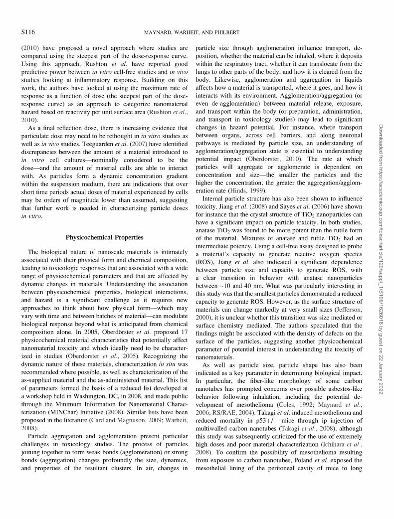
Internal particle structure has also been shown to influence
toxicity. Jiang et al. (2008) and Sayes et al. (2006) have shown
for instance that the crystal structure of TiO2 nanoparticles can
have a significant impact on particle toxicity. In both studies,
anatase TiO2 was found to be more potent than the rutile form
of the material. Mixtures of anatase and rutile TiO2 had an
intermediate potency. Using a cell-free assay designed to probe
a material’s capacity to generate reactive oxygen species
(ROS), Jiang et al. also indicated a significant dependence
between particle size and capacity to generate ROS, with
a clear transition in behavior with anatase nanoparticles
between ~10 and 40 nm. What was particularly interesting in
this study was that the smallest particles demonstrated a reduced
capacity to generate ROS. However, as the surface structure of
materials can change markedly at very small sizes (Jefferson,
2000), it is unclear whether this transition was size mediated or
surface chemistry mediated. The authors speculated that the
findings might be associated with the density of defects on the
surface of the particles, suggesting another physicochemical
parameter of potential interest in understanding the toxicity of
nanomaterials.
As well as particle size, particle shape has also been
indicated as a key parameter in determining biological impact.
In particular, the fiber-like morphology of some carbon
nanotubes has prompted concerns over possible asbestos-like
behavior following inhalation, including the potential de-
velopment of mesothelioma (Coles, 1992; Maynard et al.,2006; RS/RAE, 2004). Takagi et al. induced mesothelioma and
reduced mortality in p53þ/� mice through ip injection of
multiwalled carbon nanotubes (Takagi et al., 2008), although
this study was subsequently criticized for the use of extremely
high doses and poor material characterization (Ichihara et al.,2008). To confirm the possibility of mesothelioma resulting
from exposure to carbon nanotubes, Poland et al. exposed the
mesothelial lining of the peritoneal cavity of mice to long
S116 MAYNARD, WARHEIT, AND PHILBERT
Dow
nloaded from https://academ
ic.oup.com/toxsci/article/120/suppl_1/S109/1626018 by guest on 22 January 2022

multiwalled carbon nanotubes via ip injection and concluded
that the early pathological effects were characteristic of
asbestos-like events in producing inflammation (Poland et al.,2008). Subsequently, Ryman-Rasmussen et al. (2009) sub-
jected mice to a single inhalation exposure of multiwalled
carbon nanotubes and reported that, at a subsequent post-
exposure period, the nanotubes translocated from airspace to
sites outside the respiratory tract and embedded in the
subpleural wall and within subpleural macrophages. This
finding served to provide an indirect confirmation of the
possibility of inhaled multiwalled carbon nanotubes producing
effects both inside and outside the respiratory tract—similar to
asbestos fibers. It is interesting to note, however, that two 90-
day inhalation studies with multiwall carbon nanotubes
conducted in rats, reported by Ma-Hock et al. (2009) (Nanocyl
multiwalled carbon nanotubes) and Pauluhn (2010) (Baytubes
multiwalled carbon nanotubes), failed to find pathological
effects outside the respiratory tract. Either there is a difference
among species, the pleural effect is not particularly pronounced
or a greater focus needs to be implemented to investigate the
potential and relevance of this pleural effect (Warheit, 2009).
To add further complexity to the biological actions of
nanotubes, Kagan et al. (2010a) recently reported that carbon
nanotubes may be biodegraded via a neutrophil myeloperox-
idase mechanism under conditions of inflammation, although it
remains unclear how relevant the results of this in vitro study
are to conditions in vivo.
The question over carbon nanotube toxicity is dominated by
the physicochemical nature of the material. Carbon nanotubes
are not a homogeneous material category but rather represent
an extremely wide array of material chemistries and morphol-
ogies, determined by the number of concentric graphene walls
constituting the nanotubes, their chirality, their diameter, their
length, the density of surface defects, surface functionalization,
the presence of trace elements and other contaminants,
nanotube straightness, the degree of nanotube entanglement,
and so on. Poland et al. demonstrated the potential of one
subset of this material—long, straight multiwalled carbon
nanotubes—to show fiber-like behavior in a biological envi-
ronment (Donaldson et al., 2010). However, many forms of the
material are too short, too long, or too tangled to demonstrate
similar behavior. Nevertheless, these non-fiber–like forms of
carbon nanotubes may present their own distinct hazards
(Shvedova et al., 2003, 2008). Given evidence that the
morphology of carbon nanotube material released into the air
during handling can vary markedly from batch to batch, the
challenges of relating relevant characteristics to hazard are
complex (Maynard et al., 2007). This is a material that cannot
be adequately characterized by chemistry alone, or as a simple
fiber, in determining its potential toxicity. Rather, it epitomizes
the need for a detailed and sophisticated understanding of
nanomaterial physicochemical characteristic in understanding
potential hazard.
Biokinetics
Unlike free or loosely bound molecules, the transport,
accumulation, transformation, and clearance of nanomaterials
in the body is intimately associated with physical form as well
as chemical composition. Understanding the biokinetics of
nanomaterials provides information on internal doses to
secondary organs and is essential to designing and interpreting
in vitro studies. Oberdorster cites the well-documented
tendency of nanoparticles to translocate from primary de-
position sites to secondary organs (Oberdorster, 2010) but
cautions that uninformed interpretation of these data can lead to
misunderstanding of potential risk. Inhalation studies using 15
and 80 nm iridium nanoparticles have demonstrated the
translocation of inhaled particles to extrapulmonary organs.
However, translocation rates were the order of ~1–2%, with the
rate decreasing rapidly at larger particle sizes (Kreyling et al.,2002, 2009). Nevertheless, there is mounting evidence that
changes in physical and chemical nature at the nanoscale can
have a significant impact on biodistribution. For example,
Semmler-Behnke et al. (2008) have demonstrated a marked
difference in biodistribution of 1.4-nm diameter Au55 clusters
and 18-nm diameter gold particles administered to rats via
injection and intratracheal instillation; 24 h following iv
injection, 18-nm diameter gold particles were cleared from
the blood and predominantly accumulated in the liver and
spleen; 0.5% of the injected dose was excreted via that
hepatobiliary system, but renal excretion was extremely low. In
comparison, the 1.4-nm diameter gold clusters were excreted
by the kidneys as well as by the hepatobiliary system.
Of particular concern in recent years has been the nature of
interactions between nanoparticles and the central nervous
system (Yang et al., 2010). There is evidence that inhaled
nanoparticles can translocate to the central nervous system via
olfactory neurons following nasal deposition (Oberdorster
et al., 2004) and induce significant inflammation-related effects
(Elder et al., 2006). This appears to be a particle size and
chemistry transport route that is unique to nanometer-scale
particles and raises the possibility of previously unidentified
organ-specific doses and responses. Although data remain
inconclusive, Oberdorster hypothesizes that differential protein
adsorption on nanoparticles will affect their uptake and
transport within the central nervous system (Oberdorster,
2010). Preliminary data generated using Apolipoprotein E–
coated gold nanoparticles are consistent with increased nano-
particle translocation to the central nervous system in rats
following iv administration. However, in this study, less than
0.01% of injected particles were translocated, leaving the
authors to conclude that further confirmatory studies are
needed (Oberdorster, 2010).
Nanoparticle translocation to the central nervous system is
indicative of research on a number of fronts looking at
nanoparticle movement across tight barriers. For a number for
years, there has been concern over the ability of blood-borne
NANOTOXICOLOGY AND BEYOND S117
Dow
nloaded from https://academ
ic.oup.com/toxsci/article/120/suppl_1/S109/1626018 by guest on 22 January 2022

nanoparticles to cross the placental barrier (Saunders, 2009).
Recently, Bhabra et al. (2009) have indicated – using an
in vitro model – that blood-borne nanoparticles may be able to
exert an influence across the placental barrier without
physically crossing it. Using an in vitro system designed to
investigate cellular barriers, Bhaba et al. showed that high
concentrations of Cobalt-Chromium alloy nanoparticles on one
side of a tightly meshed layer of cells can cause measurable
DNA damage to cells on the other side. However, it remains
uncertain how relevant these data are to in vivo exposures.
The skin represents another tight barrier that has received
a high level of attention in recent years, as concerns over the
ingress of mineral nanoparticle such as TiO2 and ZnO from
sunscreens and cosmetics into the body have been raised. Early
research suggested that the potential exists for sub-micrometer
diameter particles to penetrate across the dermal barrier under
some circumstances, depending on their size and chemistry
(Ryman-Rasmussen et al., 2006; Tinkle et al., 2003). However,
the majority of studies to date suggest that under most
conditions healthy skin is an effective barrier to nanometer-
scale particles entering the body and causing adverse effects
(Choksi et al., 2010; Newman et al., 2009; Nohynek et al.,2008, 2010; Osmond and McCall, 2010; Stern and McNeil,
2008). However, there remain some uncertainties surrounding
the tightness of the skin as a barrier against nanoparticles under
varying conditions, and the impacts and clearance of particles
that may cross the barrier. Recently, Gulson et al. (2010)
exposed human volunteers to sunscreens formulated with ZnO
particles tagged with the stable isotope 68Zn. Traces of 68Zn
were found in blood and urine samples of volunteers exposed
to nanometer-scale and non-nanoscale particles, providing
evidence of Zn transport into the body. However 68Zn levels
were orders of magnitude below normal blood-borne Zn
concentrations. It also remains uncertain whether these findings
were associated with particle translocation, or particle disso-
lution and subsequent ion transport.
As has been alluded to, a possible confounding factor in
understanding the biokinetics of nanoparticles is their differ-
ential interactions with proteins within biological environ-
ments. Nanoparticles within a biological environment rapidly
acquire a coating or ‘‘corona’’ of protein molecules and there is
increasing evidence that this dynamic coating mediates the
transport of, and first order interactions with, nanoparticles
within the body (Cedervall et al., 2007a, 2007b; Ehrenberg
et al., 2009). Furthermore, there is evidence that the corona –
and thus particle biokinetics – is influenced by particle size and
chemistry (Lundqvist et al., 2008). This relatively new area of
research suggests that interactions between nanoparticles and
biological systems may be more complex and dynamic than
previously thought, requiring a more holistic understanding of
how biokinetics are influenced by particle physicochemistry
and their local environments over time.
EMERGING CHALLENGES
Although we have touched on just some of the more
prominent developments in the science of nanoscale materials
toxicology, it is clear that as understanding of how these
materials interact with biological systems increases, new
questions are being raised as to how to understand and quantify
the toxicity of increasingly sophisticated materials in the context
of identifying, assessing, and managing risks. It is also be-
coming clear that, although new questions are being prompted
by the development and commercial use of engineered nano-
materials, the challenges being faced by toxicology are not
solely confined to materials or particles with physical structure
in the range of 1–100 nm. Rather, the emergence of new
nanomaterials is highlighting the importance of material
physicochemistry in mediating biological interactions that result
in toxicity. Research to date suggests that synergism between
particle chemistry and physical form becomes increasingly
important as the features and dimensions of materials entering
the body become increasingly small. But beyond this, there are
few indicators of generalized sharp size-specific transitions in
behavior. Aufann et al. (2009). have attempted to define
a particle size region where size-specific biological behavior
unique to nanoparticles might occur. Reviewing the literature,
they concluded that particles smaller than ~30 nm in diameter
are more likely to demonstrate dramatic changes in behavior
with size. However, particle sizes at which abrupt changes in
behavior occur are clearly material dependent—as was shown
by Semmler-Behnke et al. in contrasting the biokinetics of 1.4
and 18 nm diameter gold nanoparticles (Semmler-Behnke et al.,2008). And there is little reason not to suppose that some
materials may exhibit abrupt changes in behavior above 100 nm.
Concerns over the possible novel toxicity of nanomaterials
are frequently driven by abrupt size-specific changes in
functionality that are governed by size-constrained electron
behavior—often referred to as ‘‘quantum effects.’’ Yet very
few studies have shown a clear association between nanoscale
phenomena such as quantum confinement or surface plasmon
resonances and toxicity. Instead, studies have tended to
highlight the importance of decreasing particle size and
increasing specific surface area for specific particle chemistries
in altering biological behavior. In many cases, these are
scalable effects—small particles show greater or less tendency
to behave in a certain way compared with large particles, but
their behavior is predictable from larger particles. This is the
case for most studies correlating toxicity with surface area. In
other cases, nonscalable effects are seen, such as with size-
specific translocation. Yet even here, it is unclear whether the
unusual biological behavior observed is related to the
functionality these materials are designed to exhibit or simply
a function of small size.
Yet the field of toxicology is undoubtedly facing a new and
growing challenge: How to understand and address the hazard
of intentionally engineered materials where physical form and
S118 MAYNARD, WARHEIT, AND PHILBERT
Dow
nloaded from https://academ
ic.oup.com/toxsci/article/120/suppl_1/S109/1626018 by guest on 22 January 2022

chemical composition interact synergistically to determine
biological behavior. As collaborations across diverse fields of
research lead to increasingly sophisticated new materials—
many of which will be engineered with nanoscale features—
this challenge will only grow in magnitude. Up to now,
research has been driven by small particles of conventional
materials such as TiO2, ZnO, and Ag and the occasional new
material such as carbon nanotubes. But as the science and
technology of new materials becomes increasingly sophisti-
cated, toxicologists will be faced with complex multicompo-
nent materials, hybrid materials that blur the boundaries of
biological and nonbiological components and active materials
that are designed to change behavior according to their
environment or a received set of signals (Subramanian et al.,2010). These new sophisticated materials will require a new
toxicology that recognizes the significance of physicochemistry
and dynamic (and possibly remote activated) behavior, that is
cognizant of but not constrained by the importance of the
nanoscale, and that is focused on the potential biological
impacts of the materials rather than their commercially relevant
functionality.
Within this context, we highlight three emerging challenges
to addressing the toxicology of sophisticated materials:
identifying materials that have the potential to exhibit novel
and significant toxicity, characterizing materials appropriately,
and biointeractions.
Identifying Relevant Materials
Effective problem formulation is a cornerstone of contem-
porary risk assessment and, by association, toxicology
(National Academy of Science, 2008). Nevertheless, formulat-
ing the environmental health and safety impact ‘‘problems’’
posed by sophisticated materials is not trivial. A key question is
how to delineate between the materials and products that are of
concern and those that are not. In regard to engineered
nanomaterials, the conventional approach has been to use
established definitions of nanotechnology and engineered
nanomaterials. These debates typically focus on material
functionality within a narrow size range and are designed
primarily to stimulate research and innovation leading to
economically and socially beneficial new products (NSET,
2010). However, these simple function-oriented definitions do
not always lend themselves to supporting well-defined problem
statements that frame relevant toxicology research on engi-
neered nanomaterials. For example, they do not allow easy
differentiation between functionally unique materials and
products that do not present new toxicology challenges and
functionally mundane materials and products that do present
new hazards. An example of the former might be cadmium
selenide-based quantum dots, where functionality is associated
with size-dependent quantum confinement, but hazard is more
likely associated with the composition of the quantum dots.
And an example of the latter might be the use of nanoscale
particles in a product simply on the grounds of convenience or
economy, but where particle size leads to new exposures, doses,
and hazards. This disconnect between definitions driving
research and innovation and hazard-based problem formulation
is likely to become increasingly important in the face of
increasingly sophisticated materials.
An alternative approach to addressing potential hazards
presented by sophisticated materials is to use principles that
guide scientifically grounded problem formulation. Three
principles that go some way to support science-based and
socially relevant problem formulation address emergent risk,
plausibility, and impact.
Emergent Risk
The idea of emergent risk reflects the likelihood of a new
material causing harm in a manner that is not apparent,
assessable, or manageable based on current approaches to risk
assessment and management. Examples of emergent risk
include the ability of small particles to penetrate to normally
inaccessible places, the inapplicability of established toxicol-
ogy assays to some materials, scalable behavior that is not
addressed by conventional approaches to assessing hazard, and
the possibility of abrupt scale-dependent changes in material
interactions within biological systems. This understanding of
‘‘emergence’’ is dependent on the potential of a material to
cause harm in unanticipated or poorly understood ways, rather
than its physical structure or properties per se. As such, it is not
bound by rigid definitions such as those used to define
nanotechnology or nanomaterials. Rather, it enables sophisti-
cated materials that potentially present emergent and un-
anticipated risks to human health and the environment to be
distinguished from those that probably do not.
Many of the engineered nanomaterials that have raised
concerns in recent years have shown potential to lead to
emergent risks and thus would be classified as requiring further
investigation under this principle. But the principle also
embraces more complex nanomaterials that are either in the
early stages of development, or have yet to be developed,
including active nanomaterials and self-assembling materials.
Plausibility
Plausibility captures—in qualitative terms—the science-
informed likelihood of a new material or product presenting
a risk to humans. It is based on the possible hazard of a material
and potential for exposure or release to occur. But it also
addresses the likelihood of a technology being developed and
commercialized, and it leading to emergent risks. For example,
the ‘‘gray goo’’ of self-replicating nanobots envisaged by some
(Joy, 2000) might legitimately be considered an emergent risk
but is clearly not a plausible risk. In this way, plausibility acts
as a crude but effective filter to distinguish between speculative
risks—which are legion—and credible risks—which are not.
NANOTOXICOLOGY AND BEYOND S119
Dow
nloaded from https://academ
ic.oup.com/toxsci/article/120/suppl_1/S109/1626018 by guest on 22 January 2022

Impact
Impact is an indicator of the extent to which a poorly
managed sophisticated material might cause harm or the
possible reduction in harm resulting from new research into
identifying, assessing, and managing emergent risks. It helps
provide a qualitative reality check to guard against extensive
research efforts that are unlikely to have a significant impact on
protecting human health, while ensuring that research having
the potential to make a significant difference is identified and
supported.
Together, these three principles provide a basis for de-
veloping informed and relevant approaches to problem
formulation when faced with evaluating the hazards associated
with emerging sophisticated materials. They are tools that allow
new materials which raise safety concerns to be differentiated
from those that, while they may be novel from an applications
perspective, do not present undetected, unanticipated, or
enhanced risks. The principles are technology independent
and therefore can be used to guide research independently of the
sophistication of the materials being produced or shifts in
terminology and emphasis underlying technology innovation.
Applying the principles to increasingly sophisticated materi-
als that are being envisaged, a number of groups of materials
begin to emerge that may require further study:
Materials demonstrating abrupt scale-specific changes inbiological or environmental behavior. Materials that undergo
rapid size-dependent changes in physical and chemical
properties, which in turn affect biological behavior, may present
a hazard that is not predictable from larger scale materials of the
same composition. In this case, size and form at the nanoscale
may increase or decrease hazard in a way that is currently not
well understood.
Materials capable of penetrating to normally inaccessibleplaces. Materials that, by their size, shape, and/or surface
chemistry, are able to persist in or penetrate to places in the body
that are not anticipated based on current understanding may
present emergent risks. Where there is a credible possibility of
accumulation of, exposure to, or organ/system-specific dose
associated with a material that is not expected from how the
dissolved material or larger particles of the material behave,
a plausible and emergent risk is possible.
Active materials. Materials that undergo a change in their
biological behavior in response to their local environment or
a received signal (Subramanian et al., 2010), potentially
present dynamic risks that are currently not well understood.
Self-assembling materials. Materials designed to assemble
into new structures in the body once released raise issues that
may not be captured well within current approaches to hazard
assessment.
Materials exhibiting scalable hazard that is not captured byconventional hazard assessments. Where hazard scales
according to parameters other than those normally associated
with an assessment, emergent risks may arise as dose-response
relationships are inappropriately quantified. For instance, if the
hazard presented by an inhaled material scales with the surface
area of the material and the dose-response relationship is
evaluated in terms of mass concentration, the hazard will remain
ill quantified.
In each of these examples (they are not exclusive), new
research is needed if emergent and plausible risks associated
with new sophisticated materials are to be identified,
characterized, assessed, and managed.
Physicochemical Characterization
Relevant physicochemical characterization is essential to
interpreting data from toxicity studies on sophisticated materials
if generated data are to be useful. The early research by
Oberdorster et al. on inhalation exposure to TiO2 particles using
rats showed that chemistry alone could not explain differences
in dose-response relationships for two distinct sizes of particles
with the same composition (Oberdorster, 2000); it was only
when the physical structure of the two materials was included in
the assessment that the data were reconcilable—and a single
dose response relationship relative to material surface area
emerged. But relevant physicochemical characterization is also
necessary if different studies are to be reproduced and
compared. Without it, vital information is lacking that can
prevent a robust picture of material toxicity from emerging. In
the case of the Oberdorster study, material surface area was
measured, but not discrete particle size or aggregation state. As
a result, it was initially difficult to evaluate or validate whether
the effects observed were simply because of an elevated material
surface area or were associated with the presence of discrete
nanometer-scale particles. A clearer case of data confounding
through a lack of physicochemical characterization can be
found in studies on carbon nanotubes. Despite the enormous
variation in physical and chemical properties among carbon
nanotubes from different sources (or even the same source at
different times), early toxicology studies were remarkably
vague on the precise nature of the materials being studied,
leading to irreproducible, conflicting, and ultimately uninter-
pretable data (Lam et al., 2006).
The relevance of physicochemical characterization in un-
derstanding and assessing material toxicity has received
considerable attention in recent years. A workshop in 2004,
organized by the National Institute of Environmental Health
Sciences and the University of Florida placed a strong emphasis
on the need for highly detailed materials characterization for
instance (Moudgil, 2004). These recommendations—driven in
part by materials scientists—were considered at the time to be
beyond the scope of many toxicologists. In 2005, an influential
review led by Oberdorster proposed a reduced—but still
extensive—set of physicochemical parameters that should be
S120 MAYNARD, WARHEIT, AND PHILBERT
Dow
nloaded from https://academ
ic.oup.com/toxsci/article/120/suppl_1/S109/1626018 by guest on 22 January 2022

included in nanomaterial toxicology studies (Oberdorster et al.,2005). The recommendations listed 17 parameters as either
essential or desirable in studies. Importantly, they also
considered characterization at three distinct points, recognizing
the dynamic nature of physicochemical characteristics: in the
bulk material, as-prepared for administration, and in situ. Of
these, the importance of characterizing materials in situ was
stressed as materials are capable of undergoing significant
alterations in properties once they are introduced to a biological
environment. However, it was also recognized that few
technologies currently exist that enable detailed materials
characterization within in vitro and in vivo test systems.
Although the Oberdorster recommendations were less chal-
lenging than those from Florida, they were still seen as presenting
near-insurmountable barriers to toxicologists. As a result,
discussions within the community continued to focus on
a minimum characterization set that would be both feasible and
readily adoptable. A 2008 workshop was held at the Woodrow
Wilson International Center for Scholars in Washington, DC, was
influential in mapping out such a minimum characterization set
(Table 2) (MINChar Initiative, 2008). The list was one of the first
pragmatic sets of physicochemical characterization requirements
and reflects similar thinking published elsewhere (Boverhof and
David, 2010; Card and Magnuson, 2009; Warheit, 2008).
As a result of these and other efforts, there is movement
toward expecting and including detailed physicochemical
characterization data in nanomaterial toxicology studies. This
is becoming increasingly important as methodologies are
developed to develop predictive models for engineered nano-
materials and—by extension—sophisticated materials in gen-
eral. In 2006, Maynard et al. challenged the scientific
community to work towards predictive models for nano-
material impact (Maynard et al., 2006). Four years later,
a number of initiatives are beginning to work toward this goal
(Alvarez et al., 2009; Meng et al., 2009). These approaches
depend on associating key material properties with mecha-
nisms of biological interaction and ultimately effects. To be
successful, they will depend on detailed physicochemical
characterization of the materials under test.
However, knowing what needs to be measured is only part of
the challenge, it is complemented by the need for tools to make
appropriate measurements. Here, progress is still lacking, with
even the minimum characterization requirements proposed by
initiatives like MINChar challenging toxicologists. Three
challenges in particular face the toxicology community as
increasingly sophisticated materials are developed: presenting
samples to test systems that are well-characterized, evaluating
key physicochemical properties in situ, and developing
analytical techniques that can provide useful information into
tools that are accessible to the toxicology community. Each
comes with its own challenges and it is likely to be many years
before substantial progress is made. Yet evaluating and
quantifying the toxicology of sophisticated materials will
depend on new tools and methodologies in each of these areas.
One final significant challenge exists here: developing an
understanding of the tolerance within which physicochemical
characteristics show similar or markedly different biological
behavior (National Academies, 2009). Most sophisticated
materials will be manufactured within a certain range of
physicochemical properties, ensuring functionality is achieved
without resulting in over-costly production processes. As a
result, materials will demonstrate variation in particle size,
shape, surface properties, and other characteristics, both
between batches and within samples. Understanding the
association between variations in physicochemical character-
istics and toxicity will be essential in developing the tools and
methodologies to quantify and address risks presented by
sophisticated materials. Central to this are three questions: (1)
How precisely do physicochemical characteristics need to be
measured? (2) What constitutes a significant change in
characteristics such as particle size, shape, or composition
when evaluating toxicity? and (3) How should the hazard
associated with a material representing a distribution of
physicochemical characteristic be evaluated? So far, very little
progress has been made toward addressing these issues.
Biointeractions
Over the past few years, concerns have been expressed in the
lay press and elsewhere about the ‘‘new toxicities’’ of
TABLE 2
Proposed Minimum Nanomaterial Characterization
Requirements for Use in Toxicology Studies (MINChar
Initiative, 2008)
What does the material look like?
Particle size/size distribution
Agglomeration state/aggregation
Shape
What is the material made of?
Overall composition (including chemical composition and crystal structure)
Surface composition
Purity (including levels of impurities)
What factors affect how a material interacts with its surroundings?
Surface area
Surface chemistry, including reactivity, hydrophobicity
Surface charge
Overarching considerations to take into account when characterizing
engineered nanomaterials in toxicity studies:
Stability—how do material properties change with time (dynamic stability),
storage, handling, preparation, delivery etc? Include solubility, and the rate
of material release through dissolution.
Context/media—how do material properties change in different media; i.e.
from the bulk material to dispersions to material in various biological
matrices? (‘‘as administered’’ characterization is considered to be
particularly important)
Where possible, materials should be characterized sufficiently to interpret the
response to the amount of material against a range of potentially relevant
dose metrics, including mass, surface area and number concentration.
NANOTOXICOLOGY AND BEYOND S121
Dow
nloaded from https://academ
ic.oup.com/toxsci/article/120/suppl_1/S109/1626018 by guest on 22 January 2022

nanomaterials as they interact with biological systems. These
concerns appear to be based on two potential types of
interaction: (1) emergent quantum mechanical properties of
nanomaterials may lead to novel interactions with biology and
(2) the matching of scales between biological machinery and
the engineered nanomaterial may lead to new mechanisms of
interaction. On the first assumption, little information exists in
the peer-reviewed literature that suggests a direct quantum
mechanical interaction or energy transfer between engineered
nanomaterials and biological systems in the absence of external
energy sources. Specifically, in the absence of pumping of laser
radiation deep into tissues (and thus activating size-mediated
radiation-particle-biology interactions), it does not appear that
such biophysical interactions have been detected in carbona-
ceous and other materials currently in high volume production.
Nevertheless, smaller quantities of new materials have been
recently developed that allow for interesting biophysical
interactions at this radiation/material/biology interface. In
addition to the well-known photodynamic production of ROS
for the treatment of cancer and the eradication of microbial
biofilms, etc., gold nanoshells have been used for thermal
ablation of tumor cells in deep tissues (Guerrero et al., 2010;
Halas, 2010; Orringer et al., 2009; Wu et al., 2009). More recent
developments in photonics have enabled the synthesis of
quantum-dot like structures that enable multiphoton excitation
and emission modes that may or may not produce ROS (Pecher
et al., 2010). All these approaches require the injection of
a relatively large tuned photon flux (usually generated by
a laser) into the system. Whether or not the results are purely
energetic interactions between engineered nanomaterials and
biological macromolecules or whether there are further
significant mechanisms of interaction remains to be determined.
In this respect, biophysical measurements will have to be
examined for toxicological plausibility in eliciting a pathological
or pathophysiological outcome.
The second, more widely accepted mechanism is that
nanomaterials are at the scale of biological macromolecules
and lend themselves to hybridization. Indeed, macromolecules
such as DNA have been used to solubilize highly hydrophobic
single-walled carbon nanotubes (Yamamoto et al., 2010). It is
this ‘‘matching of scales’’ that permits nucleic acids, lipids,
proteins, and likely other macromolecules to interact with and
coat the surface of engineered nanomaterials to produce harm.
For example, single-walled carbon nanotubes have been shown
to reproducibly gain access to cell nuclei and induce DNA
strand breaks or formation of micronuclei (Cveticanin et al.,2010). Perhaps it is no surprise that nanomaterials are
recognized by biological macromolecules and are adsorbed
the available surfaces. This adsorptive process is frequently
linked to the early stages of inflammation and is collectively
known as opsonization. The question remains whether nor not
engineered nanomaterials will present new ‘‘antigenic chal-
lenges’’ that will enhance or diminish immunologic function.
Inflammation is a common biological initiator or promoter of
numerous pathophysiological states that end in either acute
inflammatory disease (e.g., formation of granulomata) or act as a
subchronic/chronic initiator/promoter of more pernicious de-
generative or neoplastic disease. The long-known immunolog-
ical process of opsonization of microbial and other external
intruders has recently been shown to play a critical role in
mediating a variety of idiopathic, environmental, and iatrogenic
disorders. The complement system is composed of a tightly
regulated complex of proteins that enable host immune
responses to foreign materials. These proteins adhere to the
surface of foreign bodies, are themselves activated, and
stimulate removal of the offending article(s) by phagocytes.
Inappropriate activation of this system has been linked to a
variety of pathophysiological states including asthma and lupus
(Gonzalez et al., 2010; Sarma and Ward, 2010; Silva, 2010).
The seminal work of Dawson and Colleagues in Ireland has
led to the firmer understanding that characterization of the
chemical and physical nature of nanomaterials is simply a
necessary first step to determining the potential of the material
to subvert biological processes. The physicochemical nature of
the nanomaterial is augmented by the adhesion of a layer of
proteins, lipids, and lipoproteins that forms upon first contact
with the biological milieu (Lynch et al., 2007, 2009; Hellstrand
et al., 2009; Walczyk et al., 2010; Vauthier et al., 2009). It is,
in fact, this entire complex of nanomaterial encased in
biological macromolecules that cells encounter and to which
they respond. The picture is further complicated by the fact that
there is dynamic exchange of many macromolecules between
the external space and the surface of the nanomaterial that is
dependent on the anatomic location of the material. The sheer
complexity of the surface dynamic provides for at least three
biological outcomes: (1) the interactions between biological
fluids and matrices may defeat intended targeting regimens that
are designed to deliver highly functional engineered nano-
materials to specific anatomic sites and may increase off-target
effects, (2) the adsorption of elements of the complement
cascade may activate or inhibit inflammatory processes that
would in the normal run of things enable the body to effectively
dispose of biological and other environmental threats at the
nanoscale, or (3) the nanomaterial itself may provide a stable
platform for the inappropriate delivery of bioactive molecules to
anatomic sites and initiate cell signaling processes that trigger an
adverse (or beneficial if appropriately designed) event (Dawson
et al., 2009; Gaucher et al., 2009; Moghimi et al., 2010; Serda
et al., 2009). Given that many of the engineered nanomaterials
that are in current mass production are stable and are likely to
persist in the body, the dynamic nature of the interactions
between the nano- and biological interface add to the
complexity of what needs to be known in assessing the likely
biological outcome (Alexis et al., 2008).
The latter consideration, which is modulated by the du-
rability of nanomaterial, evokes the consideration of time as
a dose metric. In addition to the descriptions of surface
S122 MAYNARD, WARHEIT, AND PHILBERT
Dow
nloaded from https://academ
ic.oup.com/toxsci/article/120/suppl_1/S109/1626018 by guest on 22 January 2022

reactivity, chemical composition, charge, aspect ratio, etc., the
cautious toxicologist will need to have a firm understanding of
the ability of the material to be disposed of by the biological
system in which it dwells (Kagan et al., 2010a). The work of
Kagan et al. remains to be confirmed in more complex systems
that includes tangled carbon nanotubes or carbonaceous
nanomaterials that have been functionalized. Nevertheless, this
study provides an intriguing insight into the possibility that
there may exist for the most stable nanomaterials a dynamic
equilibrium between those biological processes that induce
accumulation of the material and may confer harm and those
that ultimately dispose of the material in situ (Kagan et al.,2010b). In its ultimate redux, the kinds of analysis that are
required for adequate hazard and risk assessment of engineered
nanomaterials specifically and increasingly sophisticated mate-
rials more generally are far more extensive and complex than
those required for traditional chemicals. Success in preventing
long-term harm and protect public health will require engage-
ment in multidimensional high-content analyses of the pertinent
’omics with reference to traditional metrics such as dose,
duration, and hazard. The general toxicity or off-target effects of
emerging sophisticated materials will require investigation
through selective profiling against large panels of potential
targets. At present, these kinds of studies are expensive and their
utility in estimating the risk of harm is not well characterized.
Unless the experimental design is tailored to take into account
known biological processes and is constrained to pathways that
are known to induce identifiable harm (not merely a change in
expression), the implementation of such approaches will not be
widely adopted, and our ability to improve our collective
predictive capabilities will be severely diminished (Merino
et al., 2010).
A particular challenge associated with understanding inter-
actions between nanomaterials and biological systems is that of
relating in vitro observations to in vivo behavior. Recent
research has begun to push the capacity of in vitro approaches to
rapidly screen nanomaterials for potential toxicity and to
develop potential causal relationships between characteristics
and biologically relevant behavior (George et al., 2010; Meng
et al., 2009). Yet toxicology studies in vitro carry with them
several limitations, confounding factors, and caveats that make
more complicated the incorporation of such data in the
assessment of the risk potential of nanomaterials. For example,
many published experiment apply a suspension of well-
characterized nanomaterial to cell cultures with little consider-
ation of the way in which particles behave in fluid suspensions.
In addition to the well-known behavior of agglomeration and
aggregation in aqueous environments, particles show a variety
of behaviors that include surface adsorption, changes in surface
charge and charge distribution, and nonuniform spatial
distribution. Indeed, the latter may result in the delivery of
widely differing amounts of nanoparticle to the surface of cells
in culture (Teeguarden et al., 2007). This simple physical
interaction of the nanoparticle with the suspending medium
brings into sharp focus a variety of other considerations that
cause an apparent difference in potency in vitro.
On the biological side, deficiencies in the ability of the cells in
culture to recapitulate their phenotype in the context of the intact
tissue are well known and range from the loss of biochemical
functions critical to the biotransformation/bioactivation of
xenobiotics to the morphology of the cell. Also, because most
in vitro toxicologic investigations use monocultures of epithelia,
in vitro culture models cannot engage in paracrine signaling
between, for example, mesenchymal elements of the native
tissue or dendritic cells that themselves take part in humoral
signaling with other lymphoid tissues. The latter has recently
been shown to be important in whole-body responses to
nanomaterials (Mitchell et al., 2007). Interestingly, there have
been exciting developments in the development of nano-
materials for enhanced cultured cell models that aid in the
preservation of physiologic and morphologic function. These
nanomaterials show considerable promise for neurotoxicology
and neuroscience, for example, and include nanofibers such as
polycaprolactone in the promotion of neuronal differentiation
and orientation of neuronal progenitor cells derived from human
embryonic stem cells (Mahairaki et al., 2010). Similarly,
electrospun bioactive nanomaterials that mimic the properties
of extracellular membranes have been developed and success-
fully deployed in the culturing of human renal tubular cells in
a format that recapitulates the intercellular tight junctions and
initiates formation of the brush border and expression of
biologically relevant transport mechanisms such as c-glutamyl
transpeptidase (Dankers et al., 2011). Even newer materials
have been developed that permit the dynamic inward budding of
membranes (exosomes) that encapsulate small RNA’s in a
format ready to deliver to cultured and other cell types (Pegtel
et al., 2010; Zomer et al., 2010). Undoubtedly, many of these
advances and others in nanotechnology/nanomaterials will be
useful in addressing and overcoming some of the complicating
factors and limitations of cell culture in toxicologic research.
There are on the drawing board wide variety of nano-
technologies for medical imaging and therapy. Overwhelm-
ingly, the new nanomaterial-based approaches to therapy and
imaging use nanoparticles that aim to improve the pharmaco-
kinetic/pharmacodynamic profile of existing (predominantly
hydrophobic) compounds. In that sense, these are ‘‘smart
excipients’’ or ‘‘smart carriers.’’ An advanced embodiment
of the smart carrier approach is the targeting of highly
branched dendrimers of various chemical types and incorporate
different cyclic cores such as carbopeptides, carboproteins,
octopus glycosides, inositol-based dendrimers, cyclodextrins,
calix[4]arenes, resorcarenes, cavitands, and porphyrins
(reviewed by Sebestik et al., 2010). Even the ‘‘simple’’
liposome has benefitted from advances synthesis at < 100 nm
and the ability to perform sophisticated surface chemistry
(reviewed by Jølck et al., 2010). Hydrogels of the kind
described elsewhere in this review have also been deployed
for the imaging a treatment of tumors. Perhaps not surprisingly,
NANOTOXICOLOGY AND BEYOND S123
Dow
nloaded from https://academ
ic.oup.com/toxsci/article/120/suppl_1/S109/1626018 by guest on 22 January 2022

the second generation of nanotechnologies for medicine is
already under development and have been funded by the NIH
Common Fund (http://nihroadmap.nih.gov/nanomedicine/fun-
dedresearch.asp). These technologies are frequently inspired by
the kind of solutions provided in the efficient nanomachines of
nature, for example, cellular machinery that packages proteins
with immensely massive forces, nanomachines that sense
photons and wavelengths outside those visible to the human
eye, etc. The use of newer (macro) technologies that work at the
nanometer scale, for example, photonic tweezers, femtosecond
pulsed lasers, and ultrasound, enable the development of tunable
molecular assemblies that convert imparted energy into
beneficial mechanical work aimed at repairing defects within
the cell. All these advances will, as they are deployed in
medicine and beyond, impose massive strain on the current
toxicologic paradigms used in the evaluation of chemicals and
the first generation of nanomaterials.
Considering further the intersection between beneficial use,
biointeractions, and potential biological impact, the pharma-
ceutical industry has used a variety of smart excipients for
targeted drug delivery for many years that dissolve with time
and give rise to soluble chemicals that are readily excreted in
the urine and/or feces thereby reducing the potential for
bioaccumulation and off-target effects. Other approaches use
the dissolution of the nanomaterial to reduce complement-
mediated activation following admission into the vascular
space (Gao et al., 2008). The picture is more complicated when
the surface of the material is coated with targeting ligands for
delivery of the nanodevice to a specific anatomic location or
cell type. The surfaces of copolymer blends of polylactic-
glycolic acid and polyethylene glycol nanoparticles have been
successfully grafted with a number of ligands such as herceptin
(anti-HER2 antibody). The nanoparticle itself may be loaded
with an anticancer drug or a smaller nanoparticle such as
nanosilver and permits delivery of therapeutics to a variety of
cells, tissues, or surfaces. The targeting effect has been shown
to be quantitatively controlled by two major approaches: (1)
adjusting the copolymer blend ratio of the nanoparticle matrix
with concomitant changes in the geometry of the surface and
alterations in ligand density on the surface of the nanoparticle
surface, and (2) adjusting the molar ratio of herceptin to
available free amines appearing on the nanoparticle surface.
Both these approaches (within limits) permitted a linear
relation between the concentration of nanoparticles adminis-
tered and the amount binding to tumor cells (Liu et al., 2010;
Shameli et al., 2010). Of course, in the development of a
nanomedicine, the purpose is to develop a material that is
a priori biocompatible. With the notable exception of the
nanomaterials for devices and prostheses, the synthesis of new
therapeutic agents is rarely guided by considerations such as
hardness, tensile strength, ability to absorb blunt/sharp force
trauma, etc. These factors are more frequently the domain of
consumer products and manufacturing.
The deployment of nanomaterials and other sophisticated
materials in medical and consumer products is still in its
infancy. As such, there exists a fleeting opportunity for a cross-
over between these two disparate domains and sharing of pre-
competitive data on hazard and exposure that may catalyze the
smart design of materials that minimize harmful bio-nano-
interactions and maintain the desired physicochemical proper-
ties imparted by a scale less than 100 nm.
LOOKING TO THE FUTURE
Nanotoxicology is a field that has been propelled into the
limelight by science, speculation, and a growing push toward
developing new and unusual materials in products. Its grounding
in science is clear—research shows that the size, shape, chemistry
and other physicochemical parameters of physical objects affect
how they interact with biological systems and the potential
impacts they have. Yet the uniqueness of the field in terms of the
‘‘nano’’ prefix is perhaps not so clear. Size matters—this is
indisputable. But decades of research have demonstrated that
particle size matters at the micrometer scale when it comes to
human health, as well as at the nanoscale, raising the question of
whether nanomaterial toxicity can be understood as an extension
of what we know about larger scale materials or whether there is
something unique about how nanoscale materials interact with
biology that justifies them being singled out? Research over the
past 20 years has shown that nanoscale materials can show
unexpected and unusual toxicity and that physicochemical
complexity is an important mediator of toxicity at the nanoscale.
But it also shows that we are far from developing a precise
understanding of how these parameters govern mechanisms of
interaction or how they are empirically associated with response.
In effect, the form and chemistry combined of materials entering
the body are important, but the indicators of emergent risks are
more complex than a simple size range.
The challenges of understanding how form and chemistry
mediate toxicity will become increasingly apparent as emerging
technologies lead to increasingly sophisticated materials. This is
critical where ever-more complex and multifunctional thera-
peutics are being designed at the nanoscale for introduction to
the human body. But it is also important where sophisticated
materials may enter the body via other routes—as components
of food, or through nonintentional exposure during product
manufacture, and use, or during and after disposal. Advances in
materials science, synthetic chemistry, biotechnology, and other
areas are leading to materials that demonstrate designed and
adaptable functionality; that obscure distinctions between
biological and nonbiological substances; and that have multiple
and often intricate components. Future sophisticated materials
are more likely to resemble the complexity of human-scale
engineered devices, rather than the simplicity of unique
chemical entities. And this in turn means that a more
sophisticated and systems-based approach to assessing their
toxicity and addressing their potential risks is needed.
S124 MAYNARD, WARHEIT, AND PHILBERT
Dow
nloaded from https://academ
ic.oup.com/toxsci/article/120/suppl_1/S109/1626018 by guest on 22 January 2022

In moving forward, the risk assessment paradigm remains
relevant. Ultimately, decisions need to be driven by a science-
based approach that links dose to response. However, sub-
stantial work is needed in applying the risk assessment paradigm
to new materials, so that a science-informed understanding of
how to quantify and predict the potential risks of sophisticated
materials is developed. At the same time, there is a parallel and
somewhat intertwined challenge: Quantitative toxicology and
risk assessment are unlikely to keep pace with the accelerating
development of emerging sophisticated materials, meaning
there will be a growing knowledge gap between the materials
being produced, and the knowledge needed to ensure their safe
use. Bridging this gap will require new approaches to evaluating
risk and making decisions in the face of potential risks where
there is incomplete information on exposure, hazard, and
response. And given the dynamic nature of emerging materials
as they are generated, used, disposed of and recycled, these
approaches will need to be established within a life cycle
framework (Som et al., 2010). Developing models that predict
associations between sophisticated materials, biological inter-
actions, and impacts will be a critical part of this (Maynard et al.,2006). There will also be an increasing need to push risk
assessment—and thus toxicology—upstream in the innovation
process, allowing early decisions to be made on safe and
responsible product development (Owen et al., 2009). In effect,
a new science of risk is needed that draws together the physical,
biological, and social sciences to develop an integrated approach
to the emerging challenges presented by sophisticated materials.
Since publication of Oberdorster’s and Ferin’s research on the
size-mediated response to inhaled TiO2 particles over 20 years
ago, our understanding of how physicochemical characteristics
mediate material toxicity has grown by leaps and bounds (Ferin
et al., 1990; Oberdorster et al., 1990). We can now begin to
appreciate the challenges presented by simple nanoscale
materials such as TiO2, ZnO, Ag, carbon nanotubes, and CeO2.
But these simple materials are merely the vanguard of a new era
of complex materials, where novel and dynamic functionality is
engineered into multifaceted substances. If we are to meet the
challenge of ensuring the safe use of this new generation of
substances, it is time to move beyond ‘‘nano’’ toxicology and
toward a new toxicology of sophisticated materials.
FUNDING
The University of Michigan Risk Science Center (to
A.D.M.); ES 2R01 ES08846 (to M.A.P.).
REFERENCES
Aitken, R. J., Hankin, S. M., Ross, B., Tran, C. L., Stone, V., Fernandes, T. F.,
Donaldson, K., Duffin, R., Chaudhry, Q., Wilkins, T. A., et al. (2009). In
EMERGNANO: A Review of Completed and Near Completed Environment,
Health and Safety Research on Nanomaterials and Nanotechnology. Institute
for Occupational Medicine, Edinburgh, UK.
Alexis, F., Pridgen, E., Molnar, L. K., and Farokhzad, O. C. (2008). Factors
affecting the clearance and biodistribution of polymeric nanoparticles. Mol.
Pharm. 5, 505–515.
Alvarez, P. J. P., Colvin, V., Lead, J. R., and Stone, V. (2009). Research
priorities to advance eco-responsible nanotechnology. ACS Nano 3,
1616–1619.
Auffan, M., Rose, J., Bottero, J. Y., Lowry, G. V., Jolivet, J. P., and Wiesner, M. R.
(2009). Towards a definition of inorganic nanoparticles from an environmental,
health and safety perspective. Nat. Nanotechnol. 4, 634–641.
Balashazy, I., Hofmann, W., and Heistracher, T. (2003). Local particle
deposition patterns may play a key role in the development of lung cancer. J.
Appl. Physiol 94, 1719–1725.
Balbus, J. M., Maynard, A. D., Colvin, V. L., Castranova, V., Daston, G. P.,
Denison, R. A., Dreher, K. L., Goering, P. L., Goldberg, A. M.,
Kulinowski, K. M., et al. (2007). Meeting report: hazard assessment for
nanoparticles—Report from an interdisciplinary workshop. Environ. Health
Perspect. 115, 1654–1659.
Benninghoff, A. D., and Hessler, W. (2008). Nanoparticles damage brain cells.
Available at: http://www.environmentalhealthnews.org/ehs/newscience/
nanoparticles-damage-brain-cells/. Accessed September 26, 2010.
Bhabra, G., Sood, A., Fisher, B., Cartwright, L., Saunders, M., Evans, W. H.,
Surprenant, A., Lopez-Castejon, G., Mann, S., Davis, S. A., et al. (2009).
Nanoparticles can cause DNA damage across a cellular barrier. Nat. Nano 4,
876–883. Available at: http://www.nature.com/nnano/journal/v4/n12/
suppinfo/nnano.2009.313_S1.html.
Binnig, G., Rohrer, H., Gerber, C., and Weibel, E. (1982). Surface studies by
scanning tunneling microscopy. Phys. Rev. Lett. 49, 57–61.
Boverhof, D., and David, R. (2010). Nanomaterial characterization: consid-
erations and needs for hazard assessment and safety evaluation. Anal.
Bioanal. Chem. 396, 953–961.
Brown, J. S., Zeman, K. L., and Bennett, W. D. (2010). Ultrafine particle
deposition and clearance in the healthy and obstructed lung. Am. J. Respir.
Crit. Care Med. 166, 1240–1247.
Card, J. W., and Magnuson, B. (2009). Proposed minimum characterization
parameters for studies on food and food-related nanomaterials. J. Food Sci.
74, vi–vii.
Cedervall, T., Lynch, I., Foy, M., Berggard, T., Donnelly, S. C., Cagney, G.,
Linse, S., and Dawson, K. A. (2007a). Detailed identification of plasma
proteins adsorbed on copolymer nanoparticles. Angew Chem. Int. Ed. Engl.
46, 5754–5756.
Cedervall, T., Lynch, I., Lindman, S., Berggard, T., Thulin, E., Nilsson, H.,
Dawson, K. A., and Linse, S. (2007b). Understanding the nanoparticle-
protein corona using methods to quantify exchange rates and affinities of
proteins for nanoparticles. Proc. Natl. Acad. Sci. U.S.A 104, 2050–2055.
Chalupa, D. C., Morrow, P. E., Oberdorster, G., Utell, M. J., and
Frampton, M. W. (2004). Ultrafine particle deposition in subjects with
asthma. Environ. Health Perspect. 112, 879–882.
Chemical Industry Vision 2020 Technology Partnership and the Semiconductor
Research Corporation. (2005). Joint NNI-ChI CBAN and SRC CWG5
Nanotechnology research needs recommendations. Available at: http://
www.chemicalvision2020.org/pdfs/chem-semi%20ESH%20recommendations.
pdf. Accessed October 20, 2010.
Choksi, A. N., Poonawalla, T., and Wilkerson, M. G. (2010). Nanoparticles:
a closer look at their dermal effects. J. Drugs Dermatol. 9, 475–481.
Clinton, W. J. (2000). In President Clinton’s Address to Caltech on Science
and Technology. Remarks by the President at Science and Technology Event.
California Institute of Technology Pasadena, Pasadena, CA.
Coles, G. V. (1992). Occupational risks. Nature 359, 99.
Cveticanin, J., Joksic, G., Leskovac, A., Petrovic, S., Sobot, A. V., and
Neskovic, O. (2010). Using carbon nanotubes to induce micronuclei and
NANOTOXICOLOGY AND BEYOND S125
Dow
nloaded from https://academ
ic.oup.com/toxsci/article/120/suppl_1/S109/1626018 by guest on 22 January 2022

double strand breaks of the DNA in human cells. Nanotechnology 21,
015102.
Dankers, P. Y., Boomker, J. M., Huizinga-van der Vlag, A., Wisse, E.,
Appel, W. P., Smedts, F. M., Harmsen, M. C., Bosman, A. W., Meijer, W.,
and van Luyn, M. J. (2011). Bioengineering of living renal membranes
consisting of hierarchical, bioactive supramolecular meshes and human
tubular cells. Biomaterials 32, 723–733.
Dawson, K. A., Salvati, A., and Lynch, I. (2009). Nanoparticles reconstruct
lipids. Nat. Nanotechnol. 4, 84–85.
Dockery, D. W., Pope, C. A., Xu, X., Spengler, J. D., Ware, J. H., Fay, M. E.,
Ferris, B. G., and Speizer, F. E. (1993). An association between air pollution
and mortality in six U.S. cities. N. Engl. J. Med. 329, 1753–1759.
Donaldson, K., Murphy, F. A., Duffin, R., and Poland, C. A. (2010). Asbestos,
carbon nanotubes and the pleural mesothelium: a review of the hypothesis
regarding the role of long fibre retention in the parietal pleura, inflammation
and mesothelioma. Part. Fibre Toxicol. 7.
Donaldson, K., and Poland, C. A. (2009). Nanotoxicology: new insights into
nanotubes. Nat. Nanotechnol. 4, 708–710.
Donaldson, K., Stone, V., Tran, C. L., Kreyling, W., and Borm, P. J. A. (2004).
Nanotoxicology. Occup. Environ. Med. 61, 727–728.
Drexler, E. (1986). In Engines of Creation: The Coming Era of Nanotechnol-
ogy. Anchor Books, New York, NY.
Ehrenberg, M. S., Friedman, A. E., Finkelstein, J. N., Oberdorster, G., and
McGrath, J. L. (2009). The influence of protein adsorption on nanoparticle
association with cultured endothelial cells. Biomaterials 30, 603–610.
Eigler, D. M., and Schweizer, E. K. (1990). Positioning single atoms with
a scanning tunneling microscope. Nature 344, 524–526.
Elder, A., Gelein, R., Silva, V., Feikert, T., Opanashuk, L., Carter, J., Potter, R.,
Maynard, A., Finkelstein, J., and Oberdorster, G. (2006). Translocation of
inhaled ultrafine manganese oxide particles to the central nervous system.
Environ. Health Perspect. 114, 1172–1178.
ETC Group. (2003). No Small Matter II: The Case for a Global Moratorium.
Size Matters!. ETC Group, Winnipeg, Canada.
Ferin, J., Oberdorster, G., Penney, D. P., Soderholm, S. C., Gelein, R., and
Piper, H. C. (1990). Increased pulmonary toxicity of ultrafine particles 1.
Particle clearance, translocation, morphology. J. Aerosol. Sci. 21, 381–384.
Feynman, R. P. (1960). There’s plenty of room at the bottom. Eng. Sci. 23,
22–36.
Gao, D., Xu, H., Philbert, M. A., and Kopelman, R. (2008). Bioeliminable
nanohydrogels for drug delivery. Nano Lett. 8, 3320–3324.
Gaucher, G., Asahina, K., Wang, J. H., and Leroux, J. C. (2009). Effect of
poly(N-vinyl-pyrrolidone)-block-poly(D,L-lactide) as coating agent on the
opsonization, phagocytosis, and pharmacokinetics of biodegradable nano-
particles. Biomacromolecules 10, 408–416.
George, S., Pokhrel, S., Xia, T., Gilbert, B., Ji, Z. X., Schowalter, M.,
Rosenauer, A., Damoiseaux, R., Bradley, K. A., Madler, L., et al. (2010).
Use of a rapid cytotoxicity screening approach to engineer a safer zinc oxide
nanoparticle through iron doping. ACS Nano 4, 15–29.
Gonzalez, S. F., Lukacs-Kornek, V., Kuligowski, M. P., Pitcher, L. A.,
Degn, S. E., Turley, S. J., and Carroll, M. C. (2010). Complement-dependent
transport of antigen into B cell follicles. J. Immunol. 185, 2659–2664.
Guerrero, S., Araya, E., Fiedler, J. L., Arias, J. I., Adura, C., Albericio, F.,
Giralt, E., Arias, J. L., Fernandez, M. S., and Kogan, M. J. (2010). Improving
the brain delivery of gold nanoparticles by conjugation with an amphipathic
peptide. Nanomedicine (Lond) 5, 897–913.
Gulson, B., McCall, M., Korsch, M., Laura Gomez, L., Philip Casey, P.,
Yalchin Oytam, Y., Alan Taylor, A., Lesley Kinsley, L., and Gavin
Greenoak, G. (2010). Small amounts of zinc from zinc oxide particles in
sunscreens applied outdoors are absorbed through human skin. Toxicol. Sci.
118, 140–149.
Halas, N. J. (2010). Plasmonics: an emerging field fostered by Nano Letters.
Nano Lett. 10, 3816–3822.
Hellstrand, E., Lynch, I., Andersson, A., Drakenberg, T., Dahlback, B.,
Dawson, K. A., Linse, S., and Cedervall, T. (2009). Complete high-density
lipoproteins in nanoparticle corona. FEBS J 276, 3372–3381.
Hett, A. (2004). In Nanotechnology. Small Matter, Many Unknowns. SwissRe,
Zurich, Switzerland.
Hinds, W. C. (1999). In Aerosol Technology: Properties, Behavior and
Measurement of Airborne Particles, 2nd ed. Wiley-Interscience, New York, NY.
Ichihara, G., Castranova, V., Tanioka, A., and Miyazawa, K. (2008). Letter to
the editor. J. Toxicol. Sci. 33, 38.
ICON. (2008a). Predicting Nano-Biointeractions: An International Assessment
of Nanotechnology Environment, Health and Safety Research Needs.
International Council On Nanotechnology, Houston, TX.
ICON. (2008b). Towards Predicting Nano-Biointeractions: An International
Assessment of Nanotechnology Environment. Health and Safety Research
Needs. International Council On Nanotechnology, Houston, TX.
Iijima, S. (1991). Helical microtubules of graphitic carbon. Nature 354, 56–58.
IWGN. (1999). Nanotechnology Research Directions: IWGN Workshop
Report. Vision for Nanotechnology R&D in the Next Decade. National
Science and Technology Council Committee on Technology Interagency
Working Group on Nanoscience, Engineering and Technology (IWGN),
Washington, DC.
Jefferson, D. A. (2000). The surface activity of ultrafine particles. Philos.
Trans. R. Soc. Lond. A 358, 2683–2692.
Jiang, J., Oberdorster, G., Elder, A., Gelein, R., Mercer, P., and Biswas, P.
(2008). Does nanoparticle activity depend upon size and crystal phase?
Nanotoxicology 2, 33–42.
Jølck, R. I., Feldborg, L. N., Andersen, S., Moghimi, S. M., and
Andresen, T. L. (2010). Engineering liposomes and nanoparticles for
biological targeting. Adv. Biochem. Eng. Biotechnol. Advance Access
published on November 4, 2010; doi:10.1007/10_2010_92.
Joy, B. (2000). ‘‘Why the future doesn’t need us’’ Wired Magazine. 8.04.
Available at: http://www.wired.com/wired/archive/8.04/joy.html. Accessed
September 26, 2010.
Kagan, V. E., Konduru, N. V., Feng, W. H., Allen, B. L., Conroy, J., Volkov, Y.,
Vlasova, II, Belikova, N. A., Yanamala, N., Kapralov, A., Tyurina, Y. Y., et al.
(2010a). Carbon nanotubes degraded by neutrophil myeloperoxidase induce
less pulmonary inflammation. Nat. Nanotechnol. 5, 354–359.
Kagan, V. E., Shi, J., Feng, W., Shvedova, A. A., and Fadeel, B. (2010b).
Fantastic voyage and opportunities of engineered nanomaterials: what are the
potential risks of occupational exposures? J. Occup. Environ. Med. 52,
943–946.
Kreyling, W. G., Semmler, M., Erbe, F., Mayer, P., Takenaka, S., Schulz, H.,
Oberdorster, G., and Ziesenis, A. (2002). Translocation of ultrafine insoluble
iridium particles from lung epithelium to extrapulmonary organs is size
dependent but very low. J. Toxicol. Environ. Health Part A 65, 1513–1530.
Kreyling, W. G., Semmler-Behnke, M., Seitz, J., Scymczak, W., Wenk, A.,
Mayer, P., Takenaka, S., and Oberdorster, G. (2009). Size dependence of the
translocation of inhaled iridium and carbon nanoparticle aggregates from the
lung of rats to the blood and secondary target organs. Inhal. Toxicol.
21(Suppl 1), 55–60.
Lam, C. W., James, J. T., McCluskey, R., Arepalli, S., and Hunter, R. L. (2006).
A review of carbon nanotube toxicity and assessment of potential occupational
and environmental health risks. Crit. Rev. Toxicol. 36, 189–217.
Liu, Y., Li, K., Liu, B., and Feng, S. S. (2010). A strategy for precision
engineering of nanoparticles of biodegradable copolymers for quantitative
control of targeted drug delivery. Biomaterials 31, 9145–9155.
Lundqvist, M., Stigler, J., Elia, G., Lynch, I., Cedervall, T., and Dawson, K. A.
(2008). Nanoparticle size and surface properties determine the protein corona
S126 MAYNARD, WARHEIT, AND PHILBERT
Dow
nloaded from https://academ
ic.oup.com/toxsci/article/120/suppl_1/S109/1626018 by guest on 22 January 2022

with possible implications for biological impacts. Proc. Natl. Acad. Sci.
U.S.A. 105, 14265–14270.
Luther, W. (2004). Technological analysis. Industrial application of nano-
materials—chances and risks. Future Technologies Division of VDI
Technologiezentrum GmbH, Dusseldorf, Germany.
Lux Research. (2009). Nanomaterials State of the Market Q1 2009. Lux
Research, Inc., New York, NY.
Lynch, I., Cedervall, T., Lundqvist, M., Cabaleiro-Lago, C., Linse, S., and
Dawson, K. A. (2007). The nanoparticle-protein complex as a biological
entity; a complex fluids and surface science challenge for the 21st century.
Adv. Colloid Interface Sci. 134–135, 167–174.
Lynch, I., Salvati, A., and Dawson, K. A. (2009). Protein-nanoparticle
interactions: what does the cell see? Nat. Nanotechnol. 4, 546.
Mahairaki, V., Lim, S., Christopherson, G., Nasonkin, I., Yu, C., Mao, H. Q.,
and Koliatsos, V. (2010). Nanofiber matrices promote the neuronal
differentiation of human embryonic stem cell derived-neural precursors in
vitro. Tissue Eng. Part A. Advance Access published on December 18, 2010;
doi:10.1089/ten.tea.2010.0377.
Ma-Hock, L., Treumann, S., Strauss, V., Brill, S., Luizi, F., Mertler, M.,
Wiench, K., Gamer, A. O., van Ravenzwaay, B., and Landsiedel, R. (2009).
Inhalation toxicity of multiwall carbon nanotubes in rats exposed for 3
months. Toxicol. Sci. 112, 468–481.
Maynard, A. D. (2006). In Nanotechnology: A Research Strategy for
Addressing Risk. Woodrow Wilson International Center for Scholars, Project
on Emerging Nanotechnologies, Washington, DC.
Maynard, A. D. (2007a). Nanotechnology: the next big thing, or much ado
about nothing? Ann. Occup. Hyg. 51, 1–12.
Maynard, A. D. (2007b). Nanotechnology: laying a firm foundation for
sustainable nanotechnologies. In Nanotoxicology Characterization, Dosing
and Health Effects (N. Monteiro-Riviere and C.L. Tran, Eds.), pp. 1–6.
Informa, New York, NY.
Maynard, A. D., and Aitken, R. J. (2007). Assessing exposure to airborne nan-
omaterials: current abilities and future requirements. Nanotoxicology 1, 26–41.
Maynard, A. D., and Kuempel, E. D. (2005). Airborne nanostructured particles
and occupational health. J. Nanopart. Res. 7, 587–614.
Maynard, A. D., Aitken, R. J., Butz, T., Colvin, V., Donaldson, K.,
Oberdorster, G., Philbert, M. A., Ryan, J., Seaton, A., Stone, V., et al.
(2006). Safe handling of nanotechnology. Nature 444, 267–269.
Maynard, A. D., Ku, B. K., Emery, M., Stolzenburg, M., and McMurry, P. H.
(2007). Measuring particle size-dependent physicochemical structure in airborne
single walled carbon nanotube agglomerates. J. Nanopart. Res. 9, 85–92.
Meng, H., Xia, T., George, S., and Nel, A. E. (2009). A predictive toxicological
paradigm for the safety assessment of nanomaterials. ACS Nano 3,
1620–1627.
Merino, A., Bronowska, A. K., Jackson, D. B., and Cahill, D. J. (2010). Drug
profiling: knowing where it hits. Drug Discov. Today 15, 749–756.
MINChar Initiative. (2008). Recommended Minimum Physical and Chemical
Parameters for Characterizing Nanomaterials on Toxicology Studies
Available at: http://characterizationmatters.org/parameters/. Accessed
September 25, 2010.
Mitchell, L. A., Gao, J., Wal, R. V., Gigliotti, A., Burchiel, S. W., and
McDonald, J. D. (2007). Pulmonary and systemic immune response to
inhaled multiwalled carbon nanotubes. Toxicol. Sci. 100, 203–214.
Moghimi, S. M., Andersen, A. J., Hashemi, S. H., Lettiero, B., Ahmadvand, D.,
Hunter, A. C., Andresen, T. L., Hamad, I., and Szebeni, J. (2010). Complement
activation cascade triggered by PEG-PL engineered nanomedicines and carbon
nanotubes: the challenges ahead. J. Controlled Release 146, 175–181.
Moudgil, B. (2004). In Developing Experimental Approaches for the
Evaluation of Toxicological Interactions of Nanoscale Materials. University
of Florida, Gainsville, FL.
National Academies. (2009). Review of the Federal Strategy for Nanotechnol-
ogy-Related Environmental, Health, and Safety Research. The National
Academies Press, Washington, DC.
National Academy of Science. (2008). Science and Decisions. Advancing Risk
Assessment. National Research Council Board on Environmental Studies and
Toxicology, Washington, DC.
Nel, A., Xia, T., Madler, L., and Li, N. (2006). Toxic potential of materials at
the nanolevel. Science 311, 622–627.
Newman, M. D., Stotland, M., and Ellis, J. I. (2009). The safety of nanosized
particles in titanium dioxide- and zinc oxide-based sunscreens. J. Am. Acad.
Dermatol. 61, 685–692.
NNI. (2008). Strategy for nanotechnology-related environmental, health and
safety research. National Nanotechnology Initiative, Washington, DC.
Nohynek, G. J., Antignac, E., Re, T., and Toutain, H. (2010). Safety assessment
of personal care products/cosmetics and their ingredients. Toxicol. Appl.
Pharmacol. 243, 239–259.
Nohynek, G. J., Dufour, E. K., and Roberts, M. S. (2008). Nanotechnology,
cosmetics and the skin: is there a health risk? Skin Pharmacol. Physiol. 21,
136–149.
NSET. (2006). The National Nanotechnology Initiative. Research and
Development Leading to a Revolution in Technology and Industry
Supplement to the President’s FY 2007 Budget. Subcommittee on Nanoscale
Science, Engineering and Technology, Committee on Technology, National
Science and Technology Council, Washington, DC.
NSET. (2010). The National Nanotechnology Initiative. Research and
Development Leading to a Revolution in Technology and Industry
Supplement to the President’s FY 2011 Budget. Subcommittee on Nanoscale
Science, Engineering and Technology, Committee on Technology, National
Science and Technology Council, Washington, DC.
Oberdorster, G. (2000). Toxicology of ultrafine particles: in vivo studies.
Philos. Trans. R. Soc. Lond. A Math. Phys. Eng. Sci. 358, 2719–2740.
Oberdorster, G. (2010). Safety assessment for nanotechnology and nano-
medicine: concepts of nanotoxicology. J. Int. Med. 267, 89–105.
Oberdorster, G., Ferin, J., Finkelstein, G., Wade, P., and Corson, N. (1990).
Increased pulmonary toxicity of ultrafine particles. 2. Lung lavage studies. J.
Aerosol. Sci. 21, 384–387.
Oberdorster, G., Maynard, A., Donaldson, K., Castranova, V., Fitzpatrick, J.,
Ausman, K., Carter, J., Karn, B., Kreyling, W., Lai, D., et al. (2005). Principles
for characterizing the potential human health effects from exposure to
nanomaterials: elements of a screening strategy. Part. Fiber Toxicol. 2,
doi:10.1186/1743-8977-2-8.
Oberdorster, G., Oberdorster, E., and Oberdorster, J. (2005). Nanotoxicology:
an emerging discipline evolving from studies of ultrafine particles. Environ.
Health Perspect. 113, 823–839.
Oberdorster, G., Sharp, Z., Atudorei, V., Elder, A., Gelein, R., Kreyling, W.,
and Cox, C. (2004). Translocation of inhaled ultrafine particles to the brain.
Inhal. Toxicol 16, 437–445.
Oberdorster, G., Stone, V., and Donaldson, K. (2007). Toxicology of
nanoparticles: a historical perspective. Nanotoxicology 1, 2–25.
Orringer, D. A., Koo, Y. E., Chen, T., Kopelman, R., Sagher, O., and Philbert, M. A.
(2009). Small solutions for big problems: the application of nanoparticles to
brain tumor diagnosis and therapy. Clin. Pharma. Ther. 85, 531–534.
Osmond, M. J., and McCall, M. J. (2010). Zinc oxide nanoparticles in modern
sunscreens: an analysis of potential exposure and hazard.Nanotoxicology4,15–41.
Owen, R., Baxter, D., Maynard, T., and Depledge, M. (2009). Beyond
regulation: risk pricing and responsible innovation. Environ. Sci. Technol.
43, 6902–6906.
Pauluhn, J. (2010). Subchronic 13-week inhalation exposure of rats to
multiwalled carbon nanotubes: toxic effects are determined by density of
agglomerate structures, not fibrillar structures. Toxicol. Sci. 113, 226–242.
NANOTOXICOLOGY AND BEYOND S127
Dow
nloaded from https://academ
ic.oup.com/toxsci/article/120/suppl_1/S109/1626018 by guest on 22 January 2022

PCAST. (2010). Report to the President and Congress on the Third Assessment
of the National Nanotechnology Initiative. President’s Council of Advisors
on Science and Technology, Washington, DC.
Pecher, J., Huber, J., Winterhalder, M., Zumbusch, A., and Mecking, S. (2010).
Tailor-made conjugated polymer nanoparticles for multicolor and multipho-
ton cell imaging. Biomolecules 11, 2776–2780.
Pegtel, D. M., Cosmopoulos, K., Thorley-Lawson, D. A., van Eijndhoven, M. A.,
Hopmans, E. S., Lindenberg, J. L., de Gruijl, T. D., Wurdinger, T., and
Middeldorp, J. M. (2010). Functional delivery of viral miRNAs via exosomes.
Proc. Natl. Acad. Sci. U.S.A. 7, 6328–6333.
Pekkanen, J., Peters, A., Hoek, G., Tiittanen, P., Brunekreef, B., de Hartog, J.,
Heinrich, J., Ibald-Mulli, A., Kreyling, W. G., Lanki, T., et al. (2002).
Particulate air pollution and risk of ST-segment depression during repeated
submaximal exercise tests among subjects with coronary heart disease—The
exposure and risk assessment for fine and ultrafine particles in ambient air
(ULTRA) study. Circulation 106, 933–938.
Poland, C. A., Duffin, R., Kinloch, I., Maynard, A., Wallace, W. A. H.,
Seaton, A., Stone, V., Brown, S., MacNee, W., and Donaldson, K. (2008).
Carbon nanotubes introduced into the abdominal cavity of mice show asbestos-
like pathogenicity in a pilot study. Nature Nanotechnology 3, 423–428.
Pope, C. A. I. (1996). Particulate pollution and health: a review of the Utah
valley experience. J. Exp. Anal. Environ. Epidemiol. 6, 23–34.
RCEP. (2008). Novel Materials in the Environment: The Case of Nanotech-
nology. Royal Commission on Environmental Pollution, London, UK.
RS/RAE. (2004). Nanoscience and nanotechnologies: Opportunities and
Uncertainties. The Royal Society and The Royal Academy of Engineering,
London, UK.
Rushton, E. K., Jiang, J., Leonard, S. S., Eberly, S., Castranova, V., Biswas, P.,
Elder, A., Han, X. L., Gelein, R., Finkelstein, J., et al. (2010). Concept of
assessing nanoparticle hazards considering nanoparticle dosemetric and
chemical/biological response metrics. J. Toxicol. Environ. Health Part A 73,
445–461.
Ryman-Rasmussen, J. P., Cesta, M. F., Brody, A. R., Shipley-Phillips, J. K.,
Everitt, J. I., Tewksbury, E. W., Moss, O. R., Wong, B. A., Dodd, D. E.,
Andersen, M. E., et al. (2009). Inhaled carbon nanotubes reach the
subpleural tissue in mice. Nat. Nanotechnol. 4, 747–751.
Ryman-Rasmussen, J. P., Riviere, J. E., and Monteiro-Riviere, N. A. (2006).
Penetration of intact skin by quantum dots with diverse physicochemical
properties. Toxicol. Sci. 91, 159–165.
Sarma, J. V., and Ward, P. A. (2010). The complement system. Cell Tissue Res.
343, 227–235.
Saunders, M. (2009). Transplacental transport of nanomaterials. Wiley
Interdiscip. Rev. Nanomed. Nanobiotechnol. 1, 671–684.
Sayes, C. M., Wahi, R., Kurian, P. A., Liu, Y. P., West, J. L., Ausman, K. D.,
Warheit, D. B., and Colvin, V. L. (2006). Correlating nanoscale titania structure
with toxicity: a cytotoxicity and inflammatory response study with human
dermal fibroblasts and human lung epithelial cells. Toxicol. Sci. 92, 174–185.
SCENIHR. (2005). Scientific Committee on Emerging and newly Identified
Health Risks (SCENIHR) Opinion on the Appropriateness of Existing
Methodologies to Assess the Potential Risks Associated with Engineered and
Adventitious Products of Nanotechnologies. Scientific Committee on
Emerging and newly Identified Health Risks.
SCENIHR. (2009). Risk Assessment of Products of Nanotechnologies.
Scientific Committee on Emerging and Newly Identified Health Risks,
Brussels, Belgium.
Schwartz, J., and Morris, R. (1995). Air-pollution and hospital admissions
for cardiovascular disease in Detroit, Michigan. Am. J. Epidemiol. 142,
23–35.
Schwartz, J., Dockery, D. W., and Neas, L. M. (1996). Is daily mortality
associated specifically with fine particles? J. Air Waste Manage. Assoc. 46,
927–939.
Seaton, A., MacNee, W., Donaldson, K., and Godden, D. (1995). Particulate air
pollution and acute health effects. Lancet 345, 176–178.
Sebestik, J., Niederhafner, P., and Jezek, J. (2010). Peptide and glycopeptide
dendrimers and analogous dendrimeric structures and their biomedical
applications. Amino Acids 40, 301–370.
Semmler-Behnke, M., Kreyling, W. G., Lipka, J., Fertsch, S., Wenk, A.,
Takenaka, S., Schmid, G., and Brandau, W. (2008). Biodistribution of 1.4-
and 18-nm gold particles in rats. Small 4, 2108–2111.
Serda, R. E., Go, J. H., Bhavane, R. C., Liu, X. W., Chiappini, C., Decuzzi, P.,
and Ferrari, M. (2009). The association of silicon microparticles with
endothelial cells in drug delivery to the vasculature. Biomaterials 30,
2440–2448.
Shameli, K., Ahmad, M. B., Yunus, W. M., Ibrahim, N. A., Rahman, R. A.,
Jokar, M., and Darroudi, M. (2010). Silver/poly (lactic acid) nano-
composites: preparation, characterization, and antibacterial activity. Int.
J. Nanomed. 2010, 573–579.
Shvedova, A. A., Kisin, E. R., Murray, A. R., Gandelsman, V. Z.,
Maynard, A. D., Baron, P. A., and Castranova, V. (2003). Exposure to
carbon nanotube material: assessment of the biological effects of nanotube
materials using human keratinocyte cells. J. Toxicol. Environ. Health 66,
1909–1926.
Shvedova, A. A., Kisin, E., Murray, A. R., Johnson, V. J., Gorelik, O.,
Arepalli, S., Hubbs, A. F., Mercer, R. R., Keohavong, P., Sussman, N., et al.
(2008). Inhalation vs. aspiration of single-walled carbon nanotubes in
C57BL/6 mice: inflammation, fibrosis, oxidative stress, and mutagenesis.
Am. J. Physiol. Lung Cell. Mol. Physiol. 295, L552–L565.
Silva, M. T. (2010). Neutrophils and macrophages work in concert as inducers
and effectors of adaptive immunity against extracellular and intracellular
microbial pathogens. J. Leukoc. Biol. 87, 805–813.
Som, C., Berges, M., Chaudhry, Q., Dusinska, M., Fernandes, T. F.,
Olsen, S. I., and Nowack, B. (2010). The importance of life cycle concepts
for the development of safe nanoproducts. Toxicology 269, 160–169.
Stern, S. T., and McNeil, S. E. (2008). Nanotechnology safety concerns
revisited. Toxicol. Sci. 101, 4–21.
Subramanian, V., Youtie, J., Porter, A., and Sharipa, P. (2010). Is there a shift
to "active" nanostructures? J. Nanopart. Res. 12, 1–10.
Takagi, A., Hirose, A., Nishimura, T., Fukumori, N., Ogata, A., Ohashi, N.,
Kitajima, S., and Kanno, J. (2008). Induction of mesothelioma in P53þ/-
mouse by intraperitoneal application of multi-wall carbon nanotube.
J. Toxicol. Sci. 33, 105–116.
Taniguchi, N. (1974). On the basic concept of "Nano-Technology". Proceed-
ings of International Conference in Production Engineering Tokyo, Part II
Vol. 18 Japan Society of Precision Engineering, Tokyo, Japan.
Teeguarden, J. G., Hinderliter, P. M., Orr, G., Thrall, B. D., and Pounds, J. G.
(2007). Particokinetics in vitro: dosimetry considerations for in vitro
nanoparticle toxicity assessments. Toxicol. Sci. 95, 300–312.
Tinkle, S. S., Antonini, J. M., Rich, B. A., Roberts, J. R., Salmen, R.,
DePree, K., and Adkins, E. J. (2003). Skin as a route of exposure and
sensitization in chronic beryllium disease. Environ. Health Perspect. 111,
1202–1208.
Toumey, C. (2008). Reading Feynman into nanotechnology: a text for a new
Science. Techne 13, 133–168.
Toumey, C. (2010). Tracing and disputing the story of nanotechnology. In
(G. Hodge, D. Bowman, and A. D. Maynard, Eds.), pp. 46, pp. 46–59.
Edward Elgar, Cheltenham, UK.
Vauthier, C., Lindner, P., and Cabane, B. (2009). Configuration of bovine
serum albumin adsorbed on polymer particles with grafted dextran corona.
Colloids Surf. B Biointerfaces 69, 207–215.
Walczyk, D., Bombelli, F. B., Monopoli, M. P., Lynch, I., and Dawson, K. A.
(2010). What the cell "sees" in bionanoscience. J. Am. Chem. Soc. 132,
5761–5768.
S128 MAYNARD, WARHEIT, AND PHILBERT
Dow
nloaded from https://academ
ic.oup.com/toxsci/article/120/suppl_1/S109/1626018 by guest on 22 January 2022

Wang, J., Liu, Y., Jiao, F., Lao, F., Li, W., Gu, Y., Li, Y., Ge, C., Zhou, G.,
Li, B., et al. (2008). Time-dependent translocation and potential impairment
on central nervous system by intra-nasally instilled TiO2 nanoparticles.
Toxicology 254, 82–90.
Warheit, D. B. (2008). How meaningful are the results of nanotoxicity studies in
the absence of adequate material characterization? Toxicol. Sci. 101, 183–185.
Warheit, D. B. (2009). Long-term inhalation toxicity studies with multiwalled carbon
nanotubes: closing the gaps or initiating the debate? Toxicol. Sci. 112, 273–275.
Warheit, D. B., Borm, P. J. A., Hennes, C., and Lademann, J. (2007). Testing
strategies to establish the safety of nanomaterials: conclusions of an
ECETOC workshop. Inhal. Toxicol 19, 631–643.
Warheit, D. B., Webb, T. R., Sayes, C. M., Colvin, V. L., and Reed, K. L.
(2006). Pulmonary instillation studies with nanoscale TiO2 rods and dots in
rats: toxicity is not dependent upon particle size and surface area. Toxicol.Sci. 91, 227–236.
Wichmann, H. E., and Peters, A. (2000). Epidemiological evidence of the
effects of ultrafine particle exposure. Philos. Trans. R. Soc. Lond. A 358,
2751–2768.
Wu, J. F., Hao, X., Wei, T., Kopelman, R., Philbert, M. A., and Xi, C. W. (2009).
Eradication of bacteria in suspension and biofilms using methylene blue-loaded
dynamic nanoplatforms. Antimicrob. Agents Chemother. 53, 3042–3048.
Yamamoto, Y., Fujigaya, T., Niidome, Y., and Nakashima, N. (2010).
Fundamental properties of oligo double-stranded DNA/single-walled carbon
nanotube nanobiohybrids. Nanoscale 2, 1767–1772.
Yang, Z., Liu, Z. W., Allaker, R. P., Reip, P., Oxford, J., Ahmad, Z., and
Ren, G. (2010). A review of nanoparticle functionality and toxicity on the
central nervous system. J. R. Soc. Interface 7, S411–S422.
Zomer, A., Vendrig, T., Hopmans, E. S., van Eijndhoven, M.,
Middeldorp, J. M., and Pegtel, D. M. (2010). Exosomes: fit to deliver small
RNA. Commun. Integr. Biol. 5, 447–450.
NANOTOXICOLOGY AND BEYOND S129
Dow
nloaded from https://academ
ic.oup.com/toxsci/article/120/suppl_1/S109/1626018 by guest on 22 January 2022