The neural correlates of rule implementation and ... · 1 The neural correlates of rule...
109
1 The neural correlates of rule implementation and attentional bias in the task-cuing paradigm Yiquan Shi
Transcript of The neural correlates of rule implementation and ... · 1 The neural correlates of rule...
1
The neural correlates of rule implementation
and attentional bias in the task-cuing
paradigm
Yiquan Shi
2
The neural correlates of rule implementation and
attentional bias in the task-cuing paradigm
Inaugural-Dissertation
zur Erlangung des
Doktorgrades der Philosophie an der
Ludwig-Maximilians-Universität
München
vorgelegt von
Yiquan Shi
aus München

3
Referent: Prof. Dr. Torsten Schubert, Department für Psychologie, LMU Korreferent: Prof. Dr. Hermann Müller, Department für Psychologie, LMU Tag der mündlichen Prüfung: 22. Dezember 2009
4
Table of Contents
Table of Contents ...................................................................................................................... 4
CHAPTER 1............................................................................................................................... 6
General background and research questions .............................................................................. 6
1.1 Executive control in the task-cuing paradigm.................................................................. 6
1.2 Research questions ........................................................................................................... 8
1.3 Method of isolating cue period and target period neural activity..................................... 9
1.4 Introduction of fMRI Experiment 1: .............................................................................. 11
The neural mechanisms of task rule implementation........................................................... 11
1.5 Introduction of fMRI Experiment 2 ............................................................................... 17
The preparatory attentional modulation to the posterior specific regions............................ 17
1.6 Introduction of Experiment 3: ........................................................................................ 22
CHAPTER 2............................................................................................................................. 25
fMRI Experiment 1 .................................................................................................................. 25
2.1 Research aim and expectations....................................................................................... 25
2.2 Method ........................................................................................................................... 26
2.3 Results ............................................................................................................................ 31
2.4 Summary of results......................................................................................................... 41
CHAPTER 3............................................................................................................................. 43
fMRI Experiment 2 .................................................................................................................. 43
3.1 Research hypothesis and expectations ........................................................................... 43
3.2 Methods.......................................................................................................................... 44
3.3 Results ............................................................................................................................ 52
3.4 Summary of results......................................................................................................... 67

5
CHAPTER 4............................................................................................................................. 69
Experiment 3 ............................................................................................................................ 69
4.1 Research aim .................................................................................................................. 69
4.2 Method ........................................................................................................................... 69
4.3. Expectations .................................................................................................................. 72
4.4 Results ............................................................................................................................ 73
4.4 Summary ........................................................................................................................ 76
CHAPTER 5............................................................................................................................. 80
General discussion.................................................................................................................... 80
5.1 Attentional bias, task rule activation and task preparation............................................. 80
5.2 Reengagement of executive control involved in preparation period.............................. 85
5.3 The rule-related regions ................................................................................................. 86
5.4 A higher need of control for the task rule preparation in switch trials........................... 90
5.5 The persistent activity from preceding trials in the posterior brain regions................... 93
5.6 Conclusion...................................................................................................................... 94
References ............................................................................................................................... 95
German summary ................................................................................................................... 102
Acknowledgements ................................................................................................................ 106
Curriculum Vitae ................................................................................................................. 107
Supplemental material ......................................................................................................... 108

6
CHAPTER 1
General background and research questions
1.1 Executive control in the task-cuing paradigm
The executive control system is a supervisor system which can guide, modulate and
coordinate other cognitive processing to achieve certain task goal. It plays important role for
successful goal-directed behavior especially in situations with changing task contexts.
Particularly in these changing task contexts, the requirements of flexibly activating
appropriate task rule is high. Thus the executive control for rule implementation is needed.
Moreover, efficiently focusing of attention to the task relevant characters but ignoring the task
irrelevant characters is also highly required. Thus executive control for bias of attention is
needed in this situation.
A paradigm, called task-cuing paradigm, is suitable to investigate the executive
control in the changing task contexts (e. g., Allport 1994; Meiran 1996; Rogers & Monsell
1995). In this paradigm, subjects are required to rapidly switch between two different
discrimination tasks (e. g., gender discrimination and color discrimination for colored face
picture). The current task can either be the same or different to the preceding task, which is
referred to as repetition or switch condition respectively. Before the appearance of the target,
a task-cue is presented to indicate the upcoming task, thus permitting preparation for that task
and making it possible to temporally dissociate task preparation from task execution (e. g.,
Meiran 1996). It has been shown that in the task-cuing paradigm, participants’ performance
7
benefits from a prolonged cue-target interval (CTI), which points to the ability of successful
task preparation guided by executive control (Meiran 2000; Rogers & Monsell 1995).
Particularly in this paradigm, the frequently change of task leads to ongoing changes
of the relevant task representations including the task relevant feature (e. g., face or color) and
rule knowledge. Correspondingly, the bias of attention (Meiran 2000; Monsell 2003; Rogers
& Monsell 1995) and the rule implementation (Mayr & Kliegl 2000; Rogers & Monsell 1995,
2003; Rubinstein et al. 2001) are claimed and examined to be critical cognitive components
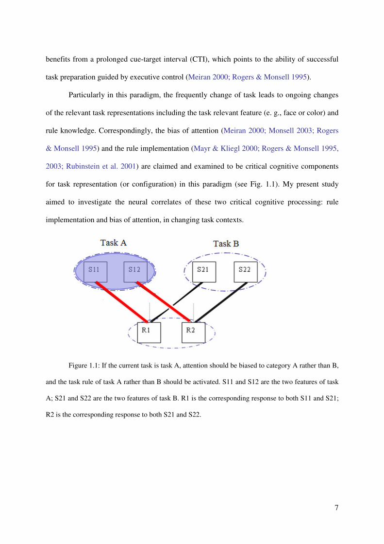
for task representation (or configuration) in this paradigm (see Fig. 1.1). My present study
aimed to investigate the neural correlates of these two critical cognitive processing: rule
implementation and bias of attention, in changing task contexts.
Figure 1.1: If the current task is task A, attention should be biased to category A rather than B,
and the task rule of task A rather than B should be activated. S11 and S12 are the two features of task
A; S21 and S22 are the two features of task B. R1 is the corresponding response to both S11 and S21;
R2 is the corresponding response to both S21 and S22.
8
1.2 Research questions
Presumably, the activation of such attentional bias and rule implementation are parts
of a more general mechanism of task preparation, which includes the prior activation of neural
modules necessary for behavior and starts long before the manifestation of the overt behavior
(Gollwitzer & Sheeran 2006; Monsell 2003; Rubinstein et al. 2001;).
A number of studies were conducted to investigate the neural basis of the broader
mechanisms of task preparation (Brass & von Cramon 2002, 2004; Gruber et al. 2006; Luks
et al. 2002; MacDonald et al. 2000; Sohn et al. 2000). These studies on task preparation
showed a large scare of cortical regions including the lateral prefrontal cortex (LPFC), the
medial frontal cortex (MeFC), pre-motor regions, and parietal regions to be part of a network
that seems to come into play when participants prepare for an upcoming sensory-motor task.
On one hand, intraparietal cortex and superior frontal cortex are identified to be
critical for attentional control by several fMRI studies (e g., Corbetta et al. 2000, 2002, 2005;
Serences et al., 2001, 2004; Serences & Yantis 2007). Notice that, the foci of attentional
control locate within the networks of task preparation. This finding fits well with the
hypothesis that attentional bias is part of the general task preparation. On the other hand, the
neural mechanisms of task rule activation in changing task contexts are still not clear yet.
Therefore the first fMRI experiment of this study was conducted mainly to investigate
whether regions specific to the mechanisms of task rule implementation can be found
(presumably) within the task preparation networks.
Again for the bias of attention, it was assumed that the top-down control biases the
“bottom-up” sensory processing via amplifying the neural representation in some
corresponding feature specific posterior regions (e g., Desimone & Duncan, 1995; Kastner &
Ungerleider, 2000). And this opinion was supported by several preceding findings (e g.,

9
Corbetta et al. 2005; Serences et al., 2001, 2004; Serences & Yantis 2007). However, if the
preparatory attentional control also biases sensory processing through a similar way is still
lacking of investigation. Therefore the second fMRI study was conducted mainly to
investigate if preparatory attentional control could bias sensory processing via the modulation
to the posterior feature specific regions’ activity.
1.3 Method of isolating cue period and target period neural activity
This study aimed to isolate the neural correlates for task rule implementation from the
brain network of general task preparation, and to find the modulation in the posterior brain
regions’ activity in the task preparation period. Considering the processing in the target period
(during task execution, i.e. after the target was presented) are temporally close to the
processing in the cue period (during task preparation, i.e. in the interval between the
presenting of cue and target). Particular method is needed for the adopted methodology,
functional magnetic resonance image (fMRI), to dissociate the task preparation-related and
task execution-related neural activity. . Studies concerned with understanding the neural
mechanisms of task preparation have often used the task-cuing paradigm in combination with
an event-related fMRI design (Brass & von Cramon 2002, 2004; Gruber et al. 2006; Luks et
al. 2002; Sohn et al. 2000). Earlier neuroimaging studies investigated preparation-related
activity by analyzing the fMRI activity during very long CTIs (e.g., up to 12.5 s) and,
therefore, their findings may have been compromised by memory load confounds (Luks et al.
2002; MacDonald et al. 2000; Sohn et al. 2000).
More recent studies isolated task preparation-related activity by measuring neural
activity separately for so-called cue-only trials, cue-target trials, and null-events in the task-
cuing paradigm (Brass & von Cramon 2002, 2004). While on cue-only trials, there is no target
10
following the cue, on cue-target trials a target requires the execution of the task, and the null-
event represents a baseline condition without any cue and target information. Because
participants do not know in advance whether or not a target will follow the cue, they have to
prepare for task execution on every type of trial, both on cue-only and on cue-target trials (see
also Corbetta et al. 2000; Weissman et al. 2005). This allows for a measurement of
preparation-related activity during the processing of cue-only trials.
With the use of cue-only trial design, Brass and von Cramon (2002) contrasted
activation in cue-only trials and in null-events and found a fronto-parietal network to be
related to task preparation. In particular, this network included regions in the dorsolateral
prefrontal cortex (DLPFC), e.g., near the inferior frontal junction point (IFJ), regions
surrounding the intraparietal sulcus (IPS), in the dorsal premotor cortex and in the pre-
supplementary motor area (pre-SMA) of the medial frontal gyrus.
The fMRI Experiments 1 and 2 in the present study adopted the cue-only trial design
and the task-cuing paradigm in order to find the neural correlates of task rule implementation
and the possible attentional modulation to feature specific posterior regions respectively.
In particular, the research aim of fMRI Experiment 1 was achieved by the applying of
a new kind of cue called “rule-cue”. In the rule-cue display, not only the task type information
but also the rule of the current task ( a set of S-R associations) were explicitly presented. Note
that, before the formal experiment, participants have received enough practice which can
make sure the task rules have been remembered well. So I expected the participants did not
use the explicit rule information in the rule-cue to learn the task rule (e g., consolidate their
memory of task rule or obtain the task rule better), rather they used this information to
implement the task rule. This hypothesis was supported by a behavioral experiment,
Experiment 3 of this study. It will be introduced after the description of the two fMRI
experiments.
11
Since the rule-cue can implement the task rule more efficiently than the task-cue
which supplies explicit task type information only, the neural activities in these two cue
conditions were compared in Experiment 1 to identify the neural correlate of rule
implementation.
1.4 Introduction of fMRI Experiment 1:
The neural mechanisms of task rule implementation
According to Miller (2001) rule knowledge is stored in the prefrontal working memory
and it contains knowledge about the stimuli, the behavioral responses, and about the context
of the situations in which a particular rule has to be applied. As introduced above, the
activation of such rule representations could be part of a more general mechanism of task
preparation, which refers to a large scare frontal-parietal network. And the aim of this present
experiment is to further find the unequivocal neural correlates of task rule implementation.
Although the findings provided a number of valuable insights into the functional
neuroanatomy of task preparation, they are not unequivocal regarding the neural correlates of
task rule activation. This is so because a task-cuing paradigm like that in Brass and von
Cramon (2002) does not permit the mechanisms of activating the specific task rules to be
distinguished from rather general task preparation (see also Ruge et al. 2009). The
presentation of the cue informed participants about the task they had to perform later upon the
presentation of the target. If the time was sufficient and the participants intended to do so,
they could either activate the current task rule or, alternatively, they could wait with the
activation of the task rule until the presentation of the imperative target. Thus, depending on
participants’ strategy, either to prepare the task rule early upon the presentation of the cue or
12
only later upon the presentation of the target, the point in time when the task rule was
activated was not sufficiently controlled.
For the present study, a so-called rule-cue was designed, in order to investigate rule-
related activation in a task-cuing paradigm. This rule-cue differs from the task-cue in earlier
studies because it conveys explicit information about the type of task and, in addition, about
the corresponding task rule (e. g., in a color discrimination task use left key for red and right
key for yellow color). Thus, while a task-cue (the sort of cue used in earlier studies) conveys
only general information about which task to perform, the rule-cue provides also specific
information about task rules, e.g. the stimulus-response (S-R) mapping, on the upcoming trial
(task-and-rule information). By administering the rule-cue randomly mixed with task-cues,
this experiment aimed to trigger processes related to the activation of the specific task rules
during task processing.
In particular, Chinese participants attended Experiment 1. They were presented with
either a color or a gender discrimination task, with the particular task specified by the
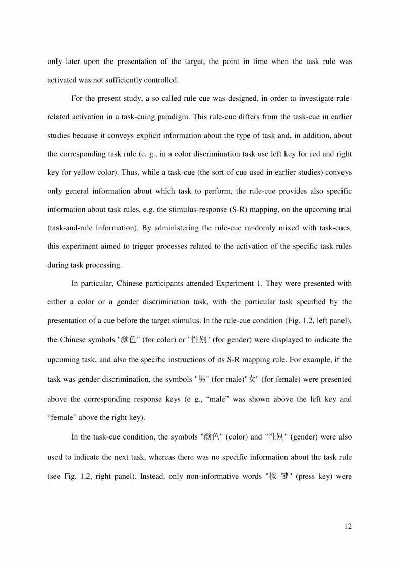
presentation of a cue before the target stimulus. In the rule-cue condition (Fig. 1.2, left panel),
the Chinese symbols "颜色" (for color) or "性别" (for gender) were displayed to indicate the
upcoming task, and also the specific instructions of its S-R mapping rule. For example, if the
task was gender discrimination, the symbols "男" (for male)"女" (for female) were presented
above the corresponding response keys (e g., “male” was shown above the left key and
“female” above the right key).
In the task-cue condition, the symbols "颜色" (color) and "性别" (gender) were also
used to indicate the next task, whereas there was no specific information about the task rule
(see Fig. 1.2, right panel). Instead, only non-informative words "按 键" (press key) were
13
presented below the task-cues, in order to make the cue display similar to that in the rule-cue
condition.
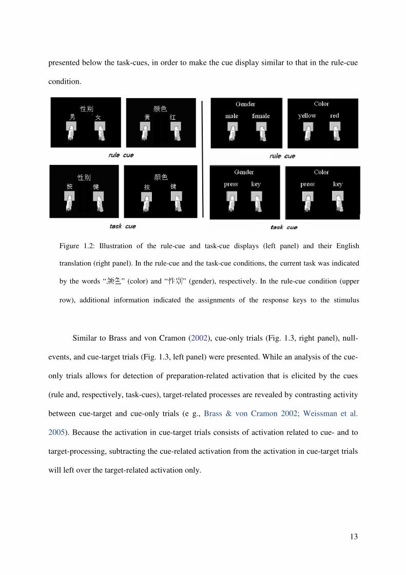
Similar to Brass and von Cramon (2002), cue-only trials (Fig. 1.3, right panel), null-
events, and cue-target trials (Fig. 1.3, left panel) were presented. While an analysis of the cue-
only trials allows for detection of preparation-related activation that is elicited by the cues
(rule and, respectively, task-cues), target-related processes are revealed by contrasting activity
between cue-target and cue-only trials (e g., Brass & von Cramon 2002; Weissman et al.
2005). Because the activation in cue-target trials consists of activation related to cue- and to
target-processing, subtracting the cue-related activation from the activation in cue-target trials
will left over the target-related activation only.
Figure 1.2: Illustration of the rule-cue and task-cue displays (left panel) and their English
translation (right panel). In the rule-cue and the task-cue conditions, the current task was indicated
by the words “颜色” (color) and “性别” (gender), respectively. In the rule-cue condition (upper
row), additional information indicated the assignments of the response keys to the stimulus
14
The distinction between rule-cues and task-cues permits rule-related neural activity to
be analyzed in the following manner. First of all, the experiment expects a significant
performance benefit from the presentation of rule-cues compared to task-cues, and rule-cues
can evoke stronger cue-related activation than task-cues, specifically, on cue-only trials. The
reason for the latter hypothesis is that, in the rule-cue condition, the cue provides explicit rule
information and this information may be activated with the cue presentation. By contrast, in
the task-cue condition, participants may postpone at least part of the rule activation processes
until later, for example, up to the time where the target is expected to appear. And even if
Figure 1.3 : Illustration of the task situation. Upper part: Left panel shows a cue-target trial
(example for the gender discrimination task). Right panel shows a cue-only trial (example for
gender discrimination task). The lower part of the figure represents the cue displays and their
English translation. The cue could either be a rule-cue (left) or a task-cue (right), (for details see
15
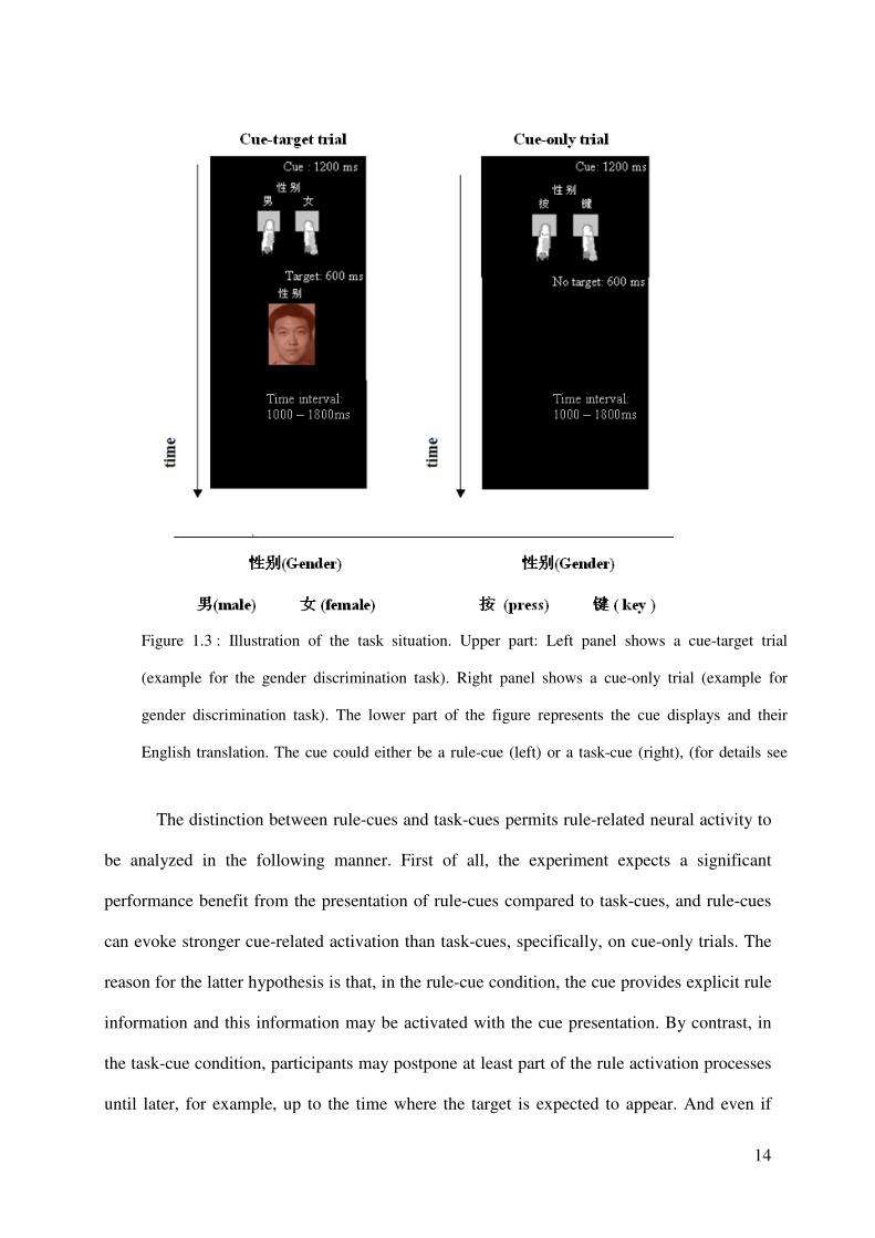
activation of the rule is not postponed, it may be less effective because the cue provides no
explicit information as to the precise task rule. Consequently, rule-related activation should be
manifest during the preparation period on cue-only trials in terms of an increased amount of
activity under the rule-cue, compared to the task-cue, condition (see the orange dotted oval in
Fig. 1.4).
Figure 1.4: The expected activation pattern in the rule-related regions
The converse pattern (of activation in rule-cue and task-cue conditions) may be
expected when considering the rule-related activation that emerges after target presentation on
cue-target trials, i.e. during task execution. It is reasonable to assume that, if participants
failed to activate the (complete) task rule right upon cue presentation, they must activate the
necessary S-R mapping rule following target presentation (Gruber et al., 2006). This would be
consistent with Gruber et al. (2006) who analyzed the neural activity under conditions of short
16
vs. long cue-target intervals (CTIs) in a task-cuing paradigm. While the time for preparing the
upcoming task was sufficient after cue presentation with long CTIs, it was insufficient with
short CTIs. The latter led to the postponement of (at least parts of) the preparation processes
until after target presentation, as indicated by an increased amount of neural activity in
preparation-related brain regions under conditions of short compared to the long CTIs (Gruber
et al. 2006; see also Brass & von Cramon 2002). In analogy to these findings, postponed rule
activation in the task-cue condition compared to the rule-cue condition was expected. This
should lead to greater activation in rule-related brain regions under task-cue, compared to the
rule-cue condition upon target presentation on cue-target trials (see the blue dotted oval in
Fig. 1.4).
In sum, the expectation include stronger neural activity related to task rule activation
in the rule-cue compared to the task-cue condition during the preparation period, and stronger
activity in rule activation-related regions during task execution in the task-cue compared to
the rule-cue condition. The common neural substrate in these two comparisons, thus,
represents those brain regions which are important for the mechanism of task rule activation
either in the task preparation or in the execution period. A method called conjunction analysis
is suitable to find the common activation of different comparisons. This experiment adopted
the conjunction analysis to find the common neural substrate about the corresponding
contrasts (i.e., preparation period (rule-cue minus task-cue), and execution period (task-cue
minus rule-cue)).

17
1.5 Introduction of fMRI Experiment 2
The preparatory attentional modulation to the posterior brain regions
Several studies (e g., Dehaene et al. 2003; Kanwisher et al. 1997; Serences et al., 2001;
Tootell et al., 1995; Zeki et al., 1991) have shown that there are several stimulus-specific
posterior brain regions who are sensitive to the presenting of certain specific stimulus (e g.,
face, or house, or motion, or number) rather than other stimulus.
In spite of the physical power of stimulus, the response of such posterior brain regions
to their specific stimuli can be modulated by attention. This had been shown by a study of
Serence et al. (2004). In that study, two spatially overlapped streams of face stimulus and
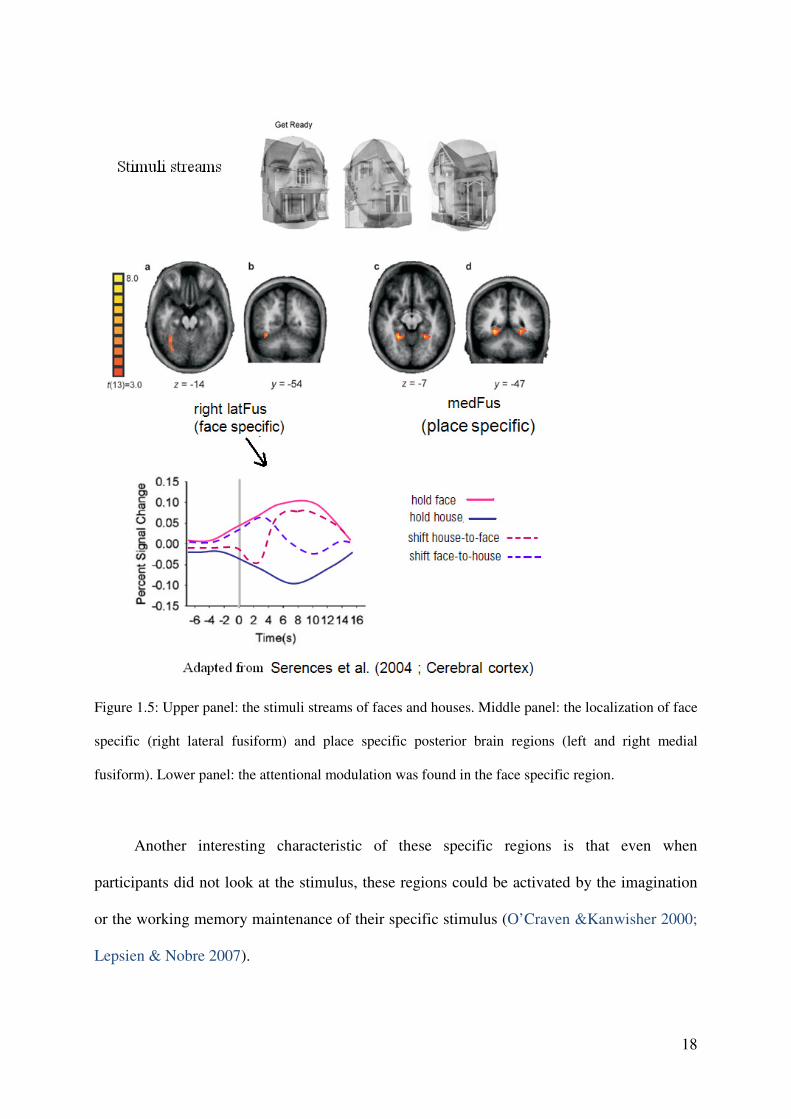
house stimulus were displayed in the screen (see Fig. 1.5, upper panel). A verbal command
was given to instruct participants attending to either houses or faces and maintaining attention
on the currently attended object stream until the next command. Attentional modulation was
found: an increased response in the face specific region (right lateral fusiform) when
participants attended to faces (see Fig. 1.5, lower panel), whereas an increased response in the
place specific region (bilateral median fusiform) when participants attended to houses
(Serences et al. 2004).
18
Figure 1.5: Upper panel: the stimuli streams of faces and houses. Middle panel: the localization of face
specific (right lateral fusiform) and place specific posterior brain regions (left and right medial
fusiform). Lower panel: the attentional modulation was found in the face specific region.
Another interesting characteristic of these specific regions is that even when
participants did not look at the stimulus, these regions could be activated by the imagination
or the working memory maintenance of their specific stimulus (O’Craven &Kanwisher 2000;
Lepsien & Nobre 2007).

19
In the cued task switch paradigm, after the presentation of the cue, the participants tend to
prepare the task in advance in order to improve their performance; the attentional control was
assumed to be an important component of such task preparation (Meiran 2000; Monsell 2003;
Rogers & Monsell 1995). Considering the fact that region could activate without the explicit
presentation of the stimuli, it seems plausible to assume that the preparatory attentional
control may lead to a modulation to the posterior specific regions. However, the investigation
for this preparatory attentional modulation and its support evidences are rare.
One relevant finding supporting this assumption was observed by Wylie et al.’s study
(2006). Here, participants responded to a colored rotating bar, either discriminated the color
of the bar or judged the bar’s rotating speed is slow or fast. Cue was given to inform about the
current task. In the preparation period, modulation was found in the color specific region but
not in the motion specific region. This finding at least partially supports the hypothesis which
implies that attentional bias can pre-activate the task relevant representations in the posterior
specific regions. For the silent of motion specific region in speed task preparation period, it
was explained the participants did not efficiently prepare the speed task. One of the possible
reason might be the motion discrimination task dose not encourage preparation. For example,
Shulman et al (2002a) either did not find motion-selective activity in middle temporal area
(MT, specific for motion) when subjects were verbally cued to attend to motion (Shulman et
al., 2002a).
Moreover, the factor of balance of task difficult was considered, It has been proposed that,
in the changing task context, if the two tasks are not balanced in difficulty, in order to perform
the non-dominant task well, participants may have the tendency to inhibit the dominant task.
This inhibition could persist into next trial (Allport et al., 1994). And such inhibition
20
processing might depress the activity in the posterior brain region, which does not fit with the
aim of this experiment.
Thus in this present study, in order to encourage efficient task preparation in both tasks,
two tasks with balanced difficulty were planned to use, and no motion related task was
considered.
In the current experiment, a gender and a number discrimination task were adopted
(male/female; bigger/smaller than five). The corresponding regions of interest (ROIs) are the
fusiform face area (FFA) (Kanwisher et al. 1997) and a region in the horizontal segment of
the intra parietal sulcus (IPS). Note that, the IPS processes multitude information in its
different parts, but the region in the horizontal segment is specific to the representation of
number. It is systematically activated whenever numbers are manipulated, independently of
number notation (Dehaene et al. 2003). In the present study, this region was called IPS
number region (IPSnum).
A preliminary behavioral experiment was conducted and showed that the selected two
tasks are not significantly different in difficulty thus efficient task preparation should take
place for both of these two tasks (see supplementary material 1), therefore pre-activation was
expected in the FFA if the task was gender discrimination and it was expected in the IPSnum
if the task was number discrimination.
This experiment tried to find the evidence for the hypothesis that the preparatory
attentional control can bias sensory processing via amplifying the activity of the task-relevant
stimulus-specific brain region. A cue-only trial design was used in fMRI Experiment 2 to
isolate the neural activity in task preparation period. The modified cued task switching with
the use of rule-cue was adopted. Both the rule-cue and task-cue provide the task type
information which allows preparatory attentional biasing, therefore the corresponding
modulation to the stimulus specific region was expected in both of the two cue conditions:
21
i.e., the increased activity in FFA in the preparation period of face task and the increased
activity in IPSnum in the preparation period of number task.
Contrary to Experiment 1, German participants attended this Experiment 2. There
might be cultural differences in strategy use and functional neuroanatomy of task rule
implementation. Therefore, it is interesting to check if the finding in Experiment 1 could be
replicated. This was a further aim of the present Experiment 2.
Moreover the present Experiment 2 can also check if the presentation of task rule
information is accompanied by activity changes in posterior sensory brain regions (larger
activity in the face specific region in the preparation period if the task is indicated by a rule-
cue than a task-cue; and similar or number task) or not.
In particular, in the main task, participants were presented with either a number or a
gender task, with the particular task specified by the presentation of a cue before the target
stimulus. In the rule-cue condition, the German symbols "ANZAHL" (for number) or
"GESICHT" (for face) were displayed to indicate the upcoming task, and also the specific
instructions of its S-R mapping rule. For example, if the task was gender discrimination, the
symbols "MANN" (for male)"FRAU" (for female) were presented above the corresponding
response keys (e g., “male” was shown above the left key and “female” above the right key).
In the task-cue condition, the symbols "ANZAHL" (number) and "GESICHT"
(gender) were also used to indicate the next task, whereas there was no specific information
about the task rule. Instead, only non-informative words “PRESS" (press key) were presented
below the task-cues, in order to make the cue display similar to that in the rule-cue condition.
Similar to fMRI Experiment 1, cue-only trials, null-events, and cue-target trials were
presented. While an analysis of the cue-only trials allows for detection of preparation-related
activation that is elicited by the cues (rule and, respectively, task-cues) in the face or number
22
task, target-related processes are revealed by contrasting activity between cue-target and cue-
only trials.
Besides of the main task, a localization task was conduct to identify the regions of
interest (ROIs) (i.e., face specific and number specific region) for each individual subject. The
activity of the face and number specific regions in the main task were analyzed in order to
find the preparatory modulation of attention. i.e., the increased activity in face specific region
in the preparation period of face task and the increased activity in number specific region in
the preparation period of number task.
In sum, this experiment aimed to find the evidence for the hypothesis that the
preparatory attentional control can bias sensory processing via amplifying the activity of the
task-relevant stimulus-specific brain region. For this aim, gender discrimination and number
discrimination tasks were selected because they both refer to specific posterior brain region
(i.e., FFA and IPSnum) and they are balanced in task difficulty (good for efficient preparation
in both tasks).
1.6 Introduction of Experiment 3:
The rule-cue was expected to activate the task rule more sufficiently in the preparation
period. However, one might argue that the rule-cue could facilitate the rule acquisition as well
as rule implementation. Therefore one might expect that the participants may also learn the
task rule from the displays of the rule-cues whereas no such learning processing occurred in
the task-cue condition because the task-cue displays supply no rule information. If this is
really the case, the rule-related finding in Experiment 1 and 2, which was resulted by the
comparison between neural activity in the rule-cue and task-cue conditions, might be
confounded by some learning effect.
23
I suggest that such learning of task rule from the rule-cue displays only take place when
the rules have not yet been obtained well. Human, unlike the monkey or chimpanzee, the
process of achieving a good acquisition of task rules was expected to be not time consuming,
only few trials should be enough for obtaining simple rule sets in the present study. Therefore
the rule-related activation found in Exp 1 and 2 should not confounded with learning effect,
because participants received enough practice before the formal experiment. In order to test
this hypothesis, Experiment 3 was conducted. Considering Experiment 1 adopted Chinese
participants and Experiment 2 adopted German participants, both Chinese and German
participants were adopted in Experiment 3. I expect the hypothesis works for both Chinese
and German participants.
The setting of rule transition factor
A task environment with unstable task rule was designed in this behavioral experiment.
Participants have to perform either gender discrimination or number discrimination (like in
the normal task-cuing paradigm); however the task rules are unstable. The task rule of face
discrimination could either be male-left, female-right or the reversed; the task rule of number
discrimination could either be bigger than five-right, smaller than five-left or the reversed.
Before every block which contains 10 trials, task rules (for face and number task) are
displayed on the screen for a relatively long period (i.e. 5.5s). Participants were instructed in
advance these rules were the rules for the following block and they had to remember the rules.
If the current task rules are the same with the rules in the preceding block, the current block is
a rule repetition block. Otherwise, the current block is a rule switch block. Moreover, within a
block, each task could either be guided by rule-cue or task-cue, thus resulting rule-cue block
or task-cue block.
24
Note that, the re-acquisition of rules is needed in the first few trials of rule switch block.
After the 5.5 seconds presenting of the rules display, participants can explicitly learn the task
rules from the cue displays in the rule-cue block but not in the task-cue block. ,
correspondently, part of the rule cue benefit should come from the rule cue facilitation to the
new rule’s acquisition. In addition, the rule cue should also refer to a better rule
implementation process than the task cue; this causes another part of rule cue benefit.
Whereas in the rule repetition block, the rule sets have been learned well already, thus no
spaces for the rule cue facilitation to the rule acquisition processing anymore. But the rule
cue facilitation to the rule implementation still exists; it produces rule cue benefit per se.
Taking them together, for the first few trials of block, because the rule cue benefit was
contributed by rule cue facilitation to rule acquisition and rule implementation both in the rule
switch block, whereas produced by rule cue facilitation to rule implementation only in the
rule repetition block. More rule cue benefit was expected in the rule switch block than in the
rule repetition block.
However, for the rest trials in a block, even it is a rule switch block, the task rules should
been acquired well already. Therefore the rule cue benefit should be produced by rule cue
facilitation to rule implementation only no matter the block is a rule repetition or rule switch
block. Thus for the rest trials of blocks, there should be identical rule cue benefit in the rule
switch and rule repetition block.
In sum, for the first few trials of blocks, more rule cue benefit was expected in the rule
switch blocks than in the rule repetition blocks. Whereas for the rest trials of blocks, identical
rule cue benefit was expected in the rule switch and rule repetition blocks.
25
CHAPTER 2
fMRI Experiment 1
2.1 Research aim and expectations
To isolate the neural correlates for task rule activation from those related to general
task preparation a new kind of cue called “rule-cue” was created and applied in the cued task
switching paradigm. While the task-cue represents merely information about the task type, the
rule-cue represents information about the task type and, in addition, explicit information about
the task rule (i.e. the set of S-R correspondences). The rule-cue was expected to activate the
task rule more sufficiently in the preparation period (main contrast 1: cue period (rule-cue
mins task-cue)), whereas in the task-cue condition, part of the task rule activation was
expected to be postponed into the task execution period, i.e. after the target was presented
(main contrast 2: target period (task-cue mins rule-cue)). The common neural substrate in
these two contrasts, thus, should represent those brain regions which are important for the
mechanism of task rule activation either in the task preparation or in the execution period.
26
2.2 Method
Subjects
Fifteen right-handed, healthy students of Peking University (recruited by
advertisement in the campus Bulletin Board System) participated in the study. Six participants
were female; participants’ ages ranged between 20 and 26 years, and all had normal or
corrected-to-normal vision. Prior the fMRI scanning session, they gave informed consent
about the investigation according to the Helsinki guidelines and the approval of the Academic
Committee of the Department of Psychology, Peking University. Participants were paid 50
yuan (about 5 Euros) for their service.
One participant’s response error rate was more than 20 %. Hence this participant’s
behavioral and fMRI data were removed from the data set. There was also a loss of the
behavioral data from one participant, due to data recording error. Thus, ultimately, 14
participants’ image data sets and thirteen participants’ behavioral data sets were available for
analysis.
Design
Paradigm and procedure. The task to be performed by the participants was either color
discrimination or gender discrimination. Each trial began with the presentation of a cue for a
fixed duration of 1200 ms, which could either be a rule-cue or a task-cue (see Fig. 1.1). Both
cues displayed an instruction for the upcoming task; however, a precise instruction about the
required task rule was provided only in the rule-cue condition (for more details, see Fig. 1.2).
On cue-only trials (n = 160 trials, of which 80 presented a rule-cue and 80 a task-cue), there
was no target following the cue offset, but only a black screen that lasted for 600 ms, and
there was no need for participants to make a response (Fig. 1.1, right panel).

27
In contrast, on cue-target trials (n = 280, of which 140 presented a rule-cue and 140 a
task-cue), the cue was followed by a colored face picture that was presented for 600 ms;
during this period, the task-cue instruction remained visible on the screen (above the target
picture) by presenting the words ‘gender’ or ‘color”, so as to reduce participants’ working
memory load for maintaining the task goal in the two conditions. Importantly, the information
presented during the execution period concerned only the task information and not the rule
information because the symbols ‘press key’ and the symbols illustrating the rule information
were not presented during the execution period (see Fig. 1.1, left panel). Participants were to
respond to either the color or the gender of the face depicted in the target display, depending
on the instruction of cue. Participants made two-alternative forced-choice responses using
either their left or their thumbs, with response sets counterbalanced across participants. After
the offset of the target picture, a black screen was presented for a variable interval of 1000,
1200, 1400, 1600, or 1800 ms. The next trial could then either be a cue-target or a cue-only
trial, that is an ‘event trial’, or a ‘null trial’ (n = 110) in which there was neither a cue nor a
target event. Together with the duration of the null trials, which were of the same duration as
the task trials, the interval between two event trials (the interval between the disappearance
(offset) of the target in the present trial and the appearance (onset) of the next cue) resulted in
2200 ms on average.
Task conditions and trial types. The present study used a 2 x 2 event-related fMRI
design. The first factor was cue type: the cue could be either a rule-cue or a task-cue. The
second factor was task transition: the task was either repeated or switched relative to the
preceding trial. Based on the instruction cue presented prior to the target, participants were
required to distinguish either the color or the gender of the face pictures. If the current task
was different from the preceding one, the current trial was classified as a switch trial; if the
current task was identical with the previous one, the current trial was classified as a repetition
28
trial. This factor was examined because rule activation (or retrieval) was hypothesized to
differ between task repetition trials and switch trials (Mayr & Kliegl 2000; Rogers & Monsell
1995, 2003; Rubinstein et al. 2001). That is, this factor was introduced to examine whether or
not preparation for a switched, compared to a repeated, task leads to a modulation of the task
rule activation.
Each one of the four conditions (rule / task-cue x task switch / repetition) consisted of
40 cue-only trials and 70 cue-target trials. In sum, there were 440 event trials, the order of
which was unpredictable for the participants. In addition, the event trials were randomly
intermixed with 110 null trials in which only a black screen was shown. The length of a null
trial varied from 2800 ms to 3600 ms, which was similar to the length of the other (task)
trials.
For each condition, the cue-related activation can be assessed by measuring the
activation on the cue-only trials, whereas target-related activation can be assessed by
calculating the contrast between the activation in corresponding cue-target minus cue-only
trials.
Stimulus and response conditions. On cue-only trials, only a black screen (i.e., no
target) was presented after the presentation of the cue and there was need to respond. On cue-
target trials, the target stimulus was a colored face picture. In order to create colored face
pictures we merged each one of the original black-white-colored face pictures (2 males and 2
females) with same-sized, faded red rectangles (RGB 187- 124- 106) and yellow rectangles
(RGB 179- 155- 111) with Photoshop software. As a result eight colored face pictures were
created, which we used as target stimuli: two yellow male faces, two red male faces, two
yellow female faces, and two red female faces (with the same face presented in either red or
yellow on different trials). Participants were informed by the cue to respond to either the color

29
or the gender of the face. The stimuli (cue and target stimuli) were located on a black
background in the centre of the screen and subtanded 5 degrees of visual angle.
Participants used their left and right thumbs for response. They were instructed
to respond as fast and as accurately as possible. For half the participants, the S-R mapping
rule was male-left, female-right and yellow-left, red-right. This was reversed for the other
half: female-left, male-right and red-left, yellow-right.
fMRI measurement
Imaging was performed with a SIEMENS TRIO 3-Tesla scanner at the Beijing MRI
Center for Brain Research. T2*-weighted echo-planar images (EPI) with blood oxygenation
level-dependent contrast were acquired (TR = 1500 ms, TE = 30 ms, flip angle = 90°, voxel
size = 3.4 x 3.4 x 5 mm3, matrix size = 64 x 64 voxels). Twenty six axial slices (thickness = 4
mm, spacing = 1 mm) were acquired parallel to the AC-PC plane, covering the whole cortex
and part of the cerebellum. The order of acquisition of the slices was interleaved. The first
five volumes (dummy volumes) were discarded because of possible instabilities in the
magnetic field at the beginning of a scan. Stimuli were displayed on a back-projection screen
mounted in the bore of the magnet behind the participant’s head by using an LCD projector.
Participants viewed the screen by wearing mirror glasses.
fMRI data analysis
Preprocessing. Preprocessing of the functional images was carried out using SPM2
(Wellcome Department of Cognitive Neurology, London, UK). Images were interpolated in
time (temporal realignment to the middle slice). In addition, they were spatially realigned to
the first volume for head movement correction, unwrapped, and then normalized to the
standard SPM2 EPI template in MNI space (resampled to 2 x 2 x 2 mm3 isotropic resolution)
30
with default normalization estimation. The data were then smoothed with a Gaussian kernel of
8 mm full-width half-maximum to account for inter-subject anatomical variability.
Then the image data were modeled by applying a general linear model (Friston et al.
1995). In event-related single-subject analyses, the 4 cue-only and the 4 cue-target conditions
were modeled as separate volumes (resulting from the factorial combination of the two cue
type (rule-cue vs. task-cue) and the types of task transition (task switch vs. task repetition).
Additionally, all error trials were selected to form an error trial volume. The resulting nine
volumes were convolved with the hemodynamic response function (HRF), and then beta
values of these regressors were estimated according to the ordinary least-squares (OLS)
method.
Whole-brain analyses. For group statistics, one-sample t-tests of contrast maps across
subjects (random-effects model treating subjects as a random variable) were computed to
indicate whether observed differences between conditions were significantly different from
zero.
In particular, two main contrasts were calculated: Contrast 1: For cue-only trials, rule-
cue minus task-cue trials, intended to isolate extra activation for a rule-cue. Contrast 2: (cue-
target trials minus cue-only trials for task-cues) minus (cue-target trials minus cue-only trials
for rule-cues), intended to isolate the extra activation related to the target processing when the
cue did not specify the rule. In a subsequent conjunction analysis, SPM5 (Nichols et al., 2005)
was used to locate the common task rule-related activation between these two main contrasts.
The way in which the remaining statistical contrasts were calculated is detailed in the
Results section. Unless stated otherwise, for one-sample t-tests, we used a statistical threshold
of p < 0.001, uncorrected, covering at least 10 contiguous voxels. We also checked all
reported activation foci with a small volume correction procedure (10 mm sphere centred at
the voxel with local maximum activation). If not otherwise noted, then the reported foci prove
31
significant at a threshold of p < 0.05 (small-volume corrected on both the voxel and the
cluster level). For the conjunction analysis, the conjunction hypothesis is "activated in
Contrast 1AND Contrast 2", then the conjunction null hypothesis is: (not activated in Contrast
1) OR (not activated in Contrast 2). The statistical threshold was p < 0.005, uncorrected, again
spanning at least 10 contiguous voxels .
2.3 Results
Behavioral results
Figure 2.1 presents group means of the RTs (left panel) and error rates (right panel) as
a function of task transition, for the two types of cue. Mean RTs and error rates were
submitted to a 2 x 2 repeated-measures ANOVA with the factors task transition and cue type.
RTs were significantly faster in the rule-cue than in the task-cue condition (main effect of cue
type, F (1,12) = 6.71, p < 0.05), which indicates that participants effectively utilized the rule-
cue information during the preparation period following cue presentation. The RT advantage
for rule-cue compared to the task-cue presentation (i.e., the ‘behavioral rule-cue effect’) was
17 ms. In addition, RTs were significantly slower for task switch than for task repetition trials
(main effect of task transition, F(1,12) = 12.96, p < 0.005), with switch costs amounting to 25
ms. With mean switch costs of 24 and 27 ms in rule-cue and task-cues conditions,
respectively, the interaction effect between cue type and task transition was not significant
(F(1, 12) = 0.114, p > 0.7).
The error rate ANOVA revealed a significant main effect of task transition (F (1, 12) =
60.91, p < 0.0001): more errors were made on task switch than on task repetition trials.
Additionally, a significant interaction between cue type and task transition was obtained
(F(1,12) = 8.84, p < 0.05). Further analyses with separate t-tests revealed elevated error rates

32
in switch compared to repetition trials in the rule-cue and task-cue conditions (both ts (12) >
4.00, both ps < 0.005), and larger switch costs (error rate switch – error rate repetition) in the
task-cue (error rate = 6.6 %) compared to the rule-cue condition (error rate = 3.8 %) (t (12) =
2.97, p < 0.05). Thus, as with the RT data, the error data indicated that participants’
performance benefited from the presentation of the rule-cue as compared to the task-cue. This
benefit was especially pronounced in conditions in which participants had to switch between
the tasks as revealed by the increased error rate in the switch compared to the repetition
condition.
Figure 2.1: Reaction time (RT) and error rates as function of task transition and cue type.
Imaging results
Cue-related activation in rule-cue and task-cue conditions
To identify the cue-related activation, we calculated the main effect for the cue-only
trials separately for the rule-cue and task-cue conditions, by fitting the empirical fMRI data to
the hemodynamic response function (HRF) described above. The resulting beta values are
33
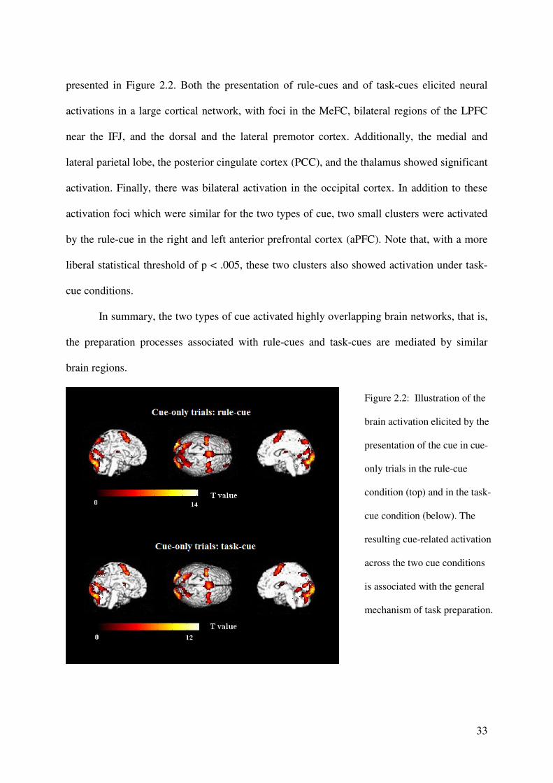
presented in Figure 2.2. Both the presentation of rule-cues and of task-cues elicited neural
activations in a large cortical network, with foci in the MeFC, bilateral regions of the LPFC
near the IFJ, and the dorsal and the lateral premotor cortex. Additionally, the medial and
lateral parietal lobe, the posterior cingulate cortex (PCC), and the thalamus showed significant
activation. Finally, there was bilateral activation in the occipital cortex. In addition to these
activation foci which were similar for the two types of cue, two small clusters were activated
by the rule-cue in the right and left anterior prefrontal cortex (aPFC). Note that, with a more
liberal statistical threshold of p < .005, these two clusters also showed activation under task-
cue conditions.
In summary, the two types of cue activated highly overlapping brain networks, that is,
the preparation processes associated with rule-cues and task-cues are mediated by similar
brain regions.
Figure 2.2: Illustration of the
brain activation elicited by the
presentation of the cue in cue-
only trials in the rule-cue
condition (top) and in the task-
cue condition (below). The
resulting cue-related activation
across the two cue conditions
is associated with the general
mechanism of task preparation.
34
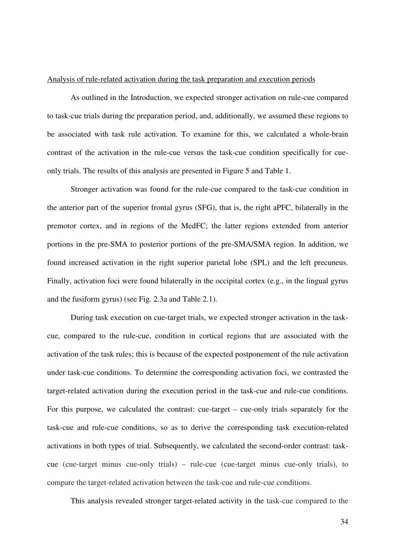
Analysis of rule-related activation during the task preparation and execution periods
As outlined in the Introduction, we expected stronger activation on rule-cue compared
to task-cue trials during the preparation period, and, additionally, we assumed these regions to
be associated with task rule activation. To examine for this, we calculated a whole-brain
contrast of the activation in the rule-cue versus the task-cue condition specifically for cue-
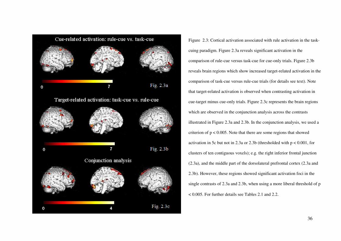
only trials. The results of this analysis are presented in Figure 5 and Table 1.
Stronger activation was found for the rule-cue compared to the task-cue condition in
the anterior part of the superior frontal gyrus (SFG), that is, the right aPFC, bilaterally in the
premotor cortex, and in regions of the MedFC; the latter regions extended from anterior
portions in the pre-SMA to posterior portions of the pre-SMA/SMA region. In addition, we
found increased activation in the right superior parietal lobe (SPL) and the left precuneus.
Finally, activation foci were found bilaterally in the occipital cortex (e.g., in the lingual gyrus
and the fusiform gyrus) (see Fig. 2.3a and Table 2.1).
During task execution on cue-target trials, we expected stronger activation in the task-
cue, compared to the rule-cue, condition in cortical regions that are associated with the
activation of the task rules; this is because of the expected postponement of the rule activation
under task-cue conditions. To determine the corresponding activation foci, we contrasted the
target-related activation during the execution period in the task-cue and rule-cue conditions.
For this purpose, we calculated the contrast: cue-target – cue-only trials separately for the
task-cue and rule-cue conditions, so as to derive the corresponding task execution-related
activations in both types of trial. Subsequently, we calculated the second-order contrast: task-
cue (cue-target minus cue-only trials) – rule-cue (cue-target minus cue-only trials), to
compare the target-related activation between the task-cue and rule-cue conditions.
This analysis revealed stronger target-related activity in the task-cue compared to the

35
rule-cue condition in most regions that had proved to be rule-related during the preparation
period in the above analysis (see section Cue-related activation). In particular, these regions
were the right anterior part of the SFG (aPFC), the right pre-motor cortex, the MeFC (i.e.,
pre-SMA), the right SPL, and the bilateral lingual and fusiform gyri. In addition to these
regions, activity was found in the LPFC, with peak activation in the right posterior MFG that
extended into the IFJ (see Fig. 2.3b and Table 2.1).
Subsequently, we performed a conjunction analysis in order to identify the regions
commonly associated with task rule activation during the preparation and the execution period
(see Fig. 2.3c and Table 2.2). This analysis was calculated across the contrasts rule-cue minus
task-cue of the cue-related activation in the preparation period, and task-cue minus rule-cue of
the target-related activation in the execution period (see the two analyses above).
This analysis revealed common activation foci in the right LPFC extending from
anterior to posterior portions of the LPFC regions near the IFJ and in anterior and more
posterior medial regions of the SFG (pre-SMA/SMA) and the MeFG. Furthermore, the two
contrasts exhibited common activity in the right SPL extending into inferior parts of the
parietal cortex (IPL), as well as common activation foci in the bilateral lingual gyrus (see Fig.
2.3c and Table 2.2). Note that there are some regions that showed activation in the
conjunction analysis but not in both of the two single contrasts (p < 0.001, for clusters of ten
contiguous voxels); e.g. the right inferior frontal junction (Fig. 2.3a), and the middle part of
the dorsolateral prefrontal cortex (Fig. 2.3a and 2.3b). However, these regions showed
significant activation foci in the two single contrasts of 2.3a and 2.3b, when using a more
liberal threshold of p < 0.005.
We propose that these regions, which proved to be activated in the conjunction
analysis, are associated with processes of task rule activation either during the preparation
period or later, during the execution period subsequent to target presentation.
36
Figure 2.3: Cortical activation associated with rule activation in the task-
cuing paradigm. Figure 2.3a reveals significant activation in the
comparison of rule-cue versus task-cue for cue-only trials. Figure 2.3b
reveals brain regions which show increased target-related activation in the
comparison of task-cue versus rule-cue trials (for details see text). Note
that target-related activation is observed when contrasting activation in
cue-target minus cue-only trials. Figure 2.3c represents the brain regions
which are observed in the conjunction analysis across the contrasts
illustrated in Figure 2.3a and 2.3b. In the conjunction analysis, we used a
criterion of p < 0.005. Note that there are some regions that showed
activation in 5c but not in 2.3a or 2.3b (thresholded with p < 0.001, for
clusters of ten contiguous voxels); e.g. the right inferior frontal junction
(2.3a), and the middle part of the dorsolateral prefrontal cortex (2.3a and
2.3b). However, these regions showed significant activation foci in the
single contrasts of 2.3a and 2.3b, when using a more liberal threshold of p
< 0.005. For further details see Tables 2.1 and 2.2.
37
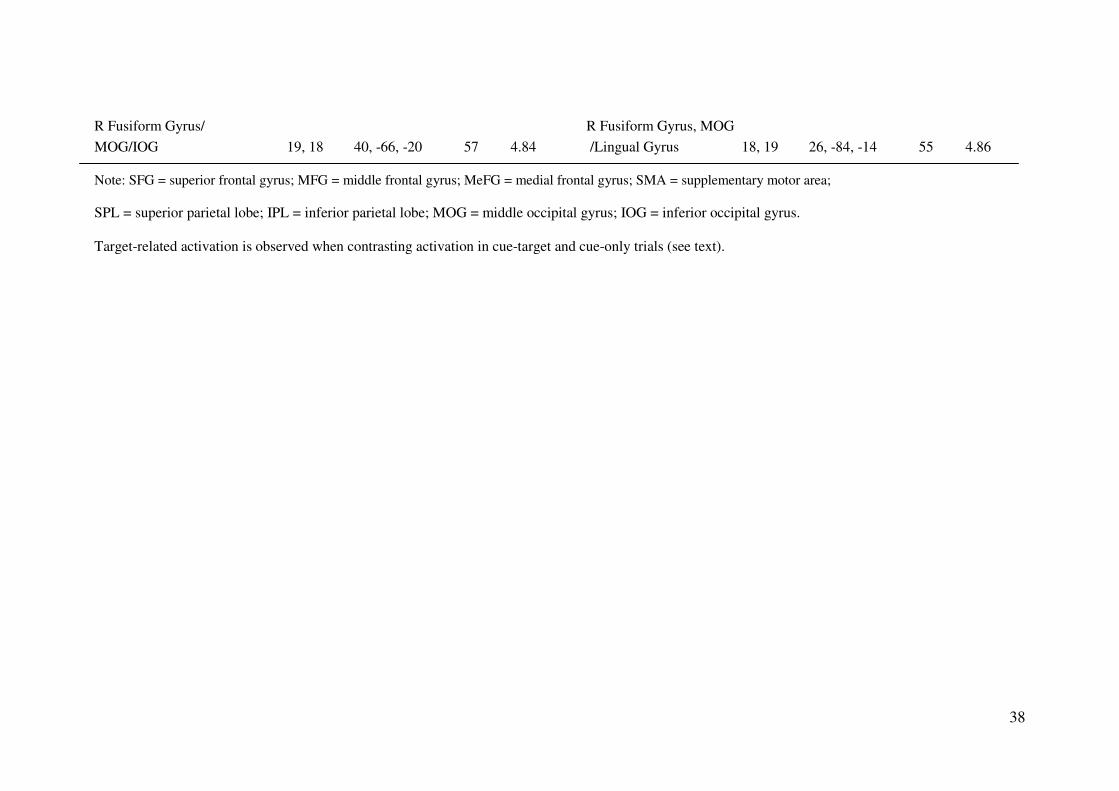
Table 2.1. Cortical activation for the comparison of rule-cue versus task-cue in cue-only trials (left) and for the comparison of task-cue versus
rule-cue for the target-related activation (right).
Cue-related activation (rule-cue- task-cue) Target-related activation (task-cue- rule-cue)
Region BA
MNI
coordinates
Voxel
number
T
max Region BA
MNI
coordinates
Voxel
number
T
max
R anterior SFG 10 38, 62, 6 186 6.15 R anterior SFG 10 36, 64, 8 67 5.36
R MFG 6 38, 2, 62 97 7.56 R MFG 6 38, 4, 64 43 7.34
L MFG/precentral gyrus 6 -48, 2, 48 23 4.66
R MFG 9, 8 56, 18, 38 97 5.13
MeFG 8 0, 50, 48 62 6.08
Medial SFG (Pre-SMA) 6 0, 34, 60 100 6.91 Medial SFG (Pre-SMA) 6 0, 18, 62 43 4.83
Medial SFG (Pre-SMA/SMA) 6 -2, 8, 72 67 5.41 MeFG(Pre-SMA) 6 -2, 12, 50 14 4.30
R SPL 7 38, -58, 56 17 3.98 R SPL 7 36, -62, 56 11 4.04
L Precuneus 7 -18, -76, 48 62 6.09
L Fusiform Gyrus/MOG 19 -44, -72, -20 49 4.88 L Fusiform Gyrus 19 -42, -68, -16 91 4.82
/MOG, IOG
R Fusiform Gyrus 37 48, -52, -24 16 4.04 R Fusiform Gyrus 37, 20 54, -58, -20 17 4.50
L MOG/IOG 19, 18 -44, -84, -12 69 5.00
L Lingual Gyrus/ L/R Lingual Gyrus
Fusiform Gyrus 17, 18 -6, -92, -16 91 5.34 /Fusiform Gyrus 18, 19 -8, -92, -18 198 5.23
R Lingual Gyrus 18 6, -86, -16 32 4.91
38
R Fusiform Gyrus/ R Fusiform Gyrus, MOG
MOG/IOG 19, 18 40, -66, -20 57 4.84 /Lingual Gyrus 18, 19 26, -84, -14 55 4.86
Note: SFG = superior frontal gyrus; MFG = middle frontal gyrus; MeFG = medial frontal gyrus; SMA = supplementary motor area;
SPL = superior parietal lobe; IPL = inferior parietal lobe; MOG = middle occipital gyrus; IOG = inferior occipital gyrus.
Target-related activation is observed when contrasting activation in cue-target and cue-only trials (see text).
39
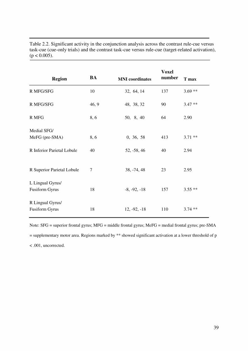
Table 2.2. Significant activity in the conjunction analysis across the contrast rule-cue versus
task-cue (cue-only trials) and the contrast task-cue versus rule-cue (target-related activation),
(p < 0.005).
Region BA MNI coordinates
Voxel
number T max
R MFG/SFG 10 32, 64, 14 137 3.69 **
R MFG/SFG 46, 9 48, 38, 32 90 3.47 **
R MFG 8, 6 50, 8, 40 64 2.90
Medial SFG/
MeFG (pre-SMA) 8, 6 0, 36, 58 413 3.71 **
R Inferior Parietal Lobule 40 52, -58, 46 40 2.94
R Superior Parietal Lobule 7 38, -74, 48 23 2.95
L Lingual Gyrus/
Fusiform Gyrus 18 -8, -92, -18 157 3.55 **
R Lingual Gyrus/
Fusiform Gyrus 18 12, -92, -18 110 3.74 **
Note: SFG = superior frontal gyrus; MFG = middle frontal gyrus; MeFG = medial frontal gyrus; pre-SMA
= supplementary motor area. Regions marked by ** showed significant activation at a lower threshold of p
< .001, uncorrected.
40
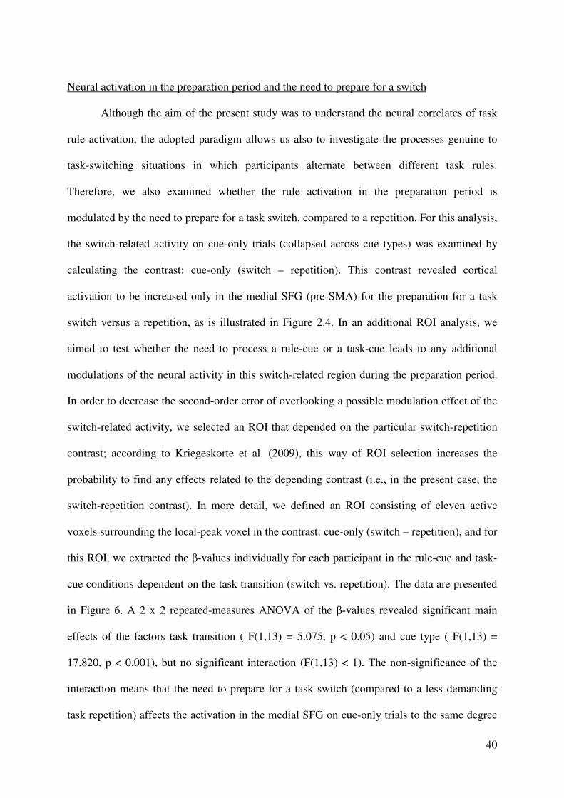
Neural activation in the preparation period and the need to prepare for a switch
Although the aim of the present study was to understand the neural correlates of task
rule activation, the adopted paradigm allows us also to investigate the processes genuine to
task-switching situations in which participants alternate between different task rules.
Therefore, we also examined whether the rule activation in the preparation period is
modulated by the need to prepare for a task switch, compared to a repetition. For this analysis,
the switch-related activity on cue-only trials (collapsed across cue types) was examined by
calculating the contrast: cue-only (switch – repetition). This contrast revealed cortical
activation to be increased only in the medial SFG (pre-SMA) for the preparation for a task
switch versus a repetition, as is illustrated in Figure 2.4. In an additional ROI analysis, we
aimed to test whether the need to process a rule-cue or a task-cue leads to any additional
modulations of the neural activity in this switch-related region during the preparation period.
In order to decrease the second-order error of overlooking a possible modulation effect of the
switch-related activity, we selected an ROI that depended on the particular switch-repetition
contrast; according to Kriegeskorte et al. (2009), this way of ROI selection increases the
probability to find any effects related to the depending contrast (i.e., in the present case, the
switch-repetition contrast). In more detail, we defined an ROI consisting of eleven active
voxels surrounding the local-peak voxel in the contrast: cue-only (switch – repetition), and for
this ROI, we extracted the β-values individually for each participant in the rule-cue and task-
cue conditions dependent on the task transition (switch vs. repetition). The data are presented
in Figure 6. A 2 x 2 repeated-measures ANOVA of the β-values revealed significant main
effects of the factors task transition ( F(1,13) = 5.075, p < 0.05) and cue type ( F(1,13) =
17.820, p < 0.001), but no significant interaction (F(1,13) < 1). The non-significance of the
interaction means that the need to prepare for a task switch (compared to a less demanding
task repetition) affects the activation in the medial SFG on cue-only trials to the same degree
41
in the rule-cue and the task-cue condition. In other words, the need to process a rule-cue or a
task-cue does not modulate the switch-related activation in the medial SFG during the
preparation period.
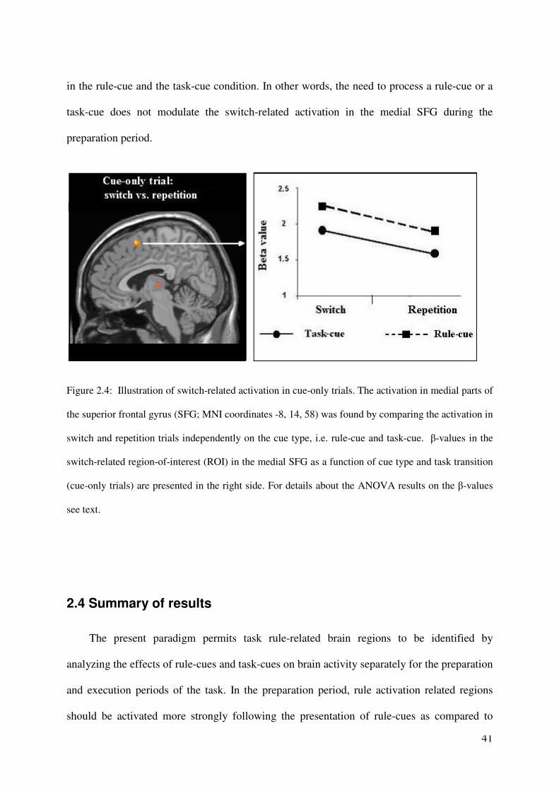
Figure 2.4: Illustration of switch-related activation in cue-only trials. The activation in medial parts of
the superior frontal gyrus (SFG; MNI coordinates -8, 14, 58) was found by comparing the activation in
switch and repetition trials independently on the cue type, i.e. rule-cue and task-cue. β-values in the
switch-related region-of-interest (ROI) in the medial SFG as a function of cue type and task transition
(cue-only trials) are presented in the right side. For details about the ANOVA results on the β-values
see text.
2.4 Summary of results
The present paradigm permits task rule-related brain regions to be identified by
analyzing the effects of rule-cues and task-cues on brain activity separately for the preparation
and execution periods of the task. In the preparation period, rule activation related regions
should be activated more strongly following the presentation of rule-cues as compared to
42
task-cues. Conversely, for the execution period, rule-related activation would be expected
specifically upon target presentation if task rules were not activated sufficiently during the
preparation period; this pattern should be revealed by the contrast of the target-related
activation in the task-cue compared to the rule-cue condition. In line with these predictions,
the conjunction analysis revealed similar frontoparietal networks of activation foci in the
corresponding contrasts, that is: the contrast of rule-cue minus task-cue for cue-only trials
(preparation period) and the contrast comparing target-related activation on task-cue versus
rule-cue trials (execution period). The common activation foci in these two contrasts included
the anterior and middle parts of the right MFG and SFG, the posterior region of the MFG near
the IFJ, regions in the medial SFG extending from anterior to posterior portions of the pre-
SMA, as well as the right SPL and IPL. All these activations conformed to the pattern
expected for cortical regions that are correlated with the mechanisms underlying task rule
activation.
43
CHAPTER 3
fMRI Experiment 2
3.1 Research hypothesis and expectations
This experiment tried to find the evidence for the hypothesis that the preparatory
attentional control can bias sensory processing via amplifying the activity of the task -
relevant stimuls-specific brain region.
The preparatory modulation was expected in the posterior stimuls-specific brain
regions (i.e., face specific and number specific regions). In particular, significant additional
activity comparing to null trials was expected to be found in the preparation period of face
task in right FFA; while in the preparation period of number task, significant additional
activity comparing to null trials was expected to be found in right IPSnum.
44
3.2 Methods
Subjects
Fourteen healthy right-handed volunteers with normal or corrected to normal vision
participated in the experiment (six males, ages 19-33, mean age: 24.9, SDV: 4.4) after
obtaining informed consent according to the Declaration of Helsinki. Each participant was
paid 20 €. Two participant’s data were excluded from the following analysis because of high
error rate (more than 20 %). Thus, ultimately, 12 participants’ data sets were available for
analysis (six male, ages 19-33, mean age: 24.4, SDV: 4.6).
Experiment setting for the main task
Paradigm and procedure. The task to be performed by the participants was either
gender discrimination (female or male) or number discrimination (bigger or smaller than five,
it is called big or small for short). Each trial began with the presentation of a cue for a fixed
duration of 1200 ms, which could either be a rule-cue or a task-cue (Fig. 3.1). Both cues
displayed an instruction for the upcoming task; however, a precise instruction about the
required task rule was provided only in the rule-cue condition (Fig. 3.1, upper panel). On cue-
only trials (n = 200 trials, of which 50 presented a rule-cue and 50 a task-cue for face task,
and the same amount for number task), there was no target following the cue offset, but only a
black screen that lasted for 600 ms, and there was no need for participants to make a response
(Fig. 3.2, right panel).
45
In contrast, on cue-target trials (n = 280, of which 70 presented a rule-cue and 70 a task-
cue for face task, and the same amount for number task), the cue was followed by picture
contains both face and number that was presented for 600 ms (Fig. 3.2). Participants were to
respond to either the number or the face depicted in the target display, depending on the
instruction of cue. Participants made two-alternative forced-choice responses using either
their left or right finger, with response sets counterbalanced across participants. After the
offset of the target picture, a black screen was presented for a variable interval of 1800, 2500,
3100, 3900, or 4600 ms. The next trial could then either be a cue-target or a cue-only trial,
that is an ‘event trial’, or a ‘null trial’ (n = 140) in which there was neither a cue nor a target
event.
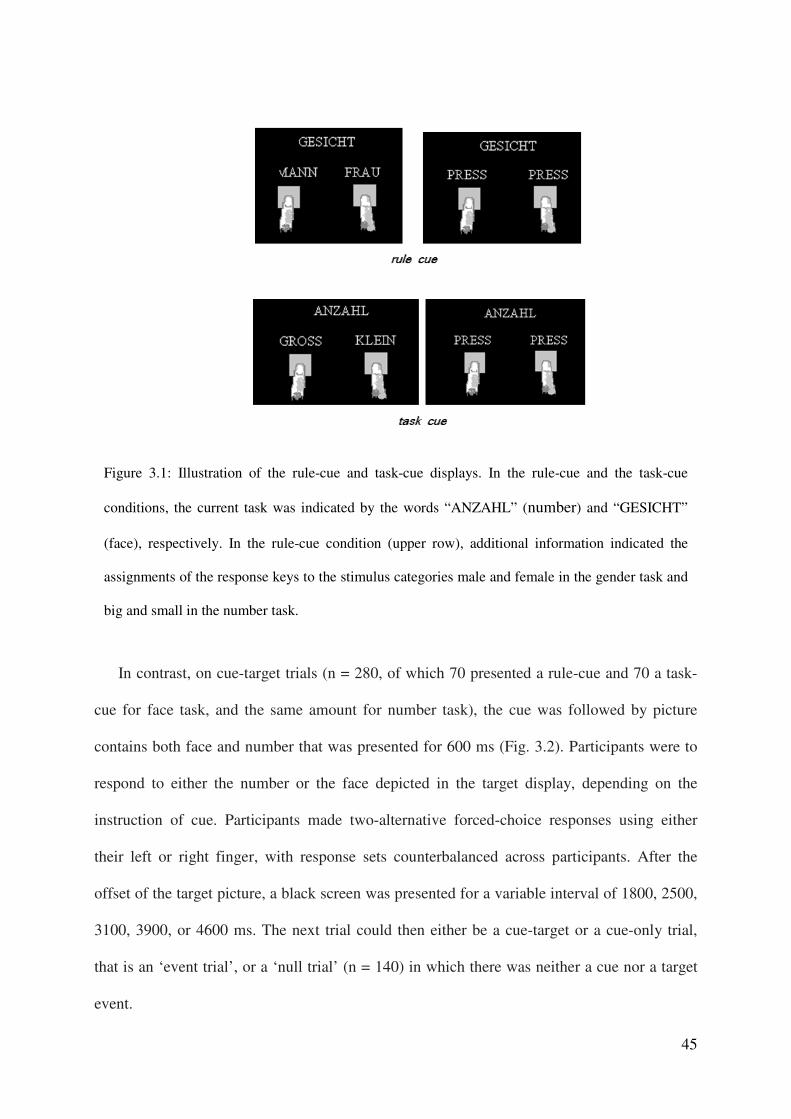
Figure 3.1: Illustration of the rule-cue and task-cue displays. In the rule-cue and the task-cue
conditions, the current task was indicated by the words “ANZAHL” (number) and “GESICHT”
(face), respectively. In the rule-cue condition (upper row), additional information indicated the
assignments of the response keys to the stimulus categories male and female in the gender task and
big and small in the number task.
46
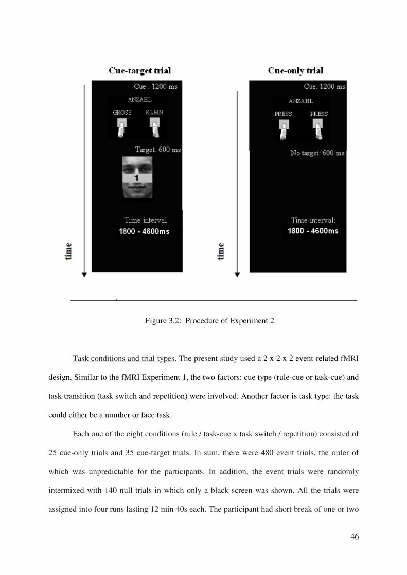
Figure 3.2: Procedure of Experiment 2
Task conditions and trial types. The present study used a 2 x 2 x 2 event-related fMRI
design. Similar to the fMRI Experiment 1, the two factors: cue type (rule-cue or task-cue) and
task transition (task switch and repetition) were involved. Another factor is task type: the task
could either be a number or face task.
Each one of the eight conditions (rule / task-cue x task switch / repetition) consisted of
25 cue-only trials and 35 cue-target trials. In sum, there were 480 event trials, the order of
which was unpredictable for the participants. In addition, the event trials were randomly
intermixed with 140 null trials in which only a black screen was shown. All the trials were
assigned into four runs lasting 12 min 40s each. The participant had short break of one or two
47
minutes between two runs. At the beginning of each run, a word “Attention” was presented
for 2 seconds to remind the participants to back to the task performing.
For each condition, the cue-related activation can be assessed by measuring the
activation on the cue-only trials, whereas target-related activation can be assessed by
calculating the contrast between the activation in corresponding cue-target minus cue-only
trials.
Stimulus and response conditions. On cue-only trials, only a black screen (i.e., no
target) was presented after the presentation of the cue and there was need to respond. On cue-
target trials, the target stimulus was a face picture with a word of number locating in the
region of nose. Two males and two females’ pictures were used, the number could be “EINS”
(one), “ZWEI”(two), “ACHT” (eight) or “NEUN”(nine). As a result 16 face-and-number
pictures were used as target stimuli. Participants were informed by the cue to respond to either
the face or the number. The stimuli (cue stimuli and target stimuli) were located on a black
background in the centre of the screen and subtanded 5 degrees of visual angle.
Participants used their left and right finger for response. They were instructed
to respond as fast and as accurately as possible. For half the participants, the S-R mapping
rule was male-left, female-right and big-left, small-right. This was reversed for the other half:
female-left, male-right and small-left, big-right.
fMRI measurement
Imaging was performed with a SIEMENS TRIO 3-Tesla scanner at the Klinikum
Großhadern (Institute for Clinical Radiology), Ludwig-Maximilians-Universität in Munich.
T2*-weighted echo-planar images (EPI) with blood oxygenation level-dependent contrast
were acquired (TR = 1500 ms, TE = 30 ms, flip angle = 80°, matrix size = 64 x 64 voxels).
Twenty three axial slices (thickness = 4 mm, spacing = 1 mm) were acquired parallel to the
48
AC-PC plane, covering the whole cortex. The order of acquisition of the slices was
interleaved. The first four volumes (dummy volumes) were discarded because of possible
instabilities in the magnetic field at the beginning of a scan. Stimuli were displayed on a back-
projection screen mounted in the bore of the magnet behind the participant’s head by using an
LCD projector. Participants viewed the screen by wearing mirror glasses.
fMRI data analysis
Preprocessing of the functional images was carried out using SPM5 (Wellcome
Department of Cognitive Neurology, London, UK). Images were interpolated in time
(temporal realignment to the middle slice). In addition, they were spatially realigned to the
first volume for head movement correction, unwrapped, and then normalized into MNI space
(resampled to 2 x 2 x 2 mm3 isotropic resolution) with default normalization estimation. The
data were then smoothed with a Gaussian kernel of 8 mm full-width half-maximum to
account for inter-subject anatomical variability.
Then the image data were modeled by applying a general linear model (Friston et al.
1995). In event-related single-subject analyses, the 8 cue-only and the 8 cue-target conditions
were modeled as separate volumes (resulting from the factorial combination of the two task
types (face vs. number), two cue types (rule-cue vs. task-cue) and the types of task transition
(task switch vs. task repetition). Additionally, the introduction which occurred at the
beginning of each run, the null trials, and all error trials were separately selected to form three
event volume. The resulting 19 volumes were convolved with the hemodynamic response
function (HRF), and then beta values of these regressors were estimated according to the
ordinary least-squares (OLS) method. The beta values of the cue-only trials were the
activation parameters analyzed in the ROI analysis, while the beta values for the cue-target
49
trials subtracted the beta values for their corresponding cue-only trials resulted the activation
parameters for target period activation, which were analyzed in the ROI analysis.
Another general linear model was built in order to replicate the findings of fMRI
Experiment 1. Similar to the fMRI Experiment 1, the 4 cue-only and the 4 cue-target
conditions were modeled as separate volumes (resulting from the factorial combination of the
two cue type (rule-cue vs. task-cue) and the types of task transition (task switch vs. task
repetition). The introduction which occurred at the beginning of each run, the null trials, and
all error trials were selected to form an event volume additionally. The resulting 11 volumes
were convolved with the hemodynamic response function (HRF), and then beta values of
these regressors were estimated according to the ordinary least-squares (OLS) method. The
for the whole brain analyses, similar conjunction analysis was performed to locate the
common task rule-related activation between these two main contrasts: Contrast 1: For cue-
only trials, rule-cue minus task-cue trials (use the threshold of p < 0.001 ); Contrast 2: (cue-
target trials minus cue-only trials for task-cues) minus (cue-target trials minus cue-only trials
for rule-cues) (use the threshold of p < 0.001 ). For the conjunction analysis, the statistical
threshold was p < 0.005, uncorrected.
Experiment design for the localization task
After the main experiment, participants performed one run of localizer tasks to
determine the individual Regions of Interest (ROIs) in the FFA and IPSnum relevant for face
processing and number processing respectively.
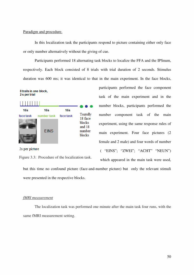
50
Paradigm and procedure.
In this localization task the participants respond to picture containing either only face
or only number alternatively without the giving of cue.
Participants performed 18 alternating task blocks to localize the FFA and the IPSnum,
respectively. Each block consisted of 8 trials with trial duration of 2 seconds. Stimulus
duration was 600 ms; it was identical to that in the main experiment. In the face blocks,
participants performed the face component
task of the main experiment and in the
number blocks, participants performed the
number component task of the main
experiment, using the same response rules of
main experiment. Four face pictures (2
female and 2 male) and four words of number
( “EINS”; “ZWEI”; “ACHT” “NEUN”)
which appeared in the main task were used,
but this time no confound picture (face-and-number picture) but only the relevant stimuli
were presented in the respective blocks.
fMRI measurement
The localization task was performed one minute after the main task four runs, with the
same fMRI measurement setting.
Figure 3.3: Procedure of the localization task.

51
fMRI data analysis
Preprocessing. Preprocessing of the functional images was carried out using SPM5
(Wellcome Department of Cognitive Neurology, London, UK). Images were spatially
realigned to the first volume for head movement correction, unwrapped, and then normalized
into MNI space (resampled to 2 x 2 x 2 mm3 isotropic resolution) with default normalization
estimation. The data were then smoothed with a Gaussian kernel of 8 mm full-width half-
maximum to account for inter-subject anatomical variability.
Then the image data were modeled by applying a general linear model (Friston et al.
1995). In event-related single-subject analyses, the face task and number task conditions were
modeled as separate volumes. Additionally, all error trials were selected to form an event
volume. The resulting three volumes were convolved with the hemodynamic response
function (HRF), and then beta values of these regressors were estimated according to the
ordinary least-squares (OLS) method.
Whole-brain analyses. For group statistics, one-sample t-tests of contrast maps across subjects
(random-effects model treating subjects as a random variable) were computed to indicate
whether observed differences between conditions were significantly different from zero. A
statistical threshold of p < 0.001, uncorrected, was used, covering at least 10 contiguous
voxels.
The contrast of ‘face – number’ and the reversed one ‘number – face’ were calculated
to find the group activity peaks in FFA and IPSnum respectively, started from which, I then
found individual face specific and number specific regions.
52
Region-of-Interest (ROI) Analyses. For the FFA and the IPSnum these ROIs were determined
in every participant. The FFA and the IPSnumber activity for the contrasts ‘face-number’ and
‘number-face’ were used for every participant. The statistical threshold used was p < 0.01,
uncorrected, covering at least 10 contiguous voxels.
Starting from the location of the group activity peak in the localization task, the nearest peak
per participant was determined as centre of the individual cube ROI mask (6mm side length).
From the voxels covered by these masks we extracted the parameter estimates from the time
series of every individual participant for all 16 task conditions (produced by the beta values,
see fMRI experiment2 _ fMRI data analysis ). The 8 activation parameters of cue period
occupied most interesting. They were compared by ANOVA with the factors task type (face
vs. number), cue type (rule-cue vs. task-cue) and task transition (task switch vs. task
repetition). The 8 parameters of cue-target trails subtracted the corresponding cue-only trails
parameters to obtain the parameters for target period activation, which then were compared by
ANOVA with the factors task type, cue type and task transition also.
3.3 Results
Behavioral Results
Mean reaction time were submitted to a 2 x 2 x 2 repeated-measures ANOVA with the
factors of task type (face or number), cue type (rule-cue or task-cue) and task transition (task
switch or repetition) (see Fig 3.4).
No significant effect was found for the main effect of task type (F(1,11) = 0.82, p
=0.38). The task difficulties of these two tasks are balanced.
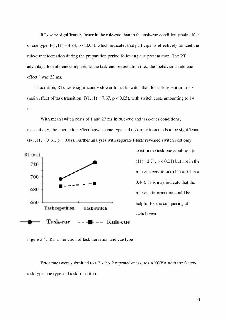
53
RTs were significantly faster in the rule-cue than in the task-cue condition (main effect
of cue type, F(1,11) = 4.84, p < 0.05), which indicates that participants effectively utilized the
rule-cue information during the preparation period following cue presentation. The RT
advantage for rule-cue compared to the task-cue presentation (i.e., the ‘behavioral rule-cue
effect’) was 22 ms.
In addition, RTs were significantly slower for task switch than for task repetition trials
(main effect of task transition, F(1,11) = 7.67, p < 0.05), with switch costs amounting to 14
ms.
With mean switch costs of 1 and 27 ms in rule-cue and task-cues conditions,
respectively, the interaction effect between cue type and task transition tends to be significant
(F(1,11) = 3.61, p = 0.08). Further analyses with separate t-tests revealed switch cost only
exist in the task-cue condition (t
(11) =2.74, p < 0.01) but not in the
rule-cue condition (t(11) = 0.1, p =
0.46). This may indicate that the
rule-cue information could be
helpful for the conquering of
switch cost.
Figure 3.4: RT as function of task transition and cue type
Error rates were submitted to a 2 x 2 x 2 repeated-measures ANOVA with the factors
task type, cue type and task transition.
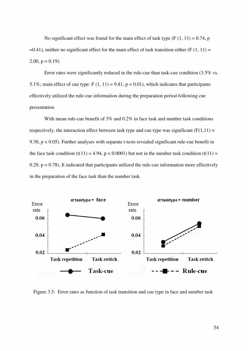
54
No significant effect was found for the main effect of task type (F (1, 11) = 0.74, p
=0.41), neither no significant effect for the main effect of task transition either (F (1, 11) =
2.00, p = 0.19).
Error rates were significantly reduced in the rule-cue than task-cue condition (3.5% vs.
5.1%; main effect of cue type: F (1, 11) = 9.81, p < 0.01), which indicates that participants
effectively utilized the rule-cue information during the preparation period following cue
presentation.
With mean rule-cue benefit of 3% and 0.2% in face task and number task conditions
respectively, the interaction effect between task type and cue type was significant (F(1,11) =
9.30, p < 0.05). Further analyses with separate t-tests revealed significant rule-cue benefit in
the face task condition (t(11) = 4.94, p < 0.0001) but not in the number task condition (t(11) =
0.29, p = 0.78). It indicated that participants utilized the rule-cue information more effectively
in the preparation of the face task than the number task.
Figure 3.5: Error rates as function of task transition and cue type in face and number task
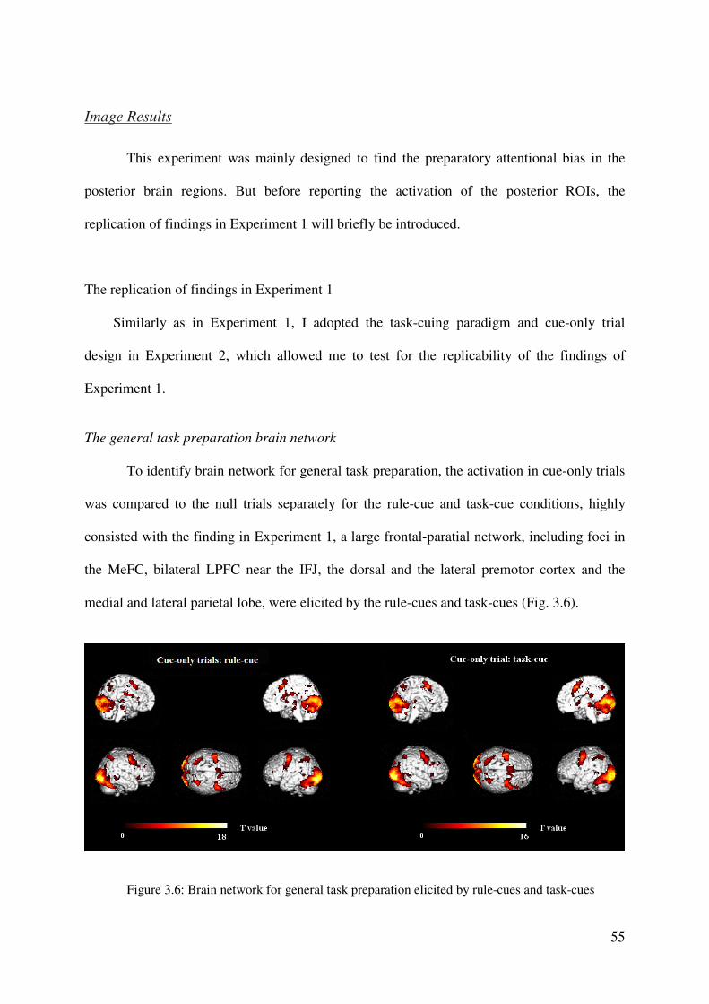
55
Image Results
This experiment was mainly designed to find the preparatory attentional bias in the
posterior brain regions. But before reporting the activation of the posterior ROIs, the
replication of findings in Experiment 1 will briefly be introduced.
The replication of findings in Experiment 1
Similarly as in Experiment 1, I adopted the task-cuing paradigm and cue-only trial
design in Experiment 2, which allowed me to test for the replicability of the findings of
Experiment 1.
The general task preparation brain network
To identify brain network for general task preparation, the activation in cue-only trials
was compared to the null trials separately for the rule-cue and task-cue conditions, highly
consisted with the finding in Experiment 1, a large frontal-paratial network, including foci in
the MeFC, bilateral LPFC near the IFJ, the dorsal and the lateral premotor cortex and the
medial and lateral parietal lobe, were elicited by the rule-cues and task-cues (Fig. 3.6).
Figure 3.6: Brain network for general task preparation elicited by rule-cues and task-cues
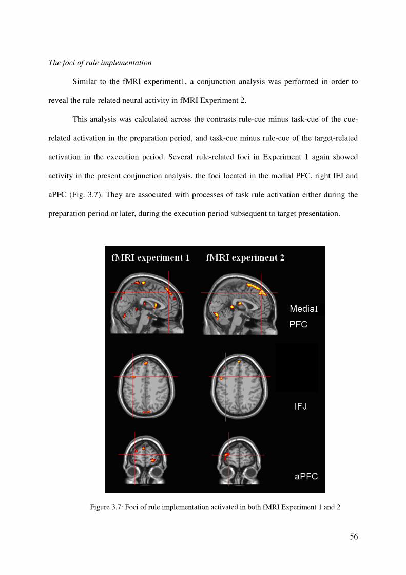
56
The foci of rule implementation
Similar to the fMRI experiment1, a conjunction analysis was performed in order to
reveal the rule-related neural activity in fMRI Experiment 2.
This analysis was calculated across the contrasts rule-cue minus task-cue of the cue-
related activation in the preparation period, and task-cue minus rule-cue of the target-related
activation in the execution period. Several rule-related foci in Experiment 1 again showed
activity in the present conjunction analysis, the foci located in the medial PFC, right IFJ and
aPFC (Fig. 3.7). They are associated with processes of task rule activation either during the
preparation period or later, during the execution period subsequent to target presentation.
Figure 3.7: Foci of rule implementation activated in both fMRI Experiment 1 and 2
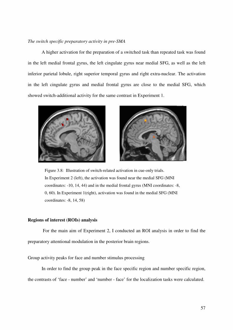
57
The switch specific preparatory activity in pre-SMA
A higher activation for the preparation of a switched task than repeated task was found
in the left medial frontal gyrus, the left cingulate gyrus near medial SFG, as well as the left
inferior parietal lobule, right superior temporal gyrus and right extra-nuclear. The activation
in the left cingulate gyrus and medial frontal gyrus are close to the medial SFG, which
showed switch-additional activity for the same contrast in Experiment 1.
Regions of interest (ROIs) analysis
For the main aim of Experiment 2, I conducted an ROI analysis in order to find the
preparatory attentional modulation in the posterior brain regions.
Group activity peaks for face and number stimulus processing
In order to find the group peak in the face specific region and number specific region,
the contrasts of ‘face - number’ and ‘number - face’ for the localization tasks were calculated.
Figure 3.8: Illustration of switch-related activation in cue-only trials.
In Experiment 2 (left), the activation was found near the medial SFG (MNI
coordinates: -10, 14, 44) and in the medial frontal gyrus (MNI coordinates: -8,
0, 60). In Experiment 1(right), activation was found in the medial SFG (MNI
coordinates: -8, 14, 58)
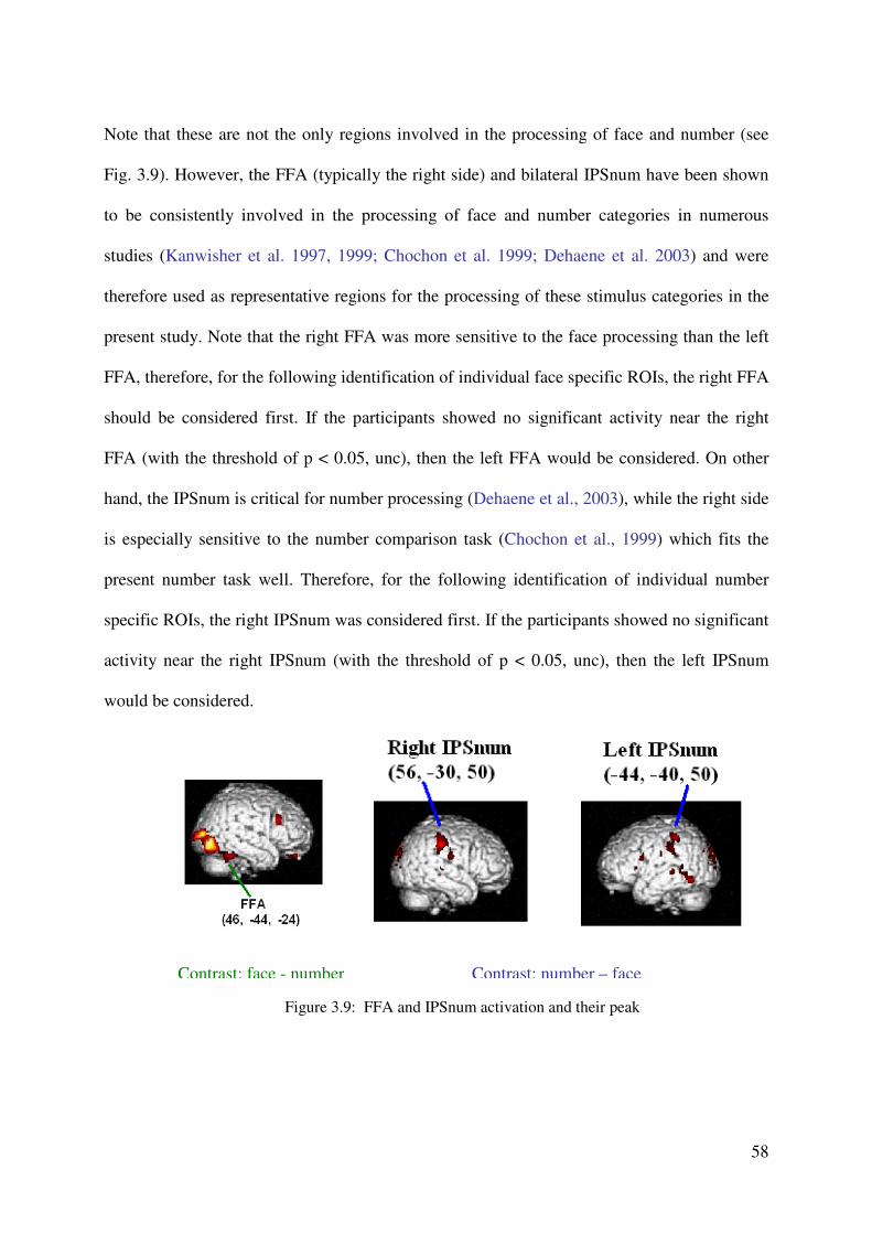
58
Note that these are not the only regions involved in the processing of face and number (see
Fig. 3.9). However, the FFA (typically the right side) and bilateral IPSnum have been shown
to be consistently involved in the processing of face and number categories in numerous
studies (Kanwisher et al. 1997, 1999; Chochon et al. 1999; Dehaene et al. 2003) and were
therefore used as representative regions for the processing of these stimulus categories in the
present study. Note that the right FFA was more sensitive to the face processing than the left
FFA, therefore, for the following identification of individual face specific ROIs, the right FFA
should be considered first. If the participants showed no significant activity near the right
FFA (with the threshold of p < 0.05, unc), then the left FFA would be considered. On other
hand, the IPSnum is critical for number processing (Dehaene et al., 2003), while the right side
is especially sensitive to the number comparison task (Chochon et al., 1999) which fits the
present number task well. Therefore, for the following identification of individual number
specific ROIs, the right IPSnum was considered first. If the participants showed no significant
activity near the right IPSnum (with the threshold of p < 0.05, unc), then the left IPSnum
would be considered.
Figure 3.9: FFA and IPSnum activation and their peak
Contrast: face - number Contrast: number – face
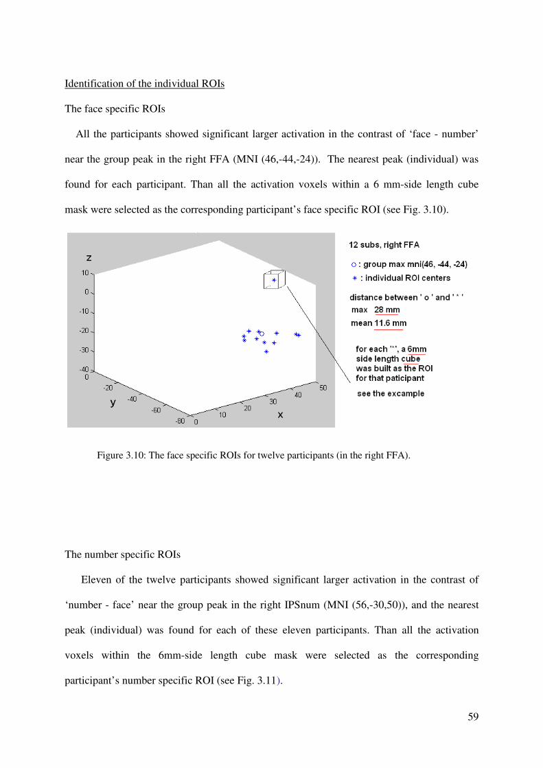
59
Identification of the individual ROIs
The face specific ROIs
All the participants showed significant larger activation in the contrast of ‘face - number’
near the group peak in the right FFA (MNI (46,-44,-24)). The nearest peak (individual) was
found for each participant. Than all the activation voxels within a 6 mm-side length cube
mask were selected as the corresponding participant’s face specific ROI (see Fig. 3.10).
Figure 3.10: The face specific ROIs for twelve participants (in the right FFA).
The number specific ROIs
Eleven of the twelve participants showed significant larger activation in the contrast of
‘number - face’ near the group peak in the right IPSnum (MNI (56,-30,50)), and the nearest
peak (individual) was found for each of these eleven participants. Than all the activation
voxels within the 6mm-side length cube mask were selected as the corresponding
participant’s number specific ROI (see Fig. 3.11).
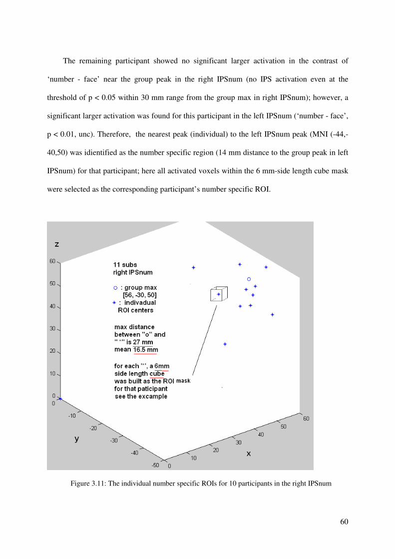
60
The remaining participant showed no significant larger activation in the contrast of
‘number - face’ near the group peak in the right IPSnum (no IPS activation even at the
threshold of p < 0.05 within 30 mm range from the group max in right IPSnum); however, a
significant larger activation was found for this participant in the left IPSnum (‘number - face’,
p < 0.01, unc). Therefore, the nearest peak (individual) to the left IPSnum peak (MNI (-44,-
40,50) was idientified as the number specific region (14 mm distance to the group peak in left
IPSnum) for that participant; here all activated voxels within the 6 mm-side length cube mask
were selected as the corresponding participant’s number specific ROI.
Figure 3.11: The individual number specific ROIs for 10 participants in the right IPSnum
61
The results of the ROI analysis
As outlined in the Introduction, preparatory attentional modulations were expected to be
found in the face and number specific region in face and number task respectively. Then the
activation parameters, especially the cue period parameters, of the face specific ROIs (right
FFA) and number specific ROIs (right IPSnum) were analyzed.
For the face specific ROIs
Cue period activity:
The activation parameters of cue-only trials were submitted to a 2 x 2 repeated-measures
ANOVA with the factors task type (face or number) and cue type (rule-cue or task-cue).
Larger activation of the FFA was found for the cue period of the face task compared to the
number task (F (1, 11) =6.25, p<0.05). Separate T tests showed that both the rule-cue and
task-cue elicited significant activity compared to the null trials (t (11) =2.23, p < 0.05; t (11)
=1.93, p < 0.05 (1 tailed; respectively) in the preparation of face task. Whereas no significant
activity compared to null trials was found for the preparation of number task in either rule-cue
or task-cue condition (both t < 1.34, p > 0.2). In line with my expectation, these findings
indicate preparatory attentional modulation in the face specific regions in both of the two cue
conditions.

62
* p<0.05
** p<0.001
Figure 3.12: Right FFA’s activity in cue period and target period
Target period activity
The target period activity in the face specific ROIs was also analyzed. The parameters of
target period activation were submitted to a 2 x 2 repeated-measures ANOVA with the factors
task type (face or number) and cue type (rule-cue or task-cue). It showed no difference for the
activity of FFA between the face and the number task (F (1, 11) = 0.172, p = 0.687). There
was no further modulation in the task execution period after the modulation in the preparation
period.
The main effect of cue type tended to be significant; in particular, in the target period the
activation tended to be larger activation in the task-cue than in rule-cue condition (F (1, 11) =
4.72, p = 0.053).
The interaction of task type and cue type tended to be significant (F (11, 1) = 4.62, p =
0.055); the following separate T tests showed larger activation in the task-cue than in the rule-
cue condition of the face task (t (11) = 2.48, p < 0.05), but no difference between these two
cue conditions in the number task (t (11) = 0.64, p = 0.53). Finally, significant additional
activity was found in the target period in all the task and cue conditions (all ts > 4.5, ps <
0.001).
Activ
atio
n p
ara
meter
63
For the number specific ROIs
As outlined in the method part, the ROIs of number processing were selected from the
region of right IPSnum for eleven of the twelve participants. Therefore, the following results
are based on the findings of these eleven ROIs in right IPSnum. Important to note that, if
including the data of the participant with the left sided ROI into the data set, the results
pattern will not change.
Cue period activity:
The activation parameters of cue-only trials were submitted to a 2 x 2 repeated-measures
ANOVA with the factors task type (face or number) and cue type (rule-cue or task-cue).
Larger activation of right IPSnum in the number task than face task in cue period was found
(F (1, 10) =5.56, p < 0.05). No significant main effect of cue type (F (1, 10) = 0.08, p = 0.78)
was found. Separate t-tests showed that both the rule cue and the task cue tend to elicit
additional activity compared to the activity in the null trials (t (10) = 1.58, p = 0.07; t (10) =
1.77, p = 0.05 respectively) in the preparation period of the number task. Whereas no
additional activity compared to that in null trials was found for the preparation to the face task
in either rule-cue or task-cue condition (both ts < 1.08, ps > 0.3).
Target period activity:
The activation parameters for the target period were submitted to a 2 x 2 repeated-
measures ANOVA with the factors task type (face or number) and cue type (rule-cue or task-
cue). The main effects of task type and cue type were not significant (both F<1.5, both
ps>0.25): no difference was found for the activity of right IPSnum in face and number task (F
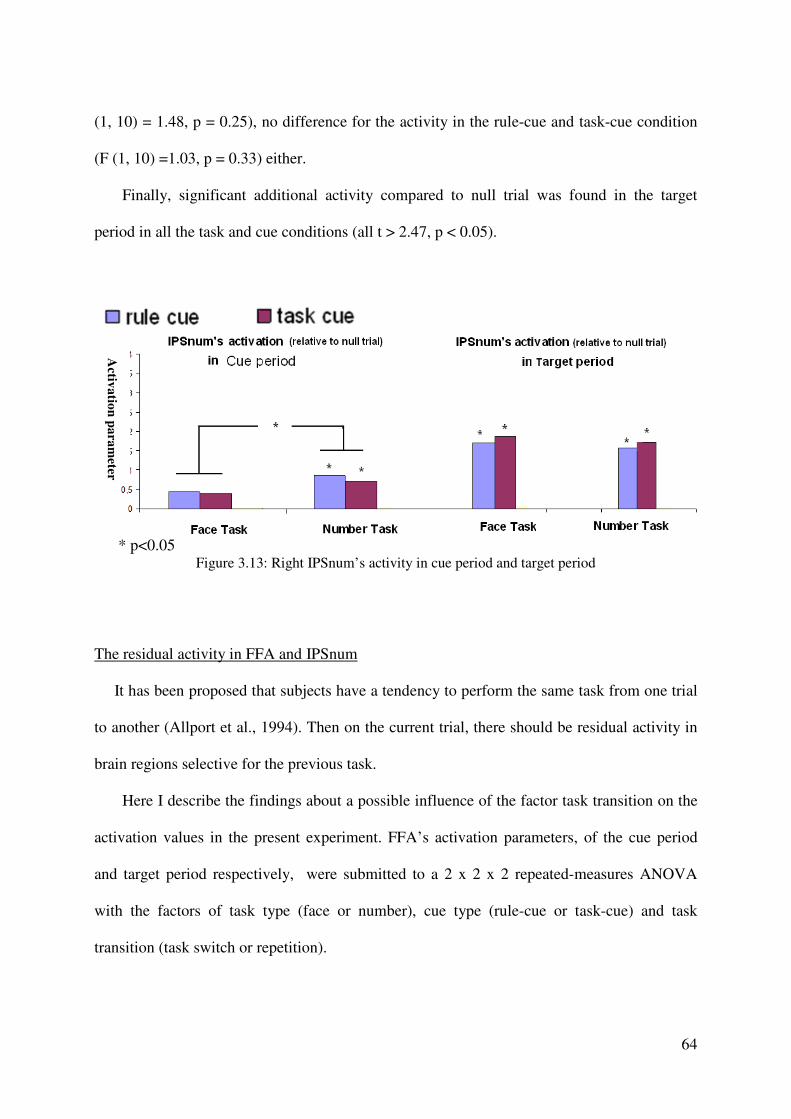
64
(1, 10) = 1.48, p = 0.25), no difference for the activity in the rule-cue and task-cue condition
(F (1, 10) =1.03, p = 0.33) either.
Finally, significant additional activity compared to null trial was found in the target
period in all the task and cue conditions (all t > 2.47, p < 0.05).
* p<0.05
Figure 3.13: Right IPSnum’s activity in cue period and target period
The residual activity in FFA and IPSnum
It has been proposed that subjects have a tendency to perform the same task from one trial
to another (Allport et al., 1994). Then on the current trial, there should be residual activity in
brain regions selective for the previous task.
Here I describe the findings about a possible influence of the factor task transition on the
activation values in the present experiment. FFA’s activation parameters, of the cue period
and target period respectively, were submitted to a 2 x 2 x 2 repeated-measures ANOVA
with the factors of task type (face or number), cue type (rule-cue or task-cue) and task
transition (task switch or repetition).
Activ
atio
n p
ara
meter
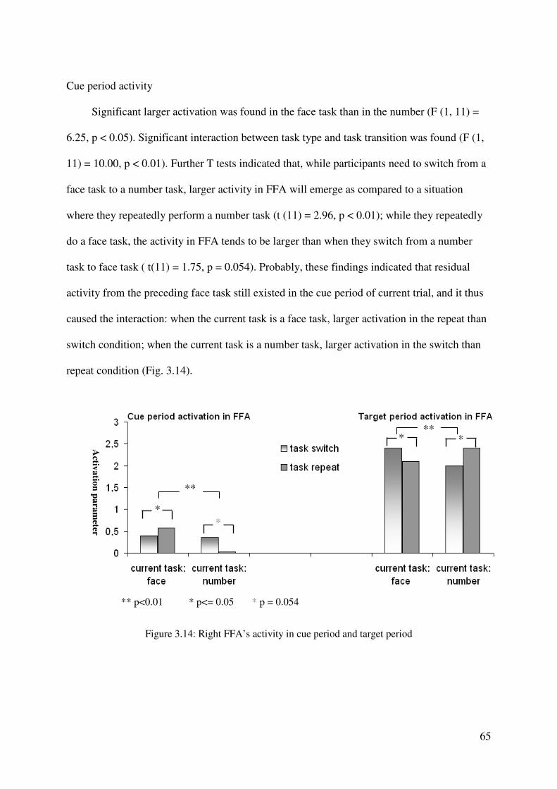
65
Cue period activity
Significant larger activation was found in the face task than in the number (F (1, 11) =
6.25, p < 0.05). Significant interaction between task type and task transition was found (F (1,
11) = 10.00, p < 0.01). Further T tests indicated that, while participants need to switch from a
face task to a number task, larger activity in FFA will emerge as compared to a situation
where they repeatedly perform a number task (t (11) = 2.96, p < 0.01); while they repeatedly
do a face task, the activity in FFA tends to be larger than when they switch from a number
task to face task ( t(11) = 1.75, p = 0.054). Probably, these findings indicated that residual
activity from the preceding face task still existed in the cue period of current trial, and it thus
caused the interaction: when the current task is a face task, larger activation in the repeat than
switch condition; when the current task is a number task, larger activation in the switch than
repeat condition (Fig. 3.14).
Figure 3.14: Right FFA’s activity in cue period and target period
Activ
atio
n p
ara
meter
*
*
*
*
** p<0.01 * p<= 0.05 * p = 0.054
**
**
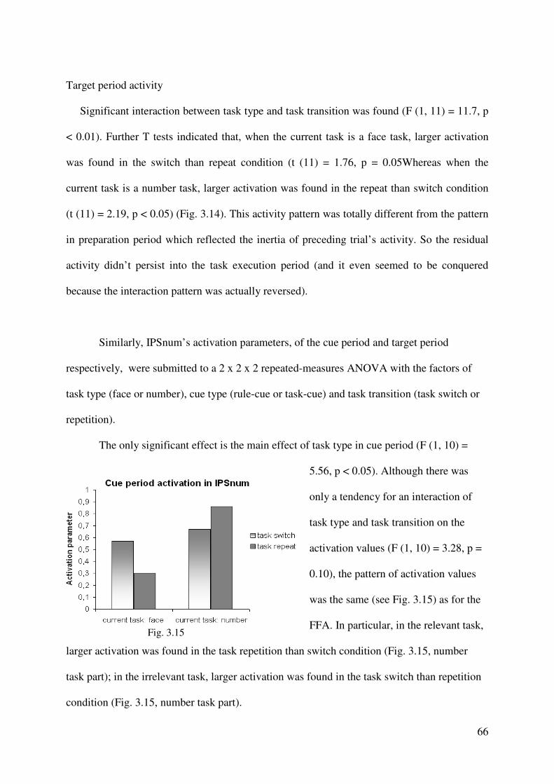
66
Target period activity
Significant interaction between task type and task transition was found (F (1, 11) = 11.7, p
< 0.01). Further T tests indicated that, when the current task is a face task, larger activation
was found in the switch than repeat condition (t (11) = 1.76, p = 0.05Whereas when the
current task is a number task, larger activation was found in the repeat than switch condition
(t (11) = 2.19, p < 0.05) (Fig. 3.14). This activity pattern was totally different from the pattern
in preparation period which reflected the inertia of preceding trial’s activity. So the residual
activity didn’t persist into the task execution period (and it even seemed to be conquered
because the interaction pattern was actually reversed).
Similarly, IPSnum’s activation parameters, of the cue period and target period
respectively, were submitted to a 2 x 2 x 2 repeated-measures ANOVA with the factors of
task type (face or number), cue type (rule-cue or task-cue) and task transition (task switch or
repetition).
The only significant effect is the main effect of task type in cue period (F (1, 10) =
5.56, p < 0.05). Although there was
only a tendency for an interaction of
task type and task transition on the
activation values (F (1, 10) = 3.28, p =
0.10), the pattern of activation values
was the same (see Fig. 3.15) as for the
FFA. In particular, in the relevant task,
larger activation was found in the task repetition than switch condition (Fig. 3.15, number
task part); in the irrelevant task, larger activation was found in the task switch than repetition
condition (Fig. 3.15, number task part).
Fig. 3.15
67
Taking the FFA’s and IPSnum’s cue period activation together, the results showed that
in the current task relevant region, larger activation was found if the current task is the same
as the preceding one than different (repetition > switch); whereas in the current task irrelevant
region, larger activation was found if the current task is the different from the preceding one
than same (switch > repetition). This effect supports that the inertia of the preceding task
processing activity could carry over to the current trial. Moreover, note that the same activity
pattern didn’t appear in the target period, which might indicate the residual activity (or
inertia) didn’t persist into the task execution.
3.4 Summary of results
In the fMRI Experiment 2, preparatory attentional modulation was found in the face
specific and number specific posterior brain regions. In particular, significant additional
activity comparing to null trials was found in the preparation period of face task in right
FFA; while in the preparation period of number task, significant additional activity
comparing to null trials was found in right IPSnum. These findings support the hypothesis
that the preparatory attentional control can bias sensory processing via amplifying the
activity of the task -relevant stimuls-specificbrain region.
For more details, this amplifying was found both in the rule-cue and task-cue
condition, and the amount of activity was not different for the cue types. These results
indicate 1) efficient attentional bias occurred in these two cue conditions. As expected,
rule-cue and task-cue both supply the information of task type thus can elicit efficient
attentional bias. 2) The additional rule information in the display, although it contains the

68
words of the stimuli features (i.e., the drawing of rules for face contains the words of
MANN and FRAU; GROSS and KLEIN in the number rule-cue), it failed to elicit
additional activity in the relevant specific regions. The participants actually used the whole
rule information to activate the task rule, which should be stored in PFC or parietal cortex.
Rather than represented single pieces of feature information in the posterior brain regions,
Another interesting finding was: the activity of either FFA or IPSnum in the task
execution period was not different for both tasks, which means no attentional modulation in
task execution period was found. This finding seems contrary to Serence et al.’s finding
(2003). But this finding may suggest that the attentional modulation is not necessarily
taking place in the task execution period. For instance, in the present experiment, after the
modulation in preparation period, attentional control didn’t re-boost in the target period.
Finally, the residual activation from preceding trial still existed in the preparation
period of the current trial; such residual activation was found in FFA and IPSnum both.
This finding referred to a bottom-up influence in sensory processing. Therefore, in this
present experiment, both the attentional modulation (top-down) and the residual activation
(bottom-up) could influence the posterior regions’ activities.
69
CHAPTER 4
Experiment 3
4.1 Research aim
This experiment aimed to prove that the learning of task rule from the rule-cue displays only
take place when the rules have not yet been obtained well, thus to support the findings of rule
implementation in Experiment 1 and 2 were not confounded by some learning processing.
4.2 Method
Subjects
22 right handed healthy students of LMU were recruited. All participants have had normal
or corrected-to-normal vision. 11 of them are German (6 male, 21-27 years old), 11 of them
are Chinese (5 male, 22-28 years old). They were paid 8 Euros each.
Paradigm and procedure.
70
The task to be performed by the participants was either gender discrimination (female or
male) or number discrimination (bigger or smaller than five, it is called big or small for short).
The task order was unpredictable for the participants. Importantly, the response rules were
changing in this experiment (female – left, male – right; small – left, big – right or male – left,
female - right; big – left, small – right). As shown in Figure 4.1, before each block which
contains ten trials, there would be a display to tell the participants the current task rules. The
rules could be the same or the reversed comparing to the preceding rules. In this display, the
current task rules were plotted, and a central symbol was given to indicate the current rule
was kept or changed (“=” for same rule, “X” for changed rule). This display lasted for 5.5
seconds, participants were asked to remember the current task rules and prepare for the
following block.
Moreover, each trial in a block was guided by rule-cue or task-cue, thus resulting rule-cue
block or task-cue block. Within every block, each trial began with the presentation of a cue
(either rule-cue or task-cue) for a fixed duration of 1500 ms. In particular, if the block is a
rule-cue block, the rule for current task was given in every cue display, whereas if the block is
a task-cue block, no such rule information was given for every trial of this block (see Fig.
4.1). After the presentation of a cue, a target stimulus (face-and-number picture) was
displayed for 800 ms, participants were asked to make a response to either the face or number
indicated by the cue. Finally, a black screen was displayed for 2700ms after the disappearance
of the target.
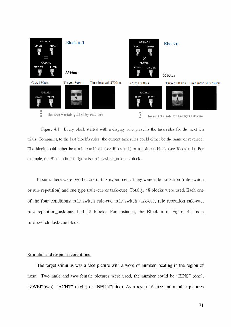
71
Figure 4.1: Every block started with a display who presents the task rules for the next ten
trials. Comparing to the last block’s rules, the current task rules could either be the same or reversed.
The block could either be a rule cue block (see Block n-1) or a task cue block (see Block n-1). For
example, the Block n in this figure is a rule switch_task cue block.
In sum, there were two factors in this experiment. They were rule transition (rule switch
or rule repetition) and cue type (rule-cue or task-cue). Totally, 48 blocks were used. Each one
of the four conditions: rule switch_rule-cue, rule switch_task-cue, rule repetition_rule-cue,
rule repetition_task-cue, had 12 blocks. For instance, the Block n in Figure 4.1 is a
rule_switch_task-cue block.
Stimulus and response conditions.
The target stimulus was a face picture with a word of number locating in the region of
nose. Two male and two female pictures were used, the number could be “EINS” (one),
“ZWEI”(two), “ACHT” (eight) or “NEUN”(nine). As a result 16 face-and-number pictures

72
were used as target stimuli. The same set of target stimuli was used for German and Chinese
participants. While the words in the task rule information display and cue display were in
German and Chinese, respectively.
All the stimuli, task rule information display before every block, cue stimuli and target
stimuli in every trial, were located on a black background in the centre of the screen, and
subtended less than 10 degrees, 5 degrees of visual angle respectively.
Participants used their left and right finger for response. They were instructed to respond
as fast and as accurately as possible.
4.3. Expectations
For the first few trials of block, a larger rule cue benefit was expected in the rule switch
than rule repetition block. Because I expected that the rule cue benefit was contributed by the
rule cue facilitation to the processing of rule acquisition and rule implementation both in the
rule switch block, whereas it was only produced by the rule cue facilitation to the rule
implementation in the rule repetition block However, for the rest trials in a block, even it is a
rule switch block, the task rules should been acquired well already. Therefore the rule cue
benefit should be produced by rule cue facilitation to rule implementation only no matter the
block is a rule repetition or rule switch block. Thus for the rest trials of blocks, identical rule
cue benefits were expected for the rule switch and rule repetition block.
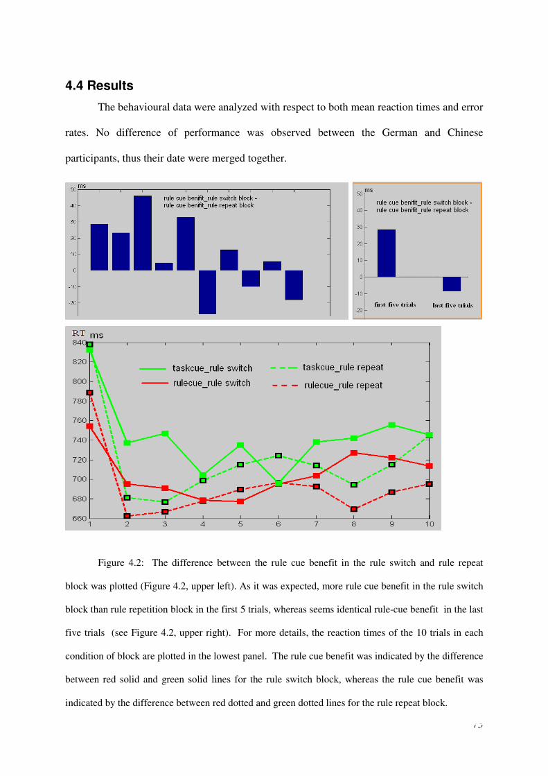
73
4.4 Results
The behavioural data were analyzed with respect to both mean reaction times and error
rates. No difference of performance was observed between the German and Chinese
participants, thus their date were merged together.
Figure 4.2: The difference between the rule cue benefit in the rule switch and rule repeat
block was plotted (Figure 4.2, upper left). As it was expected, more rule cue benefit in the rule switch
block than rule repetition block in the first 5 trials, whereas seems identical rule-cue benefit in the last
five trials (see Figure 4.2, upper right). For more details, the reaction times of the 10 trials in each
condition of block are plotted in the lowest panel. The rule cue benefit was indicated by the difference
between red solid and green solid lines for the rule switch block, whereas the rule cue benefit was
indicated by the difference between red dotted and green dotted lines for the rule repeat block.
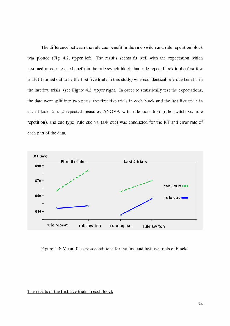
74
The difference between the rule cue benefit in the rule switch and rule repetition block
was plotted (Fig. 4.2, upper left). The results seems fit well with the expectation which
assumed more rule cue benefit in the rule switch block than rule repeat block in the first few
trials (it turned out to be the first five trials in this study) whereas identical rule-cue benefit in
the last few trials (see Figure 4.2, upper right). In order to statistically test the expectations,
the data were split into two parts: the first five trials in each block and the last five trials in
each block. 2 x 2 repeated-measures ANOVA with rule transition (rule switch vs. rule
repetition), and cue type (rule cue vs. task cue) was conducted for the RT and error rate of
each part of the data.
Figure 4.3: Mean RT across conditions for the first and last five trials of blocks
The results of the first five trials in each block
75
For the RT of correct trials (Fig. 4.3), significant main effect of cue type was found.
Subjects are faster when the task is indicated by a rule cue than a task cue (635ms vs. 670ms,
F (1, 21) = 14.84, p<0.001). Another, the reaction time in the rule switch condition is slower
than in the rule repeat condition (645 vs. 660ms), however this effect failed to be significant
(F (1, 21) = 2.66, p=0.118).
In addition and importantly, more rule cue benefit was found in the rule switch than
rule repeat condition (23ms vs. 46ms) (figure 12). And corresponding interaction between
rule transition and cue type tends to be significant (F (1, 21) = 3.55, p = 0.074). Further t test
indicate significant rule cue benefit exist in the rule switch (46ms, t(21) = 3.73, p < 0.001 )
and in the rule repeat condition (23ms, t(21) = 2.53, p < 0.05). The result indicated that the
rule cue benefit was apparently larger in the rule switch blocks than in the rule repeat blocks
in the first five trials
For the error rate, the only tend to be significant effect is the main effects of cue type.
Less error rate was found when the task is indicated by a rule cue than a task cue (3.8 vs.
5.0%, F (1, 21) = 3.12, p=0.092). Neither the main effect of rule transition (3.2 vs. 4.6%, F (1,
21) = 0.36, p = 0.55) nor the interaction between rule transition and cue type (F (1, 21) =
0.027, p = 0.87) is significant.
The results of the last five trials in each block
For the RT of correct trials (Fig. 4.3), significant main effect of cue type was found.
Subjects were faster when the task is indicated by a rule cue than a task cue (636ms vs.
662ms, F (1, 21) = 14.65, p<0.001). In addition, the reaction time in the rule switch condition
is significantly slower than in the rule repeat condition (641 vs. 658ms, F (1, 21) = 7.85,
p<0.05, =0.011).
76
As expected, no interaction between rule transition and cue type was found (F (1, 21)
= 0.027, p=0.87) (Fig. 4.3), which supported that the rule cue benefit was identical in the rule
switch and rule repetition block. Further t tests revealed nearly significant rule cue benefit in
the rule switch block (22ms, t(21) = 2.02, p = 0.056) and significant rule cue benefit in rule
repetition block (29ms, t(21) = 2.24, p < 0.05).
For the error rate, neither significant main effect nor interaction was found (all Fs <
1.7, all ps > 0.2).
4.4 Summary
As expected, rule cue benefit appeared larger in the rule switch than rule repetition block
for the first few trials of blocks. Because the rule cue benefit was contributed by the rule cue
facilitation to the processing of rule acquisition and rule implementation both in the rule
switch block, whereas it was only produced by the rule cue facilitation to the rule
implementation in the rule repetition block.
Moreover, for the rest trials in a block, even it is a rule switch block, the task rules were
acquired well already. Therefore the rule cue benefit was produced by the rule cue facilitation
to rule implementation only no matter the block was a rule repetition or rule switch block.
Correspondingly, for the rest trials of blocks, identical rule cue benefit were found in the rule
switch and rule repetition block.
77
In sum, the findings of this present experiment supported that the learning of task rule from
rule-cue displays took place when the task rules have not yet been obtained well (i.e., the first
5 trials in a block with need of task rules re-obtaining).
Note that, the task rules were stable in Experiment 1 and 2, and participants received
enough practice before their formal experiment. Therefore, there was no need for the
participants to learn the task rule from rule-cue display in the formal experiments, as a result,
the learning process had no chance to take place then to confound the findings of rule
implementation in Experiment 1 and 2.
78
CHAPTER 5
General discussion
The present study was interested in two critical components of executive control in
the changing task context. In particular, the neural evidence for preparatory attentional bias
and the functional neuroanatomy of task rules implementation. To this end, task-cuing
paradigm was adopted, in which the changing task context requires general preparation for
the upcoming task as well as the flexible activation of the appropriate task rules and
attending to appropriate target item.
In order to identify the neural correlates on rule implementation, rule-cue was devised
nd applied in the task-cuing paradigm. The rule-cue provides explicit information not only
about the type of task to be performed, but also about the specific S-R rule to be applied on
the upcoming trial. It was expected to implement the task rule more efficiently than the
task-cue who only explicitly supplies task type information in the preparation period. This
hypothesis was supported by the behavior rule-cue benefit in the two fMRI experiments
and the subtle analysis on the function of rule-cue in Experiment 3. Whereas in the task-cue
condition, part of the task rule activation was expected to be postponed into the task
execution period and/or more implementation of rule activation was needed. With the
79
comparison between rule-cue and task-cue, the rule-related foci, including the right anterior
and middle parts of MFG and SFG, the right IFJ, the pre-SMA, as well as the right SPL and
IPL, were isolate from the neural correlates for general task preparation in fMRI
Experiment 1. And the region of anterior MFG and SFG, IFJ and pre-SMA again showed
to be rule-related in fMRI Experiment 2.
The Experiment 1 of this study was inclined to conclude that the extent to which
participants prepare in advance the parameters of a future task depends on the specificity
and the amount of information provided prior to task processing. These in turn determines
the degree of activation in brain regions associated with task preparation and the following
task execution.
In order to find the evidence of attentional preparatory modulation into the stimulus-
specific posterior brain regions, the face discrimination and number discrimination tasks
were selected the fMRI Experiment 2 because they both have stimulus-specific posterior
brain region (i.e. FFA and IPSnum) and they are balanced in task difficulty which
encourage participants prepare the two task with similar extent of motivation. And another,
both the task relevant and irrelevant feature were presented in the target display in a
overlapped rather than spatially separated manner, thus to increase the need of attenional
bias in preparation period. As a result, the preparatory modulation was found in the face
specific and number specific posterior brain regions. In particular, significant additional
activity comparing to null trials was found in the preparation period of face task in right
FFA but not in the right IPSnum; while in the preparation period of number task,
significant additional activity comparing to null trials was found in right IPSnum but not in
the right FA. Further, in the task execution period, no such stimulus-specific modulation
was found in either the face or the number task. These findings support the hypothesis that
the preparatory attentional control can bias sensory processing via amplifying the activity
80
of the task-relevant stimulus-specific brain region; and suggest that the modulation may not
be necessarily needed for task execution if an efficient advanced modulation has been
encouraged to take place already.
The Experiment 2 of this study found an attentional modulation of the activities in
the posterior stimulus-specific brain regions; meanwhile the activities in these posterior
regions were also influenced by the residual activity in the preceding trial. Moreover, the
results also suggested that the modulation may not be necessarily needed for task execution
if an efficient advanced modulation has been encouraged to take place already.
5.1 Attentional bias, task rule activation and task preparation
The neural correlates
In the present fMRI Experiment 1 and 2, general preparation-related activation is
reflected in the activity elicited by cue presentation on cue-only trials, for both the rule-cue
and task-cue conditions. The presentation of these cues led to the activation of a large fronto-
parietal brain network including the MeFC, the bilateral IFJ, the dorsal and lateral premotor
cortices, the medial and lateral parietal lobe, with a relatively smaller magnitude of
activation in the bilateral anterior LPFC. This network is largely consistent with that
reported in a number of previous studies concerned with the neural correlates of task
preparation (Brass & von Cramon 2002, 2004; Gruber et al. 2006; Luks, et al. 2002;
MacDonald et al. 2000; Sohn et al. 2000).
According to the preceding image studies, the foci of frontal eye field (FEF) and IPS
subtract the preparatory space-based attentional control (e g., Corbetta et al., 2000;
Hopfinger et al., 2000; Shulman et al., 1999). And there was some evidence suggested that
81
the object-based preparatory attentional control relys on the same foci also (Shulman et al.,
1999; see also Perry & Zeki 2000; Wojciulik & Kanwisher 1999). The large scared lateral
frontal and pariatal cortex activated for the general task preparation in exp 1 and 2 involved
the FEF and IPS, which should associated with the effort of actively biasing of attention in
task preparation. In addition and importantly, in Experiment 1 and 2 of this present study,
the rule-related activation foci were a subset of this general task preparation network. In this
subset of rule-related regions, the processing of rule (cue) information led to enhanced
activation compared to the processing of task (cue) information – as a result of the explicit
rule information provided by the rule (but not the task) cues. While rule-cues are as effective
as task-cues in activating the general task goal (i.e., the type of task to be performed), they
are more powerful in activating the specific task rule. As a result, rule-cues engender
superior task preparation compared to task-cues, which is expressed in better performance
measures such as response speed and accuracy and in increased neural computations in the
related brain regions.
Influential factors for task preparation
There has been a long-standing debate concerning the extent to which participants prepare
in advance the (whole) set or only parts of relevant task parameters following cue
presentation (Brass & von Cramon 2002; Gruber et al. 2006; Luks, et al. 2002; Ruge et al.
2009; Verbruggen et al., 2007). The present findings suggest that this strongly depends on
the amount of explicit task information provided by the cue and the extent of encouraging
for advanced task preparation by task setting.
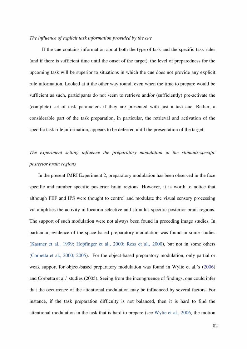
82
The influence of explicit task information provided by the cue
If the cue contains information about both the type of task and the specific task rules
(and if there is sufficient time until the onset of the target), the level of preparedness for the
upcoming task will be superior to situations in which the cue does not provide any explicit
rule information. Looked at it the other way round, even when the time to prepare would be
sufficient as such, participants do not seem to retrieve and/or (sufficiently) pre-activate the
(complete) set of task parameters if they are presented with just a task-cue. Rather, a
considerable part of the task preparation, in particular, the retrieval and activation of the
specific task rule information, appears to be deferred until the presentation of the target.
The experiment setting influence the preparatory modulation in the stimuuls-specific
posterior brain regions
In the present fMRI Experiment 2, preparatory modulation has been observed in the face
specific and number specific posterior brain regions. However, it is worth to notice that
although FEF and IPS were thought to control and modulate the visual sensory processing
via amplifies the activity in location-selective and stimulus-specific posterior brain regions.
The support of such modulation were not always been found in preceding image studies. In
particular, evidence of the space-based preparatory modulation was found in some studies
(Kastner et al., 1999; Hopfinger et al., 2000; Ress et al., 2000), but not in some others
(Corbetta et al., 2000; 2005). For the object-based preparatory modulation, only partial or
weak support for object-based preparatory modulation was found in Wylie et al.’s (2006)
and Corbetta et al.’ studies (2005). Seeing from the incongruence of findings, one could infer
that the occurrence of the attentional modulation may be influenced by several factors. For
instance, if the task preparation difficulty is not balanced, then it is hard to find the
attentional modulation in the task that is hard to prepare (see Wylie et al., 2006, the motion
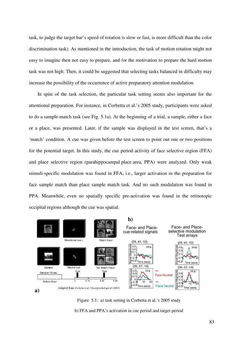
83
task, to judge the target bar’s speed of rotation is slow or fast, is more difficult than the color
discrimination task). As mentioned in the introduction, the task of motion rotation might not
easy to imagine then not easy to prepare, and /or the motivation to prepare the hard motion
task was not high. Then, it could be suggested that selecting tasks balanced in difficulty may
increase the possibility of the occurrence of active preparatory attention modulation.
In spite of the task selection, the particular task setting seems also important for the
attentional preparation. For instance, in Corbetta et al.’s 2005 study, participants were asked
to do a sample-match task (see Fig. 5.1a). At the beginning of a trial, a sample, either a face
or a place, was presented. Later, if the sample was displayed in the test screen, that’s a
‘match’ condition. A cue was given before the test screen to point out one or two positions
for the potential target. In this study, the cue period activity of face selective region (FFA)
and place selective region (parahippocampal place area, PPA) were analyzed. Only weak
stimuli-specific modulation was found in FFA, i.e., larger activation in the preparation for
face sample match than place sample match task. And no such modulation was found in
PPA. Meanwhile, even no spatially specific pre-activation was found in the retinotopic
occipital regions although the cue was spatial.
Figure 5.1: a) task setting in Corbetta et al.’s 2005 study
b) FFA and PPA’s activation in cue period and target period
a)
b)
84
These non-robust findings may due to the task setting did not encourage active
preparation for either object or space: 1) The face and house pictures are specially
dissociated thus not easy to distract each other, which may not hardly encourage the
participants bias attention to the target object category. 2) One could infer that the most
active preparation participants should include both space-based and object-based attentional
control, in particular bias their attention to the one or two position for the potential target
(space-based) and to the category of the target (object-based). However it was uncertain
that all the participants fully prepared the task in the most active manner. Actually seeing
from the results, only weak pre-activation was found in the object-selective visual cortex
and no spatially specific pre-activation was found in the retinotopic occipital regions. The
only robust pre-activation was found while contrasting the rightward- and leftward-
directing cues in a region at the intersection of the IPS and occipital sulcus. Infer from these
results, the participants might actively use the directing cue to bias attention to the correct
side (left or right), but might not very actively bias their attention to the target object
category and the exact location. Because it is easy to find the target in the correct side after
the presenting of test display (always one face and one house picture, never need to
discriminate two pictures of one category, thus easy to find the target), and after they found
the target location, the pictures from the task-irrelevant category could hardly interfere the
task (spatially dissociated). Unlike the setting of Cobetta et al.’s study, in the present fMRI
Experiment 2, participants only need to do the object-base attentional control, since all the
pictures were displayed in a same place (the center of screen), and the task relevant picture
was overlapped by the picture of task irrelevant category to increase the interference. These
settings could encourage participants biasing their attention to the target category in
advance, and correspondingly, preparatory modulations were found in the object-selective
posterior brain regions (i.e., FFA and IPSnum).
85
5.2 Reengagement of executive control involved in preparation period
The rule activation and attentional bias are considered to be critical components of
cognitive control in task cuing paradigm. In Experiment 1, the rule related regions activated
larger in the rule-cue than task-cue condition, which refer to more sufficient rule activation
in the preparation period in the rule-cue condition. In experiment2, preparatory stimulus-
specific modulation was found in the face specific and number specific regions, which refer
to the efficient preparatory attentional bias. Interestingly, in Experiment 1, the rule
implementation regions reengaged more in the task-cue than rule-cue condition, whereas in
Experiment 2, no further stimulus-specific modulation was found in the target period,
probably because the preparatory modulation was efficient in this study. These findings in
Experiment 1 and 2 are in line with some preceding findings: 1) the foci of general
anticipatory control (e g., Brass & von Cramon 2002; Gruber et al. 2006) and preparatory
attentional control (e g., Corbetta et al. 2000; Shulman et al. 2002b) reengaged in the task
execution processing. 2) Similar to the finding of Experiment 2, no attentional modulation
was found after efficient preparatory modulation (see Fig 5.1b, FFA’s activity), but if the
preparatory attention control failed to biasing the sensory processing in the stimulus-specific
region, attentional modulation was found in the target period (see Fig. 5.1b, place specific
region PPA’s activity).
All of these finding indicate that the anticipatory cognitive control in the changing task
context actually could play role not only when preparing the task but rather whenever it is
needed.
86
5.3 The rule-related regions
LPFC
The present findings indicate that a major region associated with task rule activation is
the LPFC. A number of studies that used different paradigms have shown the LPFC to be
involved in the maintenance of task-relevant information (e.g., Egner & Hirsch 2005; Kerns et
al. 2004; Koechlin et al., 2003; Koechlin & Summerfield 2007; Yeung et al. 2006).
Increased activation in dorsal regions of the LPFC (i.e., DLPFC) related to the
maintenance of task rule activation has also been shown for the Stroop paradigm. For
example, MacDonald et al. (2000) found neural activity to be increased in the DLPFC when
participants were presented with a cue that specified the task coming up next (in the Stroop
paradigm). Interestingly, the magnitude of activity increased with the expected difficulty of
this task: the DLPFC was more strongly activated when the cue indicated to participants that
they would have to process the color of a (color) word, as compared to when it signalled that
they would have to process the name of a (color) word. The present findings are in line with
these results and they show, additionally, that the degree of activity in DLPFC regions also
varies with the amount of specific information provided by the cue about the upcoming task.
An association of rule-relevant processing with regions in the LPFC has been reported
in recent fMRI studies by Bunge and colleagues (Bunge et al. 2003; see also Crone et al.
2006a, 2006b). In the Bunge et al. study, participants learned different rules of how to
respond to probe stimuli in a separate learning phase prior to the fMRI scanning session.
Similar rules (e.g., press left key if two stimuli match each other) were associated with
different types of rule-cue (verbal or symbolic cues). In the fMRI scanning session,
participants had to activate the acquired rule knowledge upon presentation of the rule-cues
and then, after a delay, process two sequentially presented probe stimuli (same, different).
87
Bunge et al. found ventral regions in the LPFC to be active during the delay after rule-cue
presentation, to be sensitive to the difficulty of the rule, and to be insensitive to the type of
rule-cue. Because of the insensitivity of these regions to the type of rule-cue, ventral LPFC
regions were assumed to be related to abstract rule knowledge; this was in contrast to regions
in the DLPFC which proved to be sensitive to the different types of rule-cue and were,
therefore, assumed to be related to the specific rule knowledge. These findings are consistent
with recent single-cell studies in monkeys in which a similar paradigm was used to assess the
representation of rule knowledge in prefrontal regions (Wallis & Miller, 2003; White & Wise,
1999).
While these studies show an involvement of LPFC regions in the retrieval of (abstract
and specific) rule knowledge from long-term memory, the present findings show that the
degree of rule-related activity may be modulated by the amount of rule information provided
by the actual rule-cue. Prior findings were not conclusive about the degree of rule activation
in the LPFC because the cues used did not permit distinguishing between general task
information and specific rule information (i.e., the different types of information provided by
the task and, respectively, rule-cues in the present study).
aPFC
While prior studies of task preparation have mainly reported preparation-related
activation in more posterior prefrontal regions, such as near the IFJ and/or the premotor
cortex (e.g., Brass & von Cramon 2002, 2004; Gruber et al. 2006), this study found additional
preparation-related activation in the right aPFC, and this activation proved to be involved in
task rule activation. Findings reported in the literature indicate a critical role of the aPFC for
difficult retrieval processes in both episodic-memory (Della-Maggiore et al. 2002; Nyberg et
88
al. 2000; Rajah et al. 1999) and working-memory tasks (Christoff & Gabrieli 2000; Leung et
al. 2005; MacLeod et al. 1998; Soto et al. 2007). According to a meta-analysis conducted by
MacLeod et al., the right aPFC tends to be activated especially in difficult WM tasks (e.g.,
when a high, rather than a low number, of items has to be maintained). In particular, the meta-
analysis of MacLeod et al. yielded mean activation foci associated with processing in difficult
WM tasks which are close (25, 61, 6; Talairach & Tournoux 1988) to the coordinates of the
aPFC activation revealed in the present study.
The finding of aPFC activation is also consistent with a study of Stern et al. (2007),
who found preparatory activation in the right aPFC in a spatial Stroop task in which the need
for rule activation was greater compared to the tasks employed in previous studies (such as
those of Brass & von Cramon 2002, 2004; Gruber et al. 2006). In more detail, in Stern et al.,
participants were presented with a direction-related word (e.g., ‘up’ or ‘down’) on an upper or
lower position of the computer screen, and they had to respond to either the direction word or
the word location according to a task-cue. Importantly, Stern et al. included only incongruent
trials (e.g., respond to the work ‘up’ presented at the lower position), which increased the
need for preparatory processes during task-cue processing as compared to studies that
intermixed congruent and incongruent conditions. Accordingly, Stern et al. (2007) concluded
that the aPFC activation observerd in their study was critical for preparation-related processes
in task performance.
Thus, based on these findings, it could be propose that, although the present task was
only moderately difficult, the external rule information provided by the rule-cues did enforce
the S-R associations to be activated and retrieved from memory to a greater degree than in the
task-cue condition. This is the likely explanation for the finding of rule-related activation in
the right aPFC in the present paradigm.
89
IFJ
Activation in posterior regions of the LPFC near the IFJ has been shown to be
associated with mechanisms involved in the ‘actualization’ of the current task representation
(e.g., by uploading new task parameters or a new task representation) in situations with
changing tasks (Brass & von Cramon, 2004; Derrfuss et al. 2005). The present findings are
consistent with this, while additionally showing that the degree to which a new task
representation is uploaded in advance depends on the specificity of the information provided
by the cue. A larger amount of presented task information permits a more complete uploading
of the task parameters required on the upcoming trial, and this is accompanied by an increased
amount of neural activity in brain areas near the IFJ.
Rule-related activation outside the lateral prefrontal cortex
Further rule-related activation was found in medial frontal regions and here
specifically within the pre-SMA, that is, in regions that are more dorsally located than regions
in the anterior cingulate cortex which are associated with mechanisms of conflict detection
and/or processing (Carter et al. 2000; Weissman et al.2005). Single-cell and tracer studies
suggest that regions of the pre-SMA receive direct input from the LPFC, while the neural
regions in the SMA proper are connected to the motor areas (Picard & Strick 2001; Tanji
1994). The specific connections to the LPFC make the pre-SMA most appropriate for the
preparation of the specific task rule during sensori-motor performance (Hikosaka et al. 1996).
In line with this, several authors have shown the pre-SMA to be involved in the acquisition
and control of arbitrary S-R associations in humans (Halsband & Freund 1990; Hikosaka et
al. 1996; Gordon et al. 1995; Picard & Strick 1996; Sakai et al. 1998, 1999) and monkeys

90
(Halsband & Passingham 1985; Halsband et al. 1994). The present study found the pre-SMA
to be activated in conditions promoting task rule activation as well as conditions of general
task preparation. However, the fact that there was additional activation in the pre-SMA in
rule-cue compared to task-cue conditions suggests that providing participants with explicit
rule information leads to an enhanced preparation of (specific) S-R associations.
The observation of rule-related activation in the parietal cortex is consistent with the
assumption that regions along the intra-parietal sulcus are involved in the activation of motor
representations which are spatially mapped to different sensory stimuli (Andersen 1987;
Caminiti et al. 1996; Schubert et al. 1998; Stein 1992). A number of neuroimaging studies
have revealed parietal activation when participants have to produce motor responses upon the
presentation of sensory stimuli in various kinds of behavioral paradigms (Bunge et al. 2002,
2003; Brass et al. 2002, 2004; Zysset et al. 2006; Snyder et al. 1997; Stelzel et al., 2008). The
present findings additionally suggest that the parietal involvement is modulated by the amount
of prior information provided about the motor response that has to be performed upon
stimulus presentation: the more information is provided about the S-R rule, the larger the
amount of neural computation in parietal areas involved in processing the required S-R
association.
5.4 A higher need of control for the task rule preparation in switch trials
A larger activation was found in exp 1 and 2 during the preparation period (cue-only
trials) for task switch compared to task repetition conditions in the medial SFG. This extends
findings of other studies, pointing to an association of this region with the switching between
tasks (e.g., Dove et al. 2000; Yeung et al. 2006). A related ROI analysis specified that the
91
observed increase of switch-related (compared to repetitions) activation in the medial SFG
region was similar in the rule-cue and task-cue conditions. Additionally, the ROI analysis
showed that the general amount of activity in switch-related regions was larger with rule-
cues than with task-cues. In our view, this latter finding suggests that, given sufficient task
rule information, the need to prepare for a task switch evokes efficient processes of reloading
the task rule information already during the preparation period of the task processing.
Rule retrieval, or activation of the currently required task set, is presumed to be an
important component of task switching (Mayr & Kliegl 2000; Rogers & Monsell 1995,
2003; Rubinstein et al. 2001). In the present study, the task rule information provided by
rule-cues may have evoked preparatory processes that included even the rule representation,
thus permitting a more complete task set reconfiguration (on switch trials) compared to the
presentation of mere task-cues. This would explain why the task switch costs (i.e., error
rates) were reduced in the rule-cue relative to the task-cue condition, and why the amount of
neural activation was increased in preparation-related regions with rule-cues compared to the
task-cues.
Some previous fMRI studies had failed to find any additional region, or even
activation, to be involved in preparing for task switches (as compared to repetitions), which
was taken to cast doubt on the assumption that switch-specific control processes are
operating during the preparation period (Brass & von Cramon 2002, 2004; Gruber et al.
2006; Ruge et al. 2005). I agree that the preparatory processes are comparable in the
anticipation of task switches and of repetitions; however, one could suggest that more
control is required when preparing for switch trials, and the corresponding additional
activation can be found in either the task preparation or the execution period, depending on
the type of cue.
92
A possible reason for the discrepant findings concerning switch-related activations in the
preparation period may lie in the different types of cues, or cue information, which were
used in studies that failed to and that did find such activations. Studies that failed to find
additional switch-related activation (see below) used arbitrary cues (e.g., in Brass et al.,
2002, a square or diamond indicating an odd/even and or a number size task), whereas this
present study used semantically unequivocal cues to indicate the upcoming task (see also
Wylie et al. 2006). It would appear plausible that, if the cue is an arbitrary shape, a
considerable amount of time needs to be spent to decode the cue and to represent the general
task goal – that is, task rule activation may be delayed and moved to the target period. This
might be the reason why studies that used such arbitrary cues failed to find any larger
activation in the preparation for task switches compared to repetitions (Brass & von Cramon
2002, 2004; Gruber et al.2006; Ruge et al. 2005). In our study, the word symbol ‘gender’
indicated the gender task and the symbol ‘color’ the color task. This use of semantic cues is
similar to the conditions in other studies which also reported additional switch-related
activity during the preparation period (e.g., Wylie et al. 2006). In the case of semantic cues,
the cue specifies the task relatively directly, making it much easier for participants to
establish the task representation (Miyake et al. 2004; Wylie et al. 2006) – and permitting
them to activate the task rule already within the preparation period. As a consequence, the
neural effort associated with the uploading of the task rule information would be increased
on task switch compared to repetition trials, and this effort may be strong enough to evoke
significant fMRI activation in the comparison of switch versus repetition trials during the
task preparation period (Wylie et al. 2006).
93
5.5 The persistent activity from preceding trials in the posterior brain regions
In the present Experiment 2, residual activation from preceding trial existed in the
preparation period in the posterior stimuls-specificbrain regions (i.e., right FFA and right
IPSnum). In preview studies for human and monkey, persistent activity was found in PFC,
but in the delay period of working memory task (e g., Bunger et al., 2003; Fuster &
Alexander 1971; Funahashi et al., 1989, 1990, 1991; Miller et al., 1996). An important
difference between the persistent activity in PFC and posterior brain regions is that the
former evident an intentional operation for task goal, while the latter is not. Rather the
persistent activity could be interference if the current task is not the same as the preceding
one. The inertia activity of the preceding trial was claimed to be a important recourse of task
switching cost (Allport 1994; Allport & Wylie 1999, 2000), and this opinion was supported
by neural evidence from Yeung et al.’s study (2006). In this study, the switch cost was
significantly correlated with the amount of activity of the task irrelevant posterior brain
regions thus provided a good example on the bottom-up influence for behavior. In the
present Experiment 2, preparatory attentional control amplified the activity in the task
relevant rather than the irrelevant posterior brain region, meanwhile, the residual activity
from preceding task still exist, these two factors together shaped the exact activity pattern in
the posterior brain regions. Such findings provided a good example to show the top-down
modulation and the bottom-up influence in sensory processing simultaneously.
94
5.6 Conclusion
This study isolated the neural correlates for task rule implimentation from those
related to general task preparation and found an preparatory attentional modulation of the
activities in the posterior stimulus-specific brain regions. Moreover, this study was inclined
to conclude that the extent to which participants prepare in advance the parameters of a
future task in turn determines the degree of activation in brain regions associated with task
preparation and the following task execution (i.e. larger activity of rule implementation was
found in the task execution period if the rule has not been sufficiently implemented in
advance; no attentional modulation of activity in the posterior brain regions was found in
the task execution period after a efficient bias of attention in task preparation).

95
References
Allport DA, Styles EA, Hsieh S (1994) Shifting intentional set: Exploring the dynamic
control of tasks. In C. Umilta & M. Moscovitch (Eds.), Attention and Performance XV, pp.
421- 452. Cambridge, MA: MIT Press.
Allport DA, Wylie G (1999) Task-switching: Positive and negative priming of task-set. In G.
W. Humphreys, J. Duncan, & A. Treisman (Eds.), Attention, space, and action: Studies in
cognitive neuroscience, pp 273-296. Oxford: Oxford University Press.
Allport DA, Wylie G (2000) “Task-switching,” stimulus-response bindings, and negative
priming. In S. Monsell & J. Driver (Eds.), Control of cognitive processes: Attention and
performance XVIII, pp 35-70. Cambridge, MA: MIT Press.
Andersen RA (1987) The role of the inferior parietal lobule in spatial perception and visual-
motor integration. In: The handbook of physiology, Sec 1, The nervous system, Vol V,
Higher functions of the brain, Ft 2 (Plum F, Mountcastle VB, Geiger SR, eds), pp 483-518.
Bethesda, MD: American Physiological Society. 58
Brass M, von Cramon DY (2002) The role of the frontal cortex in task preparation. Cereb
Cortex 12:908-914.
Brass M, von Cramon DY (2004) Decomposing components of task preparation with
functional magnetic resonance imaging. J Cogn Neurosci 16:609-620.
Bunge SA, Kahn I, Wallis JD, Miller EK, Wagner AD (2003) Neural circuits subserving the
retrieval and maintenance of abstract rules. J Neurophysiol 90:3419-3428.
Bunge SA, Hazeltine E, Scanlon MD, Rosen AC, Gabrieli JD (2002) Dissociable
contributions of prefrontal and parietal cortices to response selection. Neuroimage 17:1562-
1571.
Bunge SA (2004) How we use rules to select actions: a review of evidence from cognitive
neuroscience. Cogn Affect Behav Neurosci 4(4):564-79.
Caminiti R, Ferraina S, Johnson PB (1996) The sources of visual information to the primate
frontal lobe: a novel role for the superior parietal lobule. Cereb Cortex 6:319-328.
Carter CS, Macdonald AM, Botvinick M, Ross LL, Stenger VA, Noll D, Cohen JD (2000)
Parsing executive processes: strategic vs. evaluative functions of the anterior cingulate cortex.
Proc Natl Acad Sci USA 97:1944-1948.
Christoff K, Gabrieli JDE (2000) The frontopolar cortex and human cognition: evidence for a
rostrocaudal hierarchical organization within the human prefrontal cortex. Psychobiology
28:168-186.
Corbetta M, Kincade JM, Ollinger JM, McAvoy MP, Shulman, GL (2000) Voluntary
orienting is dissociated from target detection in human posterior parietal cortex. Nature
Neuroscience 3:292-297.
96
Corbetta M, Kincade JM, Shulman GL (2002) Neural systems for visual orienting and their
relationships to spatial working memory. J Cogn Neurosci 14:508–523.
Corbetta M, Tansy AP, Stanley CM, Astafiev SV, Snyder AZ, Shulman GL (2005) A
functional MRI study of preparatory signals for spatial location and objects.
Neuropsychologia 43:2041-2056.
Crone EA, Donohue SE, Honomichl R, Wendelken C, Bunge SA (2006) Brain regions
mediate flexible rule use during development. J Neurosci 26:11239-11247.
Crone EA, Wendelken C, Donohue SE, Bunge SA (2006) Neural evidence for dissociable
components of task-switching. Cereb Cortex 16:475-486.
Dehaene S, Piazza M, Pinel P, Cohen L (2003) Three parietal circuits for number processing.
Cognitive Neuropsychology 20:487-506.
Della-Maggiore V, Grady CL, McIntosh AR (2002) Dissecting the effect of aging on the
neural substrates of memory: deterioration, preservation or functional reorganization? Rev
Neurosci 13(2):167-181.
Derrfuss J, Brass M, Neumann J, von Cramon DY (2005) Invovlement of the inferior frontal
junction in cognitive control: Meta-analyses of switching and stroop studies. Hum Brain
Mapp 25:22-34.
Desimone R, Duncan J (1995) Neural mechanisms of selective visual attention. Annu Rev
Neurosci 18:193-222.
Dove A, Pollmann S, Schubert T, Wiggins CJ, von Cramon DY (2000) Prefrontal cortex
activation in task-switching: an event-related fMRI study. Cogn Brain Res 9:103-109.
Egner T, Hirsch J (2005) Cognitive control mechanisms resolve conflict through cortical
amplification of task-relevant information. Nat Neurosci 8:1784-1790.
Forman S, Cohen J, Fitzgerald M, Eddy W, Mintun M, Noll D (1995) Improved assessment
of significant activation in functional magnetic resonance imaging (fMRI): Use of a cluster-
size threshold. Mag Reson Med 33:636-647.
Friston KJ, Holmes AP, Worsley KJ, Poline JP, Frith CD, Frackowiak RSJ (1995) Statistical
parametric maps infunctional imaging: a general lineat approach. Hum Brain Mapp 2:189-
210.
Funahashi S, Bruce CJ, Goldman-Rakic PS (1989) Mnemonic coding of visual space in the
monkey’s dorsolateral prefrontal cortex. J Neurophysiol 61:331-349.
Funahashi S, Bruce CJ, Goldman-Rakic PS (1990) Visuospatial coding in primate prefrontal
neurons revealed by oculomotor paradigms. J Neurophysiol 63:814-831.
Funahashi S, Bruce CJ, Goldman-Rakic PS (1991) Neuronal activity related to saccadic eye
movements in the monkey’s dorsolateral prefrontal cortex. J Neurophysiol 65:1464-1483.
97
Fuster JM, Alexander G (1971) Neuron activity related to short-term memory. Science
173:652-654.
Gollwitzer PM, Sheeran P (2006) Implementation intentions and goal achievement: A
metaanalysis of effects and processes. Adv Exp Soc Psych 38:69-119.
Gordon AM, Flament D, Lee JH, Uguibil K, Kim SG, Ebner TJ (1995) Functional MRI of
cortical motor areas during sequential typing movements. Abstract 1422. Annual Meeting of
the Socirty of Neuroscience. London: MIT Press.
Gruber O, Karch S, Schlueter EK, Falkai P, and Goschke T (2006) Neural mechanisms of
advance preparation in task switching. NeuroImage 31:887-895.
Halsband U, Freund HJ (1990) Premotor cortex and conditional motor learning in man. Brain
113:207-222.
Halsband U, Matsuzaka Y, Tanji J (1994) Neuronal activity in the primate supplementary,
pre-supplementary and premotor cortex during externally and internally instructed sequential
movements. Neurosci Res 20(2):149-155.
Halsband U, Passingham RE (1985) Premotor cortex and the conditions for movement in
monkeys (Macaca fascicularis). Behav Brain Res 18(3):269-277.
Hikosaka O, Sakai K, Miyauchi S, Takino R, Sasaki Y, Putz B (1996) Activation of human
presupplementary motor area in learning of sequential procedures: A functional MRI study. J
Neurophysiol 76:617-621.
Hopfinger JB, Buonocore MH, Mangun GR (2000) The neural mechanisms of top-down
attentional control. Nature Neuroscience 3:284–291.
Kanwisher N, McDermott J, Chun MM (1997) The fusiform face area: a module in human
extrastriate cortex specialized for face perception. J Neurosci 17:4302-4311.
Kastner S, Pinsk MA, De Weerd P, Desimone R, Ungerleider LG (1999) Increased activity in
human visual cortex during directed attention in the absence of visual stimulation. Neuron
22:751–761.
Kastner S, Ungerleider LG (2000) Mechanisms of visual attention in the human cortex. Annu
Rev Neurosci 23:315-341.
Kerns JG, Cohen JD, MacDonald AW III, Cho RY, Stenger VA, Carter CS (2004) Anterior
cingulate conflict monitoring and adjustments in control. Science 303:1023-1026.
Koechlin E, Summerfield C (2007) An information theoretical approach to prefrontal
executive function. Trends Cogn Sci 11(6):229-235.
Koechlin E, Ody C, Kouneiher F (2003) The architecture of cognitive control in the human
prefrontal cortex. Science 302:1181-1185.
98
Kriegeskorte N, Simmons WK, Bellgowan PS, Baker CI (2009) Circular analysis in systems
neuroscience: the dangers of double dipping. Nat Neurosci 12:535-540.
Lavric A, Mizon GA, Monsell S (2008) Neurophysiological signature of effective
anticipatory task-set control: a task-switching investigation. Euro J Neurosci 28(5):1016-
1029.
Lepsien J, Nobre, AC (2007) Attentional modulation of object representations in working
memory. Cerebral Cortex 17(9): 2072-2083.
Leung HC, Gore JC, Goldman-Rakic PS (2005) Differential anterior prefrontal activation
during the recognition stage of a spatial working memory task. Cereb Cortex 15(11):1742-
1749.
MacDonald AW III, Cohen JD, Stenger VA, Carter CS (2000) Dissociating the role of the
dorsolateral prefrontal and anterior cingulate cortex in cognitive control. Science 288:1835-
1838.
MacLeod K, Buckner RL, Miezin FM, Petersen SE, Raichle ME (1998) Right anterior
prefrontal cortex activation during semantic monitoring and working memory. Neuroimage
7:41-48.
Mayr U, Kliegl R (2000) Task-set switching and long term memory retrieval. J Exp Psychol
Learn Mem Cogn 26:1124-1140.
Meiran N (1996) Reconfiguration of processing mode prior to task performance. J Exp
Psychol Learn Mem Cogn 22:1-20.
Meiran N (2000) Modeling cognitive control in task-switching. Psychol Res 63:234-249.
Miller EK and Cohen JD (2001) An integrative theory of prefrontal cortex function. Annu
Rev Neurosci 24:167-202.
Miller EK, Erickson CA, Desimone R (1996) Neural mechanisms of visual working memory
in prefrontal cortex of the macaque. J Neurosci 16:5154-5167.
Miller RR, Matzel LD (1988) The comparator hypothesis: A response rule for the expression
of associations. The psychology of learning and motivation 22:54-60
Miyake A, Emerson MJ, Padilla F, Ahn J (2004) Inner speech as a retrieval aid for task goals:
the effects of cue type and articulatory suppression in the random task cuing paradigm. Acta
Psychol 115:123-142.
Monsell S (2003) Task switching. Trends Cogn Sci 7:134-140.
Monsell S, Mizon GA (2006) Can the task-cuing paradigm measure an endogenous task-set
reconfiguration process? J Exp Psychol Hum Percept Perform 32:493-516.
99
Nichols T, Brett M, Andersson J, Wager T, Polinee JB (2005) Valid conjunction inference
with the minimum statistic. NeuroImage 25:653-660.
Nyberg L, Persson J, Habib R, Tulving E, McIntosh AR, Cabeza R, Houle S (2000) Large
scale neurocognitive networks underlying episodic memory. J Cogn Neurosci 12 (1):163-173.
O'Craven KM, Kanwisher N (2000) Mental imagery of faces and places activates
corresponding stiimulus- specific brain regions. J. Cogn. Neurosci 12(6): 1013-1023.
Perry RJ, Zeki S (2000) The neurology of saccades and covert shifts in spatial attention: an
event-related fMRI study. Brain 123:2273-2288.
Picard N, Strick PL (1996) Motor Areas of the Medial Wall: A review of their location and
functional activation. Cereb Cortex 6:342-353.
Picard N, Strick PL (2001) Imaging the premotor areas. Curr Opin Neurobiol 11(6):663-672.
Poline JB, Worsley KJ, Evans AC, Friston KJ (1997) Combining spatial extent and peak
intensity to test for activations in functional imaging. Neuroimage 5(2):83-96.
Rajah MN, McIntosh AR, Grady CL (1999) Frontotemporal interactions in face encoding and
recognition. Cogn Brain Res 8 (3):259-269.
Ress D, Backus BT, Heeger DJ (2000) Activity in primary visual cortex predicts performance
in a visual detection task. Nature Neuroscience 3(9):940–945.
Rogers RD, Monsell S (1995) The cost of a predictable switch between simple cognitive
tasks. J Exp Psychol Gen 124:207-231.
Rubinstein JS, Meyer DE, Evans JE (2001) Executive control of cognitive processes in task
switching. J Exp Psychol Hum Percept Perform 27:763-797.
Ruge H, Brass M, Koch I, Rubin O, Meiran N, von Cramon DY (2005) Advance preparation
and stimulus-induced interference in cued task switching: further insights from BOLD fMRI.
Neuropsychologia 43:340-355.
Ruge H, Braver, T, Meiran N (2009) Attention, intention, and strategy in preparatory control.
Neuropsychologia 47:1670-1685.
Rushworth MFS, Hadland KA, Paus T, Sipila PK (2002) Role of the human medial frontal
cortex in task switching: a combined fMRI and TMS study. J Neurophysiol 87: 2577-2592.
Sakai K, Hikosaka O, Miyauchi S, Sasaki Y, Fujimaki N, Putz B (1999) Presupplementary
motor area activation during sequence learning reflects visuo-motor association. J Neurosci
19: RC1(1-6).
100
Sakai K, Hikosaka O, Miyauchi S, Takino R, Sasaki Y and Pütz B (1998) Transition of brain
activation from frontal to parietal areas in visuo-motor sequence learning. J Neurosci
18:1827-1840.
Sakai K, Passingham RE (2003) Prefrontal interactions reflect future task operations. Nat
Neurosci 6:75-81.
Sakai K, Passingham RE (2006) Prefrontal set activation predicts rulespecific neural
processing during subsequent cognitive performance. J Neurosci 26:1211-1218.
Serences JT, Schwarzbach J, Yantis S (2001) Control mechanisms of object-based visual
attention in human cortex. Soc Neurosci Abstr 27:348-349.
Serences JT, Schwarzbach J, Courtney SM, Golay X, Yantis S (2004) Control of object-based
attention in human cortex. Cereb Cortex 14:1346-1357.
Serences JT, Yantis S (2007) Spatially selective representation of voluntary and stimulus-
driven attentional priority in human occipital, parietal, and frontal cortex. Cereb Cortex
17:284-293.
Schubert T, von Cramon, DY, Niendorf T, Pollmann S, Bublak P (1998) Cortical areas and
the control of self-determined finger movements: an fMRI study. NeuroReport 9(14):3171-
3176.
Shulman GL, D’Avossa G, Tansy AP, Corbetta M (2002a) Two attentional processes in the
parietal lobe. Cerebral Cortex 12(11):1124–1131.
Shulman GL, Ollinger JM, Akbudak E, Conturo TE, Snyder AZ, Petersen SE, Corbetta M
(1999) Areas involved in encoding and applying directional expectations to moving objects. J
Neurosci 19:9480-9496.
Shulman GL, Tansy AP, Kincade M, Petersen SE, McAvoy MP, Corbetta M (2002b)
Reactivation of networks involved in preparatory states. Cerebral Cortex 12(6):590–600.
Snyder LH, Batista AP, Andersen RA (1997) Coding of intention in the posterior parietal
cortex. Nature 386:167-170.
Sohn MH, Ursu S, Anderson JR, Stenger VA, Carter CS (2000) The role of prefrontal cortex
and posterior parietal cortex in task switching. Proc Natl Acad Sci USA 97:13448-13453.
Soto D, Humphreys GW, Rotshtein P (2007) Dissociating the neural mechanisms of memory-
based guidance of visual selection. Proc Natl Acad Sci USA 104(43):17186-17191.
Stein JF (1989) The representation of egocentric space in the posterior parietal cortex. Exp
Physiol 74:583-606.
Stelzel C, Kraft A, Brandt SA, Schubert T (2008) Dissociable effects of task order control and
task set maintenance during dual-task processing. J Cogn Neurosci 20:613-28.
101
Stern ER, Wager TD, Egner T, Hirsch J, Mangels JA (2007) Preparatory neural activity
predicts performance on a conflict task. Brain Res 11:92-102.
Talairach J, Tournoux P (1988) Co-Planar Stereotaxic Atlas of the Human Brain. Thieme
Medical Publishers, Stuttgart-New York.
Tootell RBH, Reppas JB, Dale AM, Look RB, Sereno MI, Brady TJ, Rosen BR (1995)
Functional MRI evidence for a visual motion after effect in human cortical area. MT/V5.
Nature 375:139-141.
Verbruggen F, Liefooghe B, Vandierendonck A, Demanet J (2007) Short Cue Presentations
Encourage Advance Task Preparation: A Recipe to Diminish the Residual Switch Cost. J Exp
Psychol Learn Mem Cogn 33:342-356.
Wallis JD, Miller EK (2003) From rule to response: neuronal processes in the premotor and
prefrontal cortex. J Neurophysiol 90:1790-1806.
Weissman DH, Gopalakrishnan A, Hazlett CJ, Woldorff MG (2005) Dorsal anterior cingulate
cortex resolves conflict from distracting stimuli by boosting attention toward relevant events.
Cereb Cortex 15:229-237.
Wendelken C, Bunge SA, Carter CS (2008) Maintaining structured information: an
investigation into functions of parietal and lateral prefrontal cortices. Neuropsychologia
46(2):665-78.
White IM, Wise SP (1999) Rule-dependent neuronal activity in the prefrontal cortex. Exp
Brain Res 126(3):315-335.
Wojciulik E, Kanwisher N (1999) The generality of parietal involvement in visual attention.
Neuron 23:747-764 (1999).
Wylie GR, Javitt DC, Foxe JJ (2006) Jumping the gun: is effective preparation contingent
upon anticipatory activation in task-relevant neural circuitry? Cereb Cortex 16:394-404.
Yeung N, Nystrom LE, Aronson JA, Cohen JD (2006) Between-task competition and
cognitive control in task switching. J Neurosci 26(5):1429-1438.
Zeki S, Watson JD, Lueck CJ, Friston KJ, Kennard C, Frackowiak RS (1991): A direct
demonstration of functional specialization in human visual cortex. J Neurosci 11:641–649.
Zysset S, Wendt CS, Volz KG, Neumann J, Huber O, von Cramon DY (2006) The neural
implementation of multi-attribute decision making: a parametric fMRI study with human
subjects. Neuroimage 31:1380-1388.
102
German summary
Deutsche Zusammenfassung
Die vorliegende Arbeit behandelt zwei bedeutende Bestandteile der exekutiven Kontrolle,
nämlich die funktionelle Neuroanatomie bei der Aktivierung von Aufgaberegeln und die
vorbereitende Verschiebung der Aufmerksamkeit, im Kontext von wechselnden Aufgaben.
Dabei wurde das task-cuing Paradigma angewandt, bei dem wechselnde Aufgaben eine
flexible Verschiebung der Aufmerksamkeit auf ein geeignetes Ziel sowie eine Aktivierung
der geeigneten Aufgabenregeln erfordert.
Um die funktionelle Neuroanatomie der Aktivierung von Aufgaberegeln zu finden, wurde
das fMRI Experiment 1 durchgeführt, wobei die Experimentalbedingung „rule-cue“ zum
Modulieren des Grades der Aktivierung von Aufgaberegeln gewählt wurde.
Während bei bisherigen Studien die Anwendung einer „task-cue“-Bedingung nur
Informationen über den Typ der zu erfüllenden Aufgabe bot, liefert die Anwendung der
„rule-cue“-Bedingung darüber hinaus auch Informationen über die spezifische Stimulus-
Respone-Regel, die auf die folgende Aufgabe anzuwenden ist.
Bei Anwendung der „rule-cue“-Bedingung wurde eine bessere Aktivierung der
Aufgabenregeln in der Vorbereitungsperiode erwartet. Hingegen wurde bei Anwendung der
„task-cue“-Bedingung erwartet, dass ein Teil der Aktivierung der Aufgabenregeln in die
Phase der Aufgabeausführung, nämlich nach der Präsentation des Zielobjektes, verschoben
werden könnte. Das Experiment konnte tatsächlich zeigen, dass die Reaktionszeiten der
Versuchspersonen bei Verwendung der „rule-cue“ kürzer und die Fehlerquote auch niedriger
waren als bei der „task-cue“.
103
Im fMRI Experiment 1 zeigten sich während der Vorbereitungsphase höhere Aktivierungen
in der „rule-cue“-Bedingung als in der „task-cue“-Bedingung im vorderen und mittleren Teil
des rechten mittleren frontalen Gyrus (MFG) und des superioren frontalen Gyrus (SFG), im
rechten inferioren frontalen Knotenpunkt (IFJ), im präsupplementären motorischen Areal
(pre-SMA) sowie im rechten superioren parietalen Lobus (SPL) und im inferioren parietalen
Lobus (IPL).
Zudem ließ sich in der Phase der Aufgabenausführung, d.h. während der Präsentation des
Zielobjekts, in diesen Regionen eine höhere Aktivierung in der „task-cue“-Bedingung als in
der „rule-cue“-Bedingung beobachten. Zusammenfassend ließen sich im fMRI Experiment 1
neuronale Korrelate der Aktivierung von Aufgaberegeln finden und es ließ sich zeigen, dass
diese Korrelate eine Teilmenge des Gehirnnetzwerks für die allgemeine Vorbereitung auf
Aufgaben sind.
Im fMRI Experiment 2 ließ sich die vorbereitende Verschiebung der Aufmerksamkeit in
posterioren Gehirnregionen finden, die auf die Verarbeitung von Gesichtern bzw. von
Zahlen spezialisiert sind. Insbesondere zeigte sich in der Vorbereitungsphase einer
Gesichter-Aufgabe im rechten FFA eine signifikante zusätzliche Aktivierung im Vergleich
zu Null-Trials. Bei einer Zahlen-Aufgabe hingegen zeigte sich im rechten IPSnum eine
signifikante zusätzliche Aktivierung im Vergleich zu Null-Trials. Diese Ergebnisse stützen
die Hypothese, dass die vorbereitende Kontrolle der Aufmerksamkeit die sensorische
Verarbeitung beeinflussen kann, indem sie die Aktivität der für die spezifischen Stimuli der
Aufgabe relevanten Gehirnregionen verstärkt.
Diese Verstärkung wurde sowohl unter der „rule cue“- als auch unter der „task cue“-
Bedingung gefunden und der Grad der Aktivierung variierte nicht bei den beiden
104
Bedingungen. Die Ergebnisse verdeutlichen, 1) dass eine effiziente Verschiebung der
Aufmerksamkeit bei beiden „cue“-Typen geschieht. Wie erwartet, liefern sowohl die „rule
cue“-Bedingung als auch die „task cue“-Bedingung Informationen über den Typ der
Aufgabe und ermöglichen damit die effiziente Modulation der Aufmerksamkeit. 2) Die
zusätzlichen Informationen über die Aufgabenregeln unter der „rule-cue“-Bedingung konnte
keine zusätzliche Aktivierung in den relevanten spezifischen Regionen auslösen, obwohl sie
die relevanten Stimuli-Besonderheiten verbalisierte (d.h. die „rule-cue“ der Gesichter-
Aufgabe enthielt die Worte MANN und FRAU, die „rule-cue“ der Zahlenaufgabe enthielt
die Worte GROß und KLEIN). Die Versuchspersonen wenden die gesamte Information über
die Regeln an, welche im PFC oder im parietalen Kortex gespeichert sein sollte, um die
Aufgabenregel zu aktivieren und nicht einzelne Bruchstücke der Information auf dem
Display.
Eine weitere interessante Beobachtung war: es wurde während der Phase der
Aufgabenausführung weder im FFA, noch im IPSnum eine Verschiebung der
Aufmerksamkeit gefunden, d.h. die Aktivierung des FFA in der Phase der
Aufgabenausführung war identisch mit jener in der Gesichter- und der Zahlen-Aufgabe. Dies
könnte bedeuten, dass die Kontrolle der Aufmerksamkeit nach einer effizienten
vorbereitenden Aufmerksamkeitsmodulation nicht noch einmal verstärkt wird in der Phase
der Ausführung.
Zusätzlich zu der Verschiebung der Aufmerksamkeit beeinflusste die verbleibende
Aktivierung vom vorangegangenen Trial die Aktivierung der posterioren Gehirnregionen
(d.h. die Aktivierung des FFA während der „cue“-Phase war höher, wenn die
vorangegangene Aufgabe eine Gesichter-Aufgabe war, als wenn sie eine Zahlen-Aufgabe
war). Dieses Experiment stellt ein gutes Beispiel dar, um die top-down-Modulation der

105
Aufmerksamkeit und den bottom-up-Einfluss der sensorischen Verarbeitung gleichzeitig zu
demonstrieren.
Das dritte Experiment sollte die Funktion der „rule-cue“ zeigen und die Ergebnisse zur
Regelaktivierung, die in Experiment 1 und 2 gefunden worden waren, stützen. Es ließ sich
zeigen, dass die „rule-cue“ Bedingung sowohl den Erwerb als auch die Anwendung einer
Regel erleichtern kann, jedoch ist nur die Erleichterung der Anwendung längerfristig.
Insbesondere kann die explizite Information über die Regel, bevor die Regeln gut
verinnerlicht sind, die Versuchspersonen direkt und zum richtigen Zeitpunkt an die Regel
erinnern und ihnen das Merken der Regel erleichtern. Jedoch wird die explizite Information
über die Regel nach ein paar Trials überflüssig, weil die Aufgabenregeln schnell und gut
gemerkt werden können. Auf der anderen Seite könnte die „rule-cue“-Bedingung die
Anwendung der Regel erleichtern, unabhängig davon, ob die Regel bereits erworben wurde.
Im Hinblick auf die Langfristigkeit dieser beiden Vorteile, kann man von einer kurzfristigen
Erleichterung des Regelerwerbs und von einer längerfristigen Erleichterung der
Regelanwendung ausgehen. Die Vorteile der „rule-cue“-Bedingung in den fMRI
Experimenten 1 und 2 spiegelten hauptsächlich die Erleichterung der Regelanwendung
durch die „rule-cue“ wieder.

106
Acknowledgments
This dissertation was written at the Ludwig-Maximilians University in Munich. During
the course of my dissertation, I am a student supported by the CSC-LMU project which is
cooperation between China Scholarship Council and Ludwig-Maximilians University.
I am grateful for all the support and help I have received whilst researching and writing
up this dissertation. Thanks especially go to Prof. Torsten Schubert, who supported and
supervised me every step of the way. I also want to express my special gratitude to Prof.
Hermann J. Müller for his support and encourage. Thanks Prof. Dr. Susanne Weber for
attending my defence as a committee member and asking questions associated to business
education. In addition, thank to Prof. Xiaolin Zhou, Chen Qi and Hou Ran for their help on
the conducting, designing and data analyzing of Experiment 1. Thanks also address to Ute
Coates and Thomas Meindl for help in the data collecting of Experiment 2; to Ding Yong and
Angelika Reich for their help on the German summary; to Sun Luning for help in Experiment
3’s running; to Zhou Zhiliang for support in the final stage of the thesis writing.
Last but not least, I would like to thank my parents Shi Wanqing and Chen Yingke for
their endless support during the past years.
Shi Yiquan
Munich, December 2009

107
Curiculum Vitae
Personal Information
Name: Yiquan Shi Place of birth: Hu Nan, China
Gender: Female Date of birth: 31/05/1981
Tel: (+49) 89 2180 5159 Fax: (+49) 89 2180 5211
Email: [email protected]
Address: Room 2105, Leopoldstr. 13, 80802 München
Education
2006/09 – 2009/12 Doctoral student in Department of Psychology, Ludwig Maximilians
University, Munich. Supervised by Prof. Dr. Torsten Schubert and
Prof. Dr. Herman Müller, funded by the Chinese Scholarship Council,
Ludwig-Maximilians-Universität (LMU) Munich – China Scholarship
Council (CSC) Program
2006/06 Master degree in Basic Psychology, Peking University, Beijing
2003/09-2006/06 Study of cognitive psychology at Peking University. Supervised by
Prof. Dr. Xiaolin Zhou
2003/06 Bachelor degree in information and computing science, Central South
University, Changsha
1999/09-2003/06 Study of information and computing science at Central South University
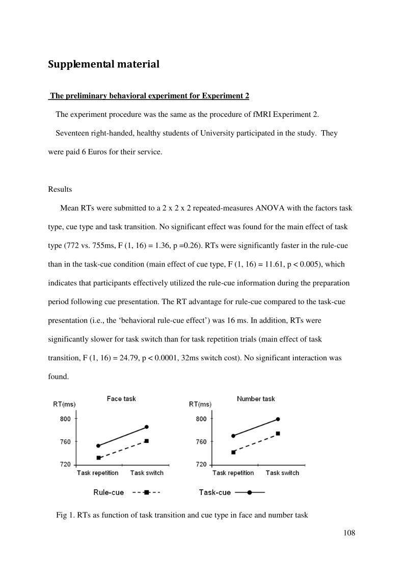
108
Supplemental material
The preliminary behavioral experiment for Experiment 2
The experiment procedure was the same as the procedure of fMRI Experiment 2.
Seventeen right-handed, healthy students of University participated in the study. They
were paid 6 Euros for their service.
Results
Mean RTs were submitted to a 2 x 2 x 2 repeated-measures ANOVA with the factors task
type, cue type and task transition. No significant effect was found for the main effect of task
type (772 vs. 755ms, F (1, 16) = 1.36, p =0.26). RTs were significantly faster in the rule-cue
than in the task-cue condition (main effect of cue type, F (1, 16) = 11.61, p < 0.005), which
indicates that participants effectively utilized the rule-cue information during the preparation
period following cue presentation. The RT advantage for rule-cue compared to the task-cue
presentation (i.e., the ‘behavioral rule-cue effect’) was 16 ms. In addition, RTs were
significantly slower for task switch than for task repetition trials (main effect of task
transition, F (1, 16) = 24.79, p < 0.0001, 32ms switch cost). No significant interaction was
found.
Fig 1. RTs as function of task transition and cue type in face and number task
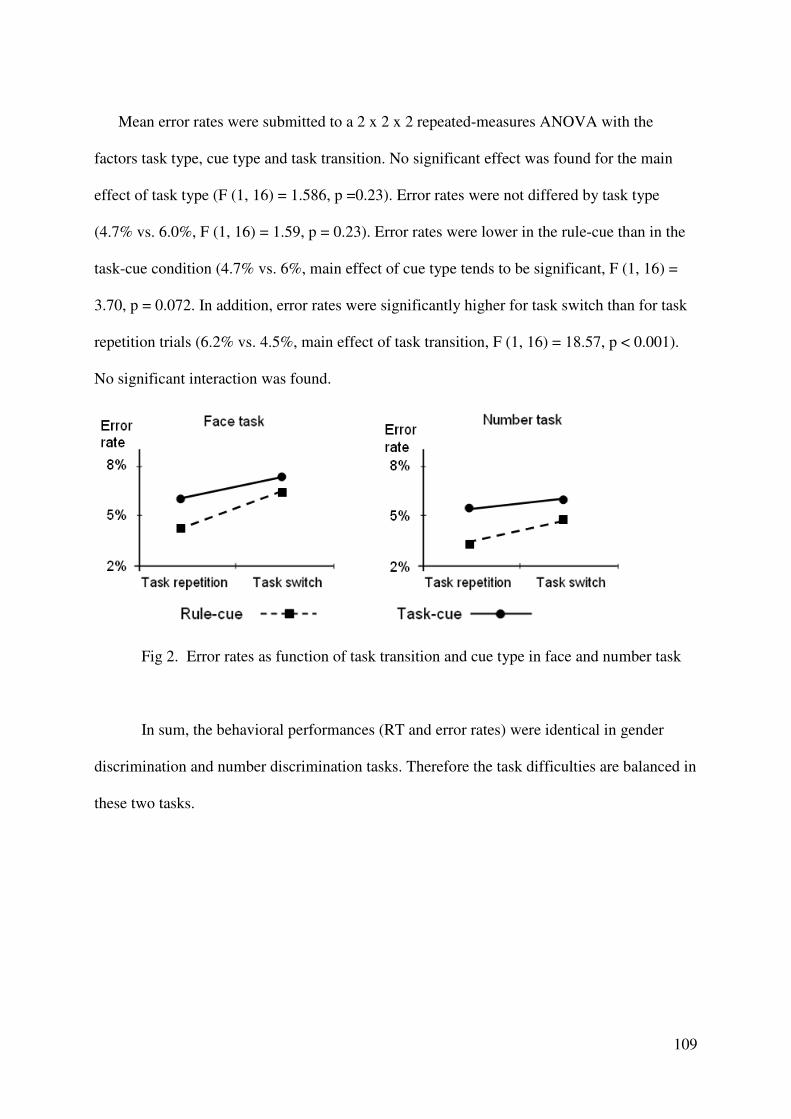
109
Mean error rates were submitted to a 2 x 2 x 2 repeated-measures ANOVA with the
factors task type, cue type and task transition. No significant effect was found for the main
effect of task type (F (1, 16) = 1.586, p =0.23). Error rates were not differed by task type
(4.7% vs. 6.0%, F (1, 16) = 1.59, p = 0.23). Error rates were lower in the rule-cue than in the
task-cue condition (4.7% vs. 6%, main effect of cue type tends to be significant, F (1, 16) =
3.70, p = 0.072. In addition, error rates were significantly higher for task switch than for task
repetition trials (6.2% vs. 4.5%, main effect of task transition, F (1, 16) = 18.57, p < 0.001).
No significant interaction was found.
Fig 2. Error rates as function of task transition and cue type in face and number task
In sum, the behavioral performances (RT and error rates) were identical in gender
discrimination and number discrimination tasks. Therefore the task difficulties are balanced in
these two tasks.
![[SfN 2013] Neural correlates of flow](https://static.fdocuments.us/doc/165x107/5564e56bd8b42afb268b4978/sfn-2013-neural-correlates-of-flow.jpg)