The Nervous System: The Spinal Cord and Spinal...
19
© 2012 Pearson Education, Inc. 14 The Nervous System: The Spinal Cord and Spinal Nerves
Transcript of The Nervous System: The Spinal Cord and Spinal...

© 2012 Pearson Education, Inc.
14 The Nervous System: The Spinal Cord and Spinal Nerves
© 2012 Pearson Education, Inc.
Introduction
• The Central Nervous System (CNS)
consists of:
• The spinal cord
• Integrates and processes information
• Can function with the brain
• Can function independently of the brain
• The brain
• Integrates and processes information
• Can function with the spinal cord
• Can function independently of the spinal cord

© 2012 Pearson Education, Inc.
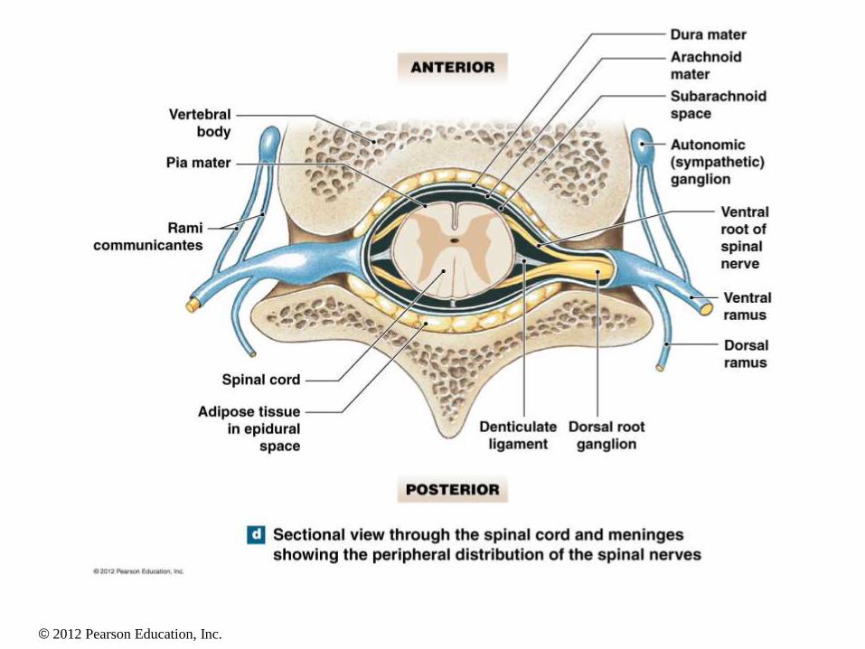
Gross Anatomy of the Spinal Cord • Central canal
• Gray matter
• Consists of cell bodies
• White matter
• Consists of axons
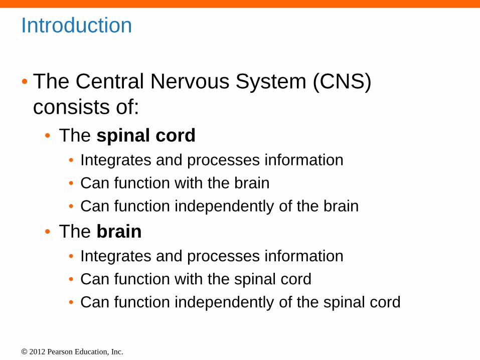
• Dorsal root and ventral root:
merge to form a spinal nerve
• Dorsal root is sensory: axons
extend from the soma within
the dorsal root ganglion
• Sensory nerves (afferent
nerves): transmit impulses
toward the spinal cord
• Ventral root is motor
• Motor nerves (efferent
nerves): transmit impulses
away from the spinal cord
© 2012 Pearson Education, Inc.

© 2012 Pearson Education, Inc.
Gross Anatomy of the Spinal Cord • Consists of:
• Cervical region
• Thoracic region
• Lumbar region
• Sacral region
• Coccygeal region
© 2012 Pearson Education, Inc.

© 2012 Pearson Education, Inc.
Spinal Meninges • Specialized membranes that
provide protection, physical
stability, and shock absorption
• Continuous with the cranial
(cerebral) meninges
• Made of three layers
• Dura mater: tough,
fibrous outermost layer
• Arachnoid mater:
middle layer
• Pia mater: innermost
layer
© 2012 Pearson Education, Inc.
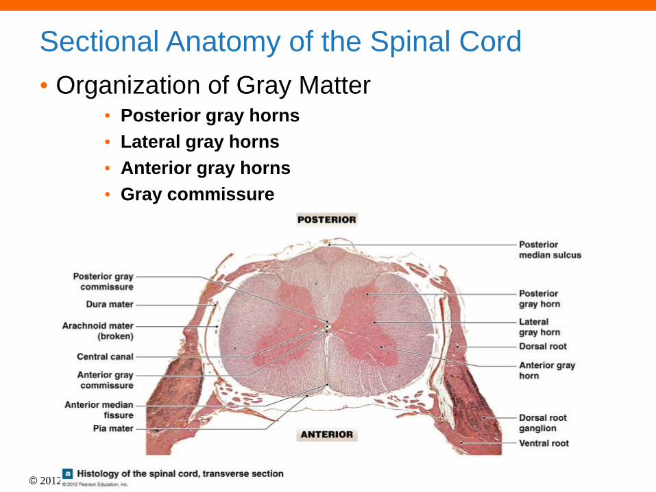
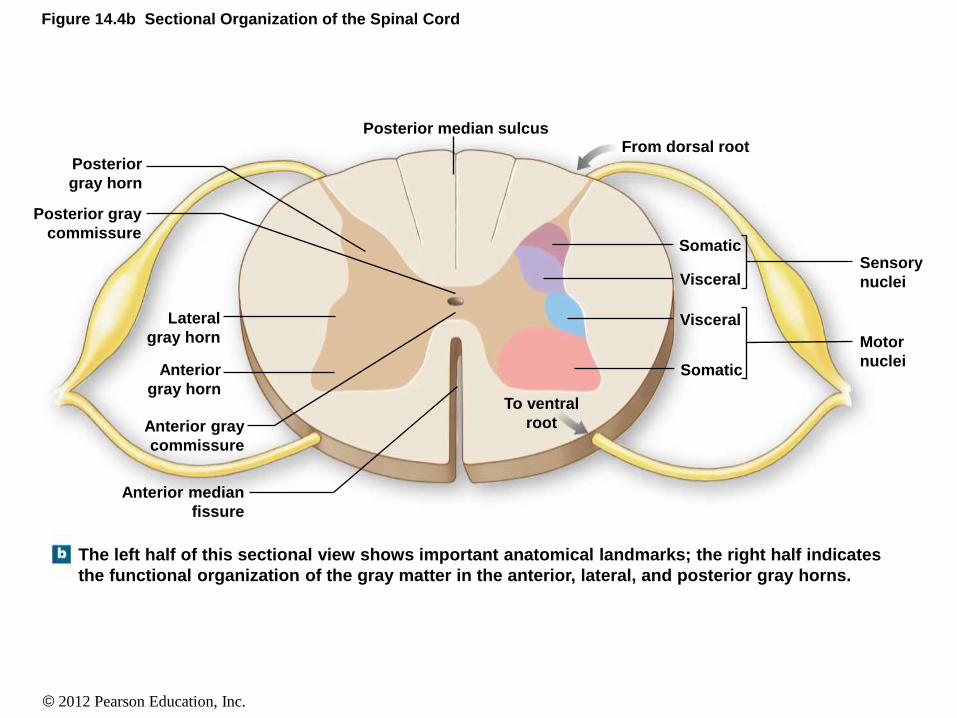
Sectional Anatomy of the Spinal Cord
• Organization of Gray Matter • Posterior gray horns
• Lateral gray horns
• Anterior gray horns
• Gray commissure
© 2012 Pearson Education, Inc.
Figure 14.4b Sectional Organization of the Spinal Cord
The left half of this sectional view shows important anatomical landmarks; the right half indicates
the functional organization of the gray matter in the anterior, lateral, and posterior gray horns.
Posterior
gray horn
Posterior gray
commissure
Lateral
gray horn
Anterior
gray horn
Anterior gray
commissure
Anterior median
fissure
To ventral
root
Posterior median sulcus From dorsal root
Sensory
nuclei
Motor
nuclei
Somatic
Somatic
Visceral
Visceral
© 2012 Pearson Education, Inc.
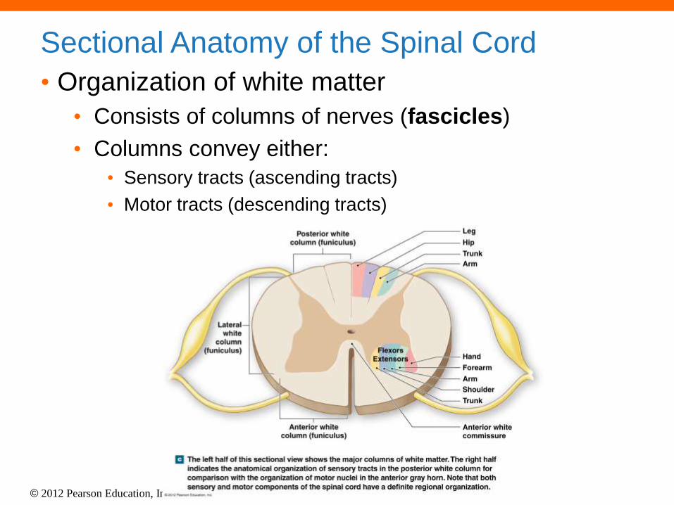
Sectional Anatomy of the Spinal Cord
• Organization of white matter
• Consists of columns of nerves (fascicles)
• Columns convey either:
• Sensory tracts (ascending tracts)
• Motor tracts (descending tracts)

© 2012 Pearson Education, Inc.
Figure 14.4c Sectional Organization of the Spinal Cord
The left half of this sectional view shows the major columns of white matter. The right half
indicates the anatomical organization of sensory tracts in the posterior white column for
comparison with the organization of motor nuclei in the anterior gray horn. Note that both
sensory and motor components of the spinal cord have a definite regional organization.
Posterior white
column (funiculus)
Anterior white
column (funiculus)
Anterior white
commissure
Flexors
Extensors
Lateral
white
column
(funiculus)
Leg
Hip
Trunk
Arm
Hand
Forearm
Arm
Shoulder
Trunk
© 2012 Pearson Education, Inc.
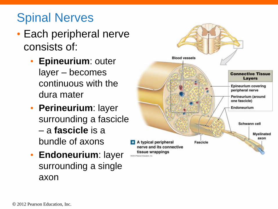
Spinal Nerves
• Each peripheral nerve
consists of:
• Epineurium: outer
layer – becomes
continuous with the
dura mater
• Perineurium: layer
surrounding a fascicle
– a fascicle is a
bundle of axons
• Endoneurium: layer
surrounding a single
axon

© 2012 Pearson Education, Inc.
Spinal Nerves
• There are 31 pairs of
spinal nerves
• 8 cervical nerves
• 12 thoracic nerves
• 5 lumbar nerves
• 5 sacral nerves
• 1 coccygeal nerve

© 2012 Pearson Education, Inc.
Spinal Nerves
• Cervical spinal nerves
emerge from C1–C8
• Thoracic spinal nerves
emerge from T1–T12
• Lumbar spinal nerves
emerge from L1–L5
• Sacral spinal nerves
emerge from S1–S5
• Coccygeal spinal nerves
emerge from Co1

© 2012 Pearson Education, Inc.
Nerve Plexuses
• There are four nerve plexuses • Cervical plexus nerves
emerge from C1–C5
• Brachial plexus nerves emerge from C5–T1
• There is not a thoracic plexus
• Lumbar plexus nerves emerge from T12–L4
• Sacral plexus nerves emerge from L4–S4
• Sometimes the lumbar and sacral are combined to form the lumbosacral plexus

© 2012 Pearson Education, Inc.
Reflexes
• Reflex
• An immediate involuntary response
• Pathway of a reflex arc
• 1. Activation of a sensory receptor
• 2. Relay of information to the CNS
• 3. Information processing
• 4. Activation of a motor neuron
• 5. Response by the effector

© 2012 Pearson Education, Inc.
Figure 14.14 A Reflex Arc
Arrival of stimulus and activation of receptor
Response by effector
Activation of a sensory neuron
Activation of a motor neuron
Information processing in CNS
Stimulus
Effector
Receptor
REFLEX ARC
Ventral root
Dorsal root
Sensation relayed to
the brain by collateral
KEY Sensory neuron (stimulated)
Excitatory interneuron
Motor neuron (stimulated)
© 2012 Pearson Education, Inc.
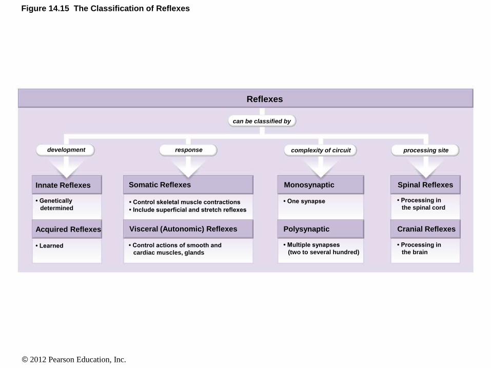
Figure 14.15 The Classification of Reflexes
Reflexes
development response
can be classified by
complexity of circuit processing site
• Processing in
the spinal cord
• Processing in
the brain
• One synapse
• Multiple synapses
(two to several hundred)
• Genetically
determined
• Learned • Control actions of smooth and
cardiac muscles, glands
• Control skeletal muscle contractions
• Include superficial and stretch reflexes
Innate Reflexes
Acquired Reflexes
Somatic Reflexes
Visceral (Autonomic) Reflexes
Spinal Reflexes
Cranial Reflexes
Monosynaptic
Polysynaptic

© 2012 Pearson Education, Inc.
Figure 14.16 Neural Organization and Simple Reflexes
CENTRAL NERVOUS SYSTEM
CENTRAL NERVOUS SYSTEM
Motor neuron
Ganglion
Sensory neuron
Sensory receptor (muscle spindle)
Skeletal muscle
Circuit 1
A monosynaptic reflex circuit involves a peripheral
sensory neuron and a central motor neuron. In this
example, stimulation of the receptor will lead to a
reflexive contraction in a skeletal muscle.
A polysynaptic reflex circuit involves a sensory neuron,
interneurons, and motor neurons. In this example, the
stimulation of the receptor leads to the coordinated
contractions of two different skeletal muscles.
Skeletal muscle 1
Skeletal muscle 2
Motor neurons
Circuit 2
Interneurons
Ganglion
Sensory neuron
Sensory receptor


![The Nervous System. Divisions of the Nervous System Central Nervous System [CNS] = Spinal Cord Brain Peripheral Nervous System [PNS]= Spinal Nerves.](https://static.fdocuments.us/doc/165x107/56649d6c5503460f94a4c71d/the-nervous-system-divisions-of-the-nervous-system-central-nervous-system.jpg)