The Nervous System The Nervous System 8 8. The Nervous System Two Organ Systems Control All the...
92
The Nervous System 8
-
Upload
amos-perkins -
Category
Documents
-
view
224 -
download
0
Transcript of The Nervous System The Nervous System 8 8. The Nervous System Two Organ Systems Control All the...
The Nervous
System
The Nervous
System88
The Nervous System
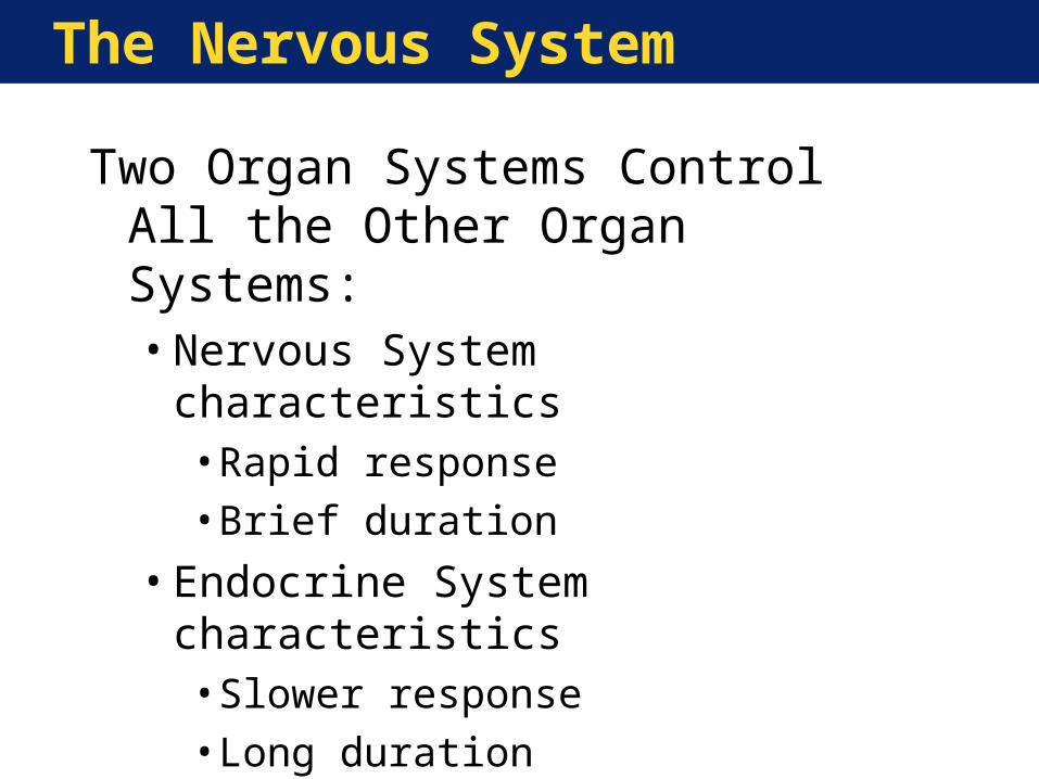
Two Organ Systems Control All the Other Organ Systems:•Nervous System characteristics
• Rapid response• Brief duration
•Endocrine System characteristics• Slower response• Long duration
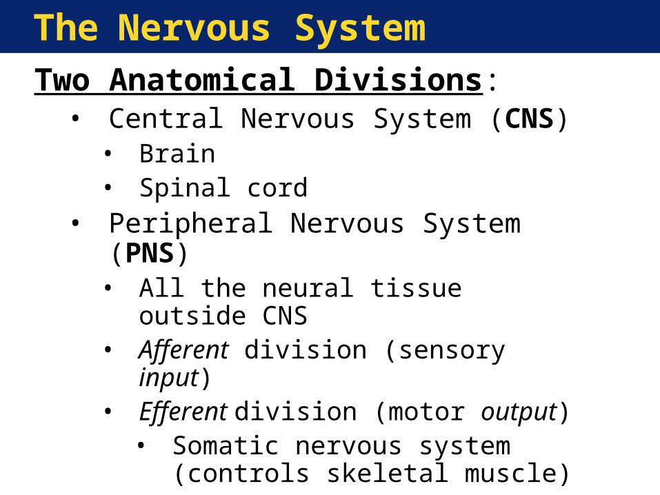
The Nervous SystemTwo Anatomical Divisions:• Central Nervous System (CNS)
• Brain• Spinal cord
• Peripheral Nervous System (PNS)• All the neural tissue outside CNS• Afferent division (sensory input)• Efferent division (motor output)
• Somatic nervous system (controls skeletal muscle)
• Autonomic nervous system (controls smooth & cardiac muscle)
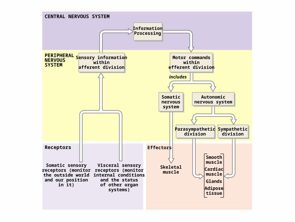
CENTRAL NERVOUS SYSTEM
PERIPHERALNERVOUSSYSTEM
Receptors
InformationProcessing
Sensory informationwithin
afferent division
Motor commandswithin
efferent division
includes
Somaticnervoussystem
Autonomicnervous system
Parasympatheticdivision
Sympatheticdivision
Skeletalmuscle
Effectors
Smoothmuscle
Cardiacmuscle
Adiposetissue
Glands
Somatic sensoryreceptors (monitorthe outside worldand our position
in it)
Visceral sensoryreceptors (monitorinternal conditions
and the statusof other organ
systems)
Neural Tissue Organization
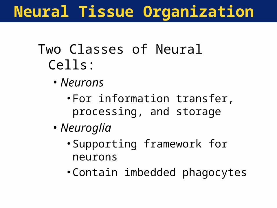
Two Classes of Neural Cells:•Neurons
• For information transfer, processing, and storage
•Neuroglia• Supporting framework for neurons• Contain imbedded phagocytes
Neural Tissue Organization
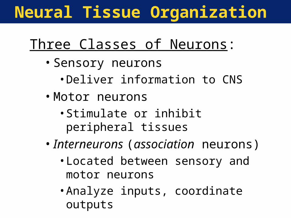
Three Classes of Neurons:•Sensory neurons
• Deliver information to CNS
•Motor neurons• Stimulate or inhibit peripheral tissues
• Interneurons (association neurons)• Located between sensory and motor
neurons• Analyze inputs, coordinate outputs
Neural Tissue Organization
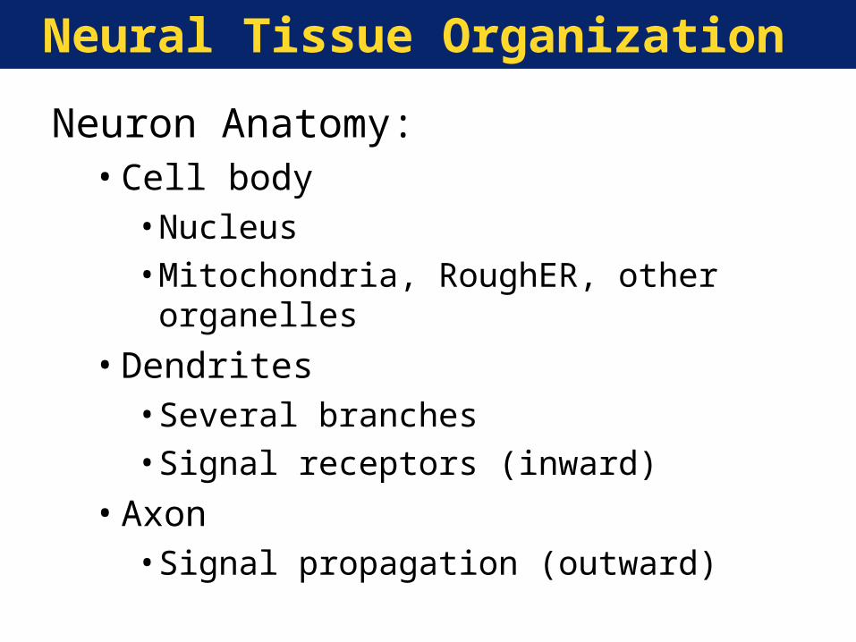
Neuron Anatomy:•Cell body
• Nucleus• Mitochondria, RoughER, other organelles
•Dendrites• Several branches• Signal receptors (inward)
•Axon• Signal propagation (outward)
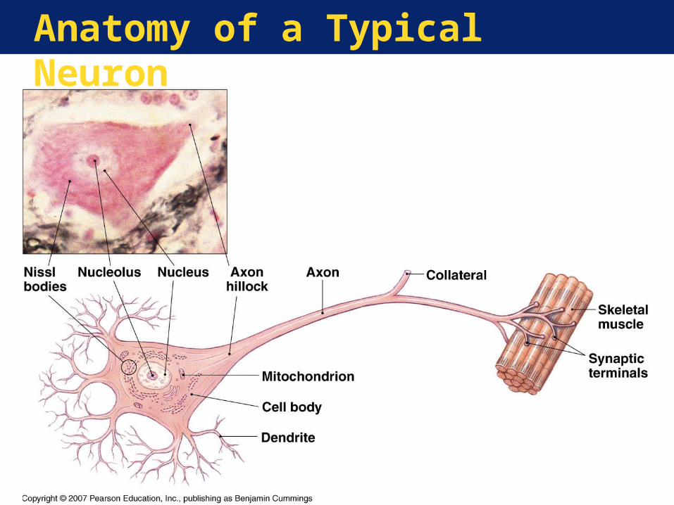
Anatomy of a Typical Neuron
Neural Tissue Organization
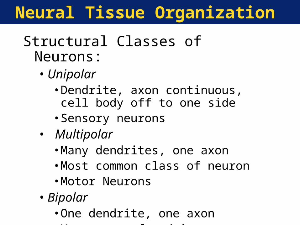
Structural Classes of Neurons:•Unipolar
• Dendrite, axon continuous, cell body off to one side
• Sensory neurons• Multipolar
• Many dendrites, one axon• Most common class of neuron• Motor Neurons
•Bipolar• One dendrite, one axon• Very rare, found in sense organs

Structural Classification of Neurons
Neural Tissue Organization
Types of Neuroglia (glia):•Astrocytes
• Part of blood-brain barrier
•Oligodendrocytes• Responsible for myelination(protective
covering) around axons
•Microglia• Phagocytic defense cells
•Ependymal cells• Lining of brain, spinal cord cavities• Source of cerebrospinal fluid
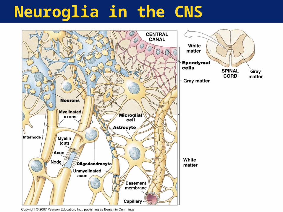
Neuroglia in the CNS
Neural Tissue Organization
Two Types of Neuroglia in the PNS:•Satellite cells
• Surround cell bodies
•Schwann cells• Surround all peripheral axons• Form myelin sheath on myelinated axons
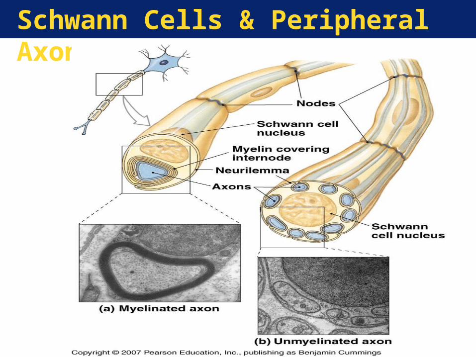
Schwann Cells & Peripheral Axons
Neural Tissue Organization
Key Note:
Neurons perform all of the communication, information processing, and control functions of the nervous system.
Neuroglia outnumber neurons and have functions essential to preserving the physical and biochemical structure of
neural tissue and the survival of neurons.
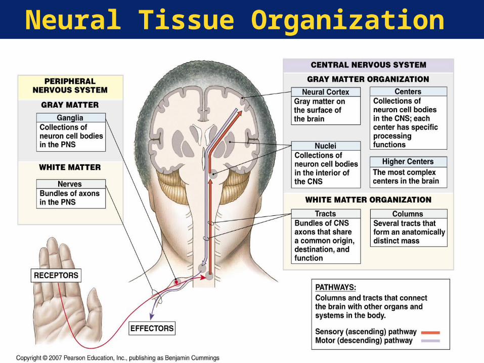
Neural Tissue Organization
Anatomic Organization of CNS Neurons:•Center—Collection of neurons with a
shared function•Nucleus—A center with a discrete
anatomical boundary•Neural cortex—Gray matter covering of
brain portions•White matter—Bundles of axons (tracts)
that share origins, destinations, and functions
Neural Tissue Organization
Anatomic Organization of PNS Neurons:•Ganglia—Groupings of neuron cell bodies•Nerve—Bundle of axons supported by
connective tissue• Spinal nerves
•To/from spinal cord• Cranial nerves
•To/from brain
Neural Tissue Organization
Neural Tissue Organization
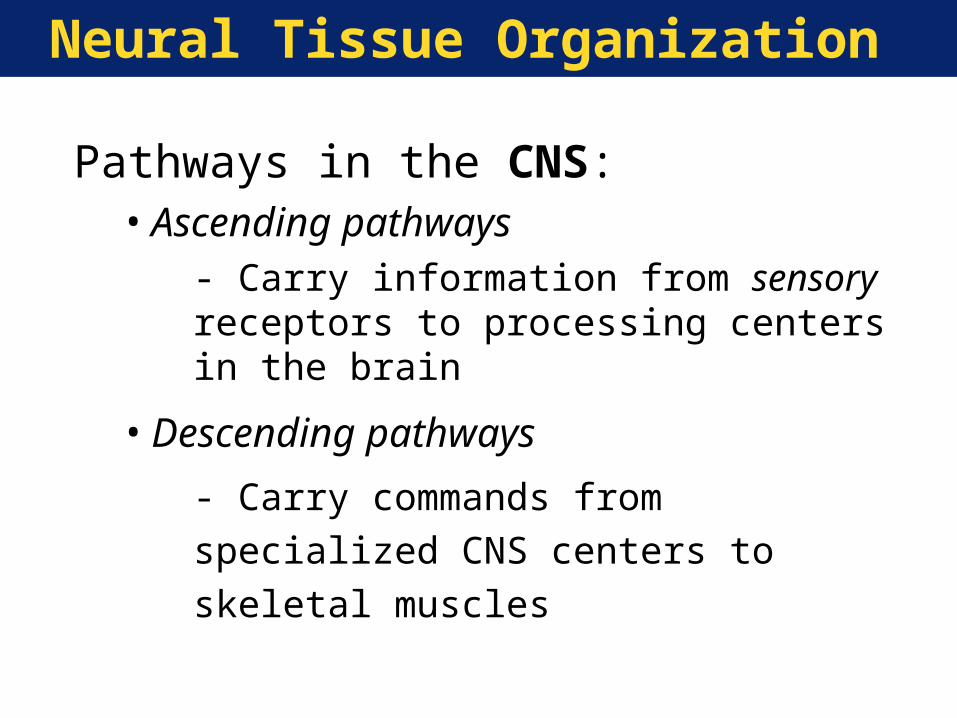
Pathways in the CNS:•Ascending pathways
- Carry information from sensory receptors to processing centers in the brain
•Descending pathways
- Carry commands from specialized CNS
centers to skeletal muscles
Neuron Function
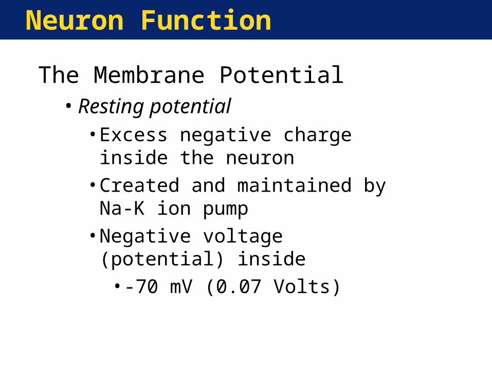
The Membrane Potential•Resting potential
• Excess negative charge inside the neuron
• Created and maintained by Na-K ion pump
• Negative voltage (potential) inside•-70 mV (0.07 Volts)
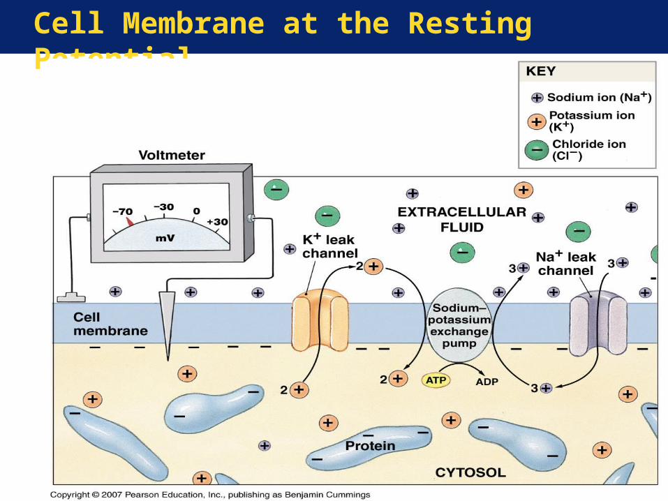
Cell Membrane at the Resting Potential
Neuron Function
A membrane potential exists across the cell membrane because:
(1) the cytosol and the extracellular fluid differ in their ionic composition, and
(2) the cell membrane is selectively permeable to these ions.
The membrane potential can quickly change, as the ionic permeability of the cell membrane changes, in response to chemical or physical stimuli.
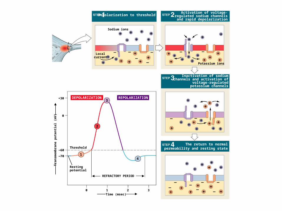
+30
0
60_
70_
Tra
nsm
emb
ran
e p
ote
nti
al (
mV
)
Threshold
Restingpotential
1
2
3
4
REFRACTORY PERIOD
DEPOLARIZATION REPOLARIZATION
Localcurrent
Depolarization to threshold
Sodium ions
Activation of voltage-regulated sodium channels
and rapid depolarization
Potassium ions
Inactivation of sodiumchannels and activation of
voltage-regulatedpotassium channels
The return to normalpermeability and resting state
Time (msec)0 1 2 3
Neuron Function
Key Note:
“Information” travels within the nervous system primarily in the form of propagated electrical signals known as action potentials. The most important information (e.g., vision, balance, movement), is carried by myelinated axons.
Neural Communication
Synapse Basics• Intercellular communication
• Axon terminal receives electrical signal from cell body
• Input to next cell• Chemical signaling to cross
synapse•Neurotransmitter release
Neural Communication
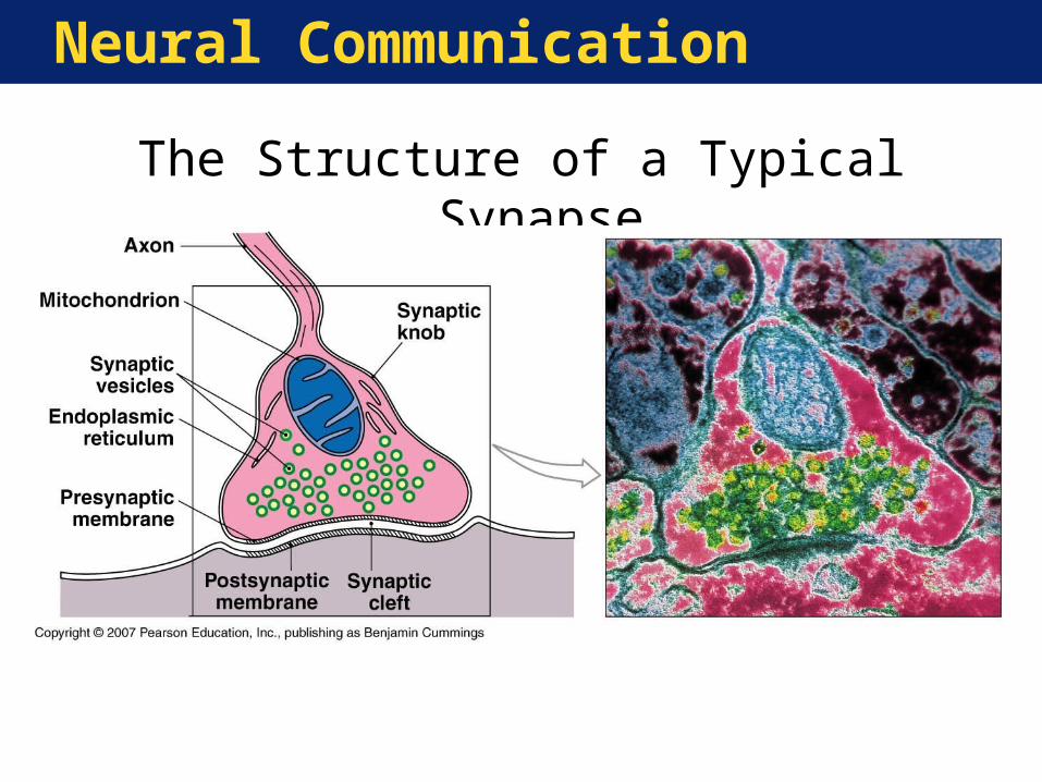
Structure of a Synapse:•Presynaptic components
• Axon terminal• Synaptic knob• Synaptic vesicles• Synaptic cleft
•Postsynaptic components• Neurotransmitter receptors
Neural Communication
The Structure of a Typical Synapse
Neural Communication
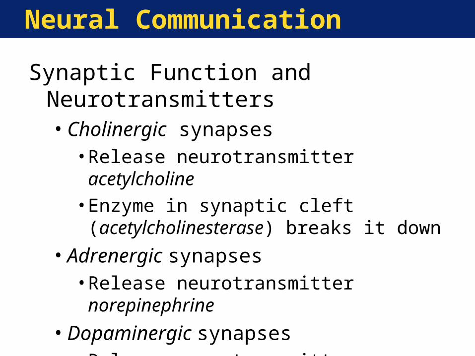
Synaptic Function and Neurotransmitters•Cholinergic synapses
• Release neurotransmitter acetylcholine• Enzyme in synaptic cleft
(acetylcholinesterase) breaks it down
•Adrenergic synapses• Release neurotransmitter norepinephrine
•Dopaminergic synapses• Release neurotransmitter dopamine
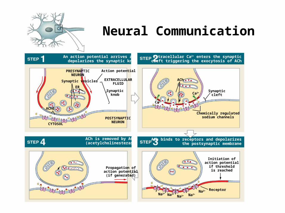
An action potential arrives anddepolarizes the synaptic knob
Action potential
ACh is removed by AChE(acetylcholinesterase)
Extracellular Ca2+ enters the synapticcleft triggering the exocytosis of ACh
ACh binds to receptors and depolarizesthe postsynaptic membrane
EXTRACELLULARFLUID
Synapticknob
PRESYNAPTICNEURON
Synaptic vesicles
ER
AChE
POSTSYNAPTICNEURONCYTOSOL
ACh
Ca2+
Ca2+Synaptic
cleft
Chemically regulatedsodium channels
Initiation ofaction potential
if thresholdis reached
ReceptorNa2+
Na2+Na2+Na2+Na2+
Propagation ofaction potential(if generated)
Neural Communication
Neural Communication
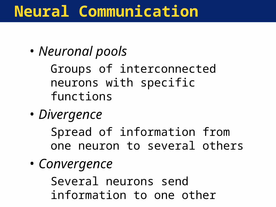
• Neuronal poolsGroups of interconnected neurons with specific functions
• DivergenceSpread of information from one neuron to several others
• ConvergenceSeveral neurons send information to one other
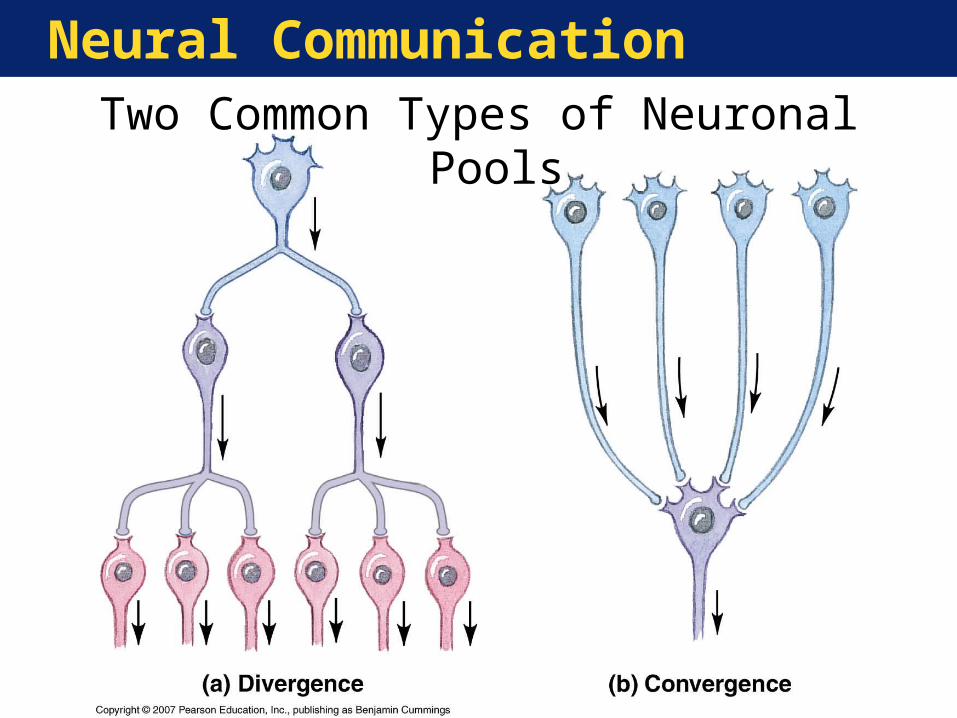
Neural CommunicationTwo Common Types of Neuronal Pools
Neural Communication
Key Note:A synaptic terminal releases a neuro-transmitter that binds to the postsynaptic cell membrane. The result is a brief, local change in the permeability of the postsynaptic cell. Many drugs affect the nervous system by stimulating neurotransmitter receptors and thus produce complex effects on perception, motor control, and emotions.
The Central Nervous System
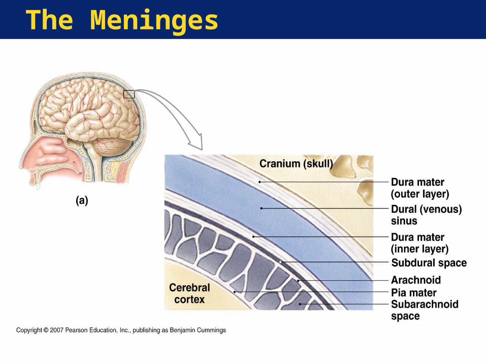
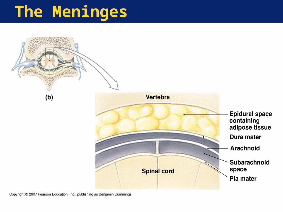
Meninges—Layers that surround and protect the brain and spinal cord (CNS)•Dura mater (“tough mother”)
• Tough, fibrous outer layer• Epidural space above dura of spinal cord
•Arachnoid (“spidery”)• Subarchnoid space• Cerebrospinal fluid
•Pia mater (“delicate mother”)• Thin inner layer
The Meninges
The Meninges
The Central Nervous System
Spinal Cord Basics:•Relays information to/from brain•Processes some information on its
own (reflexes)•Divided into 31 segments•Each segment has a pair of:
• Dorsal root ganglia• Dorsal roots• Ventral roots
•Gray matter appears as horns•White matter organized into columns
Anatomy of the Spinal Cord
Anatomy of the Spinal Cord
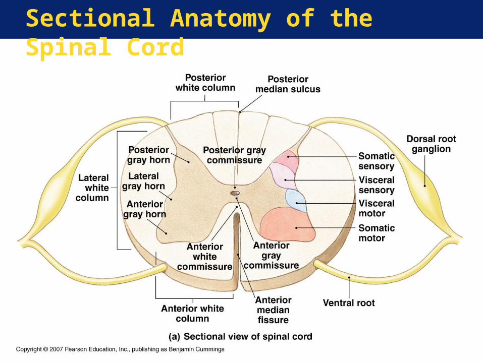
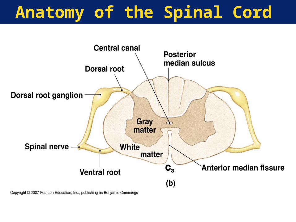
Sectional Anatomy of the Spinal Cord

Sectional Anatomy of the Spinal Cord

The Central Nervous System
Key Note:The sensory and motor nuclei (gray matter) of the spinal cord surround the central canal. Sensory nuclei are dorsal, motor nuclei are ventral. A thick layer of white matter consisting of ascending and descending axons covers the gray matter. These axons are organized into columns of axon bundles with specific functions. This highly organized structure often enables predicting the impact of particular injuries.
The Central Nervous System
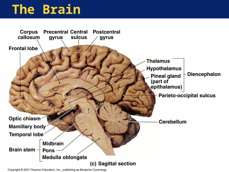
Brain Regions•Cerebrum•Diencephalon•Midbrain•Pons•Medulla oblongata•Cerebellum
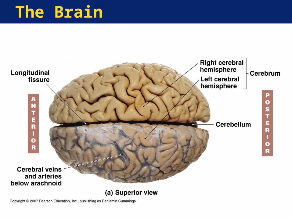
The Brain
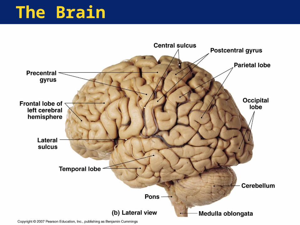
The Brain
The Brain
The Central Nervous System
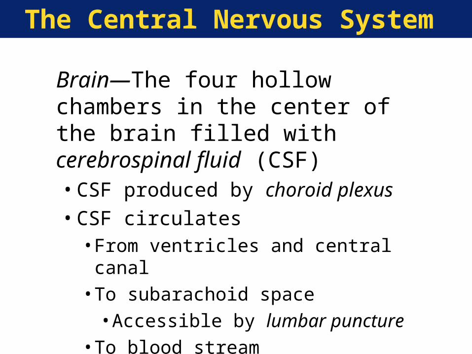
Brain—The four hollow chambers in the center of the brain filled with cerebrospinal fluid (CSF)•CSF produced by choroid plexus•CSF circulates
• From ventricles and central canal• To subarachoid space•Accessible by lumbar puncture
• To blood stream
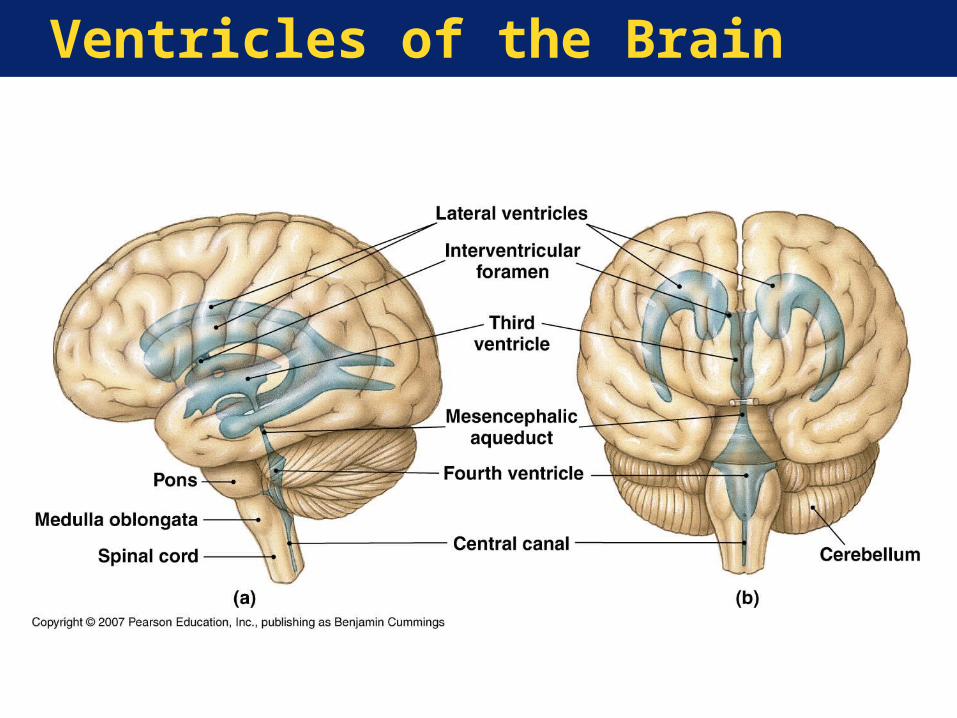
Ventricles of the Brain
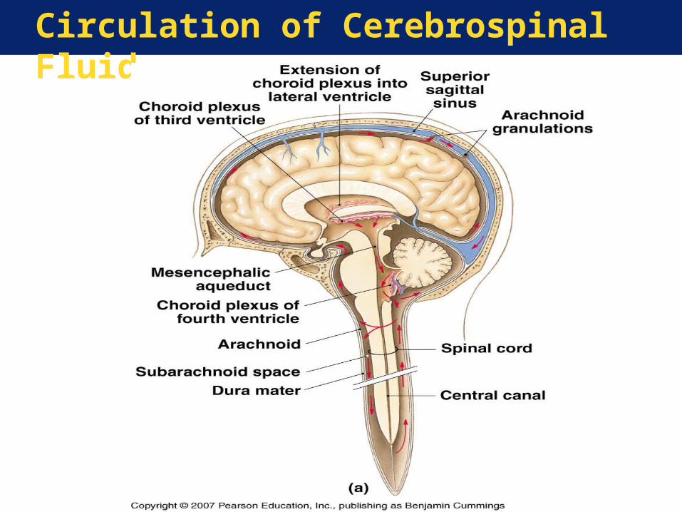
Circulation of Cerebrospinal Fluid

The Central Nervous System
Functions of the Cerebrum:•Conscious thought• Intellectual activity•Memory•Origin of complex patterns of movement
Anatomy of Cerebral Cortex:•Highly folded surface
• Elevated ridges (gyri)• Shallow depressions (sulci)
The Central Nervous System
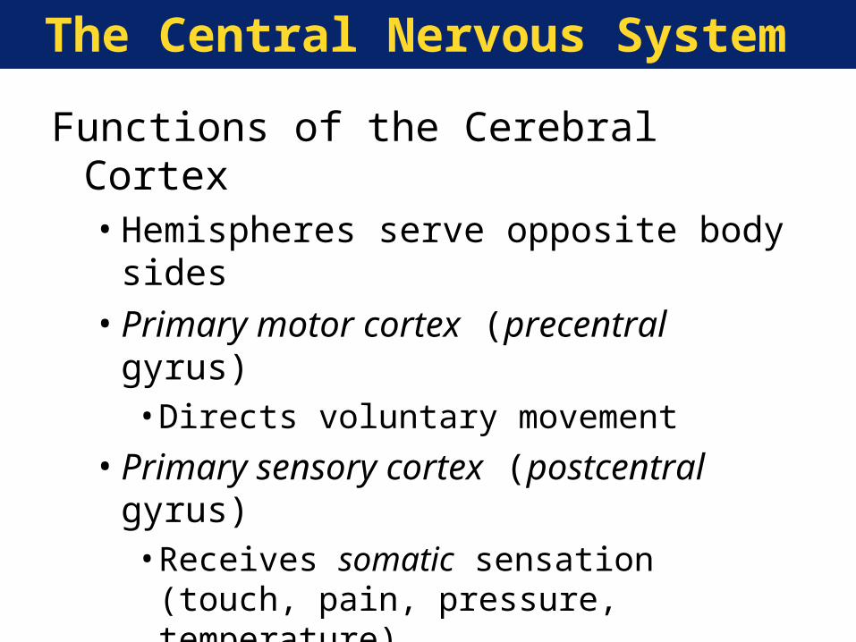
Functions of the Cerebral Cortex•Hemispheres serve opposite body sides•Primary motor cortex (precentral gyrus)
• Directs voluntary movement
•Primary sensory cortex (postcentral gyrus)• Receives somatic sensation (touch, pain,
pressure, temperature)
•Association areas• Interpret sensation• Coordinate movement
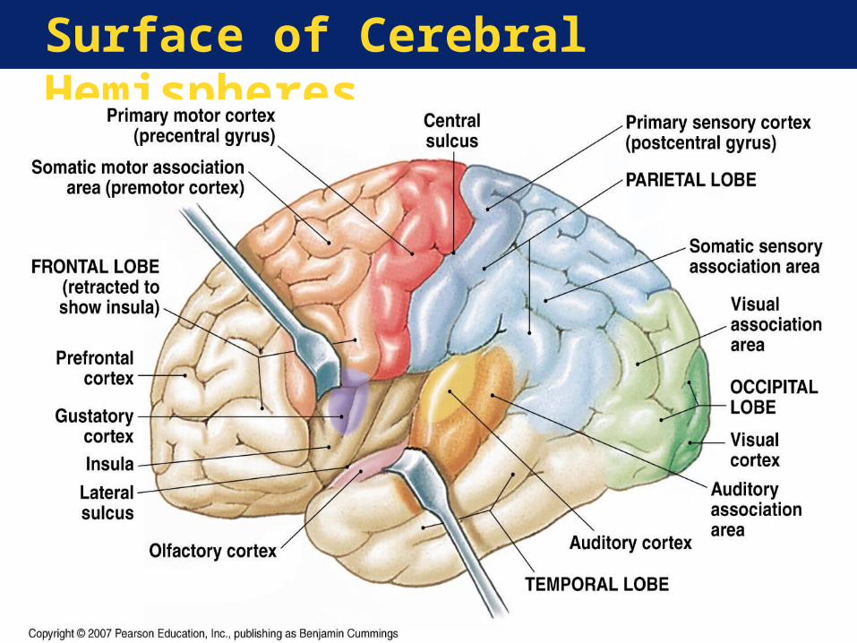
Surface of Cerebral Hemispheres
The Central Nervous System
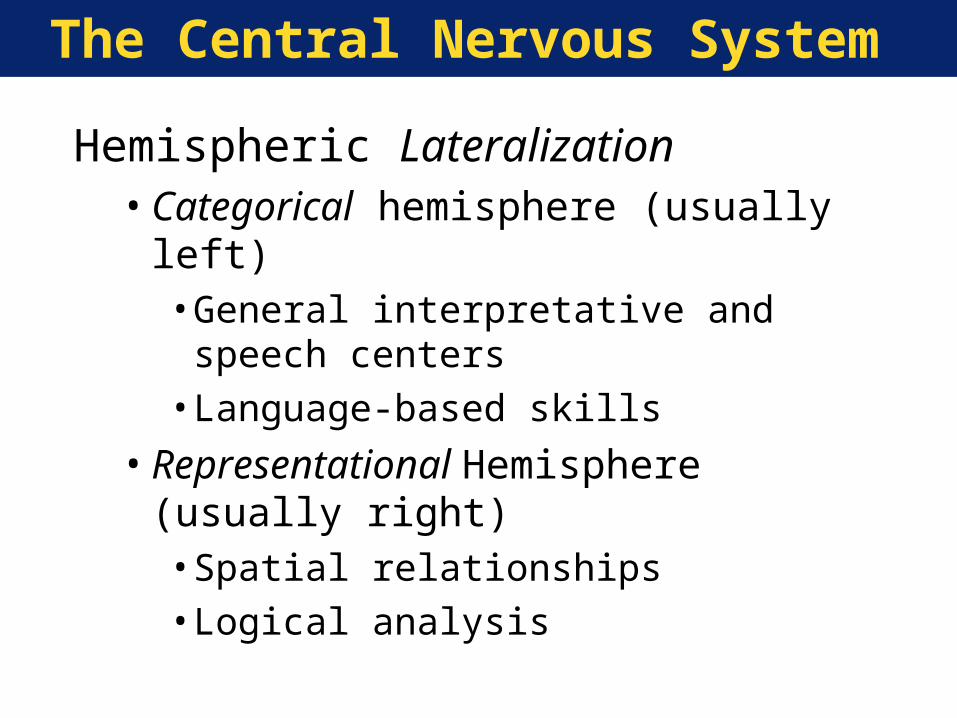
Hemispheric Lateralization•Categorical hemisphere (usually left)
• General interpretative and speech centers• Language-based skills
•Representational Hemisphere (usually right)• Spatial relationships• Logical analysis
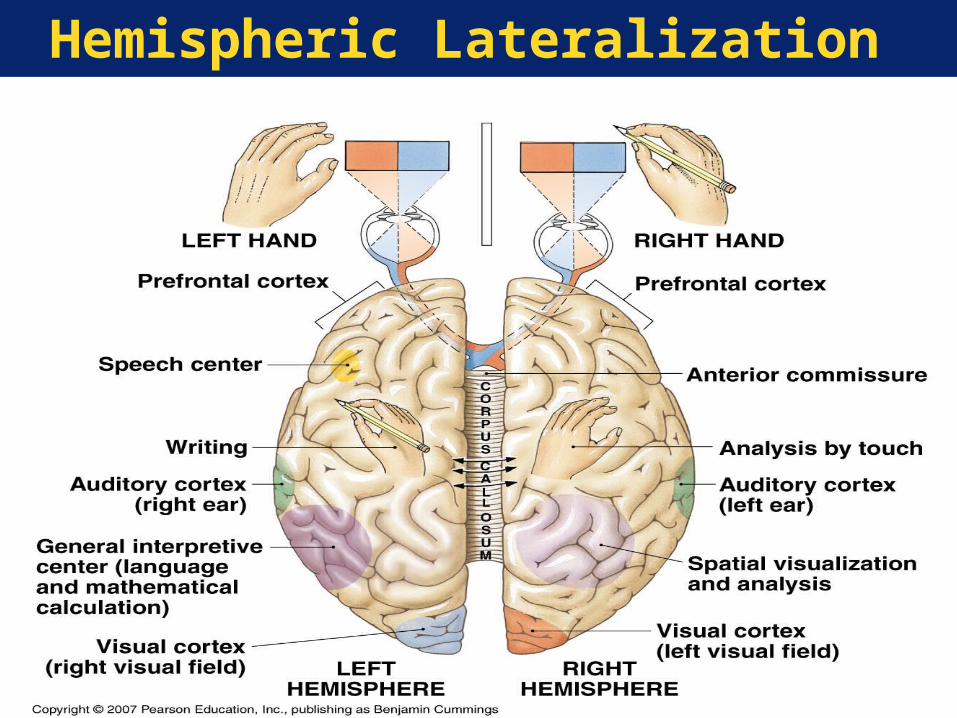
Hemispheric Lateralization
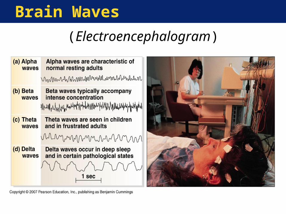
Brain Waves
(Electroencephalogram)
The Central Nervous System
The Basal Nuclei•Lie deep within central white
matter of the brain•Responsible for muscle tone•Coordinate learned movements•Coordinate rhythmic movements
(e.g., walking)
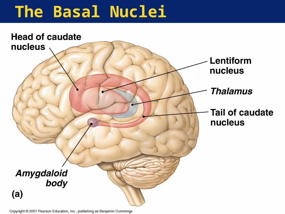
The Basal Nuclei
The Central Nervous System
Functions of the Limbic System•Establish emotions and related
drives•Link cerebral cortex intellectual
functions to brain stem autonomic functions
•Control reflexes associated with eating
•Store and retrieve long-term memories

The Limbic System
The Central Nervous System
The Diencephalon•Switching and relay center• Integration of conscious and
unconscious motor and sensory pathways
•Components include:• Epithalamus
•Choroid plexus•Pineal body
• Thalamus• Hypothalamus
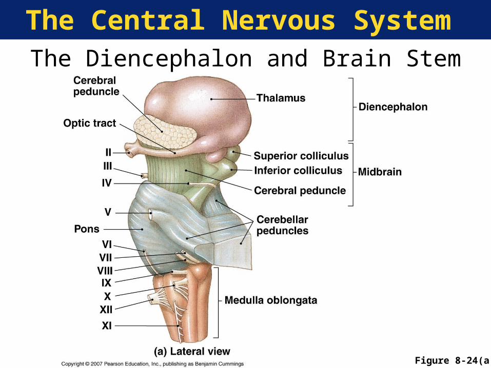
The Central Nervous SystemThe Diencephalon and Brain Stem
Figure 8-24(a)
The Central Nervous System
Functions of the Thalamus•Relay and filter all ascending (sensory)
information• Relay a small proportion to cerebral
cortex (conscious perception)• Relay most to basal nuclei and brain
stem centers
•Coordinate voluntary and involuntary motor behavior
The Central Nervous System
Functions of the Hypothalamus•Produce emotions and behavioral drives•Coordinate nervous and endocrine
systems•Secrete hormones•Coordinate voluntary and autonomic
functions•Regulate body temperature
The Central Nervous System
Anatomy and Function of the Brain Stem•Midbrain
• Process visual, auditory information• Generate involuntary movements
•Pons• Links to cerebellum• Involved in control of movement
•Medulla oblongata• Relay sensory information• Regulate autonomic function
The Central Nervous System
Functions of the Medulla Oblongata•Links brain and spinal cord•Relays ascending information to cerebral
cortex•Controls crucial organ systems by reflex
• Cardiovascular centers• Respiratory rhythmicity centers
The Central Nervous System
Anatomy and Function of the Cerebellum•Oversees postural muscles•Stores patterns of movement•Fine tunes most movements•Links to brain stem, cerebrum, spinal cord
• Communicates over cerebellar peduncles
The Central Nervous System
Key Note:The brain, a large mass of neural tissue, contains internal passageways and chambers filled with CSF. The six major regions of the brain have specific functions. As you ascend from the medulla oblongata to the cerebrum, those functions become more complex and variable. Conscious thought and intelligence are provided by the cerebral cortex.
The Peripheral Nervous System
PNS Basics•Links the CNS with the body•Carries all sensory information and motor
commands•Axons bundled in nerves•Cell bodies grouped into ganglia• Includes cranial and spinal nerves
The Peripheral Nervous System
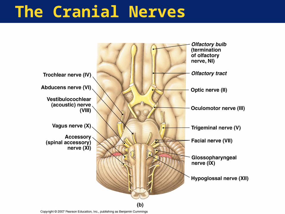
The Cranial Nerves•12 Pairs
• Connect to brain not the cord
•Olfactory (CN I)• Sense of smell
•Optic (CN II)• Sense of vision
•Oculomotor (CN III)• Eye movement
The Peripheral Nervous System
The Cranial Nerves (continued)•Trochlear (CN IV)
• Eye movement•Trigeminal (CN V)
• Eye, jaws sensation/movement•Abducens (CN VI)
• Eye movement•Facial (CN VII)
• Face, scalp, tongue sensation/movement•Vestibulocochlear (CN VIII)
• Hearing, balance
The Peripheral Nervous System
The Cranial Nerves (continued)•Glossopharyngeal (CN IX)
• Taste, swallowing
•Vagus (CN X)• Autonomic control of viscera
•Accessory (CN XI)• Swallowing, pectoral girdle movement
•Hypoglossal (CN XII)• Tongue movement

The Cranial Nerves
The Cranial Nerves
The Peripheral Nervous System
Key Note:The 12 pairs of cranial nerves are responsible for the special senses of smell, sight, and hearing/balance, and control movement of the eye, jaw, face, tongue, and muscles of the neck, back, and shoulders. They also provide sensation from the face, neck, and upper chest and autonomic innervation to thoracic and abdominopelvic organs.
The Peripheral Nervous System
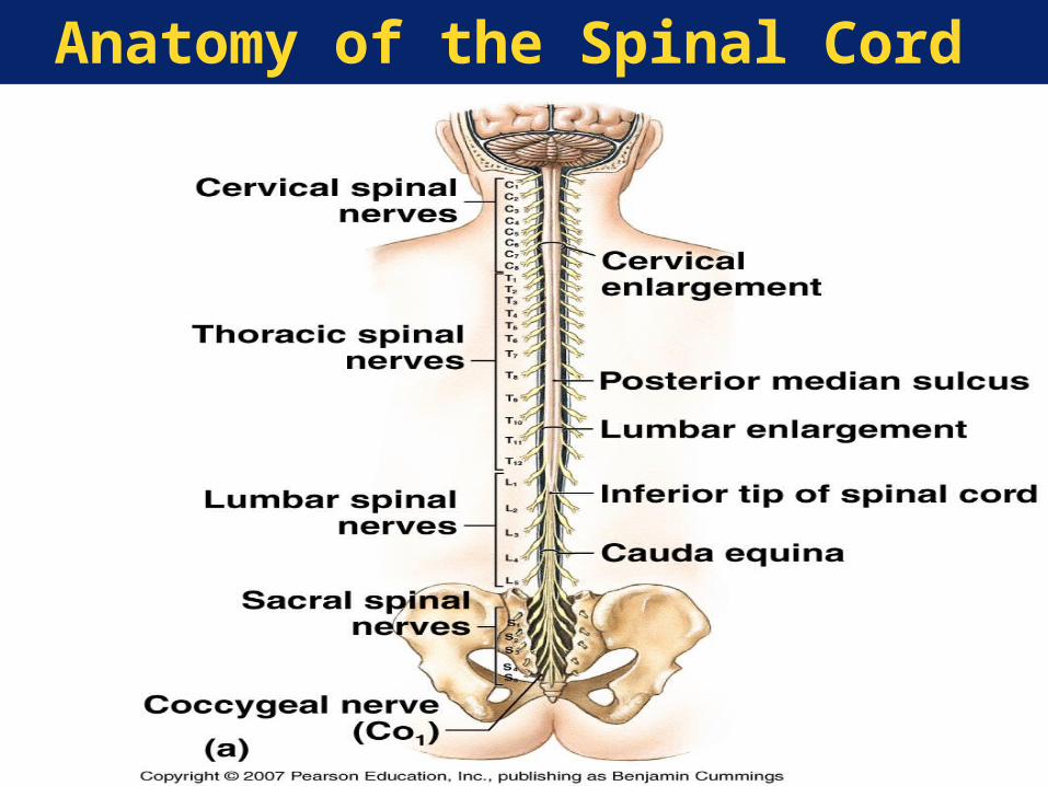
The Spinal Nerves•31 Pairs
• 8 Cervical• 12 Thoracic• 5 Lumbar• 5 Sacral
•Dermatome—Region of the body surface monitored by a pair of spinal nerves

The Peripheral Nervous System
Nerve Plexus—A complex, interwoven network of nerves•Four Large Plexuses
• Cervical plexus• Brachial plexus• Lumbar plexus• Sacral plexus
The Peripheral Nervous System
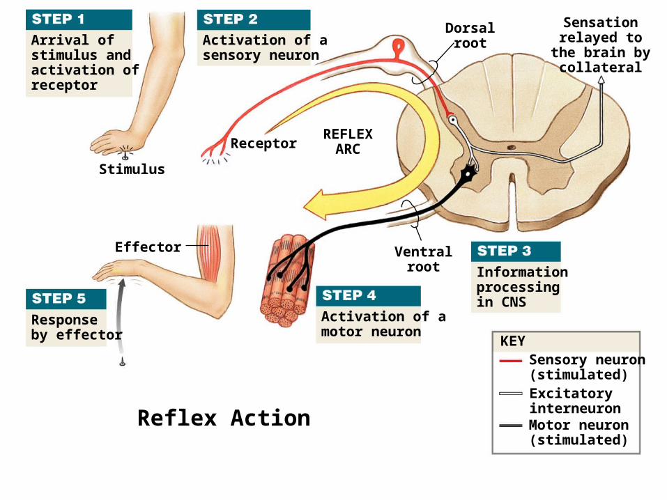
Reflex—An automatic involuntary motor response to a specific stimulus•The 5 steps in a reflex arc
• Arrival of stimulus and activation of receptor
• Activation of sensory neuron• CNS processing of information• Activation of motor neuron• Response by effector (muscle or gland)
Arrival ofstimulus andactivation ofreceptor
Stimulus
Receptor
Activation of asensory neuron
Effector
REFLEXARC
Dorsalroot
Ventralroot
Sensationrelayed to
the brain bycollateral
Activation of amotor neuron
Informationprocessingin CNS
KEYSensory neuron(stimulated)ExcitatoryinterneuronMotor neuron(stimulated)
Responseby effector
Reflex Action
The Peripheral Nervous System
Examples of Reflexes
•Monosynaptic reflex—Simplest reflex arc;
sensory neuron synapses directly on motor
neuron
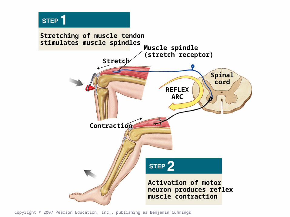
•Stretch reflex—Monosynaptic reflex to
regulate muscle length and tension
(example: patellar reflex)
•Muscle spindle—Sensory receptor in a
muscle that stimulates the stretch reflex
Copyright © 2007 Pearson Education, Inc., publishing as Benjamin Cummings
Stretching of muscle tendonstimulates muscle spindles
Stretch
Contraction
Muscle spindle(stretch receptor)
REFLEXARC
Spinalcord
Activation of motorneuron produces reflexmuscle contraction
The Peripheral Nervous System
Polysynaptic reflex—A reflex arc with at least one interneuron between the sensory afferent and motor efferent•Has a longer delay than a
monosynaptic reflex (more synapses)•Can produce more complex response•Example: flexor reflex, a withdrawal
reflex•Brain can modify spinal reflexes
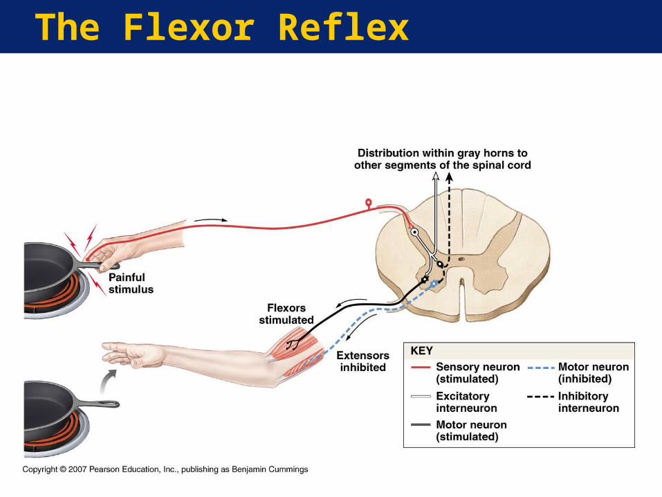
The Flexor Reflex
The Peripheral Nervous System
Key Note:
Reflexes are rapid, automatic responses to stimuli that “buy time” for the planning and execution of more complex responses that are often consciously directed.
The Peripheral Nervous System
The Autonomic Nervous System
Autonomic Nervous System
Branch of nervous system that coordinates cardiovascular, digestive, excretory, and reproductive functions
The Autonomic Nervous System
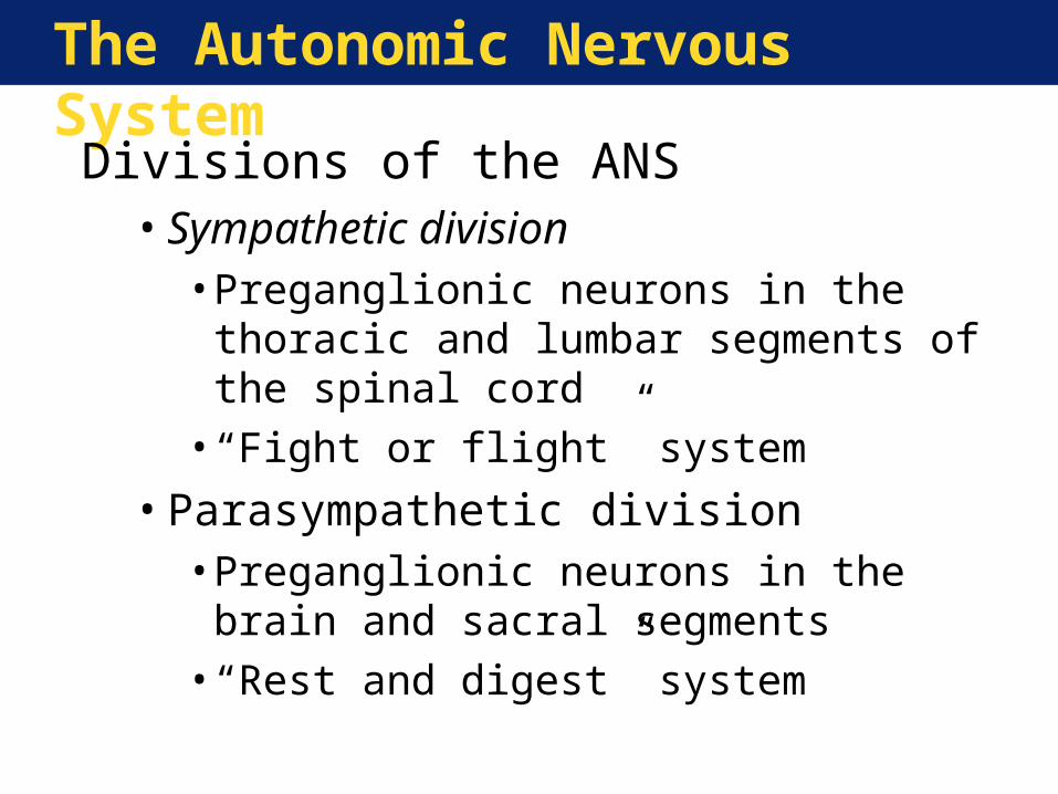
Divisions of the ANS•Sympathetic division
• Preganglionic neurons in the thoracic and lumbar segments of the spinal cord
• “Fight or flight” system
•Parasympathetic division• Preganglionic neurons in the brain and
sacral segments• “Rest and digest” system
The Autonomic Nervous System
Key Note:
The two divisions of the ANS operate largely without our awareness. The sympathetic division increases alertness, metabolic rate, and muscular abilities;
The parasympathetic division reduces metabolic rate and promotes visceral activities such as digestion.
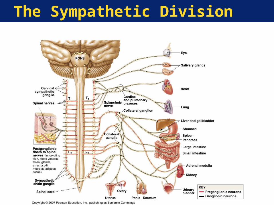
The Sympathetic Division
The Autonomic Nervous System
Effects of Sympathetic Activation•Generalized response in crises• Increased alertness•Feeling of euphoria and energy• Increased cardiovascular activity• Increased respiratory activity• Increased muscle tone
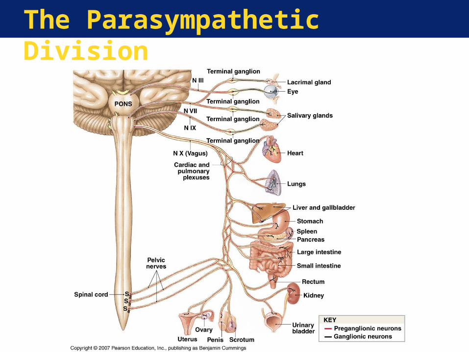
The Parasympathetic Division

The Autonomic Nervous System
Effects of Parasympathetic Activation•Relaxation•Food processing•Energy absorption•Brief effects at specific sites
The Autonomic Nervous System
Relationship between the Two Divisions:•Sympathetic division reaches visceral and
somatic structures throughout the body•Parasympathetic division reaches only
visceral structures via cranial nerves or in the abdominopelvic cavity
•Many organs receive dual innervation• In general, the two divisions produce
opposite effects on the their target organs
Aging and the Nervous System
Age-Related Changes•Reduction in brain size and weight•Loss of neurons•Decreased brain blood flow•Changes in synaptic organization of the
brain• Intracellular and extracellular changes in
CNS neurons