
The Nervous System: The Brain and Cranial Nerves AHS 101.
41
The Nervous System: The Brain and Cranial Nerves AHS 101
-
Upload
mildred-walton -
Category
Documents
-
view
213 -
download
0
Transcript of The Nervous System: The Brain and Cranial Nerves AHS 101.

The Nervous System: The Brain and Cranial Nerves
AHS 101

Regions of the BrainRegions of the Brain
Slide 7.27Copyright © 2003 Pearson Education, Inc. publishing as Benjamin Cummings
Cerebral hemispheres
Diencephalon
Brain stem
CerebellumFigure 7.12
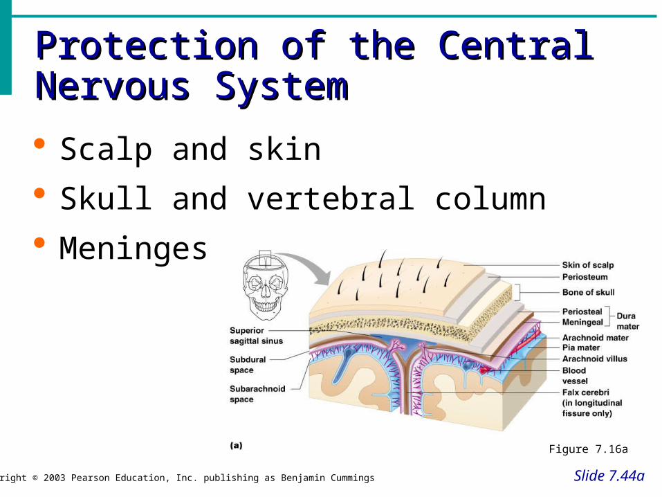
Protection of the Central Nervous Protection of the Central Nervous SystemSystem
Slide 7.44a
Copyright © 2003 Pearson Education, Inc. publishing as Benjamin Cummings
Scalp and skin
Skull and vertebral column
Meninges
Figure 7.16a

Protection of the Central Nervous Protection of the Central Nervous SystemSystem
Slide 7.44b
Copyright © 2003 Pearson Education, Inc. publishing as Benjamin Cummings
Cerebrospinal fluid
Blood brain barrier
Figure 7.16a
MeningesMeninges
Copyright © 2003 Pearson Education, Inc. publishing as Benjamin Cummings
Dura mater
Double-layered external covering
Periosteum – attached to surface of the skull
Meningeal layer – outer covering of the brain
Layers separate in certain places to form large veins called dural sinuses
Folds inward in several areas
MeningesMeninges
Slide 7.45b
Copyright © 2003 Pearson Education, Inc. publishing as Benjamin Cummings
Arachnoid layer
Middle layer
Web-like
Pia mater
Internal layer
Clings to the surface of the brain

Cerebrospinal FluidCerebrospinal Fluid
Slide 7.46Copyright © 2003 Pearson Education, Inc. publishing as Benjamin Cummings
Clear liquid similar to blood plasma composition
Formed by the choroid plexus
Forms a watery cushion to protect the brain
Circulated in arachnoid space, ventricles, and central canal of the spinal cord
Returns to blood via villi in the dural sinuses
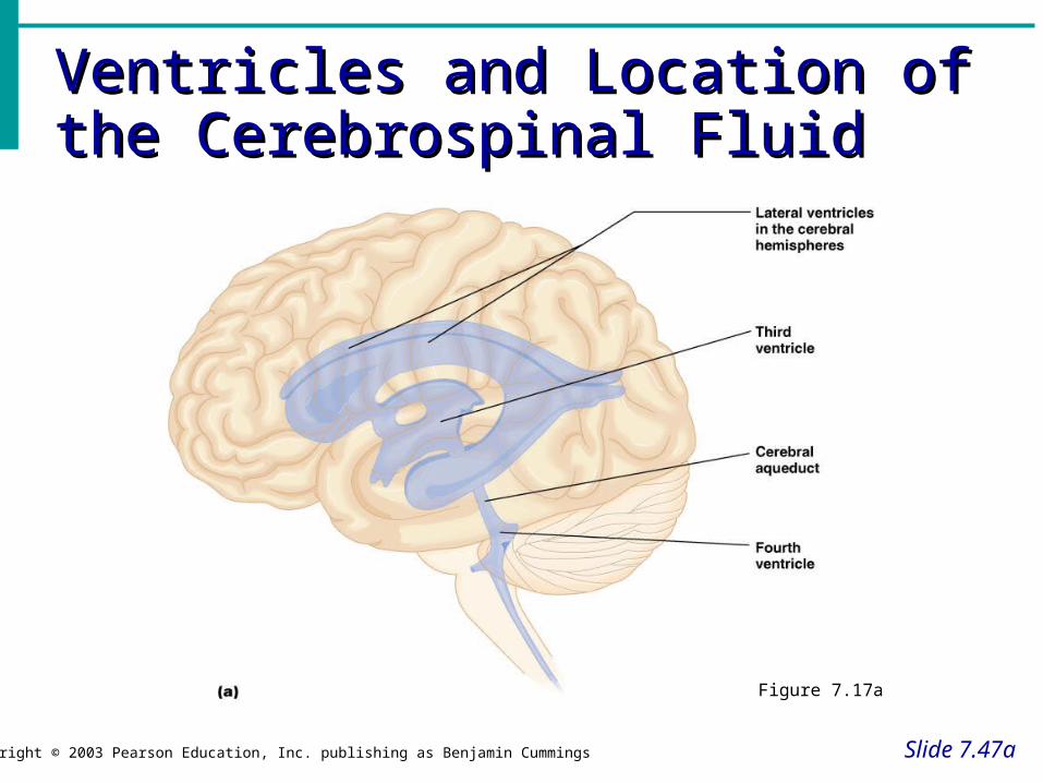
Ventricles and Location of the Ventricles and Location of the Cerebrospinal FluidCerebrospinal Fluid
Slide 7.47a
Copyright © 2003 Pearson Education, Inc. publishing as Benjamin Cummings
Figure 7.17a

Ventricles and Location of the Ventricles and Location of the Cerebrospinal FluidCerebrospinal Fluid
Slide 7.47b
Copyright © 2003 Pearson Education, Inc. publishing as Benjamin Cummings
Figure 7.17b
Blood Brain BarrierBlood Brain Barrier
Slide 7.48Copyright © 2003 Pearson Education, Inc. publishing as Benjamin Cummings
Includes the least permeable capillaries of the body
Excludes many potentially harmful substances
Useless against some substances Fats and fat soluble molecules Respiratory gases Alcohol Nicotine Anesthesia

Cerebral Hemispheres (Cerebrum)Cerebral Hemispheres (Cerebrum)
Slide 7.28a
Copyright © 2003 Pearson Education, Inc. publishing as Benjamin Cummings
Paired (left and right) superior parts of the brain
Include more than half of the brain mass
Figure 7.13a
Cerebral Hemispheres (Cerebrum)Cerebral Hemispheres (Cerebrum)
Slide 7.28b
Copyright © 2003 Pearson Education, Inc. publishing as Benjamin Cummings
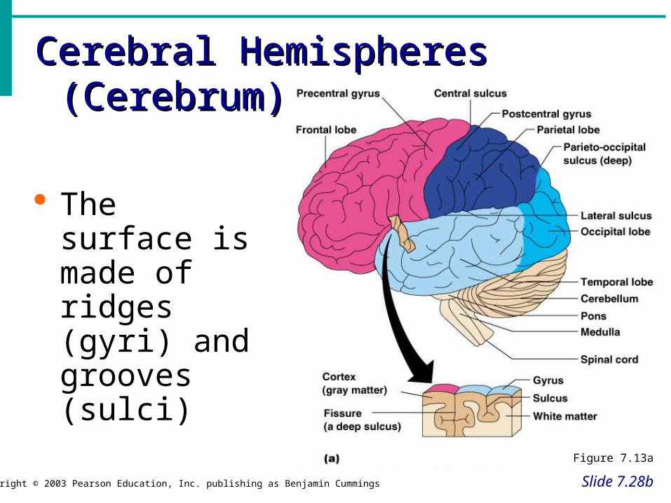
The surface is made of ridges (gyri) and grooves (sulci)
Figure 7.13a
Lobes of the CerebrumLobes of the Cerebrum
Slide 7.29a
Copyright © 2003 Pearson Education, Inc. publishing as Benjamin Cummings
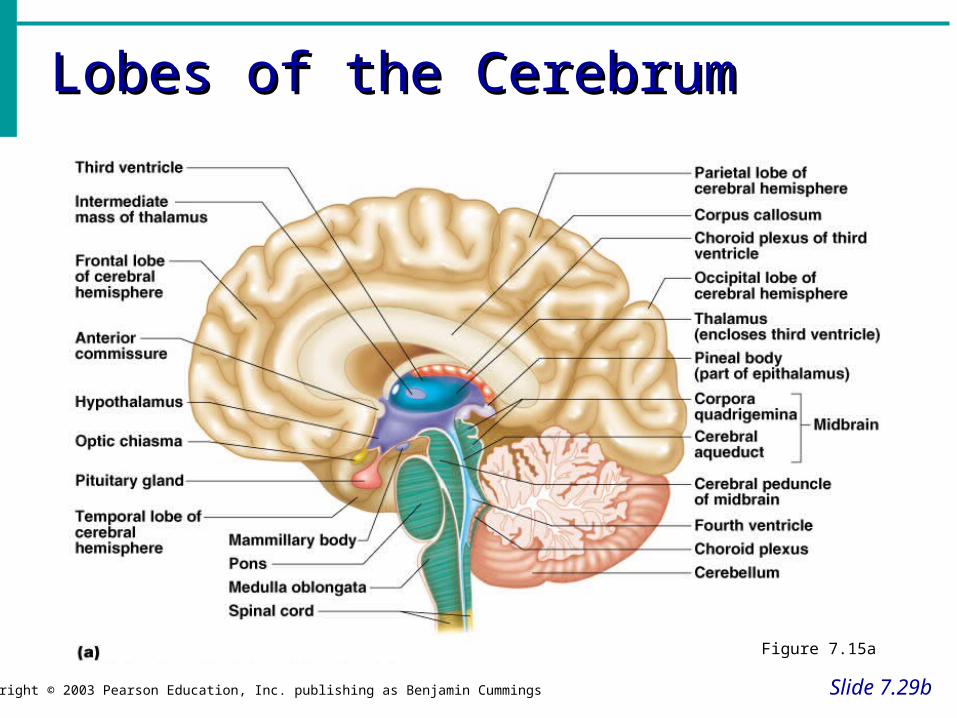
Fissures (deep grooves) divide the cerebrum into lobes
Surface lobes of the cerebrum
Frontal lobe
Parietal lobe
Occipital lobe
Temporal lobe
Lobes of the CerebrumLobes of the Cerebrum
Slide 7.29b
Copyright © 2003 Pearson Education, Inc. publishing as Benjamin Cummings
Figure 7.15a

Specialized Areas of the CerebrumSpecialized Areas of the Cerebrum
Slide 7.30Copyright © 2003 Pearson Education, Inc. publishing as Benjamin Cummings
Somatic sensory area – receives impulses from the body’s sensory receptors
Primary motor area – sends impulses to skeletal muscles
Broca’s area – involved in our ability to speak

Sensory and Motor Areas of the Sensory and Motor Areas of the Cerebral CortexCerebral Cortex
Slide 7.31Copyright © 2003 Pearson Education, Inc. publishing as Benjamin Cummings
Figure 7.14
Specialized Area of the CerebrumSpecialized Area of the Cerebrum
Slide 7.32a
Copyright © 2003 Pearson Education, Inc. publishing as Benjamin Cummings
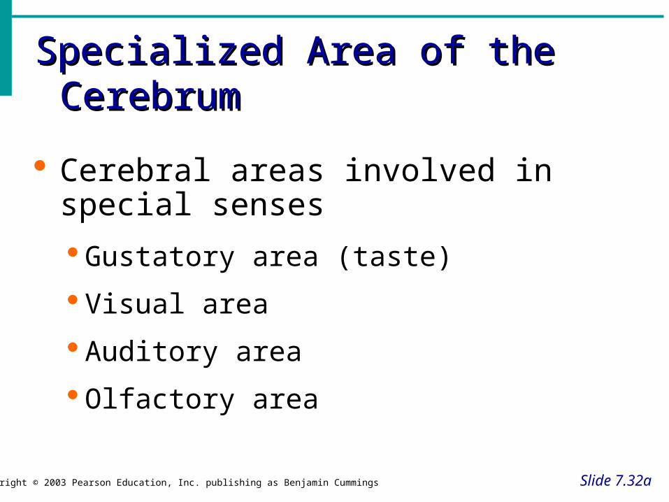
Cerebral areas involved in special senses
Gustatory area (taste)
Visual area
Auditory area
Olfactory area
Specialized Area of the CerebrumSpecialized Area of the Cerebrum
Slide 7.32b
Copyright © 2003 Pearson Education, Inc. publishing as Benjamin Cummings
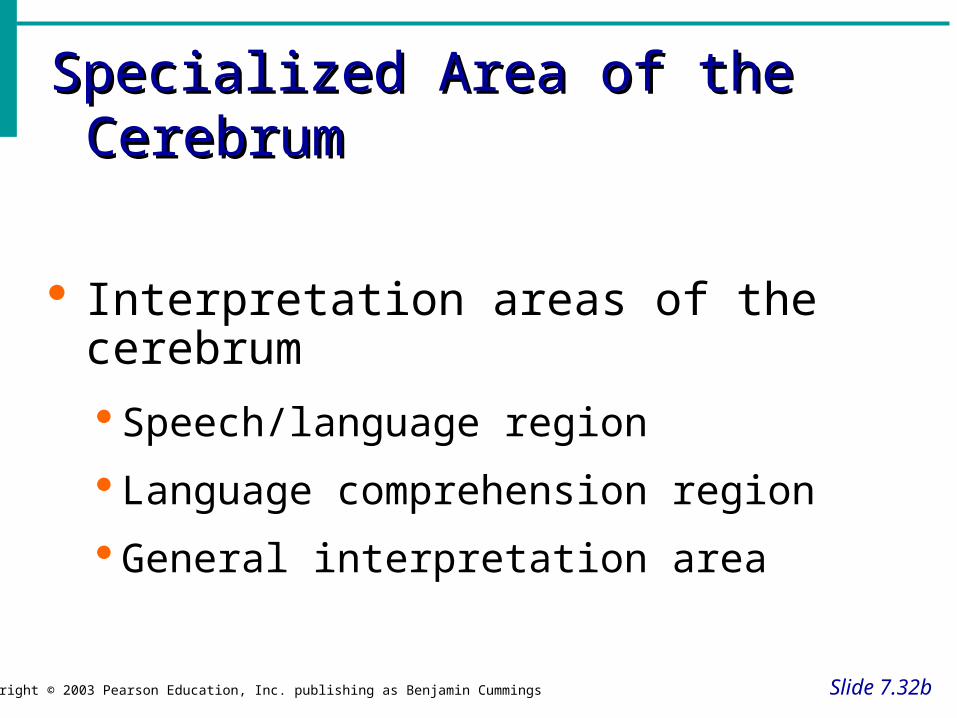
Interpretation areas of the cerebrum
Speech/language region
Language comprehension region
General interpretation area
Specialized Area of the CerebrumSpecialized Area of the Cerebrum
Slide 7.32c
Copyright © 2003 Pearson Education, Inc. publishing as Benjamin Cummings
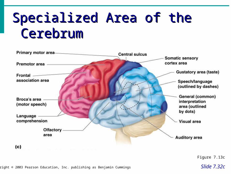
Figure 7.13c
Memory and Learning
• Short-term memory• Retention of information for few seconds or
minutes• Information lost unless reinforced
• Long-term memory• Storage of information that can be recalled
later

DiencephalonDiencephalon
Slide 7.34a
Copyright © 2003 Pearson Education, Inc. publishing as Benjamin Cummings
Sits on top of the brain stem
Enclosed by the cerebral hemispheres
Made of three parts Thalamus
Hypothalamus
Epithalamus

DiencephalonDiencephalon
Slide 7.34b
Copyright © 2003 Pearson Education, Inc. publishing as Benjamin Cummings
Figure 7.15

ThalamusThalamus
Slide 7.35Copyright © 2003 Pearson Education, Inc. publishing as Benjamin Cummings
Surrounds the third ventricle
The relay station for sensory impulses
Transfers impulses to the correct part of the cortex for localization and interpretation

HypothalamusHypothalamus
Slide 7.36a
Copyright © 2003 Pearson Education, Inc. publishing as Benjamin Cummings
Under the thalamus
Important autonomic nervous system center Helps regulate body temperature
Controls water balance
Regulates metabolism

HypothalamusHypothalamus
Slide 7.36b
Copyright © 2003 Pearson Education, Inc. publishing as Benjamin Cummings
An important part of the limbic system (emotions)
The pituitary gland is attached to the hypothalamus
EpithalamusEpithalamus
Slide 7.37Copyright © 2003 Pearson Education, Inc. publishing as Benjamin Cummings
Forms the roof of the third ventricle
Houses the pineal body (an endocrine gland)
Includes the choroid plexus – forms cerebrospinal fluid
Brain StemBrain Stem
Slide 7.38a
Copyright © 2003 Pearson Education, Inc. publishing as Benjamin Cummings
Attaches to the spinal cord
Parts of the brain stem Midbrain
Pons
Medulla oblongata

Brain StemBrain Stem
Slide 7.38b
Copyright © 2003 Pearson Education, Inc. publishing as Benjamin Cummings
Figure 7.15a

MidbrainMidbrain
Slide 7.39Copyright © 2003 Pearson Education, Inc. publishing as Benjamin Cummings
Mostly composed of tracts of nerve fibers
Has two bulging fiber tracts – cerebral peduncles
Has four rounded protrusions – corpora quadrigemina
Reflex centers for vision and hearing
Cranial nerves III and IV originate from the midbrain
PonsPons
Slide 7.40Copyright © 2003 Pearson Education, Inc. publishing as Benjamin Cummings
The bulging center part of the brain stem between the midbrain and medulla
Mostly composed of fiber tracts that carry impulses between the cerebellum and the rest of the nervous system
Includes nuclei involved in the control of breathing

Medulla OblongataMedulla Oblongata
Slide 7.41Copyright © 2003 Pearson Education, Inc. publishing as Benjamin Cummings
The lowest part of the brain stem Merges into the spinal cord Includes important fiber tracts Contains important control centers
Heart rate control Blood pressure regulation Breathing Swallowing Vomiting

Medulla Oblongata
• Motor fibers from motor cortex extend through the medulla and most cross from one side to the other.– Results in contralateral control– Right hemisphere controls muscles in the left
side of the body– Left hemisphere controls muscles in the right
side of the body

CerebellumCerebellum
Slide 7.43a
Copyright © 2003 Pearson Education, Inc. publishing as Benjamin Cummings
Two hemispheres with convoluted surfaces
Provides involuntary coordination of body movements
Helps maintain balance in standing, walking and sitting
Helps maintain muscle tone

CerebellumCerebellum
Slide 7.43b
Copyright © 2003 Pearson Education, Inc. publishing as Benjamin Cummings
Figure 7.15a

Cranial NervesCranial Nerves
Slide 7.58Copyright © 2003 Pearson Education, Inc. publishing as Benjamin Cummings
12 pairs of nerves that mostly serve the head and neck
Numbered in order, front to back
Most are mixed nerves, but three are sensory only

Distribution of Cranial NervesDistribution of Cranial Nerves
Slide 7.59Copyright © 2003 Pearson Education, Inc. publishing as Benjamin Cummings
Figure 7.21
Cranial NervesCranial Nerves
Slide 7.60Copyright © 2003 Pearson Education, Inc. publishing as Benjamin Cummings
I Olfactory nerve – sensory for smell
II Optic nerve – sensory for vision
III Oculomotor nerve – motor fibers to eye muscles
IV Trochlear – motor fiber to eye muscles
Cranial NervesCranial Nerves
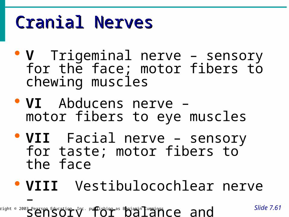
Slide 7.61Copyright © 2003 Pearson Education, Inc. publishing as Benjamin Cummings
V Trigeminal nerve – sensory for the face; motor fibers to chewing muscles
VI Abducens nerve – motor fibers to eye muscles
VII Facial nerve – sensory for taste; motor fibers to the face
VIII Vestibulocochlear nerve – sensory for balance and hearing

Cranial NervesCranial Nerves
Slide 7.62Copyright © 2003 Pearson Education, Inc. publishing as Benjamin Cummings
IX Glossopharyngeal nerve – sensory for taste; motor fibers to the pharynx
X Vagus nerves – sensory and motor fibers for pharynx, larynx, and viscera
XI Accessory nerve – motor fibers to neck and upper back
XII Hypoglossal nerve – motor fibers to tongue
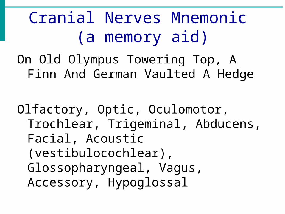
Cranial Nerves Mnemonic (a memory aid)
On Old Olympus Towering Top, A Finn And German Vaulted A Hedge
Olfactory, Optic, Oculomotor, Trochlear, Trigeminal, Abducens, Facial, Acoustic (vestibulocochlear), Glossopharyngeal, Vagus, Accessory, Hypoglossal

Aging of the Nervous System
• Brain decreases in size and weight
• Speed of processing information slows
• Movements slowed
• Memory diminishes


