THE MUSCULAR SYSTEM CHAPTER 9. FUNCTIONS OF THE MUSCULAR SYSTEM pull on bones to accomplish body...
76
THE MUSCULAR SYSTEM CHAPTER 9
-
Upload
horatio-holland -
Category
Documents
-
view
214 -
download
0
Transcript of THE MUSCULAR SYSTEM CHAPTER 9. FUNCTIONS OF THE MUSCULAR SYSTEM pull on bones to accomplish body...

THE MUSCULAR SYSTEM
CHAPTER 9

FUNCTIONS OF THE MUSCULAR SYSTEM
• pull on bones to accomplish body movements
• provide muscle tone, maintain posture
• propel body fluids and food
• generate a heartbeat

FUNCTIONS OF THE MUSCULAR SYSTEM
• generate heat
• stabilize joints
• makes up about 40% of the body’s mass, over 600 skeletal muscles
• means little mouse

A COMPARISON OF THE 3 MUSCLE TYPES:
location:
SKELETAL CARDIAC SMOOTH attached to heart walls of
bones or skin visceral organs, BV’s
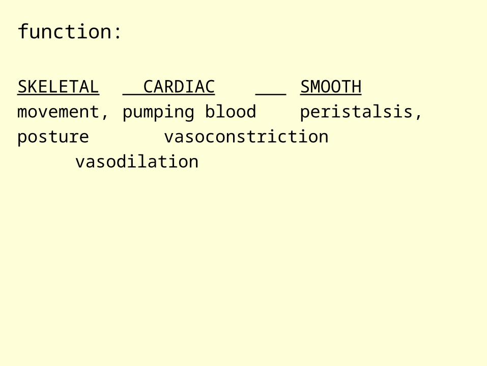
function:
SKELETAL CARDIAC SMOOTH
movement, pumping blood peristalsis,
posture vasoconstriction
vasodilation

cell shape & appearance:SKELETAL CARDIAC SMOOTH
multinucleate uninucleateuninucleate
striated striated not striated
cylindrical branched spindle-shaped
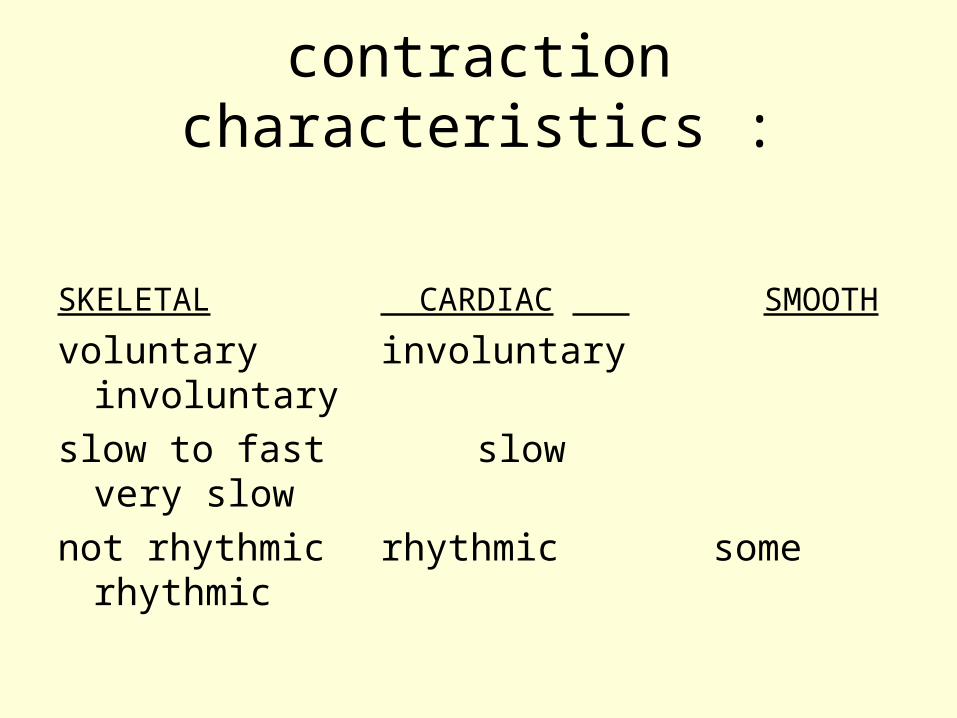
contraction characteristics :
SKELETAL CARDIAC SMOOTH
voluntary involuntary involuntary
slow to fast slow very slow
not rhythmic rhythmic some rhythmic

• skeletal muscle can contract rapidly & with great force, but it tires easily & must rest after short periods of activity
• smooth muscle contractions are slow and sustained
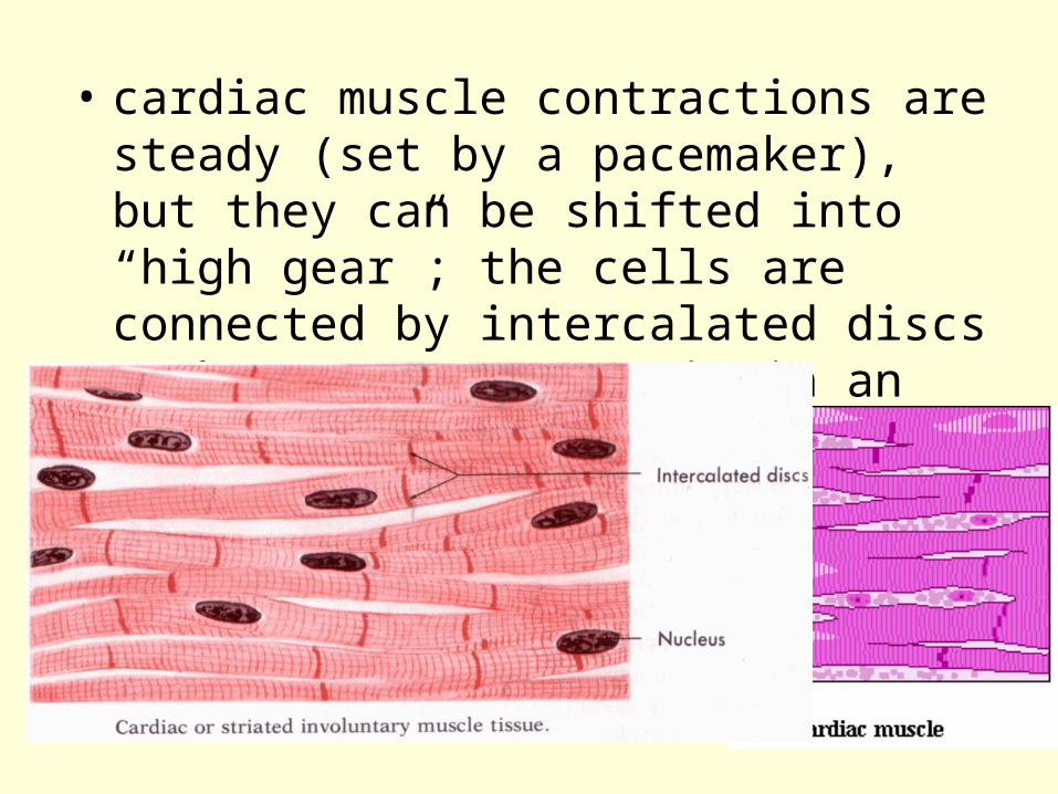
• cardiac muscle contractions are steady (set by a pacemaker), but they can be shifted into “high gear”; the cells are connected by intercalated discs & they react as a unit in an all-or-none manner

Functional characteristics of muscle
• Excitability or irritability = the ability to receive or respond to a stimulus, usually a chemical
• Contractility = the ability to shorten
• Extensibility = the ability to be stretched
• Elasticity = the ability to recoil and resume the resting length after being stretched

Gross anatomy of a skeletal muscle
• Connective tissue wrappings:– epimysium = outermost layer– perimysium = surrounds each fascicle– endomysium = surrounds each muscle fiber

Gross anatomy of a skeletal muscle
• Nerve and blood supply:– each muscle is supplied a nerve, an artery,
and a vein to supply energy & remove wastes
• Attachments:– origin = attached to the bone that does not
move– insertion = attached to the movable bone

STRUCTURE OF A SKELETAL MUSCLE
• Tendons connect muscles to bone:
STRUCTURE OF A SKELETAL MUSCLE
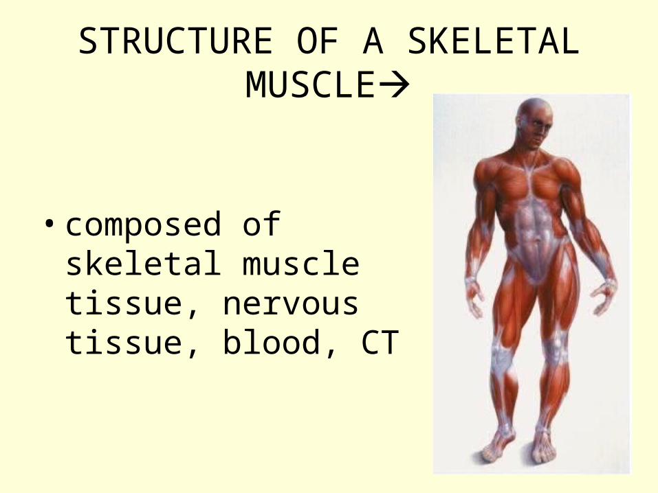
• composed of skeletal muscle tissue, nervous tissue, blood, CT

STRUCTURE OF A SKELETAL MUSCLE
• fascia = layers of CT that cover individual muscles & hold them in place; may form tendons, hooking up with a bone’s periosteum
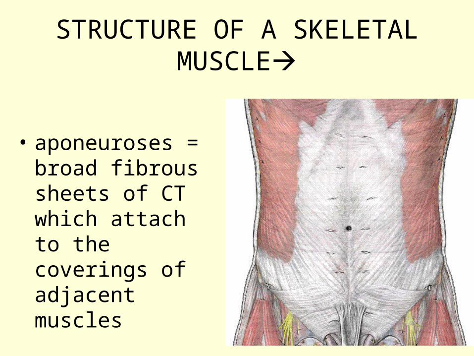
STRUCTURE OF A SKELETAL MUSCLE
• aponeuroses = broad fibrous sheets of CT which attach to the coverings of adjacent muscles

• muscles are composed of bundles of fibers (muscle cells) = fascicles
• muscle fibers are made of myofibrils

– myofibrils are made of thick & thin filaments
• myosin = thick filaments
• actin = thin filaments
– arranged in an overlapping pattern the gives striated look

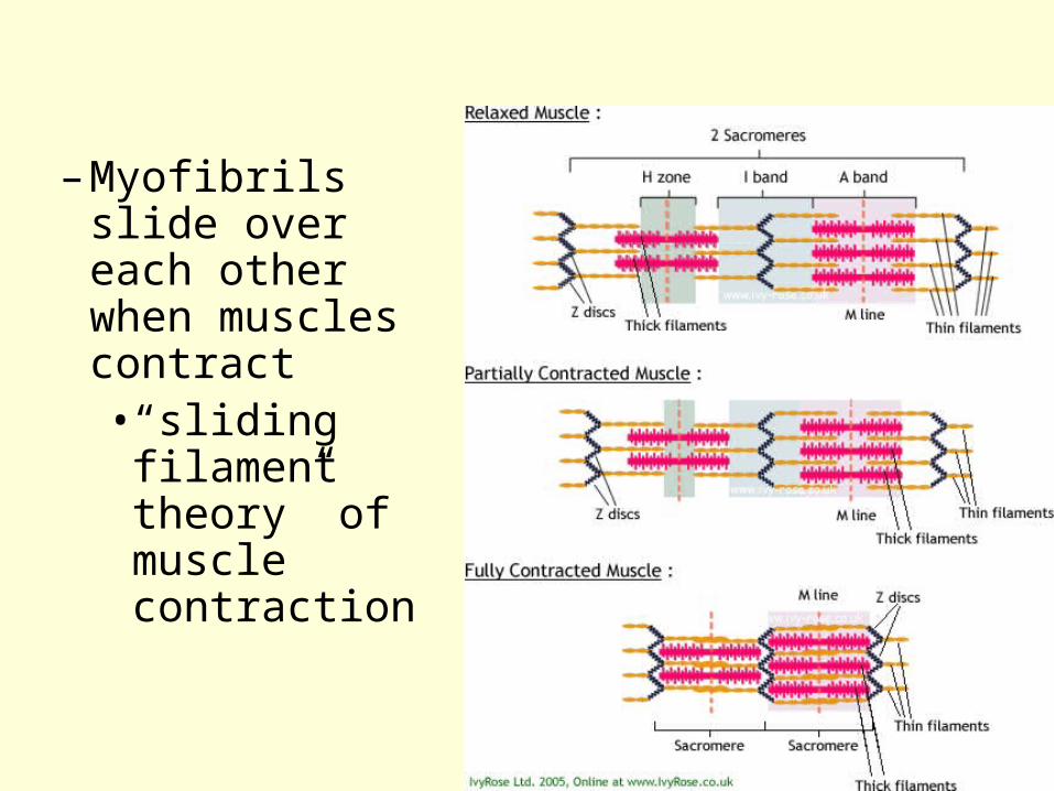
– Myofibrils slide over each other when muscles contract• “sliding
filament theory” of muscle contraction

– myofibrils consist of repeating units called sarcomeres = the segment of myofibril that extends from one Z line to the next (Z lines are what the actin filaments attach to)
– striated muscles are created by I bands (the light part) and A bands (the dark part)


neuromuscular junction is where a muscle fiber joins a motor neuron

• the tips of the motor neuron contain neurotransmitters to communicate with the muscle fiber & cause it to contract
• synaptic cleft = the gap between the nerve endings & the muscle cells
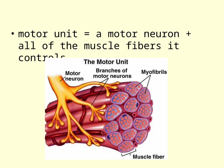
• motor unit = a motor neuron + all of the muscle fibers it controls

SKELETAL MUSCLE CONTRACTION
• myosin has “cross bridges” which fit into binding sites on the actin filaments, pulling on them & causing them to slide over the myosin

• sliding filament
• detailed crossbridges

4 steps of muscle contraction:
1- myosin cross bridge attaches to the actin myofilament
2- working stroke: the myosin head pivots & bends as it pulls on the actin filament, sliding it toward the M line
3- as new ATP attaches to the myosin head, the cross bridge detaches
4- as ATP is split into ADP + P, cocking of the myosin occurs

SKELETAL MUSCLE CONTRACTION
• muscle contraction is initiated by a nerve impulse (action potential)
• requires acetylcholine, calcium ions, & ATP
– acetylcholine is a neurotransmitter
3 energy sources of ATP
• creatine phosphate = allows immediate regeneration of ATP
• cellular respiration to break down glucose
• glycogen breaks down into glucose for cellular respiration

• aerobic cell respiration takes place in the presence of oxygen – makes 36 ATP per molecule of glucose
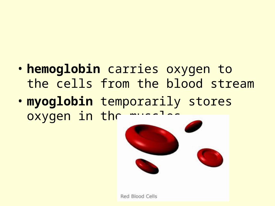
• hemoglobin carries oxygen to the cells from the blood stream
• myoglobin temporarily stores oxygen in the muscles

• during strenuous exercise, muscle cells must switch to anaerobic respiration which causes lactic acid to accumulate & cause the “burn”

• creates an oxygen debt = the amount of oxygen it takes to revert the lactic acid into glucose + restoring ATP & creatine phosphate to their original levels
• anaerobic respiration makes only 2 ATP per molecule of glucose, but it does it quickly

Energy systems used during sports:
• Requiring a surge of energy but lasts only a few seconds:– Ex = weight lifting, diving, sprinting– uses ATP and CP stores
• On-and-off burstlike activities:– Ex = tennis, soccer, 100m swim– uses anaerobic lactic acid fermentation
• Prolonged activities:– Ex = jogging, running marathons– uses aerobic respiration

• muscle fatigue = when muscles lose their ability to contract even when stimulated, usually due to lactic acid accumulation
• muscle cramps = sustained involuntary muscle contraction
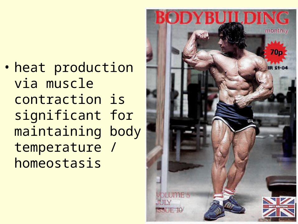
• heat production via muscle contraction is significant for maintaining body temperature / homeostasis

Effect of exercise on muscles:
• Muscle inactivity leads to weakness & muscle wasting = atrophy

Effect of exercise on muscles:
• Aerobic / endurance exercise (swimming, jogging, fast walking, biking) causes:– more efficient muscle metabolism – greater endurance, strength, resistance to fatigue– boosts overall body metabolism– more efficient neuromuscular coordination– Improves gastrointestinal mobility– enhances skeletal strength– Improves the delivery of oxygen & nutrients to all
body tissues via the CV & respiratory system

• Resistance exercise (weight lifting) causes:– Increased muscle bulk due to larger muscle fibers– more mitochondria, more myofilaments, more
glycogen storage capacity– more connective tissue between cells– significantly stronger muscles
• Cross training (alternating aerobic and anaerobic activities) promotes optimal health

MUSCULAR RESPONSES• threshold stimulus = the minimal stimulus
required to cause a muscle contraction

MUSCULAR RESPONSES• all-or-none response = skeletal muscle
fibers contract completely if they contract at all, not true for the whole muscle

MUSCULAR RESPONSES
• twitch = a single, brief, jerky contraction
• summation = a rapid series of stimuli “sums up” the contractions
• tetanus = sustained contraction with no relaxation


MUSCULAR RESPONSES• more muscle cells stimulated = a stronger
muscle contraction• recruitment = when more motor units are
recruited to respond to increasing intensity of stimulation
• summation and recruitment together can produce a sustained contraction of increasing strength

MUSCULAR RESPONSES• isotonic muscle contractions = muscles
shorten, movement occurs
• isometric muscle contractions = muscles stay the same length, no movement occurs; ex. pushing against a wall

MUSCULAR RESPONSES• rigor mortis = partial muscle contraction
that happens after death


SKELETAL MUSCLE ACTIONS
• movements depend on the type of joint the muscles are associated with & the way the muscles are attached on either side of the joint
• remember: muscles pull on bones which act as levers

SKELETAL MUSCLE ACTIONS
• origin = the end of a muscle attached to the bone that doesn’t move
• insertion = the end of the muscle attached to the bone that moves
• when muscles contract, the insertion is pulled toward the origin

SKELETAL MUSCLE ACTIONS
• prime mover = one muscle in a group that causes most of the movement
• synergist = muscles that assist the prime mover
• antagonist = causes movement opposite of the prime mover

• example: the bicep is the prime mover of elbow flexion, its antagonist is the tricep – the prime mover of elbow extension

MAJOR SKELETAL MUSCLES
• naming skeletal muscles: based on many sets of criteria– direction of the fibers
• rectus = straight, oblique = slanted
– relative size of the muscle• maximus = largest, minimus =
smallest, longus = long

MAJOR SKELETAL MUSCLES
– location of the muscle
• named for the bone such as temporalis or frontalis
– number of origins or heads
• biceps = 2, triceps = 3,
quadriceps = 4

– location of the muscle’s origin & insertion• sternocleidomastoid – sternum, clavicle,
mastoid process
– shape of the muscle• deltoid = triangle • trapezius = trapezoid
– action of the muscle• flexor, extensor, adductor

SUPERFICIAL MUSCLES OF THE HUMAN BODY
• muscles of facial expression:
– lie beneath the skin of the face & scalp, used to communicate feelings through facial expression
– includes the frontalis, orbicularis oculi, orbicularis oris, zygomaticus, platysma


SUPERFICIAL MUSCLES OF THE HUMAN BODY
• muscles of mastication:
– attach to the mandible, used for chewing
– includes the masseter & temporalis

SUPERFICIAL MUSCLES OF THE HUMAN BODY
• muscles that move the head:
– found in the neck and upper back
– includes the sternocleidomastoid, sternohyoid

SUPERFICIAL MUSCLES OF THE HUMAN BODY
• muscles that move the pectoral girdle:
– most connect the scapula to nearby bones
– includes the trapezius, serratus anterior, pectoralis minor


muscles that move the arm:
– these connect the humerus to various regions of the pectoral girdle, ribs, & vertebral column
– includes the pectoralis major, teres major, latissimus dorsi, deltoid, infraspinatus


muscles that move the forearm:
– these connect the radius & ulna to the pectoral girdle or humerus
– includes the biceps brachii, brachialis, brachioradialis, triceps brachii, supinator

SUPERFICIAL MUSCLES OF THE HUMAN BODY
• muscles that move the wrist, hand, & fingers:
– these muscles arise from the distal end of the humerus and from the radius & ulna
– includes the flexor carpi radialis, flexor carpi ulnaris, palmaris longus, extensor carpi radialis longus, extensor carpi ulnaris, and extensor digitorum


muscles of the abdominal wall:
– these muscles connect the rib cage and vertebral column to the pelvic girdle
– includes the external oblique, internal oblique, transverses abdominis, and rectus abdominis


SUPERFICIAL MUSCLES OF THE HUMAN BODY
• muscles of the pelvic outlet:
– these muscles form the floor of the pelvic cavity and fill the space within the pubic arch
– includes the bulbospongiosus and ischiocavernosus

muscles that move the thigh:
– these muscles attach to the femur and to some part of the pelvic girdle
– includes the iliopsoas, gluteus maximus, gluteus medius, gluteus minimus, tensor fasciae latae, adductor longus, adductor magnus, and gracilis

SUPERFICIAL MUSCLES OF THE HUMAN BODY
• muscles that move the leg:
– these muscles connect the tibia or fibula to the femur or pelvic girdle
– includes the biceps femoris, semitendinosis, semimembranosus, sartorius, and the quadriceps femoris group = rectus femoris, vastus lateralis, vastus medialis, vastus intermedius


SUPERFICIAL MUSCLES OF THE HUMAN BODY
• muscles that move the foot, ankle, & toes:
– these muscles attach the femur, tibia, and fibula to the bones of the foot
– includes the tibialis anterior, extensor digitorum longus, gastrocnemius, soleus, and peroneus longus