Ultrastructural Morphometrical and Immunocytochemical Analyses ...
1407
Int. J. Morphol.,31(4):1407-1414, 2013.
The Morphometrical Analysis of the External Carotid Arteryand its Branches with Multidetector Computerized
Tomography Angiography Technique
Análisis Morfométrico de la Arteria Carótida Externa y sus Ramas Mediantela Técnica de Angiografía por Tomografía Computarizada Multidetector
Musa Acar*; Ahmet Salbacak** ; Mehmet Emin Sakarya*** ; Ismail Zararsiz **** & Mahinur Ulusoy ****
ACAR, M; SALBACAK, A; SAKARYA, M. E.; ZARARSIZ, I. & ULUSOY, M. The morphometrical analysis of the external carotidartery and ıts branches with multidetector computerized tomography angiography technique. Int. J. Morphol., 31(4):1407-1414, 2013.
SUMMARY: The external carotid artery (ECA) is the main artery of the head and the neck region. Carotid bifurcation (CB),which is one of the place where the atherosclerotic plaques are most commonly seen. The surgical procedure of these plaques whichcauses cerebral vascular accident (CVA) is carotid endarterectomy. In this surgical procedure, the knowledge of the anatomical coursesand variations of the carotid artery increased the surgery performance. In our study, we aimed to introduce the course, the location and thevariation of the ECA’s and their branches. This study is carried out on multidetector computerized tomography angiography of the ECA’sof 50 men and 50 women, totally 200 ECA’s (100 right, 100 left). The measurement of the inner diameter of the common carotid artery(CCA) and the ECA was evaluated. The location of the CB was determined and its vertical distance to the gonion measured. We foundthat the superior thyroid artery (STA) originated from the CCA, the CB and the ECA. The vertical distances between the CB and the STA,lingual (LA), facial (FA) occipital (OA) were measured. The ECA and its branches were recorded. We believe that the assessment of theECA morphometrically may comprise control groups of diseases related to the vessel diameter and this data may be used as reference inclinic and surgery. Knowing the anatomical details and variations is vital to prevent unpredictable complications in surgery.
KEY WORDS: External carotid artery; Multidetector computerized tomography; Variation; Anatomy.
INTRODUCTION
The neck region is of great vital value because ofthe link between head and trunk, upper limb. The thyroidgland, trachea, jugular veins and carotid arteries are vitalorgans and do not have protective osseous structure likeother system so they are very vulnerable to firearm andknife injuries, the blood vessel and the nerves arecommonly damaged in this area (Moore & Dalley, 2007;Ozgur et al., 2008a).
The external carotid artery, feeds the structures ofthe face, the scalp, upper part of the thyroid gland and somepart of the dura mater, is main artery of the head and neckregion (Mahendrakar, 2007; Mamatha et al., 2003). TheECA, which is one of the two arteries of the CCA, bifurcateat the level of the (superior margin of the) thyroid cartilagefrom the CCA which is known carotid bifurcation (CB). The
common carotid artery divides into ECA and ICA at thecarotid triangle, and locates the anteromedial of the ICA sothe anterior branches of the ECA do not cross ICA. The ECAgives immediately its branches as the superior thyroid artery,ascending pharyngeal artery, LA, FA, OA, posterior auricu-lar artery, superficial temporal artery, maxillary arteryrespectively, after at the level of the CB (Moore & Dalley).
The carotid bifurcation is the place in whereatherosclerotic plaque is most commonly seen. Stenosis ofthe carotid arteries is important cause of the CVA. Theformation of the atherosclerotic plaques is closely related tothe anatomy of the CB which effects cervical blood flow(Takahashi et al., 1997; Schulz & Rothwell, 2001; Furlan etal., 2007; Hayashi et al., 2005). The most common treatmentof these plaques, which causes the CVA, is endartrectomy.
* Mevlana University, Department of Physical Therapy and Rehabilitation, Konya, Turkey.** Necmettin Erbakan University Meram, Faculty of Medicine Department of Anatomy, Konya, Turkey.*** Necmettin Erbakan University Meram, Faculty of Medicine Department of Radiology, Konya, Turkey.**** Mevlana University, Faculty of Medicine, Department of Anatomy, Konya, Turkey.
1408
Surgical procedures require that both atheroscleroticplaques and tunica intima are removed. In this type ofsurgical procedure, knowing the details of the anatomyand variations of the carotid arteries may increase thesurgical success. Knowledge about the CCA, ECA and itsbranches and variations show that they are very importantin radiologic diagnosis and surgery (Gluncic et al., 2001).Ligation of the ECA is performed in case of unstoppablebleeding of arteries which originated the ECA. Similarlyin surgery performed in the carotid trigonum, theknowledge of the anatomy is necessary (Gluncic et al.;Hayashi et al.).
In this study we evaluated the morphometricanalysis of ECA and its branches, the properties, the originof the branches and variations of bifurcation in adults. Weaimed to compare these feature according to sex and thesides, to detect the prevalence and to provide helpfulinformation for surgery intervention performed in theseareas in the scope of the obtained data.
MATERIAL AND METHOD
This study was carried out on images obtained withmultidetector CT angiography (MDCTA) technique, from200 sample (100 right, 100 left) of 100 patients (50 men, 50women) who were referred to the CT angiography, did nothave any carotid artery disease, in the Department ofRadiodiagnostics, Faculty of Medicine, Necmettin ErbakanUniversity.
We planned our study as three stage; the preparationof materials, the assessment of the ECA and its branchesand the examining its variations, and statistical analysis. Allof the stage was performed by the same researcher tominimize the risk of error.
In first stage of the study; patients who previouslyconsulted the hospital and underwent MDCTA of the headand neck region by using 64 slice-MDCT system (SiemensSomatom Sensation, Erlanger, Almanya, 2005) wereidentified. Then the volumetric and sub-volumetric imageswhich could be appropriate to assess the arterial structurereformatted in the sagittal, coronal, axial plane with MIP(maximum intensity projection), MPR (multiplanarreformation) and VRT (volume rendering technique) wereidentified. The measurements were performed on thesesamples. All patients were in supine position when they wereexamined by the MDCTA so the standardized data wereobtained. The obtained standardized data is important forthe reliability of the study.
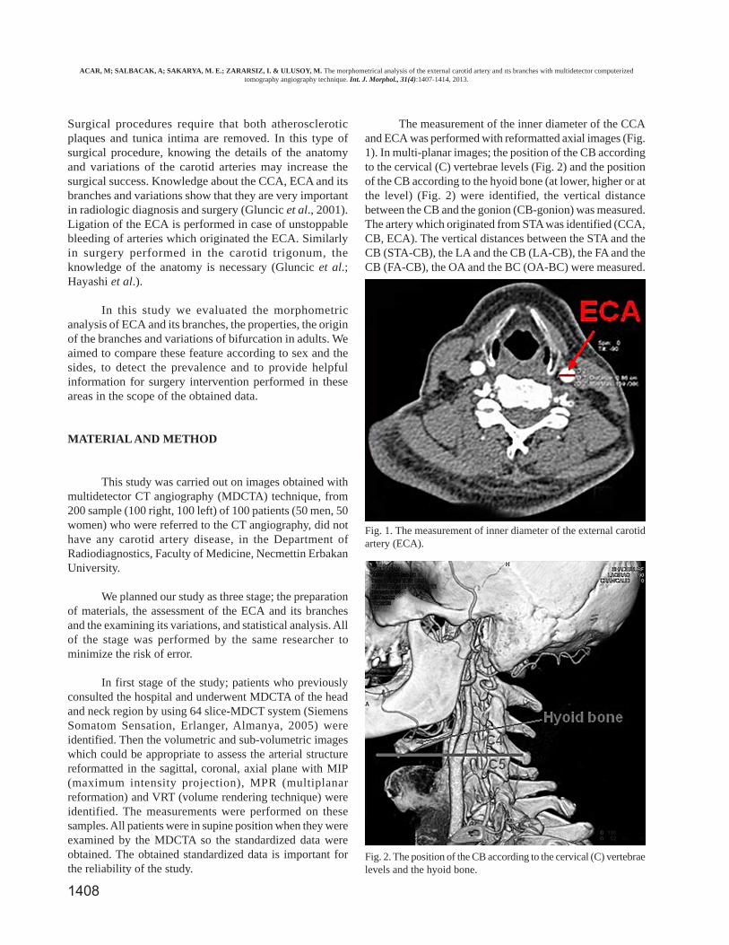
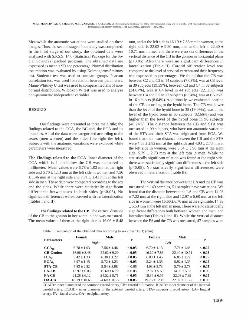
The measurement of the inner diameter of the CCAand ECA was performed with reformatted axial images (Fig.1). In multi-planar images; the position of the CB accordingto the cervical (C) vertebrae levels (Fig. 2) and the positionof the CB according to the hyoid bone (at lower, higher or atthe level) (Fig. 2) were identified, the vertical distancebetween the CB and the gonion (CB-gonion) was measured.The artery which originated from STA was identified (CCA,CB, ECA). The vertical distances between the STA and theCB (STA-CB), the LA and the CB (LA-CB), the FA and theCB (FA-CB), the OA and the BC (OA-BC) were measured.
Fig. 1. The measurement of inner diameter of the external carotidartery (ECA).
Fig. 2. The position of the CB according to the cervical (C) vertebraelevels and the hyoid bone.
ACAR, M; SALBACAK, A; SAKARYA, M. E.; ZARARSIZ, I. & ULUSOY, M. The morphometrical analysis of the external carotid artery and ıts branches with multidetector computerizedtomography angiography technique. Int. J. Morphol., 31(4):1407-1414, 2013.
1409
Meanwhile the anatomic variations were studied on theseimages. Thus, the second stage of our study was completed.In the third stage of our study, the obtained data wereanalyzed with S.P.S.S. 14.0 (Statistical Package for the So-cial Sciences) packed program. The obtained data areexpressed as mean ± SD and percentage. Normal distributionassumption was evaluated by using Kolmogorov-Smirnovtest. Student-t test was used to compare groups, Pearsoncorrelation test was used for relation between parameters.Mann-Whitney U test was used to compare medians of non-normal distribution, Wilcoxon W test was used to analyzenon-parametric independent variables.
RESULTS
Our findings were presented as three main title; thefindings related to the CCA, the BC and, the ECA and itsbranches. All of the data were categorized according to thesexes (men-women) and the sides (the right-the left).Subjects with the anatomic variations were excluded whileparameters were measured.
The Findings related to the CCA. Inner diameter of theCCA which is 1 cm below the CB was measured asmillimeter. Mean values were 6.78 ± 1.03 mm at the rightside and 6.70 ± 1.13 mm at the left side in women and 7.56± 1.46 mm at the right side and 7.71 ± 1.43 mm at the leftside in men. These data were compared according to the sexand the sides. While there were statistically significantdifferences between sex in both sides (p<0.05), Nosignificant differences were observed with the lateralization(Tables I and II).
The findings related to the CB. The vertical distanceof the CB to the gonion in horizontal plane was measured.The mean values of them at the right side is 16.06 ± 8.48
mm, and at the left side is 16.19 ± 7.86 mm in women, at theright side is 22.02 ± 9.20 mm, and at the left is 22.40 ±10.71 mm in men and there were no sex differences in thevertical distance of the CB to the gonion in horizontal plane(p<0.05). Also there were no significant differences inlateralization (Table II). Carotid bifurcation level wascompared to the level of cervical vertebra and their frequencywas expressed as percentages. We found that the CB wasbetween C2 and C3 in 14 subjects (7.03%), was at C3 levelin 39 subjects (19.59%), between C3 and C4 in 69 subjects(34.67%), was at C4 level in 44 subjects (22.11%), wasbetween C4 and C5 in 17 subjects (8.54%), was at C5 levelin 16 subjects (8.04%). Additionally, we evaluated locationof the CB according to the hyoid bone. The CB was lowerthan the level of the hyoid bone in 38 (19.09%), was at thelevel of the hyoid bone in 65 subjects (32.66%) and washigher than the level of the hyoid bone in 96 subjects(48.24%). The distance between the CB and STA wasmeasured in 99 subjects, who have not anatomic variationof the STA and their STA was originated from ECA. Wefound that the mean distance between the CB and the STAwere 4.83 ± 2.82 mm at the right side and 4.03 ± 2.73 mm atthe left side in women, were 5.54 ± 3.98 mm at the rightside, 5.79 ± 2.73 mm at the left mm in men. While nostatistically significant relation was found at the right side,there were statistically significant differences at the left side(p<0.05). No statistically significant differences wereobserved in lateralization (Table II).
The vertical distance between the LA and the CB wasmeasured in 149 samples, 51 samples have variations. Wefound that the distance between the LA and CB were 14.03± 7.22 mm at the right side and 12.97 ± 5.60 mm at the leftside in women, were 15.60 ± 6.70 mm at the right side, 14.93± 5.53 mm at the left mm in men. There were no statisticallysignificant differences both between women and men, andlateralization (Tables I and II). While the vertical distancebetween the FA and the CB was measured, 47 samples were
Female Male Female MaleParameters
RightP
LeftP
CCAID 6.78 ± 1.03 7.56 ± 1.46 < 0.05 6.70 ± 1.13 7.71 ± 1.43 < 0.05CB-Gonion 16.06 ± 8.48 22.02 ± 9.20 < 0.05 16.19 ± 7.86 22.40 ± 10.71 < 0.05ICAID 5.42 ± 1.33 6.38 ± 1.22 < 0.05 6.00 ± 1.45 6.85 ± 1.72 < 0.05ECAID 4.97 ± 1.15 5.72 ± 1.23 < 0.05 5.24 ± 1.35 5.92 ± 1.35 < 0.05STA-CB 4.83 ± 2.82 5.54 ± 3.98 > 0.05 4.03 ± 2.73 5.79 ± 2.73 < 0.05LA-CB 13.97 ± 6.05 15.60 ± 6.70 > 0.05 12.97 ± 5.60 14.93 ± 5.53 > 0.05FA-CB 21.28 ± 6.12 24.52 ± 8.71 < 0.05 18.84 ± 6.55 22.03 ± 7.99 < 0.05OA-CB 18.19 ± 10.65 24.60 ± 16.77 < 0.05 19.76 ± 12.15 22.02 ± 11.25 > 0.05
Table I. Comparison of the obtained data according to sex (mean±SD) (mm).
CCAID= inner diameter of the common carotid artery, CB= carotid bifurcation, ICAID= inner diameter of the internalcarotid artery, ECAID= inner diameter of the external carotid artery, STA= superior thyroid artery, LA= lingualartery, FA= facial artery, OA= occipital artery.
ACAR, M; SALBACAK, A; SAKARYA, M. E.; ZARARSIZ, I. & ULUSOY, M. The morphometrical analysis of the external carotid artery and ıts branches with multidetector computerizedtomography angiography technique. Int. J. Morphol., 31(4):1407-1414, 2013.
1410
excluded due to having variations. So 147 samples wereincluded this measurement. The distances were 21.28 ± 6.12mm at the right side and 18.84 ± 6.55 mm at the left side inwomen, were 24.52 ± 8.71 mm at the right side, 22.03 ±7.99 mm at the left mm in men. There were statisticallysignificant differences both between women and men, andlateralization (Tables I and II).
The vertical distance between the OA and the CBwas measured in 199 samples which have no variations. Thedistance was measured as 18.19 ± 10.65 mm at the rightside, 19.76 ± 12.15 at the left side in women and 24.60 ±16.77 mm at the right side, 22.02 ± 11.25 mm at the left sidein men. There were statistically significant differences inright side (p<0.05) but not in left side between women andmen. However No statistically significant differences wereobserved in lateralization (Tables I and II).
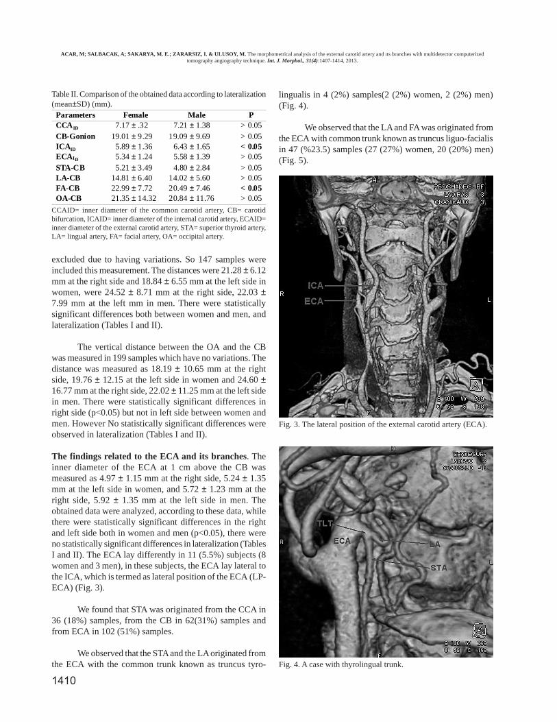
The findings related to the ECA and its branches. Theinner diameter of the ECA at 1 cm above the CB wasmeasured as 4.97 ± 1.15 mm at the right side, 5.24 ± 1.35mm at the left side in women, and 5.72 ± 1.23 mm at theright side, 5.92 ± 1.35 mm at the left side in men. Theobtained data were analyzed, according to these data, whilethere were statistically significant differences in the rightand left side both in women and men (p<0.05), there wereno statistically significant differences in lateralization (TablesI and II). The ECA lay differently in 11 (5.5%) subjects (8women and 3 men), in these subjects, the ECA lay lateral tothe ICA, which is termed as lateral position of the ECA (LP-ECA) (Fig. 3).
We found that STA was originated from the CCA in36 (18%) samples, from the CB in 62(31%) samples andfrom ECA in 102 (51%) samples.
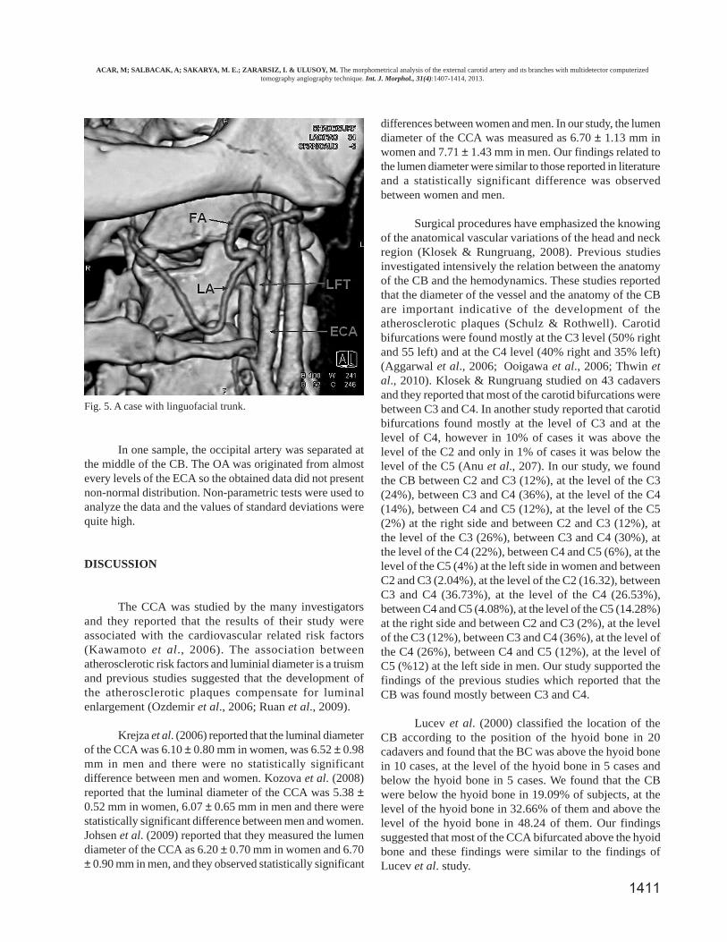
We observed that the STA and the LA originated fromthe ECA with the common trunk known as truncus tyro-
lingualis in 4 (2%) samples(2 (2%) women, 2 (2%) men)(Fig. 4).
We observed that the LA and FA was originated fromthe ECA with common trunk known as truncus liguo-facialisin 47 (%23.5) samples (27 (27%) women, 20 (20%) men)(Fig. 5).
Parameters Female Male PCCAID 7.17 ± .32 7.21 ± 1.38 > 0.05CB-Gonion 19.01 ± 9.29 19.09 ± 9.69 > 0.05ICAID 5.89 ± 1.36 6.43 ± 1.65 < 0.05ECAID 5.34 ± 1.24 5.58 ± 1.39 > 0.05STA-CB 5.21 ± 3.49 4.80 ± 2.84 > 0.05LA-CB 14.81 ± 6.40 14.02 ± 5.60 > 0.05FA-CB 22.99 ± 7.72 20.49 ± 7.46 < 0.05OA-CB 21.35 ± 14.32 20.84 ± 11.76 > 0.05
Table II. Comparison of the obtained data according to lateralization(mean±SD) (mm).
CCAID= inner diameter of the common carotid artery, CB= carotidbifurcation, ICAID= inner diameter of the internal carotid artery, ECAID=inner diameter of the external carotid artery, STA= superior thyroid artery,LA= lingual artery, FA= facial artery, OA= occipital artery.
Fig. 4. A case with thyrolingual trunk.
Fig. 3. The lateral position of the external carotid artery (ECA).
ACAR, M; SALBACAK, A; SAKARYA, M. E.; ZARARSIZ, I. & ULUSOY, M. The morphometrical analysis of the external carotid artery and ıts branches with multidetector computerizedtomography angiography technique. Int. J. Morphol., 31(4):1407-1414, 2013.
1411
differences between women and men. In our study, the lumendiameter of the CCA was measured as 6.70 ± 1.13 mm inwomen and 7.71 ± 1.43 mm in men. Our findings related tothe lumen diameter were similar to those reported in literatureand a statistically significant difference was observedbetween women and men.
Surgical procedures have emphasized the knowingof the anatomical vascular variations of the head and neckregion (Klosek & Rungruang, 2008). Previous studiesinvestigated intensively the relation between the anatomyof the CB and the hemodynamics. These studies reportedthat the diameter of the vessel and the anatomy of the CBare important indicative of the development of theatherosclerotic plaques (Schulz & Rothwell). Carotidbifurcations were found mostly at the C3 level (50% rightand 55 left) and at the C4 level (40% right and 35% left)(Aggarwal et al., 2006; Ooigawa et al., 2006; Thwin etal., 2010). Klosek & Rungruang studied on 43 cadaversand they reported that most of the carotid bifurcations werebetween C3 and C4. In another study reported that carotidbifurcations found mostly at the level of C3 and at thelevel of C4, however in 10% of cases it was above thelevel of the C2 and only in 1% of cases it was below thelevel of the C5 (Anu et al., 207). In our study, we foundthe CB between C2 and C3 (12%), at the level of the C3(24%), between C3 and C4 (36%), at the level of the C4(14%), between C4 and C5 (12%), at the level of the C5(2%) at the right side and between C2 and C3 (12%), atthe level of the C3 (26%), between C3 and C4 (30%), atthe level of the C4 (22%), between C4 and C5 (6%), at thelevel of the C5 (4%) at the left side in women and betweenC2 and C3 (2.04%), at the level of the C2 (16.32), betweenC3 and C4 (36.73%), at the level of the C4 (26.53%),between C4 and C5 (4.08%), at the level of the C5 (14.28%)at the right side and between C2 and C3 (2%), at the levelof the C3 (12%), between C3 and C4 (36%), at the level ofthe C4 (26%), between C4 and C5 (12%), at the level ofC5 (%12) at the left side in men. Our study supported thefindings of the previous studies which reported that theCB was found mostly between C3 and C4.
Lucev et al. (2000) classified the location of theCB according to the position of the hyoid bone in 20cadavers and found that the BC was above the hyoid bonein 10 cases, at the level of the hyoid bone in 5 cases andbelow the hyoid bone in 5 cases. We found that the CBwere below the hyoid bone in 19.09% of subjects, at thelevel of the hyoid bone in 32.66% of them and above thelevel of the hyoid bone in 48.24 of them. Our findingssuggested that most of the CCA bifurcated above the hyoidbone and these findings were similar to the findings ofLucev et al. study.
In one sample, the occipital artery was separated atthe middle of the CB. The OA was originated from almostevery levels of the ECA so the obtained data did not presentnon-normal distribution. Non-parametric tests were used toanalyze the data and the values of standard deviations werequite high.
DISCUSSION
The CCA was studied by the many investigatorsand they reported that the results of their study wereassociated with the cardiovascular related risk factors(Kawamoto et al., 2006). The association betweenatherosclerotic risk factors and luminial diameter is a truismand previous studies suggested that the development ofthe atherosclerotic plaques compensate for luminalenlargement (Ozdemir et al., 2006; Ruan et al., 2009).
Krejza et al. (2006) reported that the luminal diameterof the CCA was 6.10 ± 0.80 mm in women, was 6.52 ± 0.98mm in men and there were no statistically significantdifference between men and women. Kozova et al. (2008)reported that the luminal diameter of the CCA was 5.38 ±0.52 mm in women, 6.07 ± 0.65 mm in men and there werestatistically significant difference between men and women.Johsen et al. (2009) reported that they measured the lumendiameter of the CCA as 6.20 ± 0.70 mm in women and 6.70± 0.90 mm in men, and they observed statistically significant
Fig. 5. A case with linguofacial trunk.
ACAR, M; SALBACAK, A; SAKARYA, M. E.; ZARARSIZ, I. & ULUSOY, M. The morphometrical analysis of the external carotid artery and ıts branches with multidetector computerizedtomography angiography technique. Int. J. Morphol., 31(4):1407-1414, 2013.
1412
Klosek & Rungruang reported that the distancebetween the CB and the gonion is easily evaluated sothey suggest that it should be used in clinical use theyfound that the distance as 3.3 ± 0.8 cm at the right side,3.1 ± 1.2 cm at the left side in women, 3.5 ± 0.9 cm atright side, 3.9 ± 1.2 cm at the left side in men. Ozgur etal. (2008a) evaluated this distance and it was 36.3 ± 9.8mm at the right side and, 36.1 ± 9.2 mm at the left side.Our findings were 16.06 ± 8.48 mm at the right side, 16.19± 7.86 mm at the left side in women and 22.02 ± 9.20mm at the right side, 22.40 ± 10.71 mm at the left side inmen. Our study presented findings different than those ofprevious studies.
There are different surgery techniques related to theartery ligation in the carotid triangle. These techniquespositioned the ECA and the ICA to the normal anatomiccourses. Location of the ECA to the ICA laterally werereferred to the lateral position of the external carotid artery(LP-ECA). The hypoglossal nerve which located abovethe CB may be markedly compressed by the LP-ECA andits location changed laterally so this may cause neurologicalsymptoms (Ergür & I˙cke, 2005; Rusu et al., 2006). Thelateral position of the external carotid artery were firstlyreported by Hirtl in 1841 in literature. Currently thisphenomenon was not common as much as reportedpreviously. Ergür & I˙cke found that the incidence of thesubjects with the LP-ECA was %3.5. Ueda et al. (1984)reported that the incidence of the LP-ECA was 7.9% onright side, 2.3% on left side and 1% on both sides. In ourstudy, the incidence was 5.5%, 11 subjects with the LP-ECA (8 women, 3 men).
Lo et al. (2006) measured the diameter of the ECAas 5.1 mm. Similarly In our study, the diameter of the ECAwas 4.97 ± 1.15 mm in women, 5.72 ± 1.23 mm in men.
The superior thyroid artery is the surgical landmarkfor distinguishing the ECA from the ICA. Howeverprevious studies reported that the STA was separated fromboth the CB and the CCA as distinct from the ECA. Thevariations in the origin of the STA are very important insurgery. It has been reported that the accepted connectionpoint is the lower part of the STA in the ECA ligation (Loet al.; Vásquez et al., 2008). Vazquez et al. classified thevariations in origin of the STA into three groups in theircadaveric study. According to this classification the STAoriginates from the CB is type I, from the CCA is type IIand from the ECA is type III. In our study the incidence ofthe STA for type I was 49%, type II was 26.6% and typeIII was 23%. Klosek & Rungrang found that the STA wasoriginated from the CCA in 26.3% at the right side, 55.5%at the left side of women and in 11.7% at the right side,
39% at the left side of men, the remainders were originatedfrom the ECA in their study on 43 cadavers. Özgür et al.(2009) studied in 40 cadavers reported that the STA arisingfrom the level of the CB was 40%, the ECA 25% and theCCA was %35. We observed that the origin of the STAoriginated from the CCA in 14% of subjects, the BC 35%and the ECA %51 of women and from the CCA in 22% ofsubjects, the BC 27% and the ECA 51% of men. Ourfindings are not concordant with the literature.
Another variations, the common trunk from theECA which is define in the literature is thyrolingual trunk,it separates the STA and LA separated from the ECA withcommon trunk. While Vazquez et al. observed that itsfrequency was 1%, Zümre et al. (2005) reported that itfound in2.5% of cases. Hayashi et al. reported its frequencyas 1%. We observed it in 2% of samples and these trunksoriginated from the ECA.
Lo et al. reported that the mean distance betweenSTA and the BC was 5.9 mm. Özgür et al. (2009) reportedthat the distance of the STA to the CB was 3.3 ± 4.3 mm.We measured the distance between the STA to the CB intwo groups of subjects, in the first group, STA originatedfrom the ECA and its distance was 4.46 ± 2.78 mm inwomen and 5.63 ± 3.55 mm in men, as for the secondgroup, the STA originated from the CCA and its distancewas 5.02 ± 3.55 mm in women and 4.56 ± 2.30 mm inmen. In our study we measured the distance from the STAto the CB according to the origin of the STA either fromthe ECA or the CCA. The mean distances in originatedfrom the ECA were 4.46 ± 2.78 cm in women and 5.63 ±3.55 cm in men, and in originated from the CCA were5.02 ± 2.96 in women and 4.56 ± 2.30 in men.
Klosek & Rungruang examined 43 cadavers for themeasuring the distance between the LA which is anotherbranch of the ECA and the CB, and found that the meandistances were 1.5 ± 0.5 cm (right) and 1.5 ± 0.5 cm (left)in women, and 1.72 ± 0.5 cm (right) and 1.8 ± 0.6 cm.Fazan et al. (2009) reported that the mean distances were1.05 ± 0.11 cm at the right side and 1.02 ± 0.11 cm at theleft side. In the study we found that the mean distanceswere 13.48 ± 5.81 mm in women and 15.26 ± 6.10 mm inmen.
The facial artery originated from the ECAimmediately above the LA (Marx et al., 2008). Lo et al.evaluated the distance between the CB and the FA andfound that the mean distance was 22.9 mm. Ozgür et al.(2008b) evaluated this distance as 19.6 ± 8.7 mm. In ourstudy we found that the distances were 20.07 ± 6.41 mmin women and 23.30 ± 8.41 mm in men.
ACAR, M; SALBACAK, A; SAKARYA, M. E.; ZARARSIZ, I. & ULUSOY, M. The morphometrical analysis of the external carotid artery and ıts branches with multidetector computerizedtomography angiography technique. Int. J. Morphol., 31(4):1407-1414, 2013.
1413
Previous studied reported that both LA and FA wereseparated from the ECA with the common trunk known aslinguofacial trunk. Fazan et al. studied on 41 cadavers andreported that the frequencies of the linguofacial trunk were20% of cases in women and 24% of cases in men. Hayashiet al. found that there were 18% of linguofacial trunk casesin their study. We found that the incidence of this case was27% in women and 20% in men.
It is known that the OA is a branch of the ECA. Suzukiet al. (2000) reported that both OA and APA were separatedfrom the ECA by the common trunk. Aggarwal et al.identified that both the OA and the APA separated from ICAin 1 case by using angiography technique. In our study, allof the OA was originated from the ECA, the distance from
the OA to the CB was 19.76 ± 12.75 mm in women and22.02 ± 11.25 mm.
The fundamentals of our study consisted of themorphometric measurements which form content of theanatomical study and the evaluating the anatomic variation.We believe that assessment of the ECA in point ofmorphometry may comprise the control group in diseasesaffecting vessel diameter and the data may provide to formthe future preference to the clinic and the surgery. Knowinganatomic details and variations takes great advantage ofpreventing unpredictable complications during surgicalintervention. We hope that our data may be helpful both indiagnosing and treatment in the scope of presentedknowledge.
ACAR, M; SALBACAK, A; SAKARYA, M. E.; ZARARSIZ, I. & ULUSOY, M. Análisis morfométrico de la arteria carótida externay sus ramas mediante la técnica de angiografía por tomografía computarizada multidetector. Int. J. Morphol., 31(4):1407-1414, 2013.
RESUMEN: La arteria carótida externa (ACE) es la principal arteria de la cabeza y de la región del cuello. La bifurcacióncarotídea (BC) es uno de los lugares donde las placas ateroscleróticas son más frecuentes. El procedimiento quirúrgico para tratar estasplacas que causan el accidente vascular cerebral (AVC) es la endarterectomía carotídea. En este procedimiento quirúrgico, el conoci-miento de los cursos anatómicos y variaciones de la arteria carótida aumenta el rendimiento de la cirugía. El objetivo de nuestro estudiofue presentar el trayecto, localización y variación de la ACE y sus ramas. El estudio se realizó mediante angiografía multidetector portomografía computarizada de la ACE de 50 hombres y 50 mujeres, totalizando 200 ACE (100 derechas y 100 izquierdas). Se evaluaronel diámetro interior de la arteria carótida común (ACC) y la ACE. Se determinó la ubicación de la BC y se midió la distancia verticalhasta el gonion. Se observó que la arteria tiroidea superior (ATS) se originó desde la ACC, la BC y la ACA. Las distancias verticales entrela BC, y las arterias tiroídea superior, lingual (AL), facial (AF) y occipital (AO) fueron medidas. La ACE y sus ramas se registraron.Creemos que la evaluación morfométrica de la ACE puede comprender grupos de control de las enfermedades relacionadas con eldiámetro de los vasos, y estos datos pueden ser utilizados como referencia clínica y quirúrgica. El conocimiento de los detalles anatómi-cos y variaciones es de vital importancia para evitar complicaciones imprevisibles en la cirugía.
PALABRAS CLAVE: Arteria carótida externa; Tomografía computarizada multidetector ; Variación; Anatomía.
REFERENCES
Aggarwal, N. R.; Krishnamoorthy, T.; Devasia, B.; Menon, G. &Chandrasekhar, K. Variant origin of superior thyroid artery,occipital artery and ascending pharyngeal artery from acommon trunk from the cervical segment of internal carotidartery. Surg. Radiol. Anat., 28(6):650-3, 2006.
Anu, V. R.; Pai, M. M.; Rajalakshmi, R.; Latha, V. P.; Rajanigandha,V. & D'Costa, S. Clinically-relevant variations of the carotidarterial system. Singapore Med. J., 48(6):566-9, 2007.
Ergür, I. & Icke, C. Lateral position of the external carotid artery.DEÜ Tıp Fakültesi Dergisi, 19(2):107-10, 2005,
Fazan, V. P.; da Silva, J. H.; Borges, C. T.; Ribeiro, R. A.; Caetano,A. G. & Filho, O. A. An anatomical study on the lingual-facialtrunk. Surg. Radiol. Anat., 31(4):267-70, 2009.
Furlan, J. C.; de Magalhães, R. P.; de Aguiar, E. T. & Shiroma, S.
Localization of the superior laryngeal nerve during carotidendarterectomy. Surg. Radiol. Anat., 24(3-4):190-3, 2002.
Gluncic, V.; Petanjek, Z.; Marusic, A. & Gluncic, I. High bifurcationof common carotid artery, anomalous origin of ascendingpharyngeal artery and anomalous branching pattern of externalcarotid artery. Surg. Radiol. Anat., 23(2):123-5, 2001.
Hayashi, N.; Hori, E.; Ohtani, Y.; Ohtani, O.; Kuwayama, N. &Endo, S. Surgical anatomy of the cervical carotid artery forcarotid endarterectomy. Neurol. Med. Chir. (Tokyo), 45(1):25-9; discussion, 2005.
Johnsen, S. H.; Joakimsen, O.; Singh, K.; Stensland, E.; Forsdahl,S. H. & Jacobsen, B. K. Relation of common carotid arterylumen diameter to general arterial dilating diathesis and abdo-minal aortic aneurysms: the Tromsø Study. Am. J. Epidemiol.,169(3):330-8, 2009.
ACAR, M; SALBACAK, A; SAKARYA, M. E.; ZARARSIZ, I. & ULUSOY, M. The morphometrical analysis of the external carotid artery and ıts branches with multidetector computerizedtomography angiography technique. Int. J. Morphol., 31(4):1407-1414, 2013.
1414
Kawamoto, R.; Tomita, H.; Oka, Y. & Ohtsuka, N. Associationbetween risk factors and carotid enlargement. Intern. Med.,45(8):503-9, 2006.
Klosek, S. K. & Rungruang, T. Topography of carotid bifurcation:considerations for neck examination. Surg. Radiol. Anat.,30(5):383-7, 2008.
Kozakova, M.; Palombo, C.; Paterni, M.; Anderwald, C. H.; Konrad,T.; Colgan, M. P.; Flyvbjerg, A.; Dekker, J. & Relationshipbetween Insulin Sensitivity Cardiovascular risk Investigators.Body composition and common carotid artery remodeling in ahealthy population. J. Clin. Endocrinol. Metab., 93(9):3325-32,2008.
Krejza, J.; Arkuszewski, M.; Kasner, S. E.; Weigele, J.; Ustymowicz,A.; Hurst, R. W.; Cucchiara, B. L. & Messe, S. R. Carotid arterydiameter in men and women and the relation to body and necksize. Stroke, 37(4):1103-5, 2006.
Lo, A.; Oehley, M.; Bartlett, A.; Adams, D.; Blyth, P. & Al-Ali, S.Anatomical variations of the common carotid artery bifurcation.ANZ J. Surg., 76(11):970-2, 2006.
Lucev, N.; Bobinac, D.; Maric, I. & Drescik, I. Variations of the greatarteries in the carotid triangle. Otolaryngol. Head Neck Surg.,122(4):590-1, 2000.
Mahendrakar, M. A. Variation in the branching pattern of externalcarotid artery: a case report. J. Anat. Soc. India, 56:47-51, 2007.
Mamatha, T.; Rai, R.; Prabhu, L. V. & Hadimani, G.A. Anomalousbranching pattern of the external carotid artery: a case report.Rom. J. Morphol. Embryol., 51(3):593-5, 2010.
Marx, C.; Kumar, P.; Reddy, S. & Vollala, V. R. Bilateral variation offacial artery: a case report. Rom. J. Morphol. Embryol., 49(3):399-401, 2008.
Moore, L. K. & Dalley, F. A. Klinig˘e Yönelik Anatomi. Gstanbul,Nobel, 2007. pp.1000-63.
Ooigawa, H.; Nawashiro, H.; Fukui, S.; Tsuzuki, N.; Katoh, H.;Kawaguchi, T.; Kaneko, Y.; Tsutsumi, M.; Kawano, T. & Shima,K. Non-bifurcating cervical carotid artery. J. Clin. Neurosci.,13(9):944-7, 2006.
Ozdemir, H.; Artas, H.; Serhatlioglu, S. & Ogur, E. Effects ofoverweight on luminal diameter, flow velocity and intima-mediathickness of carotid arteries. Diagn. Interv. Radiol., 12(3):142-6,2006.
Ozgur, Z.; Govsa, F. & Ozgur, T. Assessment of origin characteristicsof the front branches of the external carotid artery. J. Craniofac.Surg., 19(4):1159-66, 2008a.
Ozgur, Z.; Govsa, F. & Ozgur, T. Anatomic evaluation of the carotidartery bifurcation in cadavers: implications for open andendovascular therapy. Surg. Radiol. Anat., 30(6):475-80, 2008b.
Ozgur, Z.; Govsa, F.; Celik, S. & Ozgur, T. Clinically relevantvariations of the superior thyroid artery: an anatomic guide forsurgical neck dissection. Surg. Radiol. Anat., 31(3):151-9, 2009.
Ruan, L.; Chen, W.; Srinivasan, S. R.; Sun, M.; Wang, H.; Toprak, A.& Berenson, G. S. Correlates of common carotid artery lumendiameter in black and white younger adults: the Bogalusa HeartStudy. Stroke, 40(3):702-7, 2009.
Rusu, M. C.; Vasilescu, A. & Nimigean, V. A rare anatomic variant:the lateral position of the external carotid artery. Int. J. OralMaxillofac. Surg., 35(11):1066-7, 2006.
Schulz, U. G. & Rothwell, P. M. Major variation in carotid bifurcationanatomy: a possible risk factor for plaque development? Stroke,32(11):2522-9, 2001.
Suzuki, T.; Moriyama, T.; Moriwaki, H.; Yagihashi, A.; Yajima, N. &Takahashi, G. Anomalous artery directly connecting the externaland internal carotid arteries. Ann. Anat., 182(1):59-63, 2000.
Takahashi, M.; Ashtari, M.; Papp, Z.; Patel, M.; Goldstein, J.; Maguire,W. M.; Eacobacci, T.; Khan, A. & Herman, P. G. CT angiographyof carotid bifurcation: artifacts and pitfalls in shaded-surface dis-play. AJR Am. J. Roentgenol., 168(3):813-7, 1997.
Thwin, S. S.; Soe, M. M.; Myint, M.; Than, M. & Lwin, S. Variationsof the origin and branches of the external carotid artery in a humancadaver. Singapore Med. J., 51(2):e40-2, 2010.
Ueda, S.; Kohyama, Y. & Takase, K. Peripheral hypoglossal nervepalsy caused by lateral position of the external carotid artery andan abnormally high position of bifurcation of the external andinternal carotid arteries--a case report. Stroke, 15(4):736-9, 1984.
Vázquez, T.; Cobiella, R.; Maranillo, E.; Valderrama, F. J.; McHanwell,S.; Parkin, I. & Sañudo, J. R. Anatomical variations of the supe-rior thyroid and superior laryngeal arteries. Head Neck,31(8):1078-85, 2008.
Zümre, O.; Salbacak, A.; Cicekcibasi, A. E.; Tuncer, I. & Seker, M.Investigation of the bifurcation level of the common carotid arteryand variations of the branches of the external carotid artery inhuman fetuses. Ann. Anat., 187(4):361-9, 2005.
Correspondence to:Musa ACAR, Ph.D.Department of Physical Therapy and Rehabilitation 42003Mevlana UniversityKonyaTURKEY Email: [email protected]
Received: 23-07-2013Accepted: 10-11-2013
ACAR, M; SALBACAK, A; SAKARYA, M. E.; ZARARSIZ, I. & ULUSOY, M. The morphometrical analysis of the external carotid artery and ıts branches with multidetector computerizedtomography angiography technique. Int. J. Morphol., 31(4):1407-1414, 2013.