The Monofunctional Catalase KatE of Xanthomonas ... · The Monofunctional Catalase KatE of...
11
The Monofunctional Catalase KatE of Xanthomonas axonopodis pv. citri Is Required for Full Virulence in Citrus Plants Marı ´a Laura Tondo, Silvana Petrocelli, Jorgelina Ottado, Elena G. Orellano* Molecular Biology Division, Facultad de Ciencias Bioquı ´micas y Farmace ´uticas, Instituto de Biologı ´a Molecular y Celular de Rosario, Consejo Nacional de Investigaciones Cientı ´ficas y Te ´cnicas, Universidad Nacional de Rosario, Rosario, Argentina Abstract Background: Xanthomonas axonopodis pv. citri (Xac) is an obligate aerobic phytopathogen constantly exposed to hydrogen peroxide produced by normal aerobic respiration and by the plant defense response during plant-pathogen interactions. Four putative catalase genes have been identified in silico in the Xac genome, designated as katE, catB, srpA (monofunctional catalases) and katG (bifunctional catalase). Methodology/Principal Findings: Xac catalase activity was analyzed using native gel electrophoresis and semi-quantitative RT-PCR. We demonstrated that the catalase activity pattern was regulated in different growth stages displaying the highest levels during the stationary phase. KatE was the most active catalase in this phase of growth. At this stage cells were more resistant to hydrogen peroxide as was determined by the analysis of CFU after the exposition to different H 2 O 2 concentrations. In addition, Xac exhibited an adaptive response to hydrogen peroxide, displaying higher levels of catalase activity and H 2 O 2 resistance after treatment with sub-lethal concentrations of the oxidant. In the plant-like medium XVM2 the expression of KatE was strongly induced and in this medium Xac was more resistant to H 2 O 2 . A XackatE mutant strain was constructed by insertional mutagenesis. We observed that catalase induction in stationary phase was lost meanwhile the adaptive response to peroxide was maintained in this mutant. Finally, the XackatE strain was assayed in planta during host plant interaction rendering a less aggressive phenotype with a minor canker formation. Conclusions: Our results confirmed that in contrast to other Xanthomonas species, Xac catalase-specific activity is induced during the stationary phase of growth in parallel with the bacterial resistance to peroxide challenge. Moreover, Xac catalases expression pattern is modified in response to any stimuli associated with the plant or the microenvironment it provides. The catalase KatE has been shown to have an important function for the colonization and survival of the bacterium in the citrus plant during the pathogenic process. Our work provides the first genetic evidence to support a monofunctional catalase as a virulence factor in Xac. Citation: Tondo ML, Petrocelli S, Ottado J, Orellano EG (2010) The Monofunctional Catalase KatE of Xanthomonas axonopodis pv. citri Is Required for Full Virulence in Citrus Plants. PLoS ONE 5(5): e10803. doi:10.1371/journal.pone.0010803 Editor: Ching-Hong Yang, University of Wisconsin-Milwaukee, United States of America Received January 10, 2010; Accepted April 22, 2010; Published May 24, 2010 Copyright: ß 2010 Tondo et al. This is an open-access article distributed under the terms of the Creative Commons Attribution License, which permits unrestricted use, distribution, and reproduction in any medium, provided the original author and source are credited. Funding: This work was supported by the Agencia Nacional de Promocio ´ n Cientı ´fica y Tecnolo ´ gica (ANPCyT PICT 01-12783 to Elena G. Orellano). The funders had no role in study design, data collection and analysis, decision to publish, or preparation of the manuscript. EGO and JO are staff members and MLT and SP are Fellows of the Consejo Nacional de Investigaciones Cientı ´-ficas y Tecnolo ´ gicas Te ´cnicas (CONICET, Argentina). Competing Interests: The authors have declared that no competing interests exist. * E-mail: [email protected] Introduction Aerobic organisms are usually exposed to a variety of reactive oxygen species (ROS) such as the superoxide radical (O 2 2 ), hydrogen peroxide (H 2 O 2 ), and the hydroxyl radical ( . OH), which are produced by the stepwise one-electron reduction of molecular oxygen [1,2]. Univalent reduction of O 2 leads to the production of superoxide, which may be rapidly converted to hydrogen peroxide through spontaneous dismutation or via disproportionation by the action of superoxide dismutases (SODs) [1,3]. The highly reactive hydroxyl radical is generated when hydrogen peroxide reacts with Fe 2+ in the Fenton reaction, thereby linking cellular iron status to oxidative stress. Thus, exposure to ROS is an unavoidable consequence of aerobic metabolism. However, ROS are also important components of the host immune response, and many pathogens need to prevent and overcome oxidative stress in order to establish and maintain infections [4]. The use of ROS as antimicrobial agents by the immune system is based on the high reactivity of this species with various cellular components, leading to lesions in DNA, damage to iron-sulphur clusters of key enzymes, oxidation of protein thiols and peroxidation of lipids in the invading bacteria [5,6]. To cope with the harmful effects of ROS, most aerobic organisms have evolved an arsenal of enzymes involved in either direct detoxification of ROS or repair processes of oxidatively damaged cellular components [2]. Among antioxidant enzymes, catalases (E.E. 1.11.1.6; H 2 O 2 :H 2 O 2 oxidoreductase) are central components of the detoxification pathways that prevent formation of the highly reactive hydroxyl radical by catalyzing the dismutation of H 2 O 2 to water and oxygen. Based on their enzymological properties, bacterial catalases have been classified into three types: (i ) monofunctional heme-containing catalases, PLoS ONE | www.plosone.org 1 May 2010 | Volume 5 | Issue 5 | e10803
Transcript of The Monofunctional Catalase KatE of Xanthomonas ... · The Monofunctional Catalase KatE of...

The Monofunctional Catalase KatE of Xanthomonasaxonopodis pv. citri Is Required for Full Virulence inCitrus PlantsMarıa Laura Tondo, Silvana Petrocelli, Jorgelina Ottado, Elena G. Orellano*
Molecular Biology Division, Facultad de Ciencias Bioquımicas y Farmaceuticas, Instituto de Biologıa Molecular y Celular de Rosario, Consejo Nacional de Investigaciones
Cientıficas y Tecnicas, Universidad Nacional de Rosario, Rosario, Argentina
Abstract
Background: Xanthomonas axonopodis pv. citri (Xac) is an obligate aerobic phytopathogen constantly exposed to hydrogenperoxide produced by normal aerobic respiration and by the plant defense response during plant-pathogen interactions.Four putative catalase genes have been identified in silico in the Xac genome, designated as katE, catB, srpA(monofunctional catalases) and katG (bifunctional catalase).
Methodology/Principal Findings: Xac catalase activity was analyzed using native gel electrophoresis and semi-quantitativeRT-PCR. We demonstrated that the catalase activity pattern was regulated in different growth stages displaying the highestlevels during the stationary phase. KatE was the most active catalase in this phase of growth. At this stage cells were moreresistant to hydrogen peroxide as was determined by the analysis of CFU after the exposition to different H2O2
concentrations. In addition, Xac exhibited an adaptive response to hydrogen peroxide, displaying higher levels of catalaseactivity and H2O2 resistance after treatment with sub-lethal concentrations of the oxidant. In the plant-like medium XVM2the expression of KatE was strongly induced and in this medium Xac was more resistant to H2O2. A XackatE mutant strainwas constructed by insertional mutagenesis. We observed that catalase induction in stationary phase was lost meanwhilethe adaptive response to peroxide was maintained in this mutant. Finally, the XackatE strain was assayed in planta duringhost plant interaction rendering a less aggressive phenotype with a minor canker formation.
Conclusions: Our results confirmed that in contrast to other Xanthomonas species, Xac catalase-specific activity is induced duringthe stationary phase of growth in parallel with the bacterial resistance to peroxide challenge. Moreover, Xac catalases expressionpattern is modified in response to any stimuli associated with the plant or the microenvironment it provides. The catalase KatE hasbeen shown to have an important function for the colonization and survival of the bacterium in the citrus plant during thepathogenic process. Our work provides the first genetic evidence to support a monofunctional catalase as a virulence factor in Xac.
Citation: Tondo ML, Petrocelli S, Ottado J, Orellano EG (2010) The Monofunctional Catalase KatE of Xanthomonas axonopodis pv. citri Is Required for Full Virulencein Citrus Plants. PLoS ONE 5(5): e10803. doi:10.1371/journal.pone.0010803
Editor: Ching-Hong Yang, University of Wisconsin-Milwaukee, United States of America
Received January 10, 2010; Accepted April 22, 2010; Published May 24, 2010
Copyright: � 2010 Tondo et al. This is an open-access article distributed under the terms of the Creative Commons Attribution License, which permitsunrestricted use, distribution, and reproduction in any medium, provided the original author and source are credited.
Funding: This work was supported by the Agencia Nacional de Promocion Cientıfica y Tecnologica (ANPCyT PICT 01-12783 to Elena G. Orellano). The funders hadno role in study design, data collection and analysis, decision to publish, or preparation of the manuscript. EGO and JO are staff members and MLT and SP areFellows of the Consejo Nacional de Investigaciones Cientı-ficas y Tecnologicas Tecnicas (CONICET, Argentina).
Competing Interests: The authors have declared that no competing interests exist.
* E-mail: [email protected]
Introduction
Aerobic organisms are usually exposed to a variety of reactive
oxygen species (ROS) such as the superoxide radical (O22),
hydrogen peroxide (H2O2), and the hydroxyl radical (.OH), which
are produced by the stepwise one-electron reduction of molecular
oxygen [1,2]. Univalent reduction of O2 leads to the production of
superoxide, which may be rapidly converted to hydrogen peroxide
through spontaneous dismutation or via disproportionation by the
action of superoxide dismutases (SODs) [1,3]. The highly reactive
hydroxyl radical is generated when hydrogen peroxide reacts with
Fe2+ in the Fenton reaction, thereby linking cellular iron status to
oxidative stress. Thus, exposure to ROS is an unavoidable
consequence of aerobic metabolism.
However, ROS are also important components of the host
immune response, and many pathogens need to prevent and
overcome oxidative stress in order to establish and maintain
infections [4]. The use of ROS as antimicrobial agents by the
immune system is based on the high reactivity of this species with
various cellular components, leading to lesions in DNA, damage to
iron-sulphur clusters of key enzymes, oxidation of protein thiols
and peroxidation of lipids in the invading bacteria [5,6].
To cope with the harmful effects of ROS, most aerobic
organisms have evolved an arsenal of enzymes involved in either
direct detoxification of ROS or repair processes of oxidatively
damaged cellular components [2]. Among antioxidant enzymes,
catalases (E.E. 1.11.1.6; H2O2:H2O2 oxidoreductase) are central
components of the detoxification pathways that prevent formation
of the highly reactive hydroxyl radical by catalyzing the
dismutation of H2O2 to water and oxygen. Based on their
enzymological properties, bacterial catalases have been classified
into three types: (i ) monofunctional heme-containing catalases,
PLoS ONE | www.plosone.org 1 May 2010 | Volume 5 | Issue 5 | e10803

further subdivided into the small- and large-subunit categories; (ii )
bifunctional heme-containing catalase-peroxidases, closely related
by sequence and structure to plant peroxidases; and (iii ) nonheme
or Mn-containing catalases [7,8].
Bacterial catalase levels are largely determined by two factors:
the content of H2O2 in the medium and the entry of cells into the
stationary phase of growth [7]. Most bacterial species possess
multiple catalase isozymes encoded by different genes; which are
regulated differently in terms of growth phase and response to
oxidative stress, suggesting that they may have different physio-
logical functions [7,9].
Xanthomonas axonopodis pv. citri (Xac) is a Gram negative obligate
aerobic bacterium. The organism is also the phytopathogen
responsible for citrus canker, a severe disease that affects most
commercial citrus cultivars [10,11].
One of the earliest responses to pathogen recognition in plant
defense is the so-called oxidative burst, which consists of the rapid
generation of ROS, primarily H2O2, at the site of attempted
invasion [12]. It has been reported that in plant-pathogen
incompatible interactions, accumulation of H2O2 occurs in a
biphasic manner, being the second phase of the oxidative response
responsible for the establishment of desease resistance. During
compatible interactions, in which the pathogen is capable of
colonizing a susceptible plant and causing disease, only the first
peak of lower magnitude is observed, which appears to be a non-
specific plant response to a variety of stress stimuli [13].
The capacity of phytopathogenic bacteria to multiply in host
plant tissues may be due, in part, to the ability of these organisms
to detoxify H2O2. In contrast to other active oxygen species, H2O2
can penetrate through membranes to affect a variety of cellular
processes. In this way, catalase is likely very important for Xac in
detoxifying H2O2 generated (i) endogenously through normal
aerobic respiration, and (ii) by the oxidative burst of plant cells
during plant-pathogen interactions.
Four genes encoding putative catalase enzymes have been
identified in the Xac genome (http://cancer.lbi.ic.unicamp.br/
Table 1. Bacterial strains, plasmids and primers used in this work.
Strain/plasmid Relevant genotype and description Source/reference
Strains
Xanthomonas axonopodis pv. citri
Xcc99-1330 Wild type, Apr B. I. Canteros
XackatE katE mutant of Xcc99-1330, Kmr, Apr This work
cXackatE XackatE complemented, carries pBBR1/katE, Kmr, Gmr, Apr This work
Escherichia coli
JM109 HsdR17 endA1 Recal thi gyrA96 relA1 recA1 supE44 l2D(lac-proAB), [F9, traD36, proA+B+,lacIqZDM15]
[16]
S17-1 thi, pro, hsdR, recA with RP4-2[Tc::Mu-Km::Tn7], Smr [21]
Plasmids
pGEM-T Easy PCR cloning and sequencing vector, Apr Promega
pGEM/katE pGEM-T Easy containing 440-bp fragment of katE This work
pK18mobGII pUC18 derivative, lacZa, gusA, mob site, Kmr [24]
pKmob/katE pK18mobGII containing 440-bp fragment of katE This work
pBBR1MCS-5 Broad host-range vector, Gmr [25]
pBBR1/katE pBBR1MCS-5 containing katE gene This work
Primer name Sequencea Amplified fragment
srpA-F 59 attggatccGGGCCAATACGCAGTACAAC 39 432 bp of the XAC3990 gene
srpA-R 59 aattaagcttGATTGAACGATTGCGAATACAC 39
katE-F1 59 attggatccTCGGTATTCATAGCTTCCGTTT 39 440 bp of the XAC1211 gene
katE-R1 59 aattaagcttCGTGTCCAGATACGAGAACAAC 39
katE-F2 59 TGATTCATGCGGTGAAGATGG 39 887 bp of the XAC1211 gene
katE-R2 59 GGTCTGGCTACGGAAGAACAG 39
ckatE-F 59 aattaagcttATGACATGGAGGAGCTCTGG 39 2729 bp including XAC 1211
ckatE-R 59 attggatccCTTCCCTAGGGCGGCTAC 39
katG-F 59 CTGGTGAAGGACGACGACAG 39 405 bp of the XAC1301 gene
katG-R 59 aattaagcttCAGATGGTCGAAGTAATCGTTG 39
catB-F 59 attggatccTTGAACAAGAACGTCGACAACT 39 492 bp of the XAC4029 gene
catB-R 59 aattaagcttCTTGTACAGGAACGACAGCATC 39
R16S-F 59 TGGTAGTCCACGCCCTAAACG 39 217 bp of the XAC4291 gene
R16S-R C59 TGGAAAGTTCCGTGGATGTC 39
Ap, ampicillin; Km, kanamycin; Gm, gentamycin; Sm, streptomycin.a. Capital letters correspond to nucleotides of the Xac genome sequence and small letters to nucleotides added to facilitate cloning.doi:10.1371/journal.pone.0010803.t001
Catalases from Xanthomonas
PLoS ONE | www.plosone.org 2 May 2010 | Volume 5 | Issue 5 | e10803
xanthomonas/) [14,15]. Comparative sequence analysis of their
deduced amino acid sequences indicate that srpA (XAC3990), katE
(XAC1211) and catB (XAC4029 and XAC4030) genes encode
putative monofunctional catalases while katG (XAC1301) encodes
a bifunctional catalase-peroxidase. In the present study we
investigated the Xac response to hydrogen peroxide and studied
the expression patterns of the catalase genes during the bacterial
growth cycle and in a plant-like medium. A mutant strain lacking a
functional katE gene was constructed and used to demonstrate that
KatE is the major catalase induced in Xac during the stationary
phase of growth. Furthermore, the virulence of the mutant strain
was assessed during plant-pathogen interactions with host plants
revealing the importance of this catalase in the plant colonization
process.
Materials and Methods
Bacterial strains, plasmids and growth conditionsBacterial strains and plasmids used in this study are described in
Table 1. Xac strains were routinely grown aerobically in Silva
Buddenhagen (SB) medium (5 g l21 sucrose, 5 g l21 yeast extract,
5 g l21 peptone, and 1 g l21 glutamic acid, pH 7.0) at 28uC with
shaking at 200 rpm, or on 1.5% Bacto agar-SB plates. For the in
vitro studies of pathogen responses to plant-like media, cells were
grown in nutrient broth (NB, 3 g l21 beef extract and 5 g l21 beef
peptone) and in the hrp-inducing minimal medium XVM2
(20 mM NaCl, 10 mM (NH4)2SO4, 1 mM CaCl2, 10 mM FeSO4,
5 mM MgSO4, 0.16 mM KH2PO4, 0.32 mM K2HPO4, 10 mM
fructose, 10 mM sucrose and 0.03% (w/v) casein acid hydrolysate
(casaminoacid), pH 6.7). Escherichia coli strains were grown at 37uCin Luria-Bertani (LB) medium [16]. Antibiotics were added to the
media at the following final concentrations: ampicillin (Ap)
100 mg ml21 for E. coli and 25 mg ml21 for Xac, kanamycin
(Km) 40 mg ml21 and gentamycin (Gm) 40 mg ml21 for E. coli and
20 mg ml21 for Xac. Xac strain Xcc99-1330 was kindly provided
by Blanca I. Canteros (INTA Bella Vista, Argentina).
Preparation of soluble cell extractsCell extracts were prepared from 10 ml cultures harvested by
centrifugation at 10000 g for 10 min at 4uC. Bacteria were washed
and resuspended in 500 ml of ice-cold 50 mM potassium
phosphate buffer (pH 7.0) containing 1 mM PMSF, and then
disrupted by intermittent sonication. Suspensions were clarified by
centrifugation at 12000 g for 20 min at 4uC. Protein concentra-
tions in soluble cell extracts were determined by the method of
Sedmack and Grossberg [17] with bovine serum albumin as a
standard.
Enzyme activity assay and stainingCatalase activity in cell extracts was monitored through the
decomposition of hydrogen peroxide by following the decrease in
absorbance at 240 nm [18]. The assays were performed at 25uC in
50 mM potassium phosphate buffer (pH 7.0), containing 10 mM
H2O2. An extinction coefficient of 43.6 M21 cm21 at 240 nm was
used to calculate the specific activity. One unit of catalase activity
was defined as the amount of activity required to decompose
1 mmol of H2O2 per minute under the assay conditions.
For catalase activity staining, aliquots of cell extracts
containing 25–50 mg of soluble protein were electrophoresed on
8% non-denaturing polyacrylamide gels and stained for catalase
activity as described by Scandalios [19]. To eliminate the
likelihood of multiple, potentially artifactual catalase bands,
which can be detected at higher amperage (20 to 30 mA), non-
denaturing gels were electrophoresed at 10 mA to resolve these
bands.
Recombinant DNA and microbiological techniquesAll DNA manipulations including plasmid purification, restric-
tion enzyme digestion, DNA ligation and agarose gel electropho-
resis were performed with standard techniques [16]. Total
bacterial genomic DNA from Xac was isolated using the
cetyltrimethylammonium bromide procedure [20]. Plasmids for
bacterial conjugations were transferred to Xac by biparental
mating from the broad host-range-mobilizing E. coli strain S17-1
[21]. Bacterial mixtures were spotted onto Hybond-C membranes,
placed on SB-agar and incubated for 48 h at 28uC. Membranes
were then washed and bacteria transferred to selective medium as
previously described [22].
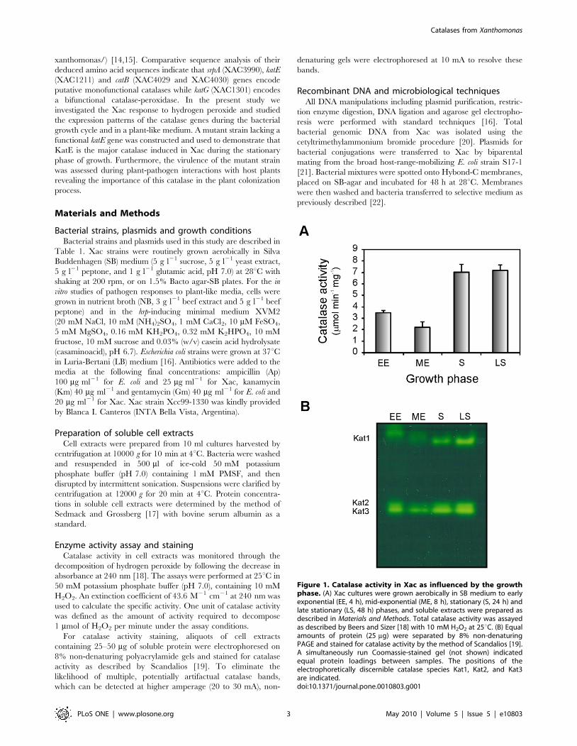
Figure 1. Catalase activity in Xac as influenced by the growthphase. (A) Xac cultures were grown aerobically in SB medium to earlyexponential (EE, 4 h), mid-exponential (ME, 8 h), stationary (S, 24 h) andlate stationary (LS, 48 h) phases, and soluble extracts were prepared asdescribed in Materials and Methods. Total catalase activity was assayedas described by Beers and Sizer [18] with 10 mM H2O2 at 25uC. (B) Equalamounts of protein (25 mg) were separated by 8% non-denaturingPAGE and stained for catalase activity by the method of Scandalios [19].A simultaneously run Coomassie-stained gel (not shown) indicatedequal protein loadings between samples. The positions of theelectrophoretically discernible catalase species Kat1, Kat2, and Kat3are indicated.doi:10.1371/journal.pone.0010803.g001
Catalases from Xanthomonas
PLoS ONE | www.plosone.org 3 May 2010 | Volume 5 | Issue 5 | e10803

Survival in the presence of hydrogen peroxideSurvival experiments were performed by subculturing Xac
overnight cultures into fresh SB medium at 2% inoculum. After 4
or 24 h of growth (early exponential and stationary phase,
respectively) aliquots of the cultures were diluted and plated on
SB-agar plates. Hydrogen peroxide was then added to the cultures
at final concentrations of 0.25 to 30 mM. After 15 min of
exposure to the oxidant, samples were removed, washed once with
fresh medium, serially diluted and plated on SB-agar plates.
To assess the H2O2 resistance of Xac in a plant-like medium,
Xac overnight cultures were subcultured into fresh NB or XVM2
media [23] at 2% inoculum and grown for 7 or 16 h to early
exponential and stationary phase respectively. Survival experi-
ments were then performed as previously described using final
concentrations of 1 and 30 mM H2O2.
For the induction experiments, Xac cultures were grown to
early exponential phase and incubated with sub-lethal concentra-
tions of hydrogen peroxide (10, 30 and 100 mM) for an additional
hour before being used in the killing experiments. After the
induction treatment, aliquots of the cultures were washed, diluted
and plated on SB-agar plates. Cultures were then treated with a
range of lethal concentrations of H2O2 (0.25 to 5 mM) for 15 min,
after which samples were removed, washed once with fresh
medium, serially diluted and plated on SB-agar plates.
In all cases, growth of liquid cultures was monitored
spectrophotometrically by optical density at 600 nm (OD600).
Colonies were counted after 48 h incubation at 28uC. The
percentage of survival was defined as the number of colony
forming units (CFU) after treatment divided by the number of
CFU prior to treatment 6100.
RNA extraction and semi-quantitative reversetranscription PCR (RT-PCR)
Total RNA of Xac cells was isolated using TRIzolH reagent
(Invitrogen), according to the manufacturer’s instructions. After
extraction, the RNA was treated with RNase-free DNase
(Promega) and its integrity was checked by agarose gel
electrophoresis. Semi-quantitative analyses of transcript levels of
katE, srpA, catB and katG were carried out using a two-step RT-
PCR approach employing the primers listed in Table 1. For
cDNA synthesis, total RNA (1 mg) was added to a 20 ml reverse
transcription reaction medium containing 4 ml 56 M-MLV
buffer (Promega), 0.5 mM dNTP mixture, 0.5 mg gene-specific
primer, 200 U M-MLV reverse transcriptase (Promega) and
incubated for 60 min at 42uC. Reverse transcription was
terminated by incubating for 5 min at 94uC. Control reactions,
where RT was omitted, were done in parallel for all the samples
to rule out the possibility of amplification from contaminating
DNA. PCR reactions were carried out with 2 ml cDNA template
under the following conditions: 25 cycles of denaturation at 94uCfor 1 min, annealing at 65uC for 1 min, and extension at 72uCfor 1 min; with a final extension step at 72uC for 5 min. The
number of cycles to be used, avoiding reaching the plateau of the
PCRs, was previously determined by taking samples at different
number of cycles during the PCR amplification step and
analyzing the products obtained by agarose gel electrophoresis.
As a constitutive control, a 217-bp fragment of 16S rRNA was
amplified using the same PCR conditions but with only 1% of the
cDNA synthesis reaction as template due to the high abundance
of 16S rRNA in total RNA extracts. RT-PCR products were
resolved on 1.5% (w/v) agarose gels, and densitometrically
quantified using Gel-Pro Analyzer Software 3.1 (Media Cyber-
netics).
Construction of the XackatE mutant strainThe XackatE mutant was constructed by insertional inactivation
of the katE gene on the chromosome by a single homologous
recombination. Primers katE-F1 and katE-R1 (Table 1), were used
to amplify a 440-bp internal fragment of the katE coding region
using Xac genomic DNA as template. The PCR product was cloned
into pGEM-T Easy vector (Promega), and the nucleotide sequence
of the insert was confirmed by automated DNA sequencing.
Subsequently, a HindIII-BamHI fragment of the PCR product was
subcloned into pK18mobGII [24], rendering pKmob/katE
(Table 1). The recombinant plasmid pKmob/katE was transferred
from E. coli strain S17-1 [21] to the Xac wild-type strain by
conjugation. Recombination of the cloned katE fragment in the
suicide plasmid with the homologous counterpart on the Xac
chromosome resulted in the disruption of the katE gene. The katE
mutant was selected on SB-agar plates containing 40 mg ml21 Km.
Figure 2. Hydrogen peroxide resistance of Xac cultures indifferent growth stages. Cells in early exponential (A) or stationary(B) phase of growth were exposed to the indicated concentrations ofH2O2 for 15 min. The number of CFU was determined for each culturebefore and after the peroxide treatment by plating of appropriatedilutions. The percentage of survival is defined as the number of CFUafter treatment divided by the number of CFU prior to treatment6100.Data are expressed as the mean 6 standard deviation of threeindependent experiments.doi:10.1371/journal.pone.0010803.g002
Catalases from Xanthomonas
PLoS ONE | www.plosone.org 4 May 2010 | Volume 5 | Issue 5 | e10803

Inactivation of katE was confirmed by PCR using specific primers
katE-F2 and katE-R2, located upstream and downstream of the
gene fragment used for the homologous recombination (Table 1).
For mutant complementation, a 2729-bp DNA fragment
containing the katE coding region and extending from 603 bp
upstream of the 59 end to 14 bp downstream of the 39 end of the
ORF was amplified using the primer pair ckatE-F and ckatE-R
(Table 1). The amplified sequence included the putative promoter
sequence of the katE gene, previously predicted with SoftBerry
(www.softberry.com). The amplified DNA fragment was then
cloned into the broad-host-range vector pBBR1MCS-5 [25] to
generate the recombinant plasmid pBBR1/katE. This plasmid was
transferred into the XackatE mutant strain by conjugation,
rendering strain cXackatE (Table 1).
Plant material and plant inoculationsOrange (Citrus sinensis cv. Valencia) was used as the host plant
for Xac. All plants were grown in a growth chamber in
incandescent light at 25uC with a photoperiod of 16 h. Overnight
cultures of Xac WT, XackatE and cXackatE were diluted in
10 mM MgCl2 to a final concentration of 105 CFU ml21. For
disease symptoms assays, bacterial suspensions were infiltrated into
leaves with needleless syringes. In planta growth assays were
performed by grinding 0.8 cm diameter leaf discs from infiltrated
leaves in 100 ml of 10 mM MgCl2, followed by serial dilutions and
plating onto SB-agar plates. Colonies were counted after 48 h
incubation at 28uC.
Results
Catalase activity pattern is regulated in different growthstages
We investigated the growth phase-dependent pattern of catalase
activity in Xac by conducting activity assays on soluble extracts
from cultures at different growth stages. A typical growth curve of
Xac in SB medium is depicted in Figure S1. As shown in
Figure 1A, the highest levels of catalase activity were observed in
the stationary and late stationary phases (with similar values of
,7 mmol min21 mg21), being approximately 2.5-fold higher than
those determined for the cultures in exponential growth.
On the other hand, equal amounts of the bacterial extracts were
separated by 8% non-denaturing PAGE and subsequently stained
for catalase activity (Figure 1B). Three distinct catalase bands were
detected throughout all stages of growth: a slow-migrating catalase
denoted Kat1, and two bands with similar electrophoretic
mobilities that were named Kat2 and Kat3. The activity level of
Kat1 increases significantly in the stationary phase of growth,
whereas the levels of Kat2 and Kat3 decline in the mid-
exponential phase and increase again as cells enter and remain
in the stationary phase.
The intensities of the bands detected in the activity gel were
measured using Gel-Pro Analyzer Software 3.1 (Media Cybernet-
ics) and the total optical density for each growth stage was
calculated. The pattern obtained was consistent with the activity
measurements depicted in Figure 1A (data not shown).
Xac in the stationary phase of growth is more resistant tohydrogen peroxide
In order to investigate whether the elevated catalase activity
observed in the stationary phase provides Xac cultures with
enhanced resistance to oxidative stress, studies of bacterial survival
in the presence of H2O2 were performed (Figure 2). In early
exponential phase, Xac was very sensitive to hydrogen peroxide
treatment, with only 0.28% survival following addition of 1 mM
H2O2 and almost no detectable CFU following treatment with
5 mM H2O2. On the other hand, Xac in stationary phase of
growth was significantly more resistant to the oxidative stress
treatment, with 95% survival following the addition of 5 mM
H2O2 and 40% survival following treatment with 30 mM H2O2.
Identification of the catalase isoforms expressed in thedifferent growth stages
Analysis of the Xac genome sequence revealed the presence of
four putative catalase genes designated as katE, srpA, catB and katG
[14,15]. Comparative sequence analysis of the encoded proteins
were performed by using ClustalX [26] and described in
Supporting Information S1 and Figures S2, S3, S4 and S5.
In order to investigate the expression profiles of the Xac catalase
genes during growth, we performed semi-quantitative RT-PCR
reactions using specific primers designed from the reported gene
Figure 3. Expression analysis of Xac catalase genes as a function of the growth phase. (A) Amplified products of the katE, srpA and katGgenes by semi-quantitative RT-PCR using RNA preparations from Xac cultures grown in SB medium to early exponential (EE, 4 h), mid-exponential(ME, 8 h), stationary (S, 24 h) and late stationary (LS, 48 h) phases. 16S rRNA was used as a loading control and to quantitate the amount of RNA in RT-PCRs. (B) Expression profiles obtained by densitometric quantification of band intensities. Experiments were performed in triplicate with similarresults; error bars indicate 61 standard deviation of the mean. IOD, integrated optical density; A.U., arbitrary units.doi:10.1371/journal.pone.0010803.g003
Catalases from Xanthomonas
PLoS ONE | www.plosone.org 5 May 2010 | Volume 5 | Issue 5 | e10803

sequences (Table 1, Figure 3). As a control for constitutive
bacterial expression a fragment of 16S rRNA was simultaneously
amplified. Expression of katE was hardly detectable at the early
and mid-exponential phases of growth, subsequently increasing to
reach 5-fold higher levels during the stationary phase. On the
other hand, expression of srpA and katG genes was detected
throughout all stages of growth, reaching maximal levels in the
mid-exponential phase and decreasing gradually towards the
stationary phase. The mRNA levels of katG were almost
undetectable in the late stationary phase. The catB gene was not
included in the figure since no product was observed in the RT-
PCR reactions under the conditions tested. To ascertain the
absence of contaminating DNA in bacterial RNA samples control
PCR reactions where RT was omitted were carried out in parallel
for all samples (data not shown).
Xac adaptive response to hydrogen peroxideThe adaptive response to oxidative stress agents is a well-
characterized phenomenon observed in many bacteria, in which
the exposure to sub-lethal levels of an oxidant leads to the
induction of genes involved in the bacterial stress response,
ultimately conferring resistance to lethal levels of the same agent or
even unrelated compounds (cross protection) [27]. The ability to
develop an adaptive response to hydrogen peroxide was
investigated in Xac by determining the catalase activity in early
exponential cultures incubated with sub-lethal concentrations of
H2O2 (10, 30 and 100 mM) for 60 min. As shown in Table 2, a 2-
fold induction of catalase activity was observed in cultures treated
with 100 mM H2O2 with respect to the untreated control cells.
Based on this observation, the resistance of bacterial cells pre-
adapted with sub-lethal levels of H2O2 to a lethal dose of the same
agent was assessed. Cultures pre-treated with 10, 30 and 100 mM
H2O2 were subsequently challenged with a killing concentration of
H2O2 (1 mM, see Figure 2A) and the percentages of survival were
determined (Figure 4A). Interestingly, a dose dependent response
was observed with these H2O2 concentrations, with a 10-fold
increase in resistance after pre-adaptation with 100 mM H2O2.
Moreover, Xac cultures pre-treated with 100 mM H2O2 for 1 hour
were then incubated with 0.25, 0.5, 1 and 5 mM H2O2 for 15 min
(Figure 4B). We found that pre-adapted cells were more resistant
than the control cells to all H2O2 concentrations tested, the
difference of survival being more pronounced as the H2O2 levels
increases. After challenge with 5 mM H2O2 survival of the pre-
adapted culture was 100-fold higher than that of the untreated
control.
A medium that mimics the environment of plantintercellular spaces modifies Xac catalase expressionpattern
As an initial approach to evaluate the involvement of catalases
during plant-pathogen interactions we determined the levels of
these enzymes in early exponential and stationary phase cultures
grown in NB, a rich standard medium, and in XVM2, a nutrient
poor medium that simulates conditions in the apoplastic space of
plants, which induces the bacterial hrp (for hypersensitive response
Table 2. Induction of catalase activity in response to sub-lethal levels of hydrogen peroxidea.
Culture Catalase activity Induction
(mmol min21 mg21 protein) (fold)
Uninduced 3.760.3 -
Induced by H2O2
10 mM 4.560.2 1.2
30 mM 5.760.2 1.5
100 mM 7.660.4 2.0
a. Xac cells were grown in SB medium to early exponential phase and exposedto the indicated concentrations of H2O2 for 1 hour. Catalase activities in solublecell extracts were measured as described in Materials and Methods.Data represent mean 6 standard deviation of three independent experiments.doi:10.1371/journal.pone.0010803.t002
Figure 4. Adaptive response of Xac to hydrogen peroxidetreatment. (A) Exponential phase cultures were adapted with theindicated concentrations of H2O2 for 60 min and then exposed to 1 mMH2O2 for 15 min. The number of CFU was determined for each culturebefore and after the treatment with 1 mM H2O2 by plating ofappropriate dilutions. The related survival is defined as the percentageof survival of the pre-adapted culture divided by the percentage ofsurvival of the untreated control. (B) Exponential phase cultures werepre-adapted with 100 mM H2O2 for 60 min. The number of CFU wasdetermined for the preadapted cultures and for the unadapted controlsand then H2O2 was added to the final concentrations indicated,followed by an incubation of 15 min. The percentage of survival wascalculated as the number of CFU after treatment divided by the numberof CFU prior to treatment 6100. Experiments were performed intriplicate; error bars indicate 61 standard deviation of the mean.doi:10.1371/journal.pone.0010803.g004
Catalases from Xanthomonas
PLoS ONE | www.plosone.org 6 May 2010 | Volume 5 | Issue 5 | e10803

and pathogenicity) gene cluster [23]. Typical growth curves of Xac
in these media are depicted in Figure S6. As shown in Table 3,
cells grown in XVM2 exhibited ,2-fold higher catalase activity
levels than cells grown in the standard medium, suggesting a
possible induction of these enzymes in the environment found in
the intercellular spaces of plant tissues. Moreover, Xac cultures
grown in XVM2 were considerably more resistant to killing when
exposed to H2O2 than those grown in NB (Table 3).
To address the question if there is a transcriptional induction of
any of the catalase genes in the XVM2 medium we performed
semiquantitative RT-PCR analysis with early exponential phase
cultures. Interestingly, different expression patterns were observed
depending on the growth conditions (Figure 5). While mRNA
levels of srpA were similar in both media, expression of katE was
,2.3-fold higher in XVM2 than in NB, whereas the levels of katG
exhibited a reduction of similar magnitude in the minimal
medium. Analysis of the catalase activity in native polyacrylamide
gels revealed that the intensity of the slow-migrating band (Kat1,
Figure 1B) was markedly raised in the plant-like medium, both in
exponential and stationary growth phases (Figure 6).
Characterization of a XackatE mutant strainHaving established that katE is transcriptionally induced in Xac
during the stationary phase of growth and in the apoplastic space
mimicking XVM2 medium, a XackatE mutant strain was then
generated by insertional mutagenesis (see Materials and Methods) and
genetically verified by PCR analysis (data not shown).
In order to assess the effect of katE disruption on the catalase
pattern, soluble extracts from the parental (WT) and mutant (katE)
strains in early exponential and stationary phases of growth were
analyzed by native gel electrophoresis and catalase staining. As
shown in Figure 7A, the upper band observed in the wild-type
strain was completely absent in the katE mutant, indicating that
this band corresponds to KatE. A complementation assay was also
carried out to validate the katE phenotype. This was done by
cloning the katE gene under the control of its own promoter
sequence in a pBBR1MCS-5 vector [25], which was then
conjugated into the katE mutant. The upper catalase band was
recovered in the resulting cXackatE strain, corroborating the
identity of this catalase (Figure 7A). The intensity of this band was
higher than the observed in the wild-type cells, probably due to the
low but still multiple copy number of the pBBR1/katE vector in
Xac cells.
The growth phase-dependent pattern of catalase activity in the
XackatE strain was then investigated by conducting assays on
soluble extracts from cultures in different stages of growth. In
contrast to wild-type bacteria, no induction was observed in
stationary phase cultures of the XackatE mutant, with a constant
average value of ,1.6 mmol min21 mg21 throughout all the
bacterial growth cycle. On the other hand, the cXackatE strain
exhibited the same pattern of wild-type cells but with higher
activity values.
Furthermore, the mutant strain was more sensitive to hydrogen
peroxide treatment in both early exponential and stationary
growth phases (Figure 7B).
The XackatE adaptive response to hydrogen peroxide was also
analyzed by determining the catalase activity in early exponential
cultures incubated with sub-lethal concentrations of the oxidant.
As was previously demonstrated for wild-type cells, a ,2-fold
Table 3. Increase in catalase activity and hydrogen peroxideresistance of Xac cells in a plant-like medium.
Medium Catalase activitya % Survivalb
(mmol min21 mg21 protein) H2O2
Early exponential phase
NB 3.260.2 0.2560.07
XVM2 7.560.3 3.460.1
Stationary phase
NB 7.260.4 3862
XVM2 16.360.6 9865
a. Xac cells were grown in the indicated media to early exponential orstationary phase and then harvested. Catalase activities in soluble cell extractswere measured as described in Materials and Methods.b. Early exponential and stationary phase cultures were exposed to 1 mM and30 mM H2O2 respectively, for 15 min. The percentage of survival is defined asthe number of CFU after treatment divided by the number of CFU prior totreatment 6100.Data represent mean 6 standard deviation of three independent experiments.doi:10.1371/journal.pone.0010803.t003
Figure 5. Expression of Xac catalase genes in the plant-mimicking XVM2 medium. (A) Amplified products of the catalase genes by semi-quantitative RT-PCR using RNA preparations from early exponential Xac cultures grown in NB and in XVM2. As a control for constitutive bacterialexpression a fragment of 16S rRNA was simultaneously amplified. (B) Expression profiles obtained by densitometric quantification of band intensities.Experiments were performed in triplicate with similar results; error bars indicate 61 standard deviation of the mean. IOD, integrated optical density;A.U., arbitrary units.doi:10.1371/journal.pone.0010803.g005
Catalases from Xanthomonas
PLoS ONE | www.plosone.org 7 May 2010 | Volume 5 | Issue 5 | e10803

induction of catalase activity was also observed in XackatE cells
treated with 100 mM H2O2 (Table 4), suggesting that KatE is not
responsible for this response.
Interaction of the XackatE mutant with host plantsIn order to assess the physiological role of KatE during the
infection process, the mutant strain was tested for its ability to trigger
disease in citrus leaves. Both wild-type bacteria and XackatE
produced typical canker lesions upon infiltration at a concentration
of 105 CFU ml21, with no differences in the time of appearance of
the first symptoms (water soaking). However, the magnitude of the
lesions and the number of cankers were significantly diminished in
the mutant strain compared to wild-type bacteria, even though the
infiltration areas and the bacterial densities were equivalent for both
strains. On the other hand, infiltration with the cXackatE strain
caused the same symptoms and a similar percentage of necrotic area
than wild-type cells (Figure 8A).
The degree of virulence of the different strains was also
evaluated by conducting bacterial growth curves in planta. As
shown in Figure 8B, the magnitudes of leaf injuries correlated with
the bacterial growths inside the host. The bacterial number of
XackatE recovered from the infected leaves was fewer than that of
the wild-type strain at each time analyzed. On the other hand,
although complementation restored the bacteria to full virulence
on citrus leaves, cXackatE growth on leaves did not reach the
values of the wild-type.
Furthermore, in order to rule out the possibility that the lower
infectivity of XackatE arise because mutant cells had already been
injured during the culture period, infection experiments were
conducted with exponentially growing cultures, in which KatE
would not be even induced. The results obtained in these
inoculations were in agreement with those previously described,
indicating that the deficiency suffered by the mutant strain arise
during the plant infection (data not shown).
Discussion
X. axonopodis pv. citri holds a strictly aerobic life style and this
physiology can lead to the intracellular generation of oxidative
stress during normal respiration on molecular oxygen. As a
pathogenic microorganism, it encounters a great deal of oxidative
stress during the infection process as well. To prevent the
accumulation of ROS generated during aerobic respiration or
plant interactions, Xac should employ versatile antioxidant
defense enzymes, including catalases. The genome of Xac has
been completely sequenced, revealing the presence of four putative
catalases, four SODs and the OxyR and SoxR sensors [14,15].
The elevated number of genes encoding for antioxidant enzymes
in this bacterium provides an indication of the overall relevance of
the antioxidant systems for its survival. In this study we focused on
the analysis of Xac catalases and their expression patterns in order
to elucidate the physiological roles that catalases play in this
microorganism.
The pattern of catalase activity was found to be growth phase-
regulated in Xac, with the highest levels detected during the
stationary phase (Figure 1). This result was unexpected because
previous reports in other Xanthomonas species showed that
maximum levels of enzyme activity were attained as the cultures
were emerging from the lag phase and subsequently declined as
growth proceeded [28–30]. On the other hand, growth into
stationary phase has been largely documented as one of the main
factors influencing catalase levels in a majority of bacteria, as these
enzymes would serve a protective role against peroxide during
periods of low metabolic activity [7,31,32]. According to our
results this may be the case for Xac, possibly suggesting different
mechanisms of catalase regulation between species of the
Xanthomonas genus. In addition, we showed that resistance levels
to H2O2 treatment also varies significantly in Xac depending on
the growth stage, with stationary phase cells being capable of
tolerating up to 30-fold higher concentrations of the oxidant than
exponentially growing cells (Figure 2).
The decrease in the activities of oxidant-scavenging enzymes,
such as catalase and SOD, observed in other Xanthomonas species
during the stationary phase of growth has lead to the proposal that
the mechanisms responsible for stationary-phase resistance to
oxidants would be independent of the levels of scavenging enzymes
[29]. In contrast, our results revealed that Xac resistance to H2O2
during the bacterial growth cycle increases in parallel with the
expression of catalase-specific activity (Figures 1 and 2), suggesting
that the mechanism of resistance to oxidants in this bacterium
differs at least partially from those reported for other species of the
Xanthomonas genus.
Figure 6. Detection of catalase activity in Xac cultures grown inNB and XVM2 media. Xac cultures were grown aerobically in NB andXVM2 media to early exponential (EE, 7 h), and stationary (S, 16 h)phases, and soluble extracts were prepared as described in Materialsand Methods. Equal amounts of protein (40 mg) were separated by 8%non-denaturing PAGE and stained for catalase activity by the method ofScandalios [19]. A simultaneously run Coomassie-stained gel (notshown) indicated equal protein loadings between samples.doi:10.1371/journal.pone.0010803.g006
Catalases from Xanthomonas
PLoS ONE | www.plosone.org 8 May 2010 | Volume 5 | Issue 5 | e10803

We also demonstrated that Xac catalase activity is regulated at
isozymes level. At all stages of Xac growth we were able to detect
three bands with catalase activity in non-denaturing gels
(Figure 1B), which exhibited differential patterns of expression
along the bacterial growth cycle, being the upper band
significantly induced during the stationary phase.
Additionally, we analyzed the expression of the complete set of
Xac catalase genes (katE, katG, catB and srpA) along the bacterial
growth cycle by RT-PCR, showing that the katE gene was strongly
induced during the stationary phase, while katG and srpA exhibited
a peak level of expression during the mid-exponential phase
(Figure 3). On the other hand, transcription of the catB gene was
not detected under the conditions tested, suggesting that the gene
may be cryptic, that is, present but not expressed in Xac. This may
be attribuited to the fact that there are two overlapping gene
fragments annotated as catB in the Xac genome sequence
(XAC4029 and XAC4030), which encode for this putative
monofunctional catalase in different open reading frames
[14,15]. However, the possibility that catB is expressed in Xac
under specific growth conditions not assayed for in this study can
not be ruled out.
Interestingly, transcript levels of katG and srpA in the mid-
exponential phase were equal to or even higher than the total
transcript levels observed during the stationary or late-stationary
phases (Figure 3). However, total catalase activity detected in the
stationary phase was significantly higher than that of the mid-
exponential phase (Figure 1A). We speculate that the apparent
discrepancy between RNA levels and catalase activities may be a
consequence of the rapid rate of bacterial duplication during the
exponential phase of the growth cycle, which may cause the
limitation of some component (e.g., heme and/or iron) essential
for the proper assembly/activity of the enzyme. A potential
mechanism of post-transcriptional regulation could also be
involved in the control of the catalase expression. Further
investigation would be necessary to probe this contention.
We have also demonstrated that interruption of the katE gene
has a marked effect on catalase activity in growth-arrested cells.
The XackatE mutant strain exhibited a constant low level of
activity throughout all the bacterial growth cycle and lower
resistance to H2O2 than wild-type cells, the difference of survival
being more pronounced during the stationary phase (Figure 7B).
These findings support the notion that KatE is the isozyme
responsible for the increase of catalase activity previously observed
for the wild-type strain during the stationary phase. Furthermore,
this catalase accounts for a considerable part of the overall
hydrogen peroxide resistance in Xac. On the other hand, the
catalase content of the XackatE mutant on non-denaturing gels
revealed the absence of the upper activity band of the wild-type
strain (Kat1 in Figure 1B), which allowed us to conclude that this
band corresponds to the KatE isozyme (Figure 7A). Moreover, the
absence of this band in both phases of growth supports the notion
that the apparently different mobility observed between lanes in
Figure 1 was only an artifactual effect of the electrophoretic run. In
addition, a decrease in the intensities of the bands with higher
electrophoretic mobilities (Kat2 and Kat3) was also observed in
these gels, suggesting that the loss of KatE influences the
expression of the other catalase isoforms.
The adaptive response to oxidative agents has been previously
proposed to play a fundamental role in plant-pathogen interac-
tions, allowing bacteria to withstand increased oxidative stress
conditions [27]. We then became interested in the adaptive
Figure 7. Catalase activity and hydrogen peroxide resistance in the XackatE mutant. (A) Xac wild-type (WT), XackatE (katE2) and cXackatE(ckatE) strains were grown aerobically in SB medium to early exponential (EE, 4 h) and stationary (S, 24 h) phases, and soluble extracts were preparedas described in Materials and Methods. Equal amounts of protein (25 mg) were separated by 8% non-denaturing PAGE and stained for catalase activityby the method of Scandalios [19]. (B) Cells in early exponential (EE) or stationary (S) phase of growth were exposed to the indicated concentrations ofH2O2 for 15 min. The number of CFU was determined for each culture before and after the peroxide treatment by plating of appropriate dilutions.The percentage of survival is defined as the number of CFU after treatment divided by the number of CFU prior to treatment 6100. Data areexpressed as the mean 6 standard deviation of three independent experiments.doi:10.1371/journal.pone.0010803.g007
Table 4. Induction of catalase activity in the XackatE mutantin response to sub-lethal levels of hydrogen peroxidea.
Culture Catalase activity Induction
(mmol min21 mg21 protein) (fold)
Uninduced 1.760.2 -
Induced by H2O2
30 mM 2.760.2 1.6
100 mM 3.260.3 1.9
a. XackatE cells were grown in SB medium to early exponential phase andexposed to the indicated concentrations of H2O2 for 1 hour. Catalase activitiesin soluble cell extracts were measured as described in Materials and Methods.Data represent mean 6 standard deviation of three independent experiments.doi:10.1371/journal.pone.0010803.t004
Catalases from Xanthomonas
PLoS ONE | www.plosone.org 9 May 2010 | Volume 5 | Issue 5 | e10803

response of Xac to H2O2, the major component of the plant
oxidative burst [13]. Our results demonstrate that Xac also
develops an adaptive response to H2O2, and the level of induced
protection correlates with the bacterial ability to induce catalase
activity during the pre-adaptation treatment (Table 2). Since the
XackatE mutant exhibited the same catalase activity induction
than wild-type cells after the oxidative treatment, we suggest that
KatE would not be involved in the adaptive response of Xac.
Adequacy of the antioxidant system may be critical for Xac
interaction with citrus plants, in order to minimize oxidative stress
and establish infection. We observed that the total catalase activity
and the resistance to H2O2 were significantly higher in the
apoplastic space mimicking XVM2 medium than in a rich
medium (NB) (Table 3). The expression analysis of the different
catalase genes indicated that the monofunctional catalase katE was
strongly induced in XVM2 (Figure 5). Consistent with this,
analysis of the upstream sequence of the katE gene revealed the
presence of an imperfect PIP box (TTCGCN14TTCGT) located
1 bp downstream of the predicted 210 promoter sequence. This
conserved plant-inducible promoter sequence motif has been
suggested to be associated with the regulation of genes induced in
planta and also in the XVM2 medium [33]. Our results indicate
that Xac catalase expression pattern is modified in response to any
stimuli associated with the plant or the microenvironment it
provides. It is still not clear if the increase in catalase activity
observed in XVM2 was only a result of higher expression of the
katE gene (due to possible differences in the catalytic properties
with respect to KatG), or enhanced enzyme activity due to post-
transcriptional regulation, or both. Nevertheless, the induction of
catalase activity in response to the plant environment may serve a
protective role against exposure to H2O2 in the early stages of
plant infection.
Furthermore, the virulence of the XackatE mutant was
considerably attenuated during the compatible interaction with
citrus plants, given that the magnitude of the damaged tissue and
the number of canker lesions were noticeably reduced compared
to the wild-type strain. The phenotypic differences observed
between both strains were consistent with the bacterial growth
curves in planta (Figure 8). The wild-type virulence was recovered
in a complemented strain (cXackatE) allowing us to conclude that
the phenotypes observed are indeed caused by the loss of KatE
function. Our results indicate that catalase KatE has an important
function in the colonization and survival of Xac in the host tissue.
Accordingly, in X. campestris pv. campestris was recently shown that a
mutant in KatG, the bifunctional catalase-peroxidase of this
bacterium, was unable to infect radish (Raphanus sativus) leaves and
cause disease [34]. The impaired ability of the XackatE mutant to
infect citrus leaves provides the first genetic evidence to support a
monofunctional catalase as a virulence factor in Xac, further
indicating that the oxidative burst may play a significant role in
pathogen growth restriction during the infection process.
Our results collectively suggest that in the apoplast environment
Xac may be more resistant to H2O2 as a consequence of KatE
induction. Our future aims are to elucidate the regulatory
pathways that orchestrate the Xac oxidative stress response during
the first stages of plant infection.
Supporting Information
Supporting Information S1 The monofunctional catalase
KatE of Xanthomonas axonopodis pv. citri is required for full virulence
in citrus plants.
Found at: doi:10.1371/journal.pone.0010803.s001 (0.06 MB
DOC)
Figure S1 Growth curve of Xac in SB medium. Xac culture was
cultivated aerobically in SB medium at 28uC with shaking at
200 rpm. Aliquots were taken at the indicated times and measured
for both optical density at 600 nm (OD600, open circles) and
colony-forming capacity on SB-agar medium (closed circles).
Found at: doi:10.1371/journal.pone.0010803.s002 (2.04 MB TIF)
Figure S2 Multiple alignment of the deduced amino acid
sequence of Xac KatE (XacE) with catalases KatE of X. campestris
pv. phaseoli (XcpE) and HPII of E. coli (EcoII), performed by using
ClustalX [26]. An asterisk indicates complete residue conserva-
tion, a colon indicates strong group conservation, a period
indicates weak group conservation, and a blank space indicates
no conservation of residues.
Found at: doi:10.1371/journal.pone.0010803.s003 (0.47 MB TIF)
Figure S3 Multiple alignment of the deduced amino acid
sequence of Xac SrpA (XacA) with catalases from X. campestris
pv. vesicatoria (Xcv), X. oryzae pv. oryzae (Xoo), P. syringae (Psy) and P.
aeruginosa (Pae), performed by using ClustalX [26]. An asterisk
indicates complete residue conservation, a colon indicates strong
Figure 8. Effect of katE disruption on pathogenicity. Xac WT (WT), XackatE (katE2) and cXackatE (ckatE) cells were inoculated at 105 CFU ml21
in 10 mM MgCl2 into the intercellular spaces of fully expanded orange leaves. (A) A representative leaf 20 days after inoculation is shown. Left panel,adaxial side; right panel, abaxial side. Dashed lines indicate the infiltrated area. (B) Bacterial growth of Xac cells in orange leaves. Values representmeans of three independent samples; error bars represent standard deviations.doi:10.1371/journal.pone.0010803.g008
Catalases from Xanthomonas
PLoS ONE | www.plosone.org 10 May 2010 | Volume 5 | Issue 5 | e10803

group conservation, a period indicates weak group conservation,
and a blank space indicates no conservation of residues.
Found at: doi:10.1371/journal.pone.0010803.s004 (0.45 MB
TIF)
Figure S4 Alignment of the deduced amino acid sequences of
Xac CatB precursor (XacBp) (A) and Xac CatB (XacB) (B) with
KatA of X. campestris pv. phaseoli (XcpA), performed by using
ClustalX [26]. An asterisk indicates complete residue conserva-
tion, a colon indicates strong group conservation, a period
indicates weak group conservation, and a blank space indicates
no conservation of residues.
Found at: doi:10.1371/journal.pone.0010803.s005 (0.47 MB TIF)
Figure S5 Multiple alignment of the deduced amino acid
sequence of Xac KatG (XacG) with catalases from X. campestris
pv. vesicatoria (Xcv) and X. campestris pv. campestris (Xcc), and the
bifunctional HPI of E. coli (EcoI), performed by using ClustalX
[26]. An asterisk indicates complete residue conservation, a colon
indicates strong group conservation, a period indicates weak group
conservation, and a blank space indicates no conservation of
residues.
Found at: doi:10.1371/journal.pone.0010803.s006 (0.60 MB TIF)
Figure S6 Growth curves of Xac in NB and XVM2 media. Xac
cultures were cultivated aerobically in these media at 28uC with
shaking at 200 rpm. Aliquots were taken at the indicated times
and measured for optical density at 600 nm (OD600).
Found at: doi:10.1371/journal.pone.0010803.s007 (1.88 MB TIF)
Acknowledgments
We are grateful to Dr. Nestor Carrillo and Dr. Adriana R. Krapp for
critical reading of the manuscript. We thank Catalina Anderson (INTA
Concordia, Argentina), Gaston Alanis and Ruben Dıaz Velez (Proyecto El
Alambrado) for the citrus plants.
Author Contributions
Conceived and designed the experiments: MLT JO EGO. Performed the
experiments: MLT SP. Analyzed the data: MLT JO EGO. Contributed
reagents/materials/analysis tools: JO EGO. Wrote the paper: MLT EGO.
References
1. Fridovich I (1978) The biology of oxygen radicals. Science 201: 875–880.
2. Imlay JA (2008) Cellular defenses against superoxide and hydrogen peroxide.Annu Rev Biochem 77: 755–776.
3. McCord JM, Fridovich I (1969) Superoxide dismutase. An enzymic function forerythrocuprein (hemocuprein). J Biol Chem 244: 6049–6055.
4. Green J, Paget MS (2004) Bacterial redox sensors. Nat Rev Microbiol 2:
954–966.5. Imlay JA, Linn S (1988) DNA damage and oxygen radical toxicity. Science 240:
1302–1309.6. Cabiscol E, Tamarit J, Ros J (2000) Oxidative stress in bacteria and protein
damage by reactive oxygen species. Int Microbiol 3: 3–8.
7. Loewen PC (1997) Bacterial catalases. In: Scandalios JG, ed. Oxidative Stressand the Molecular Biology of Antioxidant Defenses. New York: Cold Spring
Harbor Laboratory Press. pp 273–308.8. Chelikani P, Fita I, Loewen PC (2004) Diversity of structures and properties
among catalases. Cell Mol Life Sci 61: 192–208.
9. Switala J, Triggs-Raine BL, Loewen PC (1990) Homology among bacterialcatalase genes. Can J Microbiol 36: 728–731.
10. Brunings AM, Gabriel DW (2003) Xanthomonas citri: breaking de surface. MolPlant Pathol 4: 141–157.
11. Graham JH, Gottwald TR, Cubero J, Achor DS (2004) Xanthomonas axonopodis
pv. citri: factors affecting successful eradication of citrus canker. Mol Plant Pathol
5: 1–15.
12. Baker CJ, Orlandi EW (1995) Active oxygen in plant pathogenesis. Annu RevPhytopathol 33: 299–321.
13. Grant JJ, Loake GJ (2000) Role of reactive oxygen intermediates and cognateredox signaling in disease resistance. Plant Physiol 124: 21–29.
14. Van Sluys MA, Monteiro-Vitorello CB, Camargo LE, Menck CF, da Silva AC,
et al. (2002) Comparative genomic analysis of plant-associated bacteria. AnnuRev Phytopathol 40: 169–189.
15. da Silva AC, Ferro JA, Reinach FC, Farah CS, Furlan LR, et al. (2002)Comparison of the genomes of two Xanthomonas pathogens with differing host
specificities. Nature 417: 459–463.16. Sambrook J, Fritsch EF, Maniatis T (1989) Molecular cloning: A laboratory
manual. New York: Cold Spring Harbor Laboratory Press.
17. Sedmack JJ, Grossberg SE (1977) A rapid, sensitive and versatile assay forprotein using Coomassie Brilliant Blue G250. Anal Biochem 79: 544–552.
18. Beers RF, Jr., Sizer IW (1952) A spectrophotometric method for measuring thebreakdown of hydrogen peroxide by catalase. J Biol Chem 195: 133–140.
19. Scandalios JG (1968) Genetic control of multiple molecular forms of catalase in
maize. Ann N Y Acad Sci 151: 274–293.20. Murray MG, Thompson WF (1980) Rapid isolation of high molecular weight
plant DNA. Nucleic Acids Res 8: 4321–4325.21. Simon R, Priefer U, Puhler A (1983) A broad host range mobilization system for
in vivo genetic engineering: transposon mutagenesis in Gram negative bacteria.Bio/Technology 1: 784–791.
22. Dunger G, Arabolaza LN, Gottig N, Orellano EG, Ottado J (2005) Participation
of Xanthomonas axonopodis pv. citri hrp cluster in citrus canker and in non-host
plants responses. Plant Pathol 54: 781–788.
23. Wengelnik K, Bonas U (1996) HrpXv, an AraC-type regulator, activates
expression of five of the six loci in the hrp cluster of Xanthomonas campestris pv.
vesicatoria. J Bacteriol 178: 3462–3469.
24. Katzen F, Becker A, Ielmini MV, Oddo CG, Ielpi L (1999) New mobilizable
vectors suitable for gene replacement in gram-negative bacteria and their use in
mapping of the 39 end of the Xanthomonas campestris pv. campestris gum operon.
Appl Environ Microbiol 65: 278–282.
25. Kovach ME, Elzer PH, Hill DS, Robertson GT, Farris MA, et al. (1995) Four
new derivatives of the broad-host-range cloning vector pBBR1MCS, carrying
different antibiotic-resistance cassettes. Gene 166: 175–176.
26. Thompson JD, Gibson TJ, Plewniak F, Jeanmougin F, Higgins DG (1997) The
CLUSTAL_X windows interface: flexible strategies for multiple sequence
alignment aided by quality analysis tools. Nucleic Acids Res 25: 4876–4882.
27. Mongkolsuk S, Vattanaviboon P, Praitaun W (1997) Induced adaptive and
cross-protection responses against oxidative stress killing in a bacterial
phytopathogen, Xanthomonas oryzae pv. oryzae. FEMS Microbiol Lett 146:
217–221.
28. Chamnongpol S, Mongkolsuk S, Vattanaviboon P, Fuangthong M (1995)
Unusual Growth Phase and Oxygen Tension Regulation of Oxidative Stress
Protection Enzymes, Catalase and Superoxide Dismutase, in the Phytopathogen
Xanthomonas oryzae pv. oryzae. Appl Environ Microbiol 61: 393–396.
29. Vattanaviboon P, Mongkolsuk S (2000) Expression analysis and characterization
of the mutant of a growth-phase- and starvation-regulated monofunctional
catalase gene from Xanthomonas campestris pv. phaseoli. Gene 241: 259–265.
30. Chauvatcharin N, Vattanaviboon P, Switala J, Loewen PC, Mongkolsuk S
(2003) Cloning and characterization of katA, encoding the major monofunctional
catalase from Xanthomonas campestris pv. phaseoli and characterization of the
encoded catalase KatA. Curr Microbiol 46: 83–87.
31. Klotz MG, Hutcheson SW (1992) Multiple periplasmic catalases in phytopath-
ogenic strains of Pseudomonas syringae. Appl Environ Microbiol 58: 2468–2473.
32. Hanyu M, Fujimoto H, Tejima K, Saeki K (2009) Functional differences of two
distinct catalases in Mesorhizobium loti MAFF303099 under free-living and
symbiotic conditions. J Bacteriol 191: 1463–1471.
33. Koebnik R, Kruger A, Thieme F, Urban A, Bonas U (2006) Specific binding of
the Xanthomonas campestris pv. vesicatoria AraC-type transcriptional activator HrpX
to plant-inducible promoter boxes. J Bacteriol 188: 7652–7660.
34. Jittawuttipoka T, Buranajitpakorn S, Vattanaviboon P, Mongkolsuk S (2009)
The catalase-peroxidase KatG is required for virulence of Xanthomonas campestris
pv. campestris in a host plant by providing protection against low levels of H2O2.
J Bacteriol 191: 7372–7377.
Catalases from Xanthomonas
PLoS ONE | www.plosone.org 11 May 2010 | Volume 5 | Issue 5 | e10803


![Structure of the monofunctional heme catalase DR1998 from ...cmromao/Articles-pdf/2014_FebsJ_KatDR19… · Penicillium vitale (PDB 4CAT) catalases [11–14]. The three catalases from](https://static.fdocuments.us/doc/165x107/5fd1b364c2d0642f3051de92/structure-of-the-monofunctional-heme-catalase-dr1998-from-cmromaoarticles-pdf2014febsjkatdr19.jpg)

![Regulatory Mechanisms of Monofunctional and Bifunctional Anticarcinogenic Enzyme ... · (CANCER RESEARCH 48, 4776-4782, September 1, 1988] Regulatory Mechanisms of Monofunctional](https://static.fdocuments.us/doc/165x107/5f08ecbd7e708231d424630d/regulatory-mechanisms-of-monofunctional-and-bifunctional-anticarcinogenic-enzyme.jpg)