The Molecular Biology of Memory Storage
47
Bioscience Reports, Vol. 21, No. 5, October 2001 ( 2002) NOBEL LECTURE 8 DECEMBER, 2000 The Molecular Biology of Memory Storage: A Dialog Between Genes and Synapses Eric R. Kandel 1 The biology of learning, and short-term and long-term memory, as revealed by Aplysia and other organisms, is reviewed. KEY WORDS: Aplysia; learning; memory; serotonin. INTRODUCTION One of the most remarkable aspects of an animal’s behavior is the ability to modify that behavior by learning, an ability that reaches its highest form in human beings. For me, learning and memory have proven to be endlessly fascinating mental pro- cesses because they address one of the fundamental features of human activity: our ability to acquire new ideas from experience and to retain these ideas in memory. In fact, most of the ideas we have about the world and our civilization we have learned so that we are who we are in good measure because of what we have learned and what we remember. However, not all learning experiences are positive. Many psychological and emotional problems result at least in part from our experiences. In addition, specific disorders of learning and memory haunt both the infant and the adult. Down syndrome, fragile X mental retardation, age-related loss of memory, and the devastation of Alzheimer’s disease are only the familiar examples of a large number of disorders that affect memory. Throughout my career I have been interested in the biology of learning. I have been curious to know: What changes in the brain when we learn? And, once some- thing is learned, how is that information retained in the brain? I have tried to address these questions by developing a reductionist approach that would allow me to inves- tigate elementary forms of learning and memory at a cell and molecular level—as specific molecular activities within specific identified nerve cells. For a biologist like myself, interested in mental processes, the study of learning has the further appeal that, unlike other mental processes such as thought, language 1 Columbia University, College of Physicians and Surgeons, 722 West 168 Street, New York, NY10032, USA. E-mail: [email protected] The Nobel Foundation 2000. 565 0144-8463011000-05650 2002 Plenum Publishing Corporation
-
Upload
vladimir-belmont -
Category
Documents
-
view
18 -
download
2
Transcript of The Molecular Biology of Memory Storage

Bioscience Reports, Vol. 21, No. 5, October 2001 ( 2002)
NOBEL LECTURE 8 DECEMBER, 2000
The Molecular Biology of Memory Storage:A Dialog Between Genes and Synapses
Eric R. Kandel1
The biology of learning, and short-term and long-term memory, as revealed by Aplysia andother organisms, is reviewed.
KEY WORDS: Aplysia; learning; memory; serotonin.
INTRODUCTION
One of the most remarkable aspects of an animal’s behavior is the ability to modifythat behavior by learning, an ability that reaches its highest form in human beings.For me, learning and memory have proven to be endlessly fascinating mental pro-cesses because they address one of the fundamental features of human activity: ourability to acquire new ideas from experience and to retain these ideas in memory. Infact, most of the ideas we have about the world and our civilization we have learnedso that we are who we are in good measure because of what we have learned andwhat we remember. However, not all learning experiences are positive. Manypsychological and emotional problems result at least in part from our experiences.In addition, specific disorders of learning and memory haunt both the infant andthe adult. Down syndrome, fragile X mental retardation, age-related loss of memory,and the devastation of Alzheimer’s disease are only the familiar examples of a largenumber of disorders that affect memory.
Throughout my career I have been interested in the biology of learning. I havebeen curious to know: What changes in the brain when we learn? And, once some-thing is learned, how is that information retained in the brain? I have tried to addressthese questions by developing a reductionist approach that would allow me to inves-tigate elementary forms of learning and memory at a cell and molecular level—asspecific molecular activities within specific identified nerve cells.
For a biologist like myself, interested in mental processes, the study of learninghas the further appeal that, unlike other mental processes such as thought, language
1Columbia University, College of Physicians and Surgeons, 722 West 168 Street, New York, NY10032,USA. E-mail: [email protected]
The Nobel Foundation 2000.
565
0144-8463�01�1000-0565�0 2002 Plenum Publishing Corporation

566 Kandel
and consciousness, learning is relatively accessible to a cellular and molecular analy-sis. Elementary forms of learning and memory have been well characterized byclassical psychology since the work of Ivan Pavlov and Edgar Thorndike in the firsthalf of the 20th century, and these forms of learning are the most clearly delineatedand, for the experimenter, most easily controlled of any mental process.
I first became interested in the study of memory in 1950 as a result of myreadings in psychoanalysis while still an undergraduate at Harvard College. Later,during medical training, I found the psychoanalytic approach limiting because ittended to treat the brain, the organ that generates behavior, as a black box. In themid 1950s, while still in medical school, I began to appreciate that during my gener-ation the black box of the brain would be opened and that the problems of memorystorage, once the exclusive domain of psychologists and psychoanalysts, could beinvestigated with the methods of modern biology. As a result, my interest in memoryshifted from a psychoanalytic to a biological approach. As a postdoctoral fellow atthe National Institutes of Health (NIH) in Bethesda from 1957 to 1960 I focused onlearning more about the biology of the brain, and became interested in knowinghow learning produces changes in the neural networks of the brain and how a tran-sient short-term memory is converted to an enduring long-term memory.
From the beginning, my purpose in translating questions about the psychologyof learning into the empirical language of biology was not to replace the logic ofpsychology or psychoanalysis with the logic of cell and molecular biology, but totry to join these two disciplines and to contribute to a new synthesis that wouldcombine the mentalistic psychology of memory storage with the biology of neuronalsignaling. As I thought more concretely about the neural mechanisms of memorystorage, I hoped further that the biological analysis of memory might carry with it anextra bonus, that the study of memory storage might reveal new aspects of neuronalsignaling. Indeed, this has proven true. Time and again, the molecular study ofmemory has revealed novel aspects of more general biological processes.
DEVISING A RADICAL REDUCTIONIST STRATEGY TO LEARNINGAND MEMORY
At first thought, someone interested in learning and memory might be temptedto tackle the problem in its most complex and interesting form. This was theapproach that my colleague Alden Spencer and I originally had in 1958 when, atthe start of our scientific careers, we joined forces at the NIH to study the cellularproperties of the hippocampus, the part of the mammalian brain thought to be mostdirectly involved in aspects of complex memory [1]. We were initially interested in asimple question: Are the electrophysiological properties of the pyramidal cells of thehippocampus, which were thought to be the key hippocampal cells involved in mem-ory storage, fundamentally different from other neurons in the brain, such as thewell-studied motor neurons in the spinal cord involved in simple movement? In thecourse of studying the pyramidal cells of the hippocampus, it became clear to usthat all nerve cells have similar signaling properties. Therefore, the intrinsic signalingproperties of neurons would themselves not give us key insights into memory storage[17].

Molecular Biology of Memory Storage 567
Thus, the unique functions of the hippocampus had to arise not so much fromthe intrinsic properties of pyramidal neurons but from the pattern of functionalinterconnections of these cells, and how those interconnections are affected by learn-ing. To tackle that problem we needed to know how sensory information abouta learning task reaches the hippocampus, and how information processed by thehippocampus influences behavioral output. This was a formidable challenge, sincethe hippocampus has a large number of neurons and an immense number of inter-connections. It seemed unlikely that we would be able to work out in any reasonableperiod of time how the neural networks, in which the hippocampus was embedded,participate in behavior and how those networks are affected by learning.
Thus, to bring the power of modern biology to bear on the study of learning,it seemed necessary to take a very different approach—a radically reductionistapproach. Instead of studying the most complex cases, we needed to study thesimplest instances of memory storage, and to study them in animals that were mostexperimentally tractable. To do this we needed to find experimental systems in whicha simple behavioral act that could be modified by learning was controlled by a smallnumber of large and accessible nerve cells. Only in this way could we correlatechanges in the overt behavior of the animals with molecular events in identifiableneurons and examine how sensory processing in the brain is modified by learning togive rise to memories.
Such a reductionist approach was hardly new in 20th century biology. One needonly think of the use of Drosophila in genetics, of bacteria and bacteriophages inmolecular biology, and of the squid giant axon in the study of the conduction ofnerve impulses. Nevertheless, when it came to the study of behavior, many investi-gators were reluctant to use a reductionist strategy. In the 1950s and 1960s manybiologists and psychologists believed that behavior was the one area of biology inwhich the use of simple animal models, particularly invertebrate ones, was leastlikely to succeed. They argued that only higher animals exhibit interesting forms oflearning and that these forms require neuronal organizations and neuronal mechan-isms qualitatively different from those found in simple animals. As a result, anapproach to learning based on simple invertebrates was bound to fail because itwould lack relevance to mammalian and particularly to human behavior.
It was my belief at the outset, however, that concerns about the use of a simpleexperimental system to study learning were misplaced. The question was and is notwhether there is something special about the human brain; there clearly is. Rather,the question is whether the human brain and human behavior have anything at allin common with the nervous system and behavior of simpler animals. If so, thesefundamental, common principles of neuronal organization might well be studiedmore profitably in simple animals.
The answer to this second question, about commonality, was clear. By 1960,work by students of comparative behavior such as Konrad Lorenz, Niko Tinbergen,and Karl von Frisch had shown that humans share many behavioral patterns andeven simple forms of learning with simple animals [reviewed in 2]. That the evolutionof behavior and learning is conservative should not be surprising, since the evolutionof other biological functions is also conservative. There are, for example, no funda-mental functional or biochemical differences between the nerve cells and synapses
568 Kandel
of humans and those of a snail, a worm or a fly. Since behavior and learning is anexpression of nerve cell activity, it would be surprising if the learning capability ofpeople did not have some elementary features in common with the learning of snails,worms, or flies. And, if elementary forms of learning are common to all animalswith an evolved nervous system, there must be conserved features in the mechanismsof learning at the cell and molecular level, that can be studied effectively even insimple invertebrate animals.
A SIMPLE INVERTEBRATE SYSTEM THAT LENDS ITSELF TO AREDUCTIONIST APPROACH
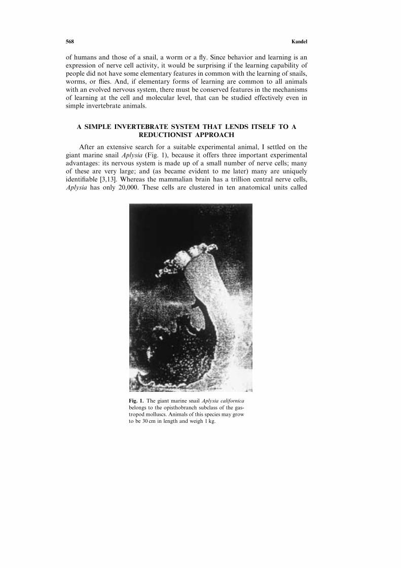
After an extensive search for a suitable experimental animal, I settled on thegiant marine snail Aplysia (Fig. 1), because it offers three important experimentaladvantages: its nervous system is made up of a small number of nerve cells; manyof these are very large; and (as became evident to me later) many are uniquelyidentifiable [3,13]. Whereas the mammalian brain has a trillion central nerve cells,Aplysia has only 20,000. These cells are clustered in ten anatomical units called
Fig. 1. The giant marine snail Aplysia californicabelongs to the opisthobranch subclass of the gas-tropod molluscs. Animals of this species may growto be 30 cm in length and weigh 1 kg.

Molecular Biology of Memory Storage 569
Fig. 2. The human brain has a million-million neurons while the brainof Aplysia has 20,000 nerve cells. Aplysia nerve cells are clusteredtogether in five major bilateral ganglia, each containing about 2000nerve cells.
ganglia, each of which contains about 2000 cells (Fig. 2). An individual ganglion,such as the abdominal ganglion, mediates not one but a family of behaviors. Thus,the simplest behaviors that can be modified by learning may involve less than 100cells. This numerical simplification made it possible to identify the specific contri-bution of individual neurons to the behavior in which they participate [13]. Inaddition to being few in number, these cells are the largest nerve cells in the animalkingdom, reaching up to 1000 µm in diameter, large enough to be seen with thenaked eye (Fig. 3). Because of their extraordinary size and their distinctive pigmen-tation, it is possible to recognize many of the cells as unique individuals. One can
Fig. 3. A photomicrograph of an abdominal ganglion of Aplysia shows the dis-tinctive pigmentation and positions of its cells. The largest cells are a millimeterin diameter and can be seen with the naked eye. Scale barG1 mm.

570 Kandel
record from these large cells for many hours without any difficulty, and the samecell can be returned to and recorded from over a period of days. The cells can easilybe dissected out for biochemical studies, so that from a single cell one can obtainsufficient mRNA to make a cDNA library. Finally, these identified cells can readilybe injected with labeled compounds, antibodies, or genetic construct procedureswhich opened up the molecular study of signal transduction within individual nervecells.
REQUIREMENT FOR A CELL-BIOLOGICAL STUDY OF MEMORYSTORAGE
Given a technically advantageous experimental system, how does a cell biologistbegin to address the problem of learning? The strategy that my colleagues and Ideveloped involved four sequential steps: (1) We first wanted to define a simplebehavior that can be modified by learning and that gives rise to memory storage.(2) We next wanted to identify the cells that make up the neural circuit of thatbehavior. (3) Within that neural circuit we then wanted to locate the critical neuronsand interconnections that had been modified by learning and that store memory.(4) Finally, we wanted to analyze the changes that occur at those sites in responseto learning and memory storage, first on the cellular and then on the molecular level[13]. I follow this outline in the discussion below.
DELINEATING A BEHAVIOR IN APLYSIA THAT IS CAPABLE OFBEING MODIFIED BY LEARNING
Irving Kupfermann and I first wanted to study the simplest possible behaviorof Aplysia [4]. We examined the animal’s behavioral capabilities and delineated avery simple defensive reflex: the withdrawal of the gill upon stimulation of thesiphon, an action that is like the quick withdrawal of a hand from a hot object. InAplysia, the gill is a respiratory organ that lies exposed in the mantle cavity. Whenthe animal is in a normal, relaxed state, the gill is partially covered by a sheet ofskin (the mantle shelf), which ends in a fleshy spout, the siphon (Fig. 1). When aweak tactile stimulus is applied to the siphon, both the siphon and gill are withdrawninto the mantle cavity and for protection under the mantle shelf [(Fig. 4); ref. 5].
Kupfermann, Harold Pinsker, and later Tom Carew, Robert Hawkins and Ifound that this simple reflex could be modified by three different forms of learning:habituation, sensitization, and classical conditioning [5, 8, 9]. As we examined thesethree forms of learning, we were struck by the resemblance each had to correspond-ing forms of memory storage in higher vertebrates and humans. As with vertebratelearning, memory storage for each type of learning in Aplysia has two phases: atransient memory that lasts minutes and an enduring memory that lasts days. Con-version of short-term to long-term memory storage requires spaced repetition—prac-tice makes perfect even in snails [8, 9, 10].
We focused initially on one type of learning, sensitization, a form of learnedfear in which a person or an experimental animal learns to respond strongly to anotherwise neutral stimulus [5, 8, 10]. For example, if a person is suddenly exposed to

Molecular Biology of Memory Storage 571
Fig. 4. A dorsal view of Aplysia showing the gill, the animal’s respiratory organ.A light touch to the siphon cavity with a fine probe causes the siphon to contractand the gill to withdraw under the protection of the mantle shelf. Here, themantle shelf is shown to be retracted for a better view of the gill. Sensitizationof the gill-withdrawal reflex, produced by applying a noxious stimulus to anotherpart of the body, such as the tail, leads to an enhancement of the withdrawalreflex of both the siphon and the gill.
an aversive stimulus, such as a gunshot going off nearby, that person will be sensit-ized by the unexpected noise. As a result, that person will be frightened and willnow startle to an otherwise innocuous stimulus like a tap on the shoulder. Similarly,on receiving an aversive shock to another part of the body such as the tail (or head),an Aplysia recognizes the stimulus as aversive and learns to enhance its defensivereflex responses to a variety of subsequent stimuli applied to the siphon, even innocu-ous stimuli [Fig. 4; 12]. The animal now remembers the shock, and the duration ofthis memory is a function of the number of repetitions of the noxious experience. Asingle shock gives rise to a memory lasting only minutes; this short-term memorydoes not require the synthesis of new protein. In contrast, four or five spaced shocksto the tail give rise to a memory lasting several days; this long-term memory doesrequire the synthesis of new protein. Further training gives rise to an even moreenduring memory lasting weeks, which also requires new protein synthesis [Fig.5; 10, 12].
Thus, just as in the complex learning in mammals [107, 108], long-term sensitiz-ation differs from the short-term process in requiring the synthesis of new proteins.This was our first clear evidence for the conservation of biochemical mechanismsbetween Aplysia and vertebrates, and it reinforced the hope that a detailed analysisof short-term memory and its transition to long-term memory in Aplysia wouldreveal molecular mechanisms of general importance.
DEFINING THE NEURAL CIRCUIT IN CELLULAR DETAIL
To analyze the cellular mechanisms of sensitization, we needed to identify theneural circuit of the gill withdrawal reflex. Kupfermann and I quickly localized the

572 Kandel
Fig. 5. Spaced repetition converts short-term memory intolong-term memory in Aplysia. In the resting state, beforesensitization training, the animal withdraws its siphon andgill only briefly in response to mild touch of the siphon.After the animal receives a single noxious shock to the tail,it withdraws its siphon and gill longer in response to thesame mild touch of the siphon. Sensitization following asingle noxious stimulus lasts about one hour. After four orfive single tail shocks the animal withdraws its gill andsiphon more powerfully and the sensitization lasts morethan a day. If the animal receives four brief trains of singleshocks a day over the course of four days, it withdraws itssiphon and gill for almost eight times as long and retainsthe memory for several weeks. [Modified from 98.]
central neuronal machinery for the reflex behavior in the animal’s abdominal gang-lion [4, 6]. Because we soon realized that many cells could be identified in everyanimal of the species [3, 4, 6, 7], we were able to give them specific names and, mostimportant, return to the same cell time and again—in both untrained and trainedanimals. In this way Kupfermann, Castellucci, Carew, Hawkins, John Byrne, and Iwere able to work out significant components of the neural circuit gill-withdrawalreflex in terms of individual cells and cell clusters. The circuit has 24 mechanorecep-tor sensory neurons that innervate the siphon skin and make direct monosynapticconnections with six gill motor cells [7, 11, 36]. The sensory neurons also madeindirect connections with the motor cells through small groups of excitatory andinhibitory interneurons [18, 19]. In addition to being identifiable, individual cells also

Molecular Biology of Memory Storage 573
proved to have surprisingly large effects on behavior [Fig. 6C; reviewed in 13, 22, 36].As we examined the neural circuit of this reflex in detail, we were struck by its
invariance—the cells that make up the circuit and their interconnections are alwaysthe same. In every animal we examined, each cell connected only to certain targetcells and not to others (Fig. 7). Carew, John Koester, Wayne Hening, and I alsofound this invariance in the neural circuitry of other behaviors in Aplysia—inking,control of the circulation, locomotion [reviewed in 13, 15]—raising a key questionin the cell-biological study of learning: How can learning occur in a neural circuitthat is precisely wired?
HOW DOES LEARNING AFFECT THE INVARIANT ELEMENTS OF THENEURAL CIRCUIT?
In his Croonian Lecture to the Royal Society of 1894, Santiago Ramon y Cajalproposed a theory of memory storage: memory is stored in the growth of new con-nections [16]. This prescient idea was neglected in good part for half a century asstudents of learning fought over newer competing ideas. First, Karl Lashley, RossAdey, Wolfgang Kohler, and a number of Gestalt psychologists proposed that learn-ing leads to changes in electric fields or chemical gradients, which they postulatedsurround neuronal populations and are produced by the aggregate activity of cellsrecruited by the learning process. Second, Alexander Forbes and Lorente de Noproposed that memory is stored dynamically by a self-reexciting chain of neurons.This idea was later championed by Donald Hebb as a mechanism for short-termmemory. Finally, Holger Hyden proposed that learning led to changes in the basecomposition of DNA or RNA. Even though there was much discussion about themerits of each of these ideas, there was no direct evidence to support any of them[reviewed in 17].
We were now in a position to address these alternative ideas by confrontingdirectly the question of how learning can occur in a circuit with fixed neuronalelements. Kupfermann, Castellucci, Carew, Hawkins, and I examined the neuralcircuit of the gill-withdrawal reflex while the animal underwent sensitization orhabituation, a form of learning in which the animal learns to ignore an innocuousstimulus to siphon when given with monotonous repetition. (We later also extendedthese studies to an examination of classical conditioning [20].) Our studies providedclear evidence for Cajal’s idea: learning results from changes in the strength ofthe synaptic connections between precisely interconnected cells [6, 7]. Thus, whilethe organism’s developmental program assures that the connections between cellsare invariant, it does not specify their precise strength. Rather, experience alters thestrength and effectiveness of these pre-existing chemical connections. Seen in theperspective of these three forms of learning, synaptic plasticity emerged as a funda-mental mechanism for information storage by the nervous system, a mechanism thatis built into the very molecular architecture of chemical synapses [95].
We soon appreciated that the synaptic strength of a chemical synapse couldbe modified in two ways: homosynaptically and heterosynaptically. Homosynapticchanges in synaptic strength occur in a synapse because of activity in the presynapticand postsynaptic neurons of that very synapse. During habituation, homosynaptic

574 Kandel
Fig. 6. The neural circuit of the gill-withdrawal reflex can be delineated in terms ofspecific identified cells. A. This dorsal view of the abdominal ganglion shows someidentified cells. The six identified motor cells to the gill are shaded brown; sevensensory neurons are shaded blue. In the figure a sensory neuron that synapses ongill motor neuron L7 is being stimulated electrically. A microelectrode in the motorneuron records the synaptic potential produced by the action potential in the sensoryneuron. B. The physiological demonstration of the direct connections between thesensory neuron and motor neuron. The sensory neuron receives input from thesiphon skin; the motor neuron makes direct connections onto the gill. The fact thatthe cells are large and identifiable allows for the mapping of connections betweenspecific identified cells. This part also shows the experimental arrangement for simul-taneously recording in a pre- and postsynaptic cell. The sensory neuron makes adirect connection onto the motor neuron, as is evident in part C. C. Individualcells make significant contributions to the reflex. Stimulating a single motor neuronproduces a detectable change in the gill, and stimulating a single sensory neuronproduces a large synaptic potential in the motor neuron. Repeated stimulation of asingle sensory neuron increases the frequency of firing in the motor neuron, leadingto a visible reflex contraction of the gill. A single tactile stimulus to the skin normallyactivates 6–8 sensory neurons, causing each to fire 1–2 action potentials. The repeti-tive firing of 10 action potentials in a single sensory neuron, designed to model thefiring of the total population, in fact simulates reasonably well the reflex behavior.

Molecular Biology of Memory Storage 575
Fig. 7. The neural circuit of the gill-withdrawal reflex. The siphon isinnervated by 24 sensory neurons that connect directly with the sixmotor neurons. The sensory neurons also connect to populations ofexcitatory and inhibitory interneurons that in turn connect with themotor neurons. Stimulating the tail activates modulatory interneuronsthat act on the terminals of the sensory neurons as well as on thoseof the excitatory interneurons. There are three classes of modulatoryneurons activated by tail stimuli: (1) neurons that release serotonin (5-HT), (2) neurons that release a peptide called the small cardioactivepeptides (SCP), and (3) the L29 cells, which release an unidentifiedmodulatory neurotransmitter. The serotonergic modulatory action isthe most important. Blocking the action of these serotonergic cellsblocks the effects of sensitizing stimuli.
changes occur in the monosynaptic connections between the sensory neurons andthe motor neurons of the gill-withdrawal reflex. Heterosynaptic changes occur in asynapse where presynaptic and postsynaptic neurons are themselves not active butthere is, instead, activity in one or more modulatory interneurons that act on thepresynaptic neurons, on the postsynaptic neurons of the synapse, or on both, tomodify the strength of their synaptic connections. During sensitization, heterosynap-tic changes are induced in the monosynaptic connections between the sensoryneurons and motor neurons of the gill-withdrawal reflex. Hawkins, Abrams, and Ilater found that these two types of regulation are recruited together in classicalconditioning [20, 21]. Classical conditioning therefore illustrated that the elementaryforms of homo- and heterosynaptic plasticity form an alphabet of basic mechanismsthat can produce combinations of plasticity with novel properties.

576 Kandel
In all three forms of learning we found particularly large changes in the synapticstrength of the direct connections between the sensory and motor neurons of thereflex. We therefore focused on this one component of the reflex and found severaladditional principles that have proven to be quite general. First, we found that thesame synaptic connection can be modulated in opposite ways by different forms oflearning. For example, habituation leads to a homosynaptic weakening of synapticconnections between the sensory neurons and their target cells, the motor neuronsand interneurons, while sensitization leads to heterosynaptic strengthening of thesesame sets of connections [7, 13, 33]. Second, learning not only leads to changes insynaptic strength, it can also affect the excitability of neurons. In the case of sensitiz-ation the excitability of the sensory (presynaptic) neurons is increased [25]. Third,the synaptic changes persist, thereby contributing to memory storage [10, 33, 94].Indeed, the same synaptic connection can store both short- and long-term memory[7, 23, 33, 43, 94, 98]. At a given synapse synaptic plasticity can either be short- orlong-lived depending on the number of spaced repetitions of the learning stimulus,and these parallel not only the behavioral changes of short-term memory [7, 43, 93].Finally, long-term memory storage involves not only a change in synaptic strength,but also anatomical changes, changes in the number of synaptic connections[57, 58, 59].
The changes at the synapse between the sensory and the motor neuron are onlya part of the changes in the neural circuit of the gill-withdrawal reflex. Importantchanges occur elsewhere in the circuit, but we have studied them less. We havefocused on the monosynaptic portion of the circuit in order to probe in depth themolecular mechanisms that contribute to learning and memory.
INITIAL STEPS TOWARDS A MOLECULAR CHARACTERIZATIONOF SENSITIZATION
What are the molecular mechanisms whereby short-term memory is established,and how it is converted to long-term memory? Initially, we focused on short-termsensitization (Fig. 8). In collaboration with James H. Schwartz, we found that thesynaptic changes, like the short-term behavior, were expressed even when proteinsynthesis was inhibited. Since the short-term changes persisted for many minutes, itseemed unlikely that they involved a simple conformational change of one or moreproteins. We therefore proposed, in 1971, that short-term memory might require aseries of sequential reactions similar to that mediated by cAMP-mediated signaling[26]. Our attention was drawn to cAMP because Sutherland, Rall and Krebs hadfound that various neurotransmitters could increase cAMP concentration in thebrain and in other tissues, and in the liver cAMP activated the cAMP-dependentprotein kinase [26]. In 1972, Schwartz, Howard Cedar, and I found that stimulationof the modulatory pathways recruited during heterosynaptic facilitation led to anincrease in cAMP in the abdominal ganglion [27]. Cedar and Schwartz next applieda number of neurotransmitter candidates and found that serotonin and dopaminecould increase levels of cAMP [29]. This made us wonder whether the population ofmodulatory interneurons, which produces the heterosynaptic facilitation that givesrise to sensitization, contains serotonergic cells.

Molecular Biology of Memory Storage 577
Fig. 8. The strength of the synaptic connectionsbetween the sensory and the motor neurons isincreased by sensitization, by serotonin, and bydirect activation in the presynaptic neurons of cAMPand PKA. A. The increase in synaptic strength pro-duced by sensitizing stimuli can be achieved by elec-trical stimuli to the tail, to the head, or to the neuralpathways from the tail or head. B. Facilitation couldbe reproduced by stimulating individual modulatorycells such as the serotonergic cells or by the appli-cation of the serotonin to the connections betweenthe sensory neurons and motor neurons. C. Facili-tation can also be produced by injecting cyclic AMPinto the sensory neurons. D. Facilitation can also beproduced by simply injecting the catalytic subunit ofPKA into the sensory neurons. In this experimentthe spikes of the sensory neuron have been artifici-ally broadened by the application of tetraethylam-monium (TEA). This reveals dramatically the furtherbroadening produced by the catalytic subunit ofPKA by the reduction of K+ currents. [From 13, 31,32, 37.]
Later, Hawkins, Castellucci, David Glanzman, and I delineated the modulatorysystem activated by a sensitizing stimulus to the tail [19, 34, 35], and confirmed thatit contains serotonergic interneurons. The systems includes two other classes ofheterosynaptic modulators, but the serotonergic neurons are most important forsensitization: blocking their actions alone blocked sensitization [34].
A DISTINCTION BETWEEN MEDIATING AND MODULATORYCIRCUITRY
In 1974, Castellucci and I next attempted to localize the change produced byhabituation and sensitization to either the presynaptic or the postsynaptic compo-nent of the synapse. We therefore applied a quantal analysis to the synaptic connec-tions between the sensory and motor cells and found that the short-termhomosynaptic depression that accompanies habituation is presynaptic, involving areduction in the amount of transmitter released from the presynaptic sensory neuron[30]. This transmitter, Nicholas Dale later showed, is glutamate [51]. Conversely, wefound that the short-term heterosynaptic facilitation that accompanies sensitizationresults from an increased release of this transmitter from the sensory neuron [31].
578 Kandel
We next found that a single brief application of serotonin could stimulate short-term sensitization (Figs. 8A and B)—by directly enhancing the release of transmitterfrom the sensory neurons, much as does the sensitization-induced facilitation—whereas dopamine could not [32]. Moreover, pharmacological inhibitors of sero-tonergic receptors blocked the effect of sensitizing stimuli, providing further evidencefor the importance of serotonin as a modulator of synaptic strength [32].
Understanding the role of serotonergic and other modulatory neurons in sensit-ization allowed us to distinguish two different types of neural circuits import forbehavior and learning: mediating and modulating (Fig. 7). A mediating circuit con-trols the gill-withdrawal reflex. It consists of the sensory neurons, interneurons, andmotor neurons of the reflex, and it can be modified homosynaptically by habituation.But other, more complex forms of learning, such as sensitization and classical con-ditioning, involve modulatory circuits, somewhat akin to the arousal or attentionalcircuitries in higher animals. In Aplysia such a modulating circuit consists of sero-tonergic and other modulatory interneurons that act upon the mediating circuitryto regulate the strength of its connections. For example, modulatory interneuronsact on the sensory neurons, including on their presynaptic terminals, to enhance theamount of glutamate released.
SHORT-TERM MEMORY INVOLVES cAMP AND THE ACTIVATIONOF PKA
The finding that serotonin acts on specific receptors in the presynaptic terminalsof the sensory neuron to enhance transmitter release, coupled with the fact thatserotonin is capable of bringing about an increase in cAMP in the abdominal gang-lion, caused us to ask whether serotonin facilitates the release of transmitter fromthe sensory neurons by acting via cAMP. We first explored this idea in 1976 whenBrunelli, Castellucci, and I injected cAMP directly into the presynaptic cells andfound that it produced presynaptic facilitation (Fig. 8C) [32, 33]. This provided themost and compelling evidence then available in the nervous system, that cAMP isinvolved in a specific physiological function and it gave us our first insight into themolecular mechanisms of short-term memory.
Marc Klein and I next asked: How does cAMP enhance transmitter release?We found that serotonin, or injected cAMP, leads to increased excitability and abroadening of the action potential by reducing a specific K+ current, allowing greaterCa2C influx into the presynaptic terminal with each action potential [24]. The greaterCa2C influx could contribute to the enhanced transmitter release. Following the leadto Paul Greengard, who had proposed that cAMP produces all of its action in thebrain through the cAMP-dependent protein kinase (PKA), Klein and I suggestedthat cAMP may lead to phosphorylation of this K+ channel by activating PKA [24].In collaborative experiments with Paul Greengard in 1980, Castellucci, Schwartz,and I tested this idea by injecting the active catalytic subunit of PKA and foundthat the catalytic subunit by itself produced broadening of the action potential andenhancement of glutamate release (Fig. 8D) [37]. Conversely, the specific peptideinhibitor of PKA (PKI) blocked the actions of serotonin. These findings provideddirect evidence for the role of PKA in short-term presynaptic facilitation [38].

Molecular Biology of Memory Storage 579
In 1981, Steven Siegelbaum arrived at Columbia, having just mastered single-channel recording during a visit to Erwin Neher and Bert Sakmann in Gottingen.In an elegant series of experiments, Siegelbaum and Joseph Camardo identified anovel K+ channel, the S-type K+ channel, and showed that it could be modulated bycAMP [39]. Then Siegelbaum and Michael Schuster used an isolated inside-out patchof membrane from the sensory neuron to show that PKA could act directly todecrease the number of channels open in that patch [40]. Later, Byrne showed thatserotonin also modulates a delayed-rectifier K+ channel whose contribution to spikebroadening proved even more important than that of the S-type channel [reviewedin 41]. The combined studies indicated that the S-type channel mediated the increasein excitability with a minor contribution to broadening, whereas the delayed-rectifierK+ channel contributed little to excitability but had a major role in spike broaden-ing. Finally, Hochner, Klein, and I—and independently Jack Byrne and his col-leagues—showed that, in addition to spike broadening, serotonin also enhancedrelease by an as-yet-unspecified action on the release machinery. Thus, serotoninleads to an increase in presynaptic cAMP, which activates PKA and leads to synap-tic strengthening through a combination of mechanisms [reviewed in 41].
LONG-TERM MEMORY INVOLVES THE NUCLEAR TRANSLOCATIONOF PKA AND MAP KINASE AND THE ACTIVATION OF CREB-1
Even earlier, in 1973, Arnold Kriegstein, an M.D.-Ph.D. student, succeeded inculturing Aplysia larvae so that we could raise Aplysia in the laboratory [28], provid-ing us with animals of any size or age at any time. This was a major advance andopened up the study of synaptic plasticity in dissociated cell culture, which requiredvery young animals [42, 43].
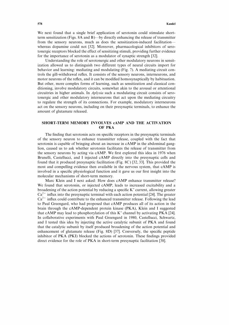
In the intact animal, one tail shock produces short-term sensitization, whichdoes not depend on protein synthesis; four to five tail shocks produce sensitizationthat lasts several days, and this long-term process requires new protein synthesis.Both are reflected in alterations in strength in the connections between the sensoryand motor neurons (Fig. 9). By substituting puffs of serotonin for tail shocks, SamSchacher, Pier Giorgio Montarolo, Philip Goelet, and I could model this behavioralprotocol in a culture dish consisting of a single sensory cell making synaptic connec-tions with a single motor cell [43]. We were able to induce both short- and long-term facilitation in this culture and found, as we had with the intact animal, thatthe long-term process differed from the short-term process in requiring the synthesisof new proteins (Fig. 10). We had now trapped, in a culture consisting of two inter-connected cells, the requirement for protein synthesis necessary to establish long-term memory.
We next used this cell culture to ask: What genes are activated to convert theshort-term process to the long-term process, and what genes are essential for themaintenance of the long-term process? We found that five spaced puffs of serotonin(simulating five spaced shocks to the tail) activate PKA, which in turn recruits themitogen-activated protein kinase (MAPK) and both translocate to the nucleus,where they activate a transcriptional cascade. The cascade begins with the transcrip-tion factor CREB-1, the c¡AMP r¡esponse e¡lement b¡ inding protein-1), so called

580 Kandel
Fig. 9. Long-term sensitization of the gill-withdrawal reflexof Aplysia involves long-term facilitation of the connectionsbetween sensory and motor neurons. A. Experimentalarrangement. The recordings on the right show representa-tive synaptic potentials in a siphon sensory neuron and agill motor neuron in a control animal and an animal thatreceived long-term sensitization training by stimulating itstail. The record was obtained one day after the end of train-ing. B. The median amplitude of the postsynaptic potential(PSP) in an identified gill motor neuron is greater in sensit-ized animals than in control animals. The effect of sensitiz-ation on the neural circuit of the gill-withdrawal reflex ismeasured by the median during of withdrawal of thesiphon. (PreGscore before training; postGscore after train-ing.) The sensitized animals were tested one day after train-ing. [Adapted from 98.]
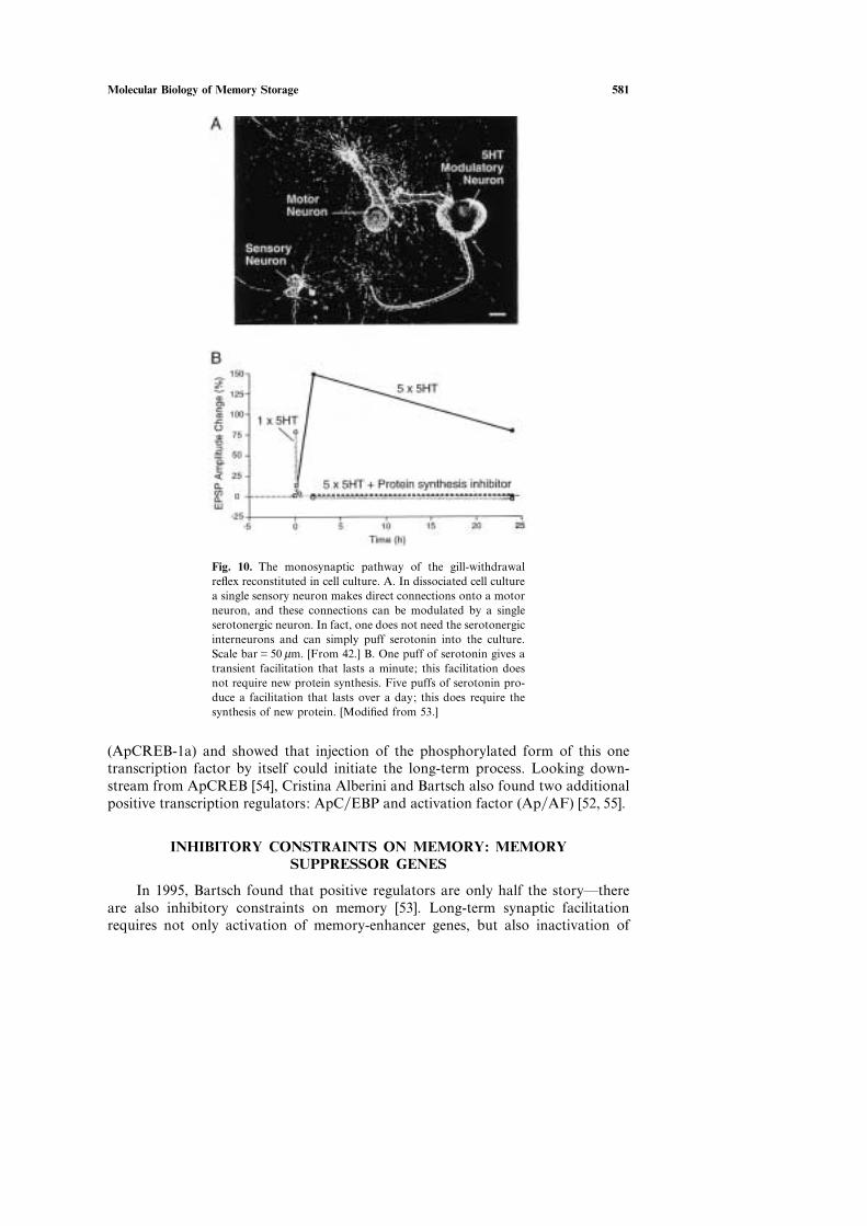
because it binds to a cAMP response element (CRE) in the promoters of targetgenes. CREB-1 leads to the activation of a set of immediate response genes, and inturn leads to the growth of new synaptic connections [44, 45, 46, 47, 49, 50, 56, 59].
The first clue to the importance of CREB in long-term memory was providedin 1990 by Pramod Dash and Binyamin Hochner [45]. They injected, into the nucleusof a sensory neuron in culture, oligonucleotides carrying the CRE DNA element,thereby titrating out CREB, and found that this selectivity blocked long-term butnot short-term facilitation (Fig. 11). Later, Dusan Bartsch cloned Aplysia CREB-1a
Molecular Biology of Memory Storage 581
Fig. 10. The monosynaptic pathway of the gill-withdrawalreflex reconstituted in cell culture. A. In dissociated cell culturea single sensory neuron makes direct connections onto a motorneuron, and these connections can be modulated by a singleserotonergic neuron. In fact, one does not need the serotonergicinterneurons and can simply puff serotonin into the culture.Scale barG50 µm. [From 42.] B. One puff of serotonin gives atransient facilitation that lasts a minute; this facilitation doesnot require new protein synthesis. Five puffs of serotonin pro-duce a facilitation that lasts over a day; this does require thesynthesis of new protein. [Modified from 53.]
(ApCREB-1a) and showed that injection of the phosphorylated form of this onetranscription factor by itself could initiate the long-term process. Looking down-stream from ApCREB [54], Cristina Alberini and Bartsch also found two additionalpositive transcription regulators: ApC�EBP and activation factor (Ap�AF) [52, 55].
INHIBITORY CONSTRAINTS ON MEMORY: MEMORYSUPPRESSOR GENES
In 1995, Bartsch found that positive regulators are only half the story—thereare also inhibitory constraints on memory [53]. Long-term synaptic facilitationrequires not only activation of memory-enhancer genes, but also inactivation of
582 Kandel
Fig. 11. Long-term facilitation requires gene transcription mediated by thetranscription factor, the cAMP response element-binding protein (CREB-1).To inhibit the action of CREB, an oligonucleotide encoding the cyclic AMPresponse element (CRE) was injected into the Aplysia sensory neuron in cul-ture. The sensory neuron in these experiments was in synaptic contact with themotor neuron. A. Experimental arrangement. A1. A single motor cell was cul-tured with two sensory cells. A2. One sensory cell was injected with the specificoligonucleotide encoding the CRE, designed to titrate out the CREB transcrip-tion factor, and the other was used as a control for injection of mutated orother oligonucleotide. B. The specific oligonucleotide blocks the serotonin-induced long-term facilitation measured 24 hr after injection (B1) but does notaffect short-term facilitation (B2). A mutated oligonucleotide encoding theCRE or an oligonucleotide encoding the heat shock enhancer and theenhancers of NFKβ do not affect long-term facilitation. [Modified from 45.]

Molecular Biology of Memory Storage 583
memory-suppressor genes. One of these, the transcription factor ApCREB-2, canrepress ApCREB-1a mediated transcription; relieving this repression lowers thethreshold for the long-term process. Indeed, Bartsch found that when antiserum thatblocks the action of ApCREB-2 is injected into the sensory neurons, a single appli-cation of serotonin, which normally produces facilitation lasting only minutes, nowproduces facilitation lasting days, accompanied by the growth of new synaptic con-nections (Fig. 12).
Thus, during long-term memory storage, a tightly controlled cascade of geneactivation is switched on, with memory-suppressor genes providing a threshold orcheckpoint for memory storage, presumably to ensure that only salient features arelearned. It is clearly an evolutionary advantage for an animal to learn and store inlong-term memory only facts important for survival, rather than retaining every-thing. Memory-suppressor genes may be regulated independently of the activators.Such independent action may be necessary to allow for the modulation of memorystorage by emotional stimuli, as occurs in ‘‘flashbulb memories’’, memories ofemotionally charged events that are recalled in detail, as if a complete picture hadbeen instantly and powerfully etched in the brain.
Once the repressive action of CREB-2 is removed and CREB-1 is activated, aset of immediate response genes is induced. One of these, the ubiquitin C-terminalhydrolase, is responsible for the proteolytic cleavage of the regulatory subunit ofPKA, thereby removing a second inhibitory constraint or memory suppressor gene.Since the regulatory subunit normally inhibits the kinase activity of the catalyticsubunit, cleavage extends the activity of the kinase for several hours [Fig. 12; refs.60, 61]. In 1982, Schwartz and I first proposed this mechanism as the simplest mol-ecular mechanism for long-term memory storage—the long-term process coopts andprolongs the action of the kinase involved in the short-term process but renders itindependent of any further signaling by serotonin or PKA [115].
THE STABLE, SELF-MAINTAINED FORM OF LONG-TERM MEMORY ISREFLECTED IN THE GROWTH OF NEW SYNAPTIC CONNECTIONS
However, this persistent kinase is only required for 10 to 12 hours. Craig Baileyand Mary Chen first showed that what makes long-term memory enduring is thegrowth of new synaptic connections, a structural change that parallels the durationof the behavioral memory [56, 57, 58, 59]. As the connections retract over time, thememory fades. A typical sensory neuron in the intact animal has about 1200 synapticvaricosities. Following long-term sensitization, the number more than doubles toabout 2600. With time the number returns to about 1500 synaptic connections(Fig. 12).
An important clue to the molecular actions that give rise to these structuralchanges came from the identification by Mark Mayford of a third class of memory-suppressor genes, the Aplysia cell adhesion molecules (ApCAMs) [62, 63], whichbelong to the immunoglobulin family of cell adhesion molecules, which includesmammalian neural cell adhesion molecule (NCAM) and Drosophila Fasciclin II.After exposure to five spaced puffs of serotonin, the concentration if ApCAM at thesurface membrane of the presynaptic sensory neuron decreases as a result of the

584 Kandel
Fig. 12. Long-term sensitization of the gill-withdrawal reflex of Aplysia leads to two major sets of changesin the sensory neurons of the reflex: (1) persistent activity of protein kinase A and (2) the growth of newsynaptic connections. In short-term sensitization (lasting minutes to hours) a single tail shock causes aone-time release of serotonin that leads to covalent modification of pre-existing proteins. The serotoninacts on a transmembrane receptor to activate adenylyl cyclase (AC) and converts ATP to the secondmessenger cAMP. In turn, cAMP activates the cyclic AMP-dependent protein kinase A (PKA), whichphosphorylates and covalently modifies a number of target substrate proteins, including K+ channels andcomponents of the exocytotic machinery of release, to enhance transmitter availability and release. Theduration of these modifications parallels the short-term memory. The cAMP-dependent protein kinase Ahas both catalytic subunits (the oval shaped structures) and regulatory subunits (the spindle-shaped struc-tures). Normally the regulatory subunits inhibit the catalytic subunits. When the level of cAMP rises, thecAMP binds to the regulatory subunit, causing it to undergo a conformational change so that it dis-sociates from and frees the catalytic subunit, allowing the freed catalytic subunit to phosphorylate sub-strate proteins in the presynaptic terminals. With repeated stimulation, the level of cAMP rises moredramatically and persists for several minutes. This frees the catalytic subunit for a sufficient period oftime to allow it to translocate to the nucleus, and in so doing it also recruits the mitogens-activatedprotein kinase (MAPK). In long-term sensitization (lasting one or more days) repeated shocks to the tailcause repeated release of serotonin that leads to the regulatory subunit being dissociated from the catalyticsubunit for sufficient time so that the catalytic subunit translocates to the nucleus where it phosphorylatesthe cyclic AMP response element-binding (CREB) protein and leads to the removal of the repressiveaction of CREB-2, which is capable of inhibiting CREB-1 perhaps by means of another protein kinase,MAP kinase which is also activated by the catalytic subunits. Once CREB-1 is activated, it activates inturn the gene that encodes a ubiquitin hydrolase, a component of a specific ubiquitin protease that leadsto the regulated proteolysis of the regulatory subunit of PKA. This cleavage of the (inhibitory) regulatorysubunit results in persistent activity of PKA, leading to persistent phosphorylation of the substrate pro-teins of PKA, including proteins involved in the short-term process. A second set of genes, activated byCREB-1, is C�EBP, which acts both by itself as a homodimer and together with activating factor (AF)as a heterodimer to give rise to the growth of new synaptic connections. In so doing it activates a numberof late genes, including elongation factor 1α (EF1α).

Molecular Biology of Memory Storage 585
internalization of the transmembrane form of ApCAM, a process that requires on-going protein synthesis. This clathrin-mediated endocytosis is blocked by mutationsin the consensus sequence for MAP kinase phosphorylation in the cytoplasmic tail[48]. Selective down-regulation of the transmembrane isoform of ApCAM causesdefasciculation; it decreases the adhesive interaction of sensory cell neurites witheach other, a prerequisite to process outgrowth and the formation of new synapticconnections.
THE cAMP, PKA, CREB SWITCH IS ALSO IMPORTANT FORLEARNING AND MEMORY IN DROSOPHILA
The pioneering work of Seymour Benzer and the subsequent studies of his stud-ents, Chip Quinn, Tim Tully, Jerry Yin, and Ronald Davis, have led to the identifi-cation of a number of genes required for memory storage in Drosophila [reviewed in97,102]. Many of the genes identified in this way are the same as those implicatedin plasticity in Aplysia. For example, Drosophila genes dunce, rutabaga, and amnesiacall encode components of the cAMP-PKA cascade. Other genes identified encodeparticipants in cell adhesion molecules similar to ApCAM [97]. Moreover, a proteinsynthesis-dependent phase of learning has been described by Tully and his col-leagues, and Yin and Tully have shown that, as in Aplysia, CREB has a critical rolein induction of longer memory [97].
IS LONG-TERM FACILITATION SYNAPSE-SPECIFIC?
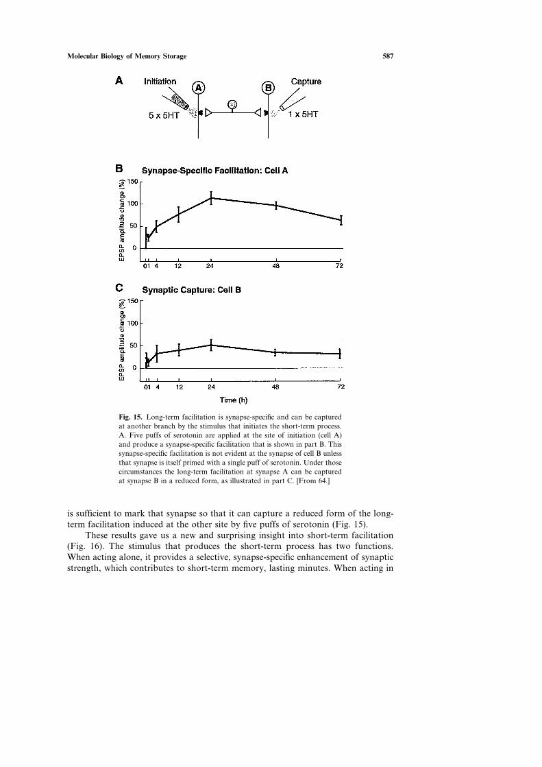
The finding of a transcriptional cascade explained why long-term memoryrequires new protein synthesis immediately after training, but it posed a new cell-biological problem. A single neuron makes hundreds of contacts on many differenttarget cells (Fig. 13). Short-term synaptic changes are synapse-specific. Since long-lasting synaptic changes require transcription and thus the nucleus, is long-termmemory storage a cell-wide process, or are there cell-biological mechanisms thatmaintain the synapse specificity of long-term facilitation?
To examine these questions at the level of individual synapses, Kelsey Martinmodified our culture system. She cultured one sensory cell with a bifurcatingn axonwith two motor neurons, forming two widely separated synapses (Fig. 14). In thisculture system, a single puff of serotonin applied to one synapse produces transientfacilitation at that synapse only, as expected [64, 65]. Five puffs of serotonin appliedto one branch produces long-lasting facilitation (72 hours) that is also restricted tothe stimulated synapse (Fig. 15). This long-lasting synapse-specific facilitationrequires CREB and also leads to structural changes. Thus, despite recruitment ofnuclear processes, long-term change in synaptic function and structure are confinedonly to those synapses stimulated by serotonin.
How does this come about? Martin, Andrea Casadio, Bailey, and I found thatfive puffs or serotonin send a signal to the nucleus to activate CREB-1, which thenappears to send proteins to all terminals; however, only those terminals that havebeen marked by serotonin can use these proteins productively for synaptic growth.Indeed, we found that one puff of serotonin to the previously unstimulated synapse

586 Kandel
Fig. 13. A single sensory neuron connects to many target cells. Therequirement of a transcriptional mechanism for long-term memoryraises the question: What is the unit lf long-term information stor-age? Is it a single synapse, as with short-term facilitation, or theentire neuron? Is there a mechanism for restricting synaptic facili-tation to some synaptic connections?
Fig. 14. This photomicrograph shows a culture system developed toexamine the action of two independent branches of a single in Aplysiasensory neuron (the small neuron in the middle) on two differentmotor neurons (large neurons). Serotonin can be selectively appliedto one and not the other of the two branches. The flow of the sero-tonin can be monitored with the dye, fast green. [From 64.]
Molecular Biology of Memory Storage 587
Fig. 15. Long-term facilitation is synapse-specific and can be capturedat another branch by the stimulus that initiates the short-term process.A. Five puffs of serotonin are applied at the site of initiation (cell A)and produce a synapse-specific facilitation that is shown in part B. Thissynapse-specific facilitation is not evident at the synapse of cell B unlessthat synapse is itself primed with a single puff of serotonin. Under thosecircumstances the long-term facilitation at synapse A can be capturedat synapse B in a reduced form, as illustrated in part C. [From 64.]
is sufficient to mark that synapse so that it can capture a reduced form of the long-term facilitation induced at the other site by five puffs of serotonin (Fig. 15).
These results gave us a new and surprising insight into short-term facilitation(Fig. 16). The stimulus that produces the short-term process has two functions.When acting alone, it provides a selective, synapse-specific enhancement of synapticstrength, which contributes to short-term memory, lasting minutes. When acting in

588 Kandel
Fig. 16. Two different functions of the short-termprocess by itself and in conjunction with the long-term process occurring at any other part within theneural tree. By itself, short-term facilitation partici-pates in short-term memory storage. In conjunctionwith long-term facilitation at any other synapse, theshort-term process markes the specific synapse towhich it is applied and allows it to capture and useproductively the proteins necessary to establish thelong-term process and to grow new synapticconnections.
conjunction with the activation of CREB initiated by a long-term process in eitherthat synapse or in any other synapse on the same neuron, the stimulus that producesthe short-term process serves to locally mark those synapses at which it occurs. Themarked synapse can then productively utilize the proteins activated by CREB forsynaptic growth to produce a persistent change in synaptic strength.
The existence of long range signaling between the synapse and the nucleus andthe nucleus and the synapse introduces a new dimension into the integrative actionof neurons that alters the rules whereby synapses are strengthened or weakened.Although long-term facilitation is synapse-specific and restricted, once transcriptionhas been activated by the long-term process, the potential for plastic change of allthe synapses of the neuron has, in fact, become altered. As a result, following theinitiation of transcription in a neuron by long-term activation of one synapse, theaction of any other synapse of that neuron is no longer determined simply by thehistory of that synapse but is also determined by the history of the transcriptionalmachinery in the nucleus. Thus, the logic of the long-term process is quite differentfrom the short-term process.

Molecular Biology of Memory Storage 589
THE NATURE OF THE LOCAL MARKING SIGNAL
How does one puff of serotonin mark a synapse for long-term change? Wefound that the synapse is marked for both long-term synaptic facilitation and forgrowth of new synaptic connections by covalent modifications of pre-existing pro-teins mediated by PKA. However, for the structural change to persist, local proteinsynthesis is required [65]. Oswald Steward’s important work in the early 1980s hadshown that dendrites contain ribosomes, and that specific mRNAs are transportedto the dendrites and translated there locally [101]. But the function of these locallytranslated mRNAs was unknown. Our experiments showed that one function wasto stabilize the synapse-specific long-term functional and structural changes.
What proteins might be important locally for the stabilization? To answer thisquestion we needed a preparation in which we could study the local mRNA andproteins in isolation, without contamination of the cell body or surrounding glialcells. Martin was able to culture several hundred sensory neurons in a dish and thencut off their cell bodies, allowing us to study the mRNAs in the processes andhow they are regulated [64]. Serotonin could stimulate translation in these isolatedprocesses. Moreover, the transcripts in these processes contained regulatorysequences consistent with their using at least two mechanisms for regulating localtranslation. These two local mechanisms of translation serve different functionalroles.
One group of transcripts, such as an isoform of α -tubulin that is important forthe assembly of microtubules for fast axonal transport and for cytoskeletal organiz-ation, has a cytoplasmic polyadenylation element (CPEP) in their 3′ untranslatedregion which, in other contexts, contributes to translational regulation by regulatingpolyadenylation. This component of translation is blocked by emetine, a generalinhibitor of protein synthesis, and is necessary for initiating the long-term process(Fig. 17). Other transcripts, such as the elongation factor EF-1α , have an oligopyri-midine tract in their 5′ untranslated region. This tract is found in a small set oftranscripts that are preferentially translated by growth factors and mitogens. Theability of these growth factors to recruit these transcripts is blocked selectively bythe drug rapamycin [reviewed in 99, 100, 101]. Indeed, we found that the stabilizationof facilitation and of the growth of new connections, both at the site of capture andthe site of initiation, are sensitive to inhibition by rapamycin [Fig. 17; and ref. 65].
A FOURTH CONSEQUENCE OF NEUROTRANSMITTER SIGNALING:REGULATING LOCAL PROTEIN SYNTHESIS
These studies thus revealed a new, fourth type, of synaptic action mediated byneurotransmitter signaling. Three of these four have emerged, at least in part, fromthe study of learning and memory. First, in 1951, Katz and Fatt opened up themodern study of chemical transmission with their discovery of inotropic receptorsthat regulate ion flux through transmitter-gated ion channels to produce fast synap-tic actions, lasting milliseconds [110]. Second, in the 1970s, metabotropic receptorswere found to activate second-messenger pathways, such as the cAMP-PKA path-way, to produce slow synaptic activity lasting minutes [111]. As we have seen in

590 Kandel
Fig. 17. The requirements for local protein synthesis differ at the sites of initiation and capture of long-term facilitation. At the site of initiation two components of local protein synthesis are required: anemetine-sensitive local protein synthesis to set up the long-term process and a rapamycin-sensitive compo-nent of local protein synthesis to maintain facilitation after 48 hr. At the site of capture, inhibitors ofcyclic AMP block the capture completely. By contrast, the rapamycin- and local protein synthesis-sensi-tive components are only required for maintenance of facilitation. [From 65.]
Aplysia, this slow synaptic action can regulate transmitter release, thereby contribu-ting to short-term memory for sensitization. Third, an even more persistent synapticaction, lasting days, results from repeated action of a modulatory transmitter suchas serotonin. With repeated applications of serotonin, second-messenger kinasestranslocate to the nucleus, where they activate a cascade of gene induction leadingto the growth of new synaptic connections. This of course raises the problem ofsynapse specificity that we have considered above. Our experiments, in the bifurcatedculture system, revealed a novel fourth action of neurotransmitters, the marking ofthe synapse and the regulation of local protein synthesis which contributes to theestablishment of synapse-specific long-term facilitation [Fig. 18].
THERE ARE TWO MAJOR TYPES OF MEMORY: THE CASE OFEXPLICIT MEMORY
I have so far considered only the simplest cases of memory storage—thoseinvolving reflexes—a form called implicit or procedural memory. Implicit memoryis memory for perceptual and motor skills and is expressed through performance,

Molecular Biology of Memory Storage 591
Fig. 18. Four different consequences of the action of neurotransmit-ters. These events show that the synapses and nucleus readily inter-act. 1. Transmitter activation of a ligand-gated ion channel leads toa rapid synaptic action. 2. Transmitter activation of a seven trans-membrane receptor and a second messenger kinase leads to a moreenduring synaptic action. 3. Repeated transmitter activation of aseven transmembrane receptor leads to the translocation of the kin-ase to the nucleus and to activation of transcription, producing apersistent synaptic action. 4. Transmitter can also activate local pro-tein synthesis to stabilize the synapse-specific facilitation.
without conscious recall of past episodes. In contrast, the memories we hold nearand dear are called explicit (or declarative) memories. These memories require con-scious recall and are concerned with memories for people, places, objects, and events.Explicit memory involves a specialized anatomical system in the medial temporallobe, and a structure deep to it, the hippocampus [Fig. 19; reviewed in 66, 94, 95].
How is explicit memory stored? We had known from the work of Louis Flexner,Bernard Agranoff, Sam Barondes, and Larry Squire that explicit memory, likeimplicit memory, has a short-term phase that does not require protein synthesis and

592 Kandel
Fig. 19. Two major forms of long-term memory: explicit (declarative) andimplicit (procedural). Explicit or declarative memory is the memory for personsand objects and requires conscious participation for recall. Implicit or proceduralmemory is the memory for perceptual and motor skills, which is perfected inperformance and does not involve conscious participation for recall.
a long-term phase that does [66]. Are these two components of memory storage alsorepresented at the cellular level? What rules govern explicit memory storage?
A decade ago, when I reached by 60th birthday, I finally gathered up my cour-age and returned to the hippocampus. A major stimulus for me was the presence inmy laboratory of Seth Grant who was eager to work with genetically modified mice[67]. When Mario Capecchi and Oliver Smithies succeeded in achieving targetedgene ablation in ES stem cells, it became clear to me that mice would now offer asuperb genetic system for relating individual genes to synaptic plasticity, on the onehand, and to complex explicit memory storage on the other. Although mice arerelatively simple mammals, they have a medial temporal lobe system, including ahippocampus, that resembles that of humans, and they use their hippocampus muchas we do to store memory of places and objects (Fig. 20).
In our work with mice we have focused on memory for extrapersonal space asa model of explicit memory, because spatial memory is well represented in rodentsand has been particularly well studied. Although we still do not know much abouthow information is transformed as it gets into and out of the hippocampus, it is wellestablished that the hippocampus contains a cellular representation of extrapersonalspace—a cognitive map of space—and that lesions of the hippocampus interferewith spatial tasks [67]. Moreover, in 1972, Terje Lømo and Tim Bliss discoveredthat the perforant path, a major pathway within the hippocampus, exhibits activity-dependent plasticity, a change now called long-term potentiation (LTP) (Fig. 21). Inthe CA1 region of the hippocampus where LTP has been studied most extensively,it had been found to be induced postsynaptically by activation of an NMDA recep-tor to glutamate. In the late 1980s Richard Morris found that blocking the NMDA

Molecular Biology of Memory Storage 593
Fig. 20. Barnes maze used in studying spatial memoryin the mouse. The mouse is put in the center of a woodenplatform. Because mice do not like open spaces they tryto find some way to move out of it. The only way theycan escape from the exposed space is to find the onehole out of 40 that leads to an escape hatch.
receptor pharmacologically not only interfered with LTP but also blocked memorystorage [69, 70].
We focused primarily on the Schaffer collateral, another pathway in the hippo-campus, not only because it has been extensively studied but also because work witha patient R.B. by Larry Squire and his colleagues had shown that a lesion restrictedto the CA1 region was sufficient to produce a significant loss of explicit memorystorage [68].
Early work on LTP in this pathway in hippocampal slices by others had focusedon the response to one or two trains of electrical stimuli. But in our work on Aplysiawe had found long-term memory and the synaptic changes that accompany it emergemost effectively with repeated stimuli. So Uwe Frey, Yan-You Huang, PeterNguyen, and I examined whether LTP changed with repeated stimulation [71, 72, 73]and found that in each of the three major hippocampal pathways LTP has phases,much like facilitation in Aplysia.
The early phase of LTP, produced by a single train of stimuli, lasts only 1–3hours and does not require new protein synthesis [reviewed in 73]; it involves co-valent modifications of pre-existing proteins that lead to the strengthening of pre-existing connections, similar in principle to short-term facilitation in Aplysia. But,as was well documented by Roger Nicoll and his colleagues, the molecular detailsof the early phase of LTP in the Schaffer collateral pathway differ from those ofshort-term facilitation in Aplysia. The influx of Ca2C through the NMDA receptorchannel leads to the activation of a Ca2C calmodulin-dependent protein kinase andthe phosphorylation of preexisting AMPA receptors, and insertion into the postsyn-aptic membrane of new AMPA receptors to glutamate [75].

594 Kandel
Fig. 21. Long-term potentiation (LTP) in the hippocam-pus. A. Three major pathways denote the direction ofthe impulse flow, each of which gives rise to LTP. Theperforant pathway from the subiculum forms excitatoryconnections with the granule cells of the dentate gyrus.The mossy fiber pathway, formed by the axons of thegranule cells of the dentate gyrus, connects the granulecells with the pyramidal cells in area CA3 of the hippo-campus. The Schaffer collateral pathway connects thepyramidal cells of the CA3 region with the pyramidalcells in the CA1 region of the hippocampus. B. The earlyand late phases of LTP in the Schaffer collateral path-way. A single train of stimuli for one second at 100 Hzelicits an early LTP, and four trains at 10-minute inter-vals elicit the late phase of LTP. The early LTP lastsabout 2 hr and the late LTP last more than 24 hr.
By contrast, repeated trains of electrical stimuli produce a late phase of LTP,which has properties quite different from early LTP and quite similar to long-termfacilitation in Aplysia (Fig. 21). The late phase of LTP persists for at least a day andrequires both translation and transcription. Like long-term facilitation in Aplysia[71, 73], the late phase of LTP is strongly modulated by a heterosynaptic input, inthis case mediated by dopamine [79]. At the Schaffer collateral synapse the latephase of LTP, like long-term storage of implicit memory, requires PKA, MAPK,and CREB, and appears to lead to the growth of new synaptic connections [Fig. 22;see refs. 71, 72, 73, 74, 76, 77, 78, 80, 81, 82].
Together with Vadim Bolshakov and Hava Golan, Siegelbaum and I examinedthe late phase of LTP on an elementary level. We stimulated a single presynapticCA3 neuron and recorded from a single CA1 postsynaptic cell, and found that the

Molecular Biology of Memory Storage 595
Fig. 22. A model for the late phase of LTP in the Schaffer collateral pathway. A single trainof action potentials initiates early LTP by activating NMDA receptors, Ca2C influx into thepostsynaptic cell, and the activation of a set of second messengers. With repeated action poten-tials the Ca2C influx also recruits an adenylyl cyclase, which activates the cAMP-dependentprotein kinase. The kinase is transported to the nucleus where it phosphorylates CREB. CREBin turn activates targets that are thought to lead to structural changes. Mutations in mice thatblock PKA or CREB reduce or eliminate the late phase of LTP. The adenylyl cyclase can alsobe modulated by dopamine signals and perhaps other modulatory inputs.
late phase requires a coordinated regulation of both pre- and postsynaptic compo-nents of the synapse. In the resting state, an action potential in a presynaptic CA3neuron released either zero or one vesicle onto the CA1 neuron. By contrast, duringthe late phase of LTP, a single action potential in a CA3 neuron released severalvesicles of transmitter onto the CA1 neuron (Fig. 23). This increase in the numberof vesicles released would seem to entail a coordinated growth of new presynapticrelease sites as well as the insertion of new clusters of postsynaptic receptors[105, 106]. Consistent with this idea, and with the properties of the late phase ofLTP, these long-term changes require new protein synthesis.
THE LATE PHASE OF LTP REQUIRES PKA AND CONTRIBUTES TOLONG-TERM EXPLICIT MEMORY FOR EXTRAPERSONAL SPACE
To explore further the specific role of PKA in late LTP and to determine itsrole in memory, Ted Abel, Mark Barad, Rusiko Bourtchouladze, Peter Nguyen, andI generated transgenic mice that express R(AB), a mutant form of the regulatorysubunit of PKA that inhibits enzyme activity [84]. To restrict expression of R(AB)to the postnatal hippocampus and other forebrain regions, we used the promoter

596 Kandel
Fig. 23. A distinction between the early and late phases of long-term potentiation in the hippocampus isevident at the level of the connection between a single CA3 cell and a single CA1 cell. [Modified from74.] A. A single CA3 cell can be stimulated to produce a single elementary synaptic potential in a CA1cell. B. Stimulating the CA3 cell repeatedly at low frequency produces an elementary response of the sizeof a minature synaptic potential or no response (a failure). The distribution of the amplitudes of manyresponses can be approximated by two Gaussian curves, one centered on zero (the failures) and the othercentered on 4 pA (the successful responses). These histograms are consistent with the synapse between asingle CA3 cell and a CA1 cell. At this synapse the CA3 cell has a single active zone from which itreleases a single vesicle in an all-or-none manner (failures or successes). In control cells there are manyfailures, i.e., the synapse has a low probability of releasing vesicles. C. In the early phase of LTP theprobability of release rises significantly. The distribution of responses is consistent with the view that asingle release site releases a vesicle with a high probability of release. D. In the late phase of LTP inducedby cAMP, the distribution of responses no longer fits two Gaussian curves but instead requires three orfour Gaussian curves, suggesting the possibility that new presynaptic active zones and postsynaptic recep-tors have grown. These effects are blocked by anisomycin, an inhibitor of protein synthesis.

Molecular Biology of Memory Storage 597
Fig. 24. A comparison of LTP in wild-type mice and mice in which PKA has been compromisedby the expression in the hippocampus of a transgene, a dominant negative inhibitor of PKA. Thisinhibitor R(AB) is a mutated form of the regulatory subunit of PKA that inhibits the catalyticsubunit but does not recognize cAMP. Two lines of mutant mice (R(AB)-1 and R(AB)-2) arecompared to the wild-type mice (WT). The mutant mice have perfectly good early LTP comparableto that of wild-type mice. Mutations in mice that block PKA or CREB reduce or eliminate lateLTP. [From 84.]
from the Ca2C�calmodulin protein kinase IIα (CaMKIIα ) gene which Mark May-ford had isolated and characterized. In these R(AB) transgenic mice the reductionin hippocampal PKA activity was paralleled by a significant decrease in late LTP,while basal synaptic transmission and early LTP remained unchanged (Fig. 24).Most interestingly, this deficit in the late phase of LTP was paralleled by behavioraldeficits in hippocampus-dependent long-term memory for context, for extrapersonalspace, whereas learning, and short-term memory, are unimpaired (Figs. 25 and 26).Thus, in the storage of explicit memory of extrapersonal space in the mammalianhippocampus, PKA plays a critical role in the transformation of short-term memoryinto long-term memory, much as it does in the storage of implicit memory in Aplysiaand Drosophila.
TOWARD A MOLECULAR BIOLOGY OF COGNITION: PKA ISREQUIRED FOR AN INTERNAL REPRESENTATION OF
EXTRAPERSONAL SPACE
Using the R(AB) mice we could now ask: What are the specific functions inspatial memory of PKA and the late phase of LTP? Why do animals with compro-mized PKA signaling have difficulty with space? [84] In addressing these questionswe were influenced by the classic studies of John O’Keefe and John Dostrovsky,who in 1971 discovered that the pyramidal cells of the hippocampus—the cells oneexamines artificially using electrical stimuli to the Schaffer collateral pathway while

598 Kandel
Fig. 25. The protocol for context conditioning. The conditioning consists of exposure to thecontext followed by a tone and then a shock. The animals are then tested 1 hr and 24 hr aftertraining. [From 84.]
studying LTP—are ‘‘place cells’’; they actually encode extrapersonal space in reallife [85]. A given pyramidal cell will fire only when the head of the mouse is in acertain part of an enclosed space: the cell’s place field. Thus, when an animal walkswithin an enclosed space, a particular subset of pyramidal cells in the hippocampusbecomes active [103]. When the animal is in different regions, different sets ofpyramidal cells become active (Fig. 27).
These findings led O’Keefe and Nadel to develop the idea that the pyramidalcells of the hippocampus form an internal neural representation, or ‘‘cognitive map,’’of the space surrounding the animal [85]. This holistic neural representation wasthought to permit the animal to solve spatial problems efficiently. When placed in anew environment, an animal develops an internal representation of the space (thecoordinated firing of a population of place cells) within minutes, and once this rep-resentation is formed it is normally stable for days. The same cell will have the samefiring field each time the animal is reintroduced to that environment. When nowplaced in a second environment, a new map is formed—again in minutes—in partfrom some of the cells that made up the map of the first environment and in partfrom pyramidal cells that had been silent previously [reviewed in 85].
It struck me that the formation of a new map resembled a learning process.The map develops with time as the animal walks around for several minutes to

Molecular Biology of Memory Storage 599
Fig. 26. Mutant mice that express the R(AB) genein the hippocampus, blocking the action of PKA,have a selective defect for long-term contextualmemory (A). Mice that express R(AB) were con-ditioned to freeze to the context in the form of a boxillustrated in Fig. 25. The mice first walked aroundfor a brief period of time and became familiar withthe context in which they walked. They then hearda sound and received a shock delivered through theelectrified grid in the floor. As a result the animalslearned to associate the context of the space withshock and to freeze when placed in the box at afuture time. These mice learned well and had goodshort-term memory at one hour for freezing to con-text. However, they no longer froze to context at24 hr after conditioning, indicating a defect in a formof long-term explicit (declarative) memory thatrequires the hippocampus. Wild-type mice exposedto anisomycin, an inhibitor of protein syntheses, dur-ing training show a similar defect for long-termmemory when tested 24 hr after conditioning (B),indicating a long-term memory defect is a form ofdeclarative memory that requires the hippocampus.[From 84.]
familiarize itself with the space, and once learned, the map of space is retained fordays and weeks. I therefore wondered whether protein synthesis and the molecularpathways underlying the late phase of LTP were important for the long-term stabil-ization of this map.
Even though both LTP and place cells had been discovered in the early 1970s,there had been no earlier attempt to link neural plasticity to a place cell mappharmacologically or genetically by exploring the role of molecules important forLTP in the formation and stabilization of place fields [86, 87, 88, 89, 103]. In one ofour initial experiments, Cliff Kentros, Robert Muller, Hawkins, and I simplyblocked LTP pharmacologically using an NMDA receptor antagonist [87]. Wefound that when placed in a new environment, animals with blocked NMDA recep-tors formed a good spatial map that was still stable one hour later. However, by24 hours, the map had become completely unstable. Most pyramidal cells no longerretained the representation of the field they had initially. This suggested that acti-vation of NMDA receptors—perhaps a step in modifying the strength of the syn-apse—is required for the long-term stabilization of a place cell map, a resultconsistent with the role for the late phase of LTP in the stabilization of a place cellmap.
We next asked: Does a selective deficit that affects only the late phase of LTPand leaves the early phase completely intact, cause a selective abnormality in thelong-term stability of place cells? Since only the late phase of LTP requires PKA,Alex Rotenberg, Muller, Abel, Hawkins, and I returned to the R(AB) transgenicmice with diminished PKA activity and a diminished form of late LTP [88]. Ifreduced activity of PKA affected the stability of place cells, R(AB) mice should be

600 Kandel
Fig. 27. Spatial memory can be studied in the mouse byrecording from individual pyramidal cells in the hippo-campus. The firing pattern of these ‘‘place’’ cells createan internal representation of the animal’s locationwithin its surrounding. A mouse is attached to arecording cable and placed inside a cylinder (49 cm indiameter by 34 cm high). The other end of the cable goesto a 235-channel commutator attached to a computer-based spike-discrimination system. The cable is alsoused to supply power to a light-emitting diode mountedon the headstage the mouse carries. The entire appar-atus is viewed with an overhead TV camera whose out-put goes to a tracking device that detects the position ofthe mouse. The output of the tracker is sent to the samecomputer used to detect spikes, so that parallel timeseries of positions and spikes are recorded. The occur-rence of spikes as a function of position is extractedfrom the basic data and is used to form two-dimensionalfiring-rate patterns that can be numerically analyzed orvisualized as color-coded firing-rate maps. Dark areasindicate regions in the circular enclosure in which thecell fires at high rates. [Based on Kandel and Squire,1998.]
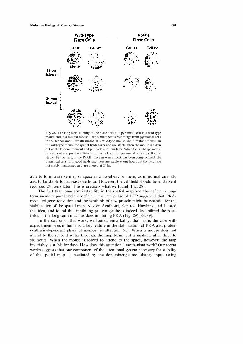
Molecular Biology of Memory Storage 601
Fig. 28. The long-term stability of the place field of a pyramidal cell in a wild-typemouse and in a mutant mouse. Two simultaneous recordings from pyramidal cellsin the hippocampus are illustrated in a wild-type mouse and a mutant mouse. Inthe wild-type mouse the spatial fields form and are stable when the mouse is takenout of the test environment and put back one hour later. When the wild-type mouseis taken out and put back 24 hr later, the fields of the pyramidal cells are still quitestable. By contrast, in the R(AB) mice in which PKA has been compromised, thepyramidal cells form good fields and these are stable at one hour, but the fields arenot stably maintained and are altered at 24 hr.
able to form a stable map of space in a novel environment, as in normal animals,and to be stable for at least one hour. However, the cell field should be unstable ifrecorded 24 hours later. This is precisely what we found (Fig. 28).
The fact that long-term instability in the spatial map and the deficit in long-term memory paralleled the deficit in the late phase of LTP suggested that PKA-mediated gene activation and the synthesis of new protein might be essential for thestabilization of the spatial map. Naveen Agnihotri, Kentros, Hawkins, and I testedthis idea, and found that inhibiting protein synthesis indeed destabilized the placefields in the long-term much as does inhibiting PKA (Fig. 29) [88, 89].
In the course of this work, we found, remarkably, that, as is the case withexplicit memories in humans, a key feature in the stabilization of PKA and proteinsynthesis-dependent phase of memory is attention [90]. When a mouse does notattend to the space it walks through, the map forms but is unstable after three tosix hours. When the mouse is forced to attend to the space, however, the mapinvariably is stable for days. How does this attentional mechanism work? Our recentworks suggests that one component of the attentional system necessary for stabilityof the spatial maps is mediated by the dopaminergic modulatory input acting

602 Kandel
Fig. 29. The group data for mice compromised in PKA,R(AB), and those in which protein synthesis is inhibited. Thetop graph shows the group data for R(AB) and wild-typemice and the bottom shows that these phenotypes are alsoobtained by inhibiting protein synthesis with anisomycin.[From 88, 89.]
through a D1�D5 receptor that activates adenylyl cyclase, cAMP, and PKA. Theactions of dopamine and other modulatory systems might, among other things,trigger the protein synthesis-dependent steps that stabilize the map.
THERE ARE ALSO INHIBITORY CONSTRAINTS ON LTP ANDEXPLICIT MEMORY STORAGE
Recently our laboratory [92] and Emmanuel Landau [116] and his colleagues havefound that the threshold for hippocampal synaptic plasticity and memory storage isdetermined by the balance between protein phosphorylation and dephosphorylation.This balance importantly involves PKA and the Ca2C-sensitive phosphatase, cal-cineurin, the initial step in a phosphatase cascade [91, 92]. To determine whetherendogenous calcineurin acts as a constraint on this balance, we inhibited calcineurinand examined the effects on synaptic plasticity and memory storage. Using the CaMkinase promoter and the doxycycline-dependent rtTA system that Mark Mayford hadfirst successfully applied to the brain, Isabelle Mansuy, Gael Malleret, Danny Winder,

Molecular Biology of Memory Storage 603
Fig. 30. Long-term potentiation requires regulation not only of kinases but also of phos-phatases. The phosphatase cascade initiated by calcineurin shuts off a phosphatase inhibitorand thereby disinhibits the protein phosphatase, which can now inhibit the kinase cascade.[Based on 92.]
Tim Bliss, and I found that a transient reduction of calcineurin activity resulted infacilitation of LTP both in ûitro and in ûiûo (Fig. 30) [92]. This facilitation persistedover several days in the intact animal, and was accompanied by enhanced learningand strengthening of short- and long-term memory on several spatial and non-spatialtasks requiring the hippocampus. The LTP and memory improvements werereversed fully by suppressing expression of the transgene through withdrawal ofdoxycycline. These results, together with previous findings by Winder and Mansuyshowing that overexpression of calcineurin impairs PKA-dependent components ofLTP and memory [83, 91], demonstrate that endogenous calcineurin can act as anegative regulator of synaptic plasticity, learning, and memory (Fig. 30).
AN OVERALL VIEW
The problem of memory storage is conveniently divided into two components:(1) the storage component of memory, the molecular mechanism whereby infor-mation is stored, and (2) the systems component of memory, the mechanisms wherebythe storage sites at each point in the explicit or implicit neural system that mediatesmemory interact to encode, store, and recall memory. I here have addressed primar-ily the storage component of memory. Our studies of this component have led totwo general conclusions.

604 Kandel
First, the research I have reviewed suggests that aspects of the storage mechan-isms—the cellular and molecular strategies used in Aplysia for storing short- andlong-term memory—are conserved from mollusk to mammals, and that the samemolecular strategies are employed in both implicit and explicit memory storage.With both implicit and explicit memory there are stages in memory that are encodedas changes in synaptic strength and that correlate with the behavioral phases ofshort- and long-term memory. The short-term synaptic changes involve covalentmodification of pre-existing proteins, leading to modification of pre-existing synapticconnections, whereas the long-term synaptic changes involve activation of geneexpression, new protein synthesis, and the formation of new connections. Whereasshort-term memory storage uses different signaling kinases for implicit and explicitmemory, long-term storage of both implicit and explicit memory uses as a coresignaling sequence PKA, MAPK, and CREB-1. We presume that this representsonly the core signaling pathway, and that at least in the mouse, additional compo-nents are recruited.
Finally, in both implicit and explicit memory the switch from short-term tolong-term memory is regulated by a number of inhibitory constraints. One suchconstraint, the balance between protein phosphorylation and dephosphorylation,determines the threshold for hippocampal synaptic plasticity and hippocampal-dependent explicit memory storage. Removing this constraint lowers the thresholdof late LTP and enhances storage of explicit memory.
Second, in addition to providing new insights into the molecular mechanismsof learning and memory storage, the molecular biological study of learning hasrevealed new features of synaptic transmission and new cell biological functions ofsynaptic signaling. For example, we have learned that modulatory transmitters ofthe brain serve as reinforcing stimuli important for synaptic plasticity related tolearning and memory storage. Different forms of learning recruit these modulatorytransmitters which can then act in one of three ways: (1) they activate second-messenger kinases that are transported to the nucleus where they initiate processesrequired for neuronal growth and long-term memory; (2) they mark the specificsynapses for capture of the long-term process and regulate local protein synthesisfor stabilization; and (3) modulatory transmitters appear to mediate, in ways we arejust beginning to understand, attentional processes required for memory formationand recall.
Most important, the study of long-term memory has made us aware of theextensive dialog between the synapse and the nucleus and the nucleus and the syn-apse (Fig. 24). The long-range interactions between the nucleus and synapse intro-duce a new level of non-Sherringtonian integration into neuronal functioning forlong-term synaptic plasticity that is different from that used by the short-term pro-cess. In the long-term process the response of a synapse is not simply determinedby its own history of activity. It is also significantly determined by the history oftranscriptional activation in the nucleus.
I started this essay by pointing out that 40 years ago, at the beginning of mycareer, I thought that a reductionist approach based on the use of a simple experi-mental system such as Aplysia might allow us to address fundamental questions inthe study of learning and memory. That was a leap of faith for which I have been

Molecular Biology of Memory Storage 605
rewarded beyond my fondest hopes. Still, the complexity of explicit memory is for-midable, and we have only begun to explore it. We as yet know little about themolecular mechanisms that initiate or stabilize the synaptic growth associated withlong-term memory. What are the signaling molecules that lead to the cytoskeletalrearrangements during synaptic remodeling? How do they relate to the moleculesinvolved with synapse formation during development? In addition, we have hereonly considered the molecular mechanisms of memory storage—mechanisms thatappeared to be shared, at least in part, by both explicit (declarative) and implicit(procedural) memory. But the storage mechanism is only one part of the memoryproblem. The more difficult part of memory—especially explicit memory—is thesystems problem of memory. We still need to seek answers to a family of importantquestions. How do different regions of the hippocampus and the medial temporallobe—the subiculum, the entorhinal, parahippocampal and perirhinal cortices–inter-act in the storage of explicit memory? How is information in any of these regionstransferred for ultimate consolidation in the neocortex? We do not, for example,understand why the initial storage of long-term memory requires the hippocampus,whereas the hippocampus is not required once a memory has been stored for weeksor months [reviewed in 95, 96]. What critical information does the hippocampusconvey to the neocortex?
We also know very little about the nature of recall of explicit (declarative)memory, a recall that requires conscious effort. These systems problems of the brainwill require more than the bottoms-up approach of molecular biology. They willalso require the top-down approaches of cognitive psychology, neurology, and psy-chiatry. Finally, they will require a set of syntheses that bridge the two approaches.
Despite these complexities, we have every reason to believe that these and otherquestions in the biology of learning will be vigorously addressed in the near future.For the biology of the mind has now captured the imagination of the scientificcommunity of the 21st century, much as the biology of the gene fascinated thescientific community of the 20th century. As the biological study of the mind comesto assume the central position within biology and medicine that it deserves, we haveevery reason to expect that a succession of brain scientists will be called to Stock-holm and honored for their own leaps of faith.
ACKNOWLEDGMENTS
I have had the privilege to work with and to learn from many gifted students,fellows, and collaborators, and I have tried throughout this lecture to acknowledgetheir contributions. But beyond specific collaborators, my science has benefited enor-mously from the interactive environment created by the Center for Neurobiologyand Behavior at the College of Physicians and Surgeons of Columbia University. Itwould be hard to find a more ideal environment in which to mature as a scientist.Specifically, I have benefited greatly from my long-standing friendship with RichardAxel, Craig Bailey, Jane Dodd, Robert Hawkins, John Koester, Thomas Jessell,James H. Schwartz, Steven Siegelbaum, and Gerald Fischbach, the current Dean ofthe College of Physicians and Surgeons. I am further grateful to John Koester forhis excellent leadership of the Center for Neurobiology and Behavior, and to David

606 Kandel
Hirsh, Sam Silverstein, and John Oldham, chairs of the three departments to whichI belong. Finally, I am indebted to Herbert Pardes who, until recently, served asDean of the College of Physicians and Surgeons.
My research has been generously supported by the Howard Hughes MedicalInstitute, the NIH, the Mathers Foundation, FRAXA, and The Lieber Trust. I amparticularly indebted to the Howard Hughes Medical Institute and its leadership,Donald Fredrickson, George Cahill, Purnell Chopin, Max Cowan, Donald Harter,and more recently Tom Cech and Gerry Rubin, whose farsighted vision has encour-aged Hughes investigators to take a long-term perspective so as to be able to tacklechallenging problems. Research on learning and memory certainly meets both ofthese criteria!
REFERENCES
1. Kandel, E. R. and Spencer, W. A. (1961) Electrophysiological properties of an archicortical neuron.In Problems in Electrobiology, Ann. N.Y. Acad. Sci. 94:570–603.
2. Thorpe, W. H. (1963) Learning and Instinct in Animals. Revised Edition. Harvard University Press:Cambridge, Mass.
3. Frazier, W. T., Kandel, E. R., Kupfermann, I., Waziri, R., and Coggeshall, R. E. (1967) Morphologi-cal and functional properties of identified neurons in the abdominal ganglion of Aplysia californica.J. Neurophysiol. 30:1288–1351.
4. Kupfermann, I. And Kandel, E. R. (1969) Neuronal controls of a behavioral response mediated bythe abdominal ganglion of Aplysia. Science 164:847–850.
5. Pinsker, H., Kupfermann, I., Castellucci, V., and Kandel, E. R. (1970) Habituation and dishabitu-ation of the gill-withdrawal reflex in Aplysia. Science 167:1740–1742.
6. Kupfermann, I., Castellucci, V., Pinsker, H., and Kandel, E. R. (1970) Neuronal correlates of habitu-ation and dishabituation of the gill-withdrawal reflex in Aplysia. Science 167:1743–1745.
7. Castellucci, V., Pinsker, H., Kupfermann, I., and Kandel, E. R. (1970) Neuronal mechanisms ofhabituation and dishabituation of the gill-withdrawal reflex in Aplysia. Science 167:1745–1748.
8. Pinsker, H. M., Hening, W. A., Carew, T. J., and Kandel, E. R. (1973) Long-term sensitization of adefensive withdrawal reflex in Aplysia. Science 182:1039–1042.
9. Carew, T. J., Pinsker, H. M., and Kandel, E. R. (1972) Long-term habituation of a defensive with-drawal reflex in Aplysia. Science 175:451–454.
10. Frost, W. N., Castellucci, V. F., Hawkins, R. D., and Kandel, E. R. (1985) Monosynaptic connec-tions made by the sensory neurons of the gill- and siphon-withdrawal reflex in Aplysia participate inthe storage of long-term memory for sensitization. Proc. Natl. Acad. Sci. USA 82:8266–8269.
11. Byrne, J., Castellucci, V., and Kandel, E. R. (1974) Receptive fields and response properties of mech-anoreceptor neurons innervating skin and mantle shelf of Aplysia. J. Neurophysiol. 37:1041–1064.
12. Castellucci, V. F., Blumenfeld, H., Goelet, P., and Kandel, E. R. (1989) Inhibitor of protein synthesisblocks long-term behavioral sensitization in the isolated gill-withdrawal reflex of Aplysia. J. Neuro-biol. 20:1–9.
13. Kandel, E. R. (1976) Cellular Basis of Behaûior: An Introduction to Behaûioral Biology. Freeman: SanFrancisco.
14. Kandel, E. R. (1978) A Cell-Biological Approach to Learning. Grass Lecture Monograph I. Society:Bethesda, Md.
15. Kandel, E. R. (1979) The Behaûioral Biology of Aplysia: A Contribution to the Comparatiûe Study ofOpisthobranch Molluscs. Freeman: San Francisco.
16. Cajal, S. R. (1894) La fine structure des centres nerveux. The Croonian Lecture. Proc. R. Soc. Lond.55:444–468.
17. Kandel, E. R. and Spencer, W. A. (1968) Cellular neurophysiological approaches in the study oflearning. Physiol. Reû. 48:65–134.

Molecular Biology of Memory Storage 607
18. Hawkins, R. D., Castellucci, V. F., and Kandel, E. R. (1981) Interneurons involved in mediationand modulation of the gill-withdrawal reflex in Aplysia. I. Identification and characterization. J.Neurophysiol. 45:304–314.
19. Hawkins, R. D., Castellucci, V. F., and Kandel, E. R. (1981) Interneurons involved in mediation andmodulation of the gill-withdrawal reflex in Aplysia. II. Identified neurons produce hetero-synapticfacilitation contributing to behavioral sensitization. J. Neurophysiol. 45:315–326.
20. Carew, T. J., Hawkins, R. D., and Kandel, E. R. (1983) Differential classical conditioning of adefensive withdrawal reflex in Aplysia californica. Science 219:397–400.
21. Hawkins, R. D., Abrams, T. W., Carew, T. J., and Kandel, E. R. (1983) A cellular mechanism ofclassical conditioning in Aplysia: Activity-dependent amplification of presynaptic facilitation. Science219:400–405.
22. Byrne, J. H., Castellucci, V. F., and Kandel, E. R. (1978) Contribution of individual mechanoreceptorsensory to defensive gill-withdrawal reflex in Aplysia. J. Neurophysiol. 41:418–431.
23. Carew, T. J., Castellucci, V. F., and Kandel, E. R. (1979) Sensitization in Aplysia: Restoration oftransmission in synapses inactivated by long-term habituation. Science 205:417–419.
24. Klein, M. and Kandel, E. R. (1980) Mechanism of calcium current modulation underlying presynap-tic facilitation and behavioral sensitization in Aplysia. Proc. Natl. Acad. Sci. USA 77:6912–6916.
25. Klein, M., Hochner, B., and Kandel, E. R. (1986) Facilitatory transmitters and cAMP can modulateaccommodation as well as transmitter release in Aplysia sensory neurons. Evidence for parallel pro-cessing in a single cell. Proc. Natl. Acad. Sci. USA 83:7994–7998.
26. Schwartz, J. H., Castellucci, V. F., and Kandel, E. R. (1971) Functioning of identified neurons andsynapses in abdominal ganglion of Aplysia in absence of protein synthesis. J. Neurophysiol. 34:939–953.
27. Cedar, H., Kandel, E. R., and Schwartz, J. H. (1972) Cyclic adenosine monophosphate in the nervoussystem of Aplysia californica. I. Increased synthesis in response to synaptic stimulation. J. Gen.Physiol. 60:558–569.
28. Kriegstein, A. R., Castellucci, V., and Kandel, E. R. (1974) Metamorphosis of Aplysia californica inlaboratory culture. Proc. Natl. Acad. Sci. USA 71:3654–3658.
29. Cedar, H. and Schwartz, J. H. (1972) Cyclic adenosine monophosphate in the nervous system ofAplysia california. II. Effect of serotonin and dopamine. J. Gen. Physiol. 60:570–587.
30. Castellucci, V. and Kandel, E. R. (1974) A quantal analysis of the synaptic depression underlyinghabituation of the gill-withdrawal reflex in Aplysia. Proc. Natl. Acad. Sci. USA 71:5004–5008.
31. Castellucci, V. and Kandel, E. R. (1976) Presynaptic facilitation as a mechanism for behavioralsensitization in Aplysia. Science 194:1176–1178.
32. Brunelli, M., Castellucci, V., and Kandel, E. R. (1976) Synaptic facilitation and behavioral sensitis-ation in Aplysia: Possible role of serotonin and cyclic AMP. Science 194:1178–1181.
33. Kandel, E. R., Brunelli, M., Byrne, J., and Castellucci, V. (1976) A common presynaptic locus forthe synaptic changes underlying short-term habituation and sensitization of the gill-withdrawal reflexin Aplysia. Cold Spring Harbor Symp. Quant. Biol. 40:465–482.
34. Glanzman, D. L., Mackey, S. L., Hawkins, R. D., Dyke, A. M., Lloyd, P. E., and Kandel, E. R.(1989) Depletion of serotonin in the nervous system of Aplysia reduces the behavioral enhancementof gill withdrawal as well as the heterosynaptic facilitation produces by tail shock. J. Neurosci.9:4200–4213.
35. Mackay, S. L., Kandel, E. R., and Hawkins, R. D. (1989) Identified serotonergic neurons LCB1 andRCB1 in the cerebral ganglia of Aplysia produce presynaptic facilitation of siphon sensory neurons.J. Neurosci. 9:4227–4235.
36. Byrne, J. H., Castellucci, V. F., Carew, T. J., and Kandel, E. R. (1978) Stimulus-response relationsand stability of mechanoreceptor and motor neurons mediating defensive gill-withdrawal reflex inAplysia. J. Neurophysiol. 41:402–417.
37. Castellucci, V. F., Kandel, E. R., Schwartz, J. H., Wilson, F. D., Nairn, A. C., and Greengard, P.(1980) Intracellular injection of the catalytic subunit of cyclic AMP-dependent protein kinase simu-lates facilitation of transmitter release underlying behavioral sensitization in Aplysia. Proc. Natl.Acad. Sci. USA 77:7492–7496.
38. Castellucci, V. F., Nairn, A., Greengard, P., Schwartz, J. H., and Kandel, E. R. (1982) Inhibitor ofadenosine 3′:5′-monophosphate-dependent protein kinase blocks presynaptic facilitation in Aplysia.J. Neurosci. 2:1673–1681.

608 Kandel
39. Siegelbaum, S., Camardo, J. S., and Kandel, E. R. (1982) Serotonin and cAMP close single K+
channels in Aplysia sensory neurons. Nature 299:413–417.40. Shuster, M. J., Camardo, J. S., Siegelbaum, S. A., and Kandel, E. R. (1985) Cyclic AMP-dependent
protein kinase closes the serotonin-sensitive K+ channels of Aplysia sensory neurons in cell-free mem-brane patches. Nature 313:392–395.
41. Byrne, J. H. and Kandel, E. R. (1995) Presynaptic facilitation revisited: State and time dependence.J. Neurosci. 16:425–435.
42. Rayport, S. G. and Schacher, S. (1986) Synaptic plasticity in vitro: Cell culture of identified Aplysianeurons mediating short-term habituation and sensitization. J. Neurosci. 6:759–763.
43. Montarolo, P. G., Goelet, P., Castellucci, V. F., Morgan, J., Kandel, E. R., and Schacher, S. (1986)A critical period for macromolecular synthesis in long-term heterosynaptic facilitation in Aplysia.Science 234:1249–1254.
44. Schacher, S., Castellucci, V. F., and Kandel, E. R. (1988) cAMP evokes long-term facilitation inAplysia sensory neurons that requires new protein synthesis. Science 240:1667–1669.
45. Dash, P. K., Hochner, B., and Kandel, E. R. (1990) Injection of cAMP-responsive element into thenucleus of Aplysia sensory neurons blocks long-term facilitation. Nature 345:718–721.
46. Bacskai, B. J. et al. (1993) Spatially resolved dynamics of cAMP and protein kinase A subunits inAplysia sensory neurons. Science 260:222–226.
47. Martin, K. C. et al. (1997) MAP kinase translocates into the nucleus of the presynaptic cell and isrequired for long-term facilitation in Aplysia. Neuron 18:899–912.
48. Bailey, C. H. et al. (1997) Mutation in the phosphorylation sites of MAP kinase blocks learning-related internalization of apCAM in Aplysia sensory neurons. Neuron 18:913–924.
49. Kaang, B.-K., Kandel, E. R., and Grant, S. G. N. (1993) Activation of cAMP-responsive genes bystimuli that produce long-term facilitation in Aplysia sensory neurons. Neuron 10:427–435.
50. Glanzman, D. L., Kandel, E. R., and Schacher, S. (1990) Target-dependent structural changesaccompanying long-term synaptic facilitation in Aplysia neurons. Science 249:799–802.
51. Dale, N. and Kandel, E. R. (1993) L-glutamate may be the fast excitatory transmitter of Aplysiasensory neurons. Proc. Natl. Acad. Sci. USA 90:7163–7167.
52. Alberini, C., Ghirardi, M., Metz, R., and Kandel, E. R. (1994) C�EBP is an immediate-early generequired for the consolidation of long-term facilitation in Aplysia. Cell 76:1099–1114.
53. Bartsch, D., et al. (1995) Aplysia CREB2 represses long-term facilitation: Relief of repression convertstransient facilitation into long-term functional and structural change. Cell 83:979–992.
54. Bartsch, D. Casadio, A., Karl, K. A., Serodio, P., and Kandel, E. R. (1998) CREB1 encodes anuclear activator, a repressor, and a cytoplasmic modulator that form a regulatory unit critical forlong-term facilitation. Cell 95:211–223.
55. Bartsch, D. et al. (2000) Enhancement of memory-related long-term facilitation by ApAF, a noveltranscription factor that acts downstream from both CREB1 and CREB2. Cell 103:595–608.
56. Bailey, C. H., Montarolo, P., Chen, M., Kandel, E. R., and Schacher, S. (1992) Inhibitors of proteinand RNA synthesis block structural changes that accompany long-term heterosynaptic plasticity inAplysia. Neuron 9:749–758.
57. Bailey, C. H. and Chen, M. (1988) Long-term memory in Aplysia modulates the total number ofvaricosities of single identified sensory neurons. Proc. Natl. Acad. Sci. USA 85:2373–2377.
58. Bailey, C. H. and Chen, M. (1989) Time course of structural changes at identified sensory neuronsynapses during long-term sensitization in Aplysia. J. Neurosci. 9:1774–1780.
59. Bailey, C. H. and Kandel, E. R. (1993) Structural changes accompanying memory storage. Annu.Reû. Physiol. 55:397–426.
60. Hedge, A. N. et al. (1997) Ubiquitin C-terminal hydrolase is an immediate-early gene essential forlong-term facilitation in Aplysia. Cell 89:115–126.
61. Chain, D. G. et al. (1999) Mechanisms for generating the autonomous cAMP-dependent proteinkinase required for long-term facilitation in Aplysia. Neuron 22:147–156.
62. Mayford, M., Barzilai, A., Keller, F., Schacher, S., and Kandel, E. R. (1992) Modulation of anNCAM-related adhesion molecule with long-term synaptic plasticity in Aplysia. Science 256:638–644.
63. Bailey, C. H., Chen, M., Keller, F., and Kandel, E. R. (1992) Serotonin-mediated endocytosis ofapCAM: An early step of learning-related synaptic growth in Aplysia. Science 256:645–649.
Molecular Biology of Memory Storage 609
64. Martin, K. C. et al. (1998) Synapse-specific transcription-dependent long-term facilitation of thesensory to motor neuron connection in Aplysia: A function for local protein synthesis in memorystorage. Cell 91:927–938.
65. Casadio, A. et al. (1999) A transient neuron-wide form of CREB-mediated long-term facilitation canbe stabilized at specific synapses by local protein synthesis. Cell 99:221–237.
66. Bacskai, B. J. et al. (1993) Spatially resolved dynamics of cAMP and protein kinase A subunits inAplysia sensory neurons. Science 260:222–226.
67. Grant, S. G. N., O’Dell, T. J., Karl, K. A., Stein, P. L., Soriano, P., and Kandel, E. R. (1992)Impaired long-term potentiation, spatial learning, and hippocampal development in fyn mutant mice.Science 258:1903–1910.
68. Squire, L. R. and Kandel, E. R. (1999) Memory: Mind to Molecules. Scientific American Books: NewYork.
69. Bliss, T. V. and Lømo, T. (1973) Long-lasting potentiation of synaptic transmission in the denatearea of the anaesthetized rabbit following stimulation of the perforant path. J. Physiol. 232:331–356.
70. Morris, R. G. M., Anderson, E., Lynch, G. S., and Baudry, M. (1986) Selective impairment oflearning and blockade of long-term potentiation by an N-methyl-D-aspartate receptor antagonist,AP5. Nature 319:774–776.
71. Frey, U., Huang, Y.-Y., and Kandel, E. R. (1993) Effects of cAMP simulate a late stage of LTP inhippocampal CA1 neurons. Science 260:1661–1664.
72. Huang, Y.-Y. and Kandel, E. R. (1994) Recruitment of long-lasting and protein kinase A-dependentlong-term potentiation in the CA1 region of hippocampus requires repeated tetanization. Learning &Memory 1:74–82.
73. Nguyen, P. V., Abel, T., and Kandel, E. R. (1994) Requirement of a critical period of transcriptionfor induction of a late phase of LTP. Science 265:1104–1107.
74. Bolshakov, V. Y., Golan, H., Kandel, E. R., and Siegelbaum, S. A. (1997) Recruitment of new sitesof synaptic transmission during the cAMP-dependent late phase of LTP at CA3-CA1 synapses in thehippocampus. Neuron 19:635–651.
75. Malenka, R. C. and Nicoll, R. A. (1997) Silent synapses speak up. Neuron 19:473–476.76. Ma, L., Zablow, L., Kandel, E. R., and Siegelbaum, S. A. (1999) Cyclic AMP induces functional
presynaptic boutons in hippocampal CA3-CA1 neuronal cultures. Nature Neurosci. 2:24–3.77. Bourtchouladze, R., Franguelli, B., Blendy, J., Cioffi, D., Schutz, G., and Silva, A. J. (1994) Deficient
long-term memory in mice with a targeted mutation of the cAMP-responsive element-binding protein.Cell 79:59–68.
78. Impey, S. et al. (1998) Cross talk between ERK and PKA is required for Ca2C stimulation of CEB-dependent transcription and ERK nuclear translocation. Neuron 21:869–883.
79. Huang, Y.-Y. and Kandel, E. R. (1995) D1�D5 receptor agonists induce a protein synthesis-depen-dent late potentiation in the CA1 region of the hippocampus. Proc. Natl. Acad. Sci. USA 92:2446–2450.
80. Muller, D. (1997) Ultrastructural plasticity of excitatory synapses. Reû. Neurosci. 8:77–93.81. English, J. D. and Sweat, J. D. (1996) Activation of p42 mitogen-activated protein kinase in hippo-
campal long term potentiation. J. Biol. Chem. 271:24329–24332.82. Engert, F. and Bonhoeffer, T. (1999) Dendritic spine changes associated with hippocampal long-term
synaptic plasticity. Nature 399:66–70.83. Winder, D. G., Mansuy, I. M., Osman, M., Moallem, T. M., and Kandel, E. R. (1998) Genetic and
pharmacological evidence for a novel, intermediate phase of long-term potentiation (I-LTP) sup-pressed by calcineurin. Cell 92:25–37.
84. Abel, T. et al. (1997) Genetic demonstration of a role for PKA in the last phase of LTP and inhippocampus-based long-term memory. Cell 88:615–626.
85. O’Keefe, J. and Nadel, L. (1978) The Hippocampus as a Cognitiûe Map. Clarendon Press: Oxford.86. Rotenberg, A., Mayford, M., Hawkins, R. D., Kandel, E. R., and Muller, R. U. (1996) Mice express-
ing activated CaMKII lack low frequency LTP and do not form stable place cells in the CA1 regionof the hippocampus. Cell 87:1351–1361.
87. Kentros, C., Hargreaves, E., Hawkins, R. D., Kandel, E. R., Shapiro, M., and Muller, R. U. (1998)Abolition of long-term stability of new hippocampal place cell maps by NMDA receptor blockade.Science 280:2121–2126.
610 Kandel
88. Rotenberg, A., Abel, T., Hawkins, R. D., Kandel, E. R., and Muller, R. U. (2000) Parallel instabilitiesof long-term potentiation, place cells, and learning causes by decreased protein kinase A activity. J.Neurosci. 20:8096–8102.
89. Agnihotri, N., Hawkins, R. D., Kandel, E. R., and Kentros, C. (2001) Protein synthesis inhibitionselectively abolishes long-term stability of hippocampal place cell maps. Soc. Neurosci. Abstr., inpress.
90. Kentros, C., Agnihotri, N., Hawkins, R., Muller, R., and Kandel, E. (2001) Stabilization of thehippocampal representation of space in mice requires attention. Soc. Neurosci. Abstr., in press.
91. Mansuy, I. M., Mayford, M., Jacob, B., Kandel, E. R., and Bach, M. E. (1990) Restricted andregulated overexpression reveals calcineurin as a key component in the transition from short-term tolong-term memory. Cell 92:39–49.
92. Mallaret, G. et al. (2001) Inducible and reversible enhancement of learning, memory, and long-termpotentiation by genetic inhibition of calcineurin. Cell 104:675–686.
93. Frey, U. and Morris, R. G. (1997) Synaptic tagging and long-term potentiation. Nature 385:533–536.
94. Castellucci, V. F., Carew, T. J., and Kandel, E. R. (1978) Cellular analysis of long-term habituationof the gill-withdrawal reflex in Aplysia. Science 202:1306–1308.
95. Milner, B., Squire, L. R., and Kandel, E. R. (1998) Cognitive neuroscience and the study of memory.Neuron 20:445–468.
96. Squire, L. R. and Zola-Morgan, S. (1991) The medial temporal lobe memory system. Science253:1380–1386.
97. Dubnau, J. and Tully, T. (1998) Gene discovery in Drosophila: New insights for learning and mem-ory. Annu. Reû. Neurosci. 21:407–444.
98. Frost, W. N., Castellucci, V. F., Hawkins, R. D., and Kandel, E. R. (1985) Monosynaptic connec-tions made by the sensory neurons of the gill- and siphon-withdrawal reflex in Aplysia participate inthe storage of long-term memory for sensitization. Proc. Natl. Acad. Sci. USA 82:8266–8269.
99. Martin, K. C., Barad, M., and Kandel, E. R. (2000) Local protein synthesis and its role in synapse-specific plasticity. Curr. Opin. Neurobiol. 10:587–592.
100. Schuman, E. M. (1997) Synapse specificity and long-term information storage. Neuron 18:339–342.101. Steward, O. (1997) mRNA localization in neurons: A multipurpose mechanism? Neuron 18:9–12.102. Weiner, J. (1999) Time, Loûe, Memory: A Great Biologist and His Quest for the Origins of Behaûior.
Knopf: New York.103. Muller, R. (1996) A quarter of a century of place cells. Neuron 17:813–822.104. McHugh, T. J., Blum, K. I., Tsein, J. Z., Tonegawa, S., and Wilson, M. A. (1996) Impaired hippo-
campal representation of space in CA1 specific NMDAR1 knockout mice. Cell 87:1339–1349.105. Bolshakov, V. Y. and Siegelbaum, S. A. (1995) Regulation of hippocampal transmitter release dur-
ing development and long-term potentiation. Science 269:1730–1734.106. Stevens, C. F. and Wang, Y. (1994) Changes in reliability of synaptic function as a mechanism for
plasticity. Nature 371:704–707.107. Ebbinghaus, H. (1885) Memory: A Contribution to Experimental Psychology (1885). Reprinted,
Dover: New York, 1963.108. Flexner, L. B. and Flexner, J. B. (1966) Effect of acetoxycyclohexamide and of an acetoxycyclohex-
amide-purymycin mixture on cerebral protein synthesis and memory in mice. Proc. Natl. Acad. Sci.USA 55:369–374.
109. Brink, J. J., Davis, R. E., and Agranoff, B. W. (1966) Effects of puromycin, acetoxycyclohexamideand actinomycin D on protein synthesis in goldfish brain. J. Neurochem. 13:889–896.
110. Fatt, P. and Katz, B. (1951) An analysis of the end-plate potential recorded with an intracellularelectrode. J. Physiol. (Lond.) 115:320–370.
111. Greengard, P. (1976) Possible role for cyclic nucleotides and phosphorylated membrane proteins inpostsynaptic actions of neurotransmitters. Nature 260:101–108.
112. Monsour, S. L., Thomas, K. R., and Capecchi, M. R. (1988) Disruption of the proto-oncogeneint-2 in mouse embryonic-derived stem cells: A general strategy for targeting mutations to non-selectible genes. Nature 336:348–352.
113. Reid, L. H., Shesely, E. G., Kin, H. S., and Smithies, O. (1991) Cotransformation and gene targetingin mouse embryonic stem cells. Mol. Cell Biol. 11:2769–2777.

Molecular Biology of Memory Storage 611
114. Koller, B. H. and Smithies, O. (1989) Inactivating the beta 2-microglobulin locus in mouse embry-onic stem cells by homoglous recombination. Proc. Natl. Acad. Sci. USA 86:8932–8935.
115. Kandel, E. R. and Schwartz, J. H. (1982) Molecular biology of learning: Modulation of transmitterrelease. Science 218:433–443.
116. Blitzer, R. D. et al. (1998) Gating of CaMKII by cAMP-regulated protein phosphatase activityduring LTP. Science 280:1940–1942.