The Mini-Crush Technique for the Treatment of Bifurcation … · The Mini-Crush Technique for the...
62
The Mini-Crush Technique for the Treatment of Bifurcation and Trifurcation Coronary Lesions Alfredo R. Galassi, MD, FACC, FSCAI, FESC Associate Professor of Cardiology Head of the Catetherization Interventional Laboratory Clinical Division of Cardiology, Ferrarotto Hospital University of Catania, Italy Angioplasty Summit TCT Asia Pacific Seoul, April 25-27, 2007
Transcript of The Mini-Crush Technique for the Treatment of Bifurcation … · The Mini-Crush Technique for the...

The Mini-Crush Techniquefor the Treatment of Bifurcation and
Trifurcation Coronary Lesions Alfredo R. Galassi, MD, FACC, FSCAI, FESC
Associate Professor of CardiologyHead of the Catetherization Interventional Laboratory
Clinical Division of Cardiology, Ferrarotto Hospital University of Catania, Italy
Angioplasty SummitTCT Asia Pacific
Seoul, April 25-27, 2007

Treatment of coronary bifurcation lesions remains anissue in terms of procedural success, MACE, TLR, restenosis and stent thrombosis
Optimal technique with DES (1 stent vs 2 stents, type of technique) is still a debate
Randomized studies are scarce, not homogeneous and executed on a small scale
Meta-analysis of these etherogeneous reports haveproven quite impossible
Coronary Bifurcation Lesions

DEFINITELY ….YES!!!! ……if the side branch is a large vesselif the side branch comes out from the main with an acute angleif the ostium or the proximal segment of the side branch have a
significant narrowingif the side branch is very difficult to be wiredif the patient is a very high risk patient and the side branch appears
relatively importantif the main branch is severely narrowed with a lot of plaque burden
… sometimes a decision should be made only following sometimes a decision should be made only following predilatationpredilatation of the main branch and of the side branchof the main branch and of the side branch!
Does The Side BranchNeed Protection by a Stent?

Treatments Are Not Equivalent
T stentingT stenting CoulottePTSPTS Crush
Technique can be divided into 2 strategies:
Simple Complex
ProvisionalT Stenting

Main Branch Side Branch0.6
0.4
0.2
0
10
20
30
40
15.5% (9/58)
8.9% (8/90)
37.9% (22/58)
11.1% (10/90)
0.21
0.47
P = 0.10 P < 0.05
P = 0.33 P < 0.001Re
sten
osis R
ate
(%)
Late
Lum
en L
oss
(mm)
0.32
0.52
No final No final KissingKissing
YesYes final final KissingKissing
Result withCrush stenting
according toperformance of final kiss:
restenosis and late loss are significantlyreduced for
the side branch
Ge et al. JACC 2005
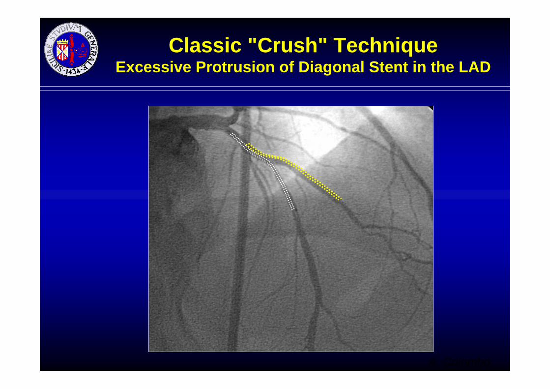
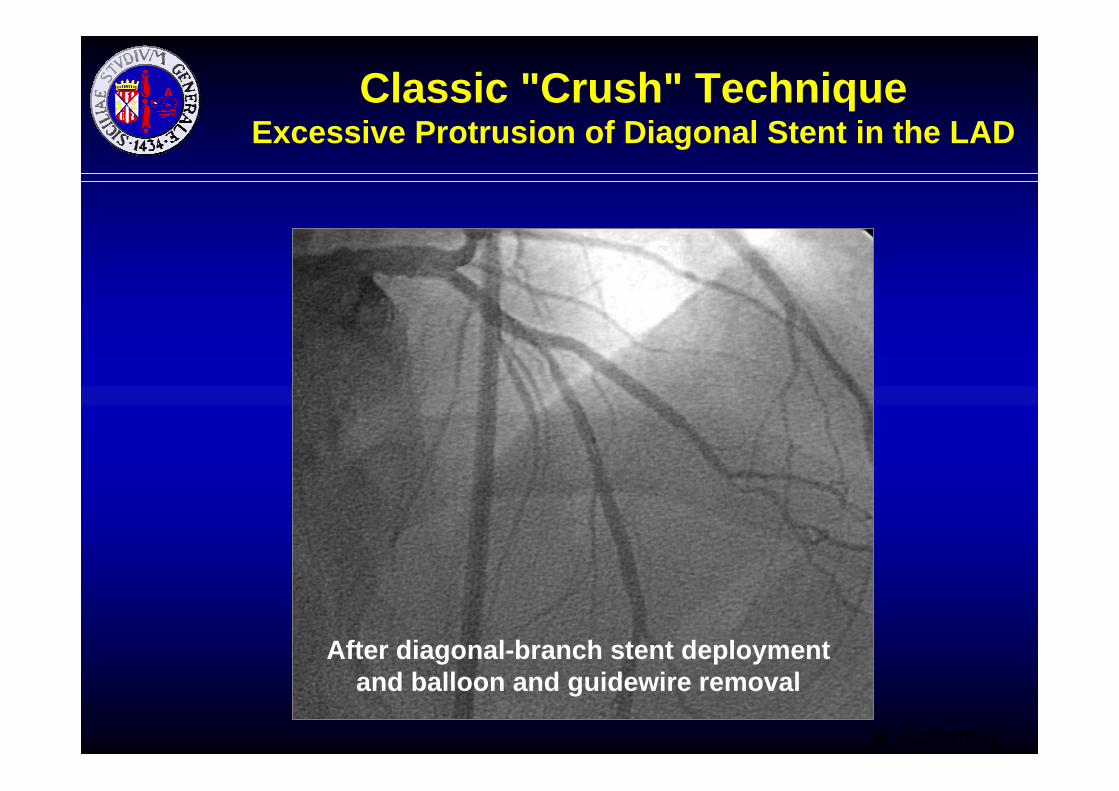
Classic "Crush" Technique Excessive Protrusion of Diagonal Stent in the LAD
A. Colombo

LADLADLAD
DiagonalDiagonalDiagonal
"Crush" Technique Excessive Protrusion of Diagonal Stent in the LAD
A. Colombo
44--5 mm5 mm

After diagonal-branch stent deploymentand balloon and guidewire removal
Classic “Crush" Technique Excessive Protrusion of Diagonal Stent in the LAD
A. Colombo
Classic "Crush" Technique Excessive Protrusion of Diagonal Stent in the LAD
After diagonal-branch stent deploymentand balloon and guidewire removal
A. Colombo

After LAD stent deployment and crushingof the diagonal-branch stent
Classical "Crush" Technique Excessive Protrusion of Diagonal Stent in the LAD
A. Colombo

"Crush" Technique Excessive Protrusion of Diagonal Stent in the LAD may cause more blood flow stagnation between the struts
DiagonalDiagonalDiagonal
LADLADLADAfter stent deployment

Mini-Crush TechniqueBench Work
courtesy of J. Ormiston(Mercy Angiography,
New Zealand)

Mini-Crush Technique Bench WorkCourtesy of J. Ormiston (Mercy Angiography, New Zealand)
30o Degree Model
AppositionApposition
StagnationStagnation
RecirculationRecirculation
DistortionDistortion
FractureFracture

Mini-Crush Technique Bench WorkCourtesy of J. Ormiston (Mercy Angiography, New Zealand)
60o Degree Model
AppositionApposition
StagnationStagnation
RecirculationRecirculation
DistortionDistortion
FractureFracture

Mini-Crush Technique Bench WorkCourtesy of J. Ormiston (Mercy Angiography, New Zealand)
90o Degree Model
AppositionApposition
StagnationStagnation
RecirculationRecirculation
DistortionDistortion
FractureFracture

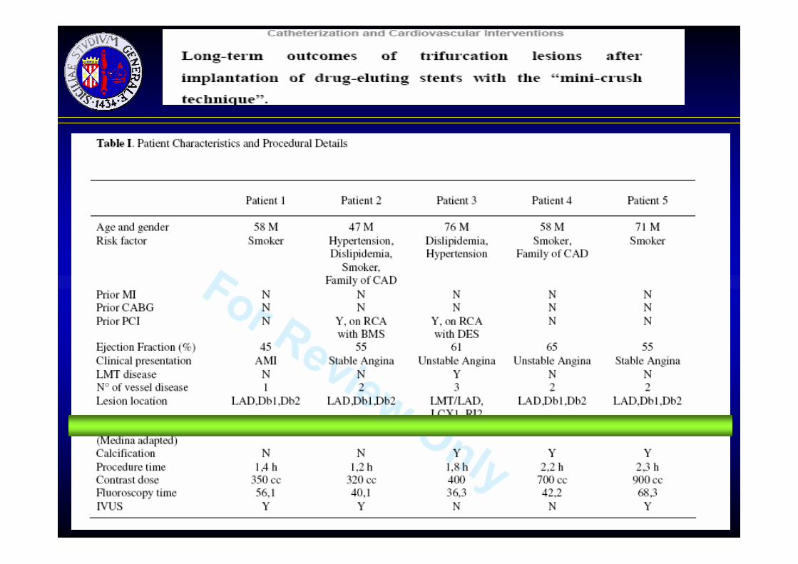
Galassi Galassi etet al.al. CatheterCatheter CardiovascCardiovasc IntervInterv in press 2007in press 2007The “Mini-Crush” Technique

14.2
Galassi Galassi etet al.al. CatheterCatheter CardiovascCardiovasc IntervInterv in press 2007in press 2007The “Mini-Crush” Technique
4.1%

Endothelialization was complete after single or overlapping BMS
Reduced with single layer DES
Further reduced by overlapping DES
Does overlapping predispose to SAT?
Courtesy of J. Ormiston

The ideal bifurcation stent or strategy should not have multiple layers with current DES
Or overlap should be limited eg with “mini-crush”
3 layers Long overlap
3 layers short overlap
Courtesy of J. Ormiston

OCT on LADShort
crushedsegment in the LAD
Courtesy of F. Prati
Post Mini-Crush of a Bifurcation LAD-D1

Post Mini-Crush Bifurcation LAD-D1
Optimized stentstruts openingby the mini-
crush technique
OCT on D1
Courtesy of F. Prati

MBproximal
MBdistall SIDE 1
SIDE 2
Proximal
Distal
0,1
0,1
0,10,1
Medina Classification for CoronaryTrifurcation Lesion (adapted)


Side Branch 1
Side Branch 2
TLR = target lesion revascularization due to restenosis (>50%) intrastent and/or 5 mm proximal and/or 5 mm distal to stent in main or side branch
TTR = target trifurcation revascularization due to restenosis (>50%) within 5 mm proximal or distal tothe carina of bifurcation, both onto the main branch and/or side branch
Trifurcation Definition

4/5 of lesion LAD-DB1-DB2 1/5 of lesion LMT-LAD-LCX-RI
Angiographic Characteristics
N° stents implanted per lesion: 3,4 ± 0,5
TLR non TTR
Final Results8-month Follow-up


Conclusions
• The “mini-crush” technique may be considered a very good refiniment of the crush technique
• Experimental bench work, as well as IVUS and OCT first clinical applications showed excellent results
• The application of this technique in the clinical setting of both bifurcation and trifurcation lesions proved to be very promising
• Now it is the time of a clinical multicenter randomized study!


Clinical Characteristics
2/5 (20%)Family History of CAD
0Previous MI
2/5 (20%)Previous PCI
54±5,8EF (mean±SD)
4/5 (80%)Smokers
0Diabetes
2/5 (20%)Dislipidemia
2/5 (20%)Hypertension
65±11,5Age (mean±SD)
5/5 (100%)Male
Pts=5
One vessel
Two vessel
Three vessel
1 1
3
StableanginaUnstableanginaAMI
2
2
1
N° of vessel disease
Clinical presentation

Is the side branch a large vessel?Does the side branch comes out from the main with an acute angle?Does the ostium or the proximal segment of the side branch have a
significant narrowing?Is the side branch very difficult to be wired?Is the patient a very high risk patient and the side branch appears
relatively important?Is the main branch severely narrowed with a lot of plaque burden?If the answer is YES, suggestion is that the operator will lean If the answer is YES, suggestion is that the operator will lean more more
towards two towards two stentsstents
… sometimes a decision should be made only following sometimes a decision should be made only following predilatationpredilatation of the main branch and of the side branchof the main branch and of the side branch!
Does The Side BranchNeed Protection by a Stent?
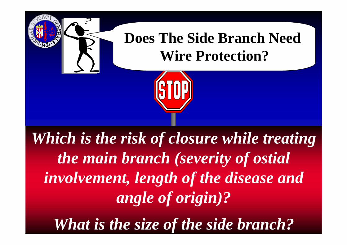
Which is the risk of closure while treatingthe main branch (severity of ostial
involvement, length of the disease and angle of origin)?
What is the size of the side branch?
Does The Side Branch NeedWire Protection?

Does The Side Branch NeedBalloon Dilatation?
…if the side branch is > 2.5 mm in diameterwith ostial disease or at risk of plaque shift elective balloon dilatation with or without
kissing balloon is advised………. but remember no oversized balloonin the side branch to prevent dissection!!!

The Sleeve Technique
JimJim etet al.al. CatheterCatheter CardiovascCardiovasc IntervInterv 20062006
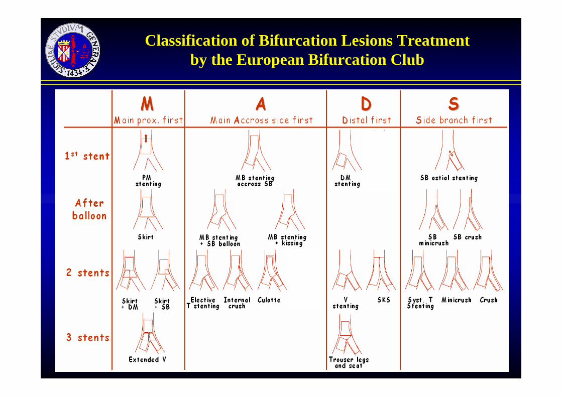
Classification of Bifurcation Lesions Treatment by the European Bifurcation Club

Classification of Bifurcation Lesions Treatment by the European Bifurcation Club

V Stenting Bench WorkCourtesy of J. Ormiston (Mercy Angiography, New Zealand)

Mini-Crush Technique Bench Work

Mini-Crush in a Trifurcation Lesion

…but remember that…… whatever strategy
you will usethe most simple and
safe technique will survive

V Stenting Bench WorkCourtesy of J. Ormiston (Mercy Angiography, New Zealand)
Stagnation area between the stents strutsRecirculation
Distortion

...........5/5 Procedural success
1/5...........Side branch 1 restenosis
0...........Late thrombosis
0...........Side branch 2 restenosis
00TBR
00CABG
1/50Total MACE
...........0Subacute thrombosis
...........0Acute thrombosis
0...........Main branch restenosis
1/50TLR
00Death
00Q-MI
0 0Non-Q MI
7-monthImmediate and30-Day
Results in the Five PatientsWith a Trifurcation Lesion

Is it so complex….?

Medina Classification
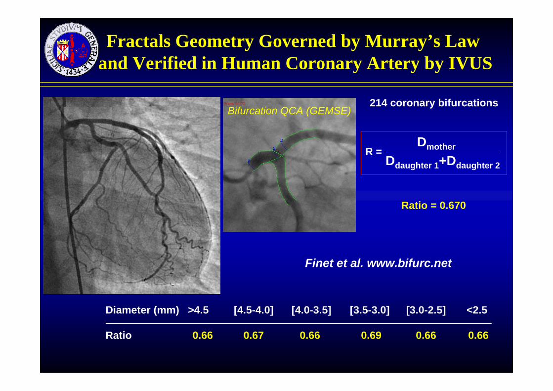
Diameter (mm) >4.5 [4.5-4.0] [4.0-3.5] [3.5-3.0] [3.0-2.5] <2.5
Ratio 0.66 0.67 0.66 0.69 0.66 0.66
214 coronary bifurcations
Fractals Geometry Governed by Murray’s Law and Verified in Human Coronary Artery by IVUS
Bifurcation QCA (GEMSE)
Ratio = 0.670
Dmother
Ddaughter 1+Ddaughter 2R =
Finet et al. www.bifurc.net

Courtesy of Drs Courtesy of Drs ReiberReiber, , KoningKoning, , TuinenburgTuinenburgLKEB and LKEB and MedisMedis
Proximal sectionProximal section
Distal1 sectionDistal1 section
Distal 2Distal 2sectionsection
Three Segments Model for the Bifurcation Analysis

Reference Diameter Calculation
Flagged central fragment
Diameters of the central fragment are automatically excluded from the calculation of the reference diameter function (flagging)
Courtesy of Drs Courtesy of Drs ReiberReiber, , KoningKoning, , TuinenburgTuinenburgLKEB and LKEB and MedisMedis

Distal 1Section
Proximal FragmentDelimiter
Spline fittedthrough the control points
Control points
Distal 1FragmentDelimiter
Courtesy of Drs Courtesy of Drs ReiberReiber, , KoningKoning, , TuinenburgTuinenburgLKEB and LKEB and MedisMedis
Side Branch Assessment

Reference Diameter for the 3 Segments
Courtesy of Drs Courtesy of Drs ReiberReiber, , KoningKoning, , TuinenburgTuinenburgLKEB and LKEB and MedisMedis

Analysis results for Side Branch
No overestimation of reference diameter → correct %diameter stenosisCourtesy of Drs Courtesy of Drs ReiberReiber, , KoningKoning, , TuinenburgTuinenburg
LKEB and LKEB and MedisMedis

Classification by the Angle of BifurcationLesions between MB and SB
Y shape
T shape
SB access:SB access: easyeasy difficultdifficult
PlaquePlaque shiftshift:: moremore lessless
>70B
AA
CC
<70B
AACC

Importance of the Bifurcation Angle “B”and Final Kissing Balloon
Dzavik et al, Am Heart J 2006
Angle>50Angle>50°° no final no final kissingkissing
Angle>50Angle>50°° final final kissingkissing
Angle<50Angle<50°° no final no final kissingkissing
Angle<50Angle<50°°final final kissingkissing

Geometrical Changes Noted During Bifurcation Stenting
D. Dvir et al WCC Barcelona 9/2006
Geometrical Changes Noted During Geometrical Changes Noted During Bifurcation Bifurcation StentingStenting
D. D. DvirDvir et al WCC Barcelona 9/2006et al WCC Barcelona 9/2006
• Bifurcation stenting causes significant geometrical changes in 3D• Two vs. one stenting technique causes most changes • 3D bifurcation reconstruction may be an important tool for planning PCI procedures and evaluating their results

Generally clinical sequelae are transient chest pain and ST-T wave changes
A small percentage of patients develop Q-waveinfarction or require emergency surgery as long as mainvessel remain patent
Non Q-wave myocardial infarction undoubtely occursfrequently (serial systematic evaluation of enzymes notavailable)
Side Branch Occlusion during PCI

RiskRisk of Acute Side of Acute Side BranchBranch OcclusionOcclusion
> 27> 27Side Side branchbranch withwith significantsignificant diseasedisease
< 4< 4Side Side branchbranch withwith minimal minimal diseasedisease
OcclusionOcclusion rate (%)rate (%)
Meier B et al. Am J Cardiol 1984; 53: 10-4
26o
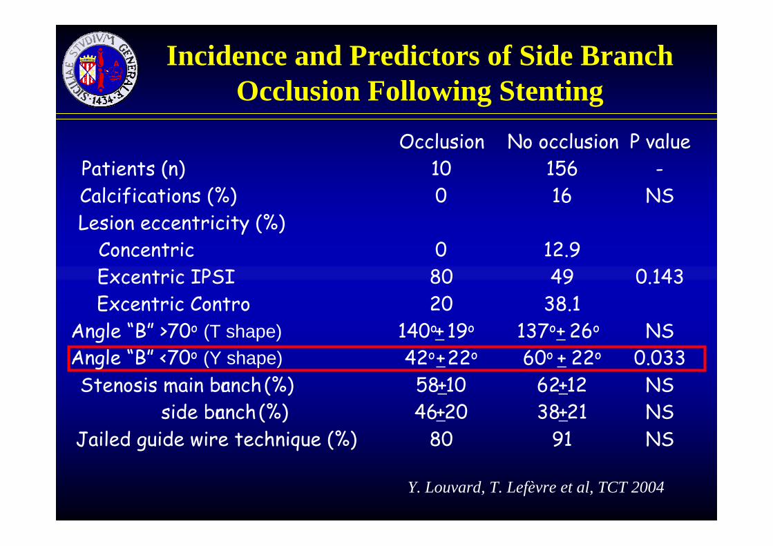
Incidence and Predictors of Side BranchOcclusion Following Stenting
Occlusion No occlusion P valuePatients (n) 10 156 -Calcifications (%) 0 16 NSLesion eccentricity (%)
Concentric 0 12.9Excentric IPSI 80 49 0.143Excentric Contro 20 38.1
Angle “B” >70o (T shape) 140o+ 19o 137o+ NS42o+22o 60o + 22o 0.033
Stenosis main branch(%) 58+10 62+12 NSside branch(%) 46+20 38+21 NS
Jailed guide wire technique (%) 80 91 NS
Angle “B” <70o (Y shape)
Y. Louvard, T. Lefèvre et al, TCT 2004

Incidence and Predictors of Side BranchOcclusion Following Stenting
Aliabadi et al, Am J Cardiol 1997
Nonthreathened side branchThreathened side branch
>50% ostial narrowing that arose fromwithin or just beyond the diseased MV
Thus, nonthreathened side branch of a small size should not be wired!!!
Side branch >2.0 mm that are at risk of closure should be protected!!!

Pre-dilatation with Kissing Balloon it avoids closure of side branch(or main vessel) by plaque shift
∅p
∅d
∅c
Common Approaches to BifurcationLesions: the Role of Kissing Balloon

Common Approaches to BifurcationLesions: the Role of Jailed Wire
Used in T shaped Bifurcations in order to favorably modify the angle between the two vessels thusfacilitating side branch re-wiring
Helps to maintain side branchpatency
In case of side branch closure assuresside branch traceability byradiopaque distal wire
• Guide wire is left inside the side branch during main vessel stenting
• Side branch guide wire is jailed between main vessel stent struts and main vessel wall

Jailed Wire Effect on Proximal Main Branch/Side Branch Angle
Baseline Wiring ° modification p valueAngle “A” > 120° (%) 77 87 - <0.02
Angle A (°) 149+17 160+18 + 11 <0.001
Angle “A” < 120° (%) 23 13 - <0.02Angle A (°) 107+11 140+19 + 33 <0.001
Y. Louvard, T. Lefèvre TCT 2003

Angiographic Predictors of Side Branch Success (Lesion <50% by QCA)
Age (years) 66±11 vs 57±8 p=0.0007Larger MB reference (mm) 3.1±0.4 vs 2.8±0.3 p=0.0085Larger SB reference (mm) 2.5±0.5 vs 2.2±0.3 p=0.0413Kissing balloon (%) 98.1 vs 76.5 p=0.0019“Jailed wire technique” (%) 92.9 vs 71.4 p=0.031
T. Lefèvre, Y. Louvard, 2003