
The Long-Term Effects of Low Level Laser Therapy (LLLT ...
55
City University of New York (CUNY) City University of New York (CUNY) CUNY Academic Works CUNY Academic Works Dissertations, Theses, and Capstone Projects CUNY Graduate Center 6-2014 The Long-Term Effects of Low Level Laser Therapy (LLLT) The Long-Term Effects of Low Level Laser Therapy (LLLT) Combined with Complex Decongestive Therapy (CDT) in the Combined with Complex Decongestive Therapy (CDT) in the Treatment of Breast Cancer Lymphedema: A Double-Blind, Treatment of Breast Cancer Lymphedema: A Double-Blind, Randomized, Placebo-Controlled Study Randomized, Placebo-Controlled Study Olivia Bramlett Graduate Center, City University of New York Igor Daysudov Graduate Center, City University of New York Toshi Odaira Graduate Center, City University of New York Bethany Rodriguez Graduate Center, City University of New York How does access to this work benefit you? Let us know! More information about this work at: https://academicworks.cuny.edu/gc_etds/798 Discover additional works at: https://academicworks.cuny.edu This work is made publicly available by the City University of New York (CUNY). Contact: [email protected]
Transcript of The Long-Term Effects of Low Level Laser Therapy (LLLT ...

City University of New York (CUNY) City University of New York (CUNY)
CUNY Academic Works CUNY Academic Works
Dissertations, Theses, and Capstone Projects CUNY Graduate Center
6-2014
The Long-Term Effects of Low Level Laser Therapy (LLLT) The Long-Term Effects of Low Level Laser Therapy (LLLT)
Combined with Complex Decongestive Therapy (CDT) in the Combined with Complex Decongestive Therapy (CDT) in the
Treatment of Breast Cancer Lymphedema: A Double-Blind, Treatment of Breast Cancer Lymphedema: A Double-Blind,
Randomized, Placebo-Controlled Study Randomized, Placebo-Controlled Study
Olivia Bramlett Graduate Center, City University of New York
Igor Daysudov Graduate Center, City University of New York
Toshi Odaira Graduate Center, City University of New York
Bethany Rodriguez Graduate Center, City University of New York
How does access to this work benefit you? Let us know!
More information about this work at: https://academicworks.cuny.edu/gc_etds/798
Discover additional works at: https://academicworks.cuny.edu
This work is made publicly available by the City University of New York (CUNY). Contact: [email protected]

The Long-term Effects of Low Level Laser Therapy (LLLT) combined with Complex Decongestive Therapy (CDT) in the treatment of Breast Cancer Lymphedema: A Double-Blind, Randomized, Placebo-Controlled Study
By
Olivia Bramlett Igor Daysudov Toshi Odaira
Bethany Rodriguez
A capstone project submitted to the Graduate Faculty in Physical Therapy in partial fulfillment of the requirements for the degree of Doctor of Physical Therapy, The City
University of New York
2014
ii
This manuscript has been read and accepted for the Graduate Faculty in Physical Therapy in satisfaction of the capstone project requirement for the degree of DPT
Date: Dr. Susan Pivko, PT, Cert. MDT, Adv. CIECP, Cert. FDM
Chair of Examining Committee (Advisor)
Date: Dr. Jeffrey Rothman Professor and Chair Department of Physical Therapy College of Staten Island/CUNY
Executive Officer
THE CITY UNIVERSITY OF NEW YORK

iii
Abstract
The Long-term Effects of Low Level Laser Therapy (LLLT) combined with Complex Decongestive Therapy (CDT) in the treatment of Breast Cancer Lymphedema: A Double-Blind, Randomized, Placebo-Controlled Study.
By
Olivia Bramlett Igor Daysudov Toshi Odaira
Bethany Rodriguez Adviser: Professor Susan Pivko PT, Cert. MDT, Adv. CIECP, Cert. FDM
Complex Decongestive Therapy (CDT), the gold standard for lymphedema
treatment, fails to demonstrate long-term efficacy. The purpose of this study was to
determine the long-term efficacy of low level laser therapy (LLLT) in reducing post-
mastectomy lymphedema when used with CDT. The experimental group received
LLLT and CDT (n = 7) while control group received sham laser and CDT (n = 7),
twice a week for 4 to 8 weeks. Percent arm circumference difference between
affected and unaffected limbs was collected over 18 months. Results revealed no
statistical difference between both groups at all time periods: 1 (p = 0.902), 2 (p =
0.535), 3 (p = 0.445), 6 (p = 0.095), 12 (p = 0.537) and 18 months (p = 0.4). Further
study with a larger sample size may prove more significant for long-term efficacy of
LLLT.
THE CITY UNIVERSITY OF NEW YORK

iv
Acknowledgements
We would like to thank our faculty advisor, Dr. Susan Pivko, for her mentorship
and guidance throughout this project for the past three years.
Thank you to Dr. Suzanne R. Babyar for her generous assistance with analyzing
data and draft editing. We would also like to acknowledge Dr. Gary Krasilovsky,
Director of the Department of Physical Therapy at Hunter College, for his additional
support.
A special thank you to the physical therapists at Rusk Institute of Rehabilitation
Medicine/NYU Langone Medical Center for allowing us the opportunity to work along
side them, sharing with us their clinical expertise, expanding our knowledge on
lymphedema and lymphedema patients, and to be part of this research endeavor: Aaron
Beattie, Tamara Bushnik PhD, FACRM., Teresa Denham, PT, MA; Mei R. Fu, PhD,
RN, ACNS-BC; Annika Ginsberg, Laurelle Kilmartin, PT, DPT, CSCS, CLT-LANA
and Ting-Ting Kuo, PT, DPT, MSPT, WCS, CLT.
Finally we would like to thank our families for their support and encouragement
in order to successfully complete this incredible milestone in our lives.

v
Table of Contents
Introduction .............................................................................................................................................. 1
Need for Study ................................................................................................................................... 12
Purpose ..................................................................................................................................................... 14
Hypothesis ........................................................................................................................................... 17
Methods .................................................................................................................................................... 18
Design ................................................................................................................................................... 18
Subjects ................................................................................................................................................ 18
Materials .............................................................................................................................................. 19
Procedures ........................................................................................................................................... 21
Results ....................................................................................................................................................... 23
Data Reduction .................................................................................................................................. 24
Table 1 .................................................................................................................................................. 24
Figure 1 ................................................................................................................................................ 25
Table 2 .................................................................................................................................................. 26
Figure 2 ................................................................................................................................................ 27
Discussion ................................................................................................................................................ 28
Future Studies .................................................................................................................................... 32
Conclusion ............................................................................................................................................... 34
Appendix .................................................................................................................................................. 35
References ................................................................................................................................................ 39

vi
List of Tables & Figures
Table 1: Average % difference of all landmarks for all participants in a sham or an
active laser group for a given follow-up visit (n = 14) ………………………………….24
Figure 1: Average Effect of Active vs. Sham Laser on Limb Circumference …...…….25
Table 2: Results of Mann-Whitney U Test to determine significance of data distribution
between laser vs. sham groups at each time period ………………………….………….26
Figure 2: Effect of active vs. sham laser on limb circumference using raw measurements
extrapolated from the Mann-Whitney U Test …………………………………………...27

1
Introduction
Cancer is a major public health issue in the United States and the rest of the
world. Currently, cancer accounts for 25% of all mortality in the United States (Siegel,
Ward, Brawley, & Jemal, 2011). Among women, the cancers of the lung, bronchi,
colorectal and breast are leading causes of cancer deaths. Breast cancer is the second
leading cause of cancer death among women, with approximately 232,600 new cases
expected for 2011 and an estimated 39,500 deaths (Siegel et al., 2011). Although there
has been a decline in incidence of breast cancer reports since 1998, the decline in rate
since 2003 has become stagnant (Siegel et al., 2011).
Treatment for breast cancer is complex. It entails a high risk of causing the onset
of lymphedema, which is considered the most dreaded, secondary pathology related to
breast cancer treatment (Fu, Ridner, & Armer, 2009). Lymphedema is swelling and
chronic inflammation that develops in the limb as a result of abnormal accumulation of
tissue proteins and interstitial fluid (Brennan, DePompolo, & Garden, 1996). Abnormal
accumulation of lymph fluid mostly occurs in the interstitial spaces of the arm, shoulder,
neck, breast or the thorax, which can lead to physical discomfort, pain, impaired function
and emotional distress (Fu et al., 2009). Of the approximately three to five million
patients in United States who suffer from lymphedema, a significant proportion have
developed lymphedema as a result of breast cancer treatment (Lawenda, Mondry, &
Johnstone, 2009).
The lymphatic system plays a critical role in maintaining homeostasis in the
body. It is a system that is responsible for ridding tissues of metabolic waste, foreign
debris and excess materials by carrying them away from the tissues to the lymph nodes

2
via circulation of lymph fluid (Mortimer, 1998). The lymphatic system is composed of
deep and superficial lymph vessels, as well as lymph nodes, which are filled with lymph
fluid (Cohen, Payne, & Tunke, 2001). Lymph fluid consists primarily of protein, water,
fatty acids, salts, white blood cells, micro-organisms and foreign debris (Lawenda et al.,
2009). Excess protein, interstitial fluid or toxic foreign debris present in the skin and
subcutaneous tissues are absorbed into lymph fluid and collected through the superficial
lymphatic system (Lawenda et al., 2009). The deep system is responsible for collecting
lymph fluid in the deeper tissues (Lawenda et al., 2009). Once waste and excess
metabolic substances are collected, lymph fluid flows from lymph capillaries into the
lymph nodes in the axillary and inguinal regions. Lymph nodes then transport the fluid to
lymphatic trunks, which will then drain lymph directly into the venous system for either
disposal of waste or reabsorption of proteins (Lawenda et al., 2009).
The lymphatic system also plays a major role in reduction and prevention of
infection. In addition to collecting excess interstitial water and proteins, the system can
pick up bacteria and other foreign antigens and transport them to lymphocytes in the
lymph node for processing (Cohen et al., 2001). In a normal physiological state, the
lymphatic system maintains a transport capacity that exceeds the volume of lymph fluid
needed for transportation. Lymphedema can be triggered due to either overload of
lymphatic fluid or dysfunction of the lymphatic vessels that decrease the transport
capacity (Lawenda et al., 2009).
Lymphedema can be classified into primary and secondary lymphedema. Primary
lymphedema refers to congenital impairment of the lymphatic system (Fu et al., 2009;
Brennan et al., 1996). Secondary lymphedema can result from trauma such as cancer,

3
cancer treatment, infection, inflammation, chronic venous insufficiency, immobility,
radiation or surgery (Fu et al., 2009; Brennan et al., 1996; Lawenda et al.,
2009). Lymphedema triggered by breast cancer treatment is classified as secondary
lymphedema.
First described as a side effect of mastectomy operations by William Halstead in
1921, secondary lymphedema related to breast cancer can be caused directly by the tumor
on the lymphatic vessels or more recently from indirect side effects of anti-cancer
therapies (Mortimer, 1998; Brennan et al., 1996). Significant impairment of lymphatic
function can occur when lymphatic structures are injured and replaced with fibrotic tissue
as a result of resection of nodes and radiation therapy that are conducted as part of cancer
treatment (Brennan et al., 1996). Lymphedema occurs more frequently in breast cancer
patients who receive both resection of lymph nodes and radiation therapy. The incidence
can be up to 23% for patients receiving a combination of sentinel lymph node biopsy
(SLNB) and radiation therapy, and up to 48% for patients receiving axillary lymph node
dissection (ALND) and radiation therapy (Lawenda et al., 2009).
Lymphedema can be classified into four stages according to skin condition and
degree of swelling (Fu et al., 2009). Stage 0 is latent or sub-clinical lymphedema, with
no visible edema but with possible, minor heaviness in the limb. Stage 1 is considered
reversible but exhibits visible edema, while stage 2 is considered non-reversible and
displays visible edema with added characteristics of pitting and fibrosis of the
skin. Stage 3 exhibits pitting edema in a largely affected area of the limb, as well as
fibrosis and leakage of lymph through damaged skin (Fu et al., 2009). Stage 3 is
considered severe but rare in upper extremity lymphedema (Fu et al., 2009).

4
The signs and symptoms of lymphedema are readily visible, significantly altering
not only the physique but also the self-perceived image of one’s physical
presentation. Lymphedema can have significant negative effects on wellness and on
one’s ability to perform activities of daily living. Range of motion of the wrist, hand and
arm are affected by the inability of muscles and joints to move freely as a result of
progressive stiffening of swollen superficial and deep tissues (Casley-Smith, Boris,
Weindorf, & Lasinski, 1998). Lymphedema can be accompanied by other distressing
symptoms such as pain, fatigue, decreased physical activity, loss of sensation of the limb,
diminished body image and other psychological distress associated with physical
disfigurement caused by this substantial swelling (Fu et al., 2009; Lawenda et al., 2009;
Cohen et al., 2001). Often individuals socially isolate themselves secondary to physical
presentation of edema and bandaging. Currently there is no cure for lymphedema, but
intervention is essential to delay onset and to eliminate further progression of the
symptoms (Lawenda et al., 2009).
Physical therapy has been traditionally used for intervention of lymphedema since
the techniques of “lymphatic massage” were first introduced in the 1930s (Casley-Smith
et al., 1998). Currently the gold standard for treatment of lymphedema is Complex
Decongestive Therapy (CDT). In one study, 35 patients with post-mastectomy
lymphedema who underwent CDT reported a 25% decrease in percent excess volume in
the affected limb when compared to the unaffected limb (Randheer, Kadambari,
Srinivasan, Bhuyaneswari, Bhanumathy, & Salaja, 2011). This study recruited subjects
following the completion of breast cancer treatment with a 4-month onset of
lymphedema. Inclusion criteria required patients to have a difference in limb volume

5
between the two arms greater than 200 ml and a difference in one point of circumference
of the two arms greater than 2 cm. In addition, participants must have been free from
reoccurring carcinoma, metastasis, cellulitis or from edema resulting from other medical
causes (Randheer et al., 2011). Parameters of interest were baseline circumference
measurements of the upper limbs and water displacement volumetry (Randheer et al.,
2011). Following the initial measurement, patients underwent CDT comprised of a 45
minute session of manual lymphatic drainage and multi-layered compression bandaging
for the duration of four times a weeks for 2 weeks. Patients were also instructed to
conduct massages on their affected limbs twice a day, apply compression bandages
throughout the day and to elevate the limb at night. After the 2 week CDT phase,
patients were followed up once a month for three months for assessment of limb volume
changes (Randheer et al., 2011). Effectiveness of CDT was calculated as the change in
absolute limb volume of the treated limb before and after the two-week phase and at each
month of follow up. The volume was also measured by calculating the difference in limb
volume between the affected and unaffected limb and expressing it in percentage
reduction of edema (Randheer et al., 2011).
CDT is comprised of the following components:
Manual Lymphatic Drainage (MLD):
Manual Lymphatic Drainage is a technique that mobilizes excess lymph fluid in
the extremities back into the body’s central lymph system to be removed. It can be
effective in decreasing edema, increasing range of motion of the limb, improving texture,
increasing circulation and diminishing the risk of cellulitis (Casley-Smith et al., 1998).

6
Compression:
Compression garments:
Based on individual needs, participants will receive a compression sleeve and
gauntlet/glove that are to be worn in the daytime during the self-management phase in
order to maintain the improvements made during intensive intervention (Casley-Smith et
al., 1998).
Multilayer Bandaging (MLB):
Multiple layers of short stretch elastic bandages are applied to the
lymphedematous limb. The tension of the bandages facilitates muscle pumping in order
to push the accumulated lymph fluid back into the body’s central lymphatic system. As
with MLD, MLB has been proven to improve the range of motion of the limb, soften
tissue texture, increase circulation and decrease risk of cellulitis (Casley-Smith et al.,
1998).
Vasopneumatic compression:
This may be used as an adjunct to treatment including MLD, MLB, and
therapeutic exercise. The compression device provides an automated sequence of
gradient compression to the limb with lymphedema. When compression is applied, it
exerts gentle, external pressure on the affected extremity in order to rid edema by
mechanically pumping the excess lymph fluid back into the center of the body (Casley-
Smith et al., 1998).

7
Exercise:
Therapeutic exercises prescribed by the physical therapist promote active
participation by the patient, in order to enhance muscle pump action to decrease the
edema in the limb (Casley-Smith et al., 1998).
Education:
Physical therapists educate the patients on posture, body mechanics, skin care and
lymphedema precautions, such as avoiding venupuncture and vital readings (blood
pressure) on the affected side. Education is crucial in order to optimize activities of daily
living and minimize risk of infection and exacerbation of lymphedema (Casley-Smith et
al., 1998).
Self-management:
When maximum improvements in girth measurement reduction and tissue
softening have been achieved, the patient is provided with a self-managed plan of care
upon discharge. This may include a combination of compression garments, MLD (self-
administered), MLB (self-administered), exercise, and a vasopneumatic compression
device (Casley-Smith et al., 1998).
Despite the empirical support of CDT, there are numerous contraindications
associated with CDT. Complex decongestive therapy can increase venous blood volume
in patients with hypertension and increase risk of injury or infection in diabetic patients
with decreased tactile sensation (Fu et al., 1996). Some absolute contraindications for
CDT are acute infections, congestive heart failure, and deep vein thrombosis. The
compression involved in CDT may exacerbate infections, increase central venous blood
volume in individuals with heart failure, and release clots in the blood vessel (Lawenda et

8
al., 2009). Use of CDT for patients with these afflictions is contraindicated. For such
patients not suitable to be treated with CDT, it is necessary to develop an alternative
mode of intervention.
Low-level laser therapy (LLLT) is a physical modality with potential for a wide
application of clinical use. It has been used to relieve common symptoms ranging from
stiffness and pain related to Achilles tendonitis, low back pain, orofacial pain, tendonitis,
neck pain, temporomandibular pain, chronic periodontis and subacromial impingement
syndrome (Yeldan, Cetin, & Ozdincler, 2009; Carcia, Martin, Houck, & Wukich, 2010;
Djavid, Mehrdad, Ghasemi, Hasan-Zadeh, Sotoodeh-Manesh, & Pouryghoub,
2007). Low-level laser therapy has also been studied for its efficacy in reducing swelling
and pain related to surgical removal of the third molar, commonly referred to as “wisdom
tooth” (Ferrante et al., 2012).
Historically both in vitro and in vivo studies have been conducted that aimed to
study the efficacy and the mechanism of low-level laser (LLL). The focus of many in
vitro studies using the LLL has been on the effect of laser irradiation on the growth and
repairing capacity of cells involved in wound healing and decreased formation of scarring
(Posten et al., 2005; Omar, Ebid, & Morsy, 2011). Most of such studies have focused on
fibroblast growth, locomotion, and production of collagen, as fibroblasts play a
significant role during the proliferative and remodeling phases of wound healing (Posten
et al., 2005). In addition, effects of LLL on monocytes and endothelial cells have also
been studied. The overarching result of these studies has shown that LLL enhances
deposition of collagen and proliferation of fibroblasts and monocytes (Posten et al.,
2005).

9
Effect of LLL on wound healing has also been investigated with in vivo
experiments using rodents as an animal model. One earlier study used a Helium-Neon
(HeNe) laser at 632.8 nm at a dose between 4 to 20 J/cm2 on surgical wound sites created
in rat models to observe wound healing differences between different doses (Kana,
Hutschenreiter, Jaina, & Waidelich, 1981). The authors observed wound healing at a
much more significant rate with application of 4 J/cm2 and also found that the lasers
increased formation of granulation tissue and deposition of collagen (Kana et al., 1981).
In addition to fibroblast activity during wound healing, many past animal studies
have also investigated the effect of LLL on tissue tensile strength, vascularization around
fractured bones, and wound healing around nerves (Posten et al., 2005). Low-level laser
therapy continues to stimulate the immune system by increasing prostaglandin levels,
reducing edema, improving lymphatic drainage motility by vasodilatation and inhibition
of platelet aggregation, and improving cellular growth for wound healing in human
subjects (Omar et al., 2011).
Historically, the therapeutic use of LLL can be traced back to a series of human
studies conducted by Mester et al. beginning in 1971, that exhibited improvement in
wound healing in chronic ulcers and neuropathic foot ulcers with application of low
energy (1 J/cm3) laser (Mester, Spiry, Szende, & Tota, 1971). Studies on the role of
LLLT in wound healing were eventually applied to and used in treating secondary
lymphedema. The U.S. Food and Drug Administration (FDA) recommended that the
dose for lymphedema treatment with laser was indicated to be 650 – 1000 nm in 2013
(Ridner, Poage-Hooper, Kanar, Doersam, Bond, & Dietrich, 2013).

10
Currently, LLL has also been focused as an effective intervention in the treatment
of post-mastectomy lymphedema (Carati, Anderson, Gannon, & Piller, 2003). Approved
by the FDA in November 2006, LLLT is a relatively new form of lymphedema treatment
that utilizes light to stimulate healing response (Anderson, Pillar, Gannon, Carati &
Angel, 2008). It has been suggested that LLL enhances lymphangiogenesis (the growth
of lymphathic vessels) and stimulates macrophage cells and breaks down fibrous tissue
(Tammela, & Alitalo, 2010; Carati et al., 2003).
One double-blind controlled trial was conducted that investigated the effect of
LLL on post-mastectomy lymphedema in 11 women with unilateral lymphedema
(Kaviani, Fateh, Nooraie, Alinagi-zadeh, & Ataie-Fashtami, 2006). The subjects were
randomly assigned to either the sham laser or laser treatment group and received laser
application in the affected arm and axillary area three times a week for 3 weeks, followed
by an 8 week interval. Laser application was repeated for three more weeks after the 8
week interval, for a total of 18 treatment sessions. The laser used was a low-energy
gallium arsenide laser at 890nm for a dose of 1.5J/cm2 (Kaviani et al., 2006). The
baseline assessment of limb circumference, range of motion, pain score and heaviness of
the affected limb were compared between the two groups during the treatment period at
3, 8, 12, 18 and 22 weeks. Data were collected regarding the difference in circumference
of the affected and unaffected limb and was measured using a non-elastic tape measure at
6 specific anatomical points. The circumference difference of the two limbs was
compared to the circumference difference at pre-treatment sessions in order to calculate
the total reduction in circumference (Kaviani et al., 2006). The total reduction in
circumference and the reduction in fluid as measured with the L-Dex scores of

11
bioimpedence in the treatment group was significantly more than in the sham group in all
sessions (p <0.001) (Kaviani et al., 2006). In addition, pain significantly decreased in the
treatment group in all sessions compared to the sham group with the exception of week 3
and 9 (p <0.001) (Kaviani et al., 2006). There was no significant difference in range of
motion and in heaviness score between the treatment and sham groups. The study does
not depict the components of the heaviness and pain score, nor does it explain at which
joints range of motion was studied.
Another study on human subjects conducted a similar double-blind, placebo
controlled trial to investigate the effect of LLL on limb volume, shoulder mobility and
handgrip strength in women with post-mastectomy lymphedema (Omar, Ebid & Morsy,
2011). The study recruited 50 participants, all with unilateral lymphedema who qualified
under the protocol and were assigned to either sham laser group or active laser
group. Low-energy gallium arsenide laser at 904nm, power of 5mW and spot size of
0.2cm2, at a final dose of 1.5J/cm2 was applied over the arm and axilla three times a week
for 12 weeks (Omar et. al., 2011). Total reduction of circumference, shoulder mobility
measured as shoulder range of motion in flexion, abduction and external rotation; grip
strength measured using a hand dynamometer were assessed at baseline prior to treatment
(week 0) and at every 4 weeks during the 12 week treatment period (week 4, 8, 12)
(Kaviani et al., 2006). The total reduction in limb circumference was significantly
greater in the treatment group than in the sham group at all assessed sessions (Kaviani et
al., 2006). Shoulder flexion and abduction significantly increased during week 8 and 12
in the treatment group, but there was no significant difference in external rotation
between the two groups during any session (Kaviani et al., 2006). Grip strength

12
improved in both groups, but the laser group exhibited significant percentage of
improvement at week 12 compared with the sham group (Kaviani et al., 2006).
Need for Study
Several double blind, placebo-controlled studies have been conducted to
investigate the efficacy of LLL on treating post-mastectomy lymphedema. However,
most studies follow up on the efficacy of LLL application for less than a year. The
interest of this research was to investigate the long lasting effect of LLL application in
preventing recurrence of lymphedema, if any, by following subjects over time after active
treatments have ceased. In addition, most studies do not assess LLL in conjunction with
the use of CDT. For patients already receiving CDT, it is worthwhile to know if the
further addition of LLL intervention would have any positive impact on treatment
outcome.
LLL has several parameters that must be defined clearly in each study, at times
complicating meaningful comparisons of results between studies. Low-level laser has 5
defining factors: power, wavelength, pulse rate, pulse duration, total irradiation time,
intensity and dose (Posten et al, 2005). Low-level laser is defined to have a power with a
range between 10-3 and 10-1 W, wavelength with a range between 300 and 10,600nm,
pulse rate with a range between 0 and 5,000 Hz, pulse duration with a range between 1 to
500 milliseconds, total irradiation time between 10 to 3,000 sec, intensity between 10-2 to
100 W/cm2, and a dose between 10-3 to 102 J/cm2 (Posten et al, 2005). Although the overall
effects of LLL seem to be positive, there is no agreement on the most optimal
physiological and physical parameters of the use of LLL that yield the best clinical

13
outcome (Kaviani et al., 2006). Further studies are necessary to help define the exact
parameter of LLL that is most optimal for clinical use in treating lymphedema.
Longitudinal data supporting CDT efficacy is limited and lends to poor carry over
when compared to more conservative treatment like compression stockings. One recent
study revealed no significant difference between percent arm reduction volume at all time
period: 3, 6, 12, and 24 weeks between a control group which donned a compression
sleeve 12 waking hours for 4 weeks and an experimental group which received CDT and
MLD for 1 hr by a specialist 5 days per week for 4 weeks (Dayes, Whelan, Julian, Parpia,
Pritchard, D’Souza, Kligman, Reise, LeBlanc, McNeely, Manchul, Wiernikowski, &
Levine, 2013). Each session followed with compression bandaging performed by a
specialist to be worn for 23 hrs and each subject was taught self-bandaging to be used
over the weekend. After 4 weeks of the active phase was over, subjects of the
experimental group received compression sleeves similar to that of the control group
(Dayes et al., 2013). Although still statistically insignificant, greater arm reduction was
seen in subjects receiving CDT for the sub group with a shorter duration of onset (Dayes
et al., 2013). Greater duration (> 1 year versus < 1 year) may explain an increased risk of
tissue fibrosis in a lymphedematous arm. Manual Lymphatic Drainage in addition to
compression stockings, may be effective. Greatest arm volume reduction may occur over
the first several weeks (approximately 3) (Dayes et al., 2013). If there is no benefit to
costly CDT, when compared to economical compression sleeves, conservative therapy
may be indicated. Complex Decongestive Therapy depends heavily on patient
compliance and incorporates MLD, daily bandaging, exercise, and skin care. During the
maintenance phase (long-term post treatment phase), the individual is responsible for

14
compression bandaging, exercise and skin care. Subjects in both sham and active laser
group may have had low rates of compliance, except for the benefits of LLLT
outweighing any decrease in efficacy due to decreased individual compliance (Javid &
Anderson, 2013).
Purpose
The purpose of this study is to determine if LLLT in conjunction with CDT can
have a long-term effect in reducing post-mastectomy lymphedema in breast cancer
survivors. The implementation of LLL is quick, easy to administer, and non-invasive;
while CDT is time consuming for patients and clinicians. Efficacy of MLD and
bandaging are also dependent on the skill level of the Clinical Lymphedema Specialist as
well as the compliance and skill level of the patients. In addition, proper technique of
bandaging with the non-dominant upper extremity can be difficult when the
lymphedatemous arm is the dominant hand. If LLLT is effective, it allows for non-
specialized clinicians to be involved in the care of people with secondary lymphedema
and is simpler to don for those whose dominant arm is affected (Ridner, Poage-Hooper,
Kanar, Doersam, Bond, & Dietrich, 2013).
Omar et al (2011) showed that there was significant improvement in reduction of
limb volume in the lymphadematous arm up to 12 weeks post LLLT treatment, compared
to a sham laser group in a double blind, randomized study. Subjects in both groups
received skin care, therapeutic exercises, compression bandaging but NOT MLD. This
study shows that rather than just a hypothesized in-direct effect of LLLT on MLD, LLLT
alone does have good efficacy (Omar et. al., 2011).
15
One double-blind, placebo-controlled study revealed no significant correlation
between the duration of lymphedema (2 – 336 months) and reduction of arm
volume. However, at three months, after two treatment bouts of LLLT (8 week rest
period between two rounds of laser treatments), mean affected limb volume was
significantly lower than that of placebo group or group receiving one bout of LLLT (8
week rest period between placebo and one round of laser treatment) (Carati, Anderson,
Gannon, & Piller, 2003). Research shows subjects who developed lymphedema in the
leg post surgery for gynecological cancer showed the effectiveness of CDT reached the
greatest reduction in Percentage Excess Volume (PEV) 3 and 6 months post CDT, but
reduction became insignificant as edema returned to pre-CDT level (2 months and 24
months) (Kim, Hwang, Kim, Chang and Lee, 2012). Percent average limb reduction had
been noted until 6 months, but did return to pre-trial level during assessment time period
at long-term defined as 12 months and 18 months (Kim et al, 2012). Moreover, one study
where subjects were divided into 2 groups –PEV < 20% (group 1) and PEV ≥ 20%
(group 2) and received 2 weeks of CDT treatment followed by maintenance phase (self-
care) and data collected at 3, 6, 12 and 24 months; PEV in group 2 was significantly
lower than baseline at all time periods, while PEV in group 1 began to increase at 6, 12,
and 24 months (Hwang, Hwang, Kim, Lee, Chang & Chu, 2013). Complex decongestive
therapy has been shown to be less effective long-term with initial PEV < 20% (Hwang et.
al., 2013).
With regard to possible development of fibrosis as one of the complications of
lymphedema, fibrotic tissue inhibits efficient MLD and fluid drainage. Another study
noted the tissue indentation resistance (TIR) by calculating the amount of force required

16
to indent tissue to a certain depth (Mayrovitz & Davey, 2011). The LLL implemented by
this research was produced by RianCorp LTU 904H – pulsed 904 nm, with an average
output of 5 mw from head size 0.2cm2. In addition, use of tonometry revealed significant
reduction in indentation forces with 5 minutes of LLLT as compared to pre LLLT forces
(Mayrovitz & Davey, 2011). A study by Ramos et al. recruited 69 subjects and showed
that rather than the timing of initiation of treatment, the biggest factor for efficacy
appeared to be PEV at baseline (Ramos, O’Donnell, & Knight, 1999).
When compared to LLLT, CDT has been criticized partly for high cost over a
long-term period and for time consumption. Complex Decongestive Therapy
incorporates an average of 60 minutes of MLD, as well as highly involved individual
participation during the maintenance phase that can yield decreased compliance
(Lasinski, Thrift, Squire, Austin, Smith, Wanchai, Green, Stewart, Cormier, & Armer,
2012). Empirical research suggests the implementation of conservative treatment of a
compression sleeve, pneumatic pump or LLL could be recommended (Haghighat, Lotfi-
Tokaldany, Maboudi, Bahadori, & Weiss, 2013; Dayes, et al., 2013).
Currently no study examines the relationship between the use of LLLT and the
possible recurrence of, or increased risk of, metastasis (Lima, Lima, Figueiredo, Carvalho
de Andrade, & Bergman, 2012). One in vitro study showed a down regulation of protein
expression responsible for cell adhesion, which can be related to metastasis (Leeuwen,
Dekker, Byers, Vermeer and Grevelink, 1996).
In consideration of the importance of promoting public health and wellness,
obesity (BMI > 30kg/m2) is a risk factor at reportedly three times greater risk for

17
developing lymphedema (Helyer, Varnic, Le, Leong, & McCready, 2010). Although
lymphedema risk is associated with numerous factors such as stage of cancer, age, type of
surgery, adjunct treatment, BMI is an important predictor for development of
lymphedema (Helyer et, al., 2010).
Educating breast cancer patients to the importance of detection of lymphedema
well into post-ALND is paramount since the average onset of post surgical lymphedema
is 36 months and some research notes onset up to 2 years later (ALND = 20% vs. SLNB
= 5.6%) (Liao, Li, Huang, Chen, Kuo, Chen, & Wei, 2013; Javid & Anderson,
2013). Teaching individuals to detect early onset of mild lymphedema is crucial as
research shows approximately 20% of mild lymphedema progresses into more severe
lymphedema in one year (Bar, Cheville, Solin, Dutta, Both, & Harris, 2010). In addition,
increase risk in lymphedema is associated with post-operative infection or delayed wound
healing and LLLT may be implicated for management of infection control and wound
management as 17-25% of breast cancer survivors are at risk for secondary lymphedema
(Lima et. al., 2012).
Hypothesis
The implementation of low level laser therapy (LLLT), in conjunction with
standard lymphedema intervention (CDT), can potentially decrease risk of infection,
decrease arm girth measurement, reduce tissue fibrosis, and enhance tissue texture in the
involved upper extremity yielding longer advantageous effects than standard CDT
intervention.

18
Methods
Design
This study is a double-blind, randomized, control trial of breast cancer survivors
with post-mastectomy lymphedema all receiving CDT. Participants were randomized
into a placebo group that received sham LLL application and a treatment group that
received active LLL application.
Subjects
The venue of this study is Rusk Institute of Rehabilitation Medicine and NYU
Clinical Cancer Center in New York, NY. A total of 16 subjects were recruited, 7 of
which were assigned to the placebo group, the other 7 assigned to the active laser group,
and 2 who dropped out after the intensive phase. Participants were recruited through
informational flyers that were circulated at Rusk Institute of Rehabilitation Medicine,
NYU Clinical Cancer Center and local community organizations (i.e. SHARE, Gilda’s
Club) throughout the 5 boroughs of New York City. Breast cancer survivors already
receiving outpatient physical therapy for post-mastectomy lymphedema were invited for
initial screening as a potential participant. Patients interested in participating in the study
proceeded through a referring physician or a nurse practitioner. Signed hard copies of
agreement of the participant’s inclusion into the study were obtained from both the
participant and the referring physician prior to the participant’s inclusion. All
participants signed the consent form to participate in the study. The NYU School of
Medicine/NYU Langone Medical Center Institutional Review Board (IRB) and Human
Research Protection Program (HRPP) committee and the Hunter College (HRPP)-IRB
committee approved this research.

19
The following criteria were required for participant inclusion to the study: female
of 21 years of age or older; affected by unilateral breast cancer which was treated by
unilateral mastectomy (no prophylactic mastectomy); lymph node dissection surgery
[sentinel lymph node biopsy (SLNB) and/or axillary lymph node dissection (ALND)];
affected with unilateral upper limb lymphedema; involved upper extremity fluid volume
that is greater than or equal to 200 mL and/or a circumferential girth measurement of the
affected limb at any 4 cm segment with a difference of 2 cm or greater in comparison to
the unaffected contralateral upper extremity; stage I or stage II lymphedema; cognitively
intact; and able to make the time commitment to participate in all treatment
sessions. Interpreters were assigned to all non-English speaking or limited English
proficiency participants who met all inclusion criteria during each treatment session for
the length of the study.
Potential participants were excluded from the study based on the following: BMI
> 40; active or metastatic cancer; currently or later undergoing adjuvant radiation or
chemotherapy therapy; restrictive active range of motion interfering with the purposes of
treatment; history of severe surgical disruption of arm; presence of primary or secondary
lymphedema in the contralateral upper extremity; active renal failure; arterial
insufficiency; congestive heart failure; chronic inflammatory conditions; use of a
pacemaker; a past medical history of deep vein thrombosis (upper extremity); past use of
CDT and/or LL; use of medication which alters fluid balance (i.e. diuretic, certain
chemotherapy); pregnancy.
Materials
The following materials were used to capture the data analyzed in this study:

20
• The LTU-904 infrared laser manufactured by RianCorp (Marleston, South
Australia) was used in this study at a wavelength of 904nm.
• Perometer Type 350 S manufactured by Pero-System Messgeraete GmbH
(Wuppertal, Germany) was used to calculate total limb volume.
• A bio-impedance analyzer, the L-Dex XCA U400 manufactured by ImpediMed
(Brisbane, Australia) was used to measure the extracellular fluid volume of
affected and unaffected limbs.
• The DF-50 BIA manufactured by ImpediMed and the InBody 230 Body
Composition Analyzer manufactured by BioSpace (Seoul, Korea) were used to
measure and calculate percent body fat and BMI of each participant.
• The Lymphedema and Breast Cancer Symptom Experience Index was used to
monitor changes in overall experience pertaining to lymphedema in each
participant as adapted from the Lymphedema and Breast Cancer Questionnaire
(LBCQ) (Armer, Radina, Porock & Culberston, 2003; Fu et al.,
2008). Participants were asked to self-assess the following lymphedema
symptoms: swelling, heaviness, breast swelling, firmness/tightness, numbness,
tenderness, aching, stiffness, impaired limb mobility, seroma formation, and arm
weakness. Presence of each symptom is expressed as either “yes” or “no” whether
they currently or recently had experienced the symptoms. Severity of each
symptom is expressed in a scale of 1 (a little) to 4 (severe). The type of symptom
distress is expressed as temporal, situational/functional, or attributive.
• The Short-Form Health Survey version 2 (SF-36v2) was used to assess physical
and mental health outcomes in each participant by monitoring changes in disease

21
burden associated with lymphedema. The SF-36v2 was developed by John E.
Ware Jr., PhD, and co-licensed by Medical Outcomes Trust (Dartmouth, NH),
Health Assessment Lab (Boston, MA), and QualityMetric Incorporated (Lincoln,
RI). It consists of 36 questions, each with five different magnitudes of
responses. An 8-scale profile is gathered with measures of functional health and
well being, physical and mental health that is a psychometric summary and a
health utility index that is based on various preferences. A median reliability
coefficient of greater than or equal to 0.8 was used to establish reliability and
validity for each of the 8 scales (Hormes, Bryan, Lytle, Gross, Ahmed, Troxel &
Schmitz, 2010; Ahmed, Prizmet, Lazovich, Schmidtz, & Folsom, 2008).
Procedures
Participants were recruited as described above. All principle investigators except
for Annika Ginsberg, unaffiliated Research Coordinator, and Dr. Tamara Bushnik,
unaffiliated Reasearch Director, both at Rusk Institute of Rehabilitation Medicine, were
blinded to the subject’s randomization and whether a participant was in a sham laser or
an active laser group.
A total of four low level lasers, two active and two placebos were prepared, each
pre-calibrated before use. The laser discrimination for sham and active laser were known
only to two unaffiliated personnel: the Research Coordinator and the Director of
Research of Rehabilitation Medicine at Rusk Institute. Recruited participants were
randomly designated to the next available slot in either Group 1 that received sham laser
application and CDT or Group 2 that received active laser application and CDT. Each
participant received an individual identifier number for use in all documentation, as well
22
as a laser number. Each of the four low level lasers were designated a number of 1
through 4.
The laser treatments were administered by one of the two co-principal
investigators who were trained in using the LLL distributed along a fabric grid. Each
participant in both groups had a personalized grid to be consistently used throughout the
study. The grid was placed over the axillary region and medial-lateral chest wall of the
involved upper quadrant implementing pre-determined anatomical landmarks to ensure
consistency from treatment to treatment. This grid was used to ensure consistent
application of the LLL among treatments and in order to treat the entire area
equally. The grids were pre-cut with 40 punctured holes. The holes were color coded
into 4 groups: red, blue, green and black. The holes alternated in color spaced ≥ 2 cm
between consecutive same-colored holes. The laser treatment followed a set of color-
coded holes rotating each treatment on a four-week cycle. Systematic treatment of the
involved area was provided by alternating the color-coded holes as follows:
• LLL treatment 1, 5, 9, 13 (red holes)
• LLL treatment 2, 6, 10, 14 (blue holes)
• LLL treatment 3, 7, 11, 15 (green holes)
• LLL treatment 4, 8, 12, 16 (black holes)
In each treatment session, LLL was applied for 1 minute each at 10 locations in
the axillary region of the lymphadematous arm as designated through the use of the
standardized grid application. Laser treatment sessions were scheduled twice a week for
4 to 8 weeks, as part of the Intensive Phase, for a total of 8 to 16 treatment sessions. The

23
duration of total weeks of treatment period was dependent on a timeline required to
achieve clinical goals. Each laser treatment session was followed by a CDT session.
The effects of sham and active laser treatment in combination with CDT on the
lymphadematous arm were investigated by conducting a time course measurement of the
following parameters: limb circumference, total limb volume, total extra-cellular fluid
volume in limb, tissue texture, percent body fat, BMI, pain intensity, symptom
experience of lymphedema, perceived health outcome, photography of limbs and
demographics/ medical information (2nd visit only). Measurement of each parameter was
taken at the 1st treatment session (baseline reading), at the 8th treatment session and at the
final treatment session. Following the last treatment, measurements were recorded at 3,
6, 12 and 18-month intervals, as part of the Maintenance Phase. See Appendix for
description on parameters.
Results
This research used Average Percent Difference of Circumferences between
affected and unaffected upper extremity (UE) as the main outcome measure.
Circumference measure is the most relevant outcome measure for comparison to other
research. It is easy to measure and is the most relevant measure to those affected with
lymphedema. Landmarks for circumference measurements were the index finger, thumb,
MCP joint, CMC joint, and wrist. Distance from wrist was calculated as 4 cm + 4i, where
i is equal to the next 4cm interval number, for a total of 9 intervals; with longer limbs, 2
more intervals were added.

24
Data Reduction
1. Percent difference of each landmark – an affected UE is compared to unaffected
• Let R be circumference of a landmark on the right UE
• Let L be circumference of a landmark on the left UE
• If right UE is affected: (R-L)/L x 100%
• If left UE is affected: (L-R)/R x 100%
2. Average percent difference across landmarks of a participant for a given visit in time –
an average of percent differences of landmarks in #1 above
3. Average percent difference of all landmarks for all participants in a sham or an active
laser group for a given follow-up visit - #2 averaged across all participants for a given
visit in time in a given participant group.
Table 1
Average % difference of all landmarks for all participants in a sham or an active laser group for a given follow-up visit (n = 14)
Visit number
Month Active (%)
Sham (%)
Sham laser subjects (n)
Active laser subjects (n)
1 1 10.86 7.85 7 7
2 2 8.42 7.17 7 7
3 3 9.06 6.38 6 7
4 6 9.82 2.52 5 5
5 12 9.38 6.79 5 6
6 18 4.73 8.81 3 3

25
Figure 1
Average Effect of Active vs. Sham Laser on Limb Circumference

26
Table 2
Results of Mann-Whitney U Test to determine significance of data distribution between laser vs. sham groups at each time period. Null Hypothesis Significance Decision
The distribution of Month 1 is the same across categories of Group.
0.902 Retain the null hypothesis.
The distribution of Month 2 is the same across categories of Group.
0.535 Retain the null hypothesis.
The distribution of Month 3is the same across categories of Group.
0.445 Retain the null hypothesis.
The distribution of Month 6 is the same across categories of Group.
0.095 Retain the null hypothesis.
The distribution of Month 12 is the same across categories of Group.
0.537 Retain the null hypothesis.
The distribution of Month 18 is the same across categories of Group.
0.400 Retain the null hypothesis.
According to Table 2, low significance in p-values suggests the results might have
happened at random and that there is no significant difference between the two groups of
participants. However, using a regression analysis a better understanding of possible
differences between the two groups of participants could be revealed. Due to low
significance values regression models are used for every group of participants and scatter
plots gauge any trends in data. No attempt to extrapolate any data points was made.
Data in Figure 1 indicates that there seemed to be a downward trend in the
difference of average percent circumference between affected and unaffected UE over an
18-month period for the participants who received an active laser treatment. For this
participant group linear regression model was used and revealed a reasonable data-trend
representation with R2 = 0.5912. This may suggest that lymphedema had a decreasing
trend over the 18-month period for this participant group.
However, data for a sham laser group fit into a parabolic regression model also
having a reasonable data-trend representation with R2 = 0.6178. Although statistically
27
insignificant, a decrease in average percent circumference was observed until 6 months,
followed by an increase in average percent circumference for 12 and 18 months, as
demonstrated by this parabolic trend. This may suggest that initially participants in this
group experienced a decreasing trend in lymphedema until about 6 months but then
lymphedema continues to increase over the long term.
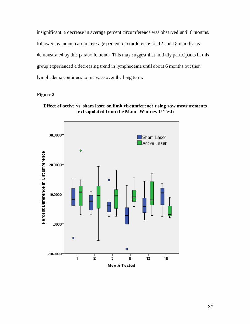
Figure 2
Effect of active vs. sham laser on limb circumference using raw measurements (extrapolated from the Mann-Whitney U Test)

28
Although statistically insignificant, greatest percent average limb circumference
difference in interquartile range and median were observed at 6 and 18 months between
the sham and active laser treatment group (figure 2).
Discussion
The purpose of this study was to determine the long-term efficacy of LLLT in
conjunction with CDT in reducing post-mastectomy lymphedema in breast cancer
survivors. Post-mastectomy lymphedema is a common complication, affecting 17 – 25%
of patients who have undergone axillary lymph node dissection (Javid & Anderson, 2013;
Lima et al., 2012). Ever since “lymphatic massage” was first established in the 1930s,
CDT has been the preferred treatment method for lymphedema (Casley-Smith et al.,
1998; Randheer et al., 2011). However, Javid and Anderson (2013) reported that recent
randomized trials have failed to confirm the efficacy of CDT over standard compression
therapy. The study by Dayes, Whelan, Julian, Parpia, Pritchard, D’Souza, Kligman,
Reise, LeBlanc, McNeely, Manchul, Wiernikowski and Levine (2013) demonstrated that
CDT had a poor long-term effect in reducing lymphedema. In their study, control groups
wore compression sleeves for 12 waking hours for 4 weeks, while the experimental group
received MLD for 1 hour by a clinical lymphedema specialist for 5 days a week for 4
weeks (Dayes et al., 2013). Both groups received patient education on skin care,
therapeutic exercise and body weight management during the follow up period. Results
indicated no significant difference between percent arm volume reduction, quality of life
measures and arm function measures at all periods of 3, 6, 12 and 24 weeks (Dayes et al.,
2013). Many authors agree that the greatest reduction in arm volume after the use of
CDT peaks in the first several weeks (Javid & Anderson, 2013).

29
Another purpose of this study was to examine the general efficacy of LLLT in
reducing lymphedema post-mastectomy. Efficacy of LLLT alone will facilitate clinical
professionals who are not licensed to carry out MLD to be involved in lymphedema care.
Opportunity exists for advanced practice nurse practitioners operating the low level lasers
in areas with shortage of clinical lymphedema specialists (Ridner, Poage-Hopper, Kanar,
Doersam, Bond, & Dietrich, 2013). However, this research aimed to determine if
LLLT, in conjunction with CDT, is still needed for long-term efficacy.
The results of this study demonstrate a general trend toward a decrease in percent
limb circumference difference over an 18 month period between the affected and
unaffected limbs in the active laser group. Despite the statistically insignificant
difference between the two groups, the decreasing trend of percent limb circumference
difference may indicate support for the alternative hypothesis that the use of LLLT in
conjunction with CDT will decrease arm girth measurement in the involved upper
extremity at 12 months when compared to sham LLLT with CDT. The indirect effect of
decrease in tissue fibrosis and direct effect of laser application may explain the arm
volume reduction in the lymphadematous arm. Work by Rufina, Lau and Cheing (2013)
demonstrated that 4 weeks of LLLT on the axillary region decreased tissue fibrosis in the
lymphadematous arm by 33%. In the likelihood that our subjects had fibrotic tissue
compositions in their lymphedematous arm, LLLT may have helped soften the fibrotic
tissue to facilitate effectiveness of LLLT. A study by Carati et al. (2003) demonstrated
the efficacy of LLLT in a double-blind, placebo-controlled study. The study had three
groups; a control group receiving only placebo laser treatment, an experimental group
receiving one dose of LLLT, and another experimental group receiving two doses of

30
LLLT with an 8-week rest period in between the doses. The results indicated no
immediate reduction of arm volume after the laser treatment, but the experimental group
receiving two doses of LLLT demonstrated significantly lower arm volume in the
affected limb than the control or the other experimental group at the 3-month period.
The results of our study also demonstrated an increasing trend in percent
circumference difference over an 18-month period between the affected and unaffected
limbs in the sham laser group receiving placebo laser treatment and conventional
CDT. The rather surprising result demonstrating the inefficacy of CDT over the 18-
month period may be explained by several factors.
The total number of 17 subjects may have been too low to delineate any
meaningful statistical significance to validate the finding of this study. It is possible that
use of CDT might have reduced the percent circumference difference between the
affected and unaffected limb if the subject numbers were higher.
Previous research has shown that the efficacy of CDT does not carry over past the
6 month follow up period, causing the arm volume to return toward the baseline level. It
examined the percent excess volume (PEV) in affected limb compared to the unaffected
limb in subjects who developed lymphedema in the lower extremity after surgical
treatment for gynecological cancer (Kim, Hwang, Kim, Change & Lee, 2012). It was
found that the effectiveness of CDT reached the greatest reduction in PEV at 3 and 6-
month post CDT, but lymphedema returned to pre-trial level at 12 and 24 months. Our
study findings were in congruence with findings by Kim et al. (2012) with average limb
reduction observed until 6 months before returning to pre-trial level at 12 and 18 months.

31
Previous research has also demonstrated that CDT has decreased long-term
effectiveness if initial PEV of subjects is less than 20%. A study by Hwang et al. (2013)
divided the treatment group into two sub groups; Group 1 composed of subjects with
initial PEV less than 20% and Group 2 composed of subjects with PEV greater than 20%.
Both groups received 2 weeks of CDT followed by a maintenance phase, during which
difference in PEV was monitored over 24 months. Their study revealed that PEV in
Group 2 was significantly lower than baseline at all time periods, while PEV in Group 1
began to increase at 6, 12, and 24 months. Our findings were in congruence with
findings by Hwang et al. (2013), as the initial PEV for our subjects was recorded as
10.86%.
Possible development of fibrosis as one of the complications of lymphedema
could have inhibited effectiveness of MLD in our study and subsequent fluid
drainage. Previous studies have demonstrated that 5 minutes of LLLT significantly
reduced indentation forces in the lymphedematous arm, indicating possible effect of
LLLT in decreasing soft tissue fibrosis (Mayrovitz & Davey, 2011). To calculate fibrosis
in skin, many authors use the tonometry method or measure the tissue indentation
resistance by calculating the amount of force required to indent tissue to a certain depth
(Mayrovitz & Davey, 2011). Tissue texture assessment by palpation, tonometry, or
calculation of tissue indentation force in future studies may reveal any presence of
fibrosis in our subjects as well as any decrease in fibrosis when LLLT in conjunction
with CDT are utilized.

32
Future Studies
Future studies could incorporate greater use of functional outcome measures as
well as a focus on effect of lymphedema severity using LLLT as opposed to the use of
standard compression sleeves. Such studies could facilitate physical therapists and other
clinicians in formulating rehabilitation goals and potentials for patients, knowing when to
make appropriate referrals to other clinical disciplines dependent on lymphedema
severity and having viable treatment alternatives for lymphedema patients inaccessible to
LLLT due to financial or geographical reasons.
Future studies could include functional outcome measures such as the Disability
of the Arm, Shoulder and Hand (DASH) outcome measure to allow assessment of
activities of daily living (ADL) affected by changes in physical function and symptoms
caused by lymphedema. In addition, future studies could measure combined upper
extremity range of motion (ROM) of shoulder flexion, abduction, and external rotation to
assess reaching tasks and self-care activities such as washing ones hair, as well as
shoulder extension and internal rotation required for donning of undergarments. Along
with goniometric measurements of functional ROM, measurements of grip strength by
hand dynamometry in functional positions may also assist physical therapists in
monitoring patient progress and formulating treatment plans (Omar et al., 2011).
Efficacy of LLLT against use of standard compression sleeves could be examined
to assess if LLLT and compression sleeves are comparable in their efficacy in reducing
lymphedema. One criticism of CDT is its high cost, which often requires 60 minutes of
MLD conducted by a certified lymphedema specialist (Lasinski et al., 2012). If LLLT
and compression sleeves demonstrate comparable efficacy, LLLT presents as another

33
viable treatment alternative for cost reduction and for patients with low compliance with
which CDT would not be appropriate.
Efficacy of LLLT on varying lymphedema severity and stages could be examined
to explore if LLLT would be a good alternative over a CDT treatment strategy for
patients with a greater PEV. A study by Ramos, O’Donnell and Knight (1999)
demonstrated that rather than the timing of treatment initiation, initial PEV at baseline is
the most significant determining factor of treatment outcome.
Future studies could also examine safety of the long-term use of LLL at currently
established wavelengths. Current recommended dose for lymphedema treatment
determined by the U.S. Food and Drug Administration is 650 to 1000 nm (Ridner et al.,
2013). However, the possible correlation between long-term use of LLL at the indicated
wavelength and increased risk of metastatic recurrence has not been studied (Lima et al.,
2012). So far, an in vitro study conducted by Leeuwen, Dekker, Byers, Vermeer and
Grevelink (1996) has shown that LLLT decreased expression of a protein that may be
responsible for cell adhesions during metastasis.
Aside from the obvious necessity of treatment advancement of lymphedema to
promote patient return to pre-morbid level of activities, the importance of patient
education on health and wellness must be advocated by all medical
professionals. Obesity, defined as body mass index (BMI) greater than 30 kg/m2, has
been shown to increase the risk of developing lymphedema by threefold (Helyer, Varnic,
Le, Leong & McCready, 2010). Although cancer stages, age, type of surgery and adjunct
treatment are considered to be associated risk factors to development of lymphedema,
BMI is an important predictor for lymphedema (Helyer et al., 2010).

34
Patient education post-mastectomy is also crucial in preventing post-surgical
complications and early detection of lymphedema. Patients should be instructed to
conduct frequent skin inspections to prevent delayed wound healing and post-operative
infections, which are associated risk factors for development of lymphedema (Javid &
Anderson, 2013). A study conducted by Lao, Li, Huang, Chen, Kuo, Chen and Wei
(2013) emphasizes the importance of patient education for early detection of
lymphedema well into the post-ALND period, as average onset of lymphedema post-
ALND is 36 months. In addition, early detection of seemingly mild lymphedema is
critical, since 20% of mild lymphedema has been shown to progress into a more severe
form of lymphedema within 1 year (Bar, Cheville, Solin, Dutta, Both & Harris, 2010).
Conclusion
Although there is not yet statistically significant evidence for the long-term
efficacy of LLLT, the most recent literature along with our findings may suggest the
following possibilities: Use of LLLT in conjunction with CDT may be indicated for long-
term management of lymphedema; Either CDT alone or LLLT with CDT may be
effective in short term management of lymphedema up to 6 months post treatment; Use
of LLLT may have a more significant effect post 6 months after the initiation of
treatment. With continuation of the study and more subjects followed to 18 months, there
may be stronger evidence to support these initial findings.

35
Appendix
Limb circumference and limb volume
Protocol previously established by Armer, et al. (2004) and Armer and Stewart (2005);
was used for circumference measurement for limb volume. Circumference of both the
affected and unaffected limbs was measured using a non-stretch tape at the following
locations: at the hand proximal to the metacarpals, wrist, and every 4 cm from the wrist
to axilla (Callaway et al., 1988; Hutzschenreuter, Wittlinger, Wittlinger, & Kurz,
1991). Non-stretch tape was used to measure the circumference in order to ensure
constant tension over soft tissue, muscle, and bony prominence, a flexible non-stretch
tape measure was used for circumferential measurement (Callaway et al., 1988; Petlund,
1991). Circumference of affected and unaffected limb was used to calculate the limb
volume of each limb. The following formula was used: V= ∑(X2 + Y2 + XY)/ 3π. V is the
sum of the limb volume, X is the circumference at one point on the limb and Y is the
circumference at a point 4 cm proximal to X. (Stantou et al., 1997).
Total limb volume
Total limb volume of affected and unaffected limb was measured using the Perometer
350S. Procedures for perometry as outlined by Armer & Stewart (2005); and as
described by the equipment manual were followed. The perometer generated a 3-
dimentional image of the limb to calculate the total limb volume. There is a standard
deviation of 8.9 ml of arm volume, less than 0.5% of LV with repeated measuring using
this method.

36
Total extra-cellular fluid volume in limb
Extra-cellular fluid volume in the affected and unaffected limb was measured using the
L-Dex XCA U400, a bio-impedance analyzer that measures resistance and impedance to
calculate extracellular fluid volume. The equipment manual provided by the
manufacturer was followed for all procedures.
Tissue texture
A physical therapist palpated the affected and unaffected limb to compare and assess
changes in skin compressibility. Skin compressibility is indicative of the extent of
fibrosis present in a limb (Piller & Thelander, 1998).
Percent of Body Fat
Percent of body fat was measured using the DF 50 BIA and InBody 230 Body
Composition Analyzer. This device sends an extremely weak electrical current of 50 kHz
and less than 500 μA through the body to determine the amount of water in each tissue.
Standard protocols established by each of the manufacturers were followed for all
measurements. Participants were requested to remove all jewelry and metal objects,
before being positioned in stand and supine position. The validity of using the BIA as the
reference method ranged from 0.84–0.96 in white women (Pineau, Guihard-Costa, &
Bocquet, 2007). The reliability (ICC) of BIA ranged from 0.97–0.99 (Jackson et al.,
1988).
Body Mass Index
Body Mass Index was calculated by entering the body weight and height into the DF50
BIA &InBody 230 device. The device uses the following formula to calculate BMI

37
based on a ratio between body weight and height: BMI = weight (lb) / height (inches)
/height (inches) x 703 (Callaway, Chumlea, & Bouchard, 1988).
Pain intensity
Pain intensity associated with lymphedema was measured using a self-reported,
numeric pain scale. Participants quantified their pain on a numeric scale from zero to
10. A rating of zero signifies no pain while a rating of 10 signifies severely debilitating
pain. It is a validated scale and a standard tool for rating pain.
Symptom experience of lymphedema
The study used the Lymphedema and Breast Cancer Symptom Experience Index
in order to monitor the participants’ overall experience of the lymphedema. This index
was a structured interview tool adapted from the Lymphedema and Breast Cancer
Questionnaire (LBCQ) (Armer, Radina, Porock, & Culberston, 2003; Fu et al,
2008). The content of the Breast Cancer Symptom Experience Index is described under
Materials.
Perceived health outcome
The Short-Form Health Survey version 2 [SF-36v2] was utilized to assess the
well-being of physical and mental health of each participant. The content of the survey is
described under Materials.
Photography of limbs
Photographs of the chest, affected and unaffected extremity were taken for each
participant in order to monitor any visual changes over the upper quadrants of the body.
Each subject was given the right to refuse the photo. If photos were taken, they were

38
managed using the Individual Identifier Number. Photos were stored in a locked cabinet
in a locked office of the Principal Investigator.
Demographics and Medical Information
A Demographics and Medical Information Tool as previously outlined by Fu,
Axelrod and Haber (2008) was used to record the age, diagnosis, treatment, nodal status,
numbers of lymph nodes removed, co-morbidities, and family medical history (breast
cancer history, breast cancer gene status, morbidity of lower extremities).
Throughout the 18 month study period, patients were instructed to report any
adverse changes to their affected limb such as rash, blister, redness swelling and/or
increased temperature of the tissue. Participants were also instructed to receive any blood
pressure evaluation, drawing of blood, injections and vaccinations on the unaffected
upper limb whenever possible.
At the end of the study, participants were contacted and informed about whether
they were part of the placebo or an intervention group. In the event that a medical
problem arose, participants were instructed to contact Teresa Denham, PT, MA, Principal
Investigator and Outpatient Physical Therapy Manager at New York University Medical
Center/Rusk Institute of Rehabilitation Medicine, at (212) 263-8466.

39
References
American Cancer Society. (2011). Cancer basics. Retrieved June 13, 2011, from http://www.cancer.org/Cancer/CancerBasics/what-is-cancer
Armer, J., Fu, M., Wainstock, J., Zagar, E., Jacobs, L. (2004)Lymphedema following
breast cancer treatment, including sentinel lymph node biopsy. Lymphology 37, 73-91. Armer, J., Radina, M., Porock, D., & Culbertson, S. (2003). Predicting breast cancer-
related lymphedema using self-reported symptoms. Nursing Research, 52(6), 370-379.
Armer, J., & Ridner, S. (2006). Measurement techniques in assessment of lymphedema.
Lymph LINK, 18(3), 1-2-4. Armer, J., & Stewart, B. (2005). A comparison of four diagnostic criteria for
lymphedema in a post-breast cancer population. Lymphatic Research & Biology, 3(4), 208-217. Bar Ad, V., Cheville, A., Solin, L., Dutta, P., Both, S., & Harris, E.. (2010) Time course of mild arm lymphedema after breast conservation treatment for
early-stage breast cancer. International Journal of Radiation Oncoloy Biology and Physics, 76(1), 85– 90.
Breastcancer.org. (2007). Axillary dissection. Retrieved June 13, 2011, from
http://breastcancer.org/treatment/surgery/lymph_node_removal/axillary_dissection.jsp
Breastcancer.org. (2008). How many nodes removed? Retrieved June 13, 2011, from
http://breastcancer.org/treatment/surgery/lymph_node_removal/number_removed.jsp
Breastcancer.org. (2008). Lymph node dissection: What to expect. Retrieved June 13,
2011, from http://breastcancer.org/treatment/surgery/lymph_node_removal/ dissection expectations.jsp
Breastcancer.org. (2008). What is breast cancer? Retrieved June 13, 2011, from
http://breastcancer.org/symptoms/understand_bc/what_is_bc.jsp

40
Breastcancer.org. (2008). Why are lymph nodes important? Retrieved June 13, 2011, from http://www.breastcancer.org/treatment/surgery/lymph_node_removal/importance.jsp
Breastcancer.org. (2009). Lymph node removal. Retrieved June 13, 2011, from
http://breastcancer.org/treatment/surgery/lymph_node_removal/ Breastcancer.org. (2010). Sentinel lymph node dissection. Retrieved June 13, 2011, from
http://breastcancer.org/treatment/surgery/lymph_node_removal/sentinel_dissection/
Breastcancer.org. (2011). Non-invasive or invasive breast cancer. Retrieved June 13,
2011, from http://breastcancer.org/symptoms/diagnosis/invasive.jsp Breastcancer.org. (2011). Symptoms & diagnosis. Retrieved June 13, 2011, from
http://breastcancer.org/symptoms/ Breastcancer.org. (2011). U.S. breast cancer statistics. Retrieved June 13, 2011, from
http://breastcancer.org/symptoms understand_bc/statistics.jsp Breastcancer.org. (2011). Vascular or lymphatic system invasion. Retrieved June 13,
2011, from http://breastcancer.org/symptoms/diagnosis/vasc_lymph_inv.jsp Brennan, M., DePompolo,R., Garden, F. (1996). Focused review: post-mastectomy lymphedema. Archives of Physical Medicine and Rehabilitation, 77(3), S74 – 80. Callaway, C., Chumlea, W., Bouchard, C, (1988). Circumferences. Anthropometric
Standardization Reference Manual, In Lohman TG, Roche AF, Martorell R, eds., 39-51.
Carati, C., Anderson, S., Gannon, B., Piller, N. (2003). Treatment of post-mastectomy lymphedema with low-level laser therapy: A double blind, placebo-controlled
trial. Cancer, 98 (6), 1114 – 1122. Carcia, C., Martin, R., Houck, J., Wukich, D. 2010. Achilles Pain, Stiffness, and Muscle
Power Deficits: Achilles Tendinitis. J Orthop Sports Phys Ther 40(9):A1-A26.

41
Casley-Smith, J., Boris, M., Weindorf, S., Lasinski, B. (1998). Treatment for lymphedema of the arm – The Casley-Smith method: A noninvasive method produces continued reduction. Cancer, 83 (12 Suppl American), 2843 – 2860.
Cetin E., Ozdincler, AR., Yeldan, I. 2009. The effectiveness of low-level laser therapy
on shoulder function in subacromial impingement syndrome. Disabil Rehabil 31(11):935-40
Cheifetz, O., Haley, L. (2010). Management of secondary lymphedema related to breast
cancer. NCBI, from http://www.ncbi.nlm.nih.gov/pmc/articles/PMC3001918/?tool=pubmed
Cohen, S., Payne, D., Tunke, R. (2001). Lymphedema: Strategies for management.
Cancer, 92(4 Suppl), 980 – 987. Dayes, I., Whelan, T., Julian, J., Parpia, S., Pritchard, K., Souza, D., Kligman, L., Reise,
D., LeBlance, L., McNeely, M. Manchul, L., Wiernikowski, J & Levine, M.N. (2013) Randomized trial of decongestive lymphatic therapy for the treatment of lymphedema in women with breast cancer. Journal of Clinical Oncology, 31 (30), 3758 – 3763.
Djavid, G., Mehrdad, R., Ghasemi, M., Hasan-Zadeh, H., Sotoodeh-Manesh,
A., Pouryaghoub, G. (2007) In chronic low back pain, low level laser therapy combined with exercise is more beneficial than exercise alone in the long-term: a randomised trial. Aust J Physiother. 53(3):155-60.
Dirican, A., Andacoglu, O., Johnson, R., McGuire, K., Mager, L., & Soran, A. (2011).
The short-term effects of low-level laser therapy in the management of breast-cancer-related lymphedema. Support Care Cancer, 19, 685-690.
Ferrante, M., Petrini, M., Trentini, P., Perfetti, G., Spoto, G. (2012). Effect of low-
level laser therapy after extraction of impacted lower third molars. Lasers Med Sci.
Foroughi, N., Dylke, E. S., Paterson, R. D., Sparrow, K. A., Fan, J., Warwick, E. B. G., et
al. (2011). Inter-rater reliability of arm circumference measurement. Lymphatic Research and Biology, 9(2), 101-107.
Frazier, T. G., Rosenberg, A. L. & Weiss, M. C. (2010). Surgery. Retrieved June 13,
2011, from http://breastcancer.org/treatments/surgery/
42
Fu, M., Axelrod, D., Haber, J. (2008). Breast cancer-related lymphedema: Information, symptoms, and risk reduction behaviors. Journal of Nursing Scholarship, 40(4), 341-348.
Fu, M., Ridner, S., Armer,J. (2009). Post-breast cancer lymphedema: Part 1. The
American Journal of Nursing, 109 (7), 48 – 54 Fu, M., Ridner, S., & Armer, J. (2009). The pathophysiology of lymphedema: Online-
only content for "post-breast cancer lymphedema: Part1". American Journal of Nursing, 06/13/2011. Retrieved from http://links.lww.com/AJN/A1
Fu, M., Ridner, S., & Armer, J. (2009). Post-breast cancer lymphedema: Part 1. The
American Journal of Nursing, 109(7),48-54. Fu, M. R., & Rosedale, M. (2009). Breast cancer survivors' experiences of lymphedema-
related symptoms. Journal of Pain and Symptom Management, 38(6), 849-859. Gary, D. E. (2007). Lymphedema diagnosis and management. Journal of the American
Academy of Nurse Practitioners, 19(2), 72-78. Gur, A., Cosut, A., Sarac, A. J., Cevik, R., Nas, K., & and Uyar, A. (2003). Efficacy of
different therapy regimes of low-power laser in painful osteoarthritis of the knee: A double-blind and randomized-controlled trial. Lasers in Surgery and Medicine, 33, 330-338.
Gur, A., Karakoc, M., Cevik, R., Sarac, A. J., & Karakoc, M. (2003). Efficacy of low
power laser therapy and exercise on pain and functions in chronic low back pain. Lasers in Surgery and Medicine, 32, 233-238.
Guyton, A. C., & Hall, J. E. (2011). The microcirculation and lymphatic system:
Capillary fluid exchange, interstitial fluid, and lymph flow. Guyton and hall textbook of medical physiology (12th ed.). Philadelphia: Saunders Elsevier, 186-189.
Haghighat, S., Lotfi-Tokaldany, M., Maboudi, A., Bahadori, A., Weiss,
J. (2013). Predictive factors of response to phase I complete decongestive therapy in upper extremity lymphedema following breast carcinoma in Iran. Lymphology. 46(2): 97-104.

43
Harmer, V. (2009) Breast cancer-related lymphoedema: Implications for primary care. - Br J Community Nurs. 14(10):S15-6, 18-9.
Helyer, L., Varnic, M., Le, L., Leong, W., McCready, D. (2010) Obesity is a risk factor
for developing postoperative lymphedema in breast cancer patients. The Breast Journal, 16, 48 – 54.
Hopkins, J., McLoda, T., Seegmiller, J., Baxter, G. (2004). Low-level laser therapy
facilitates superficial wound healing in humans: A triple-blind, sham-controlled study. Journal of Athletic Training, 39(3), 223-229.
Hutzschenreuter, P., Wittlinger, H., Wittlinger, G., Kurz, I. (1991). Post-mastectomy arm
lymphedema: Treated by manual lymph drainage and compression bandage therapy. PMR, 1(6), 166-170.
Hwang, J., Hwang, J., Kim, T., Lee, S., Chang, H., & Chu, I. (2013) Long-term effects of
complex decongestive therapy in breast cancer patients with arm lymphedema after axillary dissection. Journal of Annual Rehabilitation Medicine, 37(5), 690 – 697.
Javid, S. & Anderson, B. (2013) Mounting evidence against complex decongestive
therapy as a first-line treatment for early lymphedema. Journal of Clinical Oncology, 31(30) 3737 – 3738.
Johansson K, Albertsson M, Ingvar C, & Ekdahl C. (1999) Effects of compression
bandaging with or without manual lymph drainage treatment in patients with postoperative arm lymphedema. - Lymphology.32(3):103-10.
Kaviani, A., Fateh, M., Yousefi Nooraie, R., Alinagi-zadeh, M., Ataie-Fashtami, L.
(2006). Low- level laser therapy in the treatment of post-mastectomy lymphoedema. Lasers in Medical Science, 21 (2), 90-94.
Ko, D., Lerner, R., Klose, G., & Cosimi, A. (1998) Effective treatment of lymphedema of
the extremities. - Arch Surg;133(4):452-8. Kozanoglu, E., Basaran, S., Paydas, S., Sarpel, T. (2009). Efficacy of pneumatic
compression and low-level therapy in the treatment of post-mastectomy lyphoedema: A randomized controlled trial. Clinical Rehabilitation, 23 (2), 117-124.

44
Lasinski, B., Thrift, K., Squire, D., Austin, M., Mith, K., Wanchai, A., Green, J., Stewart, B., Cormier, J., Armer, J. (2012) A systematic review of the evidence for complete decongestive therapy in the treatment of lymphedema from 2004 – 2011. American Academy of Physical Medicine and Rehabilitation, 4, 580 – 601.
Lau, R. W. L., & Cheing, G. L. Y. (2009). Managing post-mastectomy lymphedema with
low-level laser therapy. Photomedicine and Laser Surgery, 27(5), 763-769. Lawenda, B., Mondry, T., Johnstone, P. (2009) Lymphedema: A primer on the identification and management of a chronic condition in oncologic treatment. CA:
A Cancer Journal for Clinicians, 59 (1), 8 – 24. Leal N, Carrara, H.H., Vieira, K., & Ferreira, C.H. (2009) Physiotherapy treatments for
breast cancer-related lymphedema: A literature review. - Rev Lat Am Enfermagem;17(5):730-6.
Lewy, V., Danadian, K., & Arslanian, S. (1999). Determination of body composition in
african-american children: Validation of bioelectrical impedence with dual energy X-ray absorptiometry. Journal of Pediatric Endocrinology & Metabolism: JPEM, 12(3), 443-448.
Liao, S., Li, S., Huang, H., Chen, S., Kuo, S,. Chen, D., Wei, T. (2013). The efficacy of
complex decongestive physiotherapy (CDP) and predictive factors of lymphedema severity and response to CDP in breast cancer-related lymphedema (BCRL). Breast. 22(5):703-6.
Lima, M., Lima, J., Figueiredo, M., Carvalho de Andrade M., Bergman, A. (2012). Low-
level laser therapy in secondary lymphedema after breast cancer: systematic review. Lasers Medical Science, 11(29).
Mayrovitz, H., Davey S. (2011). Changes in tissue water and indentation resistance of
lymphedematous limbs accompanying low level laser therapy (LLLT) of fibrotic skin. Lymphology. 2011 Dec;44(4):168-77.
Martín, M., Hernández, M., Avendaño, C., Rodríguez, F., Martínez, H. (2011). Manual
lymphatic drainage therapy in patients with breast cancer related lymphoedema. BMC Cancer, from http://www.biomedcentral.com/1471-2407/11/94
McNeely, M., Magee, D., Lees, A., Bagnall, K., Haykowsky, M., Hanson, J. (2004). The
addition of manual lymph drainage to compression therapy for breast cancer related lymphedema: A randomized controlled trial. Breast cancer research and

45
treatment, from http://www.springerlink.com/content/pm25575l0765836l/fulltext.html
Medical photochemical & low level laser therapy for treating lymphedema/lymphedema
Retrieved 11/22/2009, 2009, from http://www.riancorp.com/ Mester, E., Spiry, T., Szende, B. , Tota, J. (1971). “Effect of laser rays on wound
healing,” Am J Surg vol. 122, pp. 532-535, 1971 Mondry, T., Riffenburgh, R., & Johnstone, P. (2004). Prospective trial of complete
decongestive therapy for upper extremity lymphedema after breast cancer therapy. - Cancer J;10(1):42-8; Discussion 17-9.
Morrell, R. M., Halyard, M. Y., Schild, S. E., Ali, M. S., Gunderson, L. L., & Pockaj, B.
A. (2005). Breast cancer-related lymphedema. Mayo Clinic Proceedings.Mayo Clinic, 80(11), 1480-1484.
Mortimer, P.S (1998). The pathophysiology of lymphedema. Cancer, 83 (12), 2798 –
2802. Moseley, A., Carati, C., Piller, N. (2007). A systematic review of common conservative
therapies for arm lymphoedema secondary to breast cancer treatment. Annals of Oncology: Official Journal of the European Society for Medical Oncology/ESMO, 18(4), 639-646.
Nathanson, S., Wachna, D., Gilman, D., Karvelis, K., Havstad, S., Ferrara, J. (2001).
Pathways of lymphatic drainage from the breast. Annals of Surgical Oncology, 8(10), 837-843.
Omar, M., Shaheen, A., Zafar, H. (2012). A sytematic review of the effect of low level
laser therapy in the management of breast cancer-related lymphedema. Support Care Cancer, 20 (11); 2977-84.
Omar, M. T. A., Ebid, A. A., & El Morsy, A. M. (2011). Treatment of post-mastectomy
lymphedema with laser therapy: Double blind placebo control randomized study. Journal of Surgical Research, 165, 82-90.
Omron Healthcare, I. Fitness diagnostics. Retrieved 07/23, 2011, from
http://www.omronhealthcare.com/service-and-support/faq/fitness-diagnostics/

46
Omron Healthcare, I. (2008). Omron instruction manual full body sensor body composition monitor and scale model HBF-516. Bannockburn, Illinois 6001.
Omar, A., Abd-El-Gayed Ebid, A., El Morsy, A. (2011). Treatment of post-mastectomy
lymphedema with laser therapy: double blind placebo control randomized study. J Surg Res. 165(1):82-90.
Paskett, E., Dean, J., Oliveri, J., Harrop, J. (2012) Cancer-related lymphedema risk
factors, diagnosis, treatment, and impact: a review. Journal of Clinical Oncology, 30(30).
Petlund, C. (1991). Volumetry of limbs. In W.I. Olszewski, (ed). (1991). Lymph Stasis:
Pathophysiology, Diagnosis and Treatment. Boston: CRC Press, 444-51. Piller, N., Thelander, A. (1998). Treatment of chronic post-mastectomy lymphedema
with low level laser therapy: A 2.5 year follow-up. Lymphology, 31 (2), 74-86. Posten, W., Wrone, D., Dover, J., Arndt, A., Silapunt, S., Alam, M. (2005) Low-level
laser therapy for wound healing: mechanism and efficacy. Dermatol Surg 31: 334 – 340.
Price, D., McGrath, P., Rafii, A., & Buckingham, B. (1983). The validation of visual
analogue scales as ratio scale measures for chronic and experimental pain. Pain, 17(1), 45-56.
Ramos, S., O’Donnell, L., & Knight, G. (1999) Edema volume, not timing, is the key to
success in lymphedema treatment. American Journal of Surgery, 178(4) 311 - 315 Randheer, S., Kadambari, D., Srinivasan, K., Bhuyaneswari, V., Bhanumathy, M., Salaja,
R. (2011) Comprehensive decongestive therapy in post-mastectomy lymphedema: An Indian perspective. Indian J Cancer, 48: 397 – 402.
Render, R. (2007). ImpediMed FDA 501(K) form- summary ImpediMed imp XCA
extracellular fluid analysis. Retrieved July 10, 2010, from http://www.accessdata.fda.gov/cdrh_docs/pdf5/K050415.pdf
Ridner, S. (2002). Breast cancer lymphedema: Pathophysiology and risk reduction
guidelines. Oncology Nursing Forum, 29(9), 1285-1293.

47
Ridner, S., Poage-Hooper, E., Kanar, C., Doresam, J., Bond, S., Dietrich, M., (2013). A
pilot randomized clinical trial evaluating low level laser therapy as a complementary and alternative treatment to manual lymphatic drainage for breast cancer-related lymphedema. Oncology Nursing Forum, 40(4), 383-393.
Saskia, R. J., & Thiadens, R. N. (2010). What is lymphedema? Retrieved June 13, 2011, from http://lymphnet.org/lymphedemaFAQs/overview.htm
Siegel, R., Ward, E., Brawley, O., Jemal, A. (2011). The impact of eliminating socioeconomic and racial disparities on premature cancer deaths. CA: A Cancer Journal for Clinicians 61 (4), 212 – 236.
Sierla, R., Lee, T., Black, D., Kilbreath, S. (2013). Lymphedema following breast cancer: regions affected, severity of symptoms, and benefits of treatment from the patients' perspective. Clinical Journal of Oncology Nursing; 17(3), 32
Smoot, B., Johnson, M., Duda, J., Krasnoff, J., Dodd, M. (2012). Cardiorespiratory
fitness in women with and without lymphedema following breast cancer treatment. Cancer Clin Oncol, 1(1): 21-31.
Stanton, A. W., Northfield, J. W., Holroyd, B., Mortimer, P. S., & Levick, J. R. (1997).
Validation of an optoelectronic limb volumeter (perometer). Lymphology, 30(2), 77-97.
Tammela, T. and Alitalo, K. (2010). Lymphangiogenesis: Molecular mechanisms and
future promise. Cell (140) 4: 460-476. Tsai, R., Dennis, L., Lynch, C., Snetselaar, L., Zamba, G., Scott-Conner, C. (2009). The
risk of developing arm lymphedema among breast cancer survivors: A meta-analysis of treatment factors. Annals of Surgical Oncology, 16(7), 1959-1972.
Tumilty, S., Munn, J., McDonough, S., Hurley, D. A., Brasford, J. R., & Baster, G. D.
(2010). Low level laser treatment of tendinopathy: A systematic reivew with meta-analysis. Photomedicine and Laser Surgery, 28(1), 3-16.
Van Leeuwen, R., Dekker, S., Byers, H., Vermeer, B., & Grevelink, J. (1996) Modulation
of alpha 4 beta 1 and alpha 5 beta 1 integrin expression: heterogeneous effects of Q-switched ruby, Nd: YAG, and alexandrite lasers on melanoma cells in vitro. Lasers in Surgery and Medicine, 18(1), 63 – 71

48
Velanovich, V., & Szymanski, W. (1999). Quality of life of breast cancer patients with
lymphedema. American Journal of Surgery, 177(3), 184-187. Ververs, J. M., Roumen, R. M., Vingerhoets, A. J., Vreugdenhil, G., Coebergh, J. W.,
Crommelin, M. A., et al. (2001). Risk, severity and predictors of physical and psychological morbidity after axillary lymph node dissection for breast cancer. European Journal of Cancer (Oxford, England: 1990), 37(8), 991-999.
Vignes S, Porcher R, Arrault M, & Dupuy A. (2011) Factors influencing breast cancer-
related lymphedema volume after intensive decongestive physiotherapy. - Support Care Cancer (7):935-40.
Ware, J.E., Kosinski, M., Bayliss, M.S., McHorney, C.A., Rogers, W.H., Raczek, A.
(1995). Comparison of methods for the scoring and statistical analysis of SF-36® health profiles and summary measures: summary of results from the medical outcomes study. Med Care; 33 (Suppl. 4): AS264 – AS279.
Ware, J., Kosinski, M., Dewey, J. (2000). How to score version two of the SF-36 health
survey. Lincoln, RI; QualityMetric, Incorporated. Ware, J., Kosinski, M., Gandek, B., Aaronson N., Alonso J., Apolone G., Bech
P., Brazier, J., Bullinger M., Kaasa S., Leplege, A., Prieto, L., Sullivan M. (1998). The factor structure of the sf-36® Health survey in 10 countries: Results from the international quality of life assessment (IQOLA) project. J Clin Epidemiol; 51 (11): 1159 – 1165.
Williams, A. (2010) Manual lymphatic drainage: Exploring the history and evidence
base. - Br J Community Nurs. (4):S18-24. Williams, A.F., Vadgama, A., Franks, P.J., & Mortimer, P.S.. (2002) A randomized
controlled crossover study of manual lymphatic drainage therapy in women with breast cancer-related lymphoedema. - Eur J Cancer Care (Engl);11(4):254-61.
Yeldon, I., Cetin E., Ozincler, A. (2009) The effectiveness of low-level laser therapy on
shoulder function in subacromial impingement syndrome Disabil Rehabil. 31(11):935-40.


![Low Level Laser (Light) Therapy (LLLT) for Cosmetic Medicine ......Low Level Laser (Light) Therapy (LLLT) for Cosmetic Medicine and Dermatology Mossum K. Sawhney [1] and Michael R.](https://static.fdocuments.us/doc/165x107/60324a44e0ded43d400877e7/low-level-laser-light-therapy-lllt-for-cosmetic-medicine-low-level-laser.jpg)