The lncRNA VELUCTstrongly regulates viability of lung ...
12
5458–5469 Nucleic Acids Research, 2017, Vol. 45, No. 9 Published online 4 February 2017 doi: 10.1093/nar/gkx076 The lncRNA VELUCT strongly regulates viability of lung cancer cells despite its extremely low abundance Jana Seiler 1 , Marco Breinig 2 , Ma¨ ıwen Caudron-Herger 1 , Maria Polycarpou-Schwarz 1 , Michael Boutros 2 and Sven Diederichs 1,3,4,5,* 1 Division of RNA Biology & Cancer, German Cancer Research Center (DKFZ), Heidelberg, Germany, 2 Division of Signaling and Functional Genomics, German Cancer Research Center (DKFZ), Heidelberg, Germany, 3 Division of Cancer Research, Dept. of Thoracic Surgery, Medical Center –University of Freiburg, Freiburg, Germany, 4 Faculty of Medicine, University of Freiburg, Freiburg, Germany and 5 German Cancer Consortium (DKTK), Freiburg, Germany Received December 13, 2016; Revised January 24, 2017; Editorial Decision January 25, 2017; Accepted January 26, 2017 ABSTRACT Little is known about the function of most non-coding RNAs (ncRNAs). The majority of long ncRNAs (lncR- NAs) is expressed at very low levels and it is a mat- ter of intense debate whether these can be of func- tional relevance. Here, we identified lncRNAs regu- lating the viability of lung cancer cells in a high- throughput RNA interference screen. Based on our previous expression profiling, we designed an siRNA library targeting 638 lncRNAs upregulated in human cancer. In a functional siRNA screen analyzing the viability of lung cancer cells, the most prominent hit was a novel lncRNA which we called Viability En- hancing LUng Cancer Transcript (VELUCT). In sil- ico analyses confirmed the non-coding properties of the transcript. Surprisingly, VELUCT was below the detection limit in total RNA from NCI-H460 cells by RT-qPCR as well as RNA-Seq, but was robustly de- tected in the chromatin-associated RNA fraction. It is an extremely low abundant lncRNA with an RNA copy number of less than one copy per cell. Block- ing transcription with actinomycin D revealed that VELUCT RNA was highly unstable which may par- tially explain its low steady-state concentration. De- spite its extremely low abundance, loss-of-function of VELUCT with three independent experimental ap- proaches in three different lung cancer cell lines led to a significant reduction of cell viability: Next to four individual siRNAs, also two complex siPOOLs as well as two antisense oligonucleotides confirmed the strong and specific phenotype. In summary, the extremely low abundant lncRNA VELUCT is essential for regulation of cell viability in several lung cancer cell lines. Hence, VELUCT is the first example for a lncRNA that is expressed at a very low level, but has a strong loss-of-function phenotype. Thus, our study proves that at least individual low-abundant lncRNAs can play an important functional role. INTRODUCTION Protein-coding genes were long assumed to be the main and probably only molecular drivers in a cell and RNA molecules were viewed as mediator molecules and servants for processes in protein synthesis. However, deep sequenc- ing methods revealed that a large part of the human genome (∼75%) is transcribed, whereas only 1.5% encode for pro- teins (1). Long ncRNAs (lncRNAs) form a highly diverse non-coding RNA (ncRNA) class with >200 nucleotides in length that lack an open reading frame of significant length (2). The number of lncRNA genes in the human genome is still increasing, but recent analyses suggest at least numbers similar to protein-coding genes (3). The ex- pression of lncRNAs is highly regulated and depends on the developmental stage (4), on the tissue (5,6) and on cell subtypes (7,8). So far, only a small fraction of lncR- NAs is functionally characterized, but several examples are shown to play a critical role in physiological and patholog- ical processes such as cancer. Transcripts such as HOTAIR (HOx Transcript Antisense Intergenic RNA) or MALAT1 (Metastasis-Associated Lung Adenocarcinoma Transcript 1) are upregulated in lung cancer (9,10) and associated with enhanced proliferation, metastasis and poor prognosis (9– 12). However, knowledge is mostly limited to the most abun- dant lncRNAs. Contrarily, the majority of lncRNAs is of very low abundance, with many lncRNAs having a copy number of even lower than one per cell (13,14). Thus, an intense debate arose whether any of these many low abun- dant lncRNAs could play important physiological roles in a cell. Opponents of this theory argue that most of the low abundant lncRNAs are non-functional ‘junk’ (15,16), i.e. spurious RNAs that might derive from leaky transcrip- * To whom correspondence should be addressed. Tel: +49 6221 424383; Fax: +49 6221 424384; Email: [email protected] C The Author(s) 2017. Published by Oxford University Press on behalf of Nucleic Acids Research. This is an Open Access article distributed under the terms of the Creative Commons Attribution License (http://creativecommons.org/licenses/by-nc/4.0/), which permits non-commercial re-use, distribution, and reproduction in any medium, provided the original work is properly cited. For commercial re-use, please contact [email protected]
Transcript of The lncRNA VELUCTstrongly regulates viability of lung ...

5458–5469 Nucleic Acids Research, 2017, Vol. 45, No. 9 Published online 4 February 2017doi: 10.1093/nar/gkx076
The lncRNA VELUCT strongly regulates viability oflung cancer cells despite its extremely low abundanceJana Seiler1, Marco Breinig2, Maıwen Caudron-Herger1, Maria Polycarpou-Schwarz1,Michael Boutros2 and Sven Diederichs1,3,4,5,*
1Division of RNA Biology & Cancer, German Cancer Research Center (DKFZ), Heidelberg, Germany, 2Division ofSignaling and Functional Genomics, German Cancer Research Center (DKFZ), Heidelberg, Germany, 3Division ofCancer Research, Dept. of Thoracic Surgery, Medical Center – University of Freiburg, Freiburg, Germany, 4Faculty ofMedicine, University of Freiburg, Freiburg, Germany and 5German Cancer Consortium (DKTK), Freiburg, Germany
Received December 13, 2016; Revised January 24, 2017; Editorial Decision January 25, 2017; Accepted January 26, 2017
ABSTRACT
Little is known about the function of most non-codingRNAs (ncRNAs). The majority of long ncRNAs (lncR-NAs) is expressed at very low levels and it is a mat-ter of intense debate whether these can be of func-tional relevance. Here, we identified lncRNAs regu-lating the viability of lung cancer cells in a high-throughput RNA interference screen. Based on ourprevious expression profiling, we designed an siRNAlibrary targeting 638 lncRNAs upregulated in humancancer. In a functional siRNA screen analyzing theviability of lung cancer cells, the most prominent hitwas a novel lncRNA which we called Viability En-hancing LUng Cancer Transcript (VELUCT). In sil-ico analyses confirmed the non-coding properties ofthe transcript. Surprisingly, VELUCT was below thedetection limit in total RNA from NCI-H460 cells byRT-qPCR as well as RNA-Seq, but was robustly de-tected in the chromatin-associated RNA fraction. Itis an extremely low abundant lncRNA with an RNAcopy number of less than one copy per cell. Block-ing transcription with actinomycin D revealed thatVELUCT RNA was highly unstable which may par-tially explain its low steady-state concentration. De-spite its extremely low abundance, loss-of-functionof VELUCT with three independent experimental ap-proaches in three different lung cancer cell lines ledto a significant reduction of cell viability: Next tofour individual siRNAs, also two complex siPOOLsas well as two antisense oligonucleotides confirmedthe strong and specific phenotype. In summary, theextremely low abundant lncRNA VELUCT is essentialfor regulation of cell viability in several lung cancercell lines. Hence, VELUCT is the first example for a
lncRNA that is expressed at a very low level, but hasa strong loss-of-function phenotype. Thus, our studyproves that at least individual low-abundant lncRNAscan play an important functional role.
INTRODUCTION
Protein-coding genes were long assumed to be the mainand probably only molecular drivers in a cell and RNAmolecules were viewed as mediator molecules and servantsfor processes in protein synthesis. However, deep sequenc-ing methods revealed that a large part of the human genome(∼75%) is transcribed, whereas only 1.5% encode for pro-teins (1). Long ncRNAs (lncRNAs) form a highly diversenon-coding RNA (ncRNA) class with >200 nucleotidesin length that lack an open reading frame of significantlength (2). The number of lncRNA genes in the humangenome is still increasing, but recent analyses suggest atleast numbers similar to protein-coding genes (3). The ex-pression of lncRNAs is highly regulated and depends onthe developmental stage (4), on the tissue (5,6) and oncell subtypes (7,8). So far, only a small fraction of lncR-NAs is functionally characterized, but several examples areshown to play a critical role in physiological and patholog-ical processes such as cancer. Transcripts such as HOTAIR(HOx Transcript Antisense Intergenic RNA) or MALAT1(Metastasis-Associated Lung Adenocarcinoma Transcript1) are upregulated in lung cancer (9,10) and associated withenhanced proliferation, metastasis and poor prognosis (9–12). However, knowledge is mostly limited to the most abun-dant lncRNAs. Contrarily, the majority of lncRNAs is ofvery low abundance, with many lncRNAs having a copynumber of even lower than one per cell (13,14). Thus, anintense debate arose whether any of these many low abun-dant lncRNAs could play important physiological roles ina cell. Opponents of this theory argue that most of thelow abundant lncRNAs are non-functional ‘junk’ (15,16),i.e. spurious RNAs that might derive from leaky transcrip-
*To whom correspondence should be addressed. Tel: +49 6221 424383; Fax: +49 6221 424384; Email: [email protected]
C© The Author(s) 2017. Published by Oxford University Press on behalf of Nucleic Acids Research.This is an Open Access article distributed under the terms of the Creative Commons Attribution License (http://creativecommons.org/licenses/by-nc/4.0/), whichpermits non-commercial re-use, distribution, and reproduction in any medium, provided the original work is properly cited. For commercial re-use, please [email protected]

Nucleic Acids Research, 2017, Vol. 45, No. 9 5459
tion. Those junk transcripts might be rapidly removed be-cause of quality control mechanisms resulting in their lowabundance (17). Proponents of this theory argue in con-trast, that the majority of lncRNAs––although expressedat a low level––could indeed play an important role in a cell(18,19). Small amounts of RNAs might be sufficient to trig-ger downstream effects, e.g. if acting directly on the genomeat a unique allele.
Here, we identify lncRNAs that regulate the viability inlung cancer cell lines in a high-throughput RNA interfer-ence screen. We identified the novel lncRNA VELUCT (Vi-ability Enhancing LUng Cancer Transcript) which was ex-tremely low abundant and only reproducibly detectable inthe chromatin-associated RNA fraction. Nonetheless, thefunctional importance of this transcript for cell viability andproliferation was validated using multiple independent si-lencing approaches. Thus, VELUCT is an example for alncRNA that is expressed at a very low level, but has astrong phenotype upon knockdown.
MATERIALS AND METHODS
Cell lines and actinomycin D treatment
NCI-H460, NCI-H1944 and NCI-H1437 lung cancer cellswere propagated in RPMI + 10% FCS. H1944 and H1437were purchased from ATCC. H460 were authenticated us-ing Multiplex Cell Authentication by Multiplexion (Heidel-berg, Germany) as described recently (20). The SNP pro-files matched known profiles or were unique. Cells were reg-ularly tested for mycoplasma. For actinomycin D (actD)treatment, 2 × 10⁶ H460 cells were seeded in a 10 cm dishand incubated for 24 h. The medium was aspirated and 7.5ml complete medium was added containing 10 �g/ml actD(resuspended in DMSO) or the same volume of DMSO asa control.
siRNA Library
The library contained approx. 3100 single Silencer SelectsiRNAs (Life Technologies) targeting 638 lncRNAs thatwere upregulated in lung, liver and breast cancer samplesaccording to previous studies. The library was arrayed inwhite 384-well plates using a Biomek FX200 liquid han-dling system (Beckman Coulter). Each well contained 5�l of 300 nM siRNA. Column 23 and 24 of all screeningplates contained three positive siRNA controls (siCOPB2,siKIF11, siPLK1; each in duplicates per plate) and non-targeting negative siRNA controls (NC 1, NC 2, NC 3;each in quadruplicates per plate).
Transfection with silencing reagents
Cells were reverse transfected with 10 or 30 nM RNAireagents (siRNAs, siPOOLs or ASOs) in different plate for-mats. H460 cells were transfected with Dharmafect1 (Dhar-macon); H1944 and H1437 with RNAiMAX (Life Tech-nologies). For transfection in 384-well plates, 0.05 �l trans-fection reagent per well were diluted in 4.95 �l RPMI(Sigma) and incubated for 10 min (for siPOOLs (siTOOLsBiotech) and siRNAs) or 5 min (for ASOs (Exiqon)). 10�l RPMI were added and incubated with 5 �l silencing
reagent for 30 min (for siPOOLs and siRNAs) or 20 min (forASOs). Cells (1000 cells/well) in 30 �l complete mediumwere added and incubated at 37◦C, 5% CO2. Volumes weremultiplied by two for transfection in 96-well plates. If sub-cellular fractionation of transfected cells was performed, 3× 10⁶ cells were transfected in a 10 cm dish (7.5 ml final vol-ume) with 15 �l Dharmafect1 in 2.25 ml RPMI. See Sup-plementary Tables S3, S4 and S5 for sequences of siRNAs,siPOOLs and ASOs, respectively.
Subcellular fractionation, RNA isolation and DNase treat-ment
Subcellular fractionation was performed according toGagnon et al. (21). RNA was isolated using TRI reagent(Sigma) according to the manufacturer’s protocol. Wholecell RNA that was used for RNA-seq experiments was iso-lated using RNeasy Mini columns (Qiagen). DNase treat-ment of RNA was performed with Turbo DNase (ThermoScientific) with subsequent RNA purification using Phe-nol:Chloroform:Isoamyl Alcohol (25:24:1 [v/v/v]) (Roth).
rRNA depletion and RNA-seq
5 �g RNA were depleted of rRNA using the Ribo-ZeroGold rRNA Removal Kit for human, mouse and rat RNA(Illumina). RNA-seq libraries were generated using the Ag-ilent Sure Select Strand Specific RNA Library Prep for Il-lumina Multiplex Sequencing Version C.0 (Illumina). TheRNA input to generate the library was 20 ng. The single-stranded sequences were analyzed on a HiSeq 2000 V4 (Il-lumina) with 125 bp paired-end reads. The quality of thereads was assessed with the pipeline EvalRSeq that checksrRNA contamination and computes quality metrics. Af-ter adapter sequence removal, the reads were sorted for alength between 50 and 126 bp and uniquely aligned to thehuman genome v37 using Tophat2, allowing for up to twomismatches (22).
Statistical analysis
Luminescence data of the siRNA screens was statisticallyanalyzed using the R package cellHTS2 (23,24). All otherstatistical analyses were performed using Excel. For statis-tical analysis of VELUCT expression in lung cancer samplesaccording to the microarray profiling data, a paired t-test ofthe 26 paired normal and cancer tissue data was performed.Otherwise, significance was assessed using t-tests after de-termination of the variance equality using an f-test.
RESULTS
An siRNA screen to identify lncRNAs regulating cell viability
To identify lncRNAs that regulate the cell viability inlung cancer cells, an siRNA screen was performed usinga custom-made siRNA library targeting cancer-associatedlncRNAs. In order to identify tumor-related lncRNAs, amicroarray-based expression profiling of 17 000 polyadeny-lated ncRNAs was carried out in lung, liver and breast can-cer samples (Polycarpou et al., Roth et al., in preparation).Based on the profiling analysis, we identified 638 ncRNAs
5460 Nucleic Acids Research, 2017, Vol. 45, No. 9
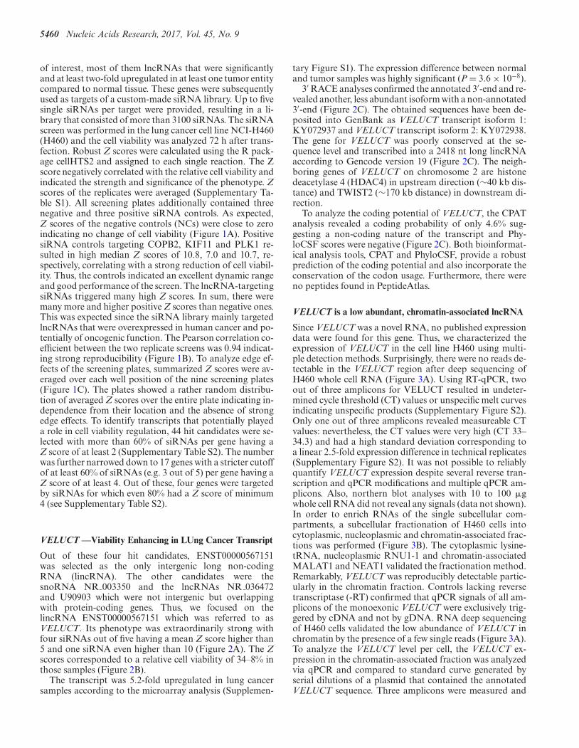
of interest, most of them lncRNAs that were significantlyand at least two-fold upregulated in at least one tumor entitycompared to normal tissue. These genes were subsequentlyused as targets of a custom-made siRNA library. Up to fivesingle siRNAs per target were provided, resulting in a li-brary that consisted of more than 3100 siRNAs. The siRNAscreen was performed in the lung cancer cell line NCI-H460(H460) and the cell viability was analyzed 72 h after trans-fection. Robust Z scores were calculated using the R pack-age cellHTS2 and assigned to each single reaction. The Zscore negatively correlated with the relative cell viability andindicated the strength and significance of the phenotype. Zscores of the replicates were averaged (Supplementary Ta-ble S1). All screening plates additionally contained threenegative and three positive siRNA controls. As expected,Z scores of the negative controls (NCs) were close to zeroindicating no change of cell viability (Figure 1A). PositivesiRNA controls targeting COPB2, KIF11 and PLK1 re-sulted in high median Z scores of 10.8, 7.0 and 10.7, re-spectively, correlating with a strong reduction of cell viabil-ity. Thus, the controls indicated an excellent dynamic rangeand good performance of the screen. The lncRNA-targetingsiRNAs triggered many high Z scores. In sum, there weremany more and higher positive Z scores than negative ones.This was expected since the siRNA library mainly targetedlncRNAs that were overexpressed in human cancer and po-tentially of oncogenic function. The Pearson correlation co-efficient between the two replicate screens was 0.94 indicat-ing strong reproducibility (Figure 1B). To analyze edge ef-fects of the screening plates, summarized Z scores were av-eraged over each well position of the nine screening plates(Figure 1C). The plates showed a rather random distribu-tion of averaged Z scores over the entire plate indicating in-dependence from their location and the absence of strongedge effects. To identify transcripts that potentially playeda role in cell viability regulation, 44 hit candidates were se-lected with more than 60% of siRNAs per gene having aZ score of at least 2 (Supplementary Table S2). The numberwas further narrowed down to 17 genes with a stricter cutoffof at least 60% of siRNAs (e.g. 3 out of 5) per gene having aZ score of at least 4. Out of these, four genes were targetedby siRNAs for which even 80% had a Z score of minimum4 (see Supplementary Table S2).
VELUCT ––Viability Enhancing in LUng Cancer Transript
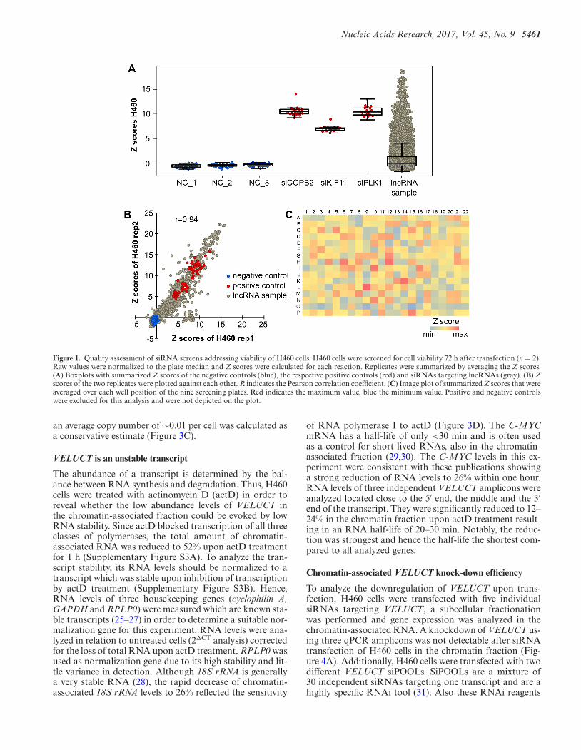
Out of these four hit candidates, ENST00000567151was selected as the only intergenic long non-codingRNA (lincRNA). The other candidates were thesnoRNA NR 003350 and the lncRNAs NR 036472and U90903 which were not intergenic but overlappingwith protein-coding genes. Thus, we focused on thelincRNA ENST00000567151 which was referred to asVELUCT. Its phenotype was extraordinarily strong withfour siRNAs out of five having a mean Z score higher than5 and one siRNA even higher than 10 (Figure 2A). The Zscores corresponded to a relative cell viability of 34–8% inthose samples (Figure 2B).
The transcript was 5.2-fold upregulated in lung cancersamples according to the microarray analysis (Supplemen-
tary Figure S1). The expression difference between normaland tumor samples was highly significant (P = 3.6 × 10⁻⁸).
3′ RACE analyses confirmed the annotated 3′-end and re-vealed another, less abundant isoform with a non-annotated3′-end (Figure 2C). The obtained sequences have been de-posited into GenBank as VELUCT transcript isoform 1:KY072937 and VELUCT transcript isoform 2: KY072938.The gene for VELUCT was poorly conserved at the se-quence level and transcribed into a 2418 nt long lincRNAaccording to Gencode version 19 (Figure 2C). The neigh-boring genes of VELUCT on chromosome 2 are histonedeacetylase 4 (HDAC4) in upstream direction (∼40 kb dis-tance) and TWIST2 (∼170 kb distance) in downstream di-rection.
To analyze the coding potential of VELUCT, the CPATanalysis revealed a coding probability of only 4.6% sug-gesting a non-coding nature of the transcript and Phy-loCSF scores were negative (Figure 2C). Both bioinformat-ical analysis tools, CPAT and PhyloCSF, provide a robustprediction of the coding potential and also incorporate theconservation of the codon usage. Furthermore, there wereno peptides found in PeptideAtlas.
VELUCT is a low abundant, chromatin-associated lncRNA
Since VELUCT was a novel RNA, no published expressiondata were found for this gene. Thus, we characterized theexpression of VELUCT in the cell line H460 using multi-ple detection methods. Surprisingly, there were no reads de-tectable in the VELUCT region after deep sequencing ofH460 whole cell RNA (Figure 3A). Using RT-qPCR, twoout of three amplicons for VELUCT resulted in undeter-mined cycle threshold (CT) values or unspecific melt curvesindicating unspecific products (Supplementary Figure S2).Only one out of three amplicons revealed measureable CTvalues: nevertheless, the CT values were very high (CT 33–34.3) and had a high standard deviation corresponding toa linear 2.5-fold expression difference in technical replicates(Supplementary Figure S2). It was not possible to reliablyquantify VELUCT expression despite several reverse tran-scription and qPCR modifications and multiple qPCR am-plicons. Also, northern blot analyses with 10 to 100 �gwhole cell RNA did not reveal any signals (data not shown).In order to enrich RNAs of the single subcellular com-partments, a subcellular fractionation of H460 cells intocytoplasmic, nucleoplasmic and chromatin-associated frac-tions was performed (Figure 3B). The cytoplasmic lysine-tRNA, nucleoplasmic RNU1-1 and chromatin-associatedMALAT1 and NEAT1 validated the fractionation method.Remarkably, VELUCT was reproducibly detectable partic-ularly in the chromatin fraction. Controls lacking reversetranscriptase (-RT) confirmed that qPCR signals of all am-plicons of the monoexonic VELUCT were exclusively trig-gered by cDNA and not by gDNA. RNA deep sequencingof H460 cells validated the low abundance of VELUCT inchromatin by the presence of a few single reads (Figure 3A).To analyze the VELUCT level per cell, the VELUCT ex-pression in the chromatin-associated fraction was analyzedvia qPCR and compared to standard curve generated byserial dilutions of a plasmid that contained the annotatedVELUCT sequence. Three amplicons were measured and
Nucleic Acids Research, 2017, Vol. 45, No. 9 5461
Figure 1. Quality assessment of siRNA screens addressing viability of H460 cells. H460 cells were screened for cell viability 72 h after transfection (n = 2).Raw values were normalized to the plate median and Z scores were calculated for each reaction. Replicates were summarized by averaging the Z scores.(A) Boxplots with summarized Z scores of the negative controls (blue), the respective positive controls (red) and siRNAs targeting lncRNAs (gray). (B) Zscores of the two replicates were plotted against each other. R indicates the Pearson correlation coefficient. (C) Image plot of summarized Z scores that wereaveraged over each well position of the nine screening plates. Red indicates the maximum value, blue the minimum value. Positive and negative controlswere excluded for this analysis and were not depicted on the plot.
an average copy number of ∼0.01 per cell was calculated asa conservative estimate (Figure 3C).
VELUCT is an unstable transcript
The abundance of a transcript is determined by the bal-ance between RNA synthesis and degradation. Thus, H460cells were treated with actinomycin D (actD) in order toreveal whether the low abundance levels of VELUCT inthe chromatin-associated fraction could be evoked by lowRNA stability. Since actD blocked transcription of all threeclasses of polymerases, the total amount of chromatin-associated RNA was reduced to 52% upon actD treatmentfor 1 h (Supplementary Figure S3A). To analyze the tran-script stability, its RNA levels should be normalized to atranscript which was stable upon inhibition of transcriptionby actD treatment (Supplementary Figure S3B). Hence,RNA levels of three housekeeping genes (cyclophilin A,GAPDH and RPLP0) were measured which are known sta-ble transcripts (25–27) in order to determine a suitable nor-malization gene for this experiment. RNA levels were ana-lyzed in relation to untreated cells (2�CT analysis) correctedfor the loss of total RNA upon actD treatment. RPLP0 wasused as normalization gene due to its high stability and lit-tle variance in detection. Although 18S rRNA is generallya very stable RNA (28), the rapid decrease of chromatin-associated 18S rRNA levels to 26% reflected the sensitivity
of RNA polymerase I to actD (Figure 3D). The C-MYCmRNA has a half-life of only <30 min and is often usedas a control for short-lived RNAs, also in the chromatin-associated fraction (29,30). The C-MYC levels in this ex-periment were consistent with these publications showinga strong reduction of RNA levels to 26% within one hour.RNA levels of three independent VELUCT amplicons wereanalyzed located close to the 5′ end, the middle and the 3′end of the transcript. They were significantly reduced to 12–24% in the chromatin fraction upon actD treatment result-ing in an RNA half-life of 20–30 min. Notably, the reduc-tion was strongest and hence the half-life the shortest com-pared to all analyzed genes.
Chromatin-associated VELUCT knock-down efficiency
To analyze the downregulation of VELUCT upon trans-fection, H460 cells were transfected with five individualsiRNAs targeting VELUCT, a subcellular fractionationwas performed and gene expression was analyzed in thechromatin-associated RNA. A knockdown of VELUCT us-ing three qPCR amplicons was not detectable after siRNAtransfection of H460 cells in the chromatin fraction (Fig-ure 4A). Additionally, H460 cells were transfected with twodifferent VELUCT siPOOLs. SiPOOLs are a mixture of30 independent siRNAs targeting one transcript and are ahighly specific RNAi tool (31). Also these RNAi reagents
5462 Nucleic Acids Research, 2017, Vol. 45, No. 9
Figure 2. Identification of the novel oncogenic lncRNA VELUCT regulating the cell viability. (A) Z scores for siRNAs targeting VELUCT in both screeningreplicates of H460 cells. (B) Relative cell viability (normalized to plate median) for siRNAs targeting VELUCT in both screening replicates of H460 cells.(C) Genomic location of the annotated and the VELUCT isoforms that were detected by 3′ RACE using chromatin-associated H460 RNA, PhyloCSFscores for all three frames on the respective strand, conservation by PhyloP and repeating elements by RepeatMasker are shown on the UCSC genomebrowser version hg19.
did not evoke a significant detectable downregulation inthe chromatin-associated fraction (Figure 4B). We hypothe-sized that this finding could either be due to a lack of knock-down efficiency or due to a lack of detectability of RNAi ef-fects in the chromatin fraction. In order to analyze the gen-eral knockdown of chromatin-associated RNAs by RNAireagents, the upstream neighboring gene HDAC4 was se-lected for comparison. It was targeted using two indepen-dent siRNAs and one siPOOL (Figure 4C). All reagentssignificantly knocked down HDAC4 expression in the cyto-plasm and nucleoplasm to 20–40%. Only one siRNA causeda slight, significant knockdown of HDAC4 by 30% in thechromatin fraction, while the other two reagents also didnot show an effect in the chromatin-associated fraction.Hence, the detectable knock-down in the cytoplasm and nu-cleoplasm was much stronger than in the chromatin frac-tions for all the reagents. This leads to the hypothesis thatRNAi reagents post-transcriptionally regulate gene expres-sion of cytoplasmic and nucleoplasmic, but not as efficientlyof chromatin-bound RNA. This could explain the lack of adetectable RNAi-induced knockdown of VELUCT in thechromatin fraction, whereas the potential downregulation
in the nucleoplasm or cytoplasm was not detectable due toits low abundance. Since nuclear transcripts are more effec-tively knocked down by antisense oligonucleotides (ASOs)(32), H460 cells were also transfected with three indepen-dent ASOs targeting VELUCT. They were designed to bindto sites close to the 5′-end, in the middle and close tothe 3′-end of the transcript (ASO 5′/m/3′). Gene expres-sion analyses in the chromatin-associated RNA fraction re-vealed a significant knockdown of VELUCT (Figure 4D).The VELUCT levels were significantly reduced by all threeASOs to 12–46% for all three tested amplicons. Interest-ingly, the single ASOs evoked deviating RNA levels for eachamplicon, which were consistent for the single replicates.For example, ASO 5′ also led to the strongest knock-downat the 5′-end, while ASO 3′ showed its strongest impact onthe 3′-end of VELUCT. To analyze a possible in cis effectof VELUCT on the expression regulation of the immedi-ate neighboring genes HDAC4 and TWIST2, we analyzedtheir chromatin-associated RNA levels upon transfection ofH460 cells with RNAi reagents targeting VELUCT. No-tably, there was no significant deregulation of the neigh-boring genes HDAC4 and TWIST2 (Supplementary Figure

Nucleic Acids Research, 2017, Vol. 45, No. 9 5463
Figure 3. The instable, low abundant VELUCT was only detectable in chromatin-associated lncRNA. (A) RNA deep sequencing was performed with wholecell RNA and chromatin-associated RNA of H460 cells. The read coverage of both runs is shown on a scale from 0 to 120 and from 0 to 5, respectively.The human genome version hg19 was used for alignment of reads. (B) Expression levels in cytoplasmic, nucleoplasmic and chromatin-associated fractionsof H460 cells were analyzed by RT-qPCR and normalized to the expression in whole cell RNA. The VELUCT m amplicon was used for detection ofVELUCT. Bars show mean ± SD (n = 3). (C) Determination of VELUCT copy number in chromatin-associated H460 RNA with three qPCR amplicons(location at 5′ end, middle or 3′ end) (n = 3). (D) H460 cells were treated with actD or DMSO (-actD) for 1 h and subsequently subcellularly fractionated.Chromatin-associated RNA levels of indicated genes were normalized to RPLP0 and relative to DMSO-treated cells. Bars show mean ± SD (n = 3).Asterisks indicate significant expression difference between treated and untreated cells. *P < 0.05, **P < 0.01, ***P < 0.001.
S4). Also expression of p21 which is negatively regulated byHDAC4 (33) did not change significantly.
Multiple independent VELUCT-specific silencing reagentsstrongly reduce viability
Since the siRNA screen already pointed to a pivotal role ofVELUCT in cell viability, more detailed experiments wereperformed in order to validate the screen. To analyze theeffect of VELUCT overexpression on H460 cell viability, aplasmid was generated that contained the annotated genesequence of VELUCT. Despite high upregulation of exoge-
nous VELUCT expression upon plasmid transfection (Sup-plementary Figure S5A), cell viability was not affected 24,48 or 72 h after transfection (Supplementary Figure S5B).In order to validate the loss-of-function screening results,independent knockdown experiments were performed withthe same settings as in the screen. Since siVELUCT 3 didnot show any phenotype in the screen, it was omitted fromfurther analyses. The screening results were validated as vi-ability of H460 cells was significantly reduced to 30–40%upon siRNA-mediated loss-of-function of VELUCT (Fig-ure 5A). To show that this effect was not only siRNA-

5464 Nucleic Acids Research, 2017, Vol. 45, No. 9
Figure 4. VELUCT knockdown was not detectable with siRNAs and siPOOLs, but with ASOs. H460 cells were transfected with 30 nM VELUCT-specificsiRNAs (A) or siPOOLs (B), 10 nM HDAC4-specific siRNAs or siPOOLs (C) or 30 nM VELUCT-specific ASOs (D) for 24 h. VELUCT RNA levels wereanalyzed in the chromatin fraction using three independent qPCR amplicons. HDAC4 levels were determined in all three subcellular fractions. All RNAlevels were normalized to cyclophilin A and relative to the respective NC. Bars show mean ± SD (n = 3–4). *P < 0.05, **P < 0.01, ***P < 0.001.

Nucleic Acids Research, 2017, Vol. 45, No. 9 5465
Figure 5. Multiple independent VELUCT-specific silencing reagents reduced viability and proliferation of H460 cells. H460 cells were transfected with 30nM VELUCT-specific siRNAs (A, D), siPOOLs (B, E) or ASOs (C, F). Cell viability (A–C) and proliferation (D–F) was measured 72 h after transfectionand normalized to the respective NC. Bars show mean ± SD (n = 3–4). *P < 0.05, **P < 0.01, ***P < 0.001.
dependent, the same experiment was performed with twoVELUCT-specific siPOOLs. Both siPOOLs significantlydecreased the cell viability even stronger to 10–20% (Fig-ure 5B). H460 cells were also transfected with VELUCT-specific ASOs in order to have a silencing approach thatwas independent of the RNAi machinery. Compared toall silencing reagents, two out of three ASOs triggered thestrongest phenotype with a reduction of cell viability to 3–6% (Figure 5C). Only the ASO that was located close to the 3′end of VELUCT did not affect cell viability. The phenotypewas time-dependent with a modest, but significant viabilitydecrease after 24 h and reaching its maximum 48 or 72 hafter transfection with all silencing reagents tested (Supple-mentary Figure S6A–C). Moreover, the effect on cell viabil-
ity was significant, but slightly weaker using 10 nM silencingreagent concentration (Supplementary Figure S6D-E), ex-cept for ASOs for which only a concentration of 30 nM trig-gered a phenotype (Supplementary Figure S6F). To validatethat the phenotype was not assay-specific, an independentreadout was performed that measured the cell proliferationupon loss-of-function of VELUCT by BrdU incorporation.Most VELUCT-specific silencing reagents significantly de-creased the cell proliferation to approx. 25% and the pheno-typic pattern was similar between proliferation and viability(Figure 5D–F). Although the cell proliferation was repro-ducibly reduced upon transfection of ASO m, the deregula-tion was not significant due to a high deviation between thebiological replicates (Figure 5F). ASO 3′ did not trigger a

5466 Nucleic Acids Research, 2017, Vol. 45, No. 9
proliferation phenotype recapitulating the viability readout.A time-dependent analysis of the proliferation revealed thatthe phenotype was significantly reduced to ∼50% already24 h after transfection, whereas the phenotype was moreprominent after 48 and 72 h (Supplementary Figure S7).The analysis of the apoptosis rate revealed that cell deathdid not contribute to the reduction of viability upon siRNAtransfection (Supplementary Figure S8).
VELUCT knockdown impairs the viability of multiple lungcancer cell lines
In order to analyze whether these results were not only rep-resentative for H460 cells, but for several lung cancer celllines, we tested multiple lung cancer cell lines for their vi-ability phenotype upon loss-of-function of VELUCT. Notall cell lines tested revealed any phenotype, but all analyzedsiRNAs targeting VELUCT significantly reduced the via-bility in H1437 and H1944 cells. This observation is con-cordant with the observation that lncRNAs are in generalexpressed and function in a tissue- and cell subtype-specificmanner (5,8). H1437 cells showed a stronger phenotypethan H1944 with a median relative cell viability of 56% ver-sus 78%, respectively (Figure 6A). The VELUCT siPOOLs-1 and -2 significantly reduced the viability in both celllines to 51 % to 78 %, respectively (Figure 6B). Strikingly,cell viability was not significantly altered upon transfectionof H1437 with all three ASOs (Figure 6C). The viabilityof H1944 cells was significantly reduced by ASO 5′ andASO m to 31% and 76%, respectively, but not by ASO 3′which was consistent with the cell line H460. Hence, theeffect on cell viability due to transfection with VELUCT-specific siRNAs, siPOOLs and partially ASOs was validatedin the lung cancer cell lines H1437 and H1944 and was thusnot restricted to the cell line H460.
In summary, eight different knockdown reagents tar-geting VELUCT in three different cell lines reduced cellgrowth as determined in two different assays––despite theextremely low abundance of VELUCT.
DISCUSSION
Although thousands of lncRNAs exist, only a minor frac-tion has been functionally characterized. A common toolto analyze several hundreds of genes for their phenotypeupon loss-of-function is an siRNA screen. However, sofar, only a few studies described siRNA screens target-ing lncRNAs (34–36). According to our knowledge, nosiRNA screen has been performed that analyzed tumor-associated lncRNAs in cancer cell lines. Thus, we designed acustom-made siRNA library that was targeting 638 tumor-associated lncRNAs. In order to reduce the impact of off-target effects on hit identification, mostly five siRNAs weredesigned for each target. Since each siRNA has a distinctrange of off-target effects, but the same on-target, a pheno-type that is observed with multiple individual siRNAs in-creases the confidence that it is due to downregulation ofthe intended target (37,38). One of the most prominent hitswas a novel gene that that we called VELUCT. Similar tomost lncRNAs (13,14,39), VELUCT was a low abundanttranscript. It was 5.2-fold upregulated in lung cancer ade-nocarcinoma according to microarray analyses. However,
it was not reproducibly detectable in whole cell RNA byRT-qPCR. This signal difference between microarray andRT-qPCR might be due to a low correlation as determinedbefore (40). Upon cellular fractionation of cells, VELUCTwas quantifiable in the chromatin-associated RNA fractionof H460 cells. This implied that VELUCT might act di-rectly on chromatin and might, e.g. regulate gene transcrip-tion as it was reported for many lncRNAs (41). On averageand as a conservative estimate, only 0.01 VELUCT RNAcopies were present in the chromatin fraction of each cell.While an underestimation for technical reasons is possible,it is highly likely that the copy number of VELUCT willremain even in a less conservative analysis lower than onecopy per cell. The low abundance suggested that VELUCTlikely acted only on single alleles. One reason for the lowabundance of VELUCT was its low stability with a half-lifeof ∼20–30 min. For comparison, the median half-life of allmammalian RNAs is 5–9 h (27,42–44), while only a minorfraction of mammalian RNAs have a half-life of <1 h (e.g.MYC or the transcription factor FOXA2) (27,45). Short-lived transcripts are reported to be tightly regulated (42) andplay a major role in processes such as transcription, cell cy-cle progression and apoptosis (27,45). Accordingly, the lowstability of VELUCT might indicate a precise temporal reg-ulation of this transcript.
Cell viability was not altered upon overexpression ofVELUCT. This might be explained by several reasons:First, VELUCT might act in cis, such as enhancer lncR-NAs (46), XIST (47) or ANRIL (48). Since cis-acting genesregulate the expression of other genes exclusively on thesame chromosome from which they are derived, a plasmid-based overexpression could not recapitulate its effect. Sec-ond, since only single VELUCT copies likely directly actedon the genome, only single alleles might be bound by lncR-NAs in order to exert their function. In case the bindingsites were already occupied, a further increase of VELUCTlevels would not promote the phenotype.
To confirm that the observed phenotype in the siRNAscreen was elicited by targeting of the VELUCT transcript,VELUCT expression was analyzed upon siRNA transfec-tion of H460 cells. However, there was no RNAi-mediatedknockdown of VELUCT detectable in the chromatin-associated fraction. Interestingly, RNAi reagents targetingthe upstream neighboring gene HDAC4 as control alsofailed to induce a detectable knockdown in the chromatinfraction, while it significantly and efficiently knocked downHDAC4 expression in the cytoplasm and nucleoplasm. Thefact that there is no detectable knockdown of chromatin-associated VELUCT might be explained by several rea-sons: On the one hand, technical limitations of RT-qPCRmight contribute to the non-detectable knockdown. Al-though VELUCT is detectable in the chromatin-associatedRNA fraction, its low abundance might still be hinderinga precise measure of RNAi-mediated downregulation. Onthe other hand, we hypothesize that the RNAi reagentscould bind to and thereby block, but do not downregu-late the chromatin-bound transcript as also observed forHDAC4. Thus, the formation of an alternative secondarystructure of VELUCT and / or the binding of other effec-tor proteins to VELUCT could be prevented. In contrast,transfection of H460 cells with independent ASOs targeting

Nucleic Acids Research, 2017, Vol. 45, No. 9 5467
Figure 6. Multiple independent VELUCT-specific silencing reagents reduced viability of other lung cancer cell lines. H1437 and H1944 cells were trans-fected with 30 nM siRNAs (A), siPOOLs (B) or ASOs (C) targeting VELUCT. Cell viability was measured 72 h after transfection and normalized to therespective NC. Bars show mean ± SD (n = 4–5). *P < 0.05, **P < 0.01, ***P < 0.001.
VELUCT evoked a significant knockdown of VELUCT to12–46%. ASOs - in contrast to RNAi reagents - might in-duce knockdown of chromatin-bound RNAs due to theirgene silencing mechanism via RNase H (49,50). The local-ization of effector proteins differs between subcellular com-partments: since RNase H is mainly localized in the nucleus(51), ASOs have a superior silencing efficiency in the nu-cleus than RNAi reagents (32). In contrast, since the RNAdegrading RISC complex is primarily localized in the cy-toplasm, RNA interference is predominantly active in thecytoplasm (52,53).
The VELUCT-specific phenotype that was observed inthe siRNA screen was confirmed in multiple validation ex-periments. To further exclude the possibility of off-targeteffects, two siPOOLs comprised of 30 independent siR-NAs confirmed the phenotype. Off-target effects are di-luted out by the low concentration of each single siRNAmaking siPOOLs highly specific with strong on-target ef-fects (31). Also, lower siRNA concentrations can reduce off-target effects (54), and the VELUCT-specific phenotype wasalso recapitulated at a 10 nM RNAi reagent concentration.RNAi reagents can nevertheless lead to a global perturba-tion of miRNA-mediated regulation due to saturation ofthe RNAi machinery (55). Again, the RNAi-independentsilencing mechanism of ASOs confirmed that no saturationof the RNAi pathway caused the observed loss of cell viabil-ity. Lastly, the effect of VELUCT loss-of-function was notrestricted to H460 cells, but also found in the lung cancercell lines H1437 and H1944 and was also reproduced witha proliferation readout. This observation may hypothesizethat VELUCT may be involved in the cell cycle. Unfortu-
nately, the low expression of VELUCT precludes more de-tailed mechanistic studies or the identification of interactionpartners for technical reasons.
Eight different knockdown reagents using two differentassays and three different cell lines make it very unlikely thatthe observed phenotypes are not specific for VELUCT. Thecentral question arising from the data presented is how toreconcile the extremely low abundance of VELUCT withits apparent functional importance. Multiple provoking hy-potheses could be raised: first, the low abundance and chro-matin association could indicate that VELUCT binds onlyto very few and specific sites in the chromatin requiring onlyvery few copies to cover all sites. Second, VELUCT couldonly be needed in a very specific state of the cell, e.g. dur-ing cell cycle or during replicative or oxidative stress or an-other condition. The very short half-life of VELUCT thenleads to the rapid clearance and low steady-state expressionof VELUCT. Lastly, VELUCT could be involved in non-cell-autonomous processes. In this case, single copies in onecell could have an impact on multiple cells amplifying itseffect. Although the low copy number of 0.01 could alsobe explained by a small subpopulation of H460 expressingVELUCT, e.g. the rare cancer stem cells, this is less likelysince the VELUCT-specific phenotype is already observableafter 24 h upon inhibition of the proliferative characteristicsby loss-of-function of VELUCT.
Low abundant, unstable transcripts have already beenidentified in yeast, where they can play important regula-tory roles, e.g. in modifications of histones (56) or chromatinremodeling (57). Although some lncRNAs have a low con-servation level, they are nevertheless functional and essen-

5468 Nucleic Acids Research, 2017, Vol. 45, No. 9
tial, such as X-inactive specific transcript (XIST) and An-tisense IGF2 receptor RNA (Air) (58). Furthermore, genesare not only conserved on the level of their sequence, butalso in their secondary structure (59).
To our knowledge, VELUCT is the first example of alncRNA that is expressed at a very low level, but never-theless has a strong phenotype upon knockdown. Thus,our study corroborates that - at least individual - lncRNAsof low abundance can execute important functions in thecell. This sheds new light on the large majority of lncRNAswhich are present at low copy numbers and have remainedunderstudied so far. Well-controlled studies for each in-dividual lncRNA are required to distinguish between the‘transcriptional noise’ of aberrant transcripts and function-ally important lncRNAs like VELUCT.
SUPPLEMENTARY DATA
Supplementary Data are available at NAR Online.
ACKNOWLEDGEMENTS
The authors would like to thank Matthias Groß andJeanette Seiler for excellent technical assistance and theDKFZ Genomics and Proteomics Core Facility (GPCF) fordeep sequencing. This study is part of the Ph.D. thesis of J.S.
FUNDING
Research in the Diederichs labs is supported by the GermanResearch Foundation [DFG Di 1421/7-1, EXC81 CellNet-works EcTop5, SFB 850]; RNA@DKFZ Cross ProgramTopic and the National Center for Tumor Diseases Hei-delberg (NCT 3.0 Integrative Projects in Basic Cancer Re-search). Funding for open access charge: Core funding.Conflict of interest statement. S.D. is co-owner of siTOOLsBiotech GmbH, Martinsried, Germany.
REFERENCES1. Djebali,S., Davis,C.A., Merkel,A., Dobin,A., Lassmann,T.,
Mortazavi,A., Tanzer,A., Lagarde,J., Lin,W., Schlesinger,F. et al.(2012) Landscape of transcription in human cells. Nature, 489,101–108.
2. Kapranov,P., Cheng,J., Dike,S., Nix,D.A., Duttagupta,R.,Willingham,A.T., Stadler,P.F., Hertel,J., Hackermuller,J.,Hofacker,I.L. et al. (2007) RNA maps reveal new RNA classes and apossible function for pervasive transcription. Science, 316, 1484–1488.
3. Iyer,M.K., Niknafs,Y.S., Malik,R., Singhal,U., Sahu,A., Hosono,Y.,Barrette,T.R., Prensner,J.R., Evans,J.R., Zhao,S. et al. (2015) Thelandscape of long noncoding RNAs in the human transcriptome.Nat. Genet., 47, 199–208.
4. Yan,L., Yang,M., Guo,H., Yang,L., Wu,J., Li,R., Liu,P., Lian,Y.,Zheng,X., Yan,J. et al. (2013) Single-cell RNA-Seq profiling ofhuman preimplantation embryos and embryonic stem cells. Nat.Struct. Mol. Biol., 20, 1131–1139.
5. Tsoi,L.C., Iyer,M.K., Stuart,P.E., Swindell,W.R., Gudjonsson,J.E.,Tejasvi,T., Sarkar,M.K., Li,B., Ding,J., Voorhees,J.J. et al. (2015)Analysis of long non-coding RNAs highlights tissue-specificexpression patterns and epigenetic profiles in normal and psoriaticskin. Genome Biol., 16, 24.
6. Cabili,M.N., Trapnell,C., Goff,L., Koziol,M., Tazon-Vega,B.,Regev,A. and Rinn,J.L. (2011) Integrative annotation of human largeintergenic noncoding RNAs reveals global properties and specificsubclasses. Genes Dev., 25, 1915–1927.
7. Liu,S.J., Nowakowski,T.J., Pollen,A.A., Lui,J.H., Horlbeck,M.A.,Attenello,F.J., He,D., Weissman,J.S., Kriegstein,A.R., Diaz,A.A.et al. (2016) Single-cell analysis of long non-coding RNAs in thedeveloping human neocortex. Genome Biol., 17, 67.
8. Kim,D.H., Marinov,G.K., Pepke,S., Singer,Z.S., He,P., Williams,B.,Schroth,G.P., Elowitz,M.B. and Wold,B.J. (2015) Single-celltranscriptome analysis reveals dynamic changes in lncRNAexpression during reprogramming. Cell Stem Cell, 16, 88–101.
9. Ji,P., Diederichs,S., Wang,W., Boing,S., Metzger,R., Schneider,P.M.,Tidow,N., Brandt,B., Buerger,H., Bulk,E. et al. (2003) MALAT-1, anovel noncoding RNA, and thymosin �4 predict metastasis andsurvival in early-stage non-small cell lung cancer. Oncogene, 22,8031–8041.
10. Liu,X.-h., Liu,Z.-l., Sun,M., Liu,J., Wang,Z.-x. and De,W. (2013) Thelong non-coding RNA HOTAIR indicates a poor prognosis andpromotes metastasis in non-small cell lung cancer. BMC Cancer, 13,464–464.
11. Wang,R., Shi,Y., Chen,L., Jiang,Y., Mao,C., Yan,B., Liu,S., Shan,B.,Tao,Y. and Wang,X. (2015) The ratio of FoxA1 to FoxA2 in lungadenocarcinoma is regulated by LncRNA HOTAIR and chromatinremodeling factor LSH. Scientific Rep., 5, 17826–17826.
12. Gutschner,T., Hammerle,M., Eißmann,M., Hsu,J., Kim,Y., Hung,G.,Revenko,A., Arun,G., Stentrup,M., Groß,M. et al. (2013) Thenoncoding RNA MALAT1 is a critical regulator of the metastasisphenotype of lung cancer cells. Cancer Res., 73, 1180–1189.
13. Derrien,T., Johnson,R., Bussotti,G., Tanzer,A., Djebali,S.,Tilgner,H., Guernec,G., Martin,D., Merkel,A., Knowles,D.G. et al.(2012) The GENCODE v7 catalog of human long noncoding RNAs:Analysis of their gene structure, evolution, and expression. GenomeRes., 22, 1775–1789.
14. Mercer,T.R., Gerhardt,D.J., Dinger,M.E., Crawford,J., Trapnell,C.,Jeddeloh,J.a., Mattick,J.S. and Rinn,J.L. (2011) Targeted RNAsequencing reveals the deep complexity of the human transcriptome.Nat. Biotechnol., 30, 99–104.
15. Eddy,S.R., Doolittle,W.F., Sapienza,C., Kidwell,M.G., Lynch,M.,Ohno,S., Orgel,L.E., Crick,F.H.C. and Thomas,C.A. (2012) TheC-value paradox, junk DNA and ENCODE. Curr. Biol., 22,R898–R899.
16. van Bakel,H., Nislow,C., Blencowe,B.J. and Hughes,T.R. (2010)Most ‘dark matter’ transcripts are associated with known genes.PLoS Biol., 8, e1000371.
17. Palazzo,A.F. and Lee,E.S. (2015) Non-coding RNA: what isfunctional and what is junk? Front. Genet., 6, 2.
18. Mercer,T.R., Dinger,M.E. and Mattick,J.S. (2009) Long non-codingRNAs: insights into functions. Nat. Rev. Genet., 10, 155–159.
19. Clark,M.B., Amaral,P.P., Schlesinger,F.J., Dinger,M.E., Taft,R.J.,Rinn,J.L., Ponting,C.P., Stadler,P.F., Morris,K.V., Morillon,A. et al.(2011) The reality of pervasive transcription. PLoS Biol., 9, e1000625.
20. Castro,F., Dirks,W.G., Fahnrich,S., Hotz-Wagenblatt,A., Pawlita,M.and Schmitt,M. (2013) High-throughput SNP-based authenticationof human cell lines. Int. J. Cancer, 132, 308–314.
21. Gagnon,K.T., Li,L., Janowski,B.A. and Corey,D.R. (2014) Analysisof nuclear RNA interference in human cells by subcellularfractionation and Argonaute loading. Nat. Protoc., 9, 2045–2060.
22. Kim,D., Pertea,G., Trapnell,C., Pimentel,H., Kelley,R. andSalzberg,S.L. (2013) TopHat2: accurate alignment of transcriptomesin the presence of insertions, deletions and gene fusions. GenomeBiol., 14, R36.
23. Boutros,M., Bras,L.P. and Huber,W. (2006) Analysis of cell-basedRNAi screens. Genome Biol., 7, R66.
24. Pelz,O., Gilsdorf,M. and Boutros,M. (2010) web cellHTS2: aweb-application for the analysis of high-throughput screening data.BMC Bioinformatics, 11, 185.
25. Kuwano,Y., Rabinovic,A., Srikantan,S., Gorospe,M., Demple,B. andNo,H. (2009) Analysis of nitric oxide-stabilized mRNAs in humanfibroblasts reveals HuR-dependent heme oxygenase 1 upregulation.Mol. Cell. Biol., 29, 2622–2635.
26. Andersen,J.B., Mazan-Mamczarz,K., Zhan,M., Gorospe,M. andHassel,B.A. (2009) Ribosomal protein mRNAs are primary targets ofregulation in RNase-L-induced senescence. RNA Biol., 6, 305–315.
27. Sharova,L.V., Sharov,A.A., Nedorezov,T., Piao,Y., Shaik,N. andKo,M.S.H. (2009) Database of mRNA half-life of 19977 genesobtained by DNA microarray analysis of pluripotent and

Nucleic Acids Research, 2017, Vol. 45, No. 9 5469
differentiating mouse embryonic stem cells supplementary data. DNARes., 16, S1.
28. Abelson,H.T., Johnson,L.F., Penman,S. and Green,H. (1974)Changes in RNA in relation to growth of the fibroblast: II. Thelifetime of mRNA, rRNA, and tRNA in resting and growing cells.Cell, 1, 161–165.
29. Berteaux,N., Aptel,N., Cathala,G., Genton,C., Coll,J., Daccache,A.,Spruyt,N., Hondermarck,H., Dugimont,T., Curgy,J.J. et al. (2008) Anovel H19 antisense RNA overexpressed in breast cancer contributesto paternal IGF2 expression. Mol. Cell Biol., 28, 6731–6745.
30. Mondal,T., Rasmussen,M., Pandey,G.K., Isaksson,A. andKanduri,C. (2010) Characterization of the RNA content ofchromatin. Genome Res., 20, 899–907.
31. Hannus,M., Beitzinger,M., Engelmann,J.C., Weickert,M.T.,Spang,R., Hannus,S. and Meister,G. (2014) SiPools: Highly complexbut accurately defined siRNA pools eliminate off-target effects.Nucleic Acids Res., 42, 8049–8061.
32. Lennox,K.A. and Behlke,M.A. (2016) Cellular localization of longnon-coding RNAs affects silencing by RNAi more than by antisenseoligonucleotides. Nucleic Acids Res., 44, 863–877.
33. Mottet,D., Pirotte,S., Lamour,V., Hagedorn,M., Javerzat,S.,Bikfalvi,A., Bellahcene,A., Verdin,E. and Castronovo,V. (2009)HDAC4 represses p21(WAF1/Cip1) expression in human cancer cellsthrough a Sp1-dependent, p53-independent mechanism. Oncogene,28, 243–256.
34. Chakraborty,D., Kappei,D., Theis,M., Nitzsche,A., Ding,L.,Paszkowski-Rogacz,M., Surendranath,V., Berger,N., Schulz,H.,Saar,K. et al. (2012) Combined RNAi and localization forfunctionally dissecting long noncoding RNAs. Nat. Methods, 9,360–362.
35. Theis,M., Chakraborty,D., Weisswange,I., Paszkowski-Rogacz,M.and Buchholz,F. (2015) Targeting human long noncoding transcriptsby endoribonuclease prepared siRNAs. J. Biomol. Screen., 20,1018–1026.
36. Negishi,M., Wongpalee,S.P., Sarkar,S., Park,J., Lee,K.Y., Shibata,Y.,Reon,B.J., Abounader,R., Suzuki,Y., Sugano,S. et al. (2014) A newlncRNA, APTR, associates with and represses the CDKN1A/p21promoter by recruiting polycomb proteins. PLoS One, 9, e95216.
37. Cullen,B.R. (2006) Enhancing and confirming the specificity of RNAiexperiments. Nat. Methods, 3, 677–681.
38. Echeverri,C.J., Beachy,P.a., Baum,B., Boutros,M., Buchholz,F.,Chanda,S.K., Downward,J., Ellenberg,J., Fraser,A.G., Hacohen,N.et al. (2006) Minimizing the risk of reporting false positives inlarge-scale RNAi screens. Nat. Methods, 3, 777–779.
39. Werner,M.S. and Ruthenburg,A.J. (2015) Nuclear fractionationreveals thousands of chromatin-tethered noncoding RNAs adjacentto active genes. Cell Rep., 12, 1089–1098.
40. Camarillo,C., Swerdel,M. and Hart,R.P. (2011) Vol. 698, pp. 419–429.41. Vance,K.W. and Ponting,C.P. (2014) Transcriptional regulatory
functions of nuclear long noncoding RNAs. Trends Genet., 30,348–355.
42. Tani,H., Mizutani,R., Salam,K.A., Tano,K., Ijiri,K., Wakamatsu,A.,Isogai,T., Suzuki,Y. and Akimitsu,N. (2012) Genome-wide
determination of RNA stability reveals hundreds of short-livednoncoding transcripts in mammals. Genome Res., 22, 947–956.
43. Schwanhausser,B., Busse,D., Li,N., Dittmar,G., Schuchhardt,J.,Wolf,J., Chen,W. and Selbach,M. (2011) Global quantification ofmammalian gene expression control. Nature, 473, 337–342.
44. Friedel,C.C., Dolken,L., Ruzsics,Z., Koszinowski,U.H. andZimmer,R. (2009) Conserved principles of mammaliantranscriptional regulation revealed by RNA half-life. Nucleic AcidsRes., 37, e115.
45. Raghavan,A., Ogilvie,R.L., Reilly,C., Abelson,M.L., Raghavan,S.,Vasdewani,J., Krathwohl,M. and Bohjanen,P.R. (2002) Genome-wideanalysis of mRNA decay in resting and activated primary human Tlymphocytes. Nucleic Acids Res., 30, 5529–5538.
46. Ørom,U.A., Derrien,T., Beringer,M., Gumireddy,K., Gardini,A.,Bussotti,G., Lai,F., Zytnicki,M., Notredame,C., Huang,Q. et al.(2010) Long noncoding RNAs with enhancer-like function in humancells. Cell, 143, 46–58.
47. Pontier,D.B. and Gribnau,J. (2011) Xist regulation and functioneXplored. Hum. Genet., 130, 223–236.
48. Congrains,A., Kamide,K., Ohishi,M. and Rakugi,H. (2013) ANRIL:molecular mechanisms and implications in human health. Int. J. Mol.Sci. 14, 1278–1292.
49. Jepsen,J.S. and Wengel,J. (2004) LNA-antisense rivals siRNA for genesilencing. Curr. Opin. Drug Discov. Dev., 7, 188–194.
50. Hutvagner,G. and Simard,M.J. (2008) Argonaute proteins: keyplayers in RNA silencing. Nat. Rev. Mol. Cell Biol., 9, 22–32.
51. Vickers,T.A. and Crooke,S.T. (2014) Antisense oligonucleotidescapable of promoting specific target mRNA reduction via competingRNase H1-dependent and independent mechanisms. PLoS One, 9,e108625.
52. Zeng,Y. and Cullen,B.R. (2002) RNA interference in human cells isrestricted to the cytoplasm. RNA, 8, 855–860.
53. Vickers,T.A., Koo,S., Bennett,C.F., Crooke,S.T., Dean,N.M. andBaker,B.F. (2003) Efficient reduction of target RNAs by smallinterfering RNA and RNase H-dependent antisense agents. Acomparative analysis. J. Biol. Chem., 278, 7108–7118.
54. Caffrey,D.R., Zhao,J., Song,Z., Schaffer,M.E., Haney,S.A.,Subramanian,R.R., Seymour,A.B. and Hughes,J.D. (2011) SiRNAoff-target effects can be reduced at concentrations that match theirindividual potency. PLoS One, 6, e21503.
55. Khan,A.A., Betel,D., Miller,M.L., Sander,C., Leslie,C.S. andMarks,D.S. (2009) Transfection of small RNAs globally perturbs generegulation by endogenous microRNAs. Nat. Biotechnol., 27, 549–555.
56. Camblong,J., Iglesias,N., Fickentscher,C., Dieppois,G. and Stutz,F.(2007) Antisense RNA stabilization induces transcriptional genesilencing via histone deacetylation in S. cerevisiae. Cell, 131, 706–717.
57. Uhler,J.P., Hertel,C. and Svejstrup,J.Q. (2007) A role for noncodingtranscription in activation of the yeast PHO5 gene. PNAS, 104,8011–8016.
58. Pang,K.C., Frith,M.C. and Mattick,J.S. (2006) Rapid evolution ofnoncoding RNAs: Lack of conservation does not mean lack offunction. Trends Genet., 22, 1–5.
59. Diederichs,S. (2014) The four dimensions of noncoding RNAconservation. Trends Genet., 30, 121–123.


![Research Paper The regulatory role of miR-107 in Coxsackie ... · regulates viral replication at the cellular level [17, 18, 19], but whether the mechanism by which lncRNA can directly](https://static.fdocuments.us/doc/165x107/60162667a0f98871eb4cf038/research-paper-the-regulatory-role-of-mir-107-in-coxsackie-regulates-viral-replication.jpg)