The Living World - Chapter 4 - Mr. O'Neil's Biology -...
62
The Living World Fourth Edition GEORGE B. JOHNSON Copyright ©The McGraw-Hill Companies, Inc. Permission required for reproduction or display PowerPoint ® Lectures prepared by Johnny El-Rady 4 Cells
Transcript of The Living World - Chapter 4 - Mr. O'Neil's Biology -...
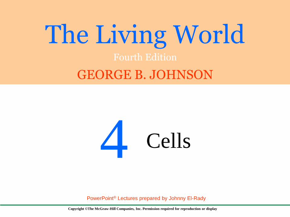
The Living World Fourth Edition
GEORGE B. JOHNSON
Copyright ©The McGraw-Hill Companies, Inc. Permission required for reproduction or display
PowerPoint® Lectures prepared by Johnny El-Rady
4 Cells

Copyright ©The McGraw-Hill Companies, Inc. Permission required for reproduction or display
4.1 Cells
Fig. 4.1 The size
of cells and their
contents
20 nm
20 mm
20 mm 2 mm 0.2 mm
2 mm
2 nm 0.2 nm
0.2 mm

Copyright ©The McGraw-Hill Companies, Inc. Permission required for reproduction or display
Robert Hooke (1665)
Examined a thin slice of cork tissue
Observed honeycombed compartments he
called cellulae (L, small rooms)
The term became cells
Matthias Schleiden and Theodor Schwann
Proposed the first two statements of the
cell theory in 1838-39
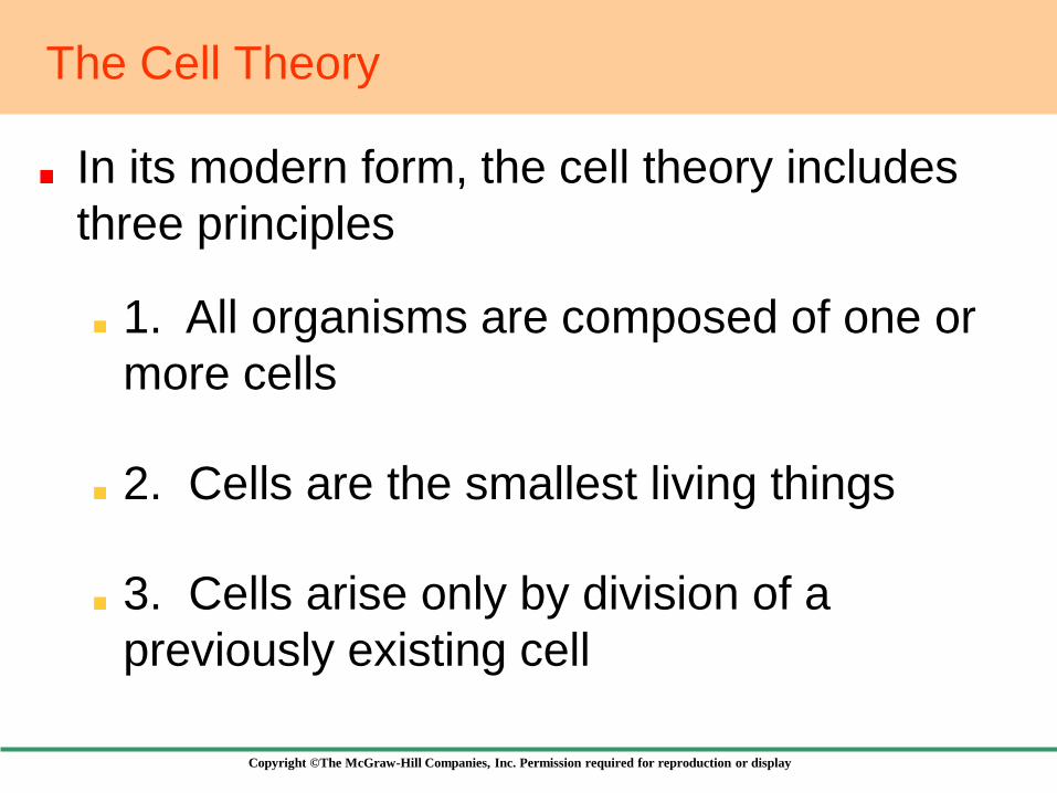
The Cell Theory
Copyright ©The McGraw-Hill Companies, Inc. Permission required for reproduction or display
In its modern form, the cell theory includes
three principles
The Cell Theory
1. All organisms are composed of one or
more cells
2. Cells are the smallest living things
3. Cells arise only by division of a
previously existing cell
Copyright ©The McGraw-Hill Companies, Inc. Permission required for reproduction or display
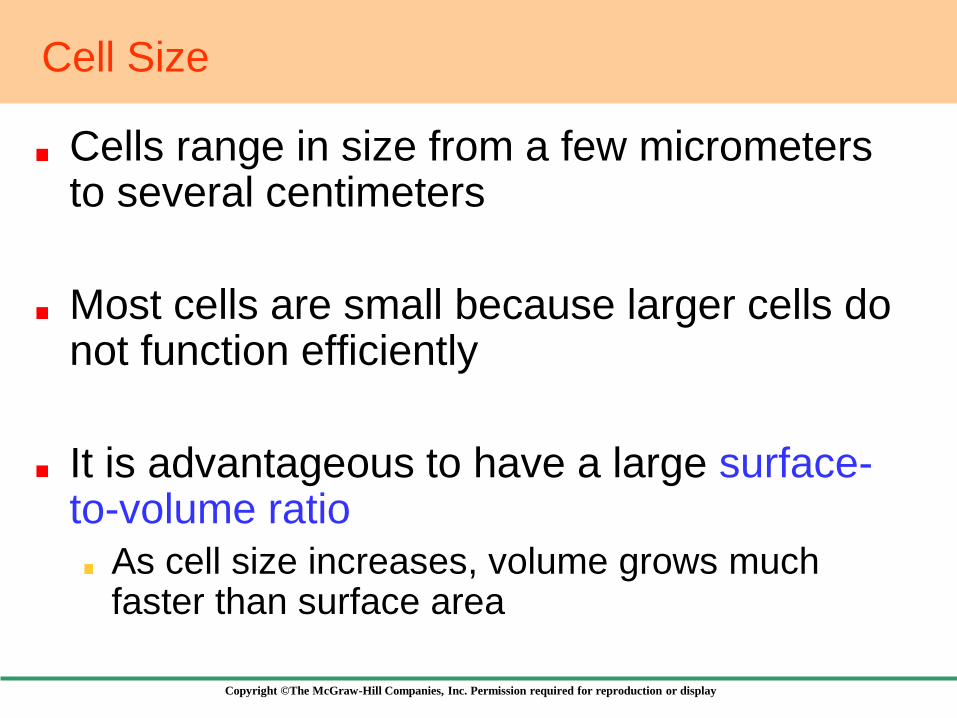
Cells range in size from a few micrometers to several centimeters
Most cells are small because larger cells do not function efficiently
It is advantageous to have a large surface-to-volume ratio
As cell size increases, volume grows much faster than surface area
Cell Size

Copyright ©The McGraw-Hill Companies, Inc. Permission required for reproduction or display
Fig. 4.2 Surface-to-volume ratio
Copyright ©The McGraw-Hill Companies, Inc. Permission required for reproduction or display
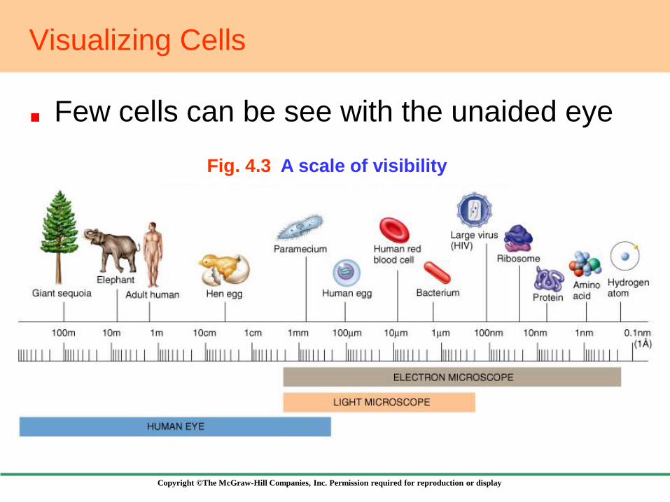
Visualizing Cells
Few cells can be see with the unaided eye
Fig. 4.3 A scale of visibility
Copyright ©The McGraw-Hill Companies, Inc. Permission required for reproduction or display
Visualizing Cells
We can’t see most cells because of the limited resolution of the human eye
Resolution is the minimum distance two points can be apart and still be seen as two points
Resolution of the human eye is 100 m
One way to increase resolution is to increase magnification, using microscopes
There are two main types of microscopes
Copyright ©The McGraw-Hill Companies, Inc. Permission required for reproduction or display
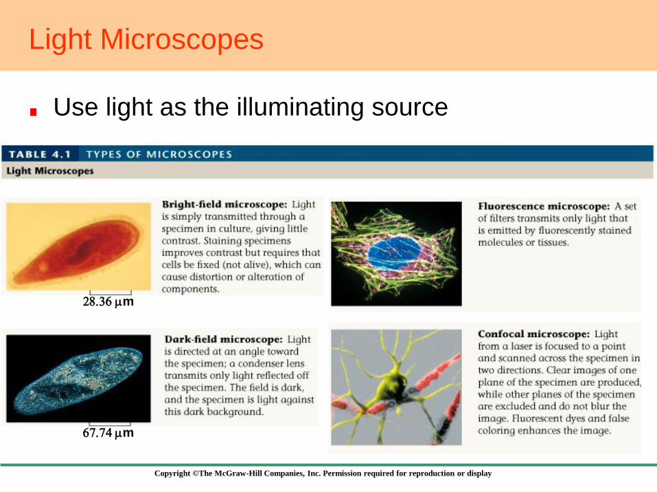
Light Microscopes
Use light as the illuminating source
28.36 mm
67.74 mm
Copyright ©The McGraw-Hill Companies, Inc. Permission required for reproduction or display
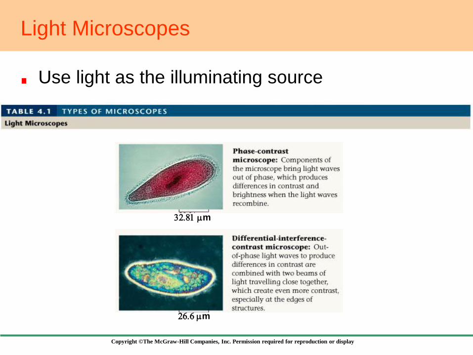
Light Microscopes
Use light as the illuminating source
32.81 mm
26.6 mm

Copyright ©The McGraw-Hill Companies, Inc. Permission required for reproduction or display
Electron Microscopes
Use a beam of electrons to produce the image
2.56 mm
6.76 mm

Copyright ©The McGraw-Hill Companies, Inc. Permission required for reproduction or display
4.2 The Plasma Membrane
Encases all living
cells
Its basic structure
is represented by
the fluid-mosaic
model
Phospholipid
bilayer with
embedded proteins
Fig. 4.4 Phospholipid structure

Copyright ©The McGraw-Hill Companies, Inc. Permission required for reproduction or display
4.2 The Plasma Membrane
In water, phospholipids spontaneously form a bilayer
Cell membranes contain zones called lipid rafts
Heavily enriched in cholesterol
Fig. 4.5

Copyright ©The McGraw-Hill Companies, Inc. Permission required for reproduction or display
Two main types
Cell-surface proteins
Project from the surface of the membrane
Act as markers or receptors
Transmembrane proteins
Extend all the way across the bilayer
Provide channels in and out of the cell
Proteins Within the Membrane

Copyright ©The McGraw-Hill Companies, Inc. Permission required for reproduction or display
Fig. 4.6 Proteins are embedded within the lipid bilayer
Copyright ©The McGraw-Hill Companies, Inc. Permission required for reproduction or display
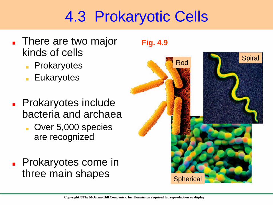
Fig. 4.9
4.3 Prokaryotic Cells
There are two major kinds of cells
Prokaryotes
Eukaryotes
Prokaryotes include bacteria and archaea
Over 5,000 species are recognized
Prokaryotes come in three main shapes
Rod
Spherical
Spiral
Copyright ©The McGraw-Hill Companies, Inc. Permission required for reproduction or display
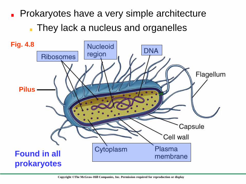
Prokaryotes have a very simple architecture
Pilus
Fig. 4.8
Found in all
prokaryotes
They lack a nucleus and organelles
Copyright ©The McGraw-Hill Companies, Inc. Permission required for reproduction or display
4.4 Eukaryotic Cells
Appeared about 1.5 billion years ago
Include all cells alive today except bacteria
and archaea
Are larger than prokaryotic cells
Have a much more complex architecture
Possess nucleus and a variety of organelles

Copyright ©The McGraw-Hill Companies, Inc. Permission required for reproduction or display
Fig. 4.10 Structure of
a plant cell
Copyright ©The McGraw-Hill Companies, Inc. Permission required for reproduction or display
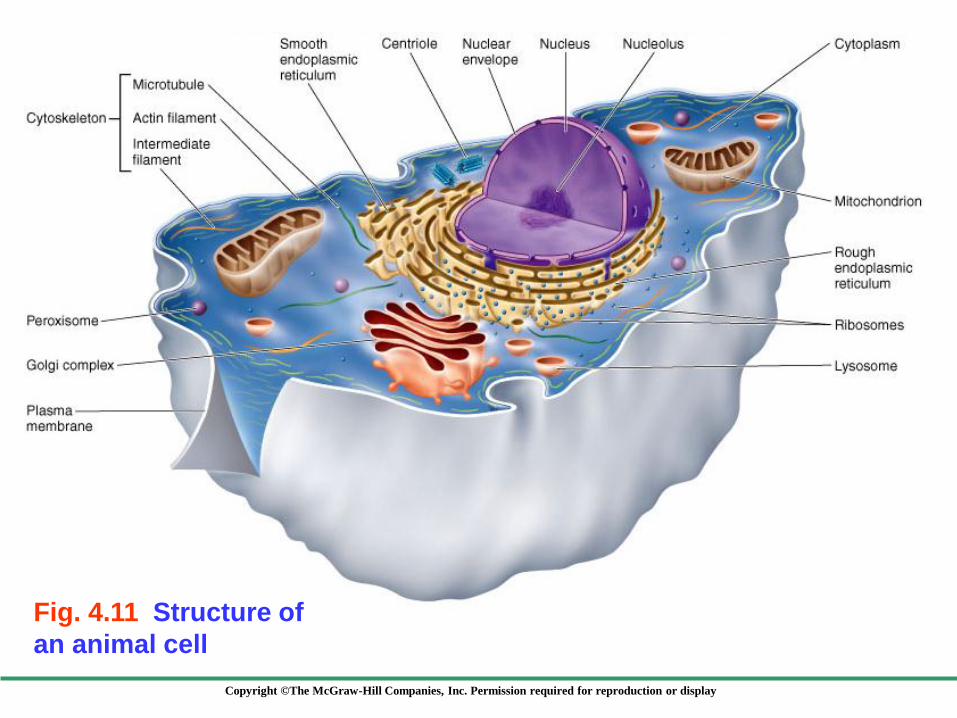
Fig. 4.11 Structure of
an animal cell
Copyright ©The McGraw-Hill Companies, Inc. Permission required for reproduction or display
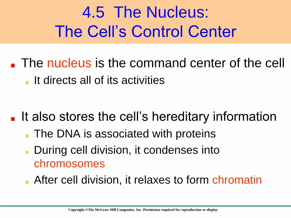
4.5 The Nucleus:
The Cell’s Control Center
The nucleus is the command center of the cell
It directs all of its activities
It also stores the cell’s hereditary information
The DNA is associated with proteins
During cell division, it condenses into
chromosomes
After cell division, it relaxes to form chromatin
Copyright ©The McGraw-Hill Companies, Inc. Permission required for reproduction or display
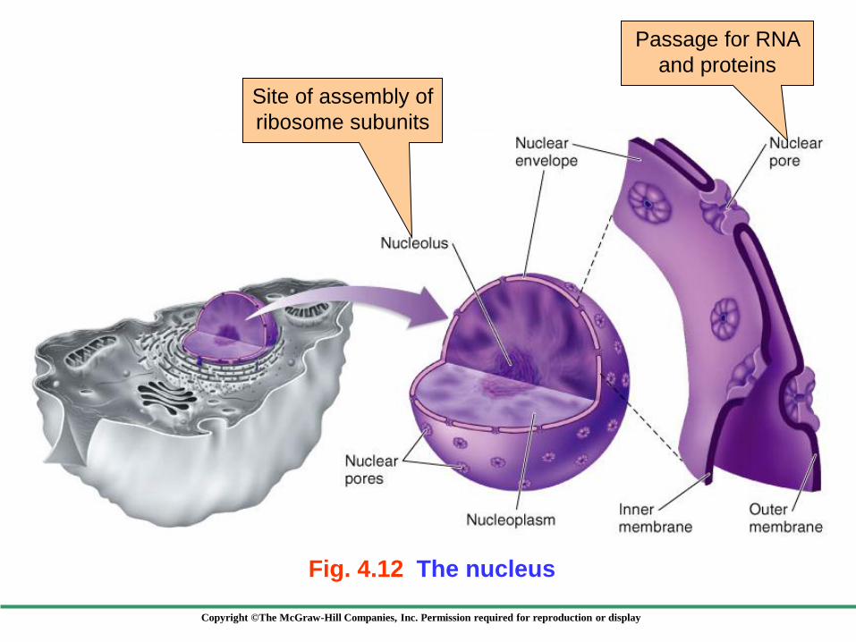
Fig. 4.12 The nucleus
Passage for RNA
and proteins
Site of assembly of
ribosome subunits
Copyright ©The McGraw-Hill Companies, Inc. Permission required for reproduction or display
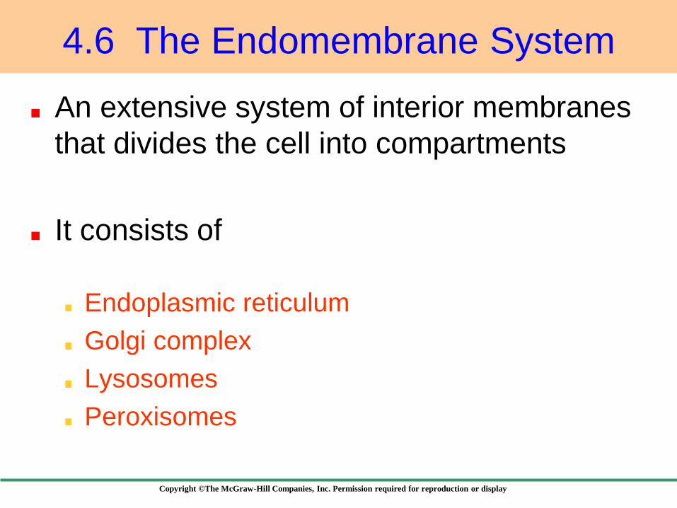
4.6 The Endomembrane System
An extensive system of interior membranes
that divides the cell into compartments
It consists of
Endoplasmic reticulum
Golgi complex
Lysosomes
Peroxisomes

Copyright ©The McGraw-Hill Companies, Inc. Permission required for reproduction or display
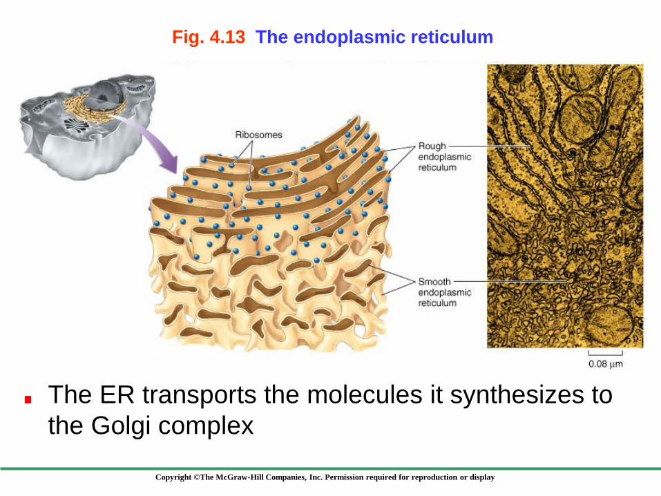
Internal membrane system creating channels and membrane-bound vesicles
Consists of two distinct regions
Rough ER Studded with ribosomes
Involved in protein synthesis
Smooth ER Embedded with enzymes
Involved in lipid and carbohydrate synthesis
Endoplasmic Reticulum (ER)
Copyright ©The McGraw-Hill Companies, Inc. Permission required for reproduction or display
The ER transports the molecules it synthesizes to
the Golgi complex
Fig. 4.13 The endoplasmic reticulum

Copyright ©The McGraw-Hill Companies, Inc. Permission required for reproduction or display
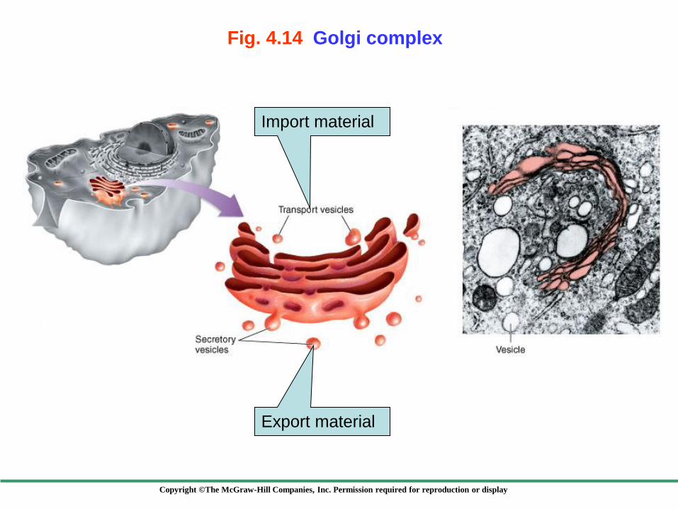
Golgi bodies are flattened stack of membranes that are scattered throughout the cytoplasm
Depending on the cell, the number of Golgi bodies ranges from a few to several hundred
These are collectively referred to as the Golgi complex
The Golgi complex collects, packages, modifes and distributes molecules
The Golgi Complex
Copyright ©The McGraw-Hill Companies, Inc. Permission required for reproduction or display
Fig. 4.14 Golgi complex
Export material
Import material

Copyright ©The McGraw-Hill Companies, Inc. Permission required for reproduction or display
Arise from the Golgi complex
They contain enzymes that break down
macromolecules
Function in intracellular digestion of
Worn-out cellular components
Substances taken into cells
The resulting material is then recycled
Lysosomes
Copyright ©The McGraw-Hill Companies, Inc. Permission required for reproduction or display
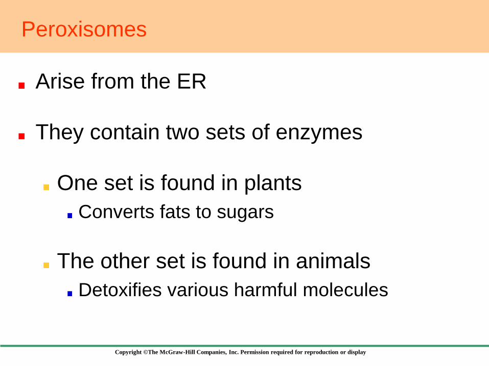
Arise from the ER
They contain two sets of enzymes
One set is found in plants
Converts fats to sugars
The other set is found in animals
Detoxifies various harmful molecules
Peroxisomes

Copyright ©The McGraw-Hill Companies, Inc. Permission required for reproduction or display
Fig. 4.15 How the endomembrane system works

Copyright ©The McGraw-Hill Companies, Inc. Permission required for reproduction or display
4.7 Organelles That Contain DNA
Two cell-like organelles contain DNA
Mitochondria
Found in almost all eukaryotes
Chloroplasts
Found only in plants and algae
Copyright ©The McGraw-Hill Companies, Inc. Permission required for reproduction or display
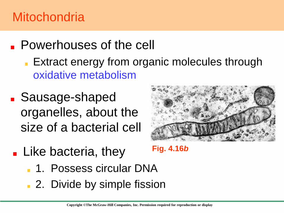
Powerhouses of the cell
Extract energy from organic molecules through
oxidative metabolism
Mitochondria
Sausage-shaped
organelles, about the
size of a bacterial cell
Like bacteria, they
1. Possess circular DNA
2. Divide by simple fission
Fig. 4.16b
Copyright ©The McGraw-Hill Companies, Inc. Permission required for reproduction or display
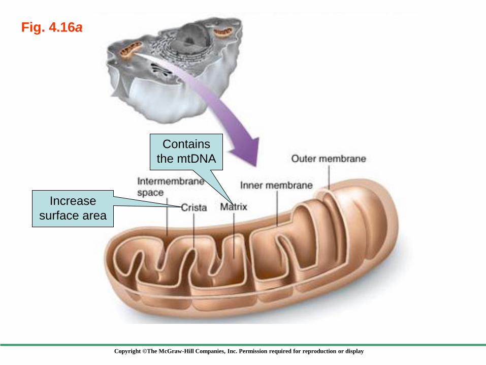
Fig. 4.16a
Increase
surface area
Contains
the mtDNA
Copyright ©The McGraw-Hill Companies, Inc. Permission required for reproduction or display
Chloroplasts
Energy-capturing centers
Sites of photosynthesis in plants and algae
Like bacteria, they
1. Possess circular DNA
2. Divide by simple fission
Like mitochondria, they are surrounded by two membranes
However, inner membrane much more complex
Copyright ©The McGraw-Hill Companies, Inc. Permission required for reproduction or display
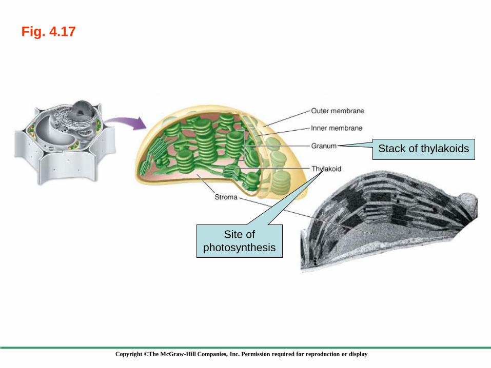
Fig. 4.17
Stack of thylakoids
Site of
photosynthesis
Copyright ©The McGraw-Hill Companies, Inc. Permission required for reproduction or display
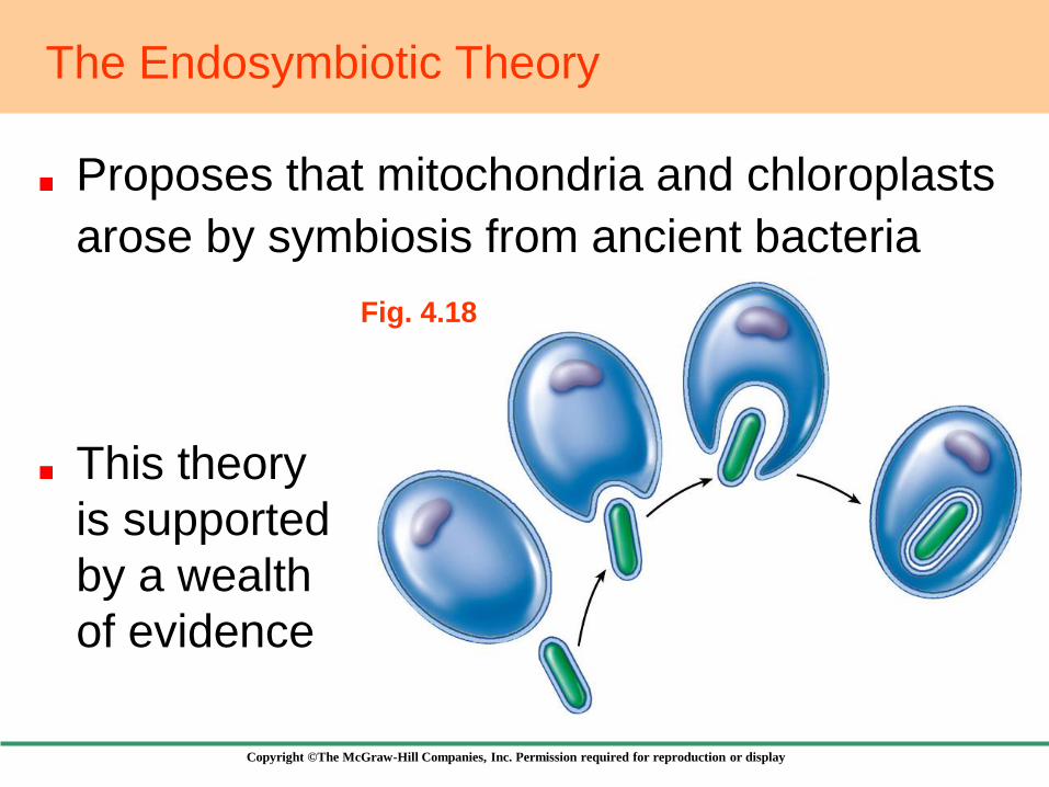
Fig. 4.18
The Endosymbiotic Theory
Proposes that mitochondria and chloroplasts
arose by symbiosis from ancient bacteria
This theory
is supported
by a wealth
of evidence

Copyright ©The McGraw-Hill Companies, Inc. Permission required for reproduction or display
4.8 The Cytoskeleton:
Interior Framework of the Cell
A dense network of protein fibers that
1. Supports the shape of the cell
2. Anchors organelles
Three different kinds of protein fibers
Microfilaments
Microtubules
Intermediate filaments

Copyright ©The McGraw-Hill Companies, Inc. Permission required for reproduction or display
Fig. 4.19
Made up of tubulin
Make up microfilaments

Copyright ©The McGraw-Hill Companies, Inc. Permission required for reproduction or display
Fig. 4.20
Centrioles
Anchor and assemble
microtubules
May have originated
as symbiotic
bacteria
Not found in higher plants and fungi
Copyright ©The McGraw-Hill Companies, Inc. Permission required for reproduction or display
Cell Movement
Essentially, all cell motion is tied to the
movement of microfilaments and microtubules
Changes in the shape of microfilaments
Enable some cells to change shape quickly
Allow some cells to crawl
Cause animal cells to divide
Copyright ©The McGraw-Hill Companies, Inc. Permission required for reproduction or display
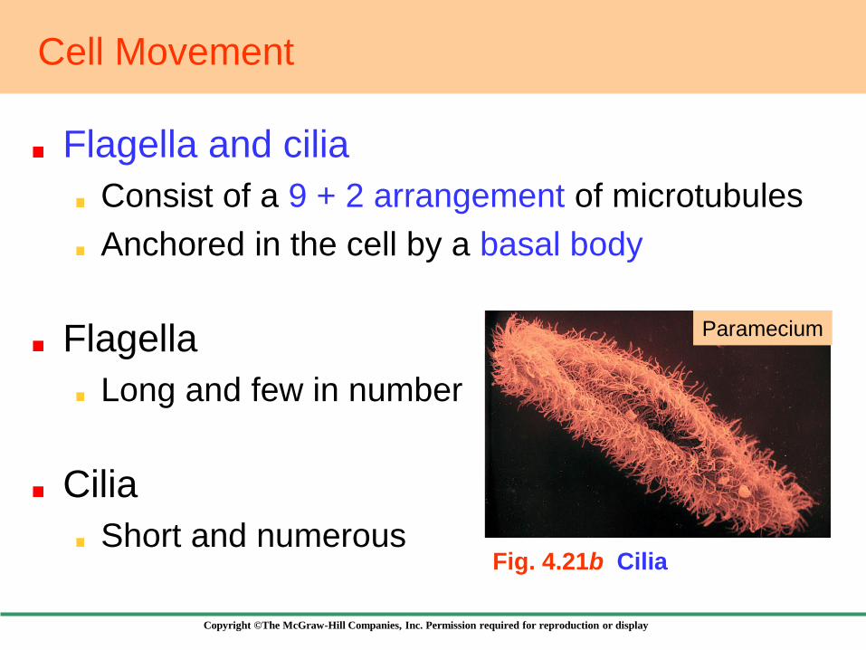
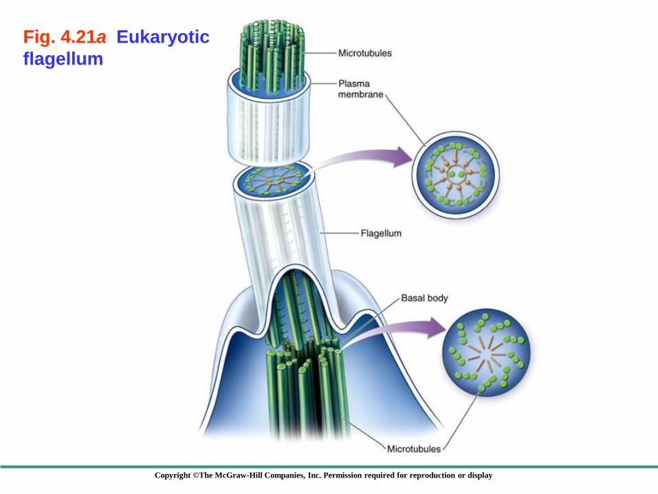
Cell Movement
Flagella and cilia
Consist of a 9 + 2 arrangement of microtubules
Anchored in the cell by a basal body
Flagella
Long and few in number
Cilia
Short and numerous Fig. 4.21b Cilia
Paramecium
Copyright ©The McGraw-Hill Companies, Inc. Permission required for reproduction or display
Fig. 4.21a Eukaryotic
flagellum
Copyright ©The McGraw-Hill Companies, Inc. Permission required for reproduction or display
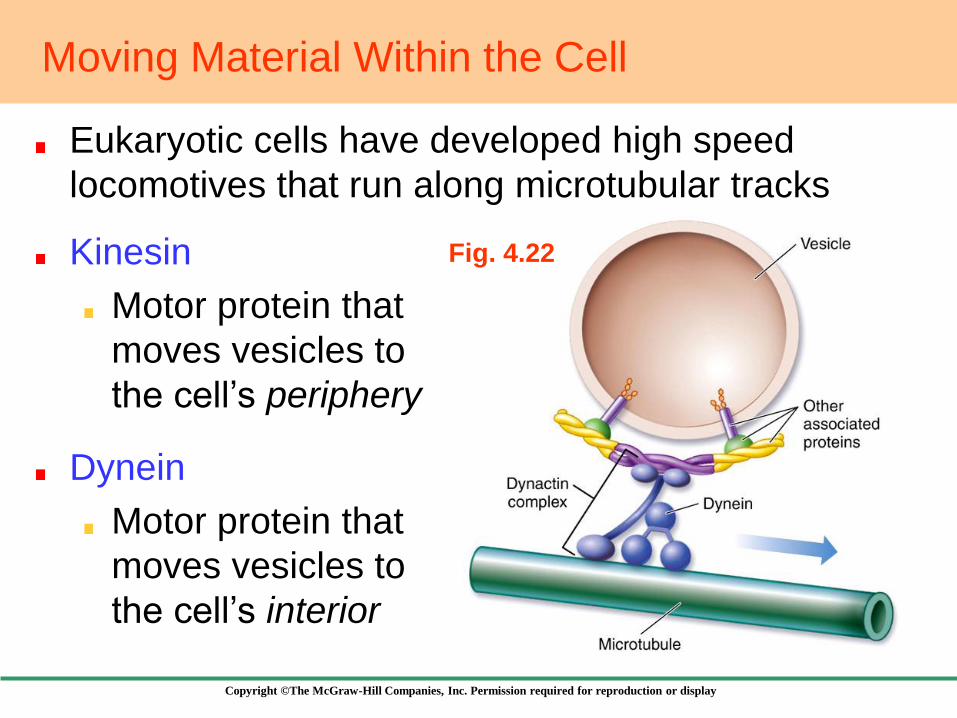
Moving Material Within the Cell
Eukaryotic cells have developed high speed
locomotives that run along microtubular tracks
Kinesin
Motor protein that
moves vesicles to
the cell’s periphery
Dynein
Motor protein that
moves vesicles to
the cell’s interior
Fig. 4.22

Copyright ©The McGraw-Hill Companies, Inc. Permission required for reproduction or display
Vacuoles
In plants
Store dissolved
substances
Can increase the
cell’s surface area
Fig. 4.23
In protists
Contractile vacuoles
are used to pump
excess water

Copyright ©The McGraw-Hill Companies, Inc. Permission required for reproduction or display
4.9 Outside the Plasma Membrane
Cell Walls
Offer protection
and support
Fungal cell walls
are made up of
chitin
Plant cell walls
are made up of
cellulose
Fig. 4.24
Glues cells
together

Copyright ©The McGraw-Hill Companies, Inc. Permission required for reproduction or display
4.9 Outside the Plasma Membrane
Extracellular Matrix
A mixture of
glycoproteins
secreted by animal
cells
Fig. 4.25
Links ECM to
the cytoskeleton
Helps coordinate
the behavior of all
cells in a tissue

Copyright ©The McGraw-Hill Companies, Inc. Permission required for reproduction or display
4.10 Diffusion and Osmosis
Diffusion is the movement of molecules down
their concentration gradient
Fig. 4.26
Equilibrium
Copyright ©The McGraw-Hill Companies, Inc. Permission required for reproduction or display
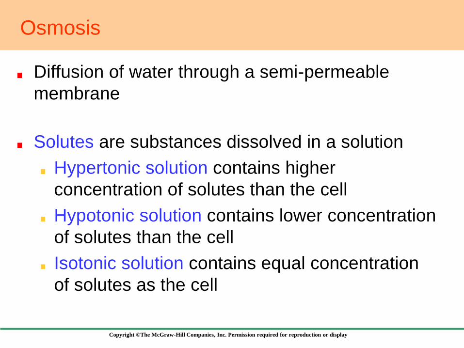
Osmosis
Diffusion of water through a semi-permeable
membrane
Solutes are substances dissolved in a solution
Hypertonic solution contains higher
concentration of solutes than the cell
Hypotonic solution contains lower concentration
of solutes than the cell
Isotonic solution contains equal concentration
of solutes as the cell
Copyright ©The McGraw-Hill Companies, Inc. Permission required for reproduction or display
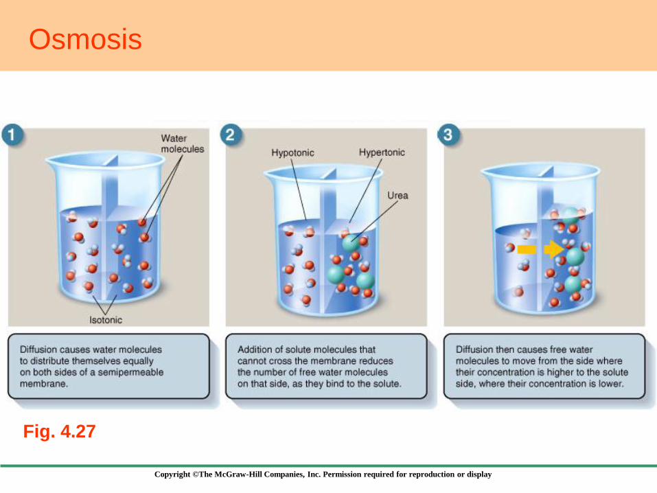
Osmosis
Fig. 4.27
Copyright ©The McGraw-Hill Companies, Inc. Permission required for reproduction or display
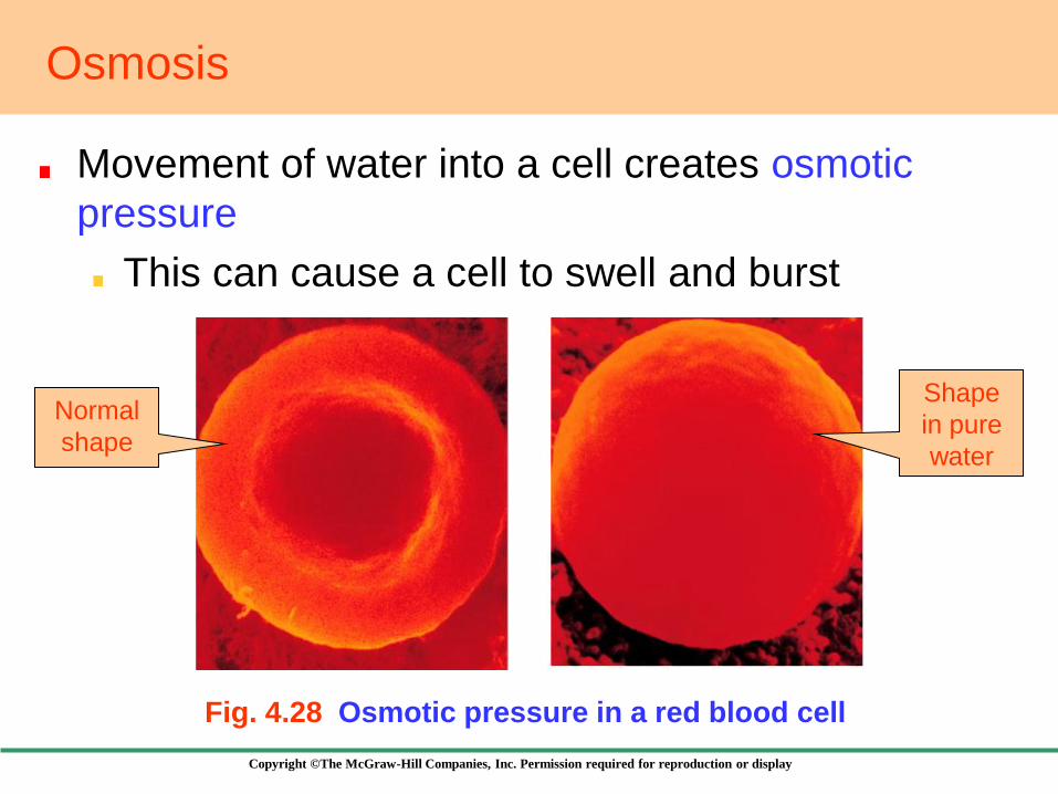
Osmosis
Movement of water into a cell creates osmotic
pressure
This can cause a cell to swell and burst
Fig. 4.28 Osmotic pressure in a red blood cell
Normal
shape
Shape
in pure
water

Copyright ©The McGraw-Hill Companies, Inc. Permission required for reproduction or display
4.11 Bulk Passage into and out of Cells
Large amounts of material can be moved in
and out of cells by membrane-bound vesicles
Exocytosis
Discharge of material from vesicles at the cell
surface
Endocytosis
The plasma membrane envelops particles and
brings them into the cell interior
Copyright ©The McGraw-Hill Companies, Inc. Permission required for reproduction or display
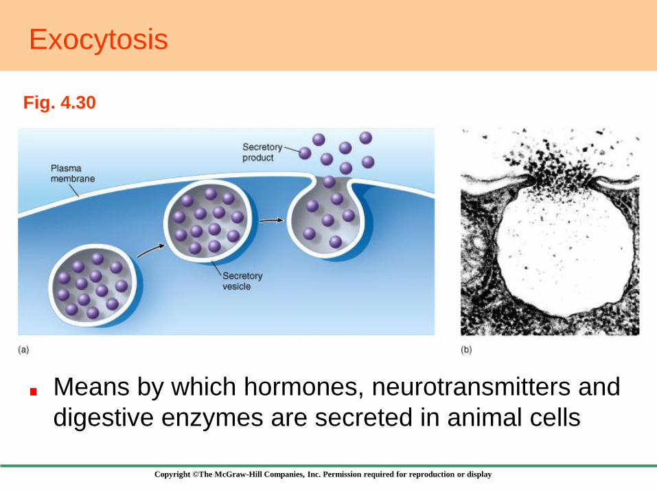
Exocytosis
Means by which hormones, neurotransmitters and
digestive enzymes are secreted in animal cells
Fig. 4.30

Copyright ©The McGraw-Hill Companies, Inc. Permission required for reproduction or display
2. Pinocytosis
Engulfment of liquid
material
Endocytosis
Fig. 4.29a
Has three major forms
1. Phagocytosis
Engulfment of
particulate material
Fig. 4.29b
Copyright ©The McGraw-Hill Companies, Inc. Permission required for reproduction or display
4.12 Selective Permeability
Cell membranes have selective permeability
They contain protein channels that allow only
certain molecules to pass
Selective Diffusion
Allows molecules to pass through open channels
in either direction
Ion channels
If the ion fits the pore, it goes through
Copyright ©The McGraw-Hill Companies, Inc. Permission required for reproduction or display
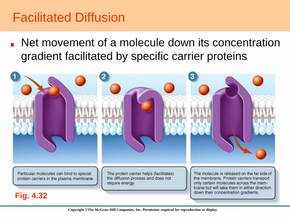
Facilitated Diffusion
Net movement of a molecule down its concentration
gradient facilitated by specific carrier proteins
Fig. 4.32
Copyright ©The McGraw-Hill Companies, Inc. Permission required for reproduction or display
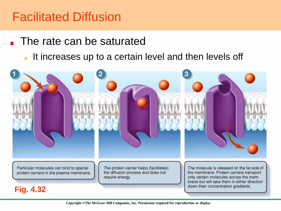
Facilitated Diffusion
The rate can be saturated
Fig. 4.32
It increases up to a certain level and then levels off
Copyright ©The McGraw-Hill Companies, Inc. Permission required for reproduction or display
Active Transport
The movement of molecules across a
membrane against a concentration gradient
This is possible by the expenditure of energy
Two types of channels are mainly used
1. Sodium-Potassium Pump
2. Proton Pump

Copyright ©The McGraw-Hill Companies, Inc. Permission required for reproduction or display
The Sodium-Potassium Pump
Fig. 4.33
Uses the energy of one ATP molecule to pump
3 Na+ outward and 2 K+ into the cell
Copyright ©The McGraw-Hill Companies, Inc. Permission required for reproduction or display
The Sodium-Potassium Pump
Leads to fewer Na+ in the cell
This concentration gradient is exploited in many
ways, including
1. The conduction of signals along nerve cells
Chapter 28
2. The transport of material into the cell
against their concentration gradient
Coupled channels
Copyright ©The McGraw-Hill Companies, Inc. Permission required for reproduction or display
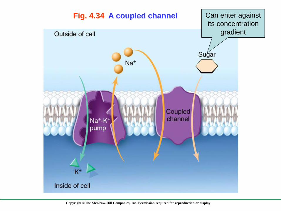
Fig. 4.34 A coupled channel Can enter against
its concentration
gradient
Copyright ©The McGraw-Hill Companies, Inc. Permission required for reproduction or display
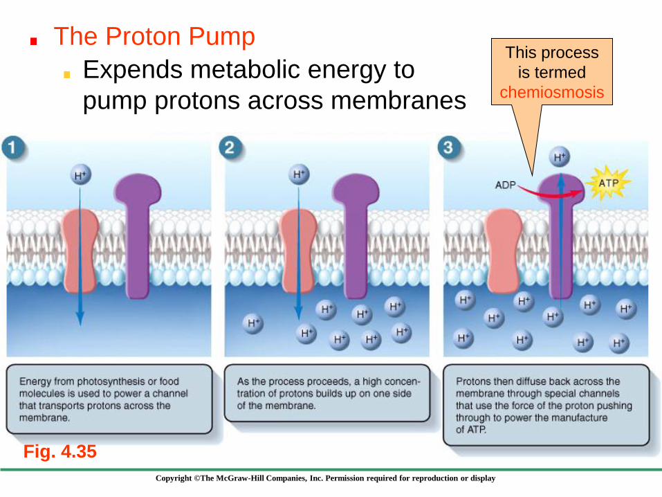
The Proton Pump
Fig. 4.35
This process
is termed
chemiosmosis
Expends metabolic energy to
pump protons across membranes

Copyright ©The McGraw-Hill Companies, Inc. Permission required for reproduction or display
How Cells Get Information
Cells sense chemical information by
means of cell surface receptor proteins
These bind specific molecules and transmit
information to the cell
Cells sense electrical information by
means of voltage-sensitive channels
These allow ions into or out of the cell in
response to electric signals