THE J BIOLOGICAL C Vol. 279, No. 3, Issue of January 16 ... Klebsiella pneumoniae ALS. The overall...
13
The Crystal Structures of Klebsiella pneumoniae Acetolactate Synthase with Enzyme-bound Cofactor and with an Unusual Intermediate* Received for publication, April 17, 2003, and in revised form, October 10, 2003 Published, JBC Papers in Press, October 13, 2003, DOI 10.1074/jbc.M304038200 Siew Siew Pang‡, Ronald G. Duggleby‡§, Richard L. Schowen¶, and Luke W. Guddat‡ From the ‡Department of Biochemistry and Molecular Biology, the University of Queensland, Brisbane, Queensland 4072, Australia and the ¶Department of Chemistry, the University of Kansas, Lawrence, Kansas 66045 Acetohydroxyacid synthase (AHAS) and acetolactate synthase (ALS) are thiamine diphosphate (ThDP)- dependent enzymes that catalyze the decarboxylation of pyruvate to give a cofactor-bound hydroxyethyl group, which is transferred to a second molecule of pyruvate to give 2-acetolactate. AHAS is found in plants, fungi, and bacteria, is involved in the biosynthesis of the branched- chain amino acids, and contains non-catalytic FAD. ALS is found only in some bacteria, is a catabolic enzyme required for the butanediol fermentation, and does not contain FAD. Here we report the 2.3-Å crystal structure of Klebsiella pneumoniae ALS. The overall structure is similar to AHAS except for a groove that accommodates FAD in AHAS, which is filled with amino acid side chains in ALS. The ThDP cofactor has an unusual con- formation that is unprecedented among the 26 known three-dimensional structures of nine ThDP-dependent enzymes, including AHAS. This conformation suggests a novel mechanism for ALS. A second structure, at 2.0 Å, is described in which the enzyme is trapped halfway through the catalytic cycle so that it contains the hy- droxyethyl intermediate bound to ThDP. The cofactor has a tricyclic structure that has not been observed previously in any ThDP-dependent enzyme, although similar structures are well known for free thiamine. This structure is consistent with our proposed mecha- nism and probably results from an intramolecular pro- ton transfer within a tricyclic carbanion that is the true reaction intermediate. Modeling of the second molecule of pyruvate into the active site of the enzyme with the bound intermediate is consistent with the stereochem- istry and specificity of ALS. The enzyme EC 2.2.1.6 (formerly EC 4.1.3.18) known both as acetolactate synthase (ALS) 1 and as acetohydroxyacid syn- thase (AHAS) catalyzes the conversion of 2 mol of pyruvate to acetolactate plus CO 2 (1). These two different names for the enzyme distinguish two forms (2) that have distinctly different properties. One difference is that AHAS requires FAD, whereas ALS is FAD-independent. However, ALS and AHAS are clearly related proteins with substantial sequence similarities and share a requirement for thiamine diphosphate (ThDP). In most flavin-containing enzymes that catalyze non-redox reactions (3), the cofactor has been shown, or is suspected, to play an internal role as an electron acceptor/donor. For exam- ple, in chorismate synthase, flavin mononucleotide participates in free radical chemistry (4). The exception is hydroxynitrile lyase, where there appears to be no internal redox role (5). In a curious parallel to ALS/AHAS, this enzyme exists in two forms, only one of which contains FAD (6). The three-dimensional structure of the FAD-dependent al- mond hydroxynitrile lyase (7) suggests that the cofactor is an evolutionary remnant of an aryl alcohol oxidase progenitor. Similarly, AHAS may have evolved from a pyruvate oxidase ancestor (8). Our determination of the structure of the FAD- dependent yeast AHAS (9) supports this idea; FAD is located close to the active site and in a position similar to that in pyruvate oxidase. The function of FAD in AHAS appears to be solely for maintenance of structural integrity. An alternative approach to unraveling the role of FAD in AHAS is to understand how the FAD-independent ALS can dispense with this cofactor. This enzyme, from Aerobacter aero- genes, was crystallized in 1967 (10), but no x-ray diffraction studies were reported. We obtained diffraction-quality crystals of the enzyme from the closely related bacterium Klebsiella pneumoniae (11); now we report the three-dimensional struc- ture of this ALS. ThDP-dependent enzymes are ubiquitous in nature and are involved in all major metabolic processes. It is generally accepted that the initial step in catalysis by these enzymes is the depro- tonation of the thiazolium ring at the C-2 atom (Fig. 1, A, I 3 II, and B). Elucidation of the structure of several ThDP-dependent enzymes (12–18) has allowed a plausible mechanism for this ionization to be proposed, involving a conserved glutamate, N-1, and the 4-amino group. Studies with cofactor analogs (19) and by mutagenesis (20 –24) support this mechanism. The high specific activity of K. pneumoniae ALS (10) corre- sponds to a k cat of 533 s 1 which is about 10-fold higher than that of any FAD-dependent AHAS (e.g. Ref. 25) showing that FAD independence does not cripple the enzyme. Quite the reverse; the FAD-dependent enzyme is unable to achieve rate enhancements that are attainable by ALS. The k cat value of K. pneumoniae ALS is significantly higher than that of any ThDP-dependent enzyme with a known structure. The k cat value is also substantially higher than the observed rate con- stant for C-2 deprotonation in Zymomonas mobilis pyruvate decarboxylase and in yeast transketolase (26), suggesting that the environment of ThDP in K. pneumoniae ALS might be especially favorable for C-2 deprotonation. * This work was supported by Grant A00105313 (to R. G. D. and L. W. G.) by the Australian Research Council. The costs of publication of this article were defrayed in part by the payment of page charges. This article must therefore be hereby marked “advertisement” in ac- cordance with 18 U.S.C. Section 1734 solely to indicate this fact. § To whom correspondence should be addressed. Tel.: 61-7-3365-4615; Fax: 61-7-3365-4699; E-mail: [email protected]. 1 The abbreviations used are: ALS, acetolactate synthase; AHAS, acetohydroxyacid synthase; DTT, dithiothreitol; r.m.s.d., root mean square deviation; ThDP, thiamine diphosphate. THE JOURNAL OF BIOLOGICAL CHEMISTRY Vol. 279, No. 3, Issue of January 16, pp. 2242–2253, 2004 © 2004 by The American Society for Biochemistry and Molecular Biology, Inc. Printed in U.S.A. This paper is available on line at http://www.jbc.org 2242 by guest on May 29, 2018 http://www.jbc.org/ Downloaded from
Transcript of THE J BIOLOGICAL C Vol. 279, No. 3, Issue of January 16 ... Klebsiella pneumoniae ALS. The overall...
The Crystal Structures of Klebsiella pneumoniae AcetolactateSynthase with Enzyme-bound Cofactor and with anUnusual Intermediate*
Received for publication, April 17, 2003, and in revised form, October 10, 2003Published, JBC Papers in Press, October 13, 2003, DOI 10.1074/jbc.M304038200
Siew Siew Pang‡, Ronald G. Duggleby‡§, Richard L. Schowen¶, and Luke W. Guddat‡
From the ‡Department of Biochemistry and Molecular Biology, the University of Queensland,Brisbane, Queensland 4072, Australia and the ¶Department of Chemistry, the University of Kansas,Lawrence, Kansas 66045
Acetohydroxyacid synthase (AHAS) and acetolactatesynthase (ALS) are thiamine diphosphate (ThDP)-dependent enzymes that catalyze the decarboxylation ofpyruvate to give a cofactor-bound hydroxyethyl group,which is transferred to a second molecule of pyruvate togive 2-acetolactate. AHAS is found in plants, fungi, andbacteria, is involved in the biosynthesis of the branched-chain amino acids, and contains non-catalytic FAD. ALSis found only in some bacteria, is a catabolic enzymerequired for the butanediol fermentation, and does notcontain FAD. Here we report the 2.3-Å crystal structureof Klebsiella pneumoniae ALS. The overall structure issimilar to AHAS except for a groove that accommodatesFAD in AHAS, which is filled with amino acid sidechains in ALS. The ThDP cofactor has an unusual con-formation that is unprecedented among the 26 knownthree-dimensional structures of nine ThDP-dependentenzymes, including AHAS. This conformation suggests anovel mechanism for ALS. A second structure, at 2.0 Å, isdescribed in which the enzyme is trapped halfwaythrough the catalytic cycle so that it contains the hy-droxyethyl intermediate bound to ThDP. The cofactorhas a tricyclic structure that has not been observedpreviously in any ThDP-dependent enzyme, althoughsimilar structures are well known for free thiamine.This structure is consistent with our proposed mecha-nism and probably results from an intramolecular pro-ton transfer within a tricyclic carbanion that is the truereaction intermediate. Modeling of the second moleculeof pyruvate into the active site of the enzyme with thebound intermediate is consistent with the stereochem-istry and specificity of ALS.
The enzyme EC 2.2.1.6 (formerly EC 4.1.3.18) known both asacetolactate synthase (ALS)1 and as acetohydroxyacid syn-thase (AHAS) catalyzes the conversion of 2 mol of pyruvate toacetolactate plus CO2 (1). These two different names for theenzyme distinguish two forms (2) that have distinctly differentproperties. One difference is that AHAS requires FAD, whereasALS is FAD-independent. However, ALS and AHAS are clearly
related proteins with substantial sequence similarities andshare a requirement for thiamine diphosphate (ThDP).
In most flavin-containing enzymes that catalyze non-redoxreactions (3), the cofactor has been shown, or is suspected, toplay an internal role as an electron acceptor/donor. For exam-ple, in chorismate synthase, flavin mononucleotide participatesin free radical chemistry (4). The exception is hydroxynitrilelyase, where there appears to be no internal redox role (5). In acurious parallel to ALS/AHAS, this enzyme exists in two forms,only one of which contains FAD (6).
The three-dimensional structure of the FAD-dependent al-mond hydroxynitrile lyase (7) suggests that the cofactor is anevolutionary remnant of an aryl alcohol oxidase progenitor.Similarly, AHAS may have evolved from a pyruvate oxidaseancestor (8). Our determination of the structure of the FAD-dependent yeast AHAS (9) supports this idea; FAD is locatedclose to the active site and in a position similar to that inpyruvate oxidase. The function of FAD in AHAS appears to besolely for maintenance of structural integrity.
An alternative approach to unraveling the role of FAD inAHAS is to understand how the FAD-independent ALS candispense with this cofactor. This enzyme, from Aerobacter aero-genes, was crystallized in 1967 (10), but no x-ray diffractionstudies were reported. We obtained diffraction-quality crystalsof the enzyme from the closely related bacterium Klebsiellapneumoniae (11); now we report the three-dimensional struc-ture of this ALS.
ThDP-dependent enzymes are ubiquitous in nature and areinvolved in all major metabolic processes. It is generally acceptedthat the initial step in catalysis by these enzymes is the depro-tonation of the thiazolium ring at the C-2 atom (Fig. 1, A, I3 II,and B). Elucidation of the structure of several ThDP-dependentenzymes (12–18) has allowed a plausible mechanism for thisionization to be proposed, involving a conserved glutamate, N-1�,and the 4�-amino group. Studies with cofactor analogs (19) and bymutagenesis (20–24) support this mechanism.
The high specific activity of K. pneumoniae ALS (10) corre-sponds to a kcat of 533 s�1 which is about 10-fold higher thanthat of any FAD-dependent AHAS (e.g. Ref. 25) showing thatFAD independence does not cripple the enzyme. Quite thereverse; the FAD-dependent enzyme is unable to achieve rateenhancements that are attainable by ALS. The kcat value ofK. pneumoniae ALS is significantly higher than that of anyThDP-dependent enzyme with a known structure. The kcat
value is also substantially higher than the observed rate con-stant for C-2 deprotonation in Zymomonas mobilis pyruvatedecarboxylase and in yeast transketolase (26), suggesting thatthe environment of ThDP in K. pneumoniae ALS might beespecially favorable for C-2 deprotonation.
* This work was supported by Grant A00105313 (to R. G. D. andL. W. G.) by the Australian Research Council. The costs of publicationof this article were defrayed in part by the payment of page charges.This article must therefore be hereby marked “advertisement” in ac-cordance with 18 U.S.C. Section 1734 solely to indicate this fact.
§ To whom correspondence should be addressed. Tel.: 61-7-3365-4615;Fax: 61-7-3365-4699; E-mail: [email protected].
1 The abbreviations used are: ALS, acetolactate synthase; AHAS,acetohydroxyacid synthase; DTT, dithiothreitol; r.m.s.d., root meansquare deviation; ThDP, thiamine diphosphate.
THE JOURNAL OF BIOLOGICAL CHEMISTRY Vol. 279, No. 3, Issue of January 16, pp. 2242–2253, 2004© 2004 by The American Society for Biochemistry and Molecular Biology, Inc. Printed in U.S.A.
This paper is available on line at http://www.jbc.org2242
by guest on May 29, 2018
http://ww
w.jbc.org/
Dow
nloaded from

Here we report, at 2.3-Å resolution, the structure of theenzyme containing ThDP and the other essential cofactorMg2�; henceforth, this complex will be denoted the “resting”enzyme. ThDP is bound in a conformation that is slightly, butsignificantly, different from that found in all other ThDP-de-pendent enzymes. We propose a mechanism for C-2 deprotona-tion that implies an active role for the sulfur atom of ThDP,similar to a suggestion by Kluger (27).
Only two of the reported structures of ThDP-dependent en-zymes have intermediates attached to the cofactor. These arepyruvate:ferredoxin oxidoreductase (28) containing the acetylradical of ThDP (formed from pyruvate) and transketolase (29)with the �-carbanion or enamine of dihydroxyethyl-ThDP(formed from hydroxypyruvate). For both enzymes, trapping ofthe intermediate results because no acceptor substrate is pres-ent to complete the catalytic cycle. For 2-ketoacid decarboxyl-ases such as pyruvate decarboxylase, there is no second sub-strate, so intermediate capture does not seem to be feasible.Superficially, the same is true for ALS/AHAS; although there isa second substrate, it cannot be omitted because it is chemi-cally identical to the first substrate. However, we realized thatthe catalytic cycle of ALS/AHAS possesses an unusual featurethat makes intermediate capture inevitable, as we shall showlater. The second structure reported here, at both 2.0- and2.3-Å resolution, is of K. pneumoniae ALS containing the inter-mediate formed after decarboxylation of the first pyruvate. Theintermediate formed appears to be the hydroxyethyl derivativeof the tricyclic form of ThDP.
MATERIALS AND METHODS
Expression and purification of K. pneumoniae ALS were as describedpreviously (11). Prior to crystallization, the enzyme was concentrated to9 mg/ml in 50 mM potassium phosphate buffer (pH 7.0) containing 1 mM
ThDP, 1 mM MgCl2, and 1 mM DTT. Crystals of the enzyme grown inhanging drops (1 �l of reservoir solution and 1 �l of protein solution)and incubated at 17 °C appeared overnight and reached their maximumsize in 2 weeks. The reservoir solution consisted of 0.1 M Na-Hepes (pH7.5–7.7), 6–8% (w/v) PEG8000, and 6–9% (v/v) ethylene glycol. Thecrystals reported previously (11) belong to the space group C2, anddiffract to 2.6-Å resolution, but in subsequent crystallization experi-ments using the same conditions, orthorhombic and triclinic crystalswere obtained (Table I). Because the C2 crystals diffract to moderateresolution only, further structural studies were pursued with the newcrystal forms. To trap the intermediate, orthorhombic and tricliniccrystals were soaked in 200 mM pyruvate (Km � 34.1 � 3.9 mM) for 1week. For cryoprotection, crystals were transferred to 5 �l of reservoirsolution containing 30% (v/v) PEG600.
For this study, three complete data sets were collected; one of theresting enzyme (i.e. without added pyruvate) and two of the enzyme-intermediate complex formed by soaking with pyruvate. An orthorhom-bic crystal was used to collect data on the resting enzyme, and data froman orthorhombic and a triclinic crystal were collected for the enzyme-
intermediate complex. The three data sets were collected on Beam-Line14D at the Advanced Photon Source in the Argonne National Labora-tory, Chicago. The data were indexed, integrated, and scaled using theprograms DENZO and SCALEPACK (30).
The structures of the enzyme-intermediate complex, in the ortho-rhombic and triclinic crystals, were solved concurrently using AMoRe(31). The search molecule for both forms was a dimer of the core region(residues 94–256, 289–383, 403–438, and 491–646) of the catalyticsubunit of yeast AHAS (PDB code 1JSC, see Ref. 9). By using the AHASdimer as the probe, two peaks in the rotation function were observed,both of which had correlation coefficients of 0.257. This compared witha correlation coefficient of 0.067 for the next highest peak. The trans-lation function positioning the second dimer relative to the first wasnext calculated. The highest peak resulted in assembly of the expectedtetramer. The correlation coefficient for this solution was 0.448. Rigidbody refinement of the model, using data from 15 to 4 Å, gave an Rfactor
of 0.424. For the orthorhombic crystal, a dimer in the asymmetric unitwas observed in the rotation and translation function calculations. Thesolution had a correlation coefficient of 0.290 and an Rfactor of 0.550.Rigid body refinement of the model, using data from 15 to 4 Å, gave acorrelation coefficient of 0.560 and an Rfactor of 0.437. The structure ofthe resting enzyme was determined using the coordinates of the ortho-rhombic enzyme-intermediate complex as a starting point. Model build-ing and refinement of the three structures were carried out using theprogram O (32) and the CNS software package (33). The final values forRfree and Rfactor and the errors in the model geometry for the threestructures are listed in Table I. The coordinates and structure factorshave been deposited with the RCSB Protein Data Bank with PDB codes1OZF (orthorhombic resting enzyme), 1OZG (orthorhombic � pyru-vate), and 1OZH (triclinic � pyruvate). Figures were generated withSETOR (34), MOLSCRIPT (35), Raster3D (36), ChemSketch (AdvancedChemistry Development Inc., Toronto, Canada), ESPript (37) and IN-SIGHT2000.1 (Accelrys).
RESULTS
Resting Enzyme
Overall Structure of the Resting Enzyme—K. pneumoniaeALS is composed of 559-residue subunits (38), thought to bearranged as a homotetramer as in its A. aerogenes counterpart(39). The recombinant ALS that we have crystallized possessesa native N-terminal sequence but has seven additional C-ter-minal residues, glutamate followed by a hexahistidine se-quence. The asymmetric unit in the resting enzyme consists ofa dimer, with the other half of the tetramer related by acrystallographic 2-fold axis of symmetry (Fig. 2A). Electrondensity for one subunit (arbitrarily designated monomer A)extends from Val7–His554, except that there is a three-residuegap from Ser184–Ala186 in a linker between two domains. Noside-chain electron density beyond the �-carbon atom was vis-ible for three polar surface residues Gln188, Lys208, and Lys225.Electron density for monomer B extends by two additionalresidues at the N terminus but has three gaps: at Val118–Gln120
(surface loop), at Ser184–Ala186 (domain linker), and at Arg362–
FIG. 1. The catalytic cycle of ALS and AHAS. The accepted view (A) is that enzyme-bound ThDP (I) ionizes to give the carbanion (II) thatthen reacts with pyruvate to give lactyl-ThDP (III). After decarboxylation, the intermediate formed (IV) reacts with a second molecule of pyruvateto give acetolactyl-ThDP (V). Release of acetolactate completes the catalytic cycle. B shows the structure of ThDP.
Crystal Structure of Acetolactate Synthase 2243
by guest on May 29, 2018
http://ww
w.jbc.org/
Dow
nloaded from
Gly363 (domain boundary). In monomer B, no side-chain elec-tron density beyond the �-carbon atom was visible for sevenpolar surface residues Gln117, Gln188, Arg228, Glu289, Arg312,Arg350, and Arg361.
Each monomer is composed of three distinct domains (Fig.2B), designated from the N terminus as the �-, �-, and �-do-mains. The �-domain extends from the observable N terminusto Ala183; the �-domain spans residues Pro193–Ser342, and the�-domain commences at Asp360 and extends to the observable Cterminus. The connector between the �- and �-domains is a9-residue random coil, partly undefined in both monomers. Theconnector between the �- and �-domains is a well defined17-residue �-helix, Pro343–Leu359 (Fig. 3, �13). The �- and�-domains are each built around a six-stranded parallel �-sheetwith the same Richardson topology of �1x, �2x, �1x, �2x,�1x, surrounded by �-helices. The �-domain has an additionalshort anti-parallel �-sheet (Fig. 3, �1/�8) derived from the N-and C-terminal sections of the domain. The �-domain has asix-stranded parallel �-sheet with a Richardson topology of�1x, �1x, �3x, �1x, �1x. This �-sheet forms part of a doubleRossmann fold. The two monomers are very similar and whenoverlaid yield a root mean square deviation (r.m.s.d.) of 0.68 Å
for 536 equivalent C� atoms. The most prominent difference isin a small loop that precedes the Val118–Gln120 gap in monomerB. The remaining differences are in the �-domain, which isshifted slightly relative to the remainder of the protein.
All ThDP-dependent enzymes have this cofactor bound at theactive site in a domain interface, and in most cases the domainsare derived from different subunits. The same is true of ALS;however, the crystallographic subunit pair does not correspondto the pair that forms the active site (Fig. 2A). Two identicalactive sites are formed at the interface between monomer Aand its symmetry partner. Similarly, monomer B and its sym-metry partner form a second pair of identical active sites. Thus,the two functional dimers are each symmetrical but slightlydifferent from one another. These differences most likely orig-inate from crystal packing forces.
Comparison with the FAD-dependent AHAS—The primaryand secondary structures of ALS are very similar to those of theFAD-dependent AHAS (Fig. 3). The three-dimensional struc-tures are also very similar. When the �-domains are overlaid(r.m.s.d. � 1.23 Å for 168 C� atoms), the only noticeable dif-ference is in a loop comprising residues Ala112–Asp123, whichadopts a different conformation in the two proteins. This region
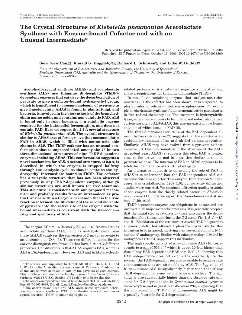
FIG. 2. Structure of K. pneumoniae ALS. A shows a ribbon diagram of the overall structure of the resting enzyme tetramer, with themonomers colored green (monomer A), red (B), blue (C), and yellow (D). There is a vertical 2-fold axis of symmetry in this view. The asymmetricunit contains monomers A and B, whereas the active sites are at the AC and BD interfaces. Monomer A is shown in B, with cylinder representationsof �-helices (red) and �-strands (turquoise) shown as arrows, connected by random coil (green). C and D compare the �-domains of ALS (C) andAHAS (D), with secondary structure indicated as in B. The residues in contact with FAD (stick model, D) in AHAS and their structural equivalentsin ALS (C) are shown in surface representation.
Crystal Structure of Acetolactate Synthase2244
by guest on May 29, 2018
http://ww
w.jbc.org/
Dow
nloaded from

FIG. 3. Structure-based sequence alignment of K. pneumoniae ALS (resting enzyme, monomer A) and yeast AHAS (monomer A, 40),drawn with the program ESPript (37). The schematics above and below the sequences represent �-helices (�1, �2, etc.), �-strands (�1, �2, etc.)and 310 helices (�1, �2, etc.), and TT represents �-turns. Residues shown in boxes are similar between the two sequences, those printed white onblack are identical, and those shown in lowercase are not visible in the electron density map of the proteins.
Crystal Structure of Acetolactate Synthase 2245
by guest on May 29, 2018
http://ww
w.jbc.org/
Dow
nloaded from

in ALS is located at the dimer-dimer interface of the tetramer.Yeast AHAS, on the other hand, does not form a homotetramerand exists as a dimer in both solution and crystals. The �-do-mains are also very similar between ALS and AHAS (r.m.s.d. �1.44 Å for 166 C� atoms). However, AHAS has a C-terminalextension as described previously (40) that is not found in ALS(Fig. 3).
The �-domains of the two proteins are the most different ofthe three domains, with a higher r.m.s.d. value (1.49 Å) forfewer (133) C� atoms. In general, this domain in both enzymeshas a similar secondary structure topology, consisting of acentral six-stranded parallel �-sheet surrounded by helices andloops. The major differences are in the connections between thestrands of the central �-sheet. An example is the polypeptidesegment connecting the �-strands 12 and 13 (Fig. 3). In yeastAHAS this segment, comprising residues Gly374 to Gly401, con-sists of two helices that are separated by a random coil. Thecorresponding region in ALS (Gly282–Ala298) is 11 residuesshorter and has two short helices, but neither corresponds tothose found in AHAS.
The most obvious difference between the structures of ALSand AHAS is that the latter contains FAD. In AHAS, theFAD-binding site is located mainly in the �-domain. FAD bindsnear the C-terminal edge of the central parallel �-sheet andforms multiple interactions with a series of loop regions foundon the surface of the domain. Most of the interacting amino acidresidues are highly conserved among AHAS sequences acrossplant and fungal and bacterial species. The surface topology ofthe binding site shows clearly that FAD, in an extended con-formation, lies in a groove that runs across the �-domain (Fig.2D). This groove structure is missing from the surface topologyof the corresponding area in the �-domain of ALS (Fig. 2C). Theabsence of the groove structure in ALS is brought about byslightly different orientations of the surface loop regions, andby amino acid substitutions to bulky hydrophobic residues atpositions that interact with FAD in AHAS.
Structure of ThDP in the Resting Enzyme—ThDP is bound ata subunit interface, anchored at one end through a magnesiumion that is octahedrally coordinated to six oxygen atoms: onefrom each phosphate group, one from the side chains of Asp447
FIG. 4. Structures and stereo electron density maps for ThDP in K. pneumoniae ALS. A shows the Fo � Fc difference electron densitymap (contoured at �5�) for the resting enzyme before ThDP is modeled. The coordinates with a planar N-3 (brown) and pyramidal N-3 (blue) areoverlaid for comparison. B and C show two views of the Fo � Fc difference electron density map before hydroxyethyl-ThDP was modeled (contouredat �5�) for the enzyme intermediate. The coordinates of the modeled tricyclic structure are shown in blue and the non-tricyclic model in brown.D, the coordinates of ThDP with a pyramidal N-3 (dark blue) and the tricyclic intermediate (light blue) are overlaid. E and F show the Fo � Fcelectron density after modeling of the tricyclic (E) and non-tricyclic (F) enzyme-intermediate with a C-2 to N-4� distance of 2.5 Å. Green densityis contoured at �3� and red density at � �3�.
Crystal Structure of Acetolactate Synthase2246
by guest on May 29, 2018
http://ww
w.jbc.org/
Dow
nloaded from

and of Asp474, the backbone of Gly476, and one water molecule.The arrangement of ligands is slightly unusual compared withother ThDP-dependent enzymes in that the amino acid side-
chain ligands are usually an aspartate and an asparagine,which are found at the two ends of the “thiamine-bindingmotif” discovered by Hawkins et al. (41). The asparagine is
FIG. 5. Proposed structures in the catalytic cycle of K. pneumoniae ALS. See text for details. I–V correspond to those in Fig. 1A.
Crystal Structure of Acetolactate Synthase 2247
by guest on May 29, 2018
http://ww
w.jbc.org/
Dow
nloaded from

highly conserved (42) with the possible exception of ALS se-quences. In Z. mobilis pyruvate decarboxylase, mutation of theequivalent asparagine to aspartate results in a drastic reduc-tion in the affinity for ThDP (43). The reason that ALS usesaspartate rather than asparagine as a magnesium ion ligand istherefore not clear. What is clear is that the presence of bothThDP and magnesium ion lays to rest the suggestion (38) thatK. pneumoniae ALS does not require these cofactors.
The first step in catalysis by ThDP-dependent enzymes iswidely believed to be the ionization of the thiazolium ring ofThDP (Fig. 1A, I3 II). In both ThDP (Fig. 1B) and the result-ing C-2 carbanions, the thiazolium ring is expected to be planarwith its aromaticity producing C-2—N-3 and C-4—C-5 bondswith lengths approaching those of double bonds. This expecta-tion is confirmed in the structures of all ThDP-dependent en-zymes containing unmodified ThDP. The bond from N-3 toC-7�, which connects to the methylaminopyrimidine ring, isnormally in the same plane as the thiazolium ring due to sp2
hybridization at N-3. This geometry is observed in the struc-tures of all ThDP-dependent enzymes containing unmodifiedThDP. In K. pneumoniae ALS the thiazolium ring is planar, butthe angle between the electron density associated with thethiazolium and methylaminopyrimidine rings is more acutethan that in any of the 26 known three-dimensional structuresof nine ThDP-dependent enzymes.
In order to fit the two rings into the observed electron densitywhile maintaining planarity at N-3 (Fig. 4A), it is necessary topropose a bond angle of 96° at C-7� and of 90° at the methylenethat links to the diphosphate tail of the cofactor. These unfa-vorable angles are each about 15° more acute than those ob-served in other ThDP-dependent enzymes, which average 111°(range 107–115°) and 107° (range 103–112°), respectively. Analternative explanation is that the structure is pyramidal atN-3 (Fig. 4A), resulting in acceptable bond angles of 110° and104°, respectively. At 2.3-Å resolution the electron density isnot sufficiently well resolved to discriminate between the twopossibilities. However, our preferred model is that with a py-ramidal N-3 because there is a reasonable mechanism thatcould give rise to this structure.
A pyramidal N-3 could arise from an improbable reductionby DTT to the thiazoline (44) or by charge migration from N-3to S-1 (Fig. 5, Ia). Kluger (27) noted the importance of Ia to theprobable structure of the carbanion. Although this S� form isexpected to be less favored than the N� form, the difference instability between the two forms may be small. In the crystalstructure of ThDP itself (45), the N� form is favored by a factorof 2.16:1 only. Moreover, the Met394 backbone carbonyl dipole,3.9 Å from S-1 but 6.3 Å from N-3, may provide some weakstabilization of the S� form. Irrespective of how it is stabilized,we suggest that the structure observed is the Ia/Ib resonancehybrid with Ib as a significant contributor. However, we cannotrule out C-2 proton dissociation giving the resonating sulfurylide/carbene (IIa/IIb).
In all ThDP-dependent enzymes, the two rings of the cofactorare held in a V conformation by a large hydrophobic residueintruding between them. For example, in yeast AHAS it isMet525 that performs this role. The structurally equivalentresidue in K. pneumoniae ALS is Met422, and the position ofthis methionine is quite similar in the two enzymes (notshown), with a C� separation of only 0.64 Å. The position ofMet422 allows a pyramidal N-3, but it does not force this con-formation. Observations on the structure of ALS containing abound intermediate, to be described below, suggest an expla-nation for the unusual conformation. This hypothesis will bedeveloped under “Discussion,” but it involves formation of thetricyclic form IIc (Fig. 5).
ALS with a Bound Intermediate
Rationale for the Addition of Pyruvate—Crystals of the en-zyme were soaked with pyruvate to convert the enzyme tointermediate IV (Fig. 1A). The overall reaction catalyzed byALS is irreversible, and it may not be immediately apparentwhy addition of pyruvate would cause the accumulation ofwhat is, ostensibly, a transient intermediate. However, whenthe catalytic cycle of the enzyme (Fig. 1A) is dissected, it can beshown that accumulation of this intermediate is not only pos-sible, it is obligatory. Addition of pyruvate initiates the cata-lytic cycle, which proceeds in a predominantly clockwise direc-tion as illustrated in Fig. 1A. In the steady state, IV representsa small fraction of the total enzyme present, but as acetolactateaccumulates, reversal of the step I 3 V becomes possible.Accumulation of acetolactate is accompanied by depletion ofpyruvate so V3 IV is increasingly favored. For as long as somepyruvate remains, all enzyme forms will coexist in steady-stateequilibrium. However, the pyruvate concentration will bedriven to zero eventually by reaction with II forming III, whichis converted irreversibly to IV. At the same time, IV 3 Vbecomes impossible because there is no pyruvate remaining.The combination of these processes forces the total active en-zyme to collect as IV.
To verify this logic, a simulation was performed (Fig. 6) byusing the program Wes (46). We do not suggest that the sim-ulation illustrated reproduces quantitatively the time course ofthe various intermediates, and we did not perform an exactsimulation because we do not know the values of the variousrate constants. However, no matter what rate constants areassumed, the simulation always terminates with IV as thefinal form. The only escape would be the release of acetalde-hyde to produce I, a reaction that is not part of the catalyticcycle and is expected to be suppressed by ALS. Even if thisreaction does occur slowly, the built-up acetolactate would en-sure that IV is immediately reformed from I via V.
Overall Structure of the Protein—Crystals in two differentspace groups (Table I) were treated with pyruvate to trap theintermediate. The triclinic form has the highest resolution at
FIG. 6. Simulation of the time-dependent distribution of en-zyme complexes as pyruvate is consumed by ALS. Complexes Iand II (Fig. 1A) were treated as a single species. The rate constantsassumed are as follows: I/II 3 III, 2.5 � 104 M�1 s�1; III 3 I/II, 1.0 �103 s�1; III 3 IV, 1.5 � 103 s�1; IV 3 V, 7.0 � 105 M�1 s�1; V 3 IV,5.0 � 101 s�1; V3 I/II, 5.5 � 102 s�1; and I/II3 V, 1.0 � 105 M�1 s�1.This combination of rate constants results in kcat and Km values of 500s�1 and 34.1 mM, respectively. Initial concentrations of pyruvate andALS were set at 200 mM and 50 �M, respectively. Simulations wereperformed with the program Wes (46). The dashed line shows pyruvateconsumption, and the solid and dotted lines show the concentrations ofI/II and IV, respectively. When pyruvate consumption exceeds 99%after about 0.7 min, there is a sharp rise in the amount of IV, and at 3min this enzyme form represents about 85% of the total. The percentageof IV rises above 98% upon continuing the simulation (not shown) for afurther 12 min and eventually reaches 100%.
Crystal Structure of Acetolactate Synthase2248
by guest on May 29, 2018
http://ww
w.jbc.org/
Dow
nloaded from
2.0 Å, and the asymmetric unit is a tetramer. For the orthor-hombic form, the asymmetric unit contains two monomers anddiffracts to 2.3 Å, the same as this form when not soaked withpyruvate. There are only marginal differences between thetriclinic and orthorhombic forms, and here we shall focus on thelatter when describing the protein structure. When we look indetail at the intermediate, the data from the triclinic form willbe used because it has the higher resolution.
Monomer A extends from Pro6 to His554 and contains nointernal gaps, whereas monomer B has one additional N-ter-minal residue and has gaps at Val118–Gln120, Gly185, andGly363. No side-chain electron density beyond the �-carbon isobserved for Lys114, Ser184, Lys208, and Lys225 in monomer Aand Gln117, Ser184, Gln188, Arg350, Arg361, and Arg362 in mon-omer B. The overall structure of the two monomers is almostidentical with an r.m.s.d. of 0.70 Å for 540 C� atoms. Compar-ing each monomer with the two of the resting enzyme, thedifferences are again rather small; of the four possible pairwisecomparisons, the highest r.m.s.d. is 0.78 Å for 536 C� atoms.When the active site regions of the resting and enzyme-inter-mediate forms are compared, the positions of all amino acidside chains are identical within the resolution of the data.
Structure of the Intermediate—The geometry of ThDP in theintermediate is subtly different from that in the resting en-zyme. One major difference is the presence of additional elec-tron density that projects from C-2 (Fig. 4, B and C), consistent
with the presence of the S-enantiomer of an hydroxyethylgroup attached to the cofactor. A further difference is that themethylaminopyrimidine and thiazolium rings are closer to oneanother than in the resting enzyme. We investigated a varietyof conformational alternatives for the cofactor, but these invari-ably left unexplained electron density between N-4� and C-2(Fig. 4F). Even when these two atoms were separated by 2.2 Åonly, closer than the sum of their van der Waals radii (2.95 Å),unexplained density remained. Our interpretation is that thereis a covalent bond connecting N-4� and C-2 (Fig. 4E).
The resulting tricyclic compound (IVc, Fig. 5) is identical todihydrothiachrome diphosphate except for the extra substitu-ent on C-2. The formation of dihydrothiachrome by cyclizationof thiamine in alkaline ethanol was first detailed by Maier andMetzler (47). Oxidation of this compound gives rise to thio-chrome (48), the fluorescent derivative that is used widely forestimating thiamine and its derivatives. Later studies havereported the formation of dihydrothiachrome in basic (49) andneutral (50) aqueous solution. It was suggested (51), before thestructure of any ThDP-dependent enzyme was known, that thetricyclic form may be catalytically important as a means ofprotecting ThDP from hydrolysis. Even though the enzymestructures published subsequently have not favored this hy-pothesis, Zoltewicz and Uray (49) do not rule it out. Similartricyclic structures may form in other ThDP-dependent en-zymes, and in this context we note that the drug omeprazole,
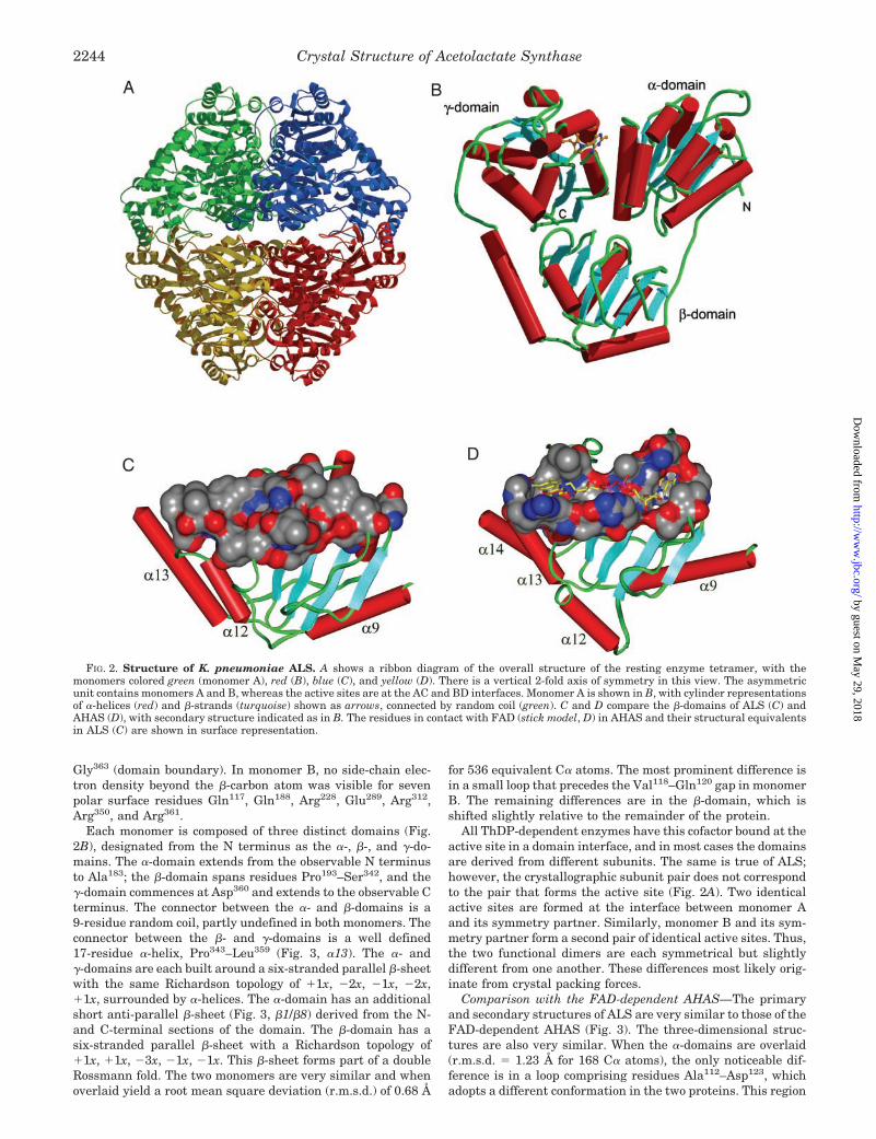
TABLE IData collection and refinement statistics
Resting Intermediate Oa Intermediate Ta
Crystal dataUnit cell length (Å) a � 116.8 a � 117.5 a � 86.3
b � 160.6 b � 160.6 b � 92.8c � 129.4 c � 129.5 c � 97.4
Unit cell angle (°) � � 90.0 � � 90.0 � � 68.0� � 90.0 � � 90.0 � � 63.5� � 90.0 � � 90.0 � � 67.7
Space group C2221 C2221 P1Crystal dimensions (mm) 0.2 � 0.05 � 0.05 0.2 � 0.05 � 0.05 0.3 � 0.3 � 0.05
Diffraction datab
Temperature (K) 100 100 100Resolution range (Å) 100–2.3 100–2.3 100–2.0Observations [I�0�(I)] 195,326 (4677) 170,273 (5047) 285040 (17,104)Unique reflections [I�0�(I)] 48,069 (2370) 48,466 (3030) 153,254 (11,633)Completeness 87.5 (43.7) 88.2 (49.3) 93.8 (71.3)Rsym
c 0.055 (0.113) 0.057 (0.126) 0.030 (0.14)I/�(I) 21.5 (5.6) 17.7 (4.5) 29.2 (4.6)
RefinementResolution limits (Å) 100–2.3 100–2.3 100–2.0Monomers per asymmetric unit 2 2 4No. atoms per asymmetric unitd
Protein non-H 8204 (24.4) 8256 (25.6) 16,260 (33.1)ThDP non-H 2 � 26 (21.1)ThDP/intermediate non-H 2 � 29 (18.7) 4 � 29 (22.9)Mg2� 2 (13.9) 2 (12.8) 4 (24.2)Phosphate non-H 2 � 5 (16.4) 2 � 5 (19.8) 4 � 5 (40.6)Diethylene glycol non-H 9 � 7 (45.0) 8 � 7 (50.0) 5 � 7 (52.9)Triethylene glycol non-H 2 � 10 (48.9)Water molecules 527 (34.1) 612 (36.3) 1335 (42.3)
Rfactor 0.165 0.162 0.191Rfree 0.215 0.214 0.228Root mean square deviations
Bond lengths (Å) 0.005 0.005 0.005Bond angles (°) 1.24 1.23 1.26
Ramachandran plot (%)Most favored 91.1 90.6 90.6Additionally allowed 8.8 9.4 9.3Generously allowed 0.1 0.0 0.1Disallowed 0.0 0.0 0.0
a Enzyme-intermediate complex, formed by soaking orthorhombic (O) or triclinic (T) crystals with 200 mM pyruvate.b Values in parentheses are statistics for the highest resolution shell, 2.38 to 2.30 Å for the orthorhombic crystals and 2.07 to 2.00 Å for the
triclinic crystal.c Rsym � ��I�I�/�I, where I is the intensity of an individual measurement of each reflection and I is the mean intensity of that reflection.d Values in parentheses are average B-factors, in Å2.
Crystal Structure of Acetolactate Synthase 2249
by guest on May 29, 2018
http://ww
w.jbc.org/
Dow
nloaded from
an analog of the tricyclic form of thiamine, inhibits Z. mobilispyruvate decarboxylase by competing with ThDP (52).
The structure that we propose has not been identified previ-ously as a possible intermediate of a ThDP-dependent enzyme.Indeed, we think that it is more likely to be a non-productivecomplex (IVc, Fig. 5) formed in a side reaction from the trueintermediate (IVb, Fig. 5). We suggest that the highly reactivetricyclic intermediate (IIc, Fig. 5) first forms prior to additionof the first pyruvate, and this reacts with the substrate formingIII that then decarboxylates to give the relatively non-reactiveenamine (IVa, Fig. 5). Because this is stable, the enzyme canpause midway through the catalytic cycle while releasing CO2
and admitting the second molecule of pyruvate. The tricyclic�-carbanion (IVb, Fig. 5) then forms, ready to react with thesecond pyruvate. However, occasionally during the prolongedincubation that occurs after the soaked crystals have used upall of the added pyruvate, a small fraction of the accumulatedIVa flickers into IVb and is converted to IVc, which is thestable product that we observe. This two-step route (IVa 3IVb3 IVc) appears to be the more likely, although we cannotrule out direct conversion of IVa to IVc.
The conversion of IVb to IVc involves protonation of C� ofthe intermediate and deprotonation of N-4�. Although thesecould be unconnected events, we suggest that there is an in-tramolecular proton transfer. The basis of this suggestion isthat the hydrogen atom on C� of IVc is in a hydrophobic region
with no suitable amino acid side chains or water molecules thatmight be a proton source. The N-4� hydrogen atom on thehydrophobic face of IVb is well positioned to act as a source ofthe C� proton, with a C to N distance of 2.4 Å.
Active Site and a Second Substrate Binding Model
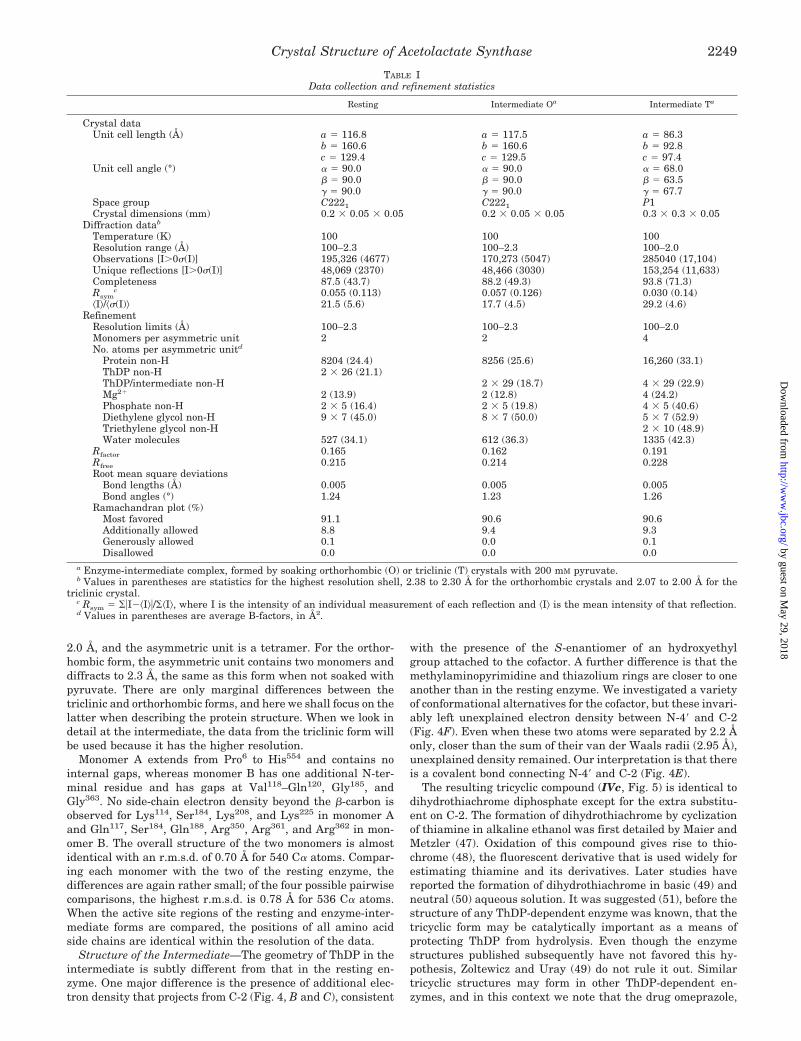
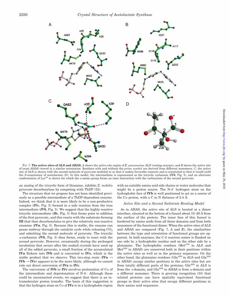
As in AHAS, the active site of ALS is located at a dimerinterface, situated at the bottom of a funnel about 15–20 Å fromthe surface of the protein. The inner face of this funnel isbordered by amino acids from all three domains and from bothmonomers of the functional dimer. When the active sites of ALSand AHAS are compared (Fig. 7, A and B), the similaritiesbetween the type and orientation of functional groups are ap-parent. In both enzymes, the C-2 reaction center is flanked onone side by a hydrophobic residue and on the other side by aglutamine. The hydrophobic residues (Met479 in ALS andMet582 in AHAS) are conserved both in their positions withinthe active sites as well as in the primary sequences. On theother hand, the glutamine residues (Gln420 in ALS and Gln202
in AHAS) occupy similar positions in the active sites but arefrom totally different parts of the proteins; Gln420 in ALS isfrom the �-domain, and Gln202 in AHAS is from �-domain anda different monomer. There is growing recognition (53) thatrelated proteins can have spatially equivalent functionalgroups in their active sites that occupy different positions intheir amino acid sequences.
FIG. 7. The active sites of ALS and AHAS. A shows the active site region of K. pneumoniae ALS (resting enzyme), and B shows the active siteof yeast AHAS viewed in a similar orientation. Residues with and without the prime symbol are derived from different monomers. C, the activesite of ALS is shown with the second molecule of pyruvate modeled in so that it makes favorable contacts and is orientated so that it would yieldthe S-enantiomer of acetolactate (D). In this model, the intermediate is represented as the tricyclic carbanion (IVb, Fig. 5), and an alternateconformation of Lys36 is shown for which the �-amino group forms an ionic interaction with the carboxylate of the second pyruvate.
Crystal Structure of Acetolactate Synthase2250
by guest on May 29, 2018
http://ww
w.jbc.org/
Dow
nloaded from

The second half of the ALS/AHAS catalytic cycle (Fig. 1A)involves the combination of the reaction intermediate (IV) withthe second substrate to yield the bound product complex (V)that then releases an acetohydroxy acid. The formation of theproduct intermediate is stereospecific, and the incoming sub-strate must be correctly orientated. This might involve an ionicinteraction between the carboxylate group and a basic residuein the active site. The only available candidate residues areLys36 in ALS and Lys251 in AHAS. As with the Gln420/Gln202
pair, the lysine residues are not from corresponding positionsin amino acid sequence, but they are found in similar locationswithin the active sites of the two enzymes. These lysines arehighly conserved within the two groups of enzymes, and theirside chains are not involved in ionic interactions with otherparts of the protein. We suggest that they are involved inbinding and orientating the second substrate.
By using the information from the crystal structures and thereaction stereochemistry, a model for the binding of the secondsubstrate in ALS is proposed (Fig. 7C). In the description of thismodel, we will refer to atoms as they are numbered in theacetolactate product (Fig. 7D), in which C-3 and C-4 are derivedfrom the first pyruvate, and C-1, C-2, and C-2a are derivedfrom the second pyruvate. We believe that the covalently boundintermediate that reacts with the second substrate is a tricyclichydroxyethyl carbanion (IVb, Fig. 5), and our proposal (Fig.7C) is based on modeling this structure. The intermediatewould be planar at C-3 with its methyl group (C-4) interactingwith Met479 and its hydroxyl group pointing toward the solventcavity. The re face of the intermediate will be buried in ahydrophobic cavity (consisting of the side chains of Met394,Met479, and the �-carbon atom of Gln420) and the si face towardthe solvent cavity where the second pyruvate is most likely tobind.
The formation of V involves the nucleophilic attack of IVb onthe C-2 atom of the second pyruvate. The product released isacetolactate with a chiral center at C-2. The S-enantiomer isrequired for branched-chain amino acid biosynthesis (54), andwe have found by circular dichroism spectroscopy (data notshown) that the products of ALS and AHAS have the samestereochemistry. Thus, in order to produce the correct enanti-omer of acetolactate, the second pyruvate must be bound toALS with its C-2 atom close to the C-3 carbanion of the inter-mediate, and orientated so that it will produce the S-enanti-omer of acetolactate.
In our model, the second pyruvate is bound in the active sitesolvent cavity and with its two carbonyl oxygens displacing twowater molecules that are present in the crystal structures ofALS containing the intermediate. The C-2 oxygen forms ahydrogen bond with the backbone nitrogen of Ala35, and theC-2a methyl group points toward Gln483. The residue corre-sponding to Gln483 in AHAS is Trp586 (Fig. 7B) and has beenshown to be involved in second substrate specificity (55). Al-though AHAS favors 2-ketobutyrate as the second substrate,ALS reacts very poorly with the larger substrate (56). We havetried to model 2-ketobutyrate into the ALS active site, but thisresults in steric clashes between the larger ethyl group of2-ketobutyrate and the extended side chain of Gln483. In orderto accommodate 2-ketobutyrate, Gln483 would have to adopt analternate conformation in which its side chain moves towardthe protein surface and out of the active site. Gln483 may playa role in ALS catalysis so this drastic change in its conforma-tion in order to bind to 2-ketobutyrate would prevent reactionfrom occurring.
The C-1 carbonyl oxygen of the modeled pyruvate is posi-tioned in place of a water molecule that is found between andhydrogen-bonded to the side chains of Gln420 and Gln483. The
nearest carboxylate oxygen of pyruvate is about 4 Å away fromthe side chain of Lys36. However, this side chain can be movedeasily so that it could form an ionic interaction with the car-boxyl group of the second pyruvate, assisting its binding andorientation. Our model positions the second pyruvate with itsC-2 atom close to the C-3 carbanion (3.8 Å) and in the rightorientation to produce 2S-acetolactate. The second substratemay be bound in a similar way in AHAS because the architec-ture of their active sites is very similar (Fig. 7, A and B) andthey produce acetolactate with the same stereochemistry.
Extraneous Ligands
Each monomer in both orthorhombic forms of the enzymecontains several 7.4 Å long sausage-shaped regions of electrondensity in the asymmetric unit, one of which in each monomeris in a pocket close to the entrance of the channel leading to theactive site. The electron density fits well to diethylene glycoland may represent an immobile segment of a much largermolecule, either the precipitant PEG8000 or the cryoprotectantPEG600. Monomer A of the triclinic form contains two mole-cules of what appears to be triethylene glycol. In this contextwe note that other crystal structures contain ligands modeledas triethylene glycol, and Lehtio et al. (57) suggest that they areobserving part of a PEG1000 molecule that was used as aprecipitant. Four of the diethylene glycol molecules in eachorthorhombic monomer form two clusters, each of which maybe part of a single PEG molecule. A geometrically plausibleethyl connection can be made between the two molecules ineach pair, and there is weak electron density in this regionsuggestive of such connections.
In all three structures, each monomer contains a tetrahedralisland of electron density in an equivalent position, which wasmodeled as a phosphate ion because the enzyme storage buffercontains 50 mM potassium phosphate. The phosphate ion islocated near the C-terminal end of helix �15/�10 at the junctionof the �- and �-domains. In the crystal structures of yeastAHAS (9, 40), a potassium ion is found at the equivalent posi-tion. In AHAS, this cation stabilizes the dipole of its interactinghelix, and it is unexpected for a phosphate ion to be found at thesame location. However, the phosphate ion in ALS is charge-neutralized by three arginine residues (Arg259, Arg352, andArg403) that surround it. Other ligands include the side chain ofGln266 and a water molecule. This phosphate ion may be func-tionally significant because it is known that ALS is activated byacetate, and this activation is reversed by phosphate and sul-fate (58, 59).
DISCUSSION
FAD and the �-Domain—The absence of FAD in ALS and itspresence in AHAS could be explained in two ways. The firstexplanation is that ALS comes from the same evolutionary lineas AHAS with both derived from an FAD-dependent pyruvateoxidase-like ancestor; subsequently, ALS has dispensed withthe requirement for FAD. Alternatively, ALS may have origi-nated from the same line as the 2-ketoacid decarboxylases thatdo not contain FAD, such as pyruvate decarboxylase, indole-pyruvate decarboxylase, and benzoylformate decarboxylase.This explanation implies that AHAS and ALS are the result ofconvergence of function with respect to the reaction catalyzed.Phylogenetic analysis of the protein sequences (60) shows thatthe ALS sequences cluster closely together and are part of alarger group that includes pyruvate oxidase and AHAS. The2-ketoacid decarboxylases form an entirely separate cluster.These data clearly indicate that ALS comes from the samepyruvate oxidase-like ancestor as AHAS. FAD has a purelystructural role in AHAS that has been supplanted in ALS byadjustment of the protein. This has involved a series of amino
Crystal Structure of Acetolactate Synthase 2251
by guest on May 29, 2018
http://ww
w.jbc.org/
Dow
nloaded from

acid substitutions and reorientations that fill the channel oc-cupied by FAD in AHAS (Fig. 2, C and D).
Structures of the enzymes mentioned above have been pub-lished (9, 13–18, 40). All have a similar three-domain structurewith a core composed of pairs of �- and �-domains, with the�-domains on the periphery. The active sites are at the junctionof the �-domain of one subunit and the �-domain of its neigh-bor. Also in this structural family is transketolase with thesame three-domain structure but arranged in the sequence �,�, and � (12).
A clear role for the �-domain has been established in pyru-vate oxidase, where it constitutes the major portion of thebinding site for FAD (14), a required participant in catalysis. Inyeast (but not Z. mobilis) pyruvate decarboxylase, an allostericactivator site that binds pyruvate, is in the �-domain (15). Wehave suggested (40) that the �-domain of yeast AHAS formspart of the binding site for the regulatory subunit of this en-zyme. For the other enzymes, the �-domain has no obviousfunction and appears to serve solely as a scaffolding to supportthe �- and �-domains. Thus, the absence of an FAD-binding sitein the �-domain of ALS reflects mutations that have occurredafter separation from the AHAS evolutionary lineage. Giventhat pyruvate decarboxylase, benzoylformate decarboxylase,indolepyruvate decarboxylase, and transketolase can constructa perfectly satisfactory �-domain scaffold without FAD, it is nogreat surprise that ALS can do the same.
If the role of the �-domain in most of these enzymes is solelyto hold the �- and �-domains together, then one might easilyimagine related enzymes that have dispensed with the �-do-main entirely by developing appropriate interactions betweenthe �- and �-domains. This appears to be the situation inphosphonopyruvate decarboxylase (61) where the protein se-quence corresponding to the �-domain is missing, whereas insulfopyruvate decarboxylase there are separate subunits thatare homologous to the �- and �-domains, with no equivalent tothe �-domain (61). Thus, this entire family of enzymes is con-structed around the �- and �-domains. The �-domain can beinvolved in regulation (yeast pyruvate decarboxylase and per-haps AHAS), structural integrity (ALS, Z. mobilis pyruvatedecarboxylase, benzoylformate decarboxylase, indolepyruvatedecarboxylase, and transketolase), or not required at all (phos-phonopyruvate decarboxylase and sulfopyruvate decarboxy-lase). Only in pyruvate oxidase does the �-domain have anindispensable role in catalysis.
ThDP—There are published structures of representatives ofmost major classes of ThDP-dependent enzymes, which showrecognizable common themes in the interactions and conforma-tion of ThDP. It is anchored to the protein through a divalentmetal ion that ligates to the diphosphate tail and a pair of polaramino acid side chains from the borders of the thiamine-bind-ing motif (41). N-1� forms a hydrogen bond to a conservedglutamate residue that plays an important role in catalysis(20–24). The cofactor is held in a V conformation by a largehydrophobic residue that projects between the thiazolium andmethylaminopyrimidine rings. This forces the 4�-amino groupinto close proximity with C-2 of the thiazolium ring. ThDP inALS displays all of these features but differs in one crucialrespect. N-3 of the thiazolium ring is pyramidal in our inter-pretation, whereas in all of the other structures it is trigonal.The protein itself does not appear to force this pyramidal con-formation, so what are the forces that bring about this uniquestructure?
An important clue comes from the structure of ALS with thetrapped hydroxyethyl intermediate, where ThDP is in its tri-cyclic form (29). A similar dihydrothiachrome compound hasbeen documented previously for thiamine in solution (47, 49,
50), but never before has it been observed in an enzyme. Su-perimposition of ThDP in the resting enzyme with that in theintermediate (Fig. 4D) demonstrates that the cofactor atomsare in very similar positions. Therefore, we suggest that theresting enzyme contains both the open and tricyclic (IIc, Fig. 5)forms of ThDP in rapid equilibrium. The proportion of thedihydrothiachrome form is small enough that it makes nomajor contribution to the electron density but large enough tofavor maintenance of N-3 in its pyramidal conformation.
We propose that the tricyclic ThDP reacts with pyruvateforming the tricyclic lactyl-ThDP (III, Fig. 5). After decarbox-ylation, the enamine (IVa, Fig. 5) is kinetically stabilizedagainst reaction with electrophiles because the forced pyrami-dal N-3 prevents delocalization of electron density from N-3 tothe nucleophilic center at the substrate-derived C-3. The activesite can then pause to release CO2 and admit the second mol-ecule of pyruvate with no risk of diversion into a non-produc-tive product. However, during the extended incubation of crys-tallization, the proximity of N-4� allows occasional reaction atC-2 of the thiazolium ring and proton transfer to the substrate-derived C-3 forming IVc (Fig. 5), the product that we havefound.
An intriguing implication of our favored structure for ThDPand mechanism is that they may explain the unusually highkcat of K. pneumoniae ALS. The enforced non-planarity at N-3,which we propose for the reasons described earlier, would re-sult in a structure in which greater positive charge is distrib-uted onto C-2, because delocalization of this charge onto N-3would not be possible and S-1 would be less able to bearpositive charge than N-3, also for the reasons given above. Theresult should be a more acidic C-2—H bond. After ionization toIIa/IIb, cyclization would then produce the nearly localizedcarbanion IIc which should exhibit very high nucleophilic re-activity toward the first pyruvate. By forcing pyramidal geom-etry at N-3, the enzyme would therefore promote C-2 reactivityand accelerate the steps leading to lactyl-ThDP (III, Fig. 5),which are rate-limiting in AHAS (62).
The prevailing evidence (1, 12–19, 26) suggests that the4�-amino group of ThDP, possibly as an imino group in a tau-tomer of the methylaminopyrimidine ring, functions as a basiccenter in C-2 deprotonation. The mechanistic proposal of Fig. 5can readily accommodate this role. This mechanism is consist-ent with the structures that we observe and is compatible withthe known chemical properties of ThDP and the catalytic prop-erties of ThDP-dependent enzymes. We speculate that in ALSwe may be observing forms of the cofactor that are relevant toother ThDP-dependent enzymes. Perhaps the high kcat of ALSfavors retention of these unusual, but crucial, conformations.Only the more relaxed structures are observed in other ThDP-dependent enzymes, because they are less able to keep ThDP inits most active state.
Acknowledgments—H.-L. Peng (National Chiao Tung University,Taiwan) and H.-Y. Chang (National Tsing Hua University, Taiwan)provided the original clone used as a source of the K. pneumoniae ALSgene. We thank R. Kluger (University of Toronto, Canada), J. V. Schloss(Kuwait University, Kuwait), and F. J. Leeper (University of Cam-bridge, UK) for useful comments on the nature and significance of theThDP structures reported here. All misinterpretations are our own. Theuse of the BioCARS sector was supported by the Australian Synchro-tron Research Program, which is funded by the Commonwealth ofAustralia under the Major National Research Facilities Program. Useof the Advanced Photon Source was supported by the United StatesDepartment of Energy, Basic Energy Sciences, Office of Energy Re-search, under Contract W-31-109-Eng-38. We thank Dr. Keith Bristerand Dr. Harry Tong for help with data collection at the AdvancedPhoton Source.
Crystal Structure of Acetolactate Synthase2252
by guest on May 29, 2018
http://ww
w.jbc.org/
Dow
nloaded from
REFERENCES
1. Duggleby, R. G., and Pang, S. S. (2000) J. Biochem. Mol. Biol. 33, 1–362. Gollop, N., Damri, B., Barak, Z., and Chipman, D. M. (1989) Biochemistry 28,
6310–63173. Bornemann, S. (2002) Nat. Prod. Rep. 19, 761–7724. Osborne, A., Thorneley, R. N. F., Abell, C., and Bornemann, S. (2000) J. Biol.
Chem. 275, 35825–358305. Vargo, D., Pokora, A., Wang, S. W., and Jorns, M. S. (1981) J. Biol. Chem. 256,
6027–60336. Wajant, H., and Effenberger, F. (1996) Biol. Chem. Hoppe-Seyler 377, 611–6177. Dreveny, I., Gruber, K., Glieder, A., Thompson, A., and Kratky, C. (2001)
Structure 9, 803–8158. Chang, Y.-Y., and Cronan, J. E., Jr. (1988) J. Bacteriol. 170, 3937–39459. Pang, S. S., Duggleby, R. G., and Guddat, L. W. (2002) J. Mol. Biol. 317,
249–26210. Stormer, F. C. (1967) J. Biol. Chem. 242, 1756–175911. Pang, S. S., Guddat, L. W., and Duggleby, R. G. (2002) Acta Crystallogr. Sect.
D Biol. Crystallogr. 58, 1237–123912. Lindqvist, Y., Schneider, G., Ermler, U., and Sundstrom, M. (1992) EMBO J.
11, 2373–237913. Dyda, F., Furey, W., Swaminathan, S., Sax, M., Farrenkopf, B., and Jordan, F.
(1993) Biochemistry 32, 6165–617014. Muller, Y. A., Schumacher, G., Rudolph, R., and Schulz, G. E. (1994) J. Mol.
Biol. 237, 315–33515. Arjunan, P., Umland, T., Dyda, F., Swaminathan, S., Furey, W., Sax, M.,
Farrenkopf, B., Gao, Y., Zhang, D., and Jordan, F. (1996) J. Mol. Biol. 256,590–600
16. Dobritzsch, D., Konig, S., Schneider, G., and Lu, G. (1998) J. Biol. Chem. 273,20196–20204
17. Hasson, M. S., Muscate, A., McLeish, M. J., Polovnikova, L. S., Gerlt, J. A.,Kenyon, G. L., Petsko, G. A., and Ringe, D. (1998) Biochemistry 37,9918–9930
18. Schutz, A., Sandalova, T., Ricagno, S., Hubner, G., Konig, S., and Schneider, G.(2003) Eur. J. Biochem. 270, 2312–2321
19. Schellenberger, A., Hubner, G., and Neef, H. (1997) Methods Enzymol. 279,131–146
20. Wikner, C., Meshalkina, L., Nilsson, U., Nikkola, M., Lindqvist, Y.,Sundstrom, M., and Schneider, G. (1994) J. Biol. Chem. 269, 32144–32150
21. Candy, J. M., Koga, J., Nixon, P. F., and Duggleby, R. G. (1996) Biochem. J.315, 745–751
22. Killenberg-Jabs, M., Konig, S., Eberhardt, I., Hohmann, S., and Hubner, G.(1997) Biochemistry 36, 1900–1905
23. Fang, R., Nixon, P. F., and Duggleby, R. G. (1998) FEBS Lett. 437, 273–27724. Bar-Ilan, A., Balan, V., Tittmann, K., Golbik, R., Vyazmensky, M., Hubner, G.,
Barak, Z., and Chipman, D. M. (2001) Biochemistry 40, 11946–1195425. Hill, C. M., Pang, S. S., and Duggleby, R. G. (1997) Biochem. J. 327, 891–89826. Kern, D., Kern, G., Neef, H., Tittmann, K., Killenberg-Jabs, M., Wikner, C.,
Schneider, G., and Hubner, G. (1997) Science 275, 67–7027. Kluger, R. (1987) Chem. Rev. 87, 863–87628. Chabriere, E., Vernede, X., Guigliarelli, B., Charon, M. H., Hatchikian, E. C.,
and Fontecilla-Camps, J. C. (2001) Science 294, 2559–256329. Fiedler, E., Thorell, S., Sandalova, T., Golbik, R., Konig, S., and Schneider, G.
(2002) Proc. Natl. Acad. Sci. U. S. A. 99, 591–595
30. Otwinowski, Z., and Minor, W. (1997) Methods Enzymol. 276, 307–32631. Navaza, J. (1994) Acta Crystallogr. Sect. A Biol. Crystallogr. 50, 157–16332. Jones, T. A., Zou, J. Y., Cowan, S. W., and Kjeldgaard, M. (1991) Acta Crys-
tallogr. Sect. A 47, 110–11933. Brunger, A. T., Adams, P. D., Clore, G. M., DeLano, W. L., Gros, P., Grosse-
Kunstleve, R. W., Jiang, J.-S., Kuszewski, J., Nilges, M., Pannu, N. S.,Read, R. J., Rice, L. M., Simonson, T., and Warren, G. L. (1988) ActaCrystallogr. Sect. D Biol. Crystallogr. 54, 905–921
34. Evans, S. V. (1993) J. Mol. Graphics 11, 134–13835. Kraulis, P. J. (1991) J. Appl. Crystallogr. 24, 946–95036. Merritt, E. A., and Bacon, D. J. (1997) Methods Enzymol. 277, 505–52437. Gouet, P., Courcelle, E., Stuart, D. I., and Metoz, F. (1999) Bioinformatics 15,
305–30838. Peng, H.-L., Wang, P.-Y., Wu, C.-M., Hwang, D.-C., and Chang, H.-Y. (1992)
Gene (Amst.) 117, 125–13039. Huseby, N.-E., Christensen, T. B., Olsen, B. R., and Størmer, F. C. (1971) Eur.
J. Biochem. (Tokyo) 20, 209–21440. Pang, S. S., Guddat, L. W., and Duggleby, R. G. (2003) J. Biol. Chem. 278,
7639–764441. Hawkins, C. F., Borges, A., and Perham, R. N. (1989) FEBS Lett. 255, 77–8242. Candy, J. M., and Duggleby, R. G. (1998) Biochim. Biophys. Acta 1385,
323–33843. Candy, J. M., and Duggleby, R. G. (1994) Biochem. J. 300, 7–1344. Zoltewicz, J. A., Dill, C. D., and Abboud, K. A. (1997) J. Org. Chem. 62,
6760–676645. Pletcher, J., and Sax, M. (1972) J. Am. Chem. Soc. 94, 3998–400546. Garcıa-Sevilla, F., Garrido-del Solo, C., Duggleby, R. G., Garcıa-Canovas, F.,
Peyro, R., and Varon, R. (2000) Biosystems 54, 151–16447. Maier, G. D., and Metzler, D. E. (1957) J. Am. Chem. Soc. 79, 4386–439148. Barger, G., Bergel, F., and Todd, A. R. (1935) Chem. Ber. 68, 2257–226249. Zoltewicz, J. A., and Uray, G. (1994) Bioorg. Chem. 22, 1–2850. Washabaugh, M. W., Yang, C. C., Hollenbach, A. D., and Chen, P. (1993)
Bioorg. Chem. 21, 170–19151. Doughty, M. B., and Lawrence, D. S. (1985) J. Chem. Soc. Chem. Commun.
454–45552. Nixon, P. F., Diefenbach, R. J., and Duggleby, R. G. (1992) Biochem. Pharma-
col. 44, 177–17953. Todd, A. E., Orengo, C. A., and Thornton, J. M. (2002) Trends Biochem. Sci. 27,
419–42654. Sylvester, S. R., and Stevens, C. M. (1979) Biochemistry 18, 4529–453155. Ibdah, M., Bar-Ilan, A., Livnah, O., Schloss, J. V., Barak, Z., and Chipman,
D. M. (1996) Biochemistry 35, 16282–1629156. Huseby, N.-E., and Størmer, F. C. (1971) Eur. J. Biochem. 20, 215–21757. Lehtio, L., Leppanen, V.-M., Kozarich, J. W., and Goldman, A. (2002) Acta
Crystallogr. Sect. D Biol. Crystallogr. 58, 2209–221258. Størmer, F. C. (1968) J. Biol. Chem. 243, 3735–373959. Holtzclaw, W. D., and Chapman, L. F. (1975) J. Bacteriol. 121, 917–92260. Bowen, T. L., Union, J., Tumbula, D. L., and Whitman, W. B. (1997) Gene
(Amst.) 188, 77–8461. Graupner, M., Xu, H., and White, R. H. (2000) J. Bacteriol. 182, 4862–486762. Tittmann, K., Golbik, R., Uhlemann, K., Khailova, L., Schneider, G., Patel, M.,
Jordan, F., Chipman, D. M., Duggleby, R. G., and Hubner, G. (2003) Bio-chemistry 42, 7885–7891
Crystal Structure of Acetolactate Synthase 2253
by guest on May 29, 2018
http://ww
w.jbc.org/
Dow
nloaded from
Siew Siew Pang, Ronald G. Duggleby, Richard L. Schowen and Luke W. GuddatEnzyme-bound Cofactor and with an Unusual Intermediate
Acetolactate Synthase withKlebsiella pneumoniaeThe Crystal Structures of
doi: 10.1074/jbc.M304038200 originally published online October 13, 20032004, 279:2242-2253.J. Biol. Chem.
10.1074/jbc.M304038200Access the most updated version of this article at doi:
Alerts:
When a correction for this article is posted•
When this article is cited•
to choose from all of JBC's e-mail alertsClick here
http://www.jbc.org/content/279/3/2242.full.html#ref-list-1
This article cites 61 references, 16 of which can be accessed free at
by guest on May 29, 2018
http://ww
w.jbc.org/
Dow
nloaded from