THE ISOLATION, IDENTIFICATION, AND CHARACTERIZATION OF ...
61
THE ISOLATION, IDENTIFICATION, AND CHARACTERIZATION OF BACTERIA ASSOCIATED WITH THE DEEP-SEA CORAL LOPHELIA PERTUSA. A thesis presented to the faculty of the Graduate School of Western Carolina University in fulfillment of the requirements for the degree of Master of Science in Biology. by S. Gold Director: Seán O’Connell Associate Professor of Biology Biology Department Committee members: Dr. Christina Kellogg, St. Petersburg Coastal and Marine Science Center Dr. Jeremy Hyman, WCU Biology Department Dr. Amanda Storm, WCU Biology Department April, 2020
Transcript of THE ISOLATION, IDENTIFICATION, AND CHARACTERIZATION OF ...

THE ISOLATION, IDENTIFICATION, AND CHARACTERIZATION OF BACTERIA
ASSOCIATED WITH THE DEEP-SEA CORAL LOPHELIA PERTUSA.
A thesis presented to the faculty of the Graduate School of Western Carolina University in
fulfillment of the requirements for the degree of Master of Science in Biology.
by
S. Gold
Director: Seán O’Connell
Associate Professor of Biology
Biology Department
Committee members:
Dr. Christina Kellogg, St. Petersburg Coastal and Marine Science Center
Dr. Jeremy Hyman, WCU Biology Department
Dr. Amanda Storm, WCU Biology Department
April, 2020

ii
ACKNOWLEDGMENTS
I would like to thank Dr. Erik Cordes, chief scientist of the Deep Search cruise, for
connecting me with Dr. Chris Kellogg and enabling collection of these samples. I’m so grateful
to Dr. Kellogg for providing me with these samples, and for her insights, constructive feedback,
and encouragement during the preparation of this thesis. I’m greatly inspired by her work and
deeply appreciative of the opportunity to collaborate with her on this project.
I would also like to thank the rest of my thesis committee, Dr. Jeremy Hyman and Dr.
Amanda Storm, and especially my advisor, Dr. Sean O’Connell. His guidance in study design,
assistance in culture work and molecular tests conducted in the lab, and support in analysis of
data and drafting of this thesis were invaluable and critical to the completion of this project. I’d
also like to thank Dr. Katherine Matthews for her aid in phylogenetic analysis of isolates, and the
Principles of General Microbiology students for their assistance in some of the culture work for
this project.
I would also like to express my gratitude for the funding sources for my graduate
research, including Western Carolina University’s Arts and Sciences Grant as well as WCU’s
Summer 2019 Graduate Research Assistantship Grant. Without these resources, my research
would not have been possible.
Finally, I would like to thank my family, friends, and fellow graduate students for their
consistent support and encouragement.

iii
TABLE OF CONTENTS
ACKNOWLEDGMENTS ........................................................................................................... ii
TABLE OF CONTENTS ........................................................................................................... iii
LIST OF TABLES ..................................................................................................................... iv
ABSTRACT .............................................................................................................................. vii
INTRODUCTION ....................................................................................................................... 1
METHODS.................................................................................................................................. 9
Site descriptions and sample collection ................................................................................... 9
Bacterial enrichment and isolation ........................................................................................ 10
Bacterial identification by 16S rRNA gene amplification and sequencing ........................... 13
Culture-based characterization of bacterial isolates .............................................................. 14
PCR analysis of genes for ammonia oxidation and nitrogen fixation ................................... 15
Statistical and phylogenetic analysis ..................................................................................... 17
RESULTS.................................................................................................................................. 18
DISCUSSION ........................................................................................................................... 31
CONCLUSION ......................................................................................................................... 37
REFERENCES CONSULTED ................................................................................................. 39
APPENDIX A. MICROBIOLOGICAL MEDIA RECIPES..................................................... 49
Medium for Ammonia Oxidizers (Modified from ATCC medium TSD-99) ....................... 49
Medium for Chitin Degraders (Modified from Murthy and Bleakley, 2012) ....................... 50
Medium for Oligotrophic Heterotrophic Marine Organisms (General) ................................ 51
APPENDIX B. SUPPLEMENTAL DATA .............................................................................. 51

iv
LIST OF TABLES
Table 1. Collection metadata for Lophelia pertusa samples. ......................................................... 9
Table 2. Isolate information for subcultures. ............................................................................... 12
Table 3. Isolate sequence matches based on 16S rRNA sequence analysis using BLAST.. ...... 19
Table 4. Antibiotic resistance of tested isolates against various antibiotics. ............................... 23
Table B1. Binary Key for Heatmap and PCA Analysis. .............................................................. 51

v
LIST OF FIGURES
Figure 1. Map showing locations of Richardson Reef Complex sample sites.. ........................... 10
Figure 2. Heatmap of morphological and physiological features of isolated cultures. ................ 23
Figure 3. Principal components analysis (PCA) of all Gram negative isolates. .......................... 25
Figure 4. Principal components analysis (PCA) of isolates cultured from chitin and DSR2B.... 26
Figure 5. Principal components analysis (PCA) of only Pseudoalteromonas. ............................ 27
Figure 6. Maximum likelihood tree of Pseudoalteromonas isolates. .......................................... 28
Figure 7. Maximum likelihood tree of Pseudoalteromonas isolates with the highest reads. ...... 29
Figure 8. Neighbor-joining tree showing Pseudoalteromonas isolates.. ..................................... 30
vi
LIST OF ABBREVIATIONS
AMO: Ammonia oxidation
AR: Antibiotic resistance
CWC: Cold-water coral
C: Carbon
DSR2B: Dilute, salty Reasoner’s 2 broth
ML: Maximum likelihood
N: Nitrogen
NJ: Neighbor joining
PCA: Principal components analysis
SIM: Sulfur, indole, motility

vii
ABSTRACT
Cold-water corals (CWC) form the structural basis of highly diverse and productive
ecosystems in the deep, dark ocean, serving as important spawning, nursery, and breeding habitat
for many fishes and invertebrates. As such, they play an important role in supporting fisheries
that humans rely on, as well as general ocean health, which is of critical importance to Earth as
the effects of climate change unfold. CWCs are heterotrophic filter feeders, and their ability to
survive in dark, oligotrophic waters may be linked to partnerships with microbial symbionts that
participate in nutrient cycling and conservation. While indirect methods–DNA sequencing,
whole genome analysis, isotopic analysis–have been used to hypothesize the roles of these
symbionts, few studies have grown cultures of associated microbes and directly observed the
metabolic processes involved in carbon and nitrogen turnover. In this study, bacteria cultured
from the globally distributed, deep-sea coral Lophelia pertusa were isolated and characterized
according to morphological and physiological characteristics. Sanger sequencing of 16S rRNA
from the isolates yielded a diversity of bacterial species in the phylum Proteobacteria. In culture,
isolates demonstrate the ability to use a variety of organics as carbon, nitrogen, and energy
sources, the most notable of which is chitin, a polymer containing both carbon and nitrogen that
is abundant in marine systems. Additionally, preliminary evidence suggests the ability of one
isolate to fix nitrogen. These findings corroborate evidence of nutrient cycling in CWCs and
support the hypothesis that microbial associates of these corals are an important aspect of their
ecophysiology and likely help fuel their productivity. Physical and physiological stress induced
by changes in the environment resulting from human activities and climate change could
influence host-microbe interactions, altering the ability of CWCs to conserve and recycle

viii
limiting resources. Loss of CWC ecosystems would mean loss of critical habitat and a globally
relevant carbon sink.

1
INTRODUCTION
The cold-water coral (CWC) Lophelia pertusa is a globally distributed, scleractinian
coral that forms the structural basis of highly diverse and productive ecosystems in the deep
ocean, serving as important spawning, nursery, and breeding habitat for many fishes and
invertebrates (Costello et al. 2005, Mortensen et al. 1995, Cordes et al. 2008). Lophelia and other
CWCs are ecosystem engineers, generating hotspots of biodiversity and organic material in an
otherwise oligotrophic deep sea environment where nutrients are limiting (Soetaert et al. 2016).
In the past decade, there has been an increase in focus on these CWCs driven mainly by
conservation concerns (Roberts and Hirshfield 2004). CWC gardens are often located in places
where large-scale commercial fishing and drilling for oil and gas are occurring, and are subject to
oil spills, dredging, and additional environmental stressors including ocean acidification and
pollution (Fisher et al. 2014). These corals are of scientific interest for a variety of reasons
including bioprospecting--discovery of new commercially useful biological compounds--and in
studying the impacts of environmental change (Maxwell 2005, Lu et al. 2015).
Corals (generally) are known for their ability to flourish in nutrient-poor environments
due to their ability to take up and recycle nutrients (Radecker et al. 2015). Corals are polytrophic,
meaning that they function at multiple trophic levels–as primary producers, primary consumers,
and secondary consumers (Muscatine and Porter 1977). This is in part due to their heterotrophic
capacity as suspension feeders, and in part due to their close association with microorganisms
capable of carrying out biological fixation of carbon and nitrogen and nutrient cycling (Wegley
et al. 2007, Radecker et al. 2015, Middleberg et al. 2015). Together, the complex assemblage of
coral animal and microbial associates are referred to as the coral holobiont. In tropical corals,

2
Symbiodinium are largely responsible for supplying their coral host with photosynthate as a
carbon and energy source for secondary production (Muscatine and Porter 1977). However, in
CWCs like L. pertusa, this role is likely played by prokaryotes. The ecology of CWCs is
fundamentally different from that of their shallow-water counterparts. CWC can live hundreds to
thousands of meters below the sea surface, well below the photic zone (Roberts et al. 2006). At
these depths, there is no ambient light, temperatures are between 4-12ºC, and there is increased
pressure (Roberts et al. 2006). Under these conditions, L. pertusa and other CWCs cannot host
photosynthetic partners. Thus, nutrient acquisition and cycling by the L. pertusa holobiont is a
key area of study.
Studies of the Lophelia holobiont have paid particular attention to identifying microbial
associates and uncovering how they are involved in nutrient cycling. Biological fixation of both
carbon and nitrogen have been demonstrated by the L. pertusa holobiont, as well as a complete
nitrogen cycle (Middleberg et al. 2015). Biological fixation is restricted to prokaryotes,
importantly implicating involvement of bacterial (and potentially archaeal) associates in these
processes. Research on L. pertusa microbial communities reveals that the coral has a
microbiome–an associated bacterial community distinct from its surrounding environment–and
that Lophelia-specific bacteria may be conserved, even among distant populations such as the
Gulf of Mexico and Trondheimsfjord in Norway (Galkiewicz et al. 2011, Kellogg et al. 2009,
Kellogg et al. 2017, Neulinger et al. 2008, Schöttner et al. 2009, van Bleijswijk et al. 2015,
Yakimov et al. 2005, Meistratzheim et al. 2016). Recent 16S rRNA amplicon analysis of DNA
from Lophelia samples taken in the Gulf of Mexico and along the Atlantic coastline corroborate
this, finding fifteen conserved bacterial operational taxonomic units (OTU) among these corals
(Kellogg et al. 2017). Molecular studies using metagenomic data have attempted to identify

3
bacterial genera and species that may be involved in various nutrient cycles based on functional
gene predictions (Neulinger et al. 2008, Kellogg et al. 2009, Kellogg et al. 2017). Taken
together, the data suggest that L. pertusa has a distinct microbiome consisting of associates
involved in carbon and nitrogen cycling.
The ability of L. pertusa to survive in dark, nutrient-poor waters appears to be dependent
on its ability to conserve limited resources and access new carbon and nitrogen sources,
processes that are necessarily mediated by bacterial and archaeal symbionts. The coral organism
provides a diversity of habitats for microbes to colonize and access to a variety of food sources.
In particular, coral mucus and the coral gastric cavity provide some of the most nutrient-rich
habitats, and may very well account for the majority of microbial diversity observed among
corals (Fernando et al. 2014, Thompson et al. 2015). Coral mucus is a complex substance made
up of carbohydrates, lipids, and proteins (Rix et al. 2016) that the coral secretes and which
provides a layer several micrometers thick of cover on the surface of the coral’s soft body
(Thompson et al. 2015, Brown and Bythell, 2005). The mucus layer is enriched by sugars and
coral waste products (such as ammonia and CO2), and by the nutrient-bearing particulates from
the surrounding water column that get trapped in it (Cole and Strathman 1973, Thompson et al.
2015). These compounds provide a source of nutrients for bacteria living on and in the mucus
layer.
The coral gastric cavity is another important habitat for microbes where nutrient cycling
is likely occurring. Field studies of L. pertusa indicate that it feeds on a broad range of food
sources including suspended particulate matter, bacteria, phytoplankton, and zooplankton, and
lab studies have confirmed these findings (Mueller et al. 2014; van Oevelen et al. 2016).
Analysis of food processing by L. pertusa reveals incorporation of 13C and 15N from several of

4
these food sources into bulk tissue, fatty acids, amino acids, and the coral skeleton, and also
reveals contribution to coral energy use and respiration (Mueller et al. 2014, van Oevelen et al.
2016). However, bacteria living in the coral gut are also predicted to participate in digestion of
ingested particles and may provide the coral with essential nutrients like vitamins and amino
acids (Thompson et al. 2015).
Cycling of carbon and nitrogen by the Lophelia holobiont serves as a means of
conserving limited resources. Functional predictions of bacteria associated with L. pertusa
suggest that they are capable of using a variety of organics as carbon, nitrogen, and energy
sources (Kellogg et al. 2017). Further functional predictions for associated bacteria suggest that
they are capable of synthesizing arginine, tyrosine, isoleucine, leucine, lysine, phenylalanine,
tryptophan, and valine, which are all amino acids found in coral mucus (Kellogg et al. 2017).
This in tandem with findings that carbon and nitrogen isotopes show up in amino acids that the
coral animal cannot synthesize (methionine, isoleucine, phenylalanine, valine, threonine
(Middleburg et al. 2015) suggest trophic transfer of microbially processed carbon and nitrogen
resources from microbial symbionts to the coral host. In addition, observation of ammonium
production and assimilation, nitrification, and denitrification in L. pertusa in the lab indicate a
complete nitrogen cycle mediated by the coral holobiont (Middleburg et al. 2015).
Indirect evidence suggests the role of microbial associates of L. pertusa in generating
new sources of organic carbon via biological fixation (Middleburg et al. 2015, Kellogg et al.
2017). Phototrophy is not an option in the deep ocean, indicating that chemotrophic prokaryotes
associated with the coral are responsible for the input of new organic material into the system,
providing the corals with an additional source of energy and carbon. Evidence of microbially
fixed carbon being assimilated into coral tissues (Middelburg et al. 2015) confirms the role of

5
bacterial symbionts in generating organic carbon that is actively used by the coral for secondary
production. The energy for carbon fixation by chemolithoautotrophs comes from oxidation of
substances such as ammonia or reduced sulfur (Middelburg et al. 2015). This is corroborated by
the presence of ammonia-oxidizing bacterial genes and 16S rRNA sequences for species that are
metabolically capable of ammonia oxidation (such as Pseudonocardia) in L. pertusa (Kellogg et
al. 2017). Culture-based studies have yet to demonstrate ammonia oxidation and carbon fixation
(Galkiewicz et al. 2011), but it is likely that microbial associates are responsible for the carbon
fixation that is supplying these corals with organic carbon in the deep sea.
Lophelia-associated bacteria are also likely to be involved in nitrogen fixation. Functional
predictions based on amplicon analysis of DNA sequences from Lophelia samples in the Gulf of
Mexico and Northern Atlantic suggest that nitrogen fixation and cycling are pathways present
among bacteria associated with Lophelia, yielding high values for nitrogen metabolism (Kellogg
et. al, 2017). Studies of Lophelia reporting gene sequences for cyanobacteria, Rhizobiales, and
the genus Vibrio–all capable of nitrogen fixation–support the presence of these microbes
(Galkiewicz et al. 2011, Kellogg et al. 2017).
In addition to nutrient cycling, the coral microbiome may play a key role in protecting the
coral animal from pathogens. The coral probiotic hypothesis proposes that the coral holobiont is
capable of developing resistance to disease (Reshef et al. 2006). The coral holobiont is proposed
to include specialized microbes that may protect the coral animal from pathogens by producing
antibiotics and/or filling particular niches, and that disruption of associated prokaryotic
communities could lead to coral disease (Rohwer et al. 2002). Evidence to support this
hypothesis includes 1) large and diverse bacterial populations are associated with coral mucus
and tissues, 2) changes seen in coral-associated bacterial populations when environmental

6
conditions change, and 3) the ability of coral to develop resistance to pathogens (Reshef et al.
2006). Results from several studies indicate that coral-associated microbial communities shift in
response to stressors such as elevated temperatures, bleaching or disease (Littman et al. 2010,
Littman et al. 2011, Kellogg et al. 2013, Frias-Lopez et al. 2002). Research conducted on coral
pathogenesis suggests that bacterial symbionts may be responsible for coral resistance to
pathogenic bacteria. Mucus-associated microbes from the coral Acropora palmata have been
shown to demonstrate antimicrobial properties and inhibit the growth of coral pathogens,
including a strain of Serratia marcescens responsible for white pox disease (Richie, 2006). The
coral Oculina patagonica has shown the ability to resist Vibrio shiloi infection over time,
suggesting evolution of a resistance mechanism in associated bacterial symbionts (Rosenberg
and Falkowitz, 2004). Additionally, genes for antibiotic resistance were identified in a
metagenomic analysis of P. asteroides and associated holobiont DNA. Specifically observed
were genes for resistance to fluoroquinolones (Wegley et al. 2007). It has also been suggested
that corals may have the ability to form associations with new microbial partners in order to
resist specific pathogens (Wegley et al. 2007).
An understanding of the prokaryotic symbionts associated with the deep-sea coral L.
pertusa may provide clues into the role of its microbiome in nutrient cycling and the protection
of these organisms. While molecular diversity surveys allow for identification of organisms and
provide functional predictions about what role they play, culture-based surveys allow for direct
testing to know what the organisms (bacteria and archaea) are capable of and how they respond
to changes to their environment. Direct observation and analysis of microbial associates of L.
pertusa can aid in understanding how these corals are able to conserve and cycle nutrients, which
likely fuels their productivity and plays an important role in supporting fisheries that humans rely

7
on as well as on general ocean health, which is of critical importance to Earth as the effects of
climate change unfold.
The main objective of this study was to culture, isolate, identify, and characterize
microbial associates of Lophelia pertusa samples from the Atlantic Ocean that may be involved
in nutrient cycling and antimicrobial properties. A culture-based study has the potential to
elucidate active metabolic processes in these bacterial symbionts. In this study, particular
attention is paid to carbon and nitrogen metabolism with the goal of identifying ways that
associated bacteria may be contributing to general organic turnover and provision of novel
sources of carbon and nitrogen for the coral host. It is hypothesized that prokaryotic organisms in
association with L. pertusa will have metabolic pathways for nitrification, which is a known
process used by the coral holobiont, and for the breakdown of chitin, which is likely an important
carbon and nitrogen source found in a variety of prey captured by the coral organism–chitin is
one of the most abundant sugar polymers in nature and is made and used by many marine
organisms including phytoplankton and zooplankton species, fungi, and crustaceans (Svitil et al.
1997). It is also predicted that associated bacteria may be resistant to various antibiotics,
suggesting their involvement protecting the coral from pathogens. A general medium was used to
encourage the growth of microbes with a wide range of organic metabolisms in addition to
enrichment media designed to select for microbes with more specific metabolic pathways for
nitrification and chitin degradation. Subsequent analysis of isolates for additional metabolic
pathways sought to provide insight into ways that bacterial associates may contribute to nutrient
cycling and trophic transfer to their coral host, while assessment of antimicrobial properties of
cultures aimed to offer evidence in support of the coral probiotic hypothesis. Isolates were
identified using 16S rRNA gene sequencing in order to associate organisms with metabolic

8
activities and offer greater insight into a Lophelia-specific microbiome. Their characterization
has the potential to illuminate a key role that microbial associates play in the nutrition and health
of these cold-water corals.

9
METHODS
Site descriptions and sample collection
Coral samples were collected by Dr. Christina Kellogg during the 2019 research cruise of
the Deep Search project (April 9–30, 2019). Samples were taken from three individual L. pertusa
colonies at two Richardson Reef Complex sites (Table 1, Figure 1). Sampling was done using the
Jason remotely operated vehicle (ROV) to collect corals at depths of 690 and 756 meters.
Environmental parameters (depth, temperature, salinity) were measured (Table 1). Samples
RB1903-J2-1128-Biobox3 and RB1903-J2-1128-Biobox6 were collected into PVC boxes such
that the corals were not in direct contract with other samples, but the lid does not seal completely
so some exposure to water column occurs over the course of the dive. Sample RB1903-J2-1129-
Q4 was collected into a single polyvinyl chloride (PVC) quiver with a rubber stopper lid that had
been cleaned with ethanol, filled with freshwater and sealed at the surface prior to deployment
(Kellogg et al. 2017). At depth, this quiver was individually opened, allowing ambient seawater
to replace the freshwater. Lophelia branches were collected and placed inside the quiver and
sealed at depth. Samples were processed upon reaching the surface using sterile technique.
Table 1. Collection metadata for L. pertusa samples.
Sample ID Date Collection
time (UTC)
Depth
(m)
Latitude Longitude Temp
(°C)
Salinity
RB1903-
J2-1128-
Biobox3
04/11/19 06:48 756 31°53.093 77°22.225 5.1 35.0

10
RB1903-
J2-1128-
Biobox6
04/11/19 05:30 756 31°52.997 77°22.338 5.5 35.0
RB1903-
J2-1129-
Q4
04/14/19 06:10 690 31°59.093 77°24.646 10 35.3
Figure 1. Map showing locations of Richardson Reef Complex sample sites. Sites in this study
were collected off the coast of Charleston, SC in the Atlantic Ocean.
Bacterial enrichment and isolation
For each sample, Lophelia pertusa pieces with a total of 20 polyps were removed from
the collection container (Biobox or Quiver) using sterile forceps, rinsed with 5 ml sterile 1XPBS
(phosphate-buffered saline) to remove any loosely associated surface bacteria, and then
transferred to a sterile aluminum weigh dish. A flame-sterilized hammer was used to smash open
11
the calyxes to expose the polyp tissue and sterile forceps were used to separate as much tissue as
possible from the skeleton. Ten ml of sterile 1XPBS was added to each dish to create a slurry.
The liquid and coral pieces from each sample were transferred to a sterile 15-ml falcon tube and
vortexed on high for 5 min to liberate bacteria from mucus, tissue, and skeleton into the
supernatant liquid. This slurry was used to inoculate selective microbiological growth media on
board the sampling vessel to promote growth of bacteria involved in nitrification, chitin
degradation, and general carbon turnover. Twenty plates total for each media type were
inoculated. One hundred µl of slurry was spread-plated onto each plate. Spread plating was done
inside a plastic box where the interior had been wiped down with 70% ethanol–the purpose being
to create a quasi-sterile space with restricted air-flow to reduce contamination since there aren’t
sterile hoods on the ship. Spreading was done with a glass rod which was dipped into ethanol,
flamed, and then touched to a part of the medium that does not contain slurry to quickly cool it,
then spreading the liquid in multiple directions until it was evenly distributed. Inoculated plates
were stored at 10oC until transfer to WCU.
Three different microbiological enrichment media were used: one medium to target
heterotrophs, one medium to select for chitin degraders, and one medium to enrich for ammonia
oxidizers. To ensure growth of microorganisms from coral samples, a general heterotrophic
medium was used to capture a wide variety of oligotrophic organisms. To simulate the nutrient
poor conditions of seawater, the general heterotrophic medium, designated as Dilute, salty R2B
(DSR2B) was made with 10% R2B modified with 2.6% NaCl added (see Appendix A). Chitin
degrading prokaryotes are chemoorganotrophic and use organic compounds as carbon and
energy sources for metabolism. A chitin-enriched media (CH) was used to target bacteria with
chitinase activity (Appendix A). Nitrifying Bacteria and Archaea are chemolithotrophic and

12
oxidize either ammonia (NH3) or nitrite (NO2-) to form nitrate (NO3-), gleaning energy from
these reactions to fix CO2. A third medium (AMO) was used to enrich for these ammonia
oxidizing microorganisms (Appendix A). Gellan gum (0.8%) was used as a solidifying agent for
all media to prevent desiccation of plates for a prolonged incubation period of several weeks.
Plates were monitored for growth, and three to five colonies from each plate were
subcultured and streaked for isolation on designated enrichment media. 109 colonies were
subcultured and streaked for isolation. Negative staining with nigrosin was used to assess
subcultures for purity. 27 cultures were selected for further evaluation based on sufficient growth
and purity. Pure cultures from chitin enrichments were transferred to dilute, salty R2B media
plates for further testing to eliminate time and resource-intensive chitin media preparation. All
cultures grown and tested on dilute, salty R2B were labeled with an “R” to distinguish them from
isolates from the AMO medium, which have only numeric values (Table 2).
Table 2. Isolate information for subcultures showing name used for each culture, the initial
culture medium, and source coral sample.
Isolate ID Enrichment medium Sample ID/Device
R1 Dilute R2B with salt RB1903-J2-1128-Biobox6
R12 Dilute R2B with salt RB1903-J2-1129-Q4
R20 Dilute R2B with salt RB1903-J2-1129-Q4
R27 Dilute R2B with salt RB1903-J2-1129-Q4
R35 Dilute R2B with salt RB1903-J2-1129-Q4
R38 Dilute R2B with salt RB1903-J2-1129-Q4
R41 Dilute R2B with salt RB1903-J2-1128-Biobox6
R9 Chitin as sole carbon source RB1903-J2-1129-Q4

13
R15 Chitin as sole carbon source RB1903-J2-1129-Q4
R17 Chitin as sole carbon source RB1903-J2-1129-Q4
R29 Chitin as sole carbon source RB1903-J2-1129-Q4
R30 Chitin as sole carbon source RB1903-J2-1129-Q4
R32 Chitin as sole carbon source RB1903-J2-1129-Q4
R33 Chitin as sole carbon source RB1903-J2-1129-Q4
R43 Chitin as sole carbon source RB1903-J2-1129-Q4
R48 Chitin as sole carbon source RB1903-J2-1129-Q4
62 Ammonia oxidation RB1903-J2-1129-Q4
63 Ammonia oxidation RB1903-J2-1129-Q4
64 Ammonia oxidation RB1903-J2-1129-Q4
65 Ammonia oxidation RB1903-J2-1129-Q4
66 Ammonia oxidation RB1903-J2-1128-Biobox6
67 Ammonia oxidation RB1903-J2-1129-Q4
68 Ammonia oxidation RB1903-J2-1129-Q4
69 Ammonia oxidation RB1903-J2-1129-Q4
70 Ammonia oxidation RB1903-J2-1129-Q4
71 Ammonia oxidation RB1903-J2-1129-Q4
72 Ammonia oxidation RB1903-J2-1129-Q4
Bacterial identification by 16S rRNA gene amplification and sequencing
DNA from bacterial isolates was extracted and amplified by PCR for full length gene
sequencing (Sanger). Submitted bacterial colonies underwent a crude NaOH lysis and PCR was
performed by GENEWIZ, Inc. (South Plainfield, New Jersey) using universal 16S primer sets to
amplify regions V1 through V9 of the 16S rRNA gene. Samples were spot checked on gel to
confirm amplification and underwent enzymatic cleanup. After cleanup primer extension

14
sequencing was performed by GENEWIZ, Inc. using Applied Biosystems BigDye version 3.1.
Both forward and reverse strands were sequenced using sequencing primers internal to the
amplicon and sequencing outward to ensure the overlap of traces. The reactions were then run on
Applied Biosystem's 3730xl DNA Analyzer. 16S rRNA gene sequences for all isolates were
analyzed using NCBI BLAST (Altschul et al. 1990) and the Ribosomal Database Project (Wang
et al. 2007) to determine the closest relatives of the isolates at the level of genus and species,
when possible. Further molecular analysis of isolates using PCR alone described below.
Culture-based characterization of bacterial isolates
All cultures except for AMO isolates were transferred to dilute, salty R2B medium for
successive culture work. Agar was used as the solidifying agent for both media as it is easier to
work with and desiccation of plates was no longer a concern. Negative and Gram staining
techniques were used to determine cell shape, size and Gram reaction. Tests for salinity (0 -
15%), pH (4 - 11), and temperature (4°C - 50°C) ranges were conducted using modified DSR2B
media to determine parameters supporting growth. Tests for motility, oxidase and catalase
activity, anaerobic growth, nitrate reduction, fermentative metabolism, and other metabolic
pathways were conducted on isolates to observe physical and metabolic properties. All culture-
based protocols were taken from Cappuccino and Sherman (2014) and supporting materials and
kits were purchased from Fisher Scientific. Tests for anaerobic growth were conducted using the
BD BBL GasPak system. Tests for catalase and Cytochrome C oxidase activity were performed
using hydrogen peroxide and BD DrySlides respectively. Tests for nitrate reduction were
conducted using nitrate broth with durham tubes modified with 3% sterile saline solution. SIM
tubes modified with 3% salt were used to test sulfur reduction, indole production, and motility.

15
Semi-solid media tubes modified with 3% salt were also used for separate motility evaluation.
Tests for casein hydrolysis, urea hydrolysis, DNase, gelatin hydrolysis, and starch hydrolysis
were all conducted using designated enrichment media modified with 3% salt. Fermentation of
glucose, lactose, sucrose, and saccharose were all conducted on separate enrichment media
modified with 3% salt using a pH color indicator to indicate fermentation of each sugar. Tests for
heterocyst formation were conducted on a nitrogen deficient enrichment medium modified with
3% salt.
Isolates were screened for antibiotic resistance and antimicrobial properties. The Kirby-
Bauer method (Hudzicki 2009) was used to assess resistance to known antibiotics using dilute
salty R2B medium in place of Mueller-Hinton agar. Isolates were tested for resistance to the
following antibiotics with standard concentrations: Tetracycline (30 µg), Clindamycin (2 µg),
Colistin (10 µg), Chloramphenicol (30 µg), Vancomycin (30 µg), Streptomycin (30 µg),
Erythromycin (15 µg), Penicillin G (10 µg), and Ampicillin (10 µg) (Table 4). The cross streak
method was used to test Lophelia-associated bacterial isolates for antimicrobial properties
(Vijayalakshmi and Jawahar, 2011; Balouri et al. 2016). Dilute, salty R2B plates were inoculated
with a lawn of Vibrio and Pseudoalteromonas isolates and then cross-streaked with single
streaks of other isolates from coral samples (Vijayalakshmi and Jawahar 2011, Davis et al. 2017,
Balouri et al. 2016). Plates were allowed to incubate for a week at room temperature and
microbial interactions were examined for signs of inhibition.
PCR analysis of genes for ammonia oxidation and nitrogen fixation
AMO isolates were screened for the presence of archaeal and bacterial 16S rRNA genes
using PCR reaction with forward primers 344F and 341F, and reverse primers 915R and 907R

16
respectively (Casamayor et al. 2000). Products were expected to be 624bp and 585pb
respectively. 25uL reaction volume consisting of 12uL Promega nuclease free water, 12.5 uL
Promega 2X master mix, and dry cell addition was used for each sample. Genes were amplified
using a thermal cycler programmed with an initial denaturation of 5 min at 94℃ followed by 35
cycles of denaturation for 1 min at 94℃, annealing for 1 min at 53.5℃, and elongation for 90 s
at 72℃, and a final elongation of 7 min at 72℃. AMO isolates were also screened for a bacterial
gene for ammonia oxidation using PCR reaction with forward primers amoA-1F and amoA-2R
using the same reaction volumes (Boyle-Yarwood et al. 2008). Expected product size was 283bp.
For ammonia oxidation PCR the thermal cycler was programmed with five minutes initial
denaturation at 94℃ followed by 35 cycles of denaturation for 1 min at 94℃, annealing for 1
min at 55℃, and elongation for 90 s at 72℃ with final elongation for 7 min at 72℃. Based on
potential growth of heterocysts on a nitrogen-deficient enrichment medium, isolates R15 and
R27 were screened for the bacterial nitrogenase nifH gene using PCR reaction with forward
primer IGK and reverse primer DVV (Gaby and Buckley 2012). Expected product size was
between 341-394bp. The same reaction volume and components were used for samples. The
thermal cycler was programmed with an initial denaturation of 5 min at 94℃ followed by 35
cycles of denaturation for 1 min at 94℃ , annealing for 1 min at 58℃, and elongation for 1 min
at 72℃, with a final elongation for 7 min at 72℃ (Gaby and Buckley 2012). All PCR products
were visualized using agarose gel electrophoresis at 100V for 20 minutes in a 1 percent agarose
gel in 1X TAE buffer. Samples were measured using the Thermo Scientific GeneRuler 100kb.
6X Orange DNA dye from Thermo Scientific was added to the ladder and samples.

17
Statistical and phylogenetic analysis
Principal components analysis (PCA) and heatmap visualization of morphological and
physiological culture characteristics were generated using ClustVis (https://biit.cs.ut.ee/clustvis/)
to examine trends in phenotypic variation among isolates. Missing data points were imputed by
the ClustVis software. A heatmap was generated for all isolates (including a single, Gram
positive isolate) to provide visual analysis of similarities and differences among phenotypic
characteristics demonstrated in culture. Separate PCA plots were generated for the 26 Gram
negative isolates and the 21 Pseudoalteromonas isolates to discern potential relationships among
the cultures.
Phylogenetic analysis of Pseudoalteromonas isolates was performed using both
maximum-likelihood (ML) and neighbor-joining (NJ) methods. The outgroup was identified as
Pseudoalteromonas bactereolytica (Bowman and McMeekin 2015). The ML analysis was
implemented by searching for the best tree using 2 simultaneous threads, followed by a rapid
bootstrap with 100 replicates. The GTR+Gamma model was used in raxmlGUI2.0 (Silvestro and
Michalak 2011). ML trees were visualized using FigTree1.4.4
(http://tree.bio.ed.ac.uk/software/figtree/). NJ trees were constructed in PAUP4.0
(https://paup.phylosolutions.com/) (Swafford 2003). Pseudoalteromonas isolates from this
Atlantic-based study were contrasted with Pseudoalteromonas isolates cultured from L. pertusa
in the Gulf of Mexico (GOM) (Galkiewicz et al. 2011). Sequences were aligned in Mega
(citation) and trimmed to an equal length of 1298bp. Additional analysis of the 5
Pseudoalteromonas isolates with the highest amount of reliable sequence data (1464bp) was
performed to potentially show greater resolution.

18
RESULTS
DNA sequencing of the isolates identified organisms from Proteobacteria and Firmicutes
with representatives from the following genera: Pseudoalteromonas, Pseudomonas, Vibrio,
Photobacterium, Pantoea, and Enterococcus (Table 3). The majority (80%) of isolates matched
to Pseudoalteromonas species (Table 3). The DSR2B medium captured the widest diversity of
organisms (Table 3) while isolates from AMO plates were restricted to Pseudoalteromonas
species (Table 3). Phylogenetic analysis of Pseudoalteromonas isolates suggests high genetic
similarity among isolates cultured from AMO and chitin media, and high genetic similarity
generally (Figure 6). Some distinction can be seen between Pseudoalteromonas strains from the
Atlantic and Gulf of Mexico (Figure 6, Figure 8). Analysis of isolates with the most complete
sequence data for 16S rRNA (1463bp) show support for genetic matches of several isolates to
type strain P. tetraodonis documented in the literature (Figure 7, Figure 8). Both ML and NJ
analyses cluster strains from the Gulf of Mexico as more closely related to one another than to
Atlantic isolates from this study (Figure 6, Figure 8). All phylogenetic analyses indicate high
genetic similarity among isolates within the genus (Figure 6, Figure 7, Figure 8).
Collectively, cultures grew over a wide range of salinities and pH values, and
demonstrated metabolism of a variety of carbon and nitrogen sources. The majority of cultures
grew between 3-12% NaCl , and between pH 4 and 11 (Figure 2). Three cultures (R1, R27, and
R38) grew without salt, and only two cultures (R38 and R15) grew anaerobically (Figure 2);
however, several cultures reduced nitrate (R27, R29, R1) and sulfate (R12, R27, R32, R9, R33,
R48) (Figure 2). Cultures demonstrated widespread ability to use a variety of organics for
growth; however, fermentation of glucose, lactose, sucrose, and saccharose was more limited
(Figure 2). Over 50% of isolates showed the ability to degrade chitin (Figure 2). Nitrate

19
reduction and urea hydrolysis were limited to only a handful of isolates (Figure 2). No evidence
of ammonia oxidation was observed in cultures (using Promega kit) or via PCR screening for
amoA and amoB genes. Of the 27 isolates, only two (R15 and R27) showed abnormal growth on
an N2-free enrichment medium. PCR for the nifH gene yielded a presumptive positive result for
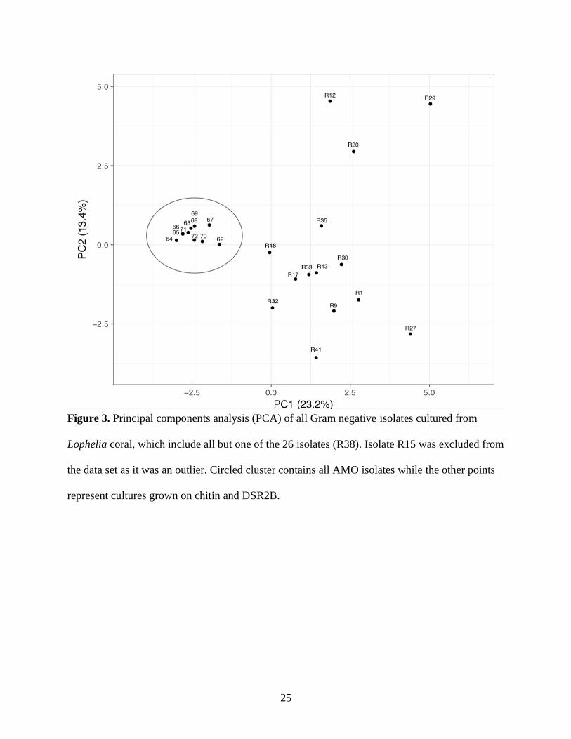
isolate R15. Isolates cultured from chitin and AMO media show similarity in their phenotypic
and metabolic characteristics (Figure 3, Figure 4). Colony morphology characteristics showed
the most dissimilarity among AMO isolates compared with cultures from other enrichments
(Figure 2, Figure 3). Among Pseudoalteromonas cultures there were colony and physiological
similarities among organisms isolated from the same enrichment medium such as colony size
and color, cell size and shape, and ability to metabolize various organic compounds (Figure 2,
Figure 5). Evidence of antibiotic resistance to one or more antibiotics was documented among a
number of isolates (Figure 2, Table 4). Most isolates and Pseudoalteromonas in particular
demonstrated resistance to Penicillin, Vancomycin, and Tetracycline (Table 4). Pantoea
demonstrated additional resistance to Ampicillin, Clindamycin, Erythromycin, and Streptomycin
(Table 4). Vibrio demonstrated resistance to Colistin (Table 4). Photobacterium demonstrated
susceptibility to Ampicillin and Tetracycline (Table 4). Results from cross-streaking were
inconclusive and failed to provide evidence of antibiotic production among L. pertusa associated
microbes.
Table 3. Isolate sequence matches based on 16S rRNA sequence analysis using BLAST.
Sequence analysis was restricted to samples from coral. Uncultured representatives are also
included for isolates due to high genetic similarity.
Closest cultured relative
Isolates Species / strain Sim (%) Accession no. Assignment Culture environment
/ host coral

20
R38 Enterococcus faecalis
strain PRG16
100 MK418939.1 Firmicutes, Bacilli Porites panamensis
R27 Enterobacter cloacae
strain PLA12
94.9 MK418921.1 Proteobacteria,
Gammaproteo-
bacteria
Pocillopora spp.
R27 Pantoea eucalypti
strain 18D
95.1 JF792085.1 Diploria strigosa
R17, R20, R30,
R32, R33, R35,
R43, R48, 62, 63,
64, 65, 66, 67, 68,
69, 70, 71, 72
Pseudoalteromonas
atlantica strain PQQ20
98.3 - 99.8 KT730056.1 Oculina patagonica
R17, R20, R30,
R32, R33, R35,
R43, R48, 62, 63,
64, 65, 66, 67, 68,
69, 70, 71, 72
Pseudoalteromonas
atlantica strain SYM2
98.2 - 99.6 KP645203.1 Free-living
Symbiodinium
cultures
R12 Pseudoalteromonas
distincta strain PQQ84
99.8 KT730063.1 Oculina patagonica
R9, R12 Pseudoalteromonas
paragorgicola strain
PQQ1
100 KT730052.1 Oculina patagonica
R9 Pseudoalteromonas
tetraodonis strain
PQQ31
100 KT730057.1 Oculina patagonica
R12, R17, R20,
R30, R32, R33,
R35, R43, R48, 62,
63, 64, 65, 66, 67,
68, 69, 70, 71, 72
Pseudoalteromonas
tetraodonis strain
PQQ5
98.3 - 99.8 KT730053.1 Oculina patagonica
R1, R41 Pseudomonas
azotoformans strain
22A
98.5 - 99.6 JF792088.1 Siderastrea siderea
R1, R41 Pseudomonas
azotoformans strain 2S
98.4 - 99.6 JF792068.1 Siderastrea siderea
R27 Serratia
proteamaculans strain
F-23
94.9 MK482654.1 Cinachyra
cavernosa (sponge)
associated with
coral in Gulf of
Mannar

21
R15 Vibrio sp. B-2-1 99.6 KT583432.1 Alcyonium
digitatum
R15 Vibrio sp. CIP 110630 99.6 HG942391.1 Corallium rubrum
R15 Vibrio sp. C-3-44 99.3 KT583560.1 Alcyonium
digitatum
R29 Photobacterium sp.
34E11
97.4 JF346761.1 Acropora palmata
Closest uncultured representative
Isolates Organism clone Sim (%) Accession no. Assignment Culture environment
/ host coral
R1, R41 Unc. bacterium clone
12F04
98.6 - 99.7 KC668970.1 Proteobacteria,
Gammaproteo-
bacteria
Stylophora pistillata
tissue
R1, R41 Unc. bacterium clone
Apal_K17
98.6 - 99.7 GU118088.1 Acropora palmata
R27 Unc. bacterium clone
Gven_A12
95.3 GU118494.1 Gorgonia ventalina
R27 Unc. bacterium clone
Gven_H08
95 GU118359.1 Gorgonia ventalina
R29 Unc. bacterium clone
RSAE3C31
97.1 JF411535.1 Platygyra carnosus
R27 Unc. bacterium clone
SGUS1048
95 FJ202675.1 Montastraea
faveolata
(aquarium)
R9, R33, 62, 71 Unc. bacterium clone
SPCiL-109
99.5 - 99.9 KC861113.1 Cinachyra
cavernosa (sponge)
associated with
coral in Gulf of
Mannar
R1, R41 Unc. marine bacterium
clone Tc-49
98.7 - 99.8 JF925029.1 Tubastraea
coccinea
R12, R17, R20,
R30, R32, R35,
R43, R48, 63, 64,
65, 66, 67,
Unc.
Pseudoalteromonas
clone CI13
98.5 - 99.9 FJ695534.1 Acropora digitifera
mucus
R9, R12, 65, 67,
68, 69, 70, 72
Unc.
Pseudoalteromonas
99.8 - 100 FJ695538.1 Acropora digitifera
mucus

22
clone CI17
R33, 62, 71 Unc.
Pseudoalteromonas
clone CI18
99.5 - 99.7 FJ695539.1 Acropora digitifera
mucus
R32, R48, 66 Unc.
Pseudoalteromonas
clone CI19
99.7 - 99.9 FJ695540.1 Acropora digitifera
mucus
R17, R20, R30,
R33, R35, R43, 62,
63, 64, 68, 69, 70,
71, 72
Unc.
Pseudoalteromonas
clone CI42
98.5 - 99.9 FJ695534.1 Acropora digitifera
mucus
R9, R12, R15,
R20, R30. R32,
R35, R43, R48, 63,
64, 65, 66, 67, 68,
69, 70, 72
Unc.
Pseudoalteromonas
clone CI47
98.6 - 100 FJ695567.1 Acropora digitifera
mucus

23
Figure 2. Heatmap of morphological and physiological features of isolated cultures from L.
pertusa. A binary system was used to designate presence or absence of a trait (Appendix B,
Supplemental Table B1). For a given trait, red indicates a positive result, blue indicates a
negative result, and white indicates no result. AR stands for antibiotic resistance.
Table 4. Antibiotic resistance of tested isolates against various antibiotics. “R” and “S” indicate
resistance and susceptibility respectively based on Kirby-Bauer protocol (Hudzicki 2009).
GenBank matches included to indicate closely matching genera of isolates. Empty cells mean not
determined.

24
Isolate ID Genbank match Amp Chl Clin Col Ery Van Pen Str Tet
R9 Pseudoalteromonas R R S
R12 Pseudoalteromonas R R R
R15 Vibrio R R
R27 Pantoea R R R R R R R
R29 Photobacterium S R R R S
R30 Pseudoalteromonas R R S
R32 Pseudoalteromonas R R
R33 Pseudoalteromonas
R35 Pseudoalteromonas R R R
R38 Enterococcus R R
R41 Pseudomonas R R R R
R43 Pseudoalteromonas R R
Key: Amp = Ampicilin, Chl = Chloramphenicol, Cln = Clindamycin, Col = Colistin, Ery =
Erythromycin, Van = Vancomycin, Pen = Penicillin, Str = Streptomycin, Tet = Tetracylin
25
Figure 3. Principal components analysis (PCA) of all Gram negative isolates cultured from
Lophelia coral, which include all but one of the 26 isolates (R38). Isolate R15 was excluded from
the data set as it was an outlier. Circled cluster contains all AMO isolates while the other points
represent cultures grown on chitin and DSR2B.

26
Figure 4. Principal components analysis (PCA) of isolates cultured from chitin and DSR2B
enrichment media. R15 was excluded from the data set as it was an outlier. The exclusion of
isolates from AMO enrichment media highlights that variation in culture characteristics of these
cultures are not highly influenced by media type.

27
Figure 5. Principal components analysis (PCA) of only isolates identified as Pseudoalteromonas
based on 16S rRNA sequencing results. R12 was excluded from the data set as it was an outlier.
Circled cluster contains all AMO isolates while the other points represent cultures grown on
chitin and DSR2B.

28
Figure 6. Maximum likelihood tree of Pseudoalteromonas isolates cultured from Atlantic
Lophelia corals along with their closest GenBank matches. Closest GenBank matches for
Pseudoalteromonas isolates from Gulf of Mexico (GOM) Lophelia corals are included and
highlighted for comparison (Galkiewicz et al. 2011). Bootstrap values represent confidence
intervals. Sequences were aligned and trimmed to an equal length of 1298bp.

29
Figure 7. Maximum likelihood tree of Pseudoalteromonas isolates with the highest sequence
reads (1464bp) cultured from Atlantic Lophelia corals along with their closest GenBank
matches. Closest GenBank matches for Pseudoalteromonas isolates from Gulf of Mexico
(GOM) Lophelia corals are included and highlighted for comparison (Galkiewicz et al. 2011).
Bootstrap values represent confidence intervals.

30
Figure 8. Neighbor-joining tree showing Pseudoalteromonas isolates cultured from Atlantic
Lophelia corals along with their closest GenBank matches. Closest GenBank matches for
Pseudoalteromonas isolates from Gulf of Mexico (GOM) Lophelia corals are included and
highlighted for comparison (Galkiewicz et al. 2011). Sequence lengths are 1298bp.

31
DISCUSSION
The coral holobiont is a symbiosis of coral animal and microbial associates, and it is
thought that both coral and microbes benefit from this relationship. The coral provides a
substrate and nutrients (food and energy sources) for microbial colonization, and microbes
provide nutrients (food and energy sources) for the coral. This study highlights some of the
microbial metabolic pathways that may be relevantly contributing to cycling of carbon and
nitrogen by the L. pertusa holobiont, which include pathways reserved to prokaryotes that can
allow for regeneration of biologically available resources for the eukaryotic coral host. It
provides direct observation of general C and N turnover and N cycling by bacteria cultured from
L. pertusa through a variety of chemoheterotrophic pathways, which has really only been
obtained indirectly by previous studies (Kellogg et al. 2009, Kellogg et al. 2017, Galkiewicz et
al. 2011, Middelburg et al. 2015). In particular, the results of this study demonstrate bacterial
metabolism of organic and inorganic substrates that are likely to be available to the coral
holobiont based on available literature on L. pertusa ecology and ecophysiology and highlights
key pathways in the nitrogen cycle that are performed specifically by microbes. Because of their
close association, bacteria living on and inside of these corals are likely to play a role in
contributing to the carbon and nitrogen budget of the coral holobiont.
Bacteria isolated in this study demonstrated the ability to degrade a wide variety of
organic materials, including various sugars, starch, casein, gelatin, lipids, chitin, urea, and DNA.
Many of these substrates are available to associated microbes as part of the L. pertusa diet,
within the mucus layer, or as metabolic coral waste products. These substrates can serve as
carbon, nitrogen, and energy sources for bacteria, and their breakdown is likely to play a role in
the cycling and retention of limited C and N resources by the coral holobiont (Wegley et al.

32
2007, Radecker et al. 2015, Middelburg et al. 2015). Among the various chemoorganotrophic
pathways demonstrated, chitinase activity is notable. Degradation of chitin was widespread
among isolates in this study, supporting bacterial chitinase activity as a function of the Lophelia
holobiont. Roughly 80% of isolates showed chitinase ability, including species of Pantoea,
Pseudoalteromonas, Pseudomonas, and Vibrio. Chitinase activity has been documented in some
species of Pseudoalteromonas, Pseudomonas, and Vibrio in soil and other freshwater and marine
environments (Grimont and Grimont 2015, Bowman and McMeekin 2015, Farmer et al. 2015,
Palleroni 2015). Chitin is ubiquitous in the marine environment. Chitinase activity among CWCs
associated microbes has been demonstrated indirectly (Neulinger et al. 2008, Yoshioka et al.
2017) and may play a role in assimilation of carbohydrates obtained from phytoplankton and
detritus (Bourne, Morrow & Webster 2016) and in defense against fungal pathogens (Kramer &
Muthukrishnan 1997).
Cultures from this study also demonstrated important metabolic pathways involved in the
cycling and regeneration of nitrogen. Nitrogen cycling is thought to be an important feature of
the L. pertusa holobiont that influences the ability of the coral to grow in the deep ocean
(Wegley et al. 2007, Radecker et al. 2015, Middleburg et al. 2015). Indirect observation of a
complete nitrogen cycle by the L. pertusa holobiont (Middleburg et al. 2015) is supported by
several metabolic pathways demonstrated by bacteria in this study. A single isolate, R15-
identified by 16S rRNA gene sequencing as a species of Vibrio-yielded a presumptive positive
for the nifH gene, potentially confirming nitrogen fixation ability in a microbial associate of L.
pertusa. Nitrogen fixation has been documented among Vibrio species cultured from Brazilian
coral Mussismilia hispida (Chimetto et al. 2008), and inferred in other studies of L. pertusa
(Galkiewicz et al. 2011, Kellogg et al. 2017). Several cultures were able to reduce nitrate to

33
nitrite, a key step in the nitrogen cycle that is facilitated solely by prokaryotes. Nitrite can be
reduced to ammonia and assimilated by the L. pertusa holobiont, or reduced all the way to
dinitrogen via denitrification, a process carried out by other microbes. Urea hydrolysis, another
important process by which bacteria can cycle nitrogen, converts urea to ammonia, which can
then be assimilated. Urea is likely an available resource to bacteria living in close association
with the coral as the result of its release into the water column by various marine organisms
living in the coral gardens (Crandall and Teece 2012). Several isolates in this study showed
urease activity, which supports the presence of this metabolic pathway within the Lophelia
holobiont.
Evidence of ammonia oxidation and carbon fixation within the L. pertusa holobiont
(Middleburg et al. 2015) and 16S rRNA sequence data for species capable of this metabolism
exist (Kellogg et al. 2017), but in spite of attempts to enrich for nitrifying bacteria and archaea,
none were cultured in this study. Their presence is likely due to evidence of ammonia oxidation
and carbon fixation within the L. pertusa holobiont (Middelburg et al. 2015) and 16S rRNA
sequence data for species capable of this metabolism. The AMO enrichment medium used in this
study was intended to capture ammonia-oxidizing organisms; however, the only organisms
cultured from this medium were bacterial species from the genus Pseudoalteromonas, which do
not demonstrate nitrifying ability or other chemolithotrophic metabolism (Bowman and
McMeekin 2015). It is likely that the trace amount of organics used to create the medium (a
ketone derivative of glutaric acid) or possible organic contaminants from the sampling
environment allowed for the growth of these organisms. Their proliferation in such a nutrient
limited environment might suggest that they are good scavengers.

34
The diversity of bacteria cultured in this study importantly confirms and expands upon
both culture-dependent and culture-independent studies of the L. pertusa microbiome. Genera
cultured in this study include Pseudoalteromonas, Pseudomonas, Vibrio, and Photobacterium,
which have been documented in association with L. pertusa (Kellogg et al. 2017, Galkiewicz et
al. 2011, Kellogg et al. 2009, Neulinger et al. 2008), and species of Pantoea and Enterococcus,
which to date have not been documented at the genus level in association with L. pertusa.
Pantoea, however, has been documented in association with corals affected by white plague
disease (Cárdenas et al. 2011), suggesting that its presence in association with L. pertusa may not
be to the coral’s benefit. With the exception of Enterococcus, these genera are all representatives
of Gammaproteobacteria, one of the most metabolically diverse bacterial phyla. Other studies
have documented the presence of these organisms as part of the L. pertusa microbiome and
discussed their metabolic potential (Kellogg et al. 2017, Galkiewicz et al. 2011, Neulinger et al.
2008), and this study has been able to confirm through direct observation some of these
metabolic pathways, including chitin degradation (Pantoea, Pseudoalteromonas, Pseudomonas,
Vibrio), sulfur reduction (Pantoea, Pseudoalteromonas), nitrogen fixation (Vibrio), nitrate
reduction (Pantoea, Pseudomonas, Vibrio), fermentative metabolism (Enterococcus,
Photobacterium, Pseudoalteromonas), and anaerobic growth (Enterococcus, Vibrio). Urease
activity (Pantoea, Pseudoalteromonas, Pseudomonas) was also documented and is an important
pathway for nitrogen recycling by the coral holobiont. These metabolic pathways are consistent
with literature on type strains of these genera (Galkiewicz et al. 2011, Bowman & McMeekin
2015, Farmer et al. 2015, Grimont et al. 2015, Palleroni 2015) and offer a more complete picture
of some of the functions of the L. pertusa holobiont.

35
Evidence from this study may offer support for a Lophelia-specific microbiome.
Alteromonadales (to which Pseudoalteromonas belongs) may be part of a conserved core
microbiome among Lophelia corals in Atlantic and GOM (Kellogg et al. 2017). As in the
culture-based study of L. pertusa associated bacteria from the Gulf of Mexico, the majority of
isolates in culture in this study of Atlantic Lophelia corals were identified as Pseudoalteromonas,
and a smaller proportion of isolates were identified as Photobacterium and Vibrio species
(Galkiewicz et al. 2011). Phylogenetic analysis of Atlantic and GOM Pseudoalteromonas
isolates indicates some genetic separation of isolates from the different locations, but very little
genetic variation overall. This separation could be connected to sample location, or it could be
the result of differences in enrichment media. The high proportion of Pseudoalteromonas
isolates taken from Lophelia corals in both locations are in alignment with evidence of some
conserved microbial associates among L. pertusa coral populations (Galkiewicz et al. 2011,
Kellogg et al. 2017). In both studies, Pseudoalteromonas species dominated among culturable
isolates. Interestingly, some Pseudoalteromonas tetraodonis species cultured from other coral
species have been shown to aid in protection against pathogenic Vibrio species (Torres et al.
2016), suggesting their role in antimicrobial production and protection of their coral host from
attack by pathogens. Taken together this suggests that Pseudoalteromonas species may be an
important part of a conserved Lophelia microbiome and may play a role in coral health.
Comparison of antibiotic resistance of isolates from the Atlantic and GOM indicates
some alignment. Susceptibility of Photobacterium to tetracycline is consistent with isolates from
the GOM (Galkiewicz et al. 2011). The majority of GOM Pseudoalteromonas isolates were
resistant to Penicillin but were all clinically susceptible to tetracycline. This is in contrast to most
Pseudoalteromonas isolates from the Atlantic, which were similarly resistant to penicillin but

36
also resistant to tetracycline. Antibiotic resistance may indicate exposure to certain antibiotics
that could (theoretically) be produced by other associated microbes and may be involved in
protecting coral from pathogens. The similarities and differences in resistance patterns among
associates of L. pertusa from the Atlantic and GOM may suggest some location-specific
differences. More generally, these results could point to some element of Lophelia coral health in
these locations that is yet to be known.
37
CONCLUSION
This study has added to knowledge about the Lophelia pertusa holobiont through culture-
dependent analysis of bacterial isolates and their ability to use various metabolic pathways
involved in carbon and nitrogen cycling, including nitrogen fixation, urea hydrolysis, nitrate
reduction, chitin degradation, and general carbon turnover. There is much still to be learned
about microbial mediation in coral nutrition and health in the deep ocean. Future research may
seek to tie isolates from this study to in situ diversity measures to determine if these organisms
are, in fact, closely associated with L. pertusa corals and whether they are metabolically active
within the functioning holobiont. While the presence and activity of diazotrophic bacteria has
been presumptively supported by this study, more work can be done to confirm these results and
to uncover additional microbial symbionts that are involved in other aspects of the nitrogen
cycle, namely nitrification, additional steps in denitrification, and ammonification. Further
studies of carbon fixation in L. pertusa are needed to better understand what microorganisms are
involved and what inorganic substrates they are using as energy sources, including nitrogen-,
sulfur-, iron-, phosphorus-based compounds. Finally, more work can be done to investigate
antibiotic activities of the Lophelia holobiont. All of these areas are importantly relevant to
understanding the function of the coral holobiont in nutrition, nutrient cycling, and overall coral
health.
Conservation and recycling of carbon, nitrogen, and other nutrients by the L. pertusa
holobiont are likely to be key aspects of its ecophysiology that allows it to grow in the deep
ocean. This has important ecological implications. Lophelia pertusa and other CWC species are
foundation species that form both the structural and trophic basis for entire ecosystems (Cordes

38
et al. 2008, van Oevelen et al. 2009). These ecosystems serve as important spawning, nursery,
and breeding habitat for many fishes and invertebrates, and they also create carbon sinks, places
where carbon gets taken up and stored in biomass (van Oevelen et al. 2009, White et al. 2012).
Physical and physiological stress induced by changes in the environment resulting from human
activities (trawling, dredging, oil drilling) and climate change (ocean acidification) could
influence host-microbe interactions, potentially altering the ability of L. pertusa to conserve and
recycle limiting nutrients. This has the potential to impact the ability of these corals to survive
and sustain ecosystems, subsequently removing important habitat for other marine organisms and
disrupting a globally relevant carbon sink.
39
REFERENCES CONSULTED
Altschul et al. (1990) Journal of Molecular Biology 215: 403-410
Balouiri, M., Sadiki, M., & Ibnsouda, S. K. (2016). Methods for in vitro evaluating
antimicrobial activity: A review. Journal of Pharmaceutical Analysis, 6(2), 71–79.
https://doi.org/10.1016/j.jpha.2015.11.005
Bourne, D. G., Morrow, K. M., & Webster, N. S. (2016). Insights into the coral
microbiome: Underpinning the health and resilience of reef ecosystems. Annual Review of
Microbiology, 70(1), 317–340. https://doi.org/10.1146/annurev-micro-102215-095440
Bowman, John P., & McMeekin, T. A. (2015). Pseudoalteromonas. Bergey’s Manual of
Systematics of Archaea and Bacteria, 1–22. https://doi.org/10.1002/9781118960608.gbm01098
Boyle-Yarwood, S. A., Bottomley, P. J., & Myrold, D. D. (2008). Community composition of
ammonia-oxidizing bacteria and archaea in soils under stands of red alder and Douglas fir in
Oregon. Environmental Microbiology, 10(11), 2956–2965. https://doi.org/10.1111/j.1462-
2920.2008.01600.x
Cappuccino, J. G., & Sherman, N. (2014). Microbiology : A laboratory Manual (10th
ed.). Glenview, IL: Pearson Education, Inc.
Cárdenas, A., Rodriguez-R, L. M., Pizarro, V., Cadavid, L. F., & Arévalo-Ferro, C.
(2011). Shifts in bacterial communities of two Caribbean reef-building coral species affected by
white plague disease. The ISME Journal, 6(3), 502–512. https://doi.org/10.1038/ismej.2011.123
Casamayor, E. O., Schafer, H., Baneras, L., Pedros-Alio, C., & Muyzer, G. (2000).
Identification of and Spatio-Temporal Differences between microbial assemblages from two
neighboring sulfurous lakes: Comparison by microscopy and denaturing gradient gel

40
electrophoresis. Applied and Environmental Microbiology, 66(2), 499–508.
https://doi.org/10.1128/aem.66.2.499-508.2000
Chimetto, L. A., Brocchi, M., Thompson, C. C., Martins, R. C. R., Ramos, H. R., &
Thompson, F. L. (2008). Vibrios dominate as culturable nitrogen-fixing bacteria of the Brazilian
coral Mussismilia hispida. Systematic and Applied Microbiology, 31(4), 312–319.
https://doi.org/10.1016/j.syapm.2008.06.001
Coles, S. L., and Strathman, R. (1973). Observations on coral mucus “floc” and their
potential trophic significance. Limnology and Oceanography. 18, 673–678.
https://doi.org/10.4319/lo.1973.18.4.0673
Cordes, E. E., McGinley, M. P., Podowski, E. L., Becker, E. L., Lessard-Pilon, S., Viada,
S. T., & Fisher, C. R. (2008). Coral communities of the deep Gulf of Mexico. Deep Sea
Research Part I: Oceanographic Research Papers, 55(6), 777–787.
https://doi.org/10.1016/j.dsr.2008.03.005
Costello, M. J., McCrea, M., Freiwald, A., Lundälv, T., Jonsson, L., Bett, B. J., . . . Allen,
D. (2005). Role of cold-water Lophelia pertusa coral reefs as fish habitat in the NE Atlantic.
Cold-Water Corals and Ecosystems, 771–805. https://doi.org/10.1007/3-540-27673-4_41
Crandall, J. B., & Teece, M. A. (2011). Urea is a dynamic pool of bioavailable nitrogen
in coral reefs. Coral Reefs, 31(1), 207–214. https://doi.org/10.1007/s00338-011-0836-1
Davis, E., Sloan, T., Aurelius, K., Barbour, A., Bodey, E., Clark, B., . . . Wildschutte, H.
(2017). Antibiotic discovery throughout the Small World Initiative: A molecular strategy to
identify biosynthetic gene clusters involved in antagonistic activity. MicrobiologyOpen, 6(3).
https://doi.org/10.1002/mbo3.435

41
Farmer, J. J., III, Michael Janda, J., Brenner, F. W., Cameron, D. N., & Birkhead, K. M.
(2015). Vibrio. Bergey’s Manual of Systematics of Archaea and Bacteria, 1–79.
https://doi.org/10.1002/9781118960608.gbm01078
Fisher, C. R., Demopoulos, A. W. J., Cordes, E. E., Baums, I. B., White, H. K., &
Bourque, J. R. (2014). Coral communities as indicators of ecosystem-level impacts of the
Deepwater Horizon spill. BioScience, 64(9), 796–807.
Frias-Lopez, J., Zerkle, A. L., Bonheyo, G. T., & Fouke, B. W. (2002). Partitioning of
Bacterial Communities between Seawater and Healthy, Black Band Diseased, and Dead Coral
Surfaces. Applied and Environmental Microbiology, 68(5), 2214–2228.
https://doi.org/10.1128/aem.68.5.2214-2228.2002
Gaby, J. C., & Buckley, D. H. (2012). A Comprehensive Evaluation of PCR Primers to
Amplify the nifH Gene of Nitrogenase. PLoS ONE, 7(7), e42149.
https://doi.org/10.1371/journal.pone.0042149
Galkiewicz, J. P., Pratte, Z. A., Gray, M. A., & Kellogg, C. A. (2011). Characterization
of culturable bacteria isolated from the cold-water coral Lophelia pertusa. FEMS Microbiology
Ecology, 77(2), 333–346. https://doi.org/10.1111/j.1574-6941.2011.01115.x
Grimont, P. A. D., & Grimont, F. (2015). Pantoea. Bergey’s Manual of Systematics of
Archaea and Bacteria, 1–14. https://doi.org/10.1002/9781118960608.gbm0115
Hudzicki, J. (2009). Kirby-Bauer disk diffusion susceptibility test protocol. American
Society for Microbiology. Cited 2019 Nov 10. Available from: www.asmscience.org
Kellogg, C. A., Goldsmith, D. B., & Gray, M. A. (2017). Biogeographic Comparison of
Lophelia-associated bacterial communities in the western Atlantic reveals conserved core
microbiome. Frontiers in Microbiology, 8. https://doi.org/10.3389/fmicb.2017.00796

42
Kellogg, C. A., Piceno, Y. M., Tom, L. M., DeSantis, T. Z., Gray, M. A., Zawada, D. G.,
& Andersen, G. L. (2013). Comparing bacterial community composition between healthy and
white plague-like disease states in Orbicella annularis using PhyloChipTM G3 microarrays. PLoS
ONE, 8(11), e79801. https://doi.org/10.1371/journal.pone.0079801
Kellogg, C. A., Lisle, J. T., & Galkiewicz, J. P. (2009). Culture-Independent
characterization of bacterial communities associated with the cold-water coral Lophelia pertusa
in the northeastern Gulf of Mexico. Applied and Environmental Microbiology, 75(8), 2294–
2303. https://doi.org/10.1128/aem.02357-08
Khripounoff, A., Caprais, J., Le Bruchec, J., Rodier, P., Noel, P., & Cathalot, C. (2014).
Deep cold-water coral ecosystems in the Brittany submarine canyons (Northeast Atlantic):
Hydrodynamics, particle supply, respiration, and carbon cycling. Limnology and Oceanography,
59(1), 87–98. https://doi.org/10.4319/lo.2014.59.1.0087
Kramer, K. J., & Muthukrishnan, S. (1997). Insect chitinases: molecular biology and
potential use as biopesticides. Insect Biochemistry and Molecular Biology, 27(11), 887–900.
https://doi.org/10.1016/s0965-1748(97)00078-7
Littman, R., Willis, B. L., & Bourne, D. G. (2011). Metagenomic analysis of the coral
holobiont during a natural bleaching event on the Great Barrier Reef. Environmental
Microbiology Reports, 3(6), 651–660. https://doi.org/10.1111/j.1758-2229.2010.00234.x
Littman, R. A., Bourne, D. G., & Willis, B. L. (2010). Responses of coral-associated
bacterial communities to heat stress differ with Symbiodinium type on the same coral host.
Molecular Ecology, 19(9), 1978–1990. https://doi.org/10.1111/j.1365-294x.2010.04620.x

43
Lu, S., Cai, W., Zhang, X., Li, X., Huang, H., Zhang, F., & Zhang, J. (2015). Black coral
as a new environmental recorder: The lead profiles in coral skeletons over the past century.
Marine Pollution Bulletin, 101(1), 226–231. https://doi.org/10.1016/j.marpolbul.2015.09.058
Maxwell, S. (2005). An aquatic pharmacy: the biomedical potential of the deep sea.
Journal of Marine Education.21(4), 31–32.
Meistertzheim, Anne.-L., Lartaud, F., Arnaud-Haond, S., Kalenitchenko, D., Bessalam,
M., Le Bris, N., & Galand, P. E. (2016). Patterns of bacteria-host associations suggest different
ecological strategies between two reef building cold-water coral species. Deep Sea Research
Part I: Oceanographic Research Papers, 114, 12–22. https://doi.org/10.1016/j.dsr.2016.04.013
Middelburg, J. J., Mueller, C. E., Veuger, B., Larsson, A. I., Form, A., & Oevelen, D.
(2015). Discovery of symbiotic nitrogen fixation and chemoautotrophy in cold-water corals.
Scientific Reports, 5(1). https://doi.org/10.1038/srep17962
Mortensen, P. B., Hovland, M., Brattegard, T., and Farestveit, R. (1995). Deep water
bioherms of the scleractinian coral Lophelia pertusa (L.) at 64◦N on the Norwegian shelf:
structure and associated megafauna. Sarsia 80, 145–158.
https://doi.org/10.1080/00364827.1995.10413586
Mortensen, P. B. (2001). Aquarium observations on the deep-water coral Lophelia
pertusa (L., 1758) (scleractinia) and selected associated invertebrates. Ophelia, 54(2), 83–104.
https://doi.org/10.1080/00785236.2001.10409457
Mueller, C. E., Larsson, A. I., Veuger, B., Middelburg, J. J., & van Oevelen, D. (2014b).
Opportunistic feeding on various organic food sources by the cold-water coral Lophelia pertusa.
Biogeosciences, 11(1), 123–133. https://doi.org/10.5194/bg-11-123-2014

44
Murthy, N. and Bleakley, B. (2012). Simplified method of preparing colloidal chitin
used for screening of chitinase-producing microorganisms. The Internet Journal of
Microbiology 10(2).
Muscatine L, Porter JW (1977) Reef corals-mutualistic symbioses adapted to nutrient-
poor environments. Bioscience 27: 454–460.
National Center for Biotechnology Information (NCBI) [Internet]. Bethesda (MD):
National Library of Medicine (US), National Center for Biotechnology Information; (1988).
Cited 2020 Jan 15. Available from: https://www.ncbi.nlm.nih.gov/
Neulinger, S. C., Jarnegren, J., Ludvigsen, M., Lochte, K., & Dullo, W.-C. (2008).
Phenotype-specific bacterial communities in the cold-water coral Lophelia pertusa (Scleractinia)
and their implications for the coral’s nutrition, health, and distribution. Applied and
Environmental Microbiology, 74(23), 7272–7285. https://doi.org/10.1128/aem.01777-08
Palleroni, Norberto J. (2015). Pseudomonas. Bergey’s Manual of Systematics of Archaea
and Bacteria, 1. https://doi.org/10.1002/9781118960608.gbm01210
Rädecker, N., Pogoreutz, C., Voolstra, C. R., Wiedenmann, J., & Wild, C. (2015).
Nitrogen cycling in corals: the key to understanding holobiont functioning? Trends in
Microbiology, 23(8), 490–497. https://doi.org/10.1016/j.tim.2015.03.008
Reshef, L., Koren, O., Loya, Y., Zilber-Rosenberg, I., & Rosenberg, E. (2006). The Coral
Probiotic Hypothesis. Environmental Microbiology, 8(12), 2068–2073.
https://doi.org/10.1111/j.1462-2920.2006.01148.x
Ritchie, K. (2006). Regulation of microbial populations by coral surface mucus and
mucus-associated bacteria. Marine Ecology Progress Series, 322, 1–14.
https://doi.org/10.3354/meps322001

45
Rix, L., de Goeij, J. M., Mueller, C. E., Struck, U., Middelburg, J. J., van Duyl, F. C., …
van Oevelen, D. (2016). Coral mucus fuels the sponge loop in warm- and cold-water coral reef
ecosystems. Scientific Reports, 6(1). https://doi.org/10.1038/srep18715
Roberts and Hirshfield (2004) Deep-sea corals: out of sight, but no longer out of mind.
Frontiers in Ecology and the Environment 2(3): 123-130. https://doi.org/10.1890/1540-
9295(2004)002[0123:DCOOSB]2.0.CO;2
Roberts, J. M. (2006). Reefs of the Deep: The Biology and Geology of Cold-Water Coral
Ecosystems. Science, 312(5773), 543–547. https://doi.org/10.1126/science.1119861
Rohwer, F., Seguritan, V., Azam, F., & Knowlton, N. (2002). Diversity and distribution
of coral-associated bacteria. Marine Ecology Progress Series, 243, 1–10.
https://doi.org/10.3354/meps243001
Rosenberg, E., & Falkovitz, L. (2004). The Vibrio shiloi Oculina patagonica model
system of coral bleaching. Annual Review of Microbiology, 58(1), 143–159.
https://doi.org/10.1146/annurev.micro.58.030603.123610
Schöttner, S., Hoffmann, F., Wild, C., Rapp, H. T., Boetius, A., & Ramette, A. (2009).
Inter- and intra-habitat bacterial diversity associated with cold-water corals. The ISME Journal,
3(6), 756–759. https://doi.org/10.1038/ismej.2009.15
Soetaert, K., Mohn, C., Rengstorf, A., Grehan, A., & van Oevelen, D. (2016). Ecosystem
engineering creates a direct nutritional link between 600m deep cold-water coral mounds and
surface productivity. Scientific Reports, 6(1). https://doi.org/10.1038/srep35057
Swofford, D. L. (2003). PAUP4.0. Phylogenetic Analysis Using Parsimony (*and Other
Methods). Version 4. Sinauer Associates, Sunderland, Massachusetts.
http://phylosolutions.com/paup-test

46
Silvestro, D., & Michalak, I. (2011). raxmlGUI: a graphical front-end for RAxML.
Organisms Diversity & Evolution, 12(4), 335–337. https://doi.org/10.1007/s13127-011-0056-0
Svitil, A. L., Chadhain, S., Moore, J. A., & Kirchman, D. L. (1997). Chitin degradation
proteins produced by the marine bacterium Vibrio harveyi growing on different forms of chitin.
Applied and Environmental Microbiology, 63(2), 408-13.
Thompson, J. R., Rivera, H. E., Closek, C. J., & Medina, M. (2015). Microbes in the
coral holobiont: partners through evolution, development, and ecological interactions. Frontiers
in Cellular and Infection Microbiology, 4. https://doi.org/10.3389/fcimb.2014.00176
Torres, M., Rubio-Portillo, E., Antón, J., Ramos-Esplá, A. A., Quesada, E., & Llamas, I.
(2016). Selection of the N-Acylhomoserine Lactone-degrading bacterium Alteromonas
stellipolaris PQQ-42 and of its potential for biocontrol in aquaculture. Frontiers in
Microbiology, 7. https://doi.org/10.3389/fmicb.2016.00646
TSD-99. (n.d.). Bicarbonate-buffered synthetic Crenarchaeota medium. Retrieved May
24, 2019, from https://www.atcc.org/products/all/TSD-99.aspx#culturemethod
van Bleijswijk, J. D. L., Whalen, C., Duineveld, G. C. A., Lavaleye, M. S. S., Witte, H.
J., & Mienis, F. (2015). Microbial assemblages on a cold-water coral mound at the SE Rockall
Bank (NE Atlantic): interactions with hydrography and topography. Biogeosciences, 12(14),
4483–4496. https://doi.org/10.5194/bg-12-4483-2015
van de Water, J. A. J. M., Melkonian, R., Voolstra, C. R., Junca, H., Beraud, E.,
Allemand, D., & Ferrier-Pagès, C. (2016). Comparative Assessment of Mediterranean
Gorgonian-Associated Microbial Communities Reveals Conserved Core and Locally Variant
Bacteria. Microbial Ecology, 73(2), 466–478. https://doi.org/10.1007/s00248-016-0858-x

47
van Oevelen, D., Mueller, C. E., Lundälv, T., & Middelburg, J. J. (2016). Food selectivity
and processing by the cold-water coral Lophelia pertusa. Biogeosciences, 13(20), 5789–5798.
https://doi.org/10.5194/bg-13-5789-2016
van Oevelen, D., Duineveld, G., Lavaleye, M., Mienis, F., Soetaert, K., & Heip, C. H. R.
(2009). The cold-water coral community as hotspot of carbon cycling on continental margins: A
food-web analysis from Rockall Bank (northeast Atlantic). Limnology and Oceanography, 54(6),
1829–1844. https://doi.org/10.4319/lo.2009.54.6.1829
Vijayalakshmi, S., & Jawahar, S. (2011). Screening of marine bacterial extracts for
antimicrobial activity. Journal of Ecotoxicology & Environmental Monitoring, 21(1), 83-86.
Wagner, H., Purser, A., Thomsen, L., Jesus, C. C., & Lundälv, T. (2011). Particulate
organic matter fluxes and hydrodynamics at the Tisler cold-water coral reef. Journal of Marine
Systems, 85(1–2), 19–29. https://doi.org/10.1016/j.jmarsys.2010.11.003
Wang, Q, G. M. Garrity, J. M. Tiedje, and J. R. Cole. 2007. Naïve Bayesian Classifier for
Rapid Assignment of rRNA Sequences into the New Bacterial Taxonomy. Appl Environ
Microbiol. 73(16):5261-5267; doi: 10.1128/AEM.00062-07 [PMID: 17586664]
Wegley, L., Edwards, R., Rodriguez-Brito, B., Liu, H., & Rohwer, F. (2007).
Metagenomic analysis of the microbial community associated with the coral Porites astreoides.
Environmental Microbiology, 9(11), 2707–2719. https://doi.org/10.1111/j.1462-
2920.2007.01383.x
Yakimov, M. M., Cappello, S., Crisafi, E., Tursi, A., Savini, A., Corselli, C., . . .
Giuliano, L. (2006). Phylogenetic survey of metabolically active microbial communities
associated with the deep-sea coral Lophelia pertusa from the Apulian plateau, Central

48
Mediterranean Sea. Deep Sea Research Part I: Oceanographic Research Papers, 53(1), 62–75.
https://doi.org/10.1016/j.dsr.2005.07.005
Yoshioka, Y., Tanabe, T., & Iguchi, A. (2017). The presence of genes encoding enzymes
that digest carbohydrates in coral genomes and analysis of their activities. PeerJ, 5, e4087.
https://doi.org/10.7717/peerj.4087

49
APPENDIX A. MICROBIOLOGICAL MEDIA RECIPES
Medium for Ammonia Oxidizers (Modified from ATCC medium TSD-99)
Composition per liter:
NaCl ………………………………………………………………………………….. 26g
MgSO4·7H2O ………………………………………………………………………… 5g
MgCl2 ……………………………………………………………………………….... 5g
CaCl2·2H2O …………………………………………………………………………... 1.5g
KBr ………………………………………………………………………………….... 0.1g
KH2PO4 ……………………………………………………………………………….. 0.4g
NaHCO3 (1M solution) ………………………………………………………………. 3 ml
FeCl EDTA (15.5 mM) ……………………………………………………………… 35ml
Trace element solution ……………………………………………………………….. 1 ml
NH4Cl (1M solution) …………………………………………………………………. 0.2ml
Alpha Glutaric Acid (100mM solution) ……………………………………………... 0.1ml
Trace element solution:
Composition per 100.00 mL:
HCl (12M) .…………………………………………………………………………. 8.33 ml
H3BO3……………………………………………………………………………….. 30mg
MnCl2·4H2O .……………………………………………………………………….. 100mg
NiCl2·6H2O.…………………………………………………………………………. 24mg
CuCl2·2H2O.…………………………………………………………………………. 2mg
ZnSO4·7H2O
.………………………………………………………………………...
144mg
(NH4)6Mo7O24·4H2O .……………………………………………………………….. 26.26mg
Preparation of trace element solution: Add components to 12M HCl and mix thoroughly. Note:
12M HCl solution was used in place of 12.5M HCl solution. Molecular concentration was
adjusted by adding volume. (NH4)6Mo7O24 ·4H2O was used in place of Na2MoO4 ·2H2O.

50
Additional ammonium was not a concern as this medium was made to target ammonia oxidizing
microbes.
Preparation of Medium: Add components distilled/deionized water and bring to volume of 1.0L.
Mix thoroughly. Distribute into tubes or flasks. Autoclave for 30 min at 15 psi pressure-121oC.
Note: for solid media, first add and dissolve 8g gellan gum and 1g CaCl into volume of
distilled/deionized water.
Medium for Chitin Degraders (Modified from Murthy and Bleakley, 2012)
Composition per liter:
NaCl …………………………………………………………………………………. 26g
K2HPO4 …………………………………………………………………………….... 0.7g
KH2PO4 ……………………………………………………………………................ 0.3g
MgSO4·5H2O ………………………………………………………………………... 0.5g
FeCl3 ……....…………………………………………………………………………. 25 µl
ZnSO4 ………………………………………………………………………………... 0.001g
MnCl2 ………………………………………………………………………………... 0.001g
Colloidal chitin solution …………………………………………………………….. 100ml
Preparation of colloidal chitin: Treat 20g of crushed chitin flakes with 150 ml of 12M HCl in a
1000ml sterile beaker under a chemical fume hood at room temperature (~25oC). Add chitin
slowly, stirring continuously with a glass pipette for 5 minutes followed by stirring for 1 minute
at 5 minute intervals for 60 minutes. Treat mixture with 2 liters of chilled deionized water to
allow colloidal chitin to precipitate. Incubate overnight at 4oC. Pass solution through two layers
of coffee filter paper. Pass approximately 3 L of deionized water through chitin trapped in filter
paper to neutralize filtrate (test that pH has risen to ~7.0). Dispose of filtrate. Press colloidal
chitin trapped in filter to remove excess moisture and place in 100 ml beaker. Use in preparation
of chitin medium. (Solution can be stored in refrigerator for up to 48 hours.)
Preparation of Medium: Add components distilled/deionized water and bring to volume of 1.0L.
(Use 900ml deionized water to account for 100 ml volume of colloidal chitin solution.) Mix
thoroughly. Distribute into tubes or flasks. Autoclave for 30 min at 15 psi pressure-121oC. Note:
for solid media, first add and dissolve 8g gellan gum and 1g CaCl into volume of
distilled/deionized water.

51
Medium for Oligotrophic Heterotrophic Marine Organisms (General)
Composition per liter:
NaCl ………………………………………………………………………………….. 26g
R2B stock solution …………………………………………………………………... 10ml
Preparation of R2B stock solution: Add 3.6g of R2B to 100 ml of distilled/deionized water. Mix
thoroughly.
Preparation of Medium: Add components distilled/deionized water and bring to volume of 1.0L.
Mix thoroughly. Distribute into tubes or flasks. Autoclave for 30 min at 15 psi pressure-121oC.
Note: for solid media, first add and dissolve 8g gellan gum and 1g CaCl into volume of
distilled/deionized water. Note: for solid media, first add and dissolve 8g gellan gum and 1g
CaCl into volume of distilled/deionized water.
APPENDIX B. SUPPLEMENTAL DATA
Table B1. Binary Key for Heatmap and PCA Analysis.
Trait Description and Binary Key
Colony size Diameter; 0 = < 1mm; 1 = 1+ mm
Colony consistency 1 0 = opaque; 1 = translucent
Colony consistency 2 0 = shiny; 1 = dull
Colony edge 0 = circular/entire; 1 = undulate
Colony elevation 0 = flat; 1 = raised
Cell size (length) Length; 0 = < 1 µm; 1 = 1+ µm
Catalase Enzyme activity; 0 = negative; 1 = positive
Oxidase Enzyme activity; 0 = negative; 1 = positive
Motility 0 = negative; 1 = positive
Anaerobic growth Growth; 0 = negative; 1 = positive
4C Growth; 0 = negative; 1 = positive
10C Growth; 0 = negative; 1 = positive

52
25C Growth; 0 = negative; 1 = positive
37C Growth; 0 = negative; 1 = positive
0% salt Growth; 0 = negative; 1 = positive
10% salt Growth; 0 = negative; 1 = positive
12.5% salt Growth; 0 = negative; 1 = positive
15% salt Growth; 0 = negative; 1 = positive
pH 3 Growth; 0 = negative; 1 = positive
pH 4 Growth; 0 = negative; 1 = positive
pH 5 Growth; 0 = negative; 1 = positive
pH 9-11 Growth; 0 = negative; 1 = positive
Glucose fermentation Metabolic activity; 0 = negative; 1 = positive
Sucrose fermentation Metabolic activity; 0 = negative; 1 = positive
Saccharose fermentation Metabolic activity; 0 = negative; 1 = positive
Lactose fermentation Metabolic activity; 0 = negative; 1 = positive
Sulfur reduction to H2S Metabolic activity; 0 = negative; 1 = positive
Acetoin production Metabolic activity; 0 = negative; 1 = positive
Urea hydrolysis Metabolic activity; 0 = negative; 1 = positive
EMB Metabolic activity (fermentation); 0 = negative; 1 = positive
Nutrient agar Metabolic activity; 0 = negative; 1 = positive
Starch hydrolysis Metabolic activity; 0 = negative; 1 = positive
Lipid hydrolysis Metabolic activity; 0 = negative; 1 = positive
Casein hydrolysis Metabolic activity; 0 = negative; 1 = positive
Gelatin hydrolysis Metabolic activity; 0 = negative; 1 = positive
DNase Metabolic activity; 0 = negative; 1 = positive
Nitrate reduction to nitrite Metabolic activity; 0 = negative; 1 = positive

53
Chitin degradation Metabolic activity; 0 = negative; 1 = positive
AR 1
Antibiotic resistance; 0 = susceptible; 1 = resistance to 1
antibiotic
AR 2+ 0 = susceptible; 1 = resistance to 2 or more antibiotics
AR 3+ 0 = susceptible; 1 = resistance to 3 or more antibiotics