The Investigation of Zinc-Dependent Enzymes in Pregnant...
11
International Journal of Science and Qualitative Analysis 2015; 1(2): 18-28 Published online June 12, 2015 (http://www.sciencepublishinggroup.com/j/ijsqa) doi: 10.11648/j.ijsqa.20150102.12 The Investigation of Zinc-Dependent Enzymes in Pregnant Women and their Correlation to Zinc Deficiency Entela Treska 1, * , Alma Daja 2 , Zhani Treska 1 1 University Hospital of Obstetrics and Gynecology “Queen Geraldine”, Tirana, Albania 2 Clinical and Biochemical Laboratory, Health Centre No. 2, Tirana, Albania Email address: [email protected] (E. Treska), [email protected] (A. Daja), [email protected] (Z. Treska) To cite this article: Entela Treska, Alma Daja, Zhani Treska. The Investigation of Zinc-Dependent Enzymes in Pregnant Women and their Correlation to Zinc Deficiency. International Journal of Science and Qualitative Analysis. Vol. 1, No. 2, 2015, pp. 18-28. doi: 10.11648/j.ijsqa.20150102.12 Abstract: Zinc is present throughout the body in low concentration, but in most tissues. It performs multiple critical functions and must be supplied at adequate levels consistently or deficiency states will result, from mild to severe. Zinc deficiency is insufficient zinc to meet the needs of biological organisms. Due to its essentiality, a lack of this trace element leads to far more severe and widespread problems. Enzymes are large biological molecules responsible for the thousands of chemical inter-conversions that sustain life. Enzymes are usually very specific as to which reactions they catalyze and the substrates that are involved in these reactions. Complementary shape, charge and hydrophilic/ hydrophobic characteristics of enzymes and substrates are responsible for this specificity. Specimens were collected at the University Hospital of Obstetrics and Gynecology “Queen Geraldine” in Tirana, Albania during a period of time from year 2011 to 2013. We took into consideration 500 cases of pregnant women from first to third trimester of pregnancy. Serum zinc was measured by using Photometry (End-Point) method, whereas zinc enzymes such as Alkaline Phosphatase, Amylase, Gamma-glutamyl Transpeptidase, Lactate Dehydrogenase, Creatine Kinase, Aspartate Amino-Transferase, Glutamic Pyruvic Transaminase, Lipase measured with the corresponding methods: Beckman Synchron LX20, Colorimetric ab102523 kit, GenWays GGT kit, nonradioactive colorimetric LDH kit, Max Discovery, Colorimetric kit, Abcam kit and Biovision kit. From 500 cases taken into consideration, 190 pregnant women (38%) had normal zinc values (70 – 120 mcg/dl) 58 pregnant women (11.6%) had zinc levels between 60 – 69.9 mcg/dl, patients which were identified as pregnant women with zinc deficiency, as a consequence of oral contraceptive use; 252 pregnant women (50.4%) with zinc values < 60 mcg/dl, identified as patients with zinc deficiency, a result of malnutrition and also urinary elimination of digestive liquids; 56 pregnant women (11.2%) with zinc values < 30 mcg/dl, identified as patient with definite deficiency as a result of different diseases like acrodermatitis enteropathica ect. According to these results, there was a positive correlation between zinc and enzymes such as ALP, CK and LDH in pregnant women suffering from hypertension, whereas a negative correlation of zinc to enzymes such GGT, AST and GPT in cases with anemia. Zinc prophylactic treatment is important before and during pregnancy. Without a proper nutritional requirement the person falls in the state of zinc deficiency. Keywords: Enzyme Assay Kit, Zinc Dependent Enzymes, Zinc and Enzymes Correlation, Zinc Deficiency, Enzyme Test Interpretation 1. Aim of the Study Evaluation of serum zinc concentration in pregnant women, from first to third trimester of pregnancy. The comparison of zinc concentration in high risk pregnant women to a control group (normal pregnant women). The expression of the correlation between zinc and zinc enzyme levels in pregnant women. Cases detection with zinc deficiency. Identification of serious problems and reasons of concentrations different from normal reference values. 2. Introduction 2.1. Introduction to Zinc Zinc is the second must abundant trace mineral in the body, after iron. It is an essential nutrient that must be ingested dietary. Not only an antioxidant, but it also helps our digestive system as well as producing certain hormones such
-
Upload
truongtuong -
Category
Documents
-
view
214 -
download
0
Transcript of The Investigation of Zinc-Dependent Enzymes in Pregnant...

International Journal of Science and Qualitative Analysis 2015; 1(2): 18-28
Published online June 12, 2015 (http://www.sciencepublishinggroup.com/j/ijsqa)
doi: 10.11648/j.ijsqa.20150102.12
The Investigation of Zinc-Dependent Enzymes in Pregnant Women and their Correlation to Zinc Deficiency
Entela Treska1, *
, Alma Daja2, Zhani Treska
1
1University Hospital of Obstetrics and Gynecology “Queen Geraldine”, Tirana, Albania 2Clinical and Biochemical Laboratory, Health Centre No. 2, Tirana, Albania
Email address: [email protected] (E. Treska), [email protected] (A. Daja), [email protected] (Z. Treska)
To cite this article: Entela Treska, Alma Daja, Zhani Treska. The Investigation of Zinc-Dependent Enzymes in Pregnant Women and their Correlation to Zinc
Deficiency. International Journal of Science and Qualitative Analysis. Vol. 1, No. 2, 2015, pp. 18-28. doi: 10.11648/j.ijsqa.20150102.12
Abstract: Zinc is present throughout the body in low concentration, but in most tissues. It performs multiple critical
functions and must be supplied at adequate levels consistently or deficiency states will result, from mild to severe. Zinc
deficiency is insufficient zinc to meet the needs of biological organisms. Due to its essentiality, a lack of this trace element
leads to far more severe and widespread problems. Enzymes are large biological molecules responsible for the thousands of
chemical inter-conversions that sustain life. Enzymes are usually very specific as to which reactions they catalyze and the
substrates that are involved in these reactions. Complementary shape, charge and hydrophilic/ hydrophobic characteristics of
enzymes and substrates are responsible for this specificity. Specimens were collected at the University Hospital of Obstetrics
and Gynecology “Queen Geraldine” in Tirana, Albania during a period of time from year 2011 to 2013. We took into
consideration 500 cases of pregnant women from first to third trimester of pregnancy. Serum zinc was measured by using
Photometry (End-Point) method, whereas zinc enzymes such as Alkaline Phosphatase, Amylase, Gamma-glutamyl
Transpeptidase, Lactate Dehydrogenase, Creatine Kinase, Aspartate Amino-Transferase, Glutamic Pyruvic Transaminase,
Lipase measured with the corresponding methods: Beckman Synchron LX20, Colorimetric ab102523 kit, GenWays GGT kit,
nonradioactive colorimetric LDH kit, Max Discovery, Colorimetric kit, Abcam kit and Biovision kit. From 500 cases taken
into consideration, 190 pregnant women (38%) had normal zinc values (70 – 120 mcg/dl) 58 pregnant women (11.6%) had
zinc levels between 60 – 69.9 mcg/dl, patients which were identified as pregnant women with zinc deficiency, as a
consequence of oral contraceptive use; 252 pregnant women (50.4%) with zinc values < 60 mcg/dl, identified as patients with
zinc deficiency, a result of malnutrition and also urinary elimination of digestive liquids; 56 pregnant women (11.2%) with zinc
values < 30 mcg/dl, identified as patient with definite deficiency as a result of different diseases like acrodermatitis
enteropathica ect. According to these results, there was a positive correlation between zinc and enzymes such as ALP, CK and
LDH in pregnant women suffering from hypertension, whereas a negative correlation of zinc to enzymes such GGT, AST and
GPT in cases with anemia. Zinc prophylactic treatment is important before and during pregnancy. Without a proper nutritional
requirement the person falls in the state of zinc deficiency.
Keywords: Enzyme Assay Kit, Zinc Dependent Enzymes, Zinc and Enzymes Correlation, Zinc Deficiency,
Enzyme Test Interpretation
1. Aim of the Study
� Evaluation of serum zinc concentration in pregnant
women, from first to third trimester of pregnancy.
� The comparison of zinc concentration in high risk
pregnant women to a control group (normal pregnant
women).
� The expression of the correlation between zinc and zinc
enzyme levels in pregnant women.
� Cases detection with zinc deficiency.
� Identification of serious problems and reasons of
concentrations different from normal reference values.
2. Introduction
2.1. Introduction to Zinc
Zinc is the second must abundant trace mineral in the
body, after iron. It is an essential nutrient that must be
ingested dietary. Not only an antioxidant, but it also helps our
digestive system as well as producing certain hormones such

International Journal of Science and Qualitative Analysis 2015; 1(2): 18-28 19
as testosterone and growth hormones. In plain language, zinc
plays a role in immune function, protein synthesis, wound
healing, DNA synthesis, and cell division. A daily intake of
zinc is required to maintain a steady state because the body
has no specialized zinc storage system [1].
2.2. The Concept of Zinc Deficiency
Zinc is present throughout the body in low concentration,
but in most tissues. It performs multiple critical functions and
must be supplied at adequate levels consistently or deficiency
states will result, from mild to severe. Zinc deficiency is
insufficient zinc to meet the needs of biological organisms.
Due to its essentiality, a lack of this trace element leads to far
more severe and widespread problems. Both, nutritional and
inherited zinc deficiency, generate similar symptoms [1], and
causes a spectrum from mild and marginal effects up to
symptoms of severe nature [2]. Human zinc deficiency was
first reported in 1961, when Iranian males were diagnosed with
symptoms including growth retardation, skin abnormalities,
and mental lethargy, attributed to nutritional zinc deficiency
[3]. Later studies with some Egyptian patients showed
remarkably similar clinical features [4]. Additional studies in
the ongoing years manifested zinc deficiency as a potentially
widespread problem in developing as well as in industrialized
nations [5].
Severe zinc deficiency can be either inherited or acquired.
The most severe of the inherited forms is acrodermatitis
enteropathica, a rare autosomal recessive metabolic disorder
resulting from a mutation in the intestinal Zip4 transporter.
Symptoms of this condition include skin lesions, alopecia,
neuropsychological disturbances, weight loss, reduced immune
function and can be lethal in the absence of treatment.
Acquired severe zinc deficiency has been observed in
patients receiving total parental nutrition without
supplementation of zinc, following excessive alcohol
ingestion, severe mal-absorption, and iatrogenic causes such as
treatment with histidine or penicillamine. The symptoms are
mostly similar to those arising during acrodermatitis
enteropathica. In mild cases of zinc deficiency, slight weight
loss and hyperammonemia were observed. One population in
which mild zinc deficiency occurs with high prevalence, even
in industrialized countries, are the elderly. Here, a significant
proportion has reduced serum zinc levels, and zinc
supplementation studies indicate that this deficiency
contributes significantly to increased susceptibility to
infectious diseases. The overall frequency of zinc deficiency
worldwide is expected to be higher than 20%. In developing
countries, it may affect more than 2 billion people.
Furthermore, it has been estimated that only 42.5% of the
elderly in the Unites States have adequate zinc intake. This
widespread occurrence combined with the variety of clinical
manifestations makes zinc deficiency a serious nutritional
problem, which has a far greater impact on human health than
the relatively infrequent intoxication with zinc.
2.3. Introduction to Enzymes and Their Specificity
Enzymes are large biological molecules responsible for the
thousands of chemical inter-conversions that sustain life.
They are highly selective catalysts, greatly accelerating both
the rate and specificity of metabolic reactions, from the
digestion of food to the synthesis of DNA. Most enzymes are
proteins, although some catalytic RNA molecules have been
identified. Enzymes adopt a specific three-dimensional
structure, and may employ organic (e.g. biotin) and inorganic
(e.g. magnesium ion) cofactors to assist in catalysis. Like all
catalysts, enzymes work by lowering the activation energy
(Ea‡) for a reaction, thus dramatically increasing the rate of
the reaction. As a result, products are formed faster and
reactions reach their equilibrium state more rapidly. Most
enzyme reaction rates are millions of times faster than those
of comparable un-catalyzed reactions. As with all catalysts,
enzymes are not consumed by the reactions they catalyze, nor
do they alter the equilibrium of these reactions. However,
enzymes do differ from most other catalysts in that they are
highly specific for their substrates.
Enzyme activity can be affected by other molecules.
Inhibitors are molecules that decrease enzyme activity;
activators are molecules that increase activity. Many drugs
and poisons are enzyme inhibitors. Activity is also affected
by temperature, pressure, chemical environment (e.g., pH),
and the concentration of substrate. Some enzymes are used
commercially, for example, in the synthesis of antibiotics. In
addition, some household products use enzymes to speed up
biochemical reactions (e.g., enzymes in biological washing
powders break down protein or fat stains on clothes; enzymes
in meat tenderizers break down proteins into smaller
molecules, making the meat easier to chew).
Enzymes are usually very specific as to which reactions
they catalyze and the substrates that are involved in these
reactions. Complementary shape, charge and hydrophilic/
hydrophobic characteristics of enzymes and substrates are
responsible for this specificity. Enzymes can also show
impressive levels of stereospecificity, regioselectivity and
chemoselectivity. Some of the enzymes showing the highest
specificity and accuracy are involved in the copying and
expression of the genome. These enzymes have "proof-
reading" mechanisms. Here, an enzyme such as DNA
polymerase catalyzes a reaction in a first step and then
checks that the product is correct in a second step. This two-
step process results in average error rates of less than 1 error
in 100 million reactions in high-fidelity mammalian
polymerases.
3. Enzymes Taken into Consideration for
the Study
3.1. Cardiac Enzymes
3.1.1. Lactate Dehydrogenase (LDH)
A lactate dehydrogenase is an enzyme present in a wide
variety of organisms, including plants and animals. It exist in
four distinct enzyme classes. Two of them are cytochrome c-
dependent enzymes, each acting on either D-lactate or L-
20 Entela Treska et al.: The Investigation of Zinc-Dependent Enzymes in Pregnant Women and their Correlation to Zinc Deficiency
lactate The other two are NAD(P)-dependent enzymes, each
acting on either D-lactate or L-lactate. Lactate dehydrogenase
catalyzes the inter-conversion of pyruvate and lactate with
concomitant inter-conversion of NADH and NAD+. It
converts pyruvate, the final product of glycolysis, to lactate
when oxygen is absent or in short supply, and it performs the
reverse reaction during the Cori cycle in the liver. At high
concentrations of lactate, the enzyme exhibits feedback
inhibition, and the rate of conversion of pyruvate to lactate is
decreased. It also catalyzes the dehydrogenation of 2-
Hydroxybutyrate, but it is a much poorer substrate than
lactate.
3.1.2. Creatine Kinase (CK)
Creatine kinase (CK), also known as creatine
phosphokinase (CPK) is an enzyme expressed by various
tissues and cell types. CK catalyses the conversion of
creatine and consumes adenosine triphosphate (ATP) to
create phosphocreatine (PCr) and adenosine diphosphate
(ADP). This enzyme reaction is reversible and thus ATP can
be generated from PCr and ADP [12]. In tissues and cells that
consume ATP rapidly, especially skeletal muscle, but also
brain, hair cells of the inner ear, spermatozoa and smooth
muscle, PCr serves as an energy reservoir for the rapid
buffering and regeneration of ATP in situ, as well as for
intracellular energy transport by the PCr shuttle or circuit.
Thus creatine kinase is an important enzyme in such tissues.
Clinically, creatine kinase is assayed in blood tests as a
marker of myocardial infarction (heart attack), muscular
dystrophy and in acute renal failure [11].
3.2. Digestive Enzymes
3.2.1. Amylase
Amylase is an enzyme that catalyses the breakdown of
starch into sugars. It is produced in the pancreas and the
glands that make saliva. When the pancreas is diseased or
inflamed, amylase releases into the blood. It is present in the
saliva of humans and some other mammals, where the
chemical process of digestion begins. Foods that contain
much starch but little sugar, such as rice and potato, taste
slightly sweet as they are chewed because amylase turns
some of their starch into sugar in the mouth. The pancreas
also makes amylase (alpha amylase) to hydrolyse dietary
starch into disaccharides and trisaccharides which are
converted by other enzymes to glucose to supply the body
with energy. Plants and some bacteria can also produce
amylase
3.2.2. Lipase
Lipase is an enzyme that catalyzes the breakdown or
hydrolysis of fats (lipids). Lipases are a subclass of the
esterases. They perform essential roles in the digestion,
transport and processing of dietary lipids (e.g. triglycerides,
fats, oils) in most, if not all, living organisms.
Most lipases act at a specific position on the glycerol
backbone of lipid substrate (A1, A2 or A3) (small intestine).
For example, human pancreatic lipase (HPL) which is the
main enzyme that breaks down dietary fats in the human
digestive system, converts triglyceride substrates found in
ingested oils to monoglycerides and two fatty acids. Several
other types of lipase activities exist in nature, such as
phospholipases and sphingomyelinases, however these are
usually treated separately from "conventional" lipases. Some
lipases are expressed secreted by pathogenic organisms
during the infection. In particular, Candida albicans has a
large number of different lipases, possibly reflecting broad
lipolytic activity, which may contribute to the persistence and
virulence of C. albicans in human tissue.
3.3. Cholelstatic Enzymes
3.3.1. Alkaline Phosphatase (ALP)
Alkaline phosphatase (ALP, ALKP) is a hydrolase enzyme
responsible for removing phosphate groups from many types
of molecules, including nucleotides, proteins, and alkaloids.
The process of removing the phosphate group is called
dephosphorylation. As the name suggests, alkaline
phosphatases are most effective in an alkaline environment. It
is sometimes used synonymously as basic phosphatise [8].
ALP is present in a number of tissues including liver, bone,
intestine, and placenta. Serum ALP is of interest in the
diagnosis of 2 main groups of conditions-hepatobiliary
disease and bone disease. Each tissue—liver, bone, placenta,
and intestine—produces a slightly different alkaline
phosphatase. These variations are called isoenzymes. In the
laboratory, alkaline phosphatase is measured as the total
amount or the amount of each of the four isoenzymes. During
pregnancy, alkaline phosphatase is made by the placenta and
leaks into the mother's bloodstream. Some tumors, however,
start production of the same kind of alkaline phosphatase
produced by the placenta. These tumors are called germ cell
tumors and include testicular cancer and certain brain tumors
[6,7].
3.3.2. Gamma-Glutamyl Transpeptidase (GGT)
Gamma-glutamyltransferase is an enzyme that transfers
gamma-glutamyl functional groups. It is found in many
tissues, the most notable one being the liver, and has
significance in medicine as a diagnostic marker. GGT
catalyzes the transfer of the gamma-glutamyl moiety of
glutathione to an acceptor that may be an amino acid, a
peptide or water (forming glutamate). GGT plays a key role
in the gamma-glutamyl cycle, a pathway for the synthesis
and degradation of glutathione and drug and xenobiotic
detoxification. This is a test which helps in determining the
level of GGT in the bloodstream. The abnormal high or low
level shows alcohol abuse, bone disease & bile duct
obstructions are the few names. As a thumb rule, the higher
the GGT level the higher would be the chances of liver
damage. There may be other situations which might lead to
this & hence, other tests will be recommended along with the
GGT test. As GGT is particularly found in the areas near
liver & other organs a raised level of GGT in the blood
indicates the flowing of the enzymes out of the liver into the
bloodstream & can be identified by its raised level. Many
International Journal of Science and Qualitative Analysis 2015; 1(2): 18-28 21
other blood tests are also performed with GGT test to figure
out the causes of damaged liver [8,9].
3.4. Liver Enzymes
3.4.1. Aspartat Aminotransferase (AST)
Aspartate aminotransferase is an enzyme that catalyzes the
conversion of α-ketoglutarate in the amino acid glutamic acid
catalyze .Without this step, the reaction would malate-
aspartate shuttle and thus the utilization of carbohydrates in
the metabolism of eukaryotes impossible.
Further, the reaction is part of the degradation of several
amino acids, because the AST is found in all living things.
We distinguish cytoplasmic and mitochondrial AST (c-AST,
m-AST), with the corresponding genes GOT1 and GOT2 .
Plants have another form in the chloroplasts . Humans
produce ASAT especially in skeletal muscle , heart muscle
and liver . The ratio of c-ASAT/m-ASAT in blood serum
suggests the condition of the heart and liver. AST catalyzes
the transfer of the common L -amino group of an amino acid
to an α-keto acid .
This occurs for example for the reaction:
L - aspartate + α-ketoglutarate ⇔ oxaloacetate + L - glutamate (1)
ASAT is required for their function of pyridoxal
phosphate ,which serves as a prosthetic group is bound to the
enzyme.
3.4.2. Glutamic-Piruvic Transaminase (GPT)
Alanine transaminase is a transaminase enzyme. It is also
called serum glutamic-pyruvic transaminase (SGPT) or
alanine aminotransferase (ALAT). ALT is found in plasma
and in various bodily tissues, but is most commonly
associated with the liver. It catalyzes the two parts of the
alanine cycle. It catalyzes the transfer of an amino group
from L-alanine to α-ketoglutarate, the products of this
reversible transamination reaction being pyruvate and L-
glutamate. ALT (and all transaminases) require the coenzyme
pyridoxal phosphate, which is converted into pyridoxamine
in the first phase of the reaction, when an amino acid is
converted into a keto acid.
Aspartate transaminase, as with all transaminases, operates
via dual substrate recognition; that is, it is able to recognize
and selectively bind two amino acids (Asp and Glu) with
different side-chains. In either case, the transaminase reaction
consists of two similar half-reactions that constitute what is
referred to as a ping-pong mechanism. In the first half-
reaction, amino acid 1 (e.g., L-Asp) reacts with the enzyme-
PLP complex to generate ketoacid 1 (oxaloacetate) and the
modified enzyme-PMP. In the second half-reaction, ketoacid
2 (α-ketoglutarate) reacts with enzyme-PMP to produce
amino acid 2 (L-Glu), regenerating the original enzyme-PLP
in the process. Formation of a racemic product (D-Glu) is
very rare.
4. Material and Methods
4.1. Method for Zinc Measurement
Serum zinc was measured by using Photometry (End-
Point) method. Specimens were collected at the University
Hospital of Obstetrics and Gynecology “Queen Geraldine” in
Tirana, Albania during a period of time from year 2011 to
2013. We took into consideration 500 cases of pregnant
women from first to third trimester of pregnancy.
4.1.1. Principle of the Method
Zinc dissociated from proteins, in particular conditions of
ionic strength gives with chromogen Nitro-PAPS a stable
colored complex, where the intensity of color is proportional
to the concentration of Zinc in the sample [17].
4.1.2. Reagents Used for the Study
REAGENT 1 (liquid) 2 x 16 mL
Buffer pH 8.2 >0.1 mol/L
Masking agents: Stabilyzers and detergents
REAGENT 2 (liquid) 1 x 8 mL
Nitro-PAPS >0.1 mmol/L
REAGENT 3 Standard (liquid) 1 x 5 mL
Zinc 200 µg/dL
4.1.3. Performance Characteristics
� Stability: The reagents, at 2-8°C, are stable up to the
expiry date shown on the package if not contaminated
during handling [18].
� Linearity: The Zinc concentration is determined
between 10-1000 µg/dL. For concentrations ≥ 1000
µg/dL dilute the sample 1:5 with saline sol., repeat the
determination and multiply the result x 5.
� Wavelength: 580 nm (570-600 nm)
� Pathlength: 1 cm
� Temperature: 37°C
� Method: end point
� Reaction: 5 minutes
� Sample/Reagent: 1/20
� Sensitivity: The minimum detectable is 10 µg/dL [19].
4.2. Methods for Enzyme Measurements
We took into consideration 8 zinc-dependent enzymes such
as: Alkaline Phosphatase (ALP), Amylase, Gamma-glutamyl
Tran speptidase (GGT), Lactat Dehydrogenase (LDH),
Creatine Kinase (CK), Aspartate Aminotransferase (AST),
Glutamic Piruvic Transaminase (GPT) and Lipase.
4.2.1. Method Used for ALP Determination
(i) Principle of the method
The Beckman Synchron LX20 method uses an enzymatic
rate by using 2-amino-2-methyl-1-propanol (AMP) buffer to
measure ALP activity in serum or plasma. In the reaction, the
ALP catalyzes the hydrolysis of the colorless organic
phosphate ester substrate, p-Nitrophenylphosphate, to the
yellow colored product p-Nitrophenol and phosphate. This

22 Entela Treska et al.: The Investigation of Zinc-Dependent Enzymes in Pregnant Women and their Correlation to Zinc Deficiency
reaction occurs at an alkaline pH of 10.3. The system
monitors the rate of change in absorbance at 410 nm over a
fixed-time interval This rate of change in absorbance is
directly proportional to the ALP activity in the serum.
Alkaline phosphatase measurements are used in the diagnosis
and treatment of liver, bone, and parathyroid disease [20,21].
(ii) Equipments used and reagent preparation
� Beckman Synchron CX Micro Sample Tube (Part
#448774)
� S/P Plastic Transfer Pipet (Cat. #P5214-10)
� S/P Brand Accutube Flange Caps (Cat. #T1226-37)
� Reagent Preparation: Beckman Synchron System ALP
Reagent (Part #442670, 200 tests/cartridge or Part
#476821, 400 tests/cartridge).
� No preparation required.
� When stored unopened at 2-8°C, the reagent is stable
until the expiration printed on the label.
� When first opened or installed on the instrument, the
reagent is stable for ten days unless the expiration date
is exceeded.
� Caustic reagent. Avoid skin contact with reagent. Use
water to wash reagent from skin.
� Standards Preparation: None required.
� Control Material
4.2.2. Method Used for Amylase Determination
(i) Principle of the method
Amylase Assay kit (Colorimetric ab102523) detects
activity of α-amylase through a two-step reaction. α-Amylase
will cleave the substrate ethylidenep NP-G7 to produce
smaller fragments that are eventually modified by α-
glucosidase, causing the release of a chromophore that can
then be measured at OD = 405 nm. The assay can detect α-
amylase content as low as 0.2 mU.
(ii) Equipments used and materials required
These materials are not included in the kit, but will be
required to successfully utilize this assay:
� MilliQwater or other type of double distilled water
(ddH2O)
� PBS
� Micro centrifuge
� Pipettes and pipette tips
� Colorimetric microplate reader equipped with filter for
OD 405nm
� 96 well plate: clear plate for colorimetric assay
� Orbital shaker
� Vortex
4.2.3. Method Used for GGT Determination
(i) Principle of the method
GenWay’s Gamma-Glutamyl Transferase Assay Kit
provides a convenient tool for sensitive detection of the GGT
in a variety of samples. The GGT in sample will recognize L-
γ-Glutamyl-pNA as a specific substrate leading to
proportional color development. The activity of GGT can be
easily quantified colorimetrically (λ = 418 nm). This assay
detects GGT activity as low as 0.5 mIU [8,9].
(ii) Kit content and reagents used
� GGT Assay Buffer WM GWB-AXR359-1
� GGT Substrate NM GWB-AXR359-2
� GGT Positive Control GWB-AXR359-3
� pNA Standard ( 2mM)
� Reaction mix
4.2.4. Method Used for LDH Determination
(i) Principle of the method
The non-radioactive colorimetric LDH assay is based on
the reduction of the tetrazolium salt MTT in a NADH-
coupled enzymatic reaction to a reduced form of MTT which
exhibits an absorption maximum at 565 nm. The intensity of
the purple color formed is directly proportional to the
enzyme activity [22].
(ii) Kit content and materials required
� Pipeting devices and accessories (e.g. multi-channel
pipettor).
� Procedure using 96-well plate: Clear bottom 96-well
plates (e.g. Corning Costar) and plate reader.
� Procedure using cuvette: Spectrophotometer and cuvets
for measuring OD 565nm.
� Substrate buffer
� NAD solution
� PMS solution
� MTT solution
� Calibrator [23,24].
4.2.5. Method Used for CK Determination
(i) Principle of the method
The MaxDiscovery™ Creatine Kinase (CK) Enzymatic
Assay Kit is a colorimetric, plate-based assay to determine
the amount of creatine kinase in serum. The kit enables
biomedical researchers to detect heart damage in mice and
rats. The test is based on a proven method for creatine kinase
determination which utilizes a coupled enzymatic assay to
specifically detect creatine kinase enzyme in fluids. It
provides accurate, proven results even in complex samples
[10,11].
(ii) Kit contents and Materials required
� Microtiter Plate 1 x 96-well Plate
� Reagent Mix bottle
� Standard vial - 20°C
� Standard Dilution Buffer
� Microtiter plate reader (with 340 nm absorbance filter)
� Microcentrifuge
� De-ionized or distilled water
� 1.5 mL microcentrifuge tubes
� Multichannel pipet (recommended)
4.2.6. Method Used for AST Determination
(i) Principle of the method
Alanine Transaminase Activity Assay Kit (colorimetric) is
a rapid and simple assay where ALT catalysis the transfer of
an amino group from alanine to α-ketoglutarate, the products
of this reversible transamination reaction being pyruvate and
glutamate [12,13]. The kit provides a rapid, simple, sensitive,
and reliable test suitable for high throughput activity assay of
International Journal of Science and Qualitative Analysis 2015; 1(2): 18-28 23
ALT with a detection limit of 10mU per well [14].
(ii) Kit components and materials required
� ALT positive control (lyophilized)
� ALT substrate (lyophilized)
� Pyruvate standard
� ALT assay buffer
� ALT enzyme mix
� OxiRed in DMSO
4.2.7. Method Used for GPT Determination
(i) Principle of the method
In Abcam’s Alanine Transaminase Activity Assay Kit,
ALT catalyzes the transfer of an amino group from alanine to
α-ketoglutarate, the products of this reversible transamination
reaction being pyruvate and glutamate. The pyruvate is
detected in a reaction that concomitantly converts a nearly
colorless probe to both color (λmax = 570 nm) and
fluorescence (Ex/Em = 535/587 nm). The kit provides a
rapid, simple, sensitive, and reliable test suitable for high
throughput activity assay of ALT with a detection limit of 10
mU per well.
(ii) Kit component and materials required
� ALT Assay Buffer
� OxiRed Probe (in DMSO)
� ALT Enzyme Mix (Lyophilized)
� ALT Substrate (Lyophilized)
� Pyruvate Standard
� ALT positive control (Lyophilized)
� Microcentrifuge
� Pipettes and pipette tips
� Fluorescent or colorimetric microplate reader
� 96 well plate
� Orbital shaker
4.2.8. Method Used for Lipase Determination
(i) Principle of the method
In BioVision’s Lipase Assay Kit, lipase hydrolyzes a
triglyceride substrate to form glycerol which is quantified
enzymatically by via monitoring a linked change in the
OxiRed probe absorbance (λ=570nm). This assay is rapid,
simple, sensitive, and reliable, as well as, suitable for high
throughput activity screening of lipase. This kit detects lipase
activity as low as 10mU per well [15,16].
(ii) Kit component and materials required
� Lipase Assay Buffer
� OxiRed Probe (in DMSO)
� Enzyme Mix (lyophilized)
� Lipase Substrate
� Glycerol Standard (100mM)
� Lipase Positive Control
5. Results and Discussion
5.1. The Results of Serum Zinc Measurements in Pregnant
Women
Serum zinc in pregnant women was measured using
Photometry (End-Point), at the Medical Laboratory “PhD
Stiljan Buzo”, in Tirana, Albania. All the laboratory values
were expressed in table 1.
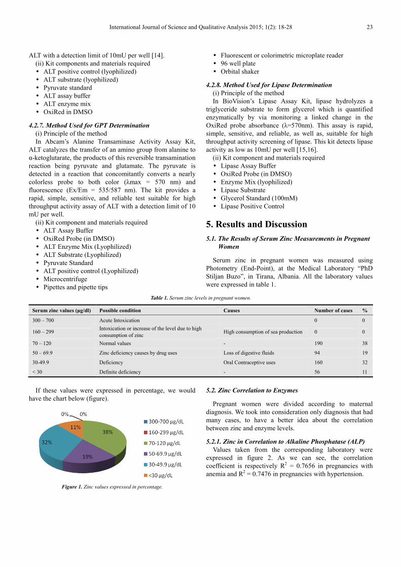
Table 1. Serum zinc levels in pregnant women.
Serum zinc values (µg/dl) Possible condition Causes Number of cases %
300 – 700 Acute Intoxication 0 0
160 – 299 Intoxication or increase of the level due to high
consumption of zinc High consumption of sea production 0 0
70 – 120 Normal values - 190 38
50 – 69.9 Zinc deficiency causes by drug uses Loss of digestive fluids 94 19
30-49.9 Deficiency Oral Contraceptive uses 160 32
< 30 Definite deficiency - 56 11
If these values were expressed in percentage, we would
have the chart below (figure).
Figure 1. Zinc values expressed in percentage.
5.2. Zinc Correlation to Enzymes
Pregnant women were divided according to maternal
diagnosis. We took into consideration only diagnosis that had
many cases, to have a better idea about the correlation
between zinc and enzyme levels.
5.2.1. Zinc in Correlation to Alkaline Phosphatase (ALP)
Values taken from the corresponding laboratory were
expressed in figure 2. As we can see, the correlation
coefficient is respectively R2 = 0.7656 in pregnancies with
anemia and R2 = 0.7476 in pregnancies with hypertension.

24 Entela Treska et al.: The Investigation of Zinc-Dependent Enzymes in Pregnant Women and their Correlation to Zinc Deficiency
a) b)
Figure 2. Correlation of zinc to ALP in different maternal diagnosis, a) anemia, b) hypertension.
5.2.2. Zinc in Correlation to Amylase
According to the figure 3, the correlation coefficient between zinc and amylase for pregnancies with abortion was R2 =
0.7033, whereas for pregnancies with hypertension R2 = 0.7472.
a) b)
Figure 3. Correlation of zinc to Amylase in different maternal diagnosis, a) abortion, b) hypertension.
5.2.3. Zinc in Correlation to Gamma-glutamyl Transpeptidase (GGT)
According to the figure 4, the correlation coeficient between zinc and GGT in pregnancies with anaemia was R2 = 0.7591,
whereas in pregnancies with hypertension R2 = 0.8385.
a) b)
Figure 4. Correlation of zinc to GGT in different maternal diagnosis, a) anemia, b) hypertension.

International Journal of Science and Qualitative Analysis 2015; 1(2): 18-28 25
5.2.4. Zinc in Correlation to Creatine Kinase (CK)
According to figure 5, the correlation coeficient between zinc and CK in pregnancies with abortion was R2 = 0.8859, in
cases with anaemia R2 = 0.8697, whereas in pregnancies with hypertension R
2 = 0.8465.
a) b)
c)
Figure 5. Correlation of zinc to CK in different maternal diagnosis, a) abortion, b) anemia, c) hypertension.
5.2.5. Zinc in Correlation to Lactat Dehydrogenase (LDH)
According to figure 6, the correlation coefficient between zinc values and LDH, in pregnancies with abortion was R2 =
0.9431, in pregnancies with anemia R2 = 0.8634, whereas in cases with hyperemesis R
2 = 0.8242.
a) b)

26 Entela Treska et al.: The Investigation of Zinc-Dependent Enzymes in Pregnant Women and their Correlation to Zinc Deficiency
c)
Figure 6. Correlation of zinc to LDH in different maternal diagnosis, a) abortion, b) anemia, c) hypertension.
5.2.6. Zinc in Correlation to Aspartate Aminotransferase (AST)
As we can see in figure 7, the correlation coeficient between zinc values and AST, in pregnancies with anaemia was R2 =
0.8035, whereas in pregnancies with hyhpertension R2 = 0.8013.
a) b)
Figure 7. Correlation of zinc to AST in different maternal diagnosis, a) anemia, b) hypertension
5.2.7. Zinc in Correlation to Glutamic Piruvic Transaminase (GPT)
According to the figure 8, the correlation coefficient between zinc and GPT in pregnancies with anemia is R2 = 0.8002,
whereas in pregnancies with hypertension is R2 = 0.8682.
a) b)
Figure 8. Correlation of zinc to GPT in different maternal diagnosis, a) anaemia, b) hypertension.

International Journal of Science and Qualitative Analysis 2015; 1(2): 18-28 27
5.2.8. Zinc in Correlation to Lipase
According to figure 9, the correlation coefficient between zinc and Lipase in pregnancies with hyperemesis is R2 = 0.9502,
whereas in partus premature pregnancies R2 = 0.8734.
a) b)
Figure 9. Correlation of zinc to Lipase in different maternal diagnosis, a) hyperemesis b) partus premature.
6. Conclusions
6.1. Conclusions about Zinc Measurements
Our study costisted of normal pregnant women, who
served as control group and of zinc deficiency pregnant
women. The maternal age was 17- 44 years old.
We analyzed 500 cases of pregnant women, from which:
� 190 pregnant women (38%) had normal zinc values
(according to lab values 70 – 120 mcg/dl)
� 58 pregnant women (11.6%) had zinc levels between 60
– 69.9 mcg/dl, these patients were identified as
pregnant women with zinc deficiency, as a consequence
of oral contraceptive use.
� 252 pregnant women (50.4%) with zinc values < 60
mcg/dl, identified as patients with zinc deficiency, a
result of which may be malnutrition and also urinary
elimination of digestive liquids.
� 56 pregnant women (11.2%) with zinc values < 30
mcg/dl, were identified as patient with definite
deficiency as a result of different diseases.
The prevalence of zinc deficiency in pregnant women was
62% (310 cases).
6.2. Conclusions about Enzyme Measurements
From the measurements done at the corresponding
laboratory, it resulted:
� There was a strong positive linear correlation (R2 > 0.7)
between zinc and ALP in both diagnosis anemia and
hypertension,
� There was strong negative linear correlation between
zinc and Amylase in both diagnosis abortion and
hypertension,
� There was a strong negative linear correlation between
zinc and GGT in both diagnosis anemia and
hypertension,
� There was a strong positive linear correlation between
zinc and CK in both in both diagnosis anemia and
hypertension, whereas a strong negative linear
correlation in pregnancies with abortion,
� There was a strong negative linear correlation between
zinc and LDH in both diagnosis abortion and anemia,
whereas a strong negative linear correlation in
pregnancies with hyperemesis,
� There was a strong negative linear correlation between
zinc and AST in pregnancies with anemia, whereas a
strong positive linear correlation in pregnancies with
hypertension,
� There was a strong negative linear correlation between
zinc and GGT in pregnancies with anemia, whereas a
strong positive linear correlation in pregnancies with
hypertension,
� There was a strong positive linear correlation between
zinc and Lipase in both diagnosis hypertension and
partus premature.
Recommendations
� An average adult woman should consume about 7
milligrams of zinc daily, while an average male should
be consuming 9.5 milligrams daily. The risk for women
to have a zinc deficiency is much greater than a man,
especially if they are malnourished because of an eating
disorder or when they are breastfeeding.
� If you are concerned about your zinc intake, taking a
good multivitamin should be sufficient. Be sure to only
take the recommended daily dose, as zinc overdoses can
also occur, which can be toxic to the body. Consuming
too much zinc can cause nausea, vomiting and fever
because too much of the mineral can interfere with how
the body processes other minerals.
� Without a proper nutritional requirement the person
falls in the state of zinc deficiency
� Zinc prophylactic treatment is important before and

28 Entela Treska et al.: The Investigation of Zinc-Dependent Enzymes in Pregnant Women and their Correlation to Zinc Deficiency
during pregnancy.
� Everyone needs zinc. Children need zinc to grow, adults
need zinc for health. Growing infants, children and
adolescents, pregnant women and lactating mothers,
athletes, vegetarians and the elderly often require more
zinc.
References
[1] Solomons NW. Mild human zinc deficiency produces an imbalance between cellmediated and humoral immunity. Nutr Rev 1998; 56:27-8.
[2] Prasad, A.S. Clinical manifestations of zinc deficiency. Annu. Rev. Nutr. 1985, 5, 341-363.
[3] Prasad, A.S.; Halsted, J.A.; Nadimi, M. Syndrome of iron deficiency anemia, hepatosplenomegaly, hypogonadism, dwarfism and geophagia. Am. J. Med. 1961, 31, 532-546.
[4] Prasad, A.S.; Miale, A.J.; Farid, Z.; Sandstead, H.H.; Schulert, A.R. Zinc metabolism in patients with the symptoms of iron deficiency, anaemia, hepatosplenomegaly, dwarfism and hypogonadism. J. Lab. Clin. Med. 1963, 61, 537-549.
[5] Tamás L, Huttová J, Mistrk I, Kogan G (2002). "Effect of Carboxymethyl Chitin-Glucan on the Activity of Some Hydrolytic Enzymes in Maize Plants". Chem. Pap. 56 (5): 326–329.
[6] Owyang C. Pancreatitis. In: Goldman L, Ausiello D, eds. Cecil Medicine. 23rd ed. Philadelphia, Pa: Saunders Elsevier; 2007:chap 147.
[7] Lange PH, Millan JL, Stigbrand T, Vessella RL, Ruoslahti E, Fishman WH (August 1982). "Placental alkaline phosphatase as a tumor marker for seminoma". Cancer Res. 42 (8): 3244–7. PMID 7093962.
[8] Tate SS, Meister A (1985). “gamma-Glutamyl transpeptidase from kidney”. Meth. Enzymol. Methods in Enzymology 113:400–419. doi:10.1016/S0076-6879(85)13053-3. ISBN 978-0-12-182013 8. PMID 2868390.
[9] Siest G, Courtay C, Oster T, Michelet F, Visvikis A, Diederich M, Wellman M (1992). "Gamma-glutamyltransferase: nucleotide sequence of the human pancreatic cDNA. Evidence for a ubiquitous gamma-glutamyltransferase polypeptide in human tissues". Biochem. Pharmacol. 43 (12): 2527–2533. doi:10.1016/0006-2952(92)90140-E. PMID 1378736.
[10] Goldblatt H (August 1969). "The effect of high salt intake on the blood pressure of rabbits". Laboratory Investigation 21 (2): 126–8. PMID 5804623
[11] Wallimann T, Wyss M, Brdiczka D, Nicolay K, Eppenberger HM (January 1992)."Intracellular compartmentation, structure and function of creatine kinase isoenzymes in tissues with high and fluctuating energy demands: the 'phosphocreatine circuit' for cellular energy homeostasis". The Biochemical Journal 281 (1): 21–40.PMC 1130636. PMID 1731757
[12] M. Panteghini: Aspartate aminotransferase isoenzymes. Clin Biochem. 23/4 / 1990 :311-ninth PMID 2225456
[13] Hirotsu K, Goto M, Okamoto A, Miyahara I (2005). "Dual substrate recognition of aminotransferases". Chem Rec 5 (3): 160–172. doi:10.1002/tcr.20042. PMID 15889412.
[14] Kochhar S, Christen P (1992). “Mechanism of racemization of amino acids by aspartate aminotransferase”. Eur J Biochem 203 (3): 563–569. doi:10.1111/j.14321033.1992.tb16584.x. PMID 1735441.
[15] Svendsen A (2000). “Lipase protein engineering”. Biochim Biophys Acta 1543 (2): 223– 228. doi:10.1016/S0167-4838(00)00239-9. PMID 11150608
[16] Goñi F, Alonso A (2002). “Sphingomyelinases: enzymology and membrane activity”. FEBS Lett 531 (1): 38–46. doi:10.1016/S0014-5793(02)03482-8. PMID 12401200Svendsen A (2000). "Lipase protein engineering". Biochim Biophys Acta 1543 (2): 223–228. doi:10.1016/S0167-4838(00)00239-9. PMID 11150608
[17] Textbook of Clinical Chemistry, Ed. by N.W. Tietz, W.B. Saunders Co., Philadelphia (1999).
[18] Young D.S., Effect of drugs on Clinical Lab. Test, 5 th Ed. AACC Press (2000).
[19] Makino T. et al., Clin. Chim. Acta, 120, 127 (1982).
[20] Beckman Synchron LX Systems Chemistry Information Manual, 2001
[21] Tietz, N.W. Textbook of Clinical Chemistry, W.B. Saunders, Philadelphia, PA (1986).
[22] Stentz R., et al. (2010). Controlled protein release from viable Lactococcus lactis cells. Appl. Environ. Microbiol. 76(9): 3026-3031.
[23] Lu F., et al. (2008). The effect of C1 inhibitor on intestinal ischemia and reperfusion injury. Am. J. Physiol. Gastrointest. Liver Physiol. 295(5): G1042-G1049.
[24] Mittal R., et al. (2009). Brain damage in newborn rat model of meningitis by Enterobacter sakazakii: a role for outer membrane protein A. Lab. Invest. 89(3): 263-277.