The Intestinal Nematodes
107
The Intestinal Nematodes Ascaris lumbricoides Common name: Giant intestinal round worm Disease: Human ascariasis
-
Upload
blueblooded23 -
Category
Documents
-
view
2.685 -
download
7
Transcript of The Intestinal Nematodes
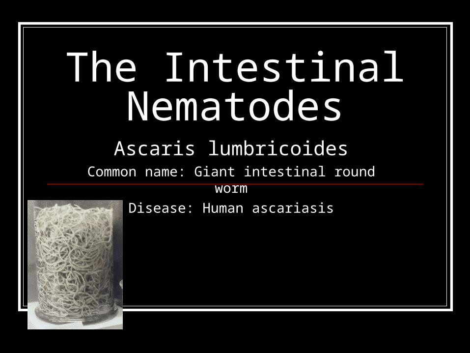
The Intestinal NematodesAscaris lumbricoides
Common name: Giant intestinal round worm
Disease: Human ascariasis

Morphology It is the largest and the most common
nematode in man. 15-30 cm by 3 mm (male). 20-45 cm by 5 mm (female). Creamy white or pinkish worm. Cylindrical, elongated, tapering gradually at
the anterior end.


Ascaris lumbricoides adult male and female © Dr Peter Darben, Queensland University of Technology clinical parasitology collection. Used with
permission
Diagnostic Features Smooth finely striated cuticle with faint
longitudinal white lateral lines. Which is seen as a whitish streak along the entire length of the body.
Terminal mouth with trilobate lips and a small triangular buccal cavity

An adult Ascaris worm. Diagnostic characteristics: tapered ends; length 15-35 cm (the females tend to be the larger ones). This worm is a female, as evidenced by the size and genital girdle (the dark circular groove at bottom area of image). Worm passed by a female child in Florida. CDC
DPDx Parasite Image Library

The male has a ventrally curved papillated posterior extremity with 2 copulatory spicules.
The female has paired reproductive organs on the posterior 2/3.
And in the anterior and middle third is a depression wherein a vagina is located.
Ova There are two types of ova: Fertilized: broadly ovoid, golden brown in
color. A. vitelline membrane: an inner non-permeable,
lipoidal layer. B. glycogen membrane: thick, transparent middle
layer. C. Albuminous/ mammillary coat: outermost layer.
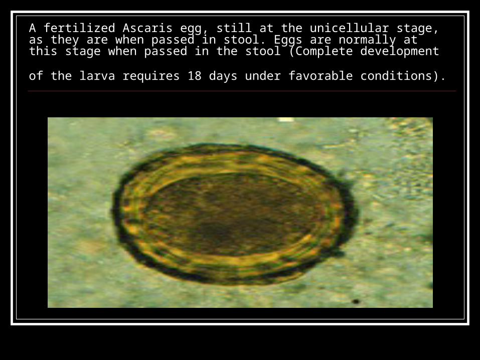
A fertilized Ascaris egg, still at the unicellular stage, as they are when passed in stool. Eggs are normally at this stage when passed in the stool (Complete development of the
larva requires 18 days under favorable conditions).

Eggs, unfertilized (left) and fertilized (right). Patient seen in Haiti. CDC
DPDx Parasite Image Library

Unfertilized: generally larger and longer, elongated or sometimes irregular in shape.
With 2 layers, relatively thinner glycogen membrane and irregular coating of albuminous layer.
The vitilline layer is absent. If mammilary coat is absent it is called
decorticated.

Larva hatching from an egg. CDC DPDx Parasite Image Library

Life cycle
Fertilized ovasoil 2 weeksembryonation ingestedcirculationheart and lungsesophagusSI

Pathology A. Larval Migration to lungs can produce:
Pneumonitis resembling asthmatic attack accompanied by marked eosinophilia (ascaris pneumonitis of loeffler’s syndrome).
Clinical manifestations:
Asthmatic type of respiration, cough, urticaria, rash, edema of the lips and eosinophilia.

Ascaris lumbricoides larva in section of lung (H&E) © Dr Peter Darben, Queensland University of Technology clinical parasitology collection. Used
with permission
B. Pathology due to Adult Worm. Abdominal pain, diarrhea, nausea, loss of
appetite. Serious and sometimes fatal effects due to
erratic migration of adult worms. They maybe regurgitated and vomited and may escape through the nostrils.
Intestinal obstruction.

Ascaris may pass the larynx and may lead to suffocation.
Or they may reach the lungs and produce pulmonary gangrene or may pass the uestachian tube and provoke otitis media.
They may invade the bile duct (obstruction), gall bladder, liver and appendix.

Diagnosis Diagnosis is made by finding the eggs in
the feces, fertilized or unfertilized by performing DFS, kato-thick, kato-katz or concentration technique.

Stool examination may give negative findings in the following conditions: When the patient is actually free from Ascaris
infection. Worms are still immature in the lumen. During larval migration When only male worms are present in the
intestine.

Treatment Piperazine citrate, pyrantel pamoate,
mebendazole, albendazole, levimazole. Piperazine and pyrantel pamoate are used
these days because these drugs have a neuromuscular blocking effect on the parasite causing paralysis.

Toxocara canis and Toxocara catti Common name: Dog ascaris and Cat ascaris Disease: toxocariasis/visceral larval migrans

Life cycle
Pathology Larval migrans can produce hemorrhage,
necrosis and granuloma of the site. Eosinophilia, liver damage, pulmonary
inflammation, and ocular problems has been observed.
Among children anemia with high WBC count.

Diagnosis Diagnosis is usually established on clinical
grounds. Examination of egg in the stool of the
patient is not useful because the egg laying adults are not present.
However, examination of fecal material from infected pets often supports the diagnosis.

Treatment Because the clinical course of VLM is so
variable, it is very difficult to evaluate efficacy of any treatment. Since the infection is self limited. Only severe cases needs to be treated. Thiabendazole appears to shorten the course of the disease.

Eggs of Toxocara canis. These eggs are passed in dog feces, especially puppies' feces. Humans do not produce or excrete eggs, and therefore eggs are not a diagnostic finding in human toxocariasis! The egg to the left is fertilized but not yet embryonated, while the egg to the right contains a well developed larva. The latter egg would be infective if ingested by a human (frequently, a child). CDC DPDx Parasite

Toxocara canis (Dog Roundworm) egg, embryonated © Dr Peter Darben, Queensland University of Technology clinical parasitology collection. Used
with permission
Trichuris trichiura Common name: whipworm
Disease: Trichuriasis

Morphology Adult They attach to the cell wall of the cecum. Colored or pinkish, the anterior end is
attenuated, whip-like, while the posterior end is more robust.
3.5-5 cm ( female) straight post. End 3-4.5 cm ( male) coiled 3600 with a
sheathed single spicule.

Trichuris trichiura adult male and female © Dr Peter Darben, Queensland University of Technology clinical parasitology collection. Used with
permission

Ova barrel shaped/lemon shaped/football
shaped or Japanese lantern shape. With a prominent bipolar plugs on both
ends. The shell is thick and composed of three
layers. With a yellowish outer and a transparent inner layer.

Egg of Trichuris trichuria as seen on wet mount. The diagnostic characteristics are: a typical barrel shape two polar plugs, that are unstained size: 50-54 µm by 22-23 µm. The external layer of the shell of the egg is yellow-brown (in contrast to the clear polar plugs). The egg is unembryonated, as eggs are when passed with the stool. CDC
DPDx Parasite Image Library

Life cycle

Eggssoil 3 weeksembryonationingestedlarvaececum

Pathology Small blood streaked diarrheic stool Abdominal pain Nausea, vomitting Anemia (hypochromic) Weight loss Rectal prolapse


Diagnosis DFS, kato-thick, kato-katz, concentration
technique
Treatment Abendazole, mebendazole, pyrantel
pamoate

Hookworms A. Necator americanus
Common name: new world hookworm, american hookworm Disease: necatoriasis, uncinariasis, tropical anemia,
“laziness”
B. Ancylostoma duodenale Common name: old world hookworm
Disease: ancylostomiasis, miner’s anemia

C. Ancylostoma braziliense Common name: cat hookworm
Disease: cutaneous larva migrans, creeping eruption
D. Ancylostoma caninum Common name: dog hookworm
Disease: cutaneous larva migrans, creeping eruption

Necator americanus is small and has a tendency to go against the general body curvature, hence a hook is formed (S shaped)
The buccal capsule is provided with a semi-lunar cutting plates.
They are also provided with long cephalic or amphidial gland that secretes a potent anticoagulant and promotes the flow of blood.
The bursa copulatrix is longer than broad with a bidigitate or a bipartite dorsal ray with long slender copulatory spicules that are fused at the tip to form a delicate barb.

Morphology Ancylostoma duodenale: contour tends to
follow the general body curvature of the body hence looks like letter C.
Large buccal capsule, equipped with two pairs of ventral teeth.

The male worms have a fan like organelle at the posterior portion known as copulatory bursa/bursa copulatrix which is short and broad with tripartite or tridigitate dorsal ray with a pair of simple, long, bristle like copulatory spicule, plain and free at the tip.

Ancylostoma braziliense one of the smaller species of hookworm with a pair of large teeth and a pair of inconspicuous median teeth in buccal capsule.
The bursa copulatrix is almost as broad as long and is supported by short lateral rays.

Ancylostoma caninum is common parasite of dogs. They have a wide buccal capsule bearing three pairs of ventral teeth. The cephalic or amphidial gland of the worm secretes anticoagulant that delays coagulation of blood. Long moderately slender rays supports the male bursa.

Life cycle
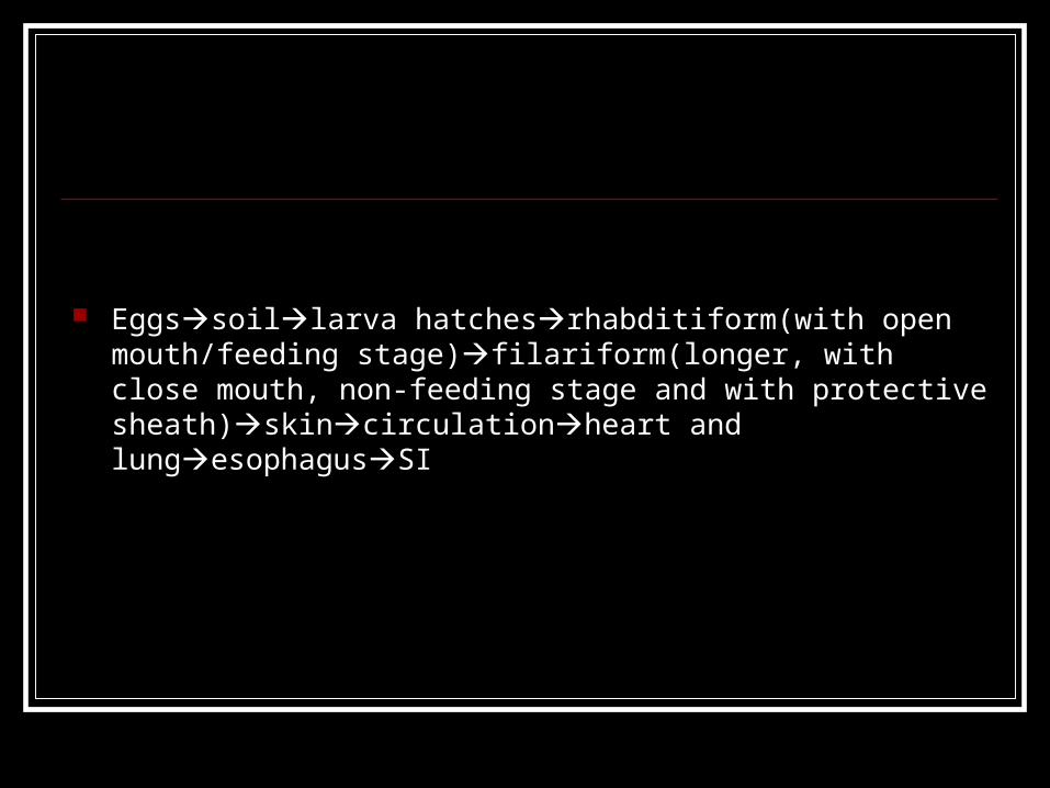
Eggssoillarva hatchesrhabditiform(with open mouth/feeding stage)filariform(longer, with close mouth, non-feeding stage and with protective sheath)skincirculationheart and lungesophagusSI

Ova Hookworm egg provided with a very thin
egg shell ( with germ cell in the process of segementation 2-8 cell stages)
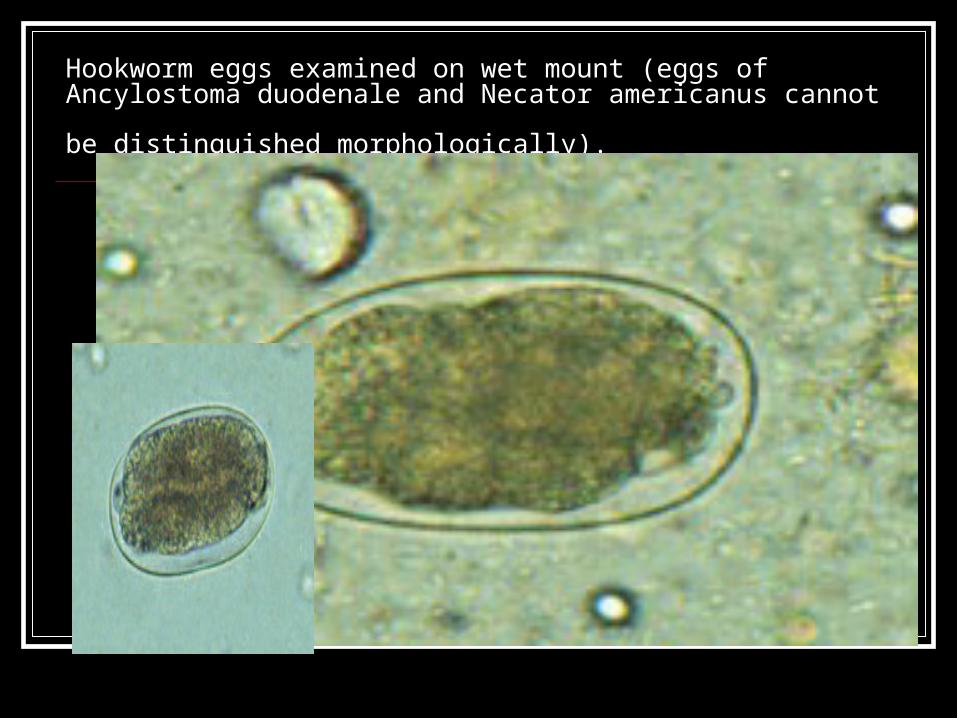
Hookworm eggs examined on wet mount (eggs of Ancylostoma duodenale
and Necator americanus cannot be distinguished morphologically).


Hookworm filariform larvae © Dr Peter Darben, Queensland University of
Technology clinical parasitology collection. Used with permission

Necator americanus adult male, posterior end © Dr Peter Darben, Queensland University of Technology clinical
parasitology collection. Used with permission


Dental Pattern
N. americanus 0 A. braziliense 1 A. caninum 3 A. duodenum 2


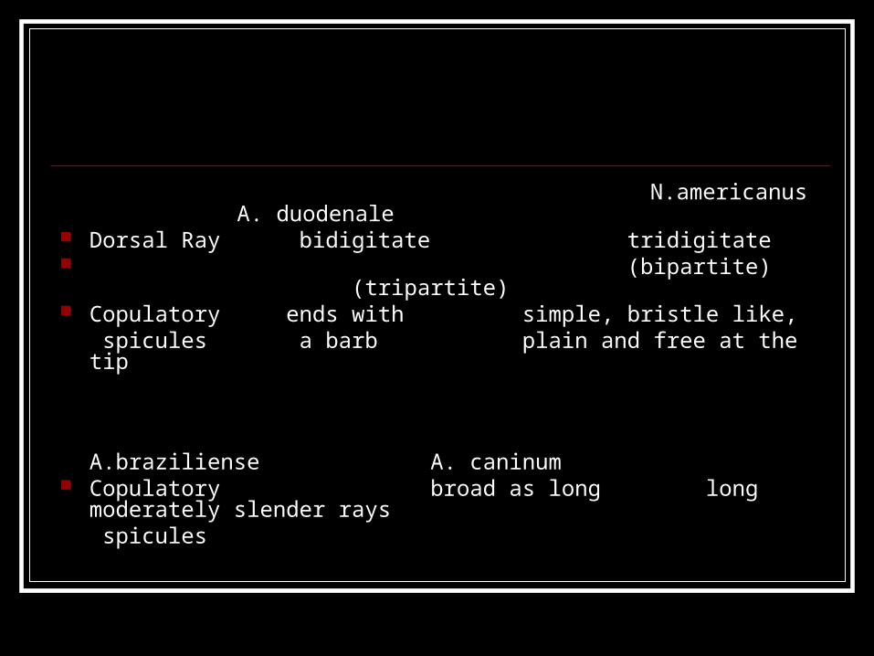
N.americanus A. duodenale Dorsal Ray bidigitate tridigitate (bipartite) (tripartite) Copulatory ends with simple, bristle like,
spicules a barb plain and free at the tip
A.braziliense A. caninum Copulatory broad as long long moderately
slender rays spicules
Pathology Ground itch/ dew itch
percutaneous entry of the infective filariform larvae often characterized by itching sensation or dermatitis. It is often severe and also known as ground itch or dew itch.

Pulmonary lesion/wakana disease
is caused by passive pulmonary migration of larval stages of hookworm. Symptoms may be similar to ascaris pneumonitis.

Creeping eruption/ cutaneous larva migrans
the infective laval stage can enter human skin but cannot pass below stratum germinativum producing serpiginous tunnel in this stratum. Common among larvae that do not normally infect human such as A. braziliense and A. caninum.
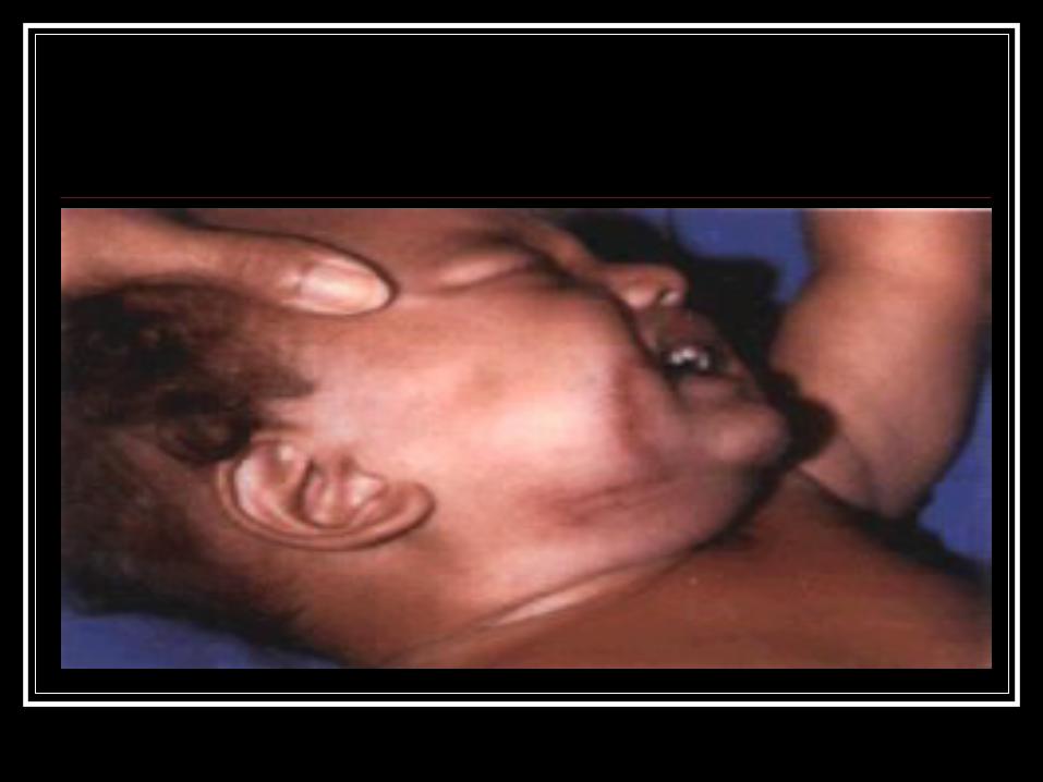


Hookworm anemia Adult worm suck the host’s blood and
mucosal substances. Microcytic hyopochromic anemia May cause hemorrhage to intestinal worm
Hypoalbuminemia due to combined loss of blood, lymph and
protein.

Treatment Albendazole, mebendazole, flubendazole Thiabendazole –topical or systemic is
successful for creeping eruption Iron therapy for anemia

Strongyloides stercoralis Common name: threadworm
Disease : strongyloidosis, cochin china diarrhea

Strongyloides stercoralis : known for its extra ordinary conditions, it is capable of both free living and parasitic existence. People are the principal host but dogs and monkeys have similar parasite.

Morphology Adult: Small, semi-transparent, with finely striated
cuticle. Male: cylindrical in shape, tail is pointed and
curved.

Female: stouter than male, ovoviviparous (they lay eggs containing larvae which are immediately hatched out (parthenogenetic) requiring no male to fertilize the eggs.
Ova The paired uteri of the adult female ova
contain single file of thin shelled transparent, partially embryonated eggss resembling “chinese lantern”.
These eggs are seldom seen on feces.

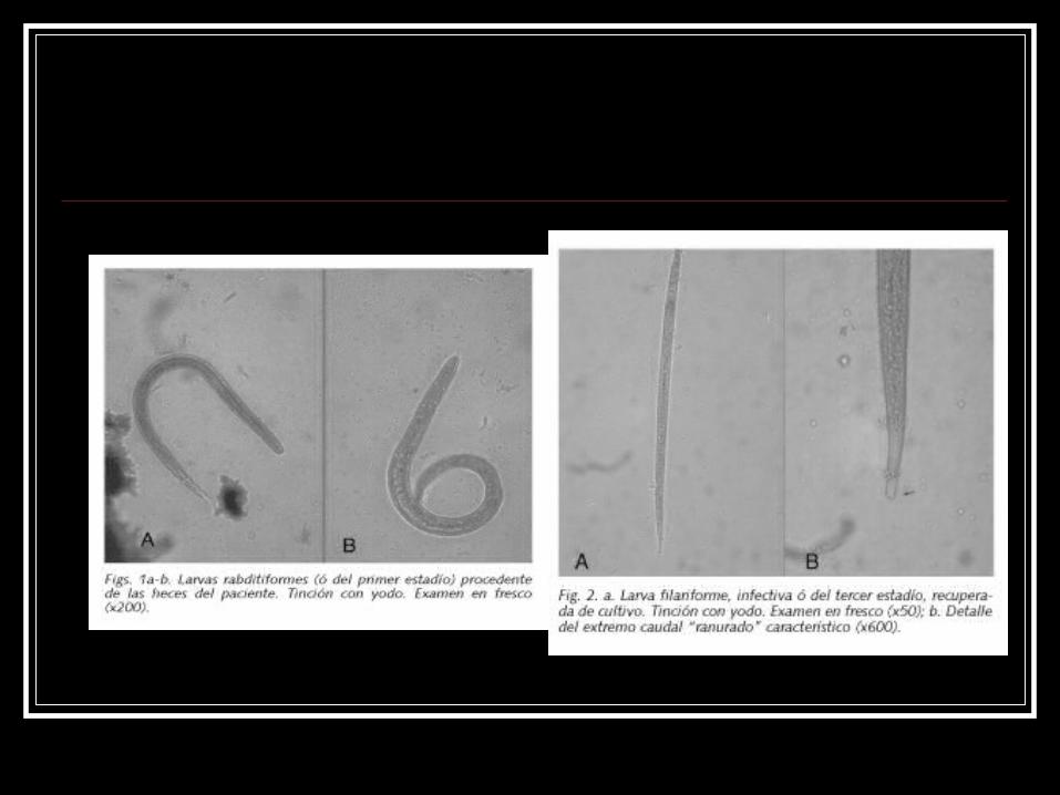
Larvae Rhabditiform: short buccal cavity, elongated
esophagus with pyriform posterior bulb and conspicuous genital primordium. Seen in feces.
Filariform: long delicate larva with long esophagus and forked notched tail.

Broncho alveolar Strogyloides stercoralis larva

Life cycle Direct SIeggs in intestinal mucosa
rhabditiformfilarifom(soil)skin blood streamheartlung esophagus
Indirect SIeggs in intestinal
mucosarhabditiform(soil)(free living adults)rhabditiform(repeat free living)/filariform(or become infective)

Autoinfection SIeggs in intestinerhabditiform
transformed into filariform penetrates intestinal mucosa blood streamheartlungesophagus

Pathology Often light and unnoticed Cutaneous infection: pruritus and erythema
at the site of penetration causing larva currens (racing larva)
Pulmonary infection: pneumonitis may develop as a result of larval migration to lungs.
Intestinal infection

Treatment
Albendazole and thiobendazole

Capillaria philippinensis Common name: Pudoc worm
Disease: Intestinal capillariasis, Pudoc’s disease

Morphology Adult: Very tiny worms the male measures 2.13 to
3.17mm with extraordinary long, smooth spicular sheath.
The female 2.5-4.3mm, the anterior portion contains esophagus (stichosoma), the posterior contains intestine and reproductive organs:

Female Capillaria Typical (oviparous): 8-10 eggs in uterus,
arranged in single row. Gives rise to a typical capillaria egg.
Atypical (larviparous): 40-45 eggs in uterus, arranged in 2-3 rows; hatch while still in uterus
Ova Typical capillaria egg: yellow in color,
moderately thick with striated egg shell and flattened bipolar plugs. Characteristically with indentation in the middle thus appear to be peanut shaped.
Atypical : thin shelled without bipolar plugs and multi-segmented or embryonated. This eggs hatch while still in utero into 1st stage larva.

Life cycle SI eggs laid by typical female
embryonation in water 3-5 days ingested by fresh water fish man ingest raw fish with larva

Pathology Persistent diarrhea and dehydration, rapid
weight loss, abdominal pain, gurgling of the stomach (borborygmus), muscle wasting, anorexia, vomiting and edema.
Malabsorption of fats and sugar Low K, Ca, carotene and TP Hypotension and cardiac failure may
develop and lead to death.

Diagnosis
DFS, conc. Technique, AECT,

Treatment
Mebendazole and abendazole
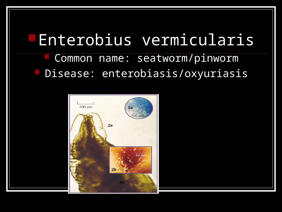
Enterobius vermicularis Common name: seatworm/pinworm
Disease: enterobiasis/oxyuriasis

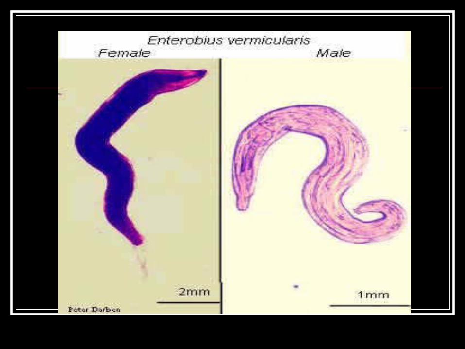
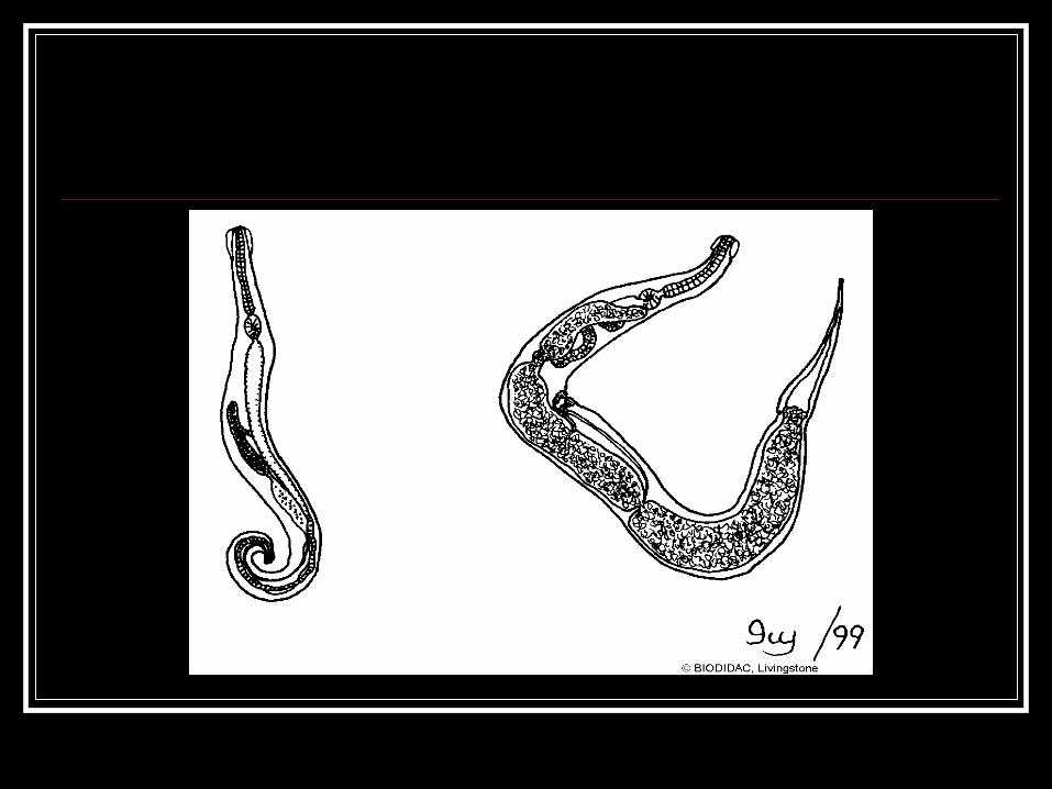
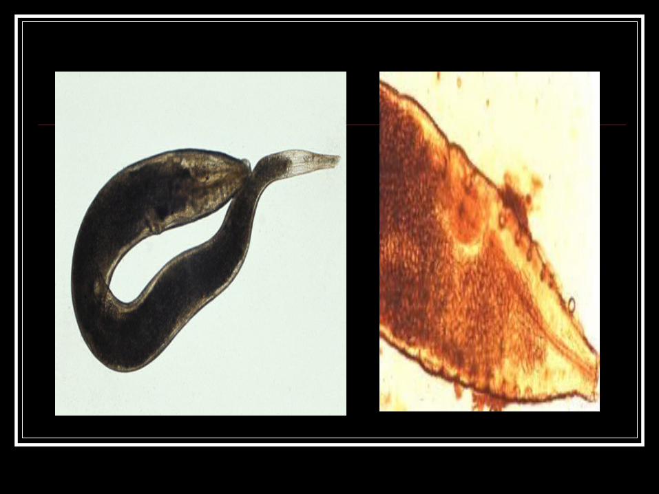
Morphology Adult: Small whitish or brownish in color, the male
measures 2-5 mm, the posterior end is strongly curved and single copulatory spicule is present.
The female measures 8-13mm with long pointed end.

The diagnostic features of adult enterobius are:
1. a pair of lateral cuticular wing like expansion at the anterior end known as cephalic alae.
2. adistinct prominent esophageal bulb.

Ova Elongated wherein the ventral side is
flattened thus the appearance similar to letter D, lopsided in appearance.
The eggs are usually embryonated when laid and mature within 6 hours after oviposition of gravid female and these are already infective.


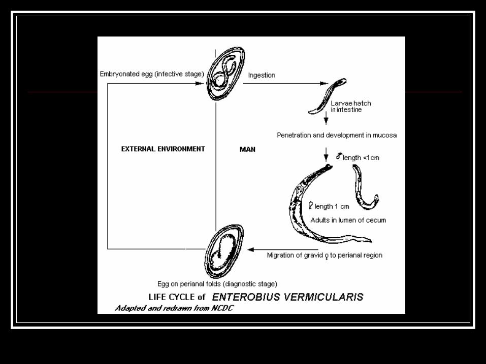

Life cycle LIfemale migrates to perianal region
deposit ebryonated eggigested or inhaledlarva hatch in duodenum migrate to LI


Pathology Insomnia, grinding of teeth Appendicitis, vulvovaginitis
The most common modes of transmission are
Hand to mouth Inhallation or airborne eggs from dust autoinfection

Diagnosis Graham scotch tape anal swab technique DFS

Treatment Abendazole, mebendazole, pyrantel
pamoate

fin