The International Student Journal of Nurse Anesthesia · extrusion, C4-5 fusion, bilateral inguinal...
67
Volume 13 Number 1 Spring 2014 The International Student Journal of Nurse Anesthesia TOPICS IN THIS ISSUE IV Acetaminophen for Preemptive Analgesia Difficult Airway Algorithm for Reintubation Clonidine & Cardiovascular Disease Transient Paraplegia after Epidural Acute Normovolemic Hemodilution Ondansetron Induced Bradycardia Cricoid Pressure Effectiveness Morbid Obesity & Pregnancy Awake Video Laryngoscopy Takotsubo Cardiomyopathy Moyamoya Disease Acute Kidney Injury Tranexamic Acid Down Syndrome Ludwig’s Angina
Transcript of The International Student Journal of Nurse Anesthesia · extrusion, C4-5 fusion, bilateral inguinal...

Volume 13 Number 1 Spring 2014
The International Student Journal of Nurse Anesthesia
TOPICS IN THIS ISSUE
IV Acetaminophen for Preemptive Analgesia
Difficult Airway Algorithm for Reintubation
Clonidine & Cardiovascular Disease
Transient Paraplegia after Epidural
Acute Normovolemic Hemodilution
Ondansetron Induced Bradycardia
Cricoid Pressure Effectiveness
Morbid Obesity & Pregnancy
Awake Video Laryngoscopy
Takotsubo Cardiomyopathy
Moyamoya Disease
Acute Kidney Injury
Tranexamic Acid
Down Syndrome
Ludwig’s Angina
INTERNATIONAL STUDENT JOURNAL OF NURSE ANESTHESIA Vol. 13 No. 1 Spring 2014
Editor
Vicki C. Coopmans, CRNA, PhD
Associate Editor Julie A. Pearson, CRNA, PhD
Editorial Board
Laura Ardizzone, CRNA, DNP Memorial Sloan Kettering Cancer Center; NY, NY
Laura S. Bonanno, CRNA, DNP Louisiana State University Health Sciences Center
Carrie C. Bowman Dalley, CRNA, MS Georgetown University
Janet A. Dewan, CRNA, PhD Northeastern University
Kären K. Embrey CRNA, EdD University of Southern California
Rhonda Gee, CRNA, DNSc Millikin University and
Decatur Memorial Hospital
Marjorie A. Geisz-Everson CRNA, PhD University of Southern Mississippi
CDR Robert Hawkins, CRNA, MBA, DNP, NC, USN Uniformed Services University
CDR Johnnie Holmes, CRNA, PhD, NC, USN Uniformed Services University
Donna Jasinski, CRNA, DNSc Georgetown University
Russell Lynn, CRNA, MSN University of Pennsylvania
MAJ Denise McFarland, CRNA, DNP, AN 2nd FST, Gelan DSP, Afghanistan
CDR Greg Nezat, CRNA, PhD, NC, USN Uniformed Services University
Teresa Norris, CRNA, EdD University of Southern California
Ilene Ottmer, CRNA, MSN University of Wisconsin Medical School & Medical Foundation
CDR Justice Parrott, CRNA, DNAP, NC, USN Uniformed Services University
Sarah Perez, CRNA, MS, MSN Washington University School of Medicine,
Barnes-Jewish Hospital; St. Louis, MO
Jo Ann Platko, CRNA, BC, PhD(c) Somnia Anesthesia, Inc.
Michael Rieker, CRNA, DNP Wake Forest Baptist Health
CDR Dennis Spence, CRNA, PhD, NC, USN Uniformed Services University
Lori Ann Winner, CRNA, MSN University of Pennsylvania
Kelly Wiltse Nicely, CRNA, PhD University of Pennsylvania
Maria Van Pelt, CRNA, MS, MSN Massachusetts General Hospital
Kathleen R. Wren, CRNA, PhD Arkansas State University

2
Contributing Editors For This Issue
Barbara Brown, CRNA, MSN Wake Forest Baptist HealthKevin C. Buettner, CRNA, PhD University of North Dakota
James Carson, CRNA, MSN Arkansas State UniversityJohn Driver, CRNA, BS Cox Health and Medical Center; Branson, MO
Michele E. Gold, CRNA, PhD University of Southern CaliforniaDavid N. Fort, CRNA, DNP Samford University
Crystal Odle, CRNA, DNAP Lincoln Memorial UniversityR. Chris Torman, CRNA, MNA Westminster CollegePaul Wilkins, CRNA, MSNA Providence Sacred Heart; Spokane, WAJasmin Ulahannan, CRNA, MSNA Parkland Hospital; Dallas, TX
Reviewers For This Issue
Amy Antalick, CRNA, MSN Washington University School of Medicine;St. Louis, MO
Jennifer Badeaux, CRNA, MN Louisiana State University Health Sciences CenterCourtney Brown, CRNA, PhD Wake Forest Baptist Health
Adam Collins, CRNA, MSNA Anesthesia Consultants of Dallas
Shelli Collins, CRNA, MSN Excel Anesthesia, PA; Dallas, TXNicole Hirst, CRNA, MSN 2nd FST, DSP Gelan, Afghanistan
David Kalil, CRNA, DNAP Louisiana State University Health Sciences Center
Connie Lorette, CRNA, PhD Northeastern UniversityNancy Perez, CRNA, MS Keck Hospital, University of Southern CaliforniaCliff Roberson, CRNA, DNP Columbia University
Erin Sebens, CRNA, MS University of Wisconsin Medical School& Medical Foundation
Mary Ellen Zerlan, CRNA, DNP Washington University School of Medicine;St. Louis, MO

3
The opinions contained in this journal are those of the authors and do not necessarily represent the opinions of the program or the University. Disclaimer for all articles authored by military personnel: The views expressed in this journal are those of the authors and do not necessarily reflect the official policy or position of their respective Military Department, Department of Defense, nor the U.S. Government. The work was prepared as part of the official duties of the military service member. Title 17 U.S.C. 105 provides that ‘Copyright protection under this title is not available for any work of the United States Government’. Title 17 U.S.C. 101 defines a United States Government work as a work prepared by a military service member or employee of the United States Government as part of that person’s official duties. Front Cover: Charles E. Gaines Jr., RN, BSN, a first year graduate student enrolled in the Nurse Anesthesia Program at Arkansas State University practices neuraxial anesthetic techniques on a porcine model. The Guide for Authors: can be found at www.aana.com by following this path: CE & Education Students Scroll down to lower right, click on Student Journal Or, use this direct link: http://www.aana.com/studentjournal

4
Table of Contents Case Reports The Use of Tranexamic Acid to Reduce Perioperative Blood Loss ...........................................6 Carrie A Cromwell, United States Army Graduate Program in Anesthesia Nursing Clonidine: Adjunct Therapy in Patients with Cardiovascular Disease ....................................9 Elizabeth H. Pflug, Wake Forest Baptist Health The Anesthetic Management of Moyamoya Disease ................................................................12 Charlene Franchi, University of Southern California Prevention of Acute Kidney Injury during Major Vascular Surgery ....................................15 Robert Kari, University of North Dakota Anesthetic Management in a Down Syndrome Patient ............................................................19 Natalie Carson, Arkansas State University Transient Paraplegia associated with Epidural Analgesia for Labor and Delivery ..............22 Robert Wayne Olson Jr., Arkansas State University Acute Normovolemic Hemodilution versus Blood Transfusion for Liver Lobectomy ..........25 Dhanya Renjith, University of Southern California Anterior Cervical Fusion and the Difficult Airway Algorithm for Reintubation ..................29 Tahira Smith, University of Southern California Ondansetron Induced Bradycardia............................................................................................32 Chalon Hollas, Arkansas State University Awake Fiberoptic Intubation for a Patient with Ludwig’s Angina ........................................35 Kelsey Bissell, Westminster College Takotsubo Cardiomyopathy: A Case of ‘Broken-Heart Syndrome’ ......................................38 Sumer Christensen, Westminster College Awake Intubation using Video Laryngoscopy for the Anticipated Difficult Airway ............41 Eric Dilg, Arkansas State University Morbid Obesity and Pregnancy: A Case Study ........................................................................44 Dawn McKinney, Lincoln Memorial University

5
Evidence-based Practice Analysis Reports Preemptive Analgesia: Intravenous Acetaminophen ...............................................................47 Justin Greenfield, Louisiana State University Health Science Center Cricoid Pressure Effectiveness: A Review of Evidence ............................................................52 Adam Peeples, Samford University Editorial ........................................................................................................................................58 Vicki C. Coopmans, CRNA, PhD Guide for Authors ........................................................................................................................59

6
The Use of Tranexamic Acid to Reduce Perioperative Blood Loss
Carrie A Cromwell, BSN United States Army Graduate Program in Anesthesia Nursing
Keywords: tranexamic acid, blood loss, antifibrinolytic
Significant consideration is required when planning the anesthetic management for patients undergoing surgery with the potential for excessive blood loss. Recent studies have shown that administering blood transfusions can be associated with severe adverse effects including higher mortality, greater postoperative morbidity, and increased health care costs.1 Measures can be taken in order to attenuate perioperative bleeding and the subsequent pathological outcomes. One of the pharmacological strategies that can be employed is the intravenous administration of the lysine analog, tranexamic acid. Case Report A 47-year-old male presented for a total hip arthroplasty related to osteoarthritis and hip pain following failed conservative therapies and medications. The patient was 190.5 cm in height and weighed 111 kg, with a BMI of 31. The patient had no known allergies. He was categorized as an American Society of Anesthesiologists (ASA) Physical Status classification 3 for hypertension controlled on lisinopril, gastroesophageal reflux disease with treatment on esomeprazole, active tobacco use, and hypothyroidism controlled with levothyroxine. Previous surgical procedures included L4-5 disc extrusion, C4-5 fusion, bilateral inguinal hernia repair, and a vasectomy with no anesthesia related complications. Preoperative laboratory values of note included hemoglobin (hgb) 13.0 g/dL and hematocrit (hct) of 35.7%. Physical
assessment was unremarkable. Preoperative medications included midazolam 2mg. Upon transfer to the operating room table, the patient was positioned supine and preoxygenated with oxygen 100% via face mask for 5 minutes while standard monitors were applied. A rapid sequence induction with cricoid pressure was performed and consisted of fentanyl 150mcg, lidocaine 100mg, propofol 200mg, and succinylcholine 100mg. A grade 1 view of the vocal cords was obtained using the GlideScope (Verathon, Inc., Bothell, WA) video laryngoscope. The trachea was intubated with a 7.5 endotracheal tube after one attempt without trauma or difficulty. Bilateral breath sounds and capnography confirmed placement, with an ETCO2 of 34mmHg. Ventilator settings included volume control mode with a tidal volume of 750 ml and respiratory rate of 10. General anesthesia was maintained with a mixture of sevoflurane 1.1% and nitrous oxide 60% with an oxygen flow of 1L/min. Following the return of twitches with the peripheral nerve stimulator, rocuronium 40mg was administered. Tranexamic acid 1g was administered over 20 minutes intravenously, followed by cefoxitin 2g. Because surgical trauma activates fibrinolysis, administration of tranexamic acid before the fibrinolytic system is activated will assist to maintain fibrin clot integrity.2 The surgical procedure ensued shortly thereafter. The surgery time was 149 minutes and without any adverse events. Total estimated blood loss was 250 ml. Neuromuscular blockade was antagonized at the end of the

7
procedure and emergence and extubation were without complication. Postoperative evaluation the following day revealed no anesthetic complications and an approximate total blood loss of 200ml. The patient was discharged on postoperative day three and did not require the transfusion of any blood products during his hospital stay. Discussion Surgical trauma, the activation of inflammatory cascades, and the transfusion of fluids and blood components can exacerbate perioperative bleeding.3 Transfusions for the replacement of blood loss can lead to adverse effects including the transmission of infectious diseases, bacterial infection, immune reactions, transfusion related acute lung injury, and mortality.3-4 Tranexamic acid is a medication that has been used and studied extensively for the reduction of perioperative blood loss. Studies support that tranexamic acid can reduce the incidence of blood transfusion in total joint arthroplasty by as much as 53%.3
Tranexamic acid is a lysine analog which inhibits fibrinolysis.3 It competitively binds to plasminogen at the lysine binding site and prevents the conversion plasminogen to plasmin.5 This prevents the breakdown of the fibrin clot by inhibiting plasmin from binding to fibrin structures after clot formation.5 By preventing fibrinolysis, fibrin clot integrity is maintained and perioperative blood loss is reduced.5
Tranexamic acid dosage protocols for bleeding prophylaxis have not been standardized; thus, the amount administered varies widely amongst insitutions.3 Doses for cardiac surgery have involved a loading dose of 10 to 30 mg/kg followed by infusion of 1 to 16 mg/kg/hr.3 Perioperative doses in a meta-analysis of studies using tranexamic
acid ranged from 10 to 135 mg/kg, with and without subsequent maintenance infusions.4 The dose used in the case discussed was a single dose of 1g, or approximately 9 mg/kg. There is no definitive plasma level of tranexamic acid that has been determined to inhibit fibrinolysis.3 The primary benefits of tranexamic acid are related to its ability to inhibit fibrinolysis, but there are also several other useful effects from the administration of this medication. In a study on the use of tranexamic acid to reduce blood loss after total knee arthroplasty, patients in the tranexamic acid group had lower D-dimer concentrations than in the control group.5 D-dimer levels are a reflection of the breakdown of fibrin by plasmin, and will be elevated with increased fibrinolysis.5 A study using tranexamic acid to reduce blood loss in total hip replacement surgery also supported a reduction in the postoperative D-dimer level.2 Additionally, it has been shown that platelet activation was not induced by tranexamic acid, and that extrinsic coagulation (PT) and intrinsic coagulation (PTT) were within normal limits and unaffected with the use of this antifibrinolytic.5 Reduced blood loss decreases the requirement for perioperative transfusions, thus alleviating the risks associated with component therapy and decreasing hospital length of stay. It has been estimated that patients who receive blood transfusions remain hospitalized an average of 2.5 days more than non-transfused patients.1 This may lead to significant reductions in health care costs. The safety of tranexamic acid in surgical patients has been examined in several studies and potential adverse effects must be considered before administration. The studies reviewed did not support an increased incidence of thromboembolic

8
events and mortality; however, the data is limited and future research should focus on this potential complication.3 A trial on the use of tranexamic acid in bleeding trauma patients demonstrated a significant reduction in mortality with no increase in thromboembolic events and a greatly reduced risk of myocardial infarction.6 The safety of tranexamic acid was evaluated in total hip and knee replacement procedures, and similar results were obtained: there were no adverse cardiovascular events in any of the study subjects, and no increase in the incidence of peripheral venous thrombosis when compared to the control group.2,5 Because of its relative safety profile, the administration of tranexamic acid was determined to be beneficial for the patient in this review. According to Jaffe, the average estimated blood loss for a total hip replacement is 500 ml.7 The total intraoperative blood loss in this case was 250 ml, which is 50% less than the average perioperative blood loss in this surgical procedure. This correlates with studies that have shown an average blood loss reduction of 53% with the use of tranexamic acid.3 The patient's hgb and hct preoperatively were 13.0 g/dL and 35.7%. On postoperative day three they were reduced to 11.8 g/dL and 35.4%, which is a decrease of only 0.9%. The median length of stay following total hip replacement surgery is eight days.8 The patient in the case reviewed was discharged on postoperative day three, which is five days less than what has been previously demonstrated.8 In this case, the transfusion of erythrocytes or other blood products were also not required during the hospital course. Implementing measures to reduce the volume of perioperative blood loss has many benefits and should be incorporated into the anesthesia plan of care. This case review
demonstrated that the use of tranexamic acid as an intravenous pharmacological agent can reduce perioperative blood loss, decrease health care costs, shorten hospital length of stay, and prevent the use of blood transfusions. References
1. Shariatmadar S, Greeson N, Candiotti K. Perioperative complications and costs associated with blood transfusions. Curr Rev Nurs Anesth. 2012;35(16):191-198.
2. Ekback G, Axelsson K, Ryttberg L, et al. Tranexamic acid reduces blood loss in total hip replacement surgery. Anesth Analg. 2000;91:1124-1130.
3. Kanake P, Eaton M. Systemic strategies for reducing blood loss in surgery. Anesthesiology News. 2013;39(1):1-7.
4. Zufferey P, Merquiol F, Laporte S, et al. Do antibrinolytics reduce allogeneic blood transfusion in orthopedic surgery. Anesthesiology. 2006;105(5):1034-1046.
5. Jansen AJ, Andreica S, Claeys M, et al. Use of tranexamic acid for an effective blood conservation strategy after total knee arthroscopy. Bri J Anaesth. 1999;83(4):596-601.
6. Ker K, Edwards P, Perel P, et al. Effect of tranexamic acid on surgical bleeding: systematic review and cumulative meta-analysis. Brit Med J. 2012;344:1-13.
7. Jaffe RA, Samuels SI. Anesthesiologists Manual of Surgical Procedures. 4th ed. Philadelphia: Lippincott Williams & Wilkins; 2009: 993.
8. Foote J, Panchoo K, Blair P, et al. Length of stay following primary total hip replacement. Ann R Coll Surg Engl. 2009;91:500-504.
Mentor: MAJ Denise McFarland, CRNA, MSN, AN

Clonidine: Adjunct Therapy in Patients with Cardiovascular Disease
Elizabeth H. Pflug, BSN Wake Forest Baptist Health
Keywords: clonidine, alpha 2 adrenergic agonist, cardiovascular disease, hypertension, analgesia Twenty-five to fifty percent of deaths in non-cardiac surgeries are related to cardiovascular complications.1 A patient who has cardiac ischemia prior to non-cardiac surgery is three times more likely to experience a postoperative myocardial infarction.2 To optimize patients’ outcomes, it is essential to assess and understand the extent of disease, provide prophylactic treatment and prepare for the most effective interventions. While there are a variety of medications available, researchers explore the use of clonidine as an adjunct therapy for patients with cardiovascular disease and hypertension. Case Report A 70-year-old, 67 kg male presented with cholecystitis and was scheduled to undergo an emergency laparoscopic cholecystectomy. The patient history included: exercise-induced angina, hypertension, coronary artery disease, mitral regurgitation, transient ischemic attacks, coronary artery bypass graft (CABG) with two stents placed in August 2002 and diabetes. His pre-operative blood pressure was 118/60 mm Hg and heart rate was 76/min. His medications included amlodipine, aspirin, clopidogrel, furosemide, glipizide, metoprolol, naproxen sodium, pantoprazole, and simvastatin. The patient had been off his clopidogrel for only two days instead of the recommended five days, therefore the patient was informed of the increased bleeding risk. The surgeon
requested that the patient receive one pack of platelets intra-operatively to help restore clotting ability and reduce bleeding. The patient had a baseline 12-lead electrocardiogram (ECG) that showed sinus rhythm with a previous inferior infarct from April 2012, a previous anterolateral infarct from February 2011, and T wave abnormalities. His ejection fraction was 40%. The patient’s history was reviewed, he was informed of anesthetic risks and concerns, and the anesthesia consent was signed. The patient was brought into the operating room and the standard ASA monitors were applied. He was pre-oxygenated via facemask with 8 L/min oxygen flow. The patient then received fentanyl 100 mcg intravenously (IV) and propofol 120 mg IV. Once the patient became apneic, mask ventilation was assumed without difficulty and rocuronium 40 mg IV was given. His blood pressure decreased and the patient received phenylephrine 240 mcg IV. The patient was atraumatically intubated with a 7.0 cuffed endotracheal tube with the assistance of a bougie. Placement was confirmed and general anesthesia maintained with 0.8% isoflurane with a 0.5 L/min of air and 0.5 liters/min of oxygen. Strict blood pressure and heart rate control were maintained within 20% of his baseline with several phenylephrine boluses of 120 mcg and an esmolol bolus of 30 mg. During the case, the patient’s systolic blood pressure increased to 160 mm Hg, clonidine 50 mcg IV was administered. The patient received the platelet transfusion without complication. Surgical blood loss was minimal.

10
During emergence from anesthesia, new ST depression was noted on the monitor in lead V4 and confirmed in diagnostic mode. Metoprolol for beta blockade, a bolus of heparin and morphine sulfate were given. The patient resumed spontaneous breathing with adequate tidal volumes and acceptable carbon dioxide levels. He was given a full neuromuscular block reversal, had four strong twitches, equal double burst, sustained tetany, he opened his eyes, he followed simple commands and then he was extubated. Oxygen was provided at 10 L/min via facemask during transportation to the recovery room. Upon arrival, a 12-lead ECG was completed and a dose of aspirin was given. The cardiologist reviewed the most recent ECG and compared it to the patient’s baseline ECG to determine a transient ischemic event had occurred and resolved. The ECG showed sinus rhythm with premature atrial contractions and ST abnormality in addition to the T wave abnormality. The patient’s blood pressure remained stable. In the recovery room, the patient was lethargic and became increasingly hypercarbic and then hypoxic, requiring reintubation. An hour later, the patient was extubated in the recovery room. At this point, he was alert, following detailed commands, breathing spontaneously with appropriate minute ventilation, vital signs were stable and the end-tidal carbon dioxide levels, measured by capnograhy, were maintained in normal range. The remainder of the hospitalization for this patient was uncomplicated and he was discharged within two days after surgery. Discussion Cardiovascular disease is prevalent and knowing that it significantly increases intra-operative risks, anesthesia practitioners must be diligent in identifying risks and treat patients to optimize their cardiac status.
Clonidine helped by blocking the sympathetic response of norepinephrine in the presynaptic neurons.3 It provides analgesic effects by affecting the alpha 2a receptors which results in reduced sympathetic responses.3 It also inhibits the triggering of nociceptive neurons when stimulated by the A-delta and C fibers, which also stops the release of substance P.5 These effects help create hemodynamic stability in the presence of surgical stimulation,4 analgesia, anxiolysis and sedation.3 The onset is quick because clonidine is highly lipid soluble, reaching its peak between sixty to ninety minutes.3
In a recent review by Chalikonda,3 clonidine was researched in four separate studies. The first evaluated the use of clonidine preoperatively, which resulted in rapid blood pressure control and no evidence of reflexive tachycardia associated with direct laryngoscopy and intubation. It also concluded that clonidine increases hemodynamic stability and reduces amount of anesthetic needed.3 The second case showed that blood pressure and heart rate decreased due to clonidine’s effect of decreasing sympathetic tone.3 The third study indicated that there is a reduction in intraoperative myocardial ischemia when clonidine is given preoperatively, but could not support that it affected the postoperative incidence of ischemia. However, the study concluded that there was a decrease in tachycardia up to five hours postoperatively.3 The fourth study of patients given clonidine preoperatively yielded results of a decrease in myocardial ischemic events from thirty-eight percent to twenty-four percent. The study found that when patients took clonidine preoperatively, there was a lower incidence of myocardial ischemia in patients with coronary artery disease.3

11
Beta blockers and alpha 2 agonists have been compared for treatment in patients with risks for cardiovascular disease. Chalikonda reviewed multiple medications and found that beta blockers and alpha2 agonists are cardioprotective in the perioperative period and suggest further studies to evaluate their use in conjunction with one another.3 Some studies have shown that beta blockers must be used judiciously due to the increased risk of stroke associated with their use.4 The use of an alternative alpha2 agonist, such as dexmedetomidine, can be considered. Due to its greater affinity for alpha 2 receptor sites it increases sedative effects, but its use is limited in that it is only available intravenously.4,5 Clonidine is commonly used because of its availability in multiple forms of administration including intravenous, oral, transdermal, intramuscular and epidural.3 Overall, clonidine can help to reduce blood pressure while offering the patient analgesia, sedation and anxiolysis.3 As an added benefit, the Blaudszun et al study found clonidine to have antiemetic effects, decreasing the incidence of post-operative nausea and vomiting.4 When considering clonidine for adjunct uses, caution should be taken in patients at risk for developing hypotension.4
Patil and Anitescu evaluated the use of clonidine on a patient who had experienced opioid induced delirium with a previous surgery and was to undergo another surgery.5 This case was performed completely without the use of opioids. Instead clonidine was used via multiple routes of administration including oral, transdermal and intravenous.5 Balanced anesthesia creates a synergistic effect by involving multiple receptors to cover various types of stimulation and pain.5 The benefit of using an alpha 2 agonist is that they do not reduce the patient’s respiratory drive or cause a decrease in neurologic
function which is ideal for patients who suffer from dementia, sleep apnea and other neurologic or respiratory illnesses.5
The patient in the case above required an emergent surgery, which increases cardiovascular risks two to five times.1 Though this patient had normal vital signs and lab work, his history of hypertension, mitral valve regurgitation, coronary artery disease, CABG with stent placement and ejection fraction of 40% indicated the need for tight blood pressure and heart rate control. Phenylephrine was chosen over ephedrine to avoid an increased heart rate and the alpha agonist will increase coronary perfusion to ensure the heart stays well perfused. Esmolol was administered to maintain heart rate on the lower end of the patient’s baseline in order to reduce the myocardial oxygen demand. During the case, the patient was given a dose of clonidine, primarily to treat an increased blood pressure, but also to provide the patient with the additional benefits associated with clonidine. In doing so, the patient can benefit from the analgesia, anxiolysis, and anti-emetic effects without concern of respiratory depression or a decrease in neurological function. The fact that the patient required reintubation in the recovery room because of the inability to maintain adequate minute ventilation could be attributed to the narcotics, the volatile agent or the anesthetic effects of hypercarbia itself. It is the responsibility of all anesthesia practitioners to be knowledgeable of the various medications that can benefit a patient with cardiovascular disease. This knowledge will assist in tailoring the anesthesia to the patient. The goal is to provide the best and safest anesthesia to patients. By utilizing adjunct medications and employing a multimodal, balanced

12
technique, the anesthetic can be specifically designed to meet the individual patient’s needs. Optimal patient outcome and surgical experience requires adequate anesthesia, analgesia and vigilance to provide the necessary interventions to maintain physiological homeostasis. Alpha 2 agonists are significant adjuncts that provide cardioprotection by maintaining hemodynamic stability, providing analgesia and sedation, which decreases opioid and anesthetic needs without causing an increase in recovery time.3-5 References 1. Butterworth JF, Mackey DC, Wasnick
JD. Anesthesia for patients with cardiovascular disease. In: Morgan & Mikhail’s Clinical anesthesiology. 5th ed. New York: McGraw-Hill Professional; 2013: 375-434.
2. Brouillette C. Anesthesia for cardiac surgery. In: Nagelhout J, Plaus K, eds.
Nurse Anesthesia. 4th ed. St. Louis: Saunders Elsevier; 2010: 504-527.
3. Chalikonda S. Alpha2-adrenergic agonists and their role in the prevention of perioperative adverse cardiac events. AANAJ. 2009;77(2):103–108.
4. Blaudszun G, Lysakowski C, Elia N, Tramèr MR. Effect of perioperative systemic α2 agonists on postoperative morphine consumption and pain intensity: systematic review and meta-analysis of randomized controlled trials. Anesthesiology. 2012;116(6):1312–1322.
5. Patil SK, Anitescu M. Opioid-free perioperative analgesia for hemicolectomy in a patient with opioid-induced delirium: a case report and review of the analgesic efficacy of the alpha-2 agonist agents. Pain Pract. 2012;12(8):656–662.
Mentor: Barabara Brown, CRNA, MSN
The Anesthetic Management of Moyamoya Disease
Charlene Franchi, MSN University of Southern California
Keywords: Moyamoya disease, subarachnoid hemorrhage, general anesthesia, STA-MCA bypass Moyamoya disease (MMD), Japanese for “puff of smoke,” is a rare, chronic cerebrovascular occlusive disease characterized by narrowing of both internal carotid arteries (ICA).1 Vessels in the brain, primarily the ICA, become constricted resulting in collateral circulation around these narrowed arteries, which appear as a “puff of smoke” on angiogram.2-4
Moyamoya disease is prevalent in Japan with an incidence of 0.54 per 100,000 people with a ratio of 1.8:1 female to male
diagnosed.1,2 This case report discusses the anesthetic management of a patient with MMD undergoing a craniotomy. Case Report A 71-year-old, 160 cm, 78 kg African American female presented for a left craniotomy to undergo a superficial temporal artery to middle cerebral artery bypass. Medical history was significant for chronic hypertension, subarachnoid hemorrhage in 2012, and bilateral ICA stenosis with 90% occlusion. An antihypertensive regimen included atenolol, amlodipine, carvedilol, hydrochlorothiazide,

13
losartan, aldactone, and furosemide. A preoperative angiogram was consistent with a “puff of smoke” appearance of collateral vessels, as seen with MMD. A prior bilateral shoulder arthroscopy was noted without anesthetic complications. All laboratory values and x-ray results were normal. The baseline hematocrit was 38%. The electrocardiogram showed normal sinus rhythm with nonspecific T- wave abnormality and the multi-gated acquisition scan revealed an ejection fraction of 65% with trace mitral, aortic, and tricuspid regurgitation. The preoperative blood pressure (BP) was 142/74 mm Hg and the physical examination revealed no neurological deficits. Upon arrival to the operating room, ASA monitors were applied and 8L/min of O2 was delivered via face mask. An intravenous induction was achieved with 2% lidocaine 60mg, propofol 120mg, and rocuronium 70 mg. The trachea was intubated and respirations were controlled with volume control mechanical ventilation: tidal volume (TV) 500 ml and a respiratory rate (RR) 9/min. A left radial arterial line and right internal jugular central venous pressure (CVP) catheter were placed under sterile technique. Baseline vital signs were: BP 141/78 mm Hg, and CVP 6cm H2O. Anesthetic maintenance included desflurane 3% with 0.5 L/min of O2 and 0.5 L/min of air and a propofol IV infusion at 75 mcg/kg/min. Mannitol 20 mg IV and dexamethasone 10mg IV were given prior to incision. An ETCO2 level of 40-45 mm Hg was maintained. The patient remained hemodynamically stable, and one arterial blood gas (ABG) sampling was performed 2 hours into the case, which revealed a PaCO2
of 48 mm Hg. CVP values remained stable (target ranges of 5-10 cm H2O). A total of 2500ml of 0.9% normal saline (NS) IV was
given. The target BP values were maintained between 135-150/65-90 mm Hg. Ondansetron 4 mg IV was given 20 min before extubation and the neuromuscular blockade was antagonized with neostigmine 3mg IV and glycopyrrolate 0.6mg IV. When extubation criteria were met, the endotracheal tube was removed without complications, followed by transfer to the postanesthesia care unit (PACU). In the PACU, the patient’s neurological status was evaluated and no deficiencies noted. The patient remained comfortable without pain, nausea, or complications from anesthesia. The patient was admitted to the intensive care unit for one day and discharged on postoperative day three. Discussion The anesthetic management of a patient with MMD undergoing a craniotomy and revascularization is complex and untraditional to the usual craniotomy treatment plan. Surgery is often complicated by ischemia or hemorrhage requiring specific intraoperative anesthetic management in order to avoid such complications.5 The perioperative management of a patient undergoing a typical craniotomy includes hyperventilation, avoidance of hypertension, and decreased fluid intake. The patient with MMD undergoing a revascularization procedure is managed differently to include a higher ETCO2, a slightly elevated BP, and increased fluid administration to maintain cerebral perfusion.5,6 While it is a goal to optimize cerebral perfusion with every patient, for this case management increased fluid administration also prevented vasospasm. Carbon dioxide is a modulator of cerebrovascular tone and its intraoperative

14
management is an essential part of the anesthetic care for the patient with MMD.5,6 Induced hypercapnia (ETCO2 greater than 40mmHg) is necessary for patients undergoing general anesthesia with MMD.2,6 It is recommended to maintain hypercapnia, especially during revascularization, to further dilate the cerebral vessels, which enhances cerebral blood flow (CBF).4,5
Intraoperatively, an ETCO2 of 45-50mmHg was maintained through a decreased RR of 9 breaths per minute and a TV of 500ml to induce cerebral vasodilation, decreasing the risk of infarction and ensuring adequate CBF.5 Hemodynamic stability has an important role in the maintenance of adequate cerebral perfusion. Hypotension has been associated with cerebral infarction and it is imperative to maintain the patient’s baseline BP to ensure cerebral perfusion, especially in this patient with ICA stenosis.5,6 The preoperative BP was 142/74 and the anesthetic management focused on maintaining the BP between 140-150/75-90. These target B/P goals were based on previous experiences of the attending anesthesiologist in the management of patients with MMD. Anesthetics such as propofol and inhalation agents should be titrated carefully to avoid hypotension, which can impair autoregulation and induce infarction.6 The anesthetic management included 3% desflurane, with a titrated propofol infusion of 75mcg/kg/min to ensure adequate anesthetic depth. If the patient’s BP decreased to a systolic value of 135, a bolus of phenylephrine 40mcg IV was given, for a total of phenylephrine 240mcg. Hypervolemia and a decrease in baseline hematocrit were maintained with 0.9% NS and a total of 2500ml of crystalloid were administered during the 5 hour case. This volume was titrated according to the
patient’s BP, CVP (ranging from 5 to 9 cmH2O), and hematocrit. A fluid bolus of 250ml was given if the blood pressure was below 135 systolic and the hematocrit was evaluated intraoperatively through an ABG during the second hour of the case. Polycythemia with associated increased viscosity is regarded as a risk factor for cerebral infarction and a higher hematocrit is associated with an increase in blood viscosity.6 Slight hypervolemia, resulting in hemodilution and a decrease in blood viscosity, can enhance cerebral perfusion.5,6
The resultant mild hemodilution is used to improve cerebral perfusion and avoid cerebral ischemia, but if excessive it can cause cerebral ischemia, due to the decrease in oxygen carrying capability of blood.6 The goal was to keep the patient adequately hydrated with a hematocrit no lower than 33%, which is approximately 15% lower than the patient’s baseline value of 38%; this is between 30% to 42% which has been suggested in the literature.5,6 An intra-operative ABG allowed for assessment of the patient’s hematocrit, fluid status, and carbon dioxide levels, which guided our anesthetic plan and prompted for the titration of RR, TV, and fluid allowance. Although this patient remained hemodynamically stable, and had no neurologic deficit postoperatively which indicated appropriate cerebral perfusion, hourly ABGs may have provided additional information regarding hematocrit, fluid status and carbon dioxide levels. Osmotic diuretics, such as mannitol are given to reduce brain volume, which makes it easier for the surgeon to gain access to the brain.2 In order to avoid a significant change in fluid volume, a lower dose of mannitol was administered to avoid excessive diuresis, which can lead to hypotension, hypovolemia, and a higher hematocrit, which can precipitate vasospasm and

15
decreased cerebral perfusion.2,6
Postoperative nausea and vomiting can cause increases in ICP, as well as cerebral vasospasm, and were avoided with administration of dexamethasone 10mg and ondansetron 4mg. Moyamoya disease is a rare condition and a customized anesthetic management plan is integral to produce positive postoperative outcomes. Optimal anesthetic management for this patient included: keeping the patient’s ETCO2 between 45-50 mm Hg, an elevated BP slightly above baseline, and mild hemodilution. Although this may seem consistent with triple “H” therapy, it is a modification of the treatment modality, and tailored toward the MMD patient, specifically with an additional focus on moderate hypercapnia. The intraoperative management of the ETCO2, BP, and volume status will help to maintain hemodynamic stability and provide a smooth anesthetic course and emergence. In conclusion, this case report provides essential principles for the management of the patient with MMD undergoing craniotomy and the incorporation of this anesthetic technique to maintain cerebral perfusion during surgery.
References 1. Burke GM, Burke AM, Sherma AK,
Hurley M,C, Batjer HH, Bendok BR. Moyamoya disease: A summary. Neurosurg Focus.2009;26(4):E11.
2. Chong CT, Manninen PH. Anesthesia for cerebral revascularization for adult moyamoya syndrome associated with sickle cell disease. J Clin Neurosci.2011;18:1709-12.
3. Smith ER, Scott RM. Surgical management of moyamoya syndrome. Skull Base. 2005;15(1):15-26.
4. Sakamoto T, Kawaguchi M, Koukichi K, Kitaguchi K, Furuya H, Karasawa J. Risk factors for neurologic deterioration after revascularization surgery in patients with moyamoya disease. Anesth Analg 1997;85:1060-5.
5. Sato K, Shirane R, Yoshimoto T. Perioperative factors related to the development of ischemic complications in patients with moyamoya disease. Childs Nerv Syst.1997;13(2):68-72.
6. Parray T, Martin TW, Siddiqui S. Moyamoya disease: a review of the disease and anesthetic management. J Neurosurg Anesthesiol.2011;23(2):100-9.
Mentor: Michele E. Gold, CRNA, PhD
Prevention of Acute Kidney Injury during Major Vascular Surgery
Robert Kari, MS
University of North Dakota
Keywords: renal, vascular, prevention, mannitol, dopamine, perioperative, abdominal aortic aneurysm Abdominal Aortic Aneurysm (AAA) is a condition that affects many individuals in the United States. According to statistics
compiled in a study by the American Heart Association, the prevalence of AAA is 1.3% of men aged 45-54 years and 12.5% of men aged 75-84. Equivalent figures for women of the same age are 0% and 5.2%.1 Open approach to AAA repair has become less common with the introduction of

16
endovascular repair. One of the major complications associated with an open AAA repair is decreased renal perfusion leading to acute kidney injury (AKI).2 Acute kidney injury is defined as a decline in kidney function evidenced by a rise in nitrogenous waste products specifically serum creatinine and blood urea nitrogen (BUN).3
Case Report A 60-year-old Caucasian male presented for open AAA repair. The patient had a vascular Lifeline ultrasound screening performed by the local Red Cross, which identified an estimated 7.22 cm AAA. A subsequent computed tomography scan revealed a non-leaking juxtarenal AAA of approximately 6.2 cm in diameter. After consultation with a general surgeon and interventional radiologist, it was determined that the proximity of the aneurysm to the renal artery and the large proximal (3.2 cm) size made endovascular AAA repair a non-option.
The patient was 188 cm in height, weighed 104 kg, and had no known drug allergies. Medical history included hyperlipidemia, hypertension, carotid artery atherosclerosis, gastroesophageal reflux disease, benign prostatic hyperplasia, anxiety, depression, former smoker (30 pack year), and prior alcohol and drug abuse. Surgical history included a tonsillectomy with denial of any personal or family history of anesthetic complications. Pertinent lab values included hemoglobin 14.8 g/dL, hematocrit 42.5%, platelets 163,000/microliter, blood urea nitrogen 17 mg/dL and creatinine 0.8 mg/dL. The patient was typed and crossed for 2 units of Packed Red Blood Cells (PRBCs). Preoperative transthoracic echocardiogram revealed normal ventricular, atrial and valve anatomy with an ejection fraction estimated at 65%. Preoperative vital signs were blood pressure 118/70, heart rate
73/min, respirations 14/min, SpO2 97%, and temperature 97.7oF.
Upon arrival to the operating room, standard monitors were applied, and the patient was placed in a sitting position with supplemental oxygen at 4 L/min via nasal cannula. Midazolam 4 mg and fentanyl 150 mcg were administered intravenously (IV) prior to placement of a thoracic epidural catheter between the eighth and ninth vertebrae. A 10 mL bolus of bupivacaine 0.125% was administered through the epidural catheter. The patient was placed supine for placement of a left radial arterial line and right internal jugular central venous catheter. Following preoxygenation with 100% oxygen, anesthesia was induced intravenously with lidocaine 80 mg, fentanyl 100 mcg, propofol 150 mg, and rocuronium 50 mg. Direct laryngoscopy was performed and the trachea was intubated with an 8.0 endotracheal tube. General anesthesia was maintained with sevoflurane and neuromuscular blockade was monitored and maintained with rocuronium.
Secondary medications prior to aortic cross clamping include heparin 5000 units and mannitol 25 g. A phenylephrine infusion was titrated to maintain a mean arterial pressure (MAP) above 60mm Hg. Following un-clamping of the aorta, MAP decreased to 50 mm Hg. A dopamine infusion of 3 mcg/kg/min was started to maintain cardiac output, support blood pressure and enhance renal perfusion. A total of 3600 mL of Lactated Ringer’s, 1400 mL of 0.9% normal saline and 480 mL of cell saver were administered maintaining central venous pressure (CVP) above 10 mm Hg. Estimated blood loss was 1000 mL and urine output was 700 mL. Spontaneous respirations returned and extubation occurred prior to transfer to the post anesthesia care unit. Subsequent chart review over the following

17
3 days indicated an increase in serum creatinine level to 1.3 mg/dL and an increase in BUN to 35 mg/dL. Values were trending towards baseline levels by the third postoperative day. Urine output was less than 0.5 mg/kg/hr on postoperative day one but significantly increased in the following days.
Discussion The development of AKI is associated with multiple risk factors including advanced age, preoperative renal function, cardiac and hepatic failure, diabetes, long standing hypertension and type of surgical procedure.4 The primary pathogenesis of perioperative AKI during AAA repair is pre-renal azotemia secondary to renal hypoperfusion. Hypovolemia, severe or sustained hypotension, and aortic cross clamping above or at the level of the renal arteries all contribute to decreased renal perfusion. Other mechanisms of injury include embolization of debris into the kidneys and surgical trauma to the renal arteries.2 One study reported 22% of patients undergoing elective AAA repair developed AKI with 4% progressing to acute renal failure. 5 Identifiable risks factors for development of AKI were intraoperative hypotension and low cardiac output. Volume loading to prevent hypovolemia should be considered one of the most effective measures to prevent AKI.6 In most cases, assessment of intravascular volume with a CVP catheter is sufficient.4 Following the establishment of a CVP baseline, subsequent CVP values can be utilized to guide volume loading to the patient. This is critical in patients who are undergoing major vascular surgery, where there are often frequent hemodynamic changes and proper fluid management is essential. In patients with poor left ventricular function, history of
congestive heart failure, or significant preoperative renal dysfunction, a pulmonary artery catheter or trans-esophageal echocardiogram should be considered.2 It is less clear which intravascular fluid should be used for volume replacement. A Cochrane review report7 of perioperative renal protection strategies indicated no apparent advantage for using crystalloids instead of colloids. Initially, crystalloids such as 0.9% normal saline and lactated Ringer’s, can be given to maintain adequate preload. If large volume replacements (> 30 mL/kg) are anticipated, normal saline should be avoided to prevent the development of hyperchloremic metabolic acidosis and hyperkalemia.4 The use of colloids is an option in severe hypovolemia where significant capillary leak is suspected. However, the use of the synthetic colloid hydroxyethyl starch (HES) is controversial due to reports that it can increase AKI.4 A review in the Cochrane database concluded there is an increased risk for AKI when using HES for volume replacement especially in the septic patient and the risks and benefits should be considered before administration8. Hemodynamic goals for adequate volume replacement include maintaining MAP at 65-75 mm Hg and even higher for individuals with preexisting hypertension. Systemic hypotension of less than 90 mm Hg is associated with the development of AKI.3 In addition; the literature suggests that a CVP of 10 mm Hg should be maintained.4 Urine output at a minimum of 0.5 mL/kg/hr would imply adequate renal perfusion. However, there is no clinical evidence suggesting adequate urine output during the perioperative period prevents AKI.7 The kidney receives approximately 20-25% of cardiac output.2 Hemodynamic changes during aortic cross clamping include

18
decreased cardiac output and decreased renal blood flow. Although MAP increases, renal perfusion pressure decreases due to the superior location of the clamp to the renal arteries. In fact, aortic clamping above the renal arteries can dramatically reduce renal blood flow by up to 80% to 90%.2 Aortic unclamping can result in a profound decrease in cardiac output and arterial blood pressure. Invasive blood pressure monitoring via an arterial catheter should be routine. First-line vasopressor agents, such as norepinephrine or phenylephrine, should be available throughout the procedure.3 There is no evidence to support the use of low-dose dopamine for renal protection during the perioperative period.7 Routine use of dopamine should be discouraged as even low doses can result in tachyarrhythmia, myocardial ischemia, and tissue necrosis.6 However, if oliguria is present and related to decreased cardiac output, administration of a positive inotrope such as dopamine may improve renal perfusion and function.4
Although studies2,7 have shown little or no benefit in the routine use of diuretics for prophylactic renal preservation, it is a widely accepted practice among anesthesia professionals. Mannitol, an osmotic diuretic, can be given prior to cross clamping of the aorta. Mannitol can increase renal cortical blood flow, reduce renal vascular congestion, scavenge toxic free radicals, promote diuresis, and reduce ischemic renal cellular edema.6 Loop diuretics, such as furosemide, can increase urine output. However, urine output is a non-specific measurement of renal function and may give a false sense of security of adequate renal perfusion. Loop diuretics may worsen hypovolemia and exacerbate renal ischemia.4 Routine use of loop diuretics in the perioperative period is not recommended.7
Although many of the suggestions reported in this discussion were utilized during the case presented, the patient still exhibited postoperative clinical signs of AKI. The extent of AKI may have been worse if preventative measures were not implemented. Debate on whether perioperative renal protective measures decrease the incidence of AKI will likely continue. The etiology of AKI is multi-factorial and reviews of current literature suggest that prophylactic prevention strategies do not completely attenuate the development of AKI. Anesthesia professionals are faced with several challenges during major vascular surgery including hemodynamic changes, fluid management, and pain control. Preservation of kidney function is a critical goal for anesthesia professionals. Acute kidney injury may be preventable if the anesthesia professional understands the causes of AKI and implements appropriate interventions, leading to improved patient outcomes. References 1. Lloyd-Jones D, Adams RJ, Brown TM,
et al. Heart disease and stroke statistics. AHA Circ. 2010;12(7):e46-e215.
2. Norris EJ. Anesthesia for vascular surgery. In: Miller RD, eds. Miller’s Anesthesia. 6th ed. Philadelphia: Elsevier. 2005:2070-2081.
3. Bajwa SJS, Sharma V. Perioperative renal protection: the strategies revisited. Ind Jo of Urol. 2012;28(3):248-255. doi:10.4103/0970-1591.102691
4. Ouellette SM. Perioperative acute kidney injury: now what? Curr Rev Nurs Anesth. 2013;35(22):269-284.
5. Tallgren M, Niemi T, Poyhia R, et al. Acute renal injury and dysfunction following elective abdominal aortic surgery. Eur J Vasc Endo Surg. 2007;33(5):550-555. DOI:10.1016/j.ejvs.2006.12.005

19
6. Tang IY, Murray PT. Prevention of perioperative acute renal function: What works? Bes Prac Res Clin Anesth. 2004;18(1):91-111. DOI: 10.1016/S1521-6896(03)00074-0
7. Zacharias M, Conlon NP, Herbison GP, Sivalingam P, Walker RJ, Hovhannisyan K. Interventions for protecting renal function in the perioperative period.
Cochrane Data Syst Rev. 2008;(4). doi:10.1002/14651858.CD003590.pub3.
8. Dart AB, Mutter TC, Ruth CA, Taback SP. Hydroxyethyl starch (HES) versus other fluid therapies: Effects on kidney function. Cochrane Database Syst Rev. 2010;(1): doi:10.1002/14651858.CD007594.pub2.
Mentor: Kevin Buettner, CRNA, PhD
Anesthetic Management in a Down Syndrome Patient
Natalie Carson, BSN Arkansas State University
Keywords: Down syndrome, trisomy 21, anesthesia, pediatric, perioperative Down syndrome (DS), also known as trisomy 21, is the most frequently occurring congenital chromosomal disorder. Its incidence is about 1 in 700 live births. DS is most often the result of an additional chromosome 21.1 Cognitive impairment, musculoskeletal defects, respiratory problems, and congenital cardiac defects are common. DS individuals have frequent infections particularly of the upper respiratory tract, which may be attributed to certain defects of the immune system (e.g. T and B cell lymphopenia).1,2 These patients are at increased risk for difficult airway management, gastroesophageal reflux (GERD), atlantoaxial instability (AAI), and paradoxical embolism.1,2 Increasing awareness and taking the proper precautions may lessen the occurrence of complications in these patients. Case Report A 23-month-old, 11.7 kg male with chronic otitis media, hearing loss, and adenoid hypertrophy presented for a bilateral
myringotomy with insertion of tympanostomy tubes, adenoidectomy, and auditory brainstem response testing. Past medical history included DS with global developmental delay, seizure disorder, and a history of ventricular septal defect (VSD). A recent echocardiogram showed no VSD. The cervical spine film evaluation was negative for AAI. Pre-anesthesia evaluation revealed gross developmental delay, lungs sounds clear to auscultation, positive for snoring, positive mouth breather, heart sounds normal, and decreased muscle tone and strength. Past surgical history included trans pars plana vitrectomy on the right eye, without complications. The patient had no known drug allergies and was currently taking levetiracetam. Nothing by mouth (NPO) status was greater than 10 hours. The child life specialist and the patient’s mother accompanied the patient to the operating room (OR). Noninvasive monitors were applied. An inhalation mask induction with sevoflurane at 8%, oxygen at 3 liters per minute, and nitrous oxide at 5 liters per minute was used to induce anesthesia. After the child achieved a loss of consciousness, the child life specialist escorted the mother

20
out of the OR. Spontaneous ventilations were maintained with a 6 mm oral airway (OA) and mask management. A 24-gauge peripheral intravenous catheter was placed in the left hand. Intravenous medications administered included glycopyrrolate 0.1 mg, dexamethasone 4 mg, morphine 1 mg, lidocaine 10 mg, and propofol 28 mg. A 500 mL bag of Lactated Ringer’s solution on microdrip tubing was administered for replacement and maintenance fluids throughout the case. The cervical spine was maintained in neutral position as direct laryngoscopy was performed with a Wis-Hipple 1.5 blade. Several ETT sizes and airway sizes were readily available. The trachea was intubated with a 4 mm uncuffed endotracheal tube without difficulty. Correct placement of the ETT was confirmed with positive end-tidal CO2, visualization of fog in the ETT, equal chest rise, and auscultation of bilateral breath sounds equal. A leak test was performed and a leak was confirmed at 20 cm H2O. Pressure support ventilation was initiated. General anesthesia was maintained with Sevoflurane at 2.6% end-tidal concentration and a mixture of oxygen at 1 liter per minute and air at 1 liter per minute. A Bair Hugger pediatric underbody forced-air warming blanket was used, and axillary temperature was monitored. After completion of the procedure, the stomach and oropharynx were suctioned with a 14 French catheter, and a 6 mm OA was placed. The trachea was extubated when the patient was fully awake, and spontaneous ventilations were maintained. The patient was transported to the post anesthesia recovery unit in the lateral position with oxygen at 4 liters per minute via a Jackson-Rees circuit. The patient was later discharged home without complications.
Discussion Down syndrome birth rates have remained fairly steady over the past decade; however, DS patients are living longer with a life expectancy of about 60 years of age. There is a high incidence of heart defects, visual impairment, hearing loss, depression, behavioral issues, infections, obesity, epilepsy, and Alzheimer’s disease in these patients.2,3 Patients with DS frequently present for surgical procedures requiring anesthesia. Commonly performed procedures include adenoidectomies, tonsillectomies, grommet insertions, ophthalmologic, dental, musculoskeletal, gastrointestinal, cardiovascular, and genitourinary operations.3Anesthetic challenges that are more prevalent among DS patients include difficult airway management, tracheal stenosis, AAI, difficult intravenous access, bradyarrhythmias, and GERD.1,2
Patients with DS have a higher risk for upper airway obstruction related to macroglossia, micrognathia, narrow hypopharynx, muscular hypotonia, increased secretions, and tonsil and adenoid hypertrophy.1,4 These patients may also present with asymptomatic and undiagnosed subglottic stenosis and tracheal stenosis that is often first detected at the time of airway assessment or intubation after the induction of anesthesia.5 ETT selection for a DS patient should be 0.5-1.0 mm diameter smaller than recommended for a child of the same age and weight.4 Furthermore, a large selection of ETTs should be available and a leak test should be performed to ensure a proper fit.6 If there is any difficulty in passing the ETT through the cords, there should be a high suspicion for subglottic or tracheal stenosis, and further evaluation by a specialist is warranted.5

21
Atlantoaxial instability is a laxity of the ligaments of the first and second cervical (C1C2) vertebral joint. Abnormal instability is known as an anterior atlantodental interval of greater than 4.5 mm. A borderline risk is evident by 3-5 mm, and 12-13 mm is usually seen in symptomatic patients. 7 Signs and symptoms that should alert the practitioner of increased risk for AAI include easy fatigability, patient preference for the sitting position, difficulty walking, abnormal gait, neck pain, limited neck mobility, incoordination, clumsiness, signs of clonus, hyperreflexia, and spasticity.1,7 Moreover, there tends to be an association between AAI and laxity of other joints such as fingers, thumbs, elbows, and knees. Neurological deficits should be documented in the preoperative assessment.4 Signs and symptoms of AAI may remain benign for many years, and rarely, they may increase in severity leading to hemiplegia, quadriplegia, and even death.4 The likelihood of AAI in DS patients varies greatly. There are as many as 20% of DS patients with AAI that are asymptomatic. Down syndrome patients undergoing general anesthesia are at increased risk for subluxation of the C1C2 joint.7 Due to the high incidence of asymptomatic patients, the head and neck of DS patients should be maintained in neutral position. A soft cervical collar could be placed after intubation as a reminder that cervical instability may exist.6 A preoperative evaluation of the cervical spine is recommended. Positive films are recommended for referral to neurosurgery and/or orthopedic surgery for evaluation of cervical stability prior to undergoing elective surgical procedures.7 In patients who are symptomatic or have a positive cervical spine film, a fiberoptic bronchoscope intubation may be warranted.1
Literature suggests that persons with DS have poor arterial and venous access. More
difficulty is encountered during attempts at arterial and peripheral venous catheterizations in infants undergoing congenital heart surgery with DS than in those without DS. Due to an increased difficulty of radial arterial cannulation, practitioners are often only successful at brachial or femoral arterial cannulation. Difficulties in radial arterial cannulation may be attributed to abnormal arterial patterns including absence or hypoplasia of the radial artery and enlargement of the anterior interosseous artery, or abnormalities of the skin and subcutaneous fat distributions in these patients. Peripheral venous cannulation in infants with DS is often performed at the external jugular or femoral vein due to many unsuccessful attempts in the four extremities. Difficulties in peripheral venous cannulation may be attributed to an abnormality in the vascular pattern, obesity, xeroderma, or skin folds at the wrists and ankles.1,8 It is recommended that attempts at vascular catheterization be left to more experienced practitioners.8
There is a higher incidence of GERD among DS patients. A thorough preoperative interview should include history of vomiting, esophagitis, and respiratory symptoms such as apnea, wheezing, and aspiration pneumonia. Anesthetic precautions in those with a positive history should include pharmacological agents to decrease the pH of stomach contents and a rapid sequence induction.4 This case report incorporated many of the recommendations in accordance with the literature. Although cervical spine films were normal, it may have been judicious to have the fiberoptic bronchoscope in the room in case we had any difficulty with intubation. Additionally, although the cervical spine was maintained in neutral position on intubation, a soft cervical collar
22
could have been placed as a reminder that cervical instability may exist. Providing care for the DS patient poses many challenges to the anesthesia practitioner. Nearly all systems are affected by DS and should be assessed when evaluating for anesthetic risks and management. Due to immune system compromise, DS patients frequent experience respiratory tract infections. This combined with higher risks of AAI, mandates a thorough assessment of the airway. References 1. Lee C, Luginbuehl I, Bissonnette B,
Mason L. Pediatric disorders. In: Stoelting R, Dierdorf S, eds. Anesthesia and Co-Existing Disease. 5th ed. Philadelphia: Churchill Livingstone; 2008:611-612.
2. Maxwell L, Goodwin S, Mancuso T, et al. Systemic disorders in infants and children. In: Motoyama E, Davis P, eds. Smith’s Anesthesia for Infants and Children. 7th ed. Philadelphia: Mosby Elsevier; 2006:1091-1092.
3. Määttä T, Määttä J, Tervo-Määttä T, Taanila A, Kaski M, Iivanainen M. Healthcare and guidelines: A population-based survey of recorded medical problems and health surveillance for people with Down syndrome. J Intellect Dev Disabil. 2011; 36(2): 118-126. doi: 10.1080/13668250.2011.570253
4. Bhattarai B, Kulkarni A, Rao S, Mairpadi A. Anesthetic consideration in downs syndrome--a review. Nepal Med Coll J. 2008; 10(3): 199-203. http://imsear.hellis.org/bitstream/123456789/46717/2/nmcj%20vol.10_no.3_Sept08p.199.pdf
5. Farrow C, Guruswamy V. Undiagnosed tracheal stenosis in a patient with Down’s syndrome. Paediatr Anaesth. 2008; 18(6): 577. doi: 10.1111/j.1460-9592.2008.02506.x.
6. Meitzner M, Skurnowicz J. Anesthetic considerations for patients with Down syndrome. AANA J. 2005; 73(2): 103-107. https://www.aana.com/newsandjournal/Documents/p103-107.pdf
7. Bhattarai B, Kulkarni A, Kalingarayar S, Upadya M. Anesthetic management of a child with Down's syndrome having atlanto axial instability. JNMA J Nepal Med Assoc. 2009; 48(173): 66-69. http://www.jnma.com.np/jnma/index.php/jnma/article/view/208
8. Sulemanji D, Donmez A, Akpek E, Alic Y. Vascular catheterization is difficult in infants with Down syndrome. Acta Anaesthesiol Scand. 2009; 53(1): 98-100. doi: 10.1111/j.1399-6576.2008.01804.x
Mentor: James Carson, CRNA, MSN
Transient Paraplegia associated with Epidural Analgesia for Labor and Delivery
Robert Wayne Olson Jr. BSN Arkansas State University
Keywords: transient paraplegia, post-anesthetic paraplegia, epidural anesthesia, neuraxial anesthesia, obstetric epidural anesthesia, anterior spinal artery syndrome,
spinal cord injury, neurology, local anesthetic, complications. Perhaps the most feared complication of

23
epidural anesthesia is paralysis. Epidural placement is associated with a 0.1% risk of transient paralysis, and a 0.02% risk of permanent paralysis.1 Mechanisms of neurological insult resulting in paralysis include direct nerve trauma, infection, and ischemia. Presented here is a case of transient flaccid paralysis associated with epidural anesthesia. Several possible etiologies are discussed, of which reversible arterial flow compromise or direct arterial injection are most likely. Case Report A healthy, 22-year-old G1 P0 female at term gestation presented in active labor. The patient had no significant health history and no contraindications to epidural anesthesia, which she requested. Laboratory values included hemoglobin of 11.3 g/dL, hematocrit of 34%, white blood cell count of 12.1x10³/uL and platelets of 164 x 103/uL. Prior to starting the procedure, the patient’s vital signs were: blood pressure 118/68, pulse 88, respiratory rate 18, temperature 98.2 degrees Fahrenheit, and SpO2 99 %. The patient received a one-liter bolus of IV fluids within 45 minutes of epidural placement. Uncomplicated epidural catheter placement was achieved at L3-4 using loss of resistance with air technique. Aspiration on the epidural catheter revealed no blood. An epidural ‘test dose’ (3 ml of 1.5% lidocaine with epinephrine 1:200,000) was negative. A bolus of 8 ml of 0.25% bupivacaine with 2 ml of fentanyl (50 mcg/ml) was prepared, to be administered in 3 ml increments with blood aspiration between increments. After a total of 2 ml of the bolus was administered, the patient complained of sudden, profound loss of sensation and motor function in both legs. It was initially
suspected that the epidural catheter was placed intrathecally, and anesthesia personnel prepared to treat the patient for a total spinal block, since a total of 5 ml of local anesthetic had been administered. However, by the end of 20 minutes the paralysis resolved, and the patient was once again feeling labor pains. Since the paralysis resolved and a second test dose was negative, a slow incremental bolus was administered. After 3 ml of the bolus was administered, the patient again complained of a sudden loss of motor function in her legs with numbness, though she did report being able to feel a sharp sensation on her legs upon assessment. The bolus was stopped, and the paralysis resolved within 20 minutes. After the second episode of transient paralysis, it was decided to discontinue bolus administrations and start a continuous infusion of 0.1% bupivacaine with 3.6mcg/ml of fentanyl at a reduced rate of 4 ml/hr. After 60 minutes of the infusion, the patient had normal motor and sensory function, but complained of pain, indicating inadequate analgesia. The epidural was once again bolused incrementally. This time a total of 10ml was administered, with no loss of motor or sensation. A continuous infusion was initiated at 10 ml an hour, and the patient remained symptom free with good pain relief until delivery. The epidural catheter was removed intact, and no further complications were assessed. Discussion Transient paraplegia following epidural anesthesia has several possible etiologies. In this case, some etiologies were considered and rejected due to the case specifics. For example, spinal cord ischemia due to hypotension is rejected since the patient’s

24
blood pressure did not fall below her baseline throughout the case. The patient was not hypovolemic, nor was she bleeding. Epidural hematoma with mass effect on nerve roots or epidural vasculature does not seem likely due to the rapid resolution of the symptoms. Direct nerve trauma is not likely for the same reason.
The segmental medullary artery, or artery of Adamkiewicz, arises from a thoracic or lumbar spinal artery at a level between T5 and L5. It enters a vertebral foramen and supplies the lower 2/3 of the anterior spinal artery.2 The anterior spinal artery supplies the anterior two thirds of the spinal cord. This portion of the cord contains the anterior and lateral spinothalamic tracts, the pyramidal tracts and the anterior horn cells. An interruption in flow to these tracts will result predominantly in loss of motor function. Pain and temperature perception may or may not be lost, but vibration and joint sensations generally remain intact.3 This phenomenon is known as anterior spinal artery syndrome.
There are several mechanisms by which the artery of Adamkiewicz can become occluded. In rare cases, the epidural catheter can enter the same foramen that the artery of Adamkiewicz passes through.3 In an original publication about this phenomena, radiographs showed that the epidural catheter exited the peridural space into a left-sided vertebral foramen, possibly sharing a very narrow space with the artery of Adamkiewicz.4 In this situation, the tip of the catheter may be stiff enough to occlude the artery directly.3 Also, arteries may become occluded due to arterial vasospasm brought on by irritation from the catheter tip.4 A third possibility is that when a catheter is sharing the lumen of an artery, injection of a very small amount of solution can raise the pressure surrounding the artery
sufficiently to temporarily occlude the artery.5
Increases in epidural pressure sufficient to occlude arterial flow can occur in other instances. Injection of large quantities (40 ml) of fluid4, or collagen adhesions in the epidural space5, have been reported as contributing causes, but are not relevant in the case at hand. However, another case report postulated that ambient temperature air injected during loss of resistance would have a subsequent increase in pressure as the air warms in a closed environment, according to Charles law.6 Air was used for loss of resistance in this case, so this is a possible cause. Intra-arterial injection of local anesthetic is another possible cause of transient paraplegia. It seems to differ from intravenous injection in that systemic effects of local anesthetic toxicity are not seen. Partly this is due to local anesthetic diffusing directly into nerve tissue through permeable capillaries, and partly it is due to the fact that the onset of paraplegia is so quick that the problem is detected before doses high enough to cause systemic effects are administered.7 In a case where local anesthetic was injected directly into a radicular artery, immediate paralysis occurred, then resolved completely within 20 minutes.7 This finding correlates with the 20-minute recovery time observed in this case. Pregnancy related factors that may predispose to intervascular placement of epidural catheters include increased epidural pressure, smaller epidural space and engorged epidural vasculature.3 Though the artery of Adamkiewicz is presumed to be at a higher level than the insertion site of an epidural, it has been observed to enter as low as L4 – L5.2 In addition, anesthesia

25
personnel commonly select an interspace that is one, two or more levels higher than assumed, increasing the chances of arterial irritation or perforation.3
One more possible mechanism for transient paralysis is what some call hysterical or dissociative paraplegia.8 Some patients going through stressful or traumatic life events have experienced a conversion disorder resulting in loss of function to extremities.8 The patient who is the subject of this report most likely suffered from a transient paraplegia either due to a temporary arterial occlusion or due to intra-arterial injection of local anesthetic, though dissociative paraplegia cannot be excluded. Occlusion may have occurred due to vasospasm, direct occlusion, or increases in pressure in the transforaminal vascular pathway or the epidural space. Intra-arterial injection appears to be a likely cause due to the correlation with other studies of a 20-minute function recovery time. It seems that a better way to manage this complication in the future would be to simply remove the epidural catheter and replace it at a different level. It is important for anesthesia personnel to be aware of the possibility for transient paraplegia to develop following epidural analgesia/anesthesia. Anesthesia personnel must consider many etiologies in order to appropriately treat transient paraplegia, provide labor analgesia, and maintain patient safety.
References: 1. Olson RL, Pellegrini JE, & Movinsky
BA. Regional anesthesia: Spinal and epidural anesthesia. In Nagelhout JJ & Plaus KL, eds. Nurse Anesthesia. 5th ed. St. Louis, MO: Elservier, 2014, 1070-1101.
2. Kennedy DJ, Dreyfuss P, Aprill CN, Bogduk N. Paraplegia following image-guided transforaminal lumbar spine epidural steroid injection: two case reports. Pain Medicine 2009;10:1389-1394.
3. Reynolds F, Neurologic Complications of Pregnancy and Neuraxial Anesthesia. In Chestnut DH, Tsen LC, Polley LS, Wong, CA, eds. Chestnut’s obstetric anesthesia: principles and practice. 4th ed. Philadelphia, PA: Mosby Elsevier; 2009:701-726.
4. Richardson J, Bedder M. Transient anterior spinal cord syndrome with continuous postoperative epidural analgesia. Anesthesiology 1990;72:764-766.
5. Eastwood DW, Anterior spinal artery syndrome after epidural anesthesia in a pregnant diabetic patient with scleredema. Anesth Analg 1991;73:90-1.
6. Vazquez-Govea A, Romo-Salas F, Martinez-Bañuelos J, Sauceda-Elenes NC, Reyes-Vaca G. Paraplegia transitoria por localizacion con aire del espacio peridural en bloqueo neuroaxial mixto. Revista Mexicana de Anestesiologia 2012;35(3):195-199.
7. Epstein, NE. The risks of epidural and transforaminal steroid injections in the spine: commentary and a comprehensive review of the literature. Surg Neurol Int 2013;4:S74-93.
8. Hirjak D, Thomann PA, Wolf RC, Weidner N, Wilder-Smith EP. Dissociative paraplegia after epidural anesthesia: a case report. Journal of Medical Case Reports 2013 7:56.
Mentor: Paul Wilkins, CRNA, MSNA

26
Acute Normovolemic Hemodilution versus Blood Transfusion for Liver Lobectomy
Dhanya Renjith, MS University of Southern California
Keywords: liver surgery, blood transfusions, acute normovolemic hemodilution, monitoring, blood loss. Hepatic surgery is associated with major blood loss and transfusion requirements. Blood transfusion during liver surgery has been shown to be associated with poor postoperative outcomes.1 A major challenge for the anesthetist during liver resection is to minimize blood transfusions to the patient while maintaining stable hemodynamics and ensuring adequate oxygenation of the tissues. Acute normovolemic hemodilution (ANH) is a technique which can be performed intraoperatively during major liver resections that has been proven to reduce allogenic blood transfusions.2
Case report A 65-year-old, 182 cm, 91 kg male presented for a right lower lobectomy of the liver for cholangiosarcoma.His medical history was significant for hypertension controlled with metoprolol, diabetes mellitus treated with an insulin sliding scale and peripheral vascular disease with a related deep vein thrombosis. The laboratory data included hemoglobin 13.5g/dL, hematocrit 40.8%, platelets 200,000/µL and blood glucose 172 mg/dL.The stress test was negative, showed an ejection fraction of 55-60% with no valvular abnormalities. The anesthetic plan included general anesthesia with an endotracheal tube, arterial line and central line placement. Informed consent was obtained. A thoracic epidural catheter was placed for postoperative pain control. The patient was premedicated with
midazolam 2mg and transported to the operating room. Standard monitors were applied and induction was initiated with lidocaine 100mg, propofol 150mg and cisatracurium 16mg. The vital signs remained stable during induction and anesthesia was continued with desflurane 6% with 1L air and 1L oxygen. A right radial arterial line, right internal jugular central line, and foley catheter were placed. A baseline arterial blood gas (ABG) showed pH 7.46, PaO2 185 mmHg, PaCO2 33.7 mmHg, base excess 0.3mmol/L, SPO2 99% and the hematocrit was 39%. Two units of blood (600 ml) were collected from the central line using collection bags containing citrate phosphate dextrose solution anticoagulant (CPDA). The removal of blood was followed by a drop in the systolic blood pressure from 105 mmHg to 80 mmHg and a decrease in the central venous pressure (CVP) from 10 mmHg to 3 mmHg. The CVP was further decreased to 1 mmHg as per the surgeon’s request by slightly raising the head of the bed. Albumin 5% (500 ml) was administered to replace the intravascular volume. A repeat ABG showed pH 7.4, hematocrit 31%, calcium level 0.97 mmol/L and base excess 0.9mmol/L. The patient received 2 g of calcium gluconate over one hour. The intravascular volume was maintained by the infusion of crystalloids, which included normal saline and plasmalyte. Fentanyl 100 mcg was titrated during the case and neuromuscular blockade was maintained with cisatracurium to a total dose of 70 mg. A phenylephrine infusion was started at 20 mcg/min and titrated to 30 mcg/min to keep the mean arterial pressure above 60 mm Hg.

27
The surgery lasted 9 hours. The collected units of blood were stored in a cooler and intermittently were gently agitated to prevent clotting. The patient received a total of 6000 ml of crystalloids and the estimated blood loss was 1700ml. The total urine output was 1100 ml. The lowest hematocrit was 26% and the patient was transfused the 2 units of the collected blood during the closure of incision. The final hematocrit was 31%. Hemodynamic stability ensued and the phenylephrine was discontinued. The patient received 1300 mcg of phenylephrine during the entire case. Ondansetron 4 mg was administered and neuromuscular blockade was antagonized with neostigmine 3 mg and glycopyrrolate 0.6 mg. The endotracheal tube was removed from the trachea, and the patient was transferred to the post anesthesia care unit. The patient remained alert with stable vital signs. The epidural catheter was utilized for postoperative pain control. Discussion Acute normovolemic hemodilution is an intraoperative blood conservation technique that involves the collection of blood from the patient under gravity. Volume is replaced with crystalloid or colloid fluids with subsequent reinfusion of the collected blood. The benefits of ANH include decreasing blood viscosity and peripheral vascular resistance, which increase cardiac output. The diluted blood which is lost during surgery contains less hemoglobin.3
The target hematocrit during ANH varies with the patient; in this case the lowest allowable hematocrit was determined to be 25%. The total allowable blood loss of 2787 ml was calculated using the formula EBV (Hi-Hf)/Hav, where EBV stands for the estimated blood volume, H1 for initial hemoglobin, Hf for final hemoglobin, and Hav for average hemoglobin.4 The estimated
blood volume was calculated as 70 ml/kg body weight. Albumin 5% was administered to manage the hypotension associated with the decrease in blood volume. The use of colloid over crystalloid for fluid replacement remains controversial, but during ANH, improved hemodynamic stability has been shown to occur with albumin 5% than ringer’s lactate in patients with coronary artery disease (CAD) on chronic beta blocker therapy undergoing coronary artery bypass grafting.5 The patient in this report had been on chronic beta-blocker therapy but had no evidence of CAD. Heart rate, arterial blood pressure, CVP, pulse oximetry, end tidal CO2, temperature, and serial ABGs were monitored intraoperatively to assess hemodynamic stability, tissue perfusion and oxygenation during ANH. Phenylephrine was necessary to maintain a MAP of 60 mm Hg. Appropriate utilization of ANH should result in a target hemoglobin that continues to provide tissue perfusion of oxygen.3 In this case, using traditional monitoring, no evidence of altered tissue perfusion was evident during the hemodilution period.
In a retrospective case review of 175 patients that underwent liver resection over a 2-year period, a CVP value of greater than 10 mmHg was associated with increased blood loss.6 In a prospective study on 30 patients undergoing hepatic resections, a CVP value of less than 5 mmHg was associated with an average blood loss less than 500ml and a CVP value greater than 5 mmHg led to an average blood loss greater than 2000 ml.7 The removal of blood during ANH also favored a lower CVP during the surgical resection. The CVP was further lowered to 1 mmHg as per the surgeon’s request during which the blood pressure was maintained with a phenylephrine drip; serial hematocrit and ABG values, and urine

28
output were monitored to evaluate tissue and organ perfusion. The 2012 Cochrane review on cardiopulmonary interventions to decrease blood loss and blood transfusions for liver resections showed hemodilution has the potential to decrease allogenic blood transfusions.8 During this case, there was no indication for an intraoperative blood transfusion since the hemodynamic parameters, the PaO2 and lactate levels, remained within normal limits without a decrease in the hemoglobin below the targeted value of 25%. The two units of collected blood were re-infused to the patient during closure of the incision, which resulted in an increase in the hematocrit from 26% to 31%. Acute normovolemic hemodilution proved to be a management technique that does not involve complex equipment and could be performed intraoperatively to prevent the administration of allogenic blood transfusions during liver surgery. The skills and experience of the anesthetist played a crucial role in selecting the ideal patient9 and establishing the end points of acute compensated anemia. The continuous measurement of cardiac output, stroke volume, stroke volume variation and mixed venous oxygen saturation might have enabled more accurate analysis of the efficacy of the technique.10 Understanding the principles of ANH therapy will be of benefit in the treatment of patients undergoing complicated surgery with major blood loss to facilitate hemodynamic stability and oxygenation of tissues with the prevention of unnecessary blood transfusion. References 1. Gruttadauria S, Chaumet MSG, Pagano
D et al. Impact of Blood Transfusion on
Early Outcome of Liver Resection for Colorectal Hepatic Metastasis. J Surg Oncol. 2011;103(2.):140-147
2. Jarnagin WR, Gonen M, Maithel SK et al. A Prospective Randomized Trial of Acute Normovolemic Hemodilution Compared to Standard Intraoperative Management in Patients undergoing Major Hepatic Resection. Ann Surg.2008; 248(3.):360-369
3. Oriani G, Parvesi M, Oriani A, Bollina I, Acute Normovolemic Hemodilution. Transfus Apher Sci.2011; 45(3.):269-274
4. American Association of Blood Banks http://www.aabb.org/resources/marketplace/Documents/063022_sam.pdf. Accessed 5/12/2013.
5. Arya VK, Nagdeva NG, Kumar A, Thingnam SK, Dhaliwal RS. Comparison of Hemodynamic Changes after Acute Normovolemic Hemodilution Using Ringer’s Lactate Versus 5% Albumin in Patients on B-blockers Undergoing Coronary Artery Bypass Surgery. J Cardiothor Vasc Anesth. 2006; 20(6.): 812-818
6. Mcnally SJ, Revie EJ, Massie LJ et al. Factors in peri-operative care that determine blood loss in liver surgery. HPB. 2012;14(4.):236-241
7. Eld EA, Sheta SA, Mansour E. Low central venous pressure anesthesia in major hepatic resection. Middle East J Anesthesiol.2005;18(2.):367-77.
8. Guruswamy KS, Li J, Vaughan J, Sharma D, Davidson BR. Cardiopulmonary interventions to decrease blood loss and blood transfusion requirements for liver resection. The Cochrane Library.2012:(5.):1-40
9. Network for Advancement of Transfusion Alternatives http://www.nataonline.com/np/432/acute-normovolemic-hemodilution-anh-practical-approach. Accessed 12/5/2013.

29
10. Kungys G, Rose DD, Fleming NW. Stroke Volume Variation During Acute Normovolemic Hemodilution. Anesth Analg. 2009;109(6.):1823-1830
Mentor: Michele E. Gold, CRNA, PhD
Anterior Cervical Fusion and the Difficult Airway Algorithm for Reintubation
Tahira Smith, MS University of Southern California
Keywords: anterior fusion, difficult airway algorithm, failed intubation, extubation Airway management can be especially difficult in the presence of anterior cervical spine surgery where patients are at risk for airway compromise in the immediate postoperative period following tracheal extubation.1 Airway management challenges begin with unstable neck pathology preoperatively, and extend to the potential for post-operative airway compromise resulting from edema or surgical compression. 2 In 2003, the American Society of Anesthesiologists (ASA) defined practice guidelines for the management of a difficult airway as “the clinical situation in which a conventionally trained anesthesiologist experiences difficulty with face mask ventilation of the upper airway, difficultly with tracheal intubation, or both” and extubation failure as “the inability to tolerate removal of the translaryngeal tube.”1 Case Report A 74-year-old, 86 kg, 178 cm male presented for an anterior bilateral cervical fusion of levels three through seven and posterior fusion of cervical spine levels two through thoracic one. The patient’s medical history included cervical disc disease with myelopathy, degenerative joint disease (DJD), hypertension, hyperlipidemia, and
depression. His medication regimen included acetazolamide, atorvastatin, escitalopram, hydrochlorothiazide, potassium chloride, and trazadone. His airway exam revealed a Mallampati III classification, adequate mouth opening with the ability to prognath, submental space of three finger breadths, mildly limited neck movement, and natural, intact dentition. Based on the patient’s medical history of cervical myelopathy, DJD with limited neck movement and his airway exam, it was decided that general anesthesia (GA) with post induction endotracheal intubation would be best accomplished with the use of a fiberoptic bronchoscope. Glycopyrrolate 0.2 mg was administered to decrease secretions. Propofol 15 mg in 5 mg incremental dosages was administered for anxiolysis prior to entering the operating room (OR). Upon entering the OR, noninvasive monitors and 100% oxygen via circuit were applied. Vital signs were stable, and GA was induced with fentanyl 100 mcg, lidocaine 80 mg and propofol 150 mg IV. Mask ventilation was successful and rocuronium 50 mg IV was administered. A 7.5mm endotracheal tube (ETT) was placed via fiberoptic scope on the first attempt while maintaining cervical spine alignment. Pre- and post-intubation somatosensory evoked potentials readings were unchanged. A right radial arterial line was placed for blood pressure monitoring. A right internal

30
jugular central venous pressure (CVP) catheter was placed using ultrasound guidance for potential infusions. Cefoxitin 2 gm and dexamethasone 10 mg were administered as per the surgeon’s request. Arterial blood gas values and CVP were within normal range and stable throughout surgery. General anesthesia was maintained with desflurane 3% inspired concentration in a mixture of oxygen 1L/min and air 1L/min and propofol 100mcg/kg/min infusion to allow for motor evoked potential monitoring. The anterior surgical approach lasted 3 hours, and the posterior surgical approach lasted 6 hours. Cervical spine immobilization was maintained during positioning in supine and prone positions. Following surgery, the patient was placed in supine position. Neostigmine 3 mg and glycopyrrolate 0.6 mg IV were administered. The patient had spontaneous respirations with tidal volumes in the range of 600 mL, sustained tetanus response on the peripheral nerve stimulator, a leak around the ETT after it was deflated, and was moving all extremities. Following extubation, partial upper airway obstruction was noted. A nasal airway was inserted with slight improvement in ventilation. Shortly thereafter, airway obstruction persisted followed by a decrease in patient responsiveness. A size 4 laryngeal mask airway (LMA) was inserted. An increase in tidal volume was noted, with the patient cooperating and following commands. After improvements in the patient’s ventilation and responsiveness, the LMA was removed and subsequently the patient again became hypercapnic, and lethargic with an ETCO2 in the 50’s, and oxygen saturation maintained greater than 95%. The LMA was thus replaced. Induction of anesthesia was not required for
placement of the LMA as the patient had become lethargic following the airway obstruction. There was no difficulty encountered in the replacement of the LMA. After a brief period of assistance with ventilation, adequate minute ventilation returned and the patient again became responsive and cooperative. Given the patient’s clear need for postoperative assistance with airway maintenance, the decision was made to perform a fiberoptic intubation through the LMA. The surgeon was present with the emergency tracheostomy tray incase a surgical airways was necessary. Oxygen was maintained with a FiO2 of 1.0, and sevoflurane was administered through the LMA at 1 MAC. Using the fiberoptic scope, an airway exchange catheter was guided into the trachea through the LMA. The LMA was removed and a 7.0 ETT was placed atraumatically over the airway exchange catheter. This was followed by the removal of the airway exchange catheter. Bilateral breath sounds were auscultated and equal, and ETCO2 was noted. The patient was then transferred to the neurosurgical intensive care unit and diagnosed with upper airway obstruction associated with posterior wall pharyngeal edema. Sedation and steroids were maintained for 48 hours until the patient’s successful extubation two days later in the OR. Discussion Extubation failure can lead to adverse outcomes, particularly in the presence of a difficult airway. Airway obstruction after anterior cervical spine surgery is a known complication that may cause extubation failure within the first 48 hours after surgery. The risk of extubation failure is increased when the length of surgery is greater than 5 hours and surgical exposure is

31
greater than 3 vertebral levels.1 In 2002, the ASA adopted the Task Force on Difficult Airway Management to develop guidelines for the management of a difficult airway and to reduce the likelihood of adverse outcomes. The guidelines include basic preparation for difficult airway management, such as having equipment available, informing the patient with a known or suspected difficult airway of increased risks associated with intubation, assigning someone to provide assistance, pre-anesthetic oxygenation by mask, and administration of supplemental oxygen throughout the management of a difficult airway.2 Although there is no formal strategy for extubation of a difficult airway, the ASA does take into consideration clinical factors that may impact ventilation. These include having a management plan in the event that adequate ventilation is not maintained after extubation, and the availability of a device that can be used for re-intubation such as a stylet, Eschmann stylet, or LMA. Combes et. al published a prospective study for the validation of the management algorithm in 2004, and noted that the use of a Eschmann stylet and or an LMA during cases of “unable to intubate or ventilate,” facilitated intubation and appropriate ventilation in most cases. 3 They concluded that adhering to the difficult airway management algorithm demonstrated its efficiency in solving most unexpected airway problems occurring in anesthetized patients. Rosenstock et. al conducted a randomized clinical trial to examine the time to intubation during awake fiberoptic versus awake video laryngoscopic tracheal intubations in patients with anticipated difficult airways. They concluded that there was no difference in time to tracheal intubation between awake flexible fiberoptic
and awake video laryngoscope in anticipated difficult airway patients. 4 It is well documented that the implementation of the ASA difficult airway algorithm has been effective in reducing mortality and anesthesia related complications. 1-5 In the presented case, a difficult airway was anticipated. The patient tolerated the planned asleep fiberoptic intubation well, and a 7.5mm ETT was placed without difficulty. The patient remained hemodynamically stable as he was placed supine at the end of surgery, and shortly thereafter reversal agents were administered to antagonize neuromuscular blocking agents. There was an LMA and fiberoptic scope on hand prior to extubation, and the surgeon remained in the OR incase of the need for a transtracheal airway. The patient met extubation criteria and the anesthesia care team agreed to perform tracheal extubation. After a brief trial, the patient failed extubation and the use of the difficult intubation algorithm was immediately instituted for reintubation. The patient’s trachea was successfully reintubated without complications. Although following the difficult airway algorithm for reintubation was effective in obtaining a patent airway, one technique that could have been instituted along with the algorithm was the placement of an ETT exchange catheter prior to the initial extubation. The exchange catheter may have maintained tracheal access for easier reintubation after the initial failed extubation, which might have been anticipated. In summary, managing this anticipated difficult reintubation after failed extubation by following the ASA difficult airway algorithm was effective. Currently, there is no formal algorithm for extubation in the

32
patient with a difficult airway. Extubation was trialed in the OR; however the patient subsequently required reintubation. This was safely and successfully achieved using the ASA difficult airway algorithm. Using the algorithm for reintubation may have averted hypoxic injury to the patient, who had full cognitive function at the time of his second extubation two days later. The implementation of the difficult airway algorithm has been effective in reducing morbidity and mortality in patients with anticipated and unanticipated difficult airways. When planning for a difficult airway intubation, basic preparation includes having a plan, having equipment available, informing the patient of increased risks associated with intubation, assigning an individual to assist, pre-oxygenation and the use of supplemental oxygenation throughout the process of difficult intubation. In a patient with suspected or known difficult airway, it is prudent to plan ahead for the possible use of the difficulty airway algorithm incase extubation should fail. References 1. Alpfelbaum J, Hagberg CA. Practice
guidelines for management of the
difficult airway, an updated report by the American society of anesthesiologists task force on management of the difficult airway. Anesthesiology 2013: 118: 251-270.
2. Cavallone LF,Vannucci A.Extubation of the difficult airway and extubation failure. Anesthesia and Analgesia 2013: 116: 368-383.
3. Combes X, Le Roux B, Suen P, Dumerat M, Motamed C, Sauvat S, Duvaldestin P, Dhonneur G. Unanticipated difficult airway in anesthetized patients. Anesthesiology 2004:100(5): 1146-1150.
4. Rosenstock C, Thogersen B, Afshari A, Christensen AL, Eriksen C, Gatke M. Awake fiberoptic or awake video laryngoscopic tracheal intubation in patients with anticipated difficult airway management. Anesthesiology 2012: 116: 1210-1216.
5. Berkow LC, Greenberg RS, Kan KH, Colantuoni E, Mark L, Flint P, Corridore M, Bhatti N, Heitmiller E. Need for emergency surgical airway reduced by a comprehensive difficult airway program. Anesthesia and Analgesia 2009: 109: 1860-1869.
Mentor: Kären K. Embrey, CRNA, EdD
Ondansetron Induced Bradycardia
Chalon Hollas, BSN Arkansas State University
Keywords: ondansetron, bradycardia, adverse reaction Ondansetron is a selective 5-HT3 receptor antagonist commonly used in the treatment of postoperative nausea and vomiting. It is estimated that for every one hundred patients who receive ondansetron intraoperatively for the prevention of
postoperative nausea and vomiting, twenty patients will not vomit who would have vomited without preemptive treatment.1 Unlike alternative antiemetic drugs that may be accompanied by severe side effects, ondansetron is relatively safe but should be used cautiously due to several documented incidences of rare but reversible extreme bradycardia.2

33
Case report This case involved a 39 year-old female who presented to the ambulatory surgery center for diagnostic left knee arthroscopy. She weighed 75 kg and was 173 cm tall. The patient was generally healthy without previous anesthetic or pertinent medical history. Oxygen was administered via facemask at 6 L/min while the standard ASA monitors were applied. After preoxygenation, an intravenous induction was performed with propofol 200 mg and lidocaine 40 mg. Upon loss of eyelash reflex, a number 3 laryngeal mask airway (LMA) was inserted into the patient’s oropharynx. Placement of the LMA was confirmed with end tidal CO2 on capnography. General anesthesia was maintained with an inhaled concentration of 2% sevoflurane and a mixture of 1 L/min air and 1 L/min oxygen. Shortly after induction, spontaneous respirations returned at a rate between 10-14 breaths per minute. Fentanyl 100 mcg was administered in divided doses. Antibiotic prophylaxis was initiated with cefazolin 1 g after induction and completed prior to surgical incision. Approximately 15 min after surgical incision, ondansetron 4 mg was administered. Within two minutes the ECG monitor displayed a heart rate (HR) of less than 20/min. A palpable pulse was present with severe sinus bradycardia. Immediately, glycopyrrolate 0.4 mg from a pre-drawn syringe was administered. After one minute the HR had not increased. Atropine 0.4 mg was subsequently given intravenously with an almost immediate increase in HR to 120/min. The patient’s respiratory pattern remained unchanged and HR returned to baseline at 60/min within five minutes. After notifying the surgeon, the case proceeded
without further incident. Emergence from anesthesia was uneventful and the LMA was removed in the operating room. After being transferred to the post anesthesia care unit and maintaining stable vital signs, the patient was discharged home on the same day. Discussion As previously discussed, ondansetron is a 5-HT3 antagonist. Ligand-gated ion channels are important sites of action for drugs, and 5-HT3 receptors play a significant role in the regulation of cations through non-selective and G-protein mediation.3 Ionotrophic 5-HT receptors are excitatory, selectively permeable to small cations and exert a variety of effects in the central nervous system including anxiolysis, analgesia, and emetic activities.1 Belonging to the gamma aminobutryic acid (GABA) superfamily, 5-HT3 receptors are extensively distributed in enteric neurons in the gastrointestinal tract and brain.1 Ondansetron prevents serotonin release from the enterochromaffin cells of the small intestine, inhibiting the vomiting reflex, through the vagal afferents in the gut, as well as, the chemoreceptor trigger zone in the brain.1,3 Animal studies have located 5-HT3 on vagal afferent nerve endings in the coronary beds.3 The specificity of these receptors are thought to cause bradycardia, hypotension, and apnea via the Von Bezhold Jarisch reflex.4 5-HT3 antagonists act on sodium and potassium channels within cardiac myocytes to prolong cardiac repolarization, widening the QRS or QT interval and increasing susceptibility to ventricular arrhythmias.3,5 In fact, ondansetron is the most potent of the 5-HT3 receptor antagonists with regard to potassium channel blockage.5 Less known is the fact

34
that ondansetron preferentially binds to the inactivated state of the cardiac sodium channels, depressing phase 0 depolarization and prolonging action potential duration by slowing conduction.5 Albeit rare, ondansetron has shown extreme bradycardia under anesthesia in a wide subset of patient ages and surgical procedures. Afonso et al2 described two cases with the first describing an 8 year old child, undergoing general anesthesia for a perianal abscess who experienced extreme bradycardia with 2 mg of intravenous ondansetron. This patient was given 0.2 mg of atropine with a subsequent correction of heart rate to 130 beats per minute. The second case was a 60 year old male undergoing total gastrectomy under general anesthesia. This patient received 0.01 mg/kg glycopyrrolate and 4 mg of ondansetron pre-induction. Sudden bradycardia developed with a heart rate of less than 20 beats per minute along with respiratory arrest and loss of consciousness. Atropine 0.6 mg IV was administered and within two minutes, full consciousness was regained and vital signs returned to normal.2 In the latter case, the preoperative administration of a significant dose of an anticholinergic should have ablated any vagal response caused by a 5-HT3 antagonist. This demonstrates that glycopyrrolate may not be a prudent first line choice for bradycardia caused by serotonin antagonism.6 There are multiple mechanisms by which ondansetron antagonizes ligand-gated channels. The extreme bradycardia experienced in this case was probably due to the complex cardiovascular effects of serotonin receptors. The complex hemodynamic effects include bradycardia or tachycardia, hypotension or hypertension, and vasoconstriction or vasodilatation.3
Blockade of 5-HT3 receptors by ondansetron will produce effects depending upon the pre-existing serotonergic activity in the parasympathetic and sympathetic limbs of the autonomic nervous system.3 In the case at hand, the initial treatment for the extreme bradycardia experienced was glycopyrrolate 0.4 mg, which had little effect. Pharmacological therapy of intraoperative bradycardia can include atropine, ephedrine, and epinephrine. Although some practitioners might consider glycopyrrolate as a first line choice, it should be indicated only for bradycardia induced via stimulation of the vagal afferent tract.6 Furthermore, the use of chronotropic drugs may lead to uncontrolled sinus tachycardia.6 In retrospect, the first drug given should have been atropine sulfate as it is more potent than glycopyrrolate in regards to its positive chronotropic effects and has a significantly faster onset.7 Bradycardia or asystole is the most common form of cardiac dysrhythmia during anesthesia and is frequently caused by uncorrected hypoxia, therefore, a high suspicion must be maintained.6 Thus, it is imperative that the causes of bradycardia must be investigated quickly with the most severe differential diagnoses ruled out first. The cardiovascular effects of inhaled anesthetics and many IV anesthetics include myocardial depression and severe hypotension. This is through direct negative inotropes effects and sensitizing the myocardium to catecholamines.6 Electrolyte disturbances can also be the root cause of severe bradycardia, as well as surgical manipulation of organs such as the rectum, uterus, mesentery, and carotid sinus.6 In this case, the patient was undergoing a surgery that was without surgical manipulation that would result in bradycardia. Ultimately, the presence of coexisting diseases, prescription

35
and nonprescription medications as well as their interactions may significantly influence pre-existing serotonergic activity making the patient susceptible to extreme reflex changes in hemodynamics with administration of ondansetron.8
References 1. Stoelting RK, Hillier SC. Pharmacology
and Physiology in Anesthetic Practice. Lippincott Williams & Wilkins; 2012:592:446-448.
2. Afonso N, Dang A, Namshikar V, Kamat S, Rataboli PV. Intravenous ondansetron causing severe bradycardia: two cases. Ann Card Anaesth. 2009;12(2):172-3. PMID: 19602754.
3. Moazzam MS, Nasreen F, Bano S, Amir SH. Symptomatic sinus bradycardia: A rare adverse effect of intravenous ondansetron. Saudi J Anaesth. 2011;5(1):96-7. PMID: 101767.
4. Kashihara K. Roles of arterial baroreceptor reflex during bezold-jarisch reflex. Curr Cardiol Rev. 2009;5(4):263-7. doi: 10.2174/157340309789317805.
5. Kuryshev YA, Brown AM, Wang L, Benedict CR, Rampe D. Interactions of the 5-hydroxytryptamine 3 antagonist class of antiemetic drugs with human cardiac ion channels. J Pharmacol Exp Ther. 2000;295(2):614-20. PMID: 11046096.
6. Laboto EB, Gravenstein N, Kirby RR. Complications in Anesthesiology. 3rd ed. Lippincott Williams & Wilkins; 2008:257-258.
7. Hata TM & Hata JS. Preoperative patient assessment and management. In Barash PG, Cullen BF, Stoelting RK, Cahalan MK, Stock MC, & Ortega R, eds. Clinical Anesthesia. 7th ed. Lippincott Williams & Wilkins; 2013:583-611.
8. Sahu S, Karna ST, Agarwal A, Ambesh SP, Srivastava A. Ondansetron causing near fatal catastrophe in a renal transplant recipient. Indian J Anaesth. 2011;55(1):81-2. doi: 10.4103/0019-5049.76582.
Mentor: Jasmin Ulahannan, CRNA, MSNA
Awake Fiberoptic Intubation for a Patient with Ludwig’s Angina
Kelsey Bissell, MS Westminster College
Keywords: Ludwig’s angina, difficult airway, awake intubation, deep neck infection, fiberoptic intubation Although contemporary dental care and antibiotic use have decreased the number of patients with Ludwig’s angina (LA) since it was identified in 1836, LA continues to pose a significant airway risk to patients and poses risks and challenges for anesthesia practitioners.1 Due to the potentially fatal complications associated with LA, the
anesthesia practitioner must recognize the diagnosis, develop an appropriate plan, and prepare accordingly to provide the safest anesthetic possible.1 Case Report A 35-year-old, 182 cm, 94 kg male inpatient presented for an emergent irrigation and debridement of the neck and tooth extraction. Preoperatively, the patient denied significant medical, surgical or anesthesia

36
history other than persistent dental problems which included poor dentition and several abscessed teeth. Social history revealed a 10 pack year smoking habit and social alcohol consumption. During his hospitalization, the infection was managed with pipercillin-tazobactam 4.5 g IV every 4 hours. The patient denied taking any other medications regularly. Laboratory results revealed an elevated white blood cell count of 21,230/L and were otherwise unremarkable. On physical exam, the patient exhibited shortness of breath, significant drooling, dysphagia, neck pain, fever and tachycardia due to the infection. The patient reported that he had been unable to swallow for greater than 24 hours. Significant edema and erythema were noted around the patient’s neck, and an airway exam revealed a Mallampati score of 4, limited mouth opening and a thyromental distance of 1 fingerbreadth. Despite the patient’s shortness of breath, he remained stable with an oxygen saturation of 98 percent without supplemental oxygen. Preoperatively, 4% lidocaine 2 mL was delivered via a nebulizer and the patient was asked to hold tongue depressors soaked with viscous 2% lidocaine adjacent to his tonsillar pillars for the anticipated awake fiberoptic intubation. The patient was also given glycopyrrolate 0.1mg IV as an antisialagogue and midazolam 2mg IV for sedation. The patient was then transferred to the operating department where standard monitors were applied and 100 percent oxygen was delivered for preoxygenation. Before instrumentation of the airway, a gag reflex was tested with a tongue depressor. After a negative gag reflex was noted, an intubating oral airway was inserted prior to placement of the fiberoptic scope. Though abnormal anatomy was noted, vocal cords were visualized and the patient was intubated with a 7.5mm endotracheal tube
(ETT). Once placement was verified by auscultation and detection of end tidal CO2, fentanyl 250 g IV, propofol 200 mg IV, rocuronium 50 mg IV were given and sevoflurane 3.0% inspired concentration was delivered via 2 L/min oxygen. Surgical anesthesia was maintained throughout the rest of the case, and the patient was transferred to ICU with an ETT at the end of the procedure after receiving additional paralytic. There, the patient remained intubated, paralyzed and sedated for the next 72 hours per surgeon request. Discussion Ludwig’s angina is a cellulitic, gangrenous infection that most commonly originates from an abscess of the second or third mandibular molars, though it may also be caused by trauma or piercings of the mouth.1-3 This infection then triggers an inflammatory process that leads to edema of the floor of the submandibular and sublingual spaces displacing the tongue superiorly and posteriorly.1,2 As one of the rare cellulitic infections that travels through fascial planes rather than the lymphatic system, LA spreads quickly and can involve the mediastinum, retro and parapharyngeal spaces.2 If left untreated, total obstruction of the oropharynx and death may result.2,4,5 Though decreased significantly from 50 percent due to antibiotics, mortality of LA can still range from 0-8.5 percent in adults and 10-17 percent in pediatrics.2,4
Signs and symptoms associated with LA can be characterized as both localized and systemic. 2 Anticipated localized signs and symptoms may include progressive swelling of the neck, induration of the posterior mouth, displacement of the tongue, drooling, dysphagia, dysphonia, restricted mouth opening, and neck and throat pain.1,2 Systemic signs and symptoms may include

37
fever, chills, poor hydration status, tachycardia.1,2,3 Evidence of poor oxygenation may be noted in severe circumstances. 2 The patient discussed in this case study displayed both localized and systemic symptoms. These symptoms included fever, chills, swelling of the neck, tachycardia, dysphagia, restricted mouth opening and profuse drooling despite aggressive antibiotic therapy of pipercillin-tazobactam IV. Historically, patients with LA were preferentially treated with an awake tracheostomy.4 However, more current literature suggests that early, aggressive antibiotic therapy combined with the use of modern equipment can safely be used to secure the airway in those with LA or other deep neck infections.4,5 Suggested alternative techniques include upright positioning, awake intubation, blind nasal intubation, retrograde intubation and the use of specialized airway equipment such as a fiberoptic scope or a video-assisted intubating device.4 An awake fiberoptic intubation was chosen as the anesthetic plan for this patient due to his excessive drooling, swollen neck and tongue, limited mouth opening, and increased BMI. After the appropriate localization of his airway with nebulized lidocaine, the patient tolerated the procedure well. Though a video-assisted intubating device has been noted in literature to be a suitable and safe alternative, it might not have been as easily tolerated in the awake patient when compared to the small, flexible fiberoptic scope. In addition to airway management, pharmacological therapy is important in the treatment of LA.3-5 Aggressive intravenous antibiotic therapy is the mainstay of LA treatment, and may be the sole required intervention for smaller infections of the neck or mouth.1,5 Often, bacterial culture
results show evidence of both aerobic and anaerobic bacteria.2 To treat this infection, pipercillin-tazobactam 4.5 g was administered IV every 4 hours. Though it was not scheduled during this patient’s surgery, it is important for anesthesia to ensure that the antibiotic dosing is maintained throughout the intraoperative period to provide adequate coverage. In some literature, administration of dexamethasone is suggested to mitigate swelling of the airway.3 Dexamethasone, a synthetic glucocorticoid, is often used for its anti-inflammatory properties, which is thought to be due to its action on the glucocorticoid receptor.6 This may have been a useful therapy that was not utilized during the patient’s intraoperative period. This case study illustrates the importance of the role of the anesthesia practitioner in the perioperative care of those with LA. Difficult airway management as well as pharmacological therapy such as antibiotics are essential for successful treatment. As the management of LA depends less on a surgical airway, it is more important than ever that the anesthesia practitioner is able to use sound clinical judgment in planning, selecting and securing a patient’s airway in a safe, controlled environment.3,4 References 1. Kremer MJ, Blair T. Ludwig angina:
Forewarned is forearmed. AANAJ. 2006;74(6): 445-450. PMID: 17236391
2. Lin HW, O’Neill A, Cunningham MJ. Ludwig’s angina in the pediatric population. Clin Pediatr 2009;48(6): 583-587. PMID: 19286617
3. Raval CB, Khan MS. Airway management in submandibular abscess patient with awake fiberoptic intubation. Middle East J Anesthesiol. 2012;21(4):647-651. PMID: 23327043

38
4. Wolfe MM, Davis, JW, Parks, SN. Is surgical airway necessary for airway management in deep neck infections and Ludwig angina? J Crit Care 2011;26(1):11-14. PMID: 20537506
5. Boscolo-Rizzo P, Da Mosto, CM. Submandibular space infection: a potentially lethal infection. Int J Infect Dis. 2009:13(3);327-333. PMID: 18952475
6. Zhang Y, Wang L, Yanhong S, et al. Conjugation of dexamethasone to C60 for the design of an anti-inflammatory nanomedicine with reduced cellular apoptosis. ACS Appl Mater Interfaces. 2013;5(11):5291-5297. PMID: 23669026
Mentor: R. Chris Torman, CRNA, MNA
Takotsubo Cardiomyopathy: A Case of ‘Broken-Heart Syndrome’
Sumer Christensen, MS Westminster College
Keywords: Takotsubo, cardiomyopathy, stress-induced, broken-heart syndrome, apical ballooning Takotsubo cardiomyopathy (TC) was first described in Japan approximately 20 years ago. The syndrome is a rarely diagnosed set of symptoms that includes an acute onset of chest pain or dyspnea, transient cardiac dysfunction that frequently has reversible wall motion abnormalities, but is without any significant obstructive coronary artery disease. It is also commonly referred to as ‘stress-induced cardiomyopathy’ or as ‘broken-heart’ syndrome. The majority of the documented cases of TC have an antecedent event of extreme emotional stress or some other type of catecholamine surge. This case report aims to describe a case of TC that was incited in the operating room, and the subsequent sequelae of the syndrome. Case Report A 56-year-old female with no past medical history presented with cholecystitis and was scheduled for an elective laparoscopic cholecystectomy. The patient weighed 72 kg
with a height of 156 cm. The patient had a previous surgical history of an appendectomy and a knee arthroscopy without any anesthetic complications and no family history of adverse anesthetic events. The patient had no known drug allergies. She was not currently taking any home medications. Cardiopulmonary exam revealed S1S2 heart sounds with no murmurs or gallops. Lung sounds were clear to auscultation. A preoperative echocardiogram (ECG) showed normal sinus rhythm at 62/min. The patient was given midazolam 2 mg intravenously (IV) in the pre-operative holding area for anxiolysis. Upon arrival to OR, the patient was assisted to the surgical table and standard monitors were applied. Preoxygenation was initiated via facemask with the patient maintaining an SpO2 of 100%. An IV induction was performed with fentanyl 150 g, lidocaine 100 mg, diprivan 120 mg, and rocuronium 45 mg. Direct laryngoscopy with a Macintosh size 3 blade provided a grade 1 view of the vocal cords and a size 7.0 mm endotracheal tube was placed. Positive end-tidal CO2 and equal bilateral breath sounds were confirmed.
39
Mechanical ventilation was initiated with volume control setting; with a tidal volume of 400 mL and a respiratory rate of 12 breaths per minute. General anesthesia was maintained with Sevoflurane 2.5% inspired concentration in a mixture of oxygen 1 L/min and air 1 L/min. Upon initial insufflation of the pneumoperitoneum it was noted that the patient became bradycardic to 32/min. The surgical team was notified and the pneumoperitoneum was released. Once the patient’s heart rate returned to 54/min, the pneumoperitoneum was re-insufflated. Upon re-insufflation, the patient immediately became bradycardic with a sudden onset of asystole. The surgical team was again notified of the asystole, no pulse could be palpated and advanced cardiopulmonary resuscitation was initiated. Epinephrine 1 mg and atropine 0.5 mg were given IV and a mini thoracotomy was performed. After one minute it was reported that there was a spontaneous return of pulsatile heartbeat via direct visualization through the thoracotomy. An arterial line was placed without difficulty. Bilateral chest tubes were placed and patient continued to be mechanically ventilated without any difficulty. The patient was then transported to the intensive care unit (ICU). A subsequent cardiac work-up demonstrated that the patient had non-specific ST changes on her 12 lead ECG, and a transthoracic echocardiogram (TTE) showed her to have an ejection fraction (EF) of 30 to 35% with moderately reduced left ventricle (LV) systolic function and that these finding were consistent with TC. During her course in the ICU she required support from an intra-aortic balloon pump (IABP), and dobutamine and norepinephrine infusions. Post-operative day 6, a repeat TTE was
performed demonstrating that an EF of greater than 70% with hyperdynamic LV systolic function and complete normalization of wall motion without the IABP or other inotropic support. Discussion Takotsubo Cardiomyopathy is a reversible stress-induced cardiomyopathy that can be triggered by severe emotional distress or a significant catecholamine release often from an acute medical illness; this is why it is frequently called “broken-heart syndrome” or “stress cardiomyopathy.” First described in Japan in 1991, it was named “Takotsubo-like cardiomyopathy” because of the appearance of the LV resembled a pot historically used in Japan used to catch octopus.1 Takotsubo Cardiomyopathy often mimics a myocardial infarction (MI) and can exhibit severe chest pain, tachypnea, pulmonary edema, shortness of breath, hemodynamic instability, tachycardia, ST segment changes, and elevated cardiac biomarkers consistent with MI.2 The hallmark of the syndrome is a transient contractile abnormality of the left ventricle causing a characteristic ballooning of the apex of the left ventricle. During systole, the ballooning of the left ventricle results in a hypokinetic apex with a narrow, hyper-contracted walls.3 The initial LV ejection fraction is most often markedly depressed, at times as low as 20%. Frequently mild elevation of troponin T or I levels occur, but does not remain distinctly elevated. Troponin T or I levels may also remain normal. Creatine kinase (CK) or CK MB levels may be normal or minimally elevated.2 The precise mechanism of this syndrome is not known. At present there are differing postulated pathologies currently being researched. One currently postulated theory

40
is that the excess of circulating catecholamines from an emotional or physical stress precipitates a diffuse coronary microvascular dysfunction, with multivessel spasm, leading to transient LV outflow tract obstruction or a direct injury to myocytes.4 One finding is that plasma catecholamines in stress-induced cardiomyopathy are markedly higher than in a myocardial infarction. Of the patients who experience the LV apical ballooning, there is not typically any evidence of clinically significant coronary artery disease. Of note, in the patient population that develops this syndrome, there is quite a gender discrepancy with 95% of the patients being postmenopausal women. This leads to the hypothesis that there may be a possible estrogen component that affects the arterial tone and vasodilation, and an excess of plasma catecholamines will lead to an exaggerated coronary spasm.5 As in this case, the patient had no previous cardiovascular history, nor any prior history suggesting problems during general anesthesia. It was possible that the elevated exogenous catecholamines introduced during the administration of epinephrine contributed to the development of TC in this patient. The optimal anesthetic management for TC is unknown. Due to the fact that many patients who develop TC do so without having any significant cardiac history, it can be difficult to predict those at risk for developing this syndrome. The recurrence rate for TC is 0 to 8% up to 5 years after the initial episode.6 The principle goal for anesthetic management for these patients is to maintain hemodynamic stability as well as avoiding any psychological stress which includes adequate anxiolysis. If there are no contraindications, beta-blockers should be a mainstay in order to decrease myocardial
oxygen consumption. Anesthetic agents that produce the least amount of myocardial depression should be used. Changes in the ECG should be monitored closely and care should be taken to avoid fluid overload. Induction and emergence of anesthesia should be as smooth as can reasonably be achieved, in order to avoid increases in catecholamine levels that may trigger TC.5 When possible, regional anesthesia may afford a good choice for anesthetic to attenuate the catecholamine release associated with induction and emergence of a general anesthetic as well as the increased myocardial depression during general anesthesia. The prognosis for TC is good, if the patient is supported through the acute phase. The overall prognosis is excellent with an in-hospital mortality of 0 to 7.7%. Recovery of the LV function to normal values typically occurs within 1 to 4 weeks.6 In this case, the patient received immediate intervention and continued supportive therapy throughout her hospital course. Recognizing TC syndrome and initiating the proper interventions early in the course to maintain hemodynamic stability likely played a role in the positive recovery of this patient. The inability to predict the patients who may be affected by TC presents a difficult variable for the anesthesia professional. Being aware of the necessary interventions and supportive care may help provide positive outcomes for these patients. References 1. Viriani S, Khan A, Mendoza C, Ferreira
A, Marchena E. Takotsubo cardiomyopathy, or broken-heart syndrome. Tex Heart Inst J. 2007;34:76-9.
2. Prasad A, Lerman A, Rihal C, et al. Apical ballooning syndrome (Tako-

41
Tsubo or stress cardiomyopathy): A mimic of acute myocardial infarction. Am Heart J. 2008;155:408-17.
3. Derrick D. The “Broken-heart syndrome”: understanding takotsubo cardiomyopathy. Crit Care Nurs. 2009;29:49-57.
4. Wittstein IS, Thiemann D, Lima J, et al. Neurohumoral features of myocardial stunning due to sudden emotional stress. N Engl J Med. 2005;352(6):539-48.
5. Balkin D, Cohen L. Takotsubo syndrome. Coron Artery Dis. 2011; 22 (3): 206-214.
6. Bybee KA, Kara T, Prasad A, et al. Systematic review: Transient left ventricular apical ballooning: A syndrome that mimics ST-segment elevation myocardial infarction. Ann Intern Med. 2004;141:858-865.
Mentor: R. Chris Torman, CRNA, MNA
Awake Intubation using Video Laryngoscopy for the Anticipated Difficult Airway
Eric Dilg, BSN Arkansas State University
Keywords: Awake intubation, difficult airway, Obesity, nebulizing lidocaine Airway management, ensuring uninterrupted oxygenation and ventilation, is a fundamental part of the practice of anesthesia. Poor outcomes can be avoided by implementing a comprehensive approach including thorough patient evaluation, multidisciplinary cooperation with a predetermined airway management strategy, skillful use of equipment, and willingness to ask for assistance. There are detrimental results accompanying difficult airways which include but is not limited to damaged dentition, brain injury, cardiopulmonary arrest and death.1 Even the most thorough assessment of the airway may not detect the possibility of a difficult intubation. Anesthesia practitioners need to always be prepared to manage an unanticipated difficult intubation. Case Report A 72-year-old, 143 kg, 178 cm, sedentary white male presented emergently to the operating room for a laparoscopic
cholecystectomy. He experienced right upper quadrant pain, which was exacerbated after eating. His medication list included amlodipine, atenolol, antiplatelet agents, atorvastatin, and insulin. The patient had no known drug allergies. Past surgical history included myocardial stenting without previous infarction, laparoscopic appendectomy, and bilateral knee replacement. The patient had stated that from previous surgeries the anesthesia practitioners had informed him of a narrow airway. His significant medical history included obstructive sleep apnea, which required him to use a continuous positive airway pressure device, hypertension, diabetes mellitus, and hyperlipidemia. Preoperative assessment revealed a thyromental distance of less than 3 cm, a mallampati class III airway, and an incisor distance of greater than 3. The patient was able to protrude his mandible beyond his maxilla and full cervical spine range of motion was noted. Oxygen at 2 L/min via nasal cannula was administered as his oxygen saturation on room air ranged from 87% to 94% and breath sounds were clear.
42
His blood pressure was 158/89 mmHg, with a heart rate of 69/min, respiratory rate of 24/min, and temperature of 99.1oF at admission. It was decided by the anesthesia team that an awake intubation using a video laryngoscope was to be performed. The patient was given a nebulizing treatment, which consisted of 5ml of 4% lidocaine. Glycopyrolate 0.2 mg IV was administered as an antisialogogue (0.2 mg IV, and 0.2 mg IM). The patient was transferred to the operating suite. He was placed in a “ramped” position to provide access to the glottic opening for successful laryngoscopy by aligning the oral, pharyngeal, and laryngeal axes. While noninvasive monitors were applied, cotton tipped swabs saturated with approximately 4ml of 4% viscous lidocaine were deeply placed in each nostril. He was instructed that the ointment was to be held in back of throat as long as possible and then could eventually be swallowed. Oxygen at 12 L/min was delivered via face mask and midazolam 2 mg and fentanyl 50 mcg IV were administered. Vital signs were noted to be stable and fentanyl 50 mcg and Propofol 30 mg IV were given. Once the gag reflex was moderated a number 3 blade on the videoscope was inserted into the oropharynx. Upon placement of the videoscope, a Grade I view was obtained by the CRNA and edematous tissue was noted around the glottic opening. After the vocal cords were visualized, a styletted number 7.5 endotracheal tube (ETT) was advanced atraumatically through the cords. After the cuff was inflated to the desired pressure, the ETT placement was confirmed by auscultating bilateral chest fields for breath sounds, visualizing equal chest rise and fall, and end tidal carbon dioxide displayed on the monitor. The ETT was secured and general anesthesia was
induced. Upon completion of the procedure the ETT was removed once the patient was fully awake. Discussion Awake intubation involves placing an endotracheal tube in the trachea while the patient continues to breathe. The foremost advantage over rapid sequence intubation (RSI) is that you do not take away the patient’s respirations or airway reflexes, which makes the process safer in many circumstances. The disadvantages are that the patient’s personality and movements, as well as the patient’s airway reflexes, must be managed. Instrumenting the back of the throat may cause gagging and vomiting, which could lead to clinically significant aspiration. Pulmonary aspiration of gastric contents during the perioperative period has significant morbidity and mortality.2 The aspiration may occur immediately before, during or after the actual act of endotracheal intubation. Fasting before elective surgery is based on the historical presumption that the absence of intake will minimize gastric fluid volume at the time of anesthesia induction, thus decreasing risks of pulmonary aspiration of gastric contents. An awake intubation should also be considered when encountering difficult airway features such as obesity, unstable neck fractures, halo devices, small / limited oral openings, or micronathia.2 After performing the preoperative assessment, anesthesia care team members determined the patient exhibited difficult airway features and was at increased risk for aspiration based upon his: a) gastric hypomotility from diabetes mellitus,2 b) morbid obesity resulting in excessive oral and pharyngeal tissues,3 and c) nausea and vomiting due to gall bladder disease.

43
Awake, flexible fiber optic intubation (FFI) is the gold standard for management of anticipated difficult tracheal intubation. However, randomized controlled trial evidenced no difference of awake tracheal intubation between flexible fiber optic scope and video laryngoscope.4 Fiber optic intubation for a known difficult airway has success rate of 87-100%.1 With topical anesthetizing agents such as nebulizing and viscous lidocaine, the need for more invasive blocks such as laryngeal and glossopharyngeal nerve blocks have diminished. To locate landmarks when using invasive techniques such as nerve blocks can be difficult when these landmarks are obscured. This is a common occurrence with morbidly obese patients. When using local anesthetic the anesthetist must consider the factor of toxicity and calculate the greatest allowable dose the patient can receive. In this case the patient received approximately 360 mg of lidocaine which was well below the maximum dose of 5 mg/kg or 715mg for this patient. The amount and rate of any topical anesthetic varies according to site of application, the amount coughed up or swallowed, and individual variation. Thus, the blood level achieved in a particular patient is unpredictable even though safe dosage limits have been observed.5 Lidocaine has shown to be rapidly absorbed from the upper airway with peak blood concentrations usually reached in 20-40 min after administration.4 The concentration of nebulized lidocaine has been shown to have various effects on patient comfort. Applications using lower concentrations of topical lidocaine (1%versus 2%) have a greater onset time as well as providing a measurably inferior airway anesthetic.5 To alleviate patients’ apprehensiveness and enhance cooperation, anesthesia practitioners must provide appropriate information about the procedure, as well as
analgesia and amnesia during the procedure. With even small doses of amnestic drugs given intravenously, 86% of patients intubated awake reported complete amnesia for the procedure when interviewed postoperatively.4
Successful awake endotracheal intubations can be performed with a video laryngoscope when suitable conditions are provided for the patient with an expected difficult airway. With moderate sedation, topical anesthetics and mucosal treatments via nebulized lidocaine, an awake intubation can be performed with success and nominal patient discomfort. References 1. Practice guidelines for management of
the difficult airway; an updated report by the ASA task force on management of the difficult airway. Anesthesiology. 2013;118(2): 1-20.
2. Heiner JS, Gabot MH. Airway management. In Nagelhout JJ, Plaus KL, eds. Nurse Anesthesia. 5th ed. St. Louis, MO: Saunders Elsevier Inc.; 2014:423-469.
3. Sinha AB, Eckmann DM. Anesthesia for bariatric surgery. In Miller RD, Dirksson LI, Fleischer LA, Wiener-Kronish JP, Young WL, eds. Miller’s Anesthesia. 7th ed. Philadelphia, PA: Churchill Livingstone Elsevier Inc.;2010:2092-2093.
4. Rosenstock CV, Thogersen B, Afshari A, Christensen A, Eriksen C, Gatke MR. (2010). Awake fiberoptic or awake video laryngoscopic. Anesthesiology. 2010;116(6):1210-16.
5. Woodruff C, Wiecaorek PM, Schricker T, Vinet B, Backman SM. Atomised lidocaine for airway topical anaesthesia in the morbidly obese: 1% compared with 2%. Anaesthesia. 2010;65(1), 12-

44
17. doi: 10.1111/j.1365-2044.2009.06126x.Epub2009. Nov 6. Accessed January 24, 2014.
Mentor: John Driver, CRNA, BS
Morbid Obesity and Pregnancy: A Case Study
Dawn McKinney, MSN Lincoln Memorial University
Keywords: obesity, pregnancy, anesthesia complications, failed neuraxial anesthesia
The prevalence of obesity is substantial and is characterized by the World Health Organization as a pandemic nutritional disorder.1, 2 Nearly half of the women in the United States of childbearing age are either overweight or obese.1-3 Obesity is classified by body mass index (BMI), which is the relationship of height to weight. BMI = weight (kilog)/height (meters2). A BMI > 30 kg/m2 is considered obese, > 40 kg/m2
morbidly obese, and > 50 kg/m2 super morbidly obese.1-7 During pregnancy, the patient’s weight at their first prenatal appointment is used for their BMI.6 Anesthetic complications in the obese parturient can be severe. This case report discusses the anesthetic management of a morbidly obese parturient who delivers by cesarean section. Case Report
A 25-year-old, gravida 3, para 2 female presented at 39 weeks gestation for a repeat cesarean section. She was 167 cm tall and weighed 178.6 kg. Her BMI was 63.4 kg/m2, classifying her as super morbidly obese. Airway assessment revealed a Mallampati class III, with full range of motion of the neck and jaw, and several broken teeth. Her neck was short and thick, with a thyromental distance of less than 4 cm. Medications included prenatal vitamins and antacids.
Medical history included childhood asthma, chronic back pain, gastroesophageal reflux, and obesity. Anesthetic options, risks, and benefits were discussed. The patient elected a combined spinal epidural technique. Vital signs preoperatively were within normal limits. Sodium citrate 30 mL by mouth and famotidine 20 mg intravenously (IV) were administered. The patient was placed in the sitting position and connected to the standard monitors. The back was prepped with betadine and draped sterilely. Landmarks were not easily identifiable, lidocaine 1% was used for local injection at the L3-L4 interspace. Attempts to locate epidural space with a 10 cm 17 gauge Touchy needle were unsuccessful. The epidural space was located with a 15 cm Touhy needle using loss of resistance technique. The option for a subarachnoid block was then eliminated due to due to the lack of a 15 cm spinal needle. The epidural catheter was easily threaded but blood was noted. The catheter was removed and a second attempt was successful with no blood or paresthesias. A test dose of lidocaine 1.5% with epinephrine 1:200,000 was injected with negative response. The catheter was secured at 17 cm. The patient was placed in a supine position with left uterine displacement. The epidural was dosed with lidocaine 2% in 5 mL increments, with the total dose being 15 mL. Surgical anesthesia was evident on the left side only after position changes, redosing

45
with an additional 5 mL, and pulling epidural catheter back. At this point, the decision was made to proceed with general anesthesia. The patient was pre-oxygenated for 5 minutes prior to rapid sequence induction with propofol 200 mg and succinylcholine 180 mg IV. With continuous cricoid pressure, using a Mac 3 blade, a grade II view was obtained and the patient was atraumatically intubated with a 6.5 endotracheal tube (ETT). A transient decrease in oxygen saturation to 90% occurred during laryngoscopy. General anesthesia was maintained with sevoflurane 1% inspired concentration with oxygen 1 L/min and air 1 L/min, and an intravenous propofol infusion titrated to effect to maintain heart rate less than 100/min and blood pressure within 20 mm Hg of the patient’s starting pressure of 121/72 mm Hg. A viable baby was delivered with Apgars of 5 and 9 at one and five minutes. After delivery, fentanyl 100 mcg, ketorolac 30 mg, cefazolin 2 g, and ondansetron 4 mg were given. After delivery of the placenta, oxytocin 20 units was added to the IV fluids. Vital signs remained stable throughout the case, with any episodes of hypotension treated with phenylephrine. At the end of the procedure, spontaneous respirations with adequate tidal volume and rate were noted. Anesthetics were discontinued, and oxygen was increased to 100%. An oral airway was placed and the oropharynx was suctioned. The patient was extubated, placed on a non-rebreather and taken to the recovery room. A hydromorphone (0.2 mg/mL) PCA was started in the recovery room to manage postoperative pain, with the demand dose being 0.1 mg, lockout of 10 min, and 4 h limit of 2 mg, considering that somnolence could be extremely detrimental in the super
obese population. Ketorolac 30 mg IV was also ordered every 6 h over 24 h. The patient and infant remained stable throughout the recovery and postpartum period and were discharged home after three days. Discussion The prevalence of obesity in general is increasing exponentially, so much that it is considered a pandemic by the World Health Organization.1,2 It is more common in women, and has become the number one contributor to mortality in the United States.1-3 According to the National Health and Nutrition Examination Survey, 34.9% of U.S. adults and nearly half of women of childbearing age are either overweight or obese.3,8 Maternal obesity has become a very common risk factor associated with pregnancy, with a prevalence of as high as 38.3%.1 Obese parturients have a 30-47% likelihood of cesarean delivery1 due to increased risk of complications during labor such as fetal distress, meconium aspiration, failure to progress, abnormal presentation, macrosomia, and shoulder dystocia.1,2,4
Clinical evidence also suggests that obese patients are more likely to need induction or augmentation of labor and have slower progress of cervical dilation.4,7 Although placement can be challenging, neuraxial anesthesia is the preferred method over general anesthesia, should cesarean delivery become necessary. Effective analgesia through epidural anesthesia provides the ability to attenuate increased cardiac output, lessen cardiac workload, attenuate the increase in ventilation due to pain in labor, decrease oxygen consumption, and provide accessibility in the case of emergent cesarean section.1,6,7 However, in obese parturients repeated attempts of insertion are common and the failure rate is high due to

46
difficulty identifying landmarks and greater risk of catheter dislodgement.1,2,4-8 The initial failure rate of epidural placement in this population has been reported to be as high as 42%.1,2 Because of this, it is recommended that an epidural be placed early in labor in this population.7
Ultrasonography is an option when epidural placement is difficult.1,6
General anesthesia in pregnant patients past the first trimester requires rapid sequence induction and tracheal intubation. Rapid desaturation due to decreased functional residual capacity and the increased risk of difficult intubation are key factors in obstetrical deaths related to general anesthesia.1-8 Tracheal intubation can be difficult in the parturient due to enlarged breasts, increased chest diameter, and airway edema.6,7 The incidence of failed intubation is about ten times higher in the pregnant population when compared to the general population.2,5 Obesity in pregnancy increases difficulties in laryngoscopy due to fat deposition in the neck, back, and airway and the rates of difficult intubation have been reported to be as high as 33%.1,6 Proper positioning with folded blankets under the patient’s shoulders can significantly improve laryngoscopic view.6,7
Fortunately, with this patient, the intubation was not extremely difficult and there were no complications due to the general anesthetic. Preoxygenation is extremely important in the obese parturient because rapid desaturation can occur as much as three times faster in these patients.5,7 Even though this patient was pre-oxygenated for a full five minutes before induction of anesthesia and the intubation was not delayed, her oxygen saturation quickly dropped to 90% during laryngoscopy. Rapid desaturation coupled with the increased risk of difficult intubation are reasons that deaths
related to general anesthesia in the pregnant patient mainly stem from airway problems.
There are numerous anesthetic complications that can occur with a morbidly obese parturient. Ideally, the anesthesia team should be notified before admission so that a plan can be developed. Early neuraxial anesthesia can be advantageous and is proven to be much safer for these patients than general anesthesia, regardless of the type of neuraxial technique chosen. Unfortunately, multiple attempts of neuraxial anesthesia were unsuccessful for this patient. Perhaps a more experienced practitioner, or the use of ultrasonography would have been beneficial. References
1. Saravanakumar K, Rao SG, Cooper GM. Obestity and obstetric anaesthesia. Anaesthesia, 2006; 61: 36-48.
2. Saravanakumar K, Rao, SG, Cooper GM. The challenges of obesity and obstetric anesthesia. Curr Opin Obstet Gynecol, 2006; 18: 631-635.
3. Pereira L. Surgery in the obese pregnant patient. Clin Obstet Gynecol, 2009; 52(4): 546-556.
4. Ramachenderan J, Bradford J, McLean M. Maternal obesity and pregnancy complications: A review. Aust N Z J Obstet Gynaecol, 2008; 48: 228-235.
5. Lee JS. Airway disasters in obstetric anesthesia. Seminars in Anesthesia, Perioperative Medicine and Pain, 2001; 20(3): 212-218.
6. Tan T, Sia AT. Anesthesia considerations in the obese gravida. Semin Perinatol, 2011; 5(21): 350-354.
7. James CF. Obesity and pregnancy: Obstetric and anesthetic implications. Current Reviews for PeriAnesthesia Nurses, 2001; 23(19): 221-232.
47
8. Stotland NE. Obesity and pregnancy. BMJ, 2009; 338: 107-110.
Mentor: Crystal Odle, CRNA, DNAP
Preemptive Analgesia: Intravenous Acetaminophen
Justin Greenfield, MN Louisiana State University Health Science Center
Keywords: preemptive analgesia, intravenous acetaminophen, acute postoperative pain, intravenous paracetamol Uncontrolled acute postoperative pain can cause additional stress and discomfort for patients. Studies have shown that 80% of patients experience moderate pain postoperatively with 50% experiencing uncontrolled pain. Inappropriate pain management can lead to surgical complications and additional physiological stress. Acute postoperative pain can lead to poor wound healing, decreased gastrointestinal motility and can increase the risk of myocardial ischemia.1 Uncontrolled acute pain increases a patient’s risk for chronic pain, which can be more difficult to manage. An increase in research and attention has been devoted to understanding pain physiology better and to improve the management of acute postoperative pain.2 Opioids provide effective pain relief, but potential side effects such as nausea, vomiting, constipation, and urinary retention may be uncomfortable for patients. In addition, sedation and respiratory depression, which may occur with the use of opioids, increase patient risks for serious complications.3 Non-steroidal anti-inflammatory drugs (NSAIDs) such as acetaminophen, aspirin, and selective cycooxygenase-2 inhibitors can been used to treat acute pain while sparing the patient the risk of opioid side effects. The long onset of action for oral
NSAIDs provides little relief for acute pain and is unfeasible for preemptive pain control in the preoperative period. 9 Ketorolac and ibuprofen have been the only non-opioid intravenous (IV) analgesics available in the United States. However, these medications have been associated with increased bleeding, delayed bone growth, and renal toxicity, which limits their use in certain surgical procedures and patient populations.3 Intravenous acetaminophen was approved by the US Food and Drug Administration (FDA) in November 2010 for the management of acute pain and fever in the adult patient.3 Acetaminophen is a synthetic, nonopioid analgesic and antipyretic., derived from p-aminophenol. Unlike other non-steroidal antipyretic and analgesics, acetaminophen has a central site of action. IV administration of acetaminophen provides a quick onset of analgesia with no affect on platelet function, surgical bleeding, or kidney function. Additionally the opioid sparing effects and minimal drug interactions may improve acute postoperative pain management. A review of current literature was conducted to evaluate the use of IV acetaminophen for preemptive analgesia for the management of acute postoperative pain in patients undergoing general anesthesia for a surgical procedure. Methodology Evidence-based Practice Model A PICO format guides the clinical question for search criteria. The PICO parameters

48
included: P (patient population) = patients receiving general anesthesia for a surgical procedure, I (current intervention) = patients receiving IV acetaminophen in the preoperative period, C (contracting intervention) = patients not receiving IV acetaminophen, O (outcome of interest) = improvement in acute postoperative pain. Purpose The purpose of this review is to examine the use of preoperative administration of intravenous acetaminophen for the management of acute postoperative pain. The clinical questions that guided this literature review included: What is the safety and effectiveness of preoperative IV acetaminophen administration for postoperative pain control, and does preoperative IV acetaminophen administration decrease the opioid requirement for pain control while reducing the side effects of opioid administration? Search terms Preoperative IV acetaminophen, preoperative IV paracetamol, IV acetaminophen, acute postoperative pain. Search models Electronic database search using PubMed, Cochrane library, EBSCOhost from years 2007-2012, published in English. Level of evidence A total of five randomized controlled trials were selected providing level II evidence Literature Review The goal of preemptive analgesia is to administer pain medication prior to the surgical incision. Moon et al. conducted a study to evaluate the analgesic effects of preoperative IV acetaminophen on hydromorphine
consumption, pain scores, and side effects in patients undergoing elective abdominal hysterectomy. The study selected 71 women with an age range of 20-65, ASA 1 or 2, and undergoing elective abdominal hysterectomy. Individuals were randomly given acetaminophen 2 g IV 30 minutes prior to surgery (group A) or normal saline 100 ml placebo (group C). Age, body weight, height, duration of anesthesia, postoperative nausea and vomiting factor, and ASA status was not significantly different between the two groups. The use of preoperative IV acetaminophen had significant reduction (P < 0.013) in the amount of 24 hour hydromorphine consumption (2.9 mg vs. 4.2 mg) compared to the placebo group. The occurrence of postoperative nausea and vomiting in group A was 36%. In comparison, group C had an incident rate of 60% for postoperative nausea and vomiting. However, there was no significant difference in the use of rescue antiemetic. Additionally, sedation scores and occurrence of side effects were not notably different between the two groups. The pain intensity was not significantly reduced with group A. A limitation noted in this study was the use of acetaminophen IV 2 g loading dose; this does not permit for standard redosing of 1 g every 6 hours maintaining the 4 g maximum daily dose.4 Choudhuri and Uppal compared the analgesic efficacy of intravenous fentanyl alone versus intravenous fentanyl plus paracetamol for postoperative pain relief after laparoscopic cholecystectomy. Eighty patients between the age of 18-70, ASA 1-2, and scheduled for laparoscopic cholecystectomy were randomly divided into two groups. Group F receive normal saline 100 ml intravenously before induction and group P received paracetamol 1 g intravenously in a 100 ml container before induction. The groups were measured on

49
the degree of sedation, fentanyl consumption, and the incidence of nausea and vomiting. The use of rescue analgesia was significantly higher in group F compared to group P. Furthermore, group F had a significantly lower mean time to the first dose of rescue analgesia. No significant difference was found in the incidence of sedation and post operative nausea and vomiting. The limitations of this study include that it was not blind and PCA pumps were not used post operatively.5 Hong et al. conducted a randomized, double blind, placebo-controlled trial to determine if repeated intravenous paracetamol decreased postoperative pain and rescue analgesic requirements after robot-assisted endoscopic thyroidectomy via the transaxillary approach. The study randomly divided 124 females with an age range of 21-60, ASA I or II into two treatment groups. Patients and nursing staff were both blind to each patient’s respective treatment group. Patients in the experimental group received 1 gm paracetamol intravenously 1 hour prior to surgery and every 6 hours post operation for 24 hours. Patients in the control group received normal saline 100 ml 1 hour before surgery and every 6 hours post operation for 24 hours. Pain scores were assessed postoperatively using an 11 point verbal analog pain score at 1, 3, 6, 12, 48, and 72 hours after surgery. Pain scores were significantly lower in the paracetamol group at 1, 3, 6, and 24 hour intervals after surgery. Additionally, patients in the paracetamol group received less rescue analgesics with less post operative nausea and vomiting.6 Wininger et al. evaluated the analgesic efficacy and safety of repeat IV acetaminophen doses by conducting a randomized double blind, placebo controlled study that compared two different dosing
regimens. The morning after undergoing abdominal laparoscopic surgery 244 patients from 17 different sites across the United States from 2008-2007 were randomly assigned to 1 of 4 groups. Patients either received IV acetaminophen 1 g every 6 hours, IV acetaminophen 650 mg every 4 hours, IV placebo 100 ml every 6 hours, or IV placebo 65 ml every 4 hours. The efficacy of acetaminophen was measured using the sum of pain index after 24 hours (SPID24) based on the verbal analog pain score (VAS). The 1 g intravenous acetaminophen group had significant pain improvement (-194.1 to -45.2 mm, P<0.007) compared to both placebo groups. The mean time to significant pain relief was 24.9 minutes with the 1 g acetaminophen group, 29.1 minutes in the 650 mg acetaminophen group, and 53.9 minutes in the combined placebo groups. The median time to the first rescue medication between the acetaminophen 1000 mg and 650 mg groups was not significantly different but still longer than the combined placebo groups (10.4 and 16.4 vs. 9.3 hours). No significant statistical differences were observed between the acetaminophen groups and the placebo groups in regard to the amount of rescue medication consumption over the 24 hour period. Treatment emergent adverse effects (TEAEs) were measured for 7 days after surgery. Laboratory assessment, vital signs, and physical examinations were performed by standard methods. No significant clinical changes in vital signs, physical examinations, and changes in liver function test values were noted during this study.3 Arici et al. conducted a study with 90 patients undergoing a total abdominal hysterectomy to assess the postoperative analgesic effects of paracetamol 1 g preoperative or intraoperative compared to a controlled placebo. Patients were randomly
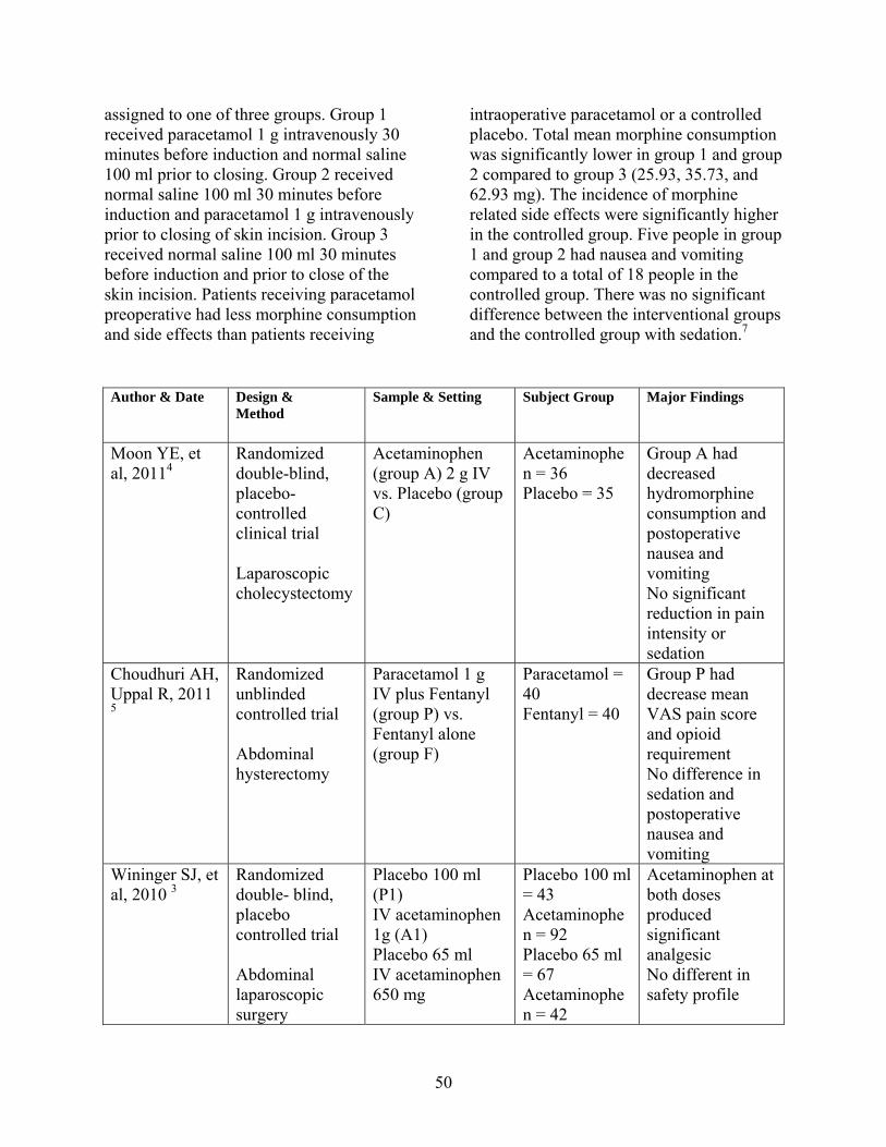
50
assigned to one of three groups. Group 1 received paracetamol 1 g intravenously 30 minutes before induction and normal saline 100 ml prior to closing. Group 2 received normal saline 100 ml 30 minutes before induction and paracetamol 1 g intravenously prior to closing of skin incision. Group 3 received normal saline 100 ml 30 minutes before induction and prior to close of the skin incision. Patients receiving paracetamol preoperative had less morphine consumption and side effects than patients receiving
intraoperative paracetamol or a controlled placebo. Total mean morphine consumption was significantly lower in group 1 and group 2 compared to group 3 (25.93, 35.73, and 62.93 mg). The incidence of morphine related side effects were significantly higher in the controlled group. Five people in group 1 and group 2 had nausea and vomiting compared to a total of 18 people in the controlled group. There was no significant difference between the interventional groups and the controlled group with sedation.7
Author & Date Design & Method
Sample & Setting Subject Group Major Findings
Moon YE, et al, 20114
Randomized double-blind, placebo-controlled clinical trial Laparoscopic cholecystectomy
Acetaminophen (group A) 2 g IV vs. Placebo (group C)
Acetaminophen = 36 Placebo = 35
Group A had decreased hydromorphine consumption and postoperative nausea and vomiting No significant reduction in pain intensity or sedation
Choudhuri AH, Uppal R, 2011 5
Randomized unblinded controlled trial Abdominal hysterectomy
Paracetamol 1 g IV plus Fentanyl (group P) vs. Fentanyl alone (group F)
Paracetamol = 40 Fentanyl = 40
Group P had decrease mean VAS pain score and opioid requirement No difference in sedation and postoperative nausea and vomiting
Wininger SJ, et al, 2010 3
Randomized double- blind, placebo controlled trial Abdominal laparoscopic surgery
Placebo 100 ml (P1) IV acetaminophen 1g (A1) Placebo 65 ml IV acetaminophen 650 mg
Placebo 100 ml = 43 Acetaminophen = 92 Placebo 65 ml = 67 Acetaminophen = 42
Acetaminophen at both doses produced significant analgesic No different in safety profile

51
Conclusions In the United States, the standard dosing of acetaminophen IV is 1 g over 15 minutes every 4 to 6 hours with a maximum daily dose of 4 g. Compared to rectal administration that has a peak plasma level of 3.5-4.5 hours and oral administration at 45-60 minutes, the peak plasma concentration of IV administration occurs immediately after the 15 minute infusion is complete. Clinical analgesic effects of IV administration occur within 5 minutes.2 The administration of IV acetaminophen provides more rapid onset and peak efficacy because of rapid cerebrospinal absorption.1 In a review of current literature, the administration of IV acetaminophen in the preoperative period has been shown in the majority of studies to reduce the opioid consumption and decrease postoperative pain levels. The mechanism of acetaminophen for pain relief is still not completely understood. Acetaminophen is believed to be a central nervous system acting cycloxygenase inhibitor sparing
peripheral prostaglandins and involves the serotoninergic inhibitory descending pathway.1,4,6 Literature has shown that acetaminophen is more effective in the treatment of somatic pain compared to visceral pain. The presence of inflammation stimulates afferent fibers causing visceral pain. Acetaminophen provides weak anti-inflammatory effects limiting its effects on visceral pain. An abdominal hysterectomy is primarily visceral pain whereas spinal and orthopedic surgeries are primarily somatic pain. Additionally somatic pain is accompanied by an automatic reflex, such as nausea and vomiting. 6 A reduction in postoperative nausea and vomiting with acetaminophen administration may be contributed with a decrease in opioid consumption and reducing secondary effects of somatic pain. The safety profile of IV acetaminophen is similar to oral administration, but provides an advantage with patients with liver damage. Unlike oral administration, IV acetaminophen is distributed systemically instead of fully passing through the liver,
Arici S, et al, 2009 7
Randomized controlled trial
Paracetamol 1 g before incision Paracetamol 1 g before closure Placebo
Paracetamol 1 g before incision = 30 Paracetamol 1gm before closure = 30 Placebo = 30
Group 1 had significantly less morphine consumption than group 2 and group 3, Group 1 and 2 had significantly less morphine related side effects than group 3
Hong JY, et al, 20106
Randomized double-blind, placebo-controlled clinical trial
Paracetamol 1 g Placebo
Paracetamol = 63 Placebo = 61
Paracetamol was associated with a decrease in rescue analgesia and postoperative nausea and vomiting

52
thereby exposing the liver to less acetaminophen. Bleeding, renal toxicity, and gastrointestinal bleeding have not been associated with intravenous acetaminophen use, providing a safer drug profile over NSAIDs.2 The use of IV acetaminophen as a preemptive analgesic has been shown to be a safe, efficient, and effective part of a multimode treatment plan for postoperative pain. The majority of trials have repeatedly shown that acetaminophen administered preoperatively can decrease the need for intraoperative and postoperative opioid administration. Acetaminophen may be most effective in patients with a history of opioid-related side effects. References 1. Jahr JS, Lee VK. Intravenous
acetaminophen. Anesthesiology Clinics. 2010;28:619-645.
2. Smith HS. Perioperative intravenous Acetaminophen. Pain Medicine. 2011;12:961-981.
3. Wininger SJ, Miller H, Minkowitz HS, Royal MA, Ang RY, Breitmeyer JB, Singla NK. A randomized, double blind, placebo-cobtrolled, multicente, repeat-dose study of two intravenous acetaminophen dosing regiments for the
treatment of pain after abdominal laparoscopic surgery. Clinical Therapeutics. November 2010;32:2348-2369
4. Moon YE, Lee YK, Lee j, Moon DE. The effects of preoperative intravenous acetaminophen in patients undergoing abdominal hysterectomy. Gynecology and Obstetrics. 2011;284:1455-1460.
5. Choudhuri AH, Uppal R. A comparison between intravenous paracetamol plus fentanyl and intravenous fentanyl alone for postoperative analgesia during laparoscopic cholecystectomy. Anesthesia: Essay and Research. 2011;5. Doi:10.4103/0259-1162.9477.
6. Hong JY, Kim WO, Chung WY, Yum JS, Kil HK. Paracetamol reduces postoperative pain and rescue analgesic demand after robot-assissted endoscopic thyroidectomy by the transaxillary approach. World Journal of Surgery. January 2010;34:521-526.
7. Arici S, Gurbet A, Turker G, Yavascaoglu B, Sahin S. Preemptive analgesia effects of intravenous paracetamol in total abdominal hysterectomy. Agri. April 2009; 21(2):54-61
Mentor: Laura S. Bonanno, CRNA, DNP
Cricoid Pressure Effectiveness: A Review of Evidence
Adam Peeples, BSN Samford University
Keywords: cricoid pressure, rapid sequence induction, Sellick maneuver, pulmonary aspiration, gastric regurgitation Cricoid pressure (CP), which was first described by Dr. Sellick in 1961, is a procedure that is routinely performed in the prophylaxis for pulmonary aspiration as part
of a rapid sequence induction (RSI). Sellick described his maneuver as “occlusion of the upper esophagus by backward pressure on the cricoid ring against the bodies of cervical vertebrae to prevent gastric contents from reaching the pharynx.”1 After trying his maneuver on a cadaver, Sellick then tried the same maneuver during anesthesia

53
induction of 26 patients at high risk of aspiration. None of the patients experienced regurgitation or vomiting when the pressure was applied, and 3 patients had immediate reflux into the pharynx upon the release of the pressure after tracheal intubation.2
Sellick interpreted his study as suggestive evidence for the effectiveness of CP in preventing regurgitation.3 Because pulmonary aspiration is the leading cause of airway related anesthetic death4, this technique has been adopted by clinicians, emergency physicians, paramedics, and anesthetists worldwide. Although CP is considered a standard of care in patients at increased risk of pulmonary aspiration, much controversy exists related to its safety and effectiveness.4
Understanding the role of CP in anesthesia involves the understanding of the different aspects such as anatomical relationships, the impact of CP on airway management, physiological relation to regurgitation, and the modes of application, which include the amount of force required for effective CP. Anatomically the cricoid cartilage lies between the thyroid cartilage and the first tracheal ring. The cricoid cartilage is shaped like a signet ring and lies at the vertebral level of C5 and C6 in adults. The esophagus lies posterior to the cricoid cartilage. The basis of CP relies on the direct compression of the esophageal lumen by the cricoid cartilage, which lies directly anterior to the esophagus.2
Documented cases of fatal regurgitation and aspiration have been reported despite the use of CP. This has resulted in continued questions regarding whether CP should be abandoned altogether.2 This report will explore the published evidence related to the safety and effectiveness of CP.
Methodology Evidence-based Practice Model The Iowa Model of Evidence-based Practice to Promote Quality Care was used to guide the evidence search. The PICO format was used to formulate the clinical question being studied. Relevant research and related literature was assembled, critiqued, and synthesized for use in clinical practice. The following parameters were considered: P (patient population) = patients requiring rapid sequence induction I (current intervention) = cricoid pressure C (comparison) = patients not receiving cricoid pressure O (outcome) = prevention of pulmonary aspiration of gastric contents Purpose The purpose for the review of the evidence was to determine the safety and efficacy of the use of CP during RSI. The question that guided the review of evidence was: In patients requiring rapid sequence induction, is the use of cricoid pressure safe and effective in the prevention of pulmonary aspiration of gastric contents? Search Terms Cricoid pressure, Sellick maneuver, rapid sequence induction, pulmonary aspiration, endotracheal intubation Search Methods This report details a comprehensive literature review that was carried out using a cross-search of online databases including PubMed, EBSCOhost, CINAHL, Cochrane Library, MEDLINE, and reference lists of relevant articles retrieved by the electronic search.

54
Levels of Evidence The primary evidence cited in this review was obtained from a closed claims analysis, prospective observational studies, case reports, and meta-analyses. Literature Review Even though CP is widely accepted as the standard of care for RSI, clinicians have recently questioned its effectiveness due to several limitations including: (a) making tracheal intubation and mask ventilation difficult or impossible due to airway alteration, (b) causing esophageal rupture and other subsequent injuries, and (c) the risk of pulmonary aspiration of gastric contents despite the use of CP, as evidenced by many documented case reports.5 Airway Alteration In Sellick’s original article, he described the use of “firm” pressure, but did not quantify the actual force needed to occlude the esophagus.1 According to Asai and Vanner6, most problems with CP occur when too much pressure is applied. Asai and Vanner state that CP of more than 20 newtons (N) can cause retching in an awake patient, which can potentially lead to pulmonary aspiration or esophageal rupture. Additionally, studies have suggested that CP of more than 40 N after loss of consciousness can cause airway obstruction and cause difficulty with tracheal intubation. Much controversy currently exists as to the exact amount of CP needed to safely and effectively occlude the esophagus without causing adverse effects.6 Sellick’s initial reports in 1961 had several limitations, including small sample size, unblinded, nonrandomized, and uncontrolled, with the technique’s proponent as the single author.1 In a descriptive magnetic resonance imaging (MRI) study, Boet et al.7 observed the
anatomical effect of CP on the occlusion of the esophageal lumen in 20 conscious volunteers using MRI. Incomplete esophageal occlusion was seen in 62.5% of individuals when proper CP was applied. From the results of this study, authors concluded that effective application of CP by an experienced operator frequently resulted in lateral deviation of the esophagus and incomplete occlusion of the esophageal lumen. This study suggests that utilizing CP for esophageal occlusion is possibly ineffective, requiring further evaluation utilizing functional studies.7 However, this study has many limitations including small sample size and non-anesthetized volunteers. Therefore, it is questionable if the results of this study can be applied to the general population. Hans-Jaochim3 describes a study in which CP applied at a force of 20-30 N caused difficult ventilation with a laryngeal mask airway in 50-73% of patients. Without the use of CP, all patients studied could be ventilated without difficulty, indicating that CP may cause substantial airway obstruction.3 Ellis, Harris, and Zideman8 state that even well controlled, appropriately applied CP can adversely affect ventilation and cause airway obstruction. The authors in this study examined 10 published articles in which CP increased peak inspiratory pressure, decreased tidal volumes, and prevented ventilation.8
Injury CP can cause harmful complications with the most severe being esophageal rupture. Ellis, Harris, and Zideman8 describe a case report of a patient vomiting during the application of CP, subsequently causing a ruptured esophagus.8 In addition, the authors also reported 3 cases in which CP caused

55
fracture of the cricoid cartilage, including one leading to fatal airway obstruction. There have also been case reports of subconjunctival hemorrhage from coughing and goiter hemorrhage as a result of CP. Longanthan and Liu9 describe the potential harm of CP in patients with cervical spine or laryngeal trauma. During the application of CP, significant movement of the cervical spine can occur, possibly leading to spinal cord injury. However, there is no conclusive study that demonstrates this.9
Pulmonary Aspiration Aspiration of gastric contents continues to be a complication during RSI. CP is a standard of care in cases where aspiration of gastric contents is considered a potential risk. However, a number of recent studies have raised questions regarding the efficacy of CP in preventing aspiration.7
Pulmonary aspiration has been documented in several cases, despite the use of CP. In a study surveying 139 anesthesiologists, 10% of the anesthesiologists surveyed reported regurgitation despite the use of CP. Failure of CP to prevent regurgitation was caused by incorrect application of CP, anatomical changes caused by CP, and individual anatomical differences.3 This was further demonstrated in a study by Smith et al.10 who challenged the assumption that the cricoid, esophagus, and vertebral body are all aligned in the same plane. The results of
the study revealed that the esophagus was lateral to the cricoid in more than 50% of the population studied. When CP was applied, MRI displayed an unobstructed section of the esophagus between the airway and vertebral body, which could possibly provide a passage for regurgitation of gastric contents.10 A prospective observational study of 4,891 cesarean sections requiring a general anesthetic, found that of the 61% of the cases in which CP was applied, 24 patients vomited or regurgitated on induction of anesthesia. Nine of these parturients died due to complications of regurgitation.11 In addition, Bailie12 presented a closed claims analysis reviewing 129 claims in which aspiration of gastric contents occurred. CP was used in 45% of 74 claims in which aspiration of gastric contents occurred during the induction of anesthesia. Fifteen claims reported permanent disabling injuries.12
The studies described above provide no evidence that CP prevents gastric regurgitation or aspiration. Although similar in their conclusions, they are not without limitations. Limitations include no randomization, inconsistent or unidentified CP practitioners, CP performed on awake vs. anesthetized subjects and unknown training of the CP practitioners.
Study Patient Group Study Type Outcomes Key Results Weaknesses Sellick 19611
26 high risk anesthesia cases in which CP was applied
Observational study
Incidence of reflux of gastric contents when CP released post-intubation of trachea
In 3 out of 26 cases, release of CP was followed by immediate reflux of gastric contents into pharynx
Observational study, small sample size. Conducted in 1961, using anesthetic techniques available at that time

56
Ellis 20078
Clinical and cadaver studies
Meta-analysis Evidence of aspiration Airway patency Gastric insufflation Airway/soft tissue injury
No high quality studies proving that CP is beneficial in preventing aspiration Some studies report aspiration despite CP 10 published articles suggesting CP reduced tidal volumes, increased peak inspiratory pressures, and prevented ventilation during mask ventilation Studies providing evidence that CP reduces gastric insufflation during mask ventilation Case reports of cricoid injury/soft tissue injury when CP was applied
No search strategy given No inclusion criteria No assessment of quality of studies Ventilation strategies used were different from those recommended today No assessment of quality of studies
Smith 201010
22 healthy volunteers
Observational study where MRI scans of necks were taken with and without the application of CP
Esophageal displacement laterally relative to the cricoid without CP Esophageal displacement laterally relative to the cricoid with CP
52.6% of cases 90.5% of cases
Healthy, awake volunteers were used, not relaxed patients undergoing RSI Findings may be influenced by muscle tone and the swallowing reflex

57
Table 1. Recent literature regarding the use of CP during RSI. Conclusions Currently, the application of CP is a strongly debated topic. Whereas some authors strongly endorse the technique and believe in its effectiveness, others believe that CP should be abandoned because it adds to patients’ risks with no evidence of any gained benefit.2 Although the use of CP makes intuitive sense, its scientific basis is weak. Although there is some limited evidence that CP may prevent regurgitation of gastric contents, the maneuver is associated with considerable risks.3
A lack of evidence from randomized controlled trials (RCT) is a major limitation to this report. No published RCTs studying the effectiveness of CP in preventing aspiration were found on this topic. Many ethical considerations make this type of study difficult to conduct. Because CP is still considered a standard of care, performing a RSI without CP could create an avenue for many lawsuits and debates. If CP continues to be the standard of care in RSI, proper training should be required of all clinicians who utilize this maneuver.
By today’s standards, CP “would not be considered an evidence-based practice.”3
References 1. Sellick BA. Cricoid pressure to control
regurgitation of stomach contents during induction of anaesthesia. Lancet. 1961;2:404-406.
2. El-Orbany M, Connolly LA. Rapid sequence induction and intubation: current controversy. Anesth Analg. 2010;110:1318-1325.
3. Hans-Jaochim P. Cricoid pressure: an alternative view. Semin Anesth, Perio M. 2005;24:120-126.
4. Cook TM, MacDougall-Davis SM. Complications and failure of airway management. BJA. 2012;109(1):68-85.
5. Ovassapian A, Salem RM. Sellick’s maneuver: to do or not to do. Anesth Analg. 2009;109(5):1360-1362.
6. Asai T, Vanner RG. Safe use of cricoid pressure. Anaesthesia. 1999;54(1):1-3.
7. Boet S, Duttchen K, Chan J, et al. Cricoid pressure provides incomplete esophageal occlusion associated with lateral deviation: a magnetic resonance imaging study. J Emerg Med. 2012.;42:606-611.
8. Ellis D, Harris T, Zideman D. Cricoid pressure in emergency department rapid sequence tracheal intubations: a risk-benefit analysis. Ann Emerg Med. 2007;50(6):653-
Unopposed esophagus without CP Unopposed esophagus with CP Lateral laryngeal displacement Airway compression
47.4% of cases 71.4% of cases 71.4% of cases 81% of cases
Small sample size
58
665. 9. Loganathan N, Liu E. Cricoid pressure:
ritual or effective measure? Singapore Med J. 2012;53(9):620-622.
10. Smith KJ, Dobranowski J, Yip G, Dauphin A, Choi P. Cricoid pressure displaces the esophagus: an observational study using magnetic resonance imaging. Anesthesiology. 2003;9(1):60-64.
11. Fenton P, Reynolds F. Life-saving or ineffective? An observational study of the
use of cricoid pressure and maternal outcome in an African setting. Int J Obstet Anesth. 2008;18:106-110.
12. Bailie R, Stephens L, Warner M, Warner M, Domino K. Liability and risk factors associated with aspiration: closed claims analysis.
Mentor: David N. Fort, CRNA, DNP
Editorial We had a very productive meeting of the ISJNA at the last Assembly of School Faculty in San Diego with two dozen CRNAs in attendance – what a great turnout! A number of items were discussed, including ways to increase dissemination of the journal, recruiting new volunteers, a review and update of the author guidelines, and consideration of new submission items to name a few. The ISJNA is in its 13th year of circulation and continues to grow, and the Fall 2013 issue was the largest since 2009. We can always use more reviewers – if you are interested please contact me! Best,
Vicki C. Coopmans, CRNA, PhD Editor
“The International Student Journal of Nurse Anesthesia is produced exclusively for publishing the work of nurse anesthesia students. It is intended to be basic and introductory in its content. Its goal is to introduce the student to the world of writing for publication; to improve the practice of nurse anesthesia and the safety of the patients entrusted to our care.”
To access prior issues of the ISJNA visit the following link:
www.aana.com/studentjournal

59
INTERNATIONAL STUDENT JOURNAL OF NURSE ANESTHESIA GUIDE FOR AUTHORS
MISSION STATEMENT The International Student Journal of Nurse Anesthesia is produced exclusively for publishing the work of nurse anesthesia students. It is intended to be basic and introductory in its content. Its goal is to introduce the student to the world of writing for publication; to improve the practice of nurse anesthesia and the safety of the patients entrusted to our care. ITEMS ACCEPTED FOR PUBLICATION Case reports, research abstracts, evidence-based practice (EBP) analysis reports, and letters to the editor may be submitted. These items must be authored by a student under the guidance of an anesthesia practitioner mentor (CRNA or physician). The mentor must submit the item for the student and serve as the contact person during the review process. Items submitted to this journal should not be under consideration with another journal. We encourage authors and mentors to critically evaluate the topic and the quality of the writing. If the topic and the written presentation are beyond the introductory publication level we strongly suggest that the article be submitted to a more prestigious publication such as the AANA Journal. ITEM PREPARATION & SUBMISSION Student authors prepare case reports, abstracts, EBP analysis reports, and letters to the editor with the guidance of a mentor. Only students may be authors. Case and EBP analysis reports must be single-authored. Abstracts may have multiple authors. Mentors should take an active role in reviewing the item to ensure appropriate content, writing style, and format prior to submission. The original intent of this journal was to publish items while the author is still a student. In order to consistently meet this goal, all submissions must be received by the editor at least 3 months prior to the author’s date of graduation. PEER REVIEW Items submitted for publication are initially reviewed by the editor. Items may be rejected, or returned to the mentor with instructions for the author to revise and resubmit prior to initiation of the formal review process. All accepted submissions undergo a formal process of blind review by at least two ISJNA reviewers. After review, items may be accepted without revision, accepted with revision, or rejected with comments. General guidelines 1. Items for publication must adhere to the American Medical Association Manual of Style (AMA, the same guide
utilized by the AANA Journal and such prominent textbooks as Nurse Anesthesia by Nagelhout and Plaus). The review process will not be initiated on reports submitted with incorrect formatting and will be returned to the mentor for revision. Please note the following: a. Use of abbreviations is detailed in Section 14. Spell out acronyms/initialisms when first used. If you are
using the phrase once, do not list the acronym/initialism at all. b. Instructions regarding units of measure can be found in Section 18. In most cases The International System
of Units (SI) is used. Abbreviations for units of measure do not need to be spelled out with first use. Some examples: height/length should be reported in cm, weight in kg, temperature in oC, pressure in mm Hg or cm H20.
c. In general, first use of pulmonary/respiratory abbreviations should be expanded, with the following exceptions: O2, CO2, PCO2, PaCO2, PO2, PaO2. Please use SpO2 for oxygen saturation as measured by pulse oximetry.
d. Use the nonproprietary (generic) name of drugs - avoid proprietary (brand) names. Type generic names in lowercase. When discussing dosages state the name of the drug, then the dosage (midazolam 2 mg).
e. Use of descriptive terms for equipment and devices is preferred. If the use of a proprietary name is necessary (for clarity, or if more than one type is being discussed), give the name followed by the manufacturer and location in parenthesis:
“A GlideScope (Verathon Inc., Bothell, WA) was used to . . . .” Please note, TM and ® symbols are not used per the AMA manual.
f. Examples of referencing are included later in this guide.

60
2. Report appropriate infusion rates and gas flow rates: a. When reporting infusion rates report them as mcg/kg/min or mg/kg/min. In some cases it may be
appropriate to report dose or quantity/hr (i.e. insulin, hyperalimentation). If a mixture of drugs is being infused give the concentration of each drug and report the infusion rate in ml/min.
b. Keep the gas laws in mind when reporting flow rates. Report the liter flows of oxygen and nitrous oxide and the percent of the volatile agent added to the gas mixture. Statements such as “40% oxygen, 60% nitrous oxide and 3% sevoflurane” do not = 100% and are thus incorrect. For example, “General anesthesia was maintained with sevoflurane 3% inspired concentration in a mixture of oxygen 1 L/min and air 1 L/min”.
3. Only Microsoft Word file formats will be accepted with the following criteria: a. Font - 12 point, Times New Roman b. Single-spacing (except where indicated), paragraphs separated with a double space (do not indent) c. One-inch margins d. Place one space after the last punctuation of sentences. End the sentence with the period before placing the
superscript number for the reference. e. Do not use columns, bolds (except where indicated), or unconventional lettering styles or fonts. f. Do not use endnote/footnote formats.
4. Do not use Endnotes or similar referencing software. Please remove all hyperlinks within the text. 5. Avoid jargon.
a. ‘The patient was reversed’ - Did you physically turn the patient around and point him in the opposite direction? “Neuromuscular blockade was antagonized.”
b. The patient was put on oxygen. "Oxygen was administered by face mask." c. The patient was intubated and put on a ventilator. “The trachea was intubated and respiration was
controlled by a mechanical ventilator. d. The patient had been on Motrin for three days. “The patient had taken ibuprofen for three days.” e. Avoid the term “MAC” when referring to a sedation technique - the term sedation (light, moderate, heavy,
unconscious) sedation may be used. Since all anesthesia administration is monitored, the editors prefer to use specific pharmacology terminology rather than reimbursement terminology.
6. Use the words “anesthesia professionals” or “anesthesia practitioners” when discussing all persons who administer anesthesia (avoid the reimbursement term “anesthesia practitioners”)
7. References a. Again, the AMA Manual of Style must be adhered to for reference formatting. b. All should be within the past 8 years, except for seminal works essential to the topic being presented. c. Primary sources are preferred. d. All items cited must be from peer-reviewed sources – use of internet sources must be carefully considered
in this regard. e. Numbering should be positioned at the one-inch margin – text should begin at 1.25”.
8. See each item for additional information. 9. Heading for each item (Case Report, Abstract, EBPA Report) must adhere to the following format: Title (bold, centered, 70 characters or less) [space] Author Name (centered, include academic credentials only) Name of Nurse Anesthesia Program (centered) [space] Anticipated date of graduation (italics, centered, will be removed prior to publication) E-mail address (italics, centered, will be removed prior to publication) [space, left-justify from this point forward] Keywords: (‘Keywords:’ in bold, followed by keywords (normal font) that can be used to identify the report in an internet search.) Case Reports The student author must have had a significant role in the conduct of the case. The total word count should be between 1200 – 1400 words. References do not count against the word count. Case reports with greater than 1400

61
words will be returned to the mentor for revision prior to initiation of the review process. The following template demonstrates the required format for case report submission. Heading (see #9 above in General Guidelines) [space] A brief introductory paragraph of less than 100 words to focus the reader’s attention. This may include historical background, demographics or epidemiology (with appropriate references) of the problem about to be discussed. It is written in the present tense. Although it is introductory, the heading word ‘Introduction’ is not used. Be certain to cite references in this section, especially statistics and demographics pertaining to your topic. [space] Case Report (bold, 400-500 words) [space] This portion discusses the case performed in 400 words or less, and is written in the past tense. Do not justify actions or behaviors in this section; simply report the events as they unfolded. Present the case in an orderly sequence. Some aspects need considerable elaboration and others only a cursory mention.
Patient description: height, weight, age, gender. History of present illness Statement of co-existing conditions/diseases Mention the current medications, generic names only. (Give dosage and schedule only if that information is
pertinent to the consequences of the case.) Significant laboratory values, x-rays or other diagnostic testing pertinent to the case. Give the units after the
values (eg. Mmol/L or mg/dL). Physical examination/Pre-anesthesia evaluation - significant findings only. Include the ASA Physical Status
and Mallampati Classification only if pertinent to the case. Anesthetic management (patient preparation, induction, maintenance, emergence, post-operative recovery).
Despite the detail presented here it is only to help the author organize the structure of the report. Under most circumstances if findings/actions are normal or not contributory to the case then they should not be described. Events significant to the focus of the report should be discussed in greater detail. The purpose of the case report is to set the stage (and ‘hook’ the reader) for the real point of your paper which is the discussion and teaching/learning derived from the case. [space] Discussion (bold, 600-800 words) [space] Describe the anesthesia implications of the focus of the case report citing current literature. Describe the rationale for your actions and risk/benefits of any options you may have had. This section is not merely a pathophysiology review that can be found in textbooks. Relate the anesthesia literature with the conduct of your case noting how and why your case was the same or different from what is known in the literature. Photographs are discouraged unless they are essential to the article. Photos with identifiable persons must have a signed consent by the person photographed forwarded to the editor via first class mail. Diag must have permission from original author. This is the most important part of the article. In terms of space and word count this should be longer than the case presentation. End the discussion with a summary lesson you learned from the case, perhaps what you would do differently if you had it to do over again. [space] References (bold) [space] A minimum of 5 references is recommended, with a maximum of 8 allowed. No more than 2 textbooks may be included in the reference list, and all references should be no older than 8 years, except for seminal works essential to the topic. This is also an exercise in evaluating and using current literature. [space] Mentor: (bold, followed by mentor name and credentials in normal text) E-mail address (italics, will be removed prior to publication) Research Abstracts Research abstracts are limited to 500 words. References are not desired but may be included if considered essential. Note that this abstract is different from a research proposal. This abstract reports the outcome of your study. Use the same format described for the case report with the exception of the section headings:
62
Heading (see #9 above in General Guidelines) [space] Introduction (bold) [space] A brief introductory paragraph including purpose and hypotheses. [space] Methods (bold) [space] Include research design and statistical analyses used [space] Results (bold) [space] Present results – do not justify or discuss here. [space] Discussion (bold) [space] Discuss results [space] References (bold) [space] Not required, but a maximum of 5 references is allowed. [space] Mentor: (bold, followed by mentor name and credentials in normal text) E-mail address (italics, will be removed prior to publication) EBP Analysis Reports Evidence-based practice analysis reports are limited to 3000 words. Please do not include an abstract. The report should provide a critical evaluation of a practice pattern in the form of a clinical question about a specific intervention and population. The manuscript should:
1. Articulate the practice issue and generate a concise question for evidence-based analysis. A focused foreground question following either the PICO or SPICE format should be used.
2. Describe the methods of inquiry used in compiling the data. 3. Critically analyze the quality of research reviewed and applicability to different practice settings. 4. Draw logical conclusions regarding appropriate translation of research into practice.
The same general format guidelines apply with the exception of the section headings as below. Please note that text books and non-peer reviewed internet sources should be avoided, and sources of reference should be less than 8 years old unless they are seminal works specifically related to your topic of inquiry: Heading (see #9 above in General Guidelines) [space] Introduction (bold) [space] Briefly introduce the reader to the practice issue or controversy, describe the scope or significance or problem, and identify the purpose of your analysis. Describe the theoretical, conceptual, or scientific framework that supports your inquiry. [space] Methodology (bold) [space] Include the format used for formulating the specific question you seek to answer, search terms and methods used, and levels of evidence. [space] Literature Analysis (bold) [space]

63
Review and critique the pertinent and current literature, determining scientific credibility and limitations of studies reviewed. Your synthesis table would be included in this section. Your review and discussion of the literature should logically lead to support a practice recommendation. Subheadings may be used if desired. [space] Conclusions (bold) [space] Summarize the salient points that support the practice recommendation and make research-supported recommendations that should improve the practice issue, while also acknowledging any limitations or weaknesses [space] References [bold] [space] A minimum of 8 references is recommended, with a maximum of 12 allowed. Letters to the Editor Students may write letters to the editor topics of interest to other students. Topics may include comments on previously published articles in this journal. Personally offensive, degrading or insulting letters will not be accepted. Suggested alternative approaches to anesthesia management and constructive criticisms are welcome. The length of the letters should not exceed 100 words and must identify the student author and anesthesia program. AMA MANUAL OF STYLE The following is brief introduction to the AMA Manual of Style reference format along with some links to basic, helpful guides on the internet. The website for the text is http://www.amamanualofstyle.com/oso/public/index.html. It is likely your institution’s library has a copy on reserve. http://www.docstyles.com/amastat.htm#Top http://healthlinks.washington.edu/hsl/styleguides/ama.html Journal names should be in italics and abbreviated according to the listing in the PubMed Journals Database. The first URL below provides a tutorial on looking up correct abbreviations for journal titles; the second is a link to the PubMed where you can perform a search. http://www.nlm.nih.gov/bsd/viewlet/search/journal/journal.html http://www.ncbi.nlm.nih.gov/pubmed The International Student Journal of Nurse Anesthesia (ISJNA) is not listed in the PubMed Database. For the purpose of citing the ISJNA in this Journal use “Int Student J Nurse Anesth” as the abbreviation. The titles of text books are also printed in italics. Please pay close attention to ensure correct punctuation. Journals Note there is a comma after the first initials until the last author, which has a period. If there are six or less authors cite all six. If there are more than six authors cite only the first three followed by “et al.” Only the first word of the title of the article is capitalized. The first letters of the major words of the journal title are capitalized. There is no space between the year, volume number, issue number, and page numbers. If there is no volume or issue number, use the month. If there is an issue number but no volume number use only the issue number (in parentheses). The pages are inclusive - do not omit digits. Some journals (and books) may be available both as hard copies and online. When referencing a journal that has been accessed online, the DOI (digital object identifier) or PMID (PubMed identification number) should be included (see example below). Journal, 6 or fewer authors: Hamdan A, Sibai A, Rameh C, Kanazeh G. Short-term effects of endotracheal intubation on voice. J Voice. 2007;21(6):762-768. Journal, more than 6 authors: Chen C, Nguyen MD, Bar-Meir E, et al. Effects of vasopressor administration on the outcomes of microsurgical breast reconstruction. Ann Plast Surg. 2010;65(1):28-31. PMID: 20548236.

64
Texts There is a difference in citing a text with one or more authors from a text with one or more editors. Texts that are edited give credit to the authors of the chapters. They must be annotated and the inclusive pages of the chapter are noted. Texts that are authored do not have different chapter authors, the chapter is not cited by heading but the inclusive pages where the information was found are cited, unless the entire book is cited. Text: Stoelting R, Dierdorf S. Anesthesia and Co-Existing Disease. 3rd ed. Philadelphia: Churchill Livingstone; 1993:351-354. Chapter from a text: Burkard J, Olson RL, Vacchiano CA. Regional anesthesia. In Nagelhout JJ, Plaus KL, eds. Nurse Anesthesia. 4th ed. St. Louis:Elsevier; 2010:977-1030 Each chapter was written by a different author. Note the chapter’s author gets the prominent location. The chapter title is cited; “editor” is abbreviated in a lowercase. The word “edition” is also abbreviated and in lower case. The inclusive pages of the chapter are cited. Electronic references Only established, peer-reviewed sources may be referenced. Please do not reference brochures or informational websites where a peer-review process cannot be confirmed. Authors are cautioned to not copy and paste from these without full credit and quotation marks where appropriate. Electronic references are cited using the following format: Author (or if no author, the name of the organization responsible for the site). Title. Name of journal or website. Year;vol(issue no.):inclusive pages. doi: or URL. Published [date]. Updated [date]. Accessed [date]. For online journals, the accessed date may be the only date available, and in some cases no page numbers. Examples: Kamangar N, McDonnell MS. Pulmonary embolism. eMedicine. http://www.emedicine.com/med/topic1958.htm. Updated August 25, 2009. Accessed September 9, 2009. Gupta A, Aggarwal N, Sharma D. Ultrasound guided ilioinguinal block. The Internet Journal of Anesthesiology. 2011;29(1). http://www.ispub.com/journal/the_internet_journal_of_anesthesiology/volume_29_number_1/article/ultrasound-guided-ilioinguinal-block.html. Accessed August 1, 2011.
ACADEMIC INTEGRITY Issues of academic integrity are the primary responsibility of the author and mentor. Accurate and appropriate acknowledgement of sources is expected. Any violation will be cause for rejection of the article. “Plagiarism is defined as the act of passing off as one's own the ideas, writings, or statements of another. Any act of plagiarism is a serious breach of academic standards, and is considered an offense against the University subject to disciplinary action. Any quotation from another source, whether written, spoken, or electronic, must be bound by quotation marks and properly cited. Any paraphrase (a recapitulation of another source's statement or idea in one's own words) or summary (a more concise restatement of another's ideas) must be properly cited.” http://grad.georgetown.edu/pages/reg_7.cfm HOW TO SUBMIT AN ITEM Manuscripts must be submitted by the mentor of the student author via e-mail to [email protected] as an attachment. The subject line of the e-mail should be “Submission to Student Journal”. The item should be saved in the following format – two-three word descriptor of the article_author’s last name_school abbreviation_mentor’s last name_date (e.g. PedsPain_Smyth_GU_Pearson_5.19.09)

65
REVIEW AND PUBLICATION If the editor does not acknowledge receipt of the item within one week, assume that it was not received and please inquire. Upon receipt, the Editor will review the submission for compliance with the Guide to Authors. If proper format has not been following the item will be returned to the mentor for correction. This is very important as all reviewers serve on a volunteer basis. Their time should be spent ensuring appropriate content, not making format corrections. It is the mentor’s responsibility to ensure formatting guidelines have been followed prior to submission. Once the item has been accepted for review the Editor will send a blinded copy to a Section Editor, who will then coordinate a blinded review by two reviewers who are not affiliated with the originating program. The reviewers recommend publication to the Section Editor or make recommendations for changes to be addressed by the author. The Section Editor will return the item to the Editor, who will return it to the mentor for appropriate action (revision, approval to print). If the article is returned to the author for repair it is usually to answer a specific question related to the case that was not clear in the narrative or it asks the author to provide a reference for a statement. Every effort is made to place the returned article in the earliest next issue. The goal is for all articles submitted by students to be published while the author is still a student. Therefore, deadlines must be met and the entire process must be efficient. If an item is not ready for publication within 3 months after the student author has graduated it will no longer be eligible for publication. For this reason it is recommended that case reports be submitted at least 4-6 months prior to the student author’s anticipated graduation date. Mentors of the papers may be asked to serve as reviewers of case reports by student authors from other prog and will be listed as contributing editors for the issue in which the item is published. PHOTOS Photos of students for the front cover of the Journal are welcome. Include a legend describing the activity and who is in the photo and identify the photographer. Only digital photos of high quality will be accepted via email to [email protected]. There must be a follow up hard copy signed by all present in the photo, as well as the photographer/ owner of the original photo, giving consent to publish the photo. Mail that consent to:
Vicki C. Coopmans, CRNA, PhD Goldfarb School of Nursing at Barnes-Jewish College 4483 Duncan Ave., Mailstop 90-36-697 St. Louis, MO 63110

66
SUBMISSION CHECK LIST
___ AMA Manual of Style and other format instructions are adhered to. ___ Total word count not exceeded (1400 for case report, 500 for abstract, 3000 for EBPA). ___ The item is one continuous Word document without artificially created page breaks. ___ Verbatim phrases and sentences are quoted and referenced. ___ All matters that are not common knowledge to the author are referenced. ___ Generic names for drugs and products are used throughout and spelled correctly in lower-case. ___ Units are designated for all dosages, physical findings, and laboratory results. ___ Endnotes, footnotes not used. ___ Jargon is absent. Heading ___ Concise title less than 70 characters long ___ Author name, credentials, nurse anesthesia program, graduation date and email are included. ___ Five Keywords are provided Case Report ___ Introduction is less than 100 words. ___ Case Report section states only those facts vital to the account (no opinions or rationale) ___ Case report section is 400-500 words and not longer than the discussion. ___ Discussion section is 600-800 words. ___ Discussion of the case management is based on a review of current literature ___ Discussion concludes with lessons learned and how the case might be better managed in the future. Abstract ___ The 500 word count maximum is not exceeded. ___ Abstract reports the outcome of your study. ___ Includes Introduction, Methods, Results, and Conclusion sections. EBPA Report ___ The 3000 word count maximum is not exceeded. ___ A critical evaluation of a practice pattern in the form of a precise clinical question about a specific intervention and
population is presented. ___ A focused foreground question following either the PICO or SPICE format is used. ___ Includes Introduction, Methodology, Literature Analysis, and Conclusion sections. References ___ AMA Style for referencing is used correctly. ___ Reference numbers are sequenced beginning with one and superscripted. ___ References are from anesthesia and other current primary source literature. ___ All inclusive pages are cited, texts as well as journals. ___ Journal titles are abbreviated as they appear in the PubMed Journals Database. ___ Number of references adheres to specific item guidelines. ___ Internet sources are currently accessible, reputable, and peer reviewed. Transmission ___ The article is sent as a attachment to [email protected] ___ The file name is correctly formatted (e.g. PedsPain_Smyth_GU_Pearson_5.19.09) ___ It is submitted by the mentor with cc to the student author ___ The words "Submission to Student Journal" are in the subject heading.