The Influence of Various Physiological Responses on ...
130
Louisiana State University LSU Digital Commons LSU Historical Dissertations and eses Graduate School 1978 e Influence of Various Physiological Responses on Ratings of Perceived Exertion Before and Aſter Training. David Richard Carter Louisiana State University and Agricultural & Mechanical College Follow this and additional works at: hps://digitalcommons.lsu.edu/gradschool_disstheses is Dissertation is brought to you for free and open access by the Graduate School at LSU Digital Commons. It has been accepted for inclusion in LSU Historical Dissertations and eses by an authorized administrator of LSU Digital Commons. For more information, please contact [email protected]. Recommended Citation Carter, David Richard, "e Influence of Various Physiological Responses on Ratings of Perceived Exertion Before and Aſter Training." (1978). LSU Historical Dissertations and eses. 3273. hps://digitalcommons.lsu.edu/gradschool_disstheses/3273
Transcript of The Influence of Various Physiological Responses on ...

Louisiana State UniversityLSU Digital Commons
LSU Historical Dissertations and Theses Graduate School
1978
The Influence of Various Physiological Responseson Ratings of Perceived Exertion Before and AfterTraining.David Richard CarterLouisiana State University and Agricultural & Mechanical College
Follow this and additional works at: https://digitalcommons.lsu.edu/gradschool_disstheses
This Dissertation is brought to you for free and open access by the Graduate School at LSU Digital Commons. It has been accepted for inclusion inLSU Historical Dissertations and Theses by an authorized administrator of LSU Digital Commons. For more information, please [email protected].
Recommended CitationCarter, David Richard, "The Influence of Various Physiological Responses on Ratings of Perceived Exertion Before and After Training."(1978). LSU Historical Dissertations and Theses. 3273.https://digitalcommons.lsu.edu/gradschool_disstheses/3273

INFORMATION TO USERS
This was produced from a copy of a document sent to us for microfilming. While the most advanced technological means to photograph and reproduce this document have been used, the quality is heavily dependent upon the quality of the material submitted.
The following explanation of techniques is provided to help you understand markings or notations which may appear on this reproduction.
1 .T he sign or “ target” for pages apparently lacking from the document photographed is “Missing Page(s)” . If it was possible to obtain the missing page(s) or section, they are spliced into the film along with adjacent pages. This may have necessitated cutting through an image and duplicating adjacent pages to assure you of complete continuity.
2. When an image on the film is obliterated with a round black mark it is an indication that the film inspector noticed either blurred copy because of movement during exposure, or duplicate copy. Unless we meant to delete copyrighted materials that should not have been filmed, you will find a good image of the page in the adjacent frame.
3. When a map, drawing or chart, etc., is part of the material being photographed the photographer has followed a definite method in “sectioning” the material. It is customary to begin filming at the upper left hand corner of a large sheet and to continue from left to right in equal sections with small overlaps. If necessary, sectioning is continued again—beginning below the first row and continuing on until complete.
4. For any illustrations that cannot be reproduced satisfactorily by xerography, photographic prints can be purchased at additional cost and tipped into your xerographic copy. Requests can be made to our Dissertations Customer Services Department.
5. Some pages in any document may have indistinct print. In all cases we have filmed the best available copy.
UniversityMicrofilms
International3 0 0 N. Z EE B R O A D, ANN A R B O R , Ml 4 8 1 0 6 18 B E D F O R D ROW, L O ND O N WC1R 4 EJ , E N G L A N D
7911561C A R T E R v D A V I D R I C H A R D
T H E I N F L U E N C E OF V A R I O U S P H Y S I O L O G I C A L R E S P O N S E S ON R A T I N G S OF P E R C E I V E D E X E R T I O N B E F O R E A N D A F T E R T R A I N I N G .T H E L O U I S I A N A S T A T E U N I V E R S I T Y A N D A G R I C U L T U R A L A N D M E C H A N I C A L C O L . , P H . D . * 1 9 7 8
UniversityMicrofilms
International 300 n / i e h h o a u . a n n a r b o r . m i 3b i o g

PLEASE NOTE:
In a l l cases th is material has been filmed in the best possible way from the available copy. Problems encountered with th is document have been iden t i f ied here with a check mark .
1. Glossy photographs ^2. Colored i l l u s t r a t i o n s j/3. Photographs with dark background ________
4. I l lu s t r a t io n s are poor copy ________
5. Print shows through as there is tex t on both sides of page _________
6. In d is t in c t , broken or small p r in t on several pages _________ throughout
7. Tightly bound copy with p r in t lo s t in spine ____
8. Computer pr in tout pages with in d is t in c t p r in t ________
9. Page(s) ________ lacking when material received, and not availablefrom school or author ________
10. Page(s) seem to be missing in numbering only as tex tfollows ________
11. Poor carbon copy ________
12. Not original copy, several pages with blurred type ________
13. Appendix pages are poor copy _______
14. Original copy with l ig h t type ________
15. Curling and wrinkled pages ________
16. Other
UniversityMicrofilms
International3 0 0 N Z E E B RD. . A N N A R B O R . Ml a s 106 ' 3131 7 6 1 - 4 7 0 0

THE INFLUENCE OF VARIOUS PHYSIOLOGICAL RESPONSES ON RATINGS
OF PERCEIVED EXERTION BEFORE AND AFTER TRAINING
A Dissertation
Submitted to the Graduate Faculty of the Louisiana State University and
Agricultural and Mechanical College in partial fulfillment of the requirements for the degree of
Doctor of Philosophy
in
The Department of Health, Physical,
and Recreation Education
byDavid R. Carter
B.S., Lamar University, 1973 M.S., Lamar University, 1974
December, 1978

ACKNOWLEDGEMENTS
The author wishes to express his deep appreciation to Dr. Jack
K. Nelson for his professional guidance and unending assistance in the
preparation of this investigation. For the assistance provided by
Dr. Mike Stone in the collection of the data, the author expresses his
gratitude.
A special thank you is extended to Dr. Barton Farthing, Dr.
Robert Mathews, Dr. Ralph Steben, and Dr. Jerry Thomas for their
advice and patience in the preparation of this dissertation.
One must note the efforts of Dr. Pat Crawford and members of
the School of Veterinary Medicine and the Department of Animal Sciences
for their time and assistance in this investigation.
Finally, and most importantly, the author must acknowledge the
efforts of his wife, Lynn. Her role in the completion of this work is
inestimable.

TABLE OF CONTENTS
Page
ACKNOWLEDGEMENTS . .
LIST OF TABLES . . .
LIST OF FIGURES . . .
ABSTRACT ...........
CHAPTER
I . INTRODUCTION
Review of Literature ...........................Development and Validation of Perceived
Exertion ...................................Training Influences on Ratings of Perceived
Exertion ...................................Psychological Influences on Ratings of
Perceived Exertion ........................Physiological Influences on Ratings of
Perceived Exertion ........................
Purpose of the Study . . .
Research Hypotheses . . .
Operational Definitions
Delimitations of the Study
Limitations of the Study ,
Significance of the Study
II. METHODS ....................
Overview of the Study , .
Selection of Subjects . .
Instrumentation .........
11
v
vi
viii
1
5
5
6
7
812
12
13
15
15
16
18
18
18
19
iii

CHAPTER Page
Testing Procedure . . . . . .................... . . . 26
Training P r o c e d u r e s ........... 33
Statistical Analysis of the Data ................ 35
III. R E S U L T S ................................................... 37
Means and Standard D e v i a t i o n s ........................ 37
Correlations . . . . . ................................. 37
Analysis of Variance of Ratings of PerceivedExertion and Cardio-pulmonary Measures ............. 42
Analysis of Variance of Sodium, Potassium andLactic Acid ............. 51
Stepwise Regression . . . . . . . . .................. 51
Alternative Statistical Interpretation ................ 53
IV. DISCUSSION................................................. 55
The Relationship Between RPE, HR and MetabolicV a r i a b l e s ................. 55
Influence of Training on the Physiological Factors . . 57
Influences of Physiological Variables in PredictingR P E ................................................... 62
S u m m a r y .......................... 63
R E F E R E N C E S .......................................................... 65
A P P E N D I C E S .......................................................... 70
V I T A ....................................................................116
iv

LIST OF TABLES
Table
1.Page
Means and Standard Deviations for the RPE Preand Post T e s t s ........................................... 38
v

LIST OF FIGURES
Figure Page
1. Gunnar A. V. Borg's RPE S c a l e ............................. 20
2. Monark Bicycle Ergometer Model 850........................ 22
3. Quinton Exercise Cardio-tachometer model 609 andPre-amplifier Supply........................................ 23
4. KL Engineering model S-300 Spirometer .................... 24
5. Beckman LB-2 Medical Gas Analyzer and OM-11Oxygen Analyzer ............................................. 25
6 . Subject fitted with head gear, three way valve,Spirometer Head, Electrodes and cardio-tachometerpre-amplifier supply........................................ 27
7. Test subject undergoing RPE test p r o t o c o l ............... 29
8 . Subject pointing to the numerical rating ofperceived exertion........................................... 30
9. Blood sample being drawn from the anticubital vein. . . . 32
10. Container of ice containing numbered centrifuge tubes for lactic acid samples, with the 4cc vacutainers forsodium and potassium samples situated in front........... 34
11. Mean Heart rates for each time interval during thepre test and post test...................................... 39
12. Mean RPE by time plot for each time interval duringthe pre test and post test.................................. 40
13. Mean HR and RPE changes between the pre and post tests. . 43
14. Mean RQ for each time interval during the pre test andpost t e s t .................................................... 44
15. Mean OXP for each time interval during the pre testand post t e s t ............................................... 46
vi

Figure Page
16. Mean VO2 1/min. for each time interval during thepre test and post test...................................... 47
17. Mean VCO2 1/min. for each time interval during thepre test and post test..................................... . 48
18. Mean VO2 ml/kg/min. for each time interval during thepre and post t e s t .......................................... 49
19. Mean VCO2 ml/kg/min. for each time interval during thepre and post t e s t .......................................... 50
20. Mean Heart Rates plotted against mean RPE's .............. 56
vii

ABSTRACT
The influence of certain physiological responses on Ratings
of Perceived Exertion (RPE) at specific points in an exercise bout
were investigated in conjunction with the influence of bicycle train
ing on the RPE. Twelve male college subjects were randomly selected
from beginning weight training courses. Mean physical characterisics+ + for subjects were age, 20.43 (- 2.58) years; height, 100.02 (- 9.01)
cm; and weight, 74.59 (- 7.63) kg.
Subjects (N = 12) were administered a Physical Work Capacity
test (PWC^q q ) in order to establish a work load criterion (HR = 180
bpm) for the 6 min. RPE test protocol utilizing a 60 rpm cadence on
the bicycle ergometer. Heart rate (HR), oxygen consumption (VO^
1/min., VC^ ml/kg/min.), and carbon dioxide production (VCC^ 1/min.,
VCO^ ml/kg/min.) data were recorded at each 30 sec. interval in the
6 min. exercise bout. Respiratory quotient (RQ) and oxygen pulse
(OXP) were computed from the metabolic data. Two minutes after
completion of the 6 min. work bout, a 6 cc blood sample was drawn
from the anticubital vein for the determination of blood lactate (LA) ,
sodium (Na), and potassium (K+ ) concentration.
After completion of the pre test an individualized bicycle
training program was designed. Training consisted of pedalling on the
bicycle ergometer 10 min./day, 3 days/week for 5 weeks. Work loads
by weeks were based on percentages of the PWCL OA estimate (i.e. 80%loUPWCiso f°r week 1 to 120% PWC^g^ for week 5). Upon completion of the

training phase a post test evaluation was administered utilizing the
same protocol as established for the pre test.
Split plot analysis of variance, factorial analysis of vari
ance, and stepwise multiple regressions were utilized to determine
differences occuring as a result of training and the importance of
the physiological variables in predicting the RPE. Split plot analysis
revealed that HR, RPE, and RQ significantly decreased as a result of
training. Oxygen consumption (VC>2 1/min., VC>2 ml/kg/min.), VC02
1/min., VC02 ml/kg/min., and OXP were significantly increased as a
result of training.
Factorial analysis revealed significant decreases in LA and
Na concentrations. No significant changes in K+ concentration were
revealed.
Stepwise regression models (N = 4) for two, 3 min. time
periods within each 6 min. bout accounted for 45 to 68 percent of
the variance for predicting the RPE. Pre test regressions appeared
to be better predictors of the RPE than post test regressions. There
fore, training appeared to alter the predictive ability of the
physiological factors responsible for lowering the RPE for the post
test. Thus, RPE appears to be a multifaceted and multistructural
composite of physiological and perhaps psychological phenomena.
ix

CHAPTER I
INTRODUCTION
Man is regarded as a psycho-somatic unit in which all psycho
logical events have corresponding physiological responses (Borg, 1973).
Because of the psycho-somatic nature of human performance, research
has drawn upon a number of disciplines in order to evaluate responses
to various stimulus situations.
Borg and Noble (1974) advocated that success in physical per
formance depends on (1) physiological and morphological endowments
and (2 ) psychological resources including the information and decision
making processes. Information and decision making processes involved
in physical performance rely heavily on perceptual cues. Such percep
tual cues allow an individual to regulate work intensity so as to
satisfy the specific goals and requirements of the activity.
Gunnar A. V. Borg (1962, 1973) developed a 21 point graded
scale which made possible direct individual comparisons of the percep
tion of exertion. At every second number on the scale, a corresponding
verbal expression was placed, such as 3 = "very, very light" and 19 =
"very, very laborious." The scale was found to be functional and
correlations of .80 to .90 were found with heart rates of subjects
tested on a bicycle ergometer. The original scale was later changed
to a 15 point graded category scale with numeric values ranging from
6 to 20 in order to match the variations in heart rate from 60 to 200
1
bpm. The scale was based on the knowledge that heart rate increases
linearly in relation to work load on a bicycle ergometer (Borg,
1973). The new scale, which is called the Ratings of Perceived Exer
tion Scale (RPE-Scale) or "Borg Scale" has been used extensively
throughout the world (Borg & Noble, 1974).
In an effort to explain how exertion is perceived, researchers
have studied various physiological and psychological components. Many
of the researchers have concluded that the overall perception of
exertion represents a Gestalt or an integration of various sensations
and feelings (Borg, 1962; Borg & Noble, 1974; Ekblom & Goldborg,
1971; Henriksson, Knuttgen & Bonde-Peterson, 1972; Noble, Metz,
Pandolf, Bell, Cafarelli & Sime, 1973a; Noble, Metz, Pandolf &
Cafarelli, 1973b; Pandolf, 1972; Pandolf, Burse & Goldman, 1975;
Pandolf & Noble, 1973). Sensations from the muscles, skin, joints
and circulatory and respiratory feedback all appear to influence the
perception of exertion (Borg, 1962; Pandolf et al., 1973). Because
the various areas of the body appear to contribute differently to
the perception of exertion, Ekblom and Goldborg (1971) proposed a
two factor theory consisting of general fatigue (cardio-respiratory)
and local fatigue (muscle). The two factor theory attempted to
explain the differences in the RPEs when different modes of exercis
ing were employed. For example, while walking or running, the general
cardio-respiratory feedback appeared to dominate the perception of
exertion while during cycling the local muscular fatigue dominated.
In an effort to identify whether the general or the local feeling
3
of fatigue dominate the perception of exertion, many researchers
have isolated specific physiological components known to be
associated with exertion and have tried to assess their contribution
to the RPE. Physiological variables of heart rate (HR), respiratory
rate (RR), oxygen consumption (VO^), carbon dioxide production
(VCC^), tidal volume, blood flow, blood lactate (LA), sodium (Na),
and potassium (K*) have been explored. Researchers have hypothesized
that RPE was influenced differently depending on the intensity of
the exercise and the relative importance of the physiological variables
at that particular intensity. When the intensity is submaximal, the
RPE is influenced primarily from the general or cardiovascular senses,
but when maximal or near maximal intensity is utilized the local
feeling of exertion appear to dominate. Since workloads of 50%
maximum oxygen consumption or greater are normally utilized in RPE
testing, the local feelings of exertion would generally be more
dominant.
In a study which supports the two factor theory, Allen and
Pandolf (1976) found that local factors dominate the perception of
exertion during work. They reported that for moderate work (50% VO^
max) and heavy work (80% VO^ max) inspired oxygen concentration sig
nificantly affected the RPE, and that blood lactate was shown to be
the prime cue for RPE. Ekblom and Goldborg (1971), Henriksson et al.
(1972), Pandolf and Noble (1973), and Robertson, McCarthy, and
Gillespie (1976) also reported that local factors appear to dominate
the perception of exertion when compared to an overall feeling of
4
exertion. Even though local factors have been shown to dominate the
perception of exertion, it was not clear which physiological compon
ents contributed the greatest amount of exertion information for
the interpretation of the RPE. Cafarelli and Noble (1975) stated
that ventilation was not important for the selection of RPE at low
exercise intensities where all cues probably come from working muscle.
Oxygen uptake, however, became more important at the upper reaches
as indicated by greater differences in RPE.
Very little information has been reported with regard to the
effect of training on RPE. However, it is well known that training
alters physiological responses. Some of the physiological measures
typically studied and their responses to training are as follows:
(1) Oxygen consumption (VO2 1/min. and VC^ ml/kg/min.) is considered
the single most important predictor of aerobic fitness. (2) Respira
tory quotient (RQ) predicts the relative contribution of the energy
sources being utilized during performance. (3) Oxygen pulse (OXP)
is the amount of O2 which can be delivered to working muscle per
heart beat and is considered to be an excellent indicator of
aerobic fitness. (4) Sodium (Na) is an electrolyte which helps
maintain a balanced condition in body fluids. Marked changes in Na
have been linked to fatigue in subjects due to the ionic imbalance
in body fluids induced by exercise. (5) Marked disturbances in K+
concentration in body fluids have been linked to muscle fatigue.
Within active muscle, K+ concentration has been shown to drop ini
tiating an increase in H+ concentration, thus influencing an

5
increase in the permeability of the cell membrane. Therefore the
coupled sodium-potassium pump may be less efficient during muscular
activity. (6) Lactic acid builds up during anaerobic glycolysis
and tends to inhibit muscle contraction. This build up continues
during heavy work and will eventually lead to the cessation of
exercise. Perceived exertion cues rely on either general or local
sensations arising from physiological alterations, but it is not
clear which physiological alterations significantly affect the RPE.
Therefore, this study will attempt to investigate and identify the
relative influences of certain physiological responses which contri
bute to the Ratings of Perceived Exertion before and after a training
program.
Review of Literature
The literature has been divided into the following categories:
(1) development and validation of perceived exertion, (2) training
influences on Ratings of Perceived Exertion, (3) psychological influ
ences on Ratings of Perceived Exertion, and (4) physiological influ
ences on Ratings of Perceived Exertion.
Development and Validation of Perceived Exertion
Several studies reported correlation coefficients from .77 to
.90 between RPE scores and HR using the Borg Scale (Arstila &
Wendelin, 1974; Bar-or, Skinner, Buskirk & Borg, 1972; Borg, 1962,
1970; Borg & Linderholm, 1967; Skinner, Borg & Buskirk, 1970; Ulner,
Janz & Lollgen, 1977).
Gamberale (1972) explored the relationship between perceived

6
exertion and HR in physical work, where different muscle groups are
involved. The relationship between the RPE and HR was found to be
linear. Ulner et al. (1977) reported very high correlation coeffi
cients (_r = .93) between HR and RPE utilizing 10 subjects, again
confirming the results of previous investigations which attempted
to establish the validity and reliability of the RPE test.
Arstila and Wendelin (1974) reported high correlation coeffi
cients between RPE scores and HR not only with Borg's 15 point graded
category scale (Borg II) (_r = .87) but also with their own Bars-
scale (r = .80). Using test-retest to estimate reliability, a
correlation coefficient of r = .88 was obtained for the Bars-scale
and _r = . 94 for the Borg scale. Arstila and Wendelin concluded
that both tests resulted in highly linear, repeatable and mutually
comparable results. The Borg Scale, however, demonstrated the best
performance in many respects, and thus it was suggested to be the
preferable test.
Training Influences on Ratings of Perceived Exertion
Fox, McKenzie, and Cohen (1975b) evaluated the metabolic and
circulatory responses to submaximal exercise of 15 trained and
untrained college males on RPE. Significant decreases were found in
VO^, HR, and LA for the arms and legs with an increased SV. The
results indicated that the specificity of training alters various
physiological responses and that the control mechanisms responsible
for the RPE appeared to be indirectly mediated by skeletal muscles.
Therefore, RPE appeared to be primarily influenced by the local

7
factors associated with muscle fatigue. Patton, Morgan, and Vogel
(1975) evaluated the effect of the level of physical fitness (cross-
sectional) as well as chronic physical training on the perception of
exertion (RPE) and concluded that the perception of the intensity of
absolute work does not differ according to the level of fitness when
studied cross-sectionally, and that significant reductions in per
ceived exertion occur following training. The two studies suggest
that training does alter certain specific physiological responses
to exercise to varying degrees depending upon the types of exercise
administered. Therefore, it is also reasonable to assume that the
physiological responses provide various cues necessary for the indivi
dual to evaluate and interpret perception of exertion.
Psychological Influences on Ratings of Perceived Exertion
To assess various psychological attributes, Morgan (1973b)
evaluated personality traits, depression traits, anxiety levels, and
somatic depression for each volunteer subject through the use of a
test battery. A modified version of the Borg scale was used to
assess the RPE. He found psychometric variables to interact with
the RPE, and recommended that when studying RPE a psychobiological
approach be utilized.
Robertson et al. (1975) studied augmenters and reducers while
cycling. Augmenters have been found to consistently magnify percep
tions of incoming stimuli, whereas reducers tend to decrease what
they perceive. The study sought to determine if augmenters consist
ently assigned higher RPE scores to a given work level than did

8
reducers. No significant differences were found between groups in
aerobic fitness and physiological responses. However, between group
differences in RPE scores were significant and RPE was found to be
significantly higher in the augmenters than in the reducers. Since
the responses between groups reduced the likelihood of a physio
logical cause of RPE differences, sensory augmentation and reduction
appears to be more influential on RPE scores at less stressful work
loads and became less influential as the stress increased.
Docktor and Sharkey (1971) and Frankenhaeuser, Post,
Nordheden, and Sjoeberg (1969) designed studies to determine the
effects of catecholamine excretion in the urine, on RPE and various
physiological parameters. Docktor and Sharkey (1971) hypothesized
that the RPE for a given work load would be decreased due to a
reduction in the incoming stimuli (habitation), improved physical
fitness, and/or task familiarity. The RPE during the sixth min.
and the 150 bpm rating did not significantly change as a result of
a five week training period. However, the time to reach 150 bpm
increased significantly. Thus, the perception related to HR was the
same even though the work load was increased. It was further noted
that vanilmandilic acid (VMA) (including adrenaline and noradrenaline)
decreased with increased training when HR was held constant.
Decreased adrenaline indicated a decrease in the mental stress aspect
of the work test while no change in noradrenaline was noted.
Physiological Influences on Ratings of Perceived Exertion
Ekblom and Goldborg (1971) proposed a two factor theory to

9
help explain the variation in RPE during different types of work.
The theory includes a local factor controlled by the feelings of
strain in the working muscles and a central factor which is comprised
primarily of the cardio-pulmonary system. Ekblom and Goldborg (1971)
stated that when one of the factors becomes pronounced (psychologi
cal or physiological), it appears to dominate the perception of
exertion. There is, however, disagreement as to which factors
actually influence the perception of exertion under different types
of exercise. For example, Kay and Shephard (1969) suggest that the
central factor appears to dominate the perception of exertion while
performing treadmill work. Ekblom and Goldborg (1971), Henriksson
et al. (1972), and Pandolf and Noble (1973) have indicated that the
local factors appear to be the dominant influencing factors of the
perception of exertion while performing on the bicycle ergometer.
Kinsman and Weiser (1975) proposed a pyramidal schema for
the underlying factors influencing the perception of exertion. The
basis for the perception of exertion appears to be the integration
of a multitude of various physiological responses.
Several investigators have studied the effects of heart rate
on the RPE (Borg and Linderholm, 1967; Ekblom and Goldborg, 1971;
Noble et al., 1973b; Pandolf et al., 1972; Patton et al., 1975).
Pandolf et al. (1972) manipulated heart rate through the use of
environmental heat. Treatments were designed to produce equal heart
rates for unequal work loads. Perceived exertion does not seem to be
a function of a single physiological parameter such as heart rate,

10
but possibly involves a more integrated set of physiological para
meters. Similar conclusions were reached by Noble et al. (1973b).
Ekblom and Goldborg (1971) reached a slightly different conclusion
after studying the relationship between the RPE and different physio
logical variables under the following three conditions: (1) altera
tion of HR by use of autonomic nervous system blocking agents, (2)
different types of physical work, and (3) before and after an eight
week training period (i.e., cross country running 5-7 days per week).
Heart Rate was found to mirror the physical strain subjectively
experienced in most work loads but was altered during the treatments
utilizing the blocking agents; therefore, HR did not appear to be
the primary factor for the establishment of the RPE.
Borg and Linderholm (1967) noted that after testing various
age groups, exercise at a given pulse rate was perceived to be
heavier by old subjects than by young ones. Therefore, pulse rate
did not appear to be the only factor influencing the RPE.
Patton et al. (1975) compared RPE and HR between active and
less active army service men. Both groups underwent a six min.
run at 6 mph, 0% grade on the treadmill. Heart rate increased
linearly with time, reaching a steady state in both groups by five
min. Perception of an absolute workload was not reflected by dif
ferences in fitness due to training as demonstrated by VC^ max and
submax HR. Training, at least running 2-4 miles per day, does not
alter the underlying physiological responses responsible for alter
ing the RPE scores.

11
Numerous investigations have been performed in an effort to
identify the differences between running and cycling, (Cafarelli &
Noble, 1975); walking and running, (Horstman, Morgan, Cymerman, &
Stokes, 1975; Michael, Durnin, Wormsley, Whitelaw, & Norgan, 1972);
cycling, (Pandolf & Noble, 1973; Stamford & Noble, 1974; Robertson
et al., 1976); and treadmill work (Michael & Hackett, 1972).
After looking at the changes in VE, HR, RPE, VO2 , and venous
lactate concentration, Michael et al. (1972) noted that walking and
running resulted in comparable physiological and perceptual responses
when the same relative intensity was used. The exception to this was
that ventilation parameters were lower during walking than running.
When subjects were given an opportunity to regulate their own work
bout for 15 min., the physiological data appeared to be the key for
the work loads selected. Michael and Hackett (1972) reported that the
selection of work intensities on the treadmill vs. bicycle exercise
were not similar since VO2 , ventilation and HR were consistently lower
on the bicycle ergometer. Oxygen debt was also not related since
treadmill O2 debt was double that of the bicycle exercise even though
the subjects felt they were exercising at the same level.
Pandolf and Noble (1973) investigated the Ratings of Perceived
Exertion at various pedalling speeds with equivalent power outputs and
then evaluated the relationship between pedalling speed (i.e. 40, 60
and 80 rpm cadence) and physiological factors responsible for an
individual's subjective estimate of exertion while cycling. Robert
son's study (1976) was similar in that power outputs were held
12
constant while pedalling rates were assigned at random (i.e. 40, 60 or
80 rpm cadence). Pandolf and Noble (1974) found that the RPE at
equivalent power outputs were negatively related to pedalling speed
although the difference between 60 and 80 rpms was not significant.
Differences in RPE were more pronounced at the higher power outputs.
These cumulative results indicate that when testing subjects on a
bicycle erogometer, power output adjustments should be between 60 and
80 rpms rather than 50 and 60 rpms.
Stamford and Noble (1974) reported that (1) RPE scores differ
significantly among pedalling rates even though metabolic costs are
equivalent, (2 ) pedalling at 60 rpms was perceived as the least stress
ful rate, and (3) 60 rpms was not perceived to be significantly more
stressful than intermittent work performed at 40 or 80 rpms in spite
of the elevated metabolic cost for continuous work. Thus they sug
gest that factors other than metabolic cost may strongly influence RPE.
Again local factors are indicated as possible contributors to RPE
scores.
Purpose of the Study
The purpose of this study is (1) to investigate and identify the
relative influences of certain physiological responses to the Ratings
of Perceived Exertion at specific points in an exercise bout and (2) to
explore the influence of bicycle training on Ratings of Perceived
Exertion.
Research Hypotheses
1. The RPE will be influenced differently by VC>2 , VCO2 , HR,
13
oxygen pulse (OXP), respiratory quotient (RQ), Na, K+, and LA at dif
ferent stages in the exercise bout. Oxygen consumption and VCO^ will
be more influential on the RPE during the latter stage of exercise.
Heart rate will be the single most influential variable on RPE. Res
piratory quotient will be more influential on the RPE in the early
stage of exercise while OXP will be more influential during the
latter stage. Sodium, K+ and LA will be more influential during the
latter stage. The RPE will be lower in the post test due to altera
tions of these physiological components.
2. On the post test heart rate (HR) at the same relative work
load will decrease as a result of training, lowering the RPE.
3. Oxygen consumption (VO^) will increase at the same relative
load, decreasing the RPE. Carbon dioxide production will increase as
a result of training due to the increased consumption of 0£ during
heavy work, thus lowering the RPE.
4. Lactic acid (LA) will decrease as a result of training,
lowering the RPE.
Operational Definitions
Rating of Perceived Exertion
The RPE is the subjective perception of exertion expressed as
a numeric value from 6 to 21.
Oxygen Consumption
The amount of oxygen consumed per minute, expressed in liters
(VO^ 1/min.) and in relation to body weight (V0£ ml/kg/min.) was
computed by utilizing the formulas in Appendix El for each 30 sec.
interval of the 6 minute work bout.

14
Carbon Dioxide Production
Carbon dioxide production per minute (VCO^ 1/min. and VCO^
ml/kg/min.) was computed for each 30 sec. interval of the 6 min. work
bout utilizing formulas from Appendix El.
Respiratory Quotient
Respiratory quotient (RQ) is the ratio of C0_ produced to 0-vco2consumed, — — . It was computed for each 30 sec. interval of the 6
2min. work bout.
Oxygen Pulse
Oxygen pulse is the amount of 0 taken out of the blood per
pulse beat and is computed by dividing the oxygen consumption by the
heart rate taken every 30 seconds.
Sodium
Sodium (Na) was measured from a sample of whole blood which
was drawn from the anticubital vein two minutes after the cessation
of exercise. Sodium is expressed in milli equivalents per liter
(mEq/1).
Potassium
Potassium (K+) concentration in whole blood was determined
from a venous sample (anticubital) drawn 2 minutes after the cessa
tion of exercise. Potassium (K+) is expressed in milli equivalents
per liter (mEq/1).
Lactic Acid
Lactic Acid (LA) concentration in deproteinized whole blood
was determined from a sample taken 2 minutes after termination of
exercise. Lactic acid is expressed in milligrams per dilution (mg/dl).

15
Delimitations of the Study
The following were recognized delimitations of the study:
1. The subjects for this study were limited to 12 male volun
teers enrolled in a basic weight training course at Louisiana State
University, Baton Rouge, Louisiana, during the spring semester of
1978.
2. The measurement of perceived exertion under testing condi
tions was limited to bicycle ergometer exercise of six min. duration
utilizing Borg's scale for the interpretation of perceived exertion.
3. The work load was limited to a maximal heart rate of 180
beats per minute.
4. The training program was confined to exercise bouts on the
bicycle ergometer.
5. Blood samples for the analysis of lactic acid, Na, and K+
and their influences on RPE were drawn two minutes after termination
of the bicycle ergometer testing period.
Limitations of the Study
This study was conducted with the following limitations:
1. Subjects in the study were instructed not to engage in
strenuous physical activity, eat, smoke, or consume alcoholic beverages
for at least two hours prior to testing. Vigorous exercise was also
to be avoided on the testing day. However, it was not possible to
insure that all 12 subjects abided by the requested guidelines.
2. The length of the exercise bout and the continuous nature
of the exercise could possibly invoke different psycho-physical res
ponses than found in other forms and durations of exercise.

16
3. The inability to sample blood for analysis throughout the
exercise bouts may have masked any significant influences of those
physiological responses at specific points in the exercise.
Significance of the Study
Perception of effort, as related to exercise, is of concern to
researchers, practitioners, and subjects who deal with performance and
performance variables. Perception of effort appears to be a limiting
factor in performance, whether the program is prescribed or volunteer.
Because the perception of exertion increases as an individual per
forms, Kinsman and Weiser (1975), Michael et al. (1972), Pandolf et
al. (1972), and Patton et al. (1975) have suggested that the percep
tion of exertion is influenced by various physiological factors. Few
attempts have been made to date, however, to identify the physiological
variables responsible for influencing the Rating of Perceived
Exertion (RPE) to the greatest extent at specific points in an exer
cise bout.
It is well known that training enhances physical performance.
A significant portion of the increased performance may be attributed
to alterations in the physiological profile of the subject. However,
it is not clear how training affects RPE. Moreover, it is unclear as
to whether or not training promotes a substantial reduction in the RPE
as it directly correlates with a reduction in physiological changes.
Therefore, the effects of training on RPE should be further investigat
ed.
Because RPE relies on incoming perceptual data supplied through
physiological adjustments, the RPE might be functioning as a monitor as

17
well as a stress indicator. Perhaps, if specific influencing factors
are identified and the subject is trained to recognize the monitoring
signals, then possibly individual adjustments can be made to enhance
performance.
There is a need for controlled studies in which the physio
logical factors can be measured and their relative influence on RPE
estimated. Training effects on physiological factors as related to
RPE also need further investigation.

CHAPTER II
METHODS
Overview of the Study
The study was conducted at Louisiana State University, Baton
Rouge, Louisiana, during the spring semester of 1978. The subjects
ranged in age from 18 to 26 years.
Prior to the training program, subjects were tested for Ratings
of Perceived Exertion (RPE) on the bicycle ergometer. Heart rate,
RPE, and respiratory data were collected throughout the testing
periods at 30 sec. intervals. Blood samples were collected after
termination of the exercise bout for analysis of blood lactate,
sodium, and potassium. Upon termination of the training period sub
jects were tested on the same parameters as in the initial test.
A stepwise regression analysis was utilized in order to deter
mine the statistical significance of the independent variables (heart
rate, respiratory data, sodium, potassium and blood lactate) on the
dependent variable (RPE). Analysis of the data was performed for both
the pre and post tests. A split plot analysis of variance was em
ployed to test the effects of the training period on the variables
and on the Ratings of Perceived Exertion.
Selection of Subjects
Subjects for this study were 12 males enrolled in a basic
weight training course in the Department of Health, Physical, and
18

19
Recreation Education at Louisiana State University, Baton Rouge,
Louisiana, during the spring semester of 1978. Mean physical charac
teristics of the subjects were: age, 20.42 (+2.58) years; height,
100.02 (+9.01) cm; and weight, 74.59 (+7.63) kg. The volunteer
subjects underwent a PWC1 test in order to establish the maximal J 180work load (HR^g^) which could be maintained for 6 minutes on the
bicycle ergometer.
An abstract of the purpose and procedures to be used in the
study and a Standard Departmental medical consent form were given to
all subjects.
Instrumentation
Borg Scale
The 15 point graded category scale as developed by Gunnar A. V.
Borg was utilized for quantifying the ratings of perceived exertion
(Figure 1).
^ 1 8 0The PWC.OA test was utilized to estimate the physical working loU
capacity of the subject. The test consisted of two consecutive 6
min. bicycle ergometer rides in which the work loads were selected to
produce heart rates of approximately 140 and 170 beats per min.
Working capacity was then calculated by plotting the heart rate
against the work load at the end of each trial. From the intersection
of the diagonal line with the a vertical line was drawn to the
predicted work load (PWC-OA) .loUMonark Bicycle Ergometer, Model 850
The Monark Bicycle Ergometer was employed as the testing and

« u w9 VEM UfiKI
1 1 u m u w r12
m m *
• S sA
Figure 1. Gunnar A. V. Borg's RPE Scale

21
training instrument. The workload in kilogram meters per minute
(kgm/min.) was the product of the net force multiplied by the distance
traveled per unit of time (Figure 2).
Stop Watch
One stop watch was utilized for the time measurements. The
stop watch was accurate to .1 of a sec.
Cardio-tachometer
A model QI-609 Exercise Cardio-tachometer was utilized to mea
sure heart rate responses of the subjects. Measurement of heart rate
by the cardio-tachometer involved the utilization of shielded leads
connected to an isolation pre-amplifier which was attached to a waist
belt and connected to the main display panel by a standard electrical
jack. The second component consisted of the control panel and dis
play unit, which provided instantaneous or 20 sec. averaging of heart
rate data (Figure 3).
Pre-amplifier Power Supply
The power supply for the cardio-tachometer pre-amplifier was
two 9.0 volt mercury batteries (Mallory Type TRI46X or equivalent).
Spirometer
A KL Engineering Model S-300 spirometer was utilized to mea
sure inspired ventilation gas during exercise (Figure 4).
Gas Analyzers
Beckman Model LB-2 and 0M-11 Medical Gas Analyzers were uti
lized to analyze the expired carbon dioxide and oxygen, respectively
(Figure 5).

22
V3-Si" '}
Figure 2. Monark Bicycle Ergometer Model 850

Figure 3. Quinton Exercise Cardio-tachometer
model 609 and Pre-amplifier Supply

24
Figure 4. KL Engineering model S-300 Spirometer

25
>BBIW6Blg8S8l8lll8B8BBgSEB8! p ^ g ^
/*«* enmk immiA •IWW f t* 'I A W * ' MIHMIM*-sr»# i»^*ir4in ii!«
Figure 5. Beckman LB-2 Medical Gas Analyzer (top)
and OM-11 Oxygen Analyzer (bottom)

Thermometer
An indoor thermometer was located above the testing area. Room
temperature was maintained between 72 and 78 degrees Fahrenheit (22
and 25 degrees Centigrade).
Metronome
An electronic metronome was utilized to set the pedalling
cadence for the bicycle ergometer.
Testing Procedure
General Procedure
Each subject's heart rate and ventilation gases were moni
tored throughout the perceived exertion test. The bicycle ergometer
was utilized to regulate the designated work loads for each subject.
Prior to the application of the electrodes, the skin surface
area at the site of each electrode attachment was thoroughly
cleansed and shaved to ensure maximum bonding and electrical conduc
tion between the subject's body and the electrode. The pre
amplifier belt was positioned around the subject's waist and the
shielded leads were connected to the proper electrode with alligator
clips. Upon completion of the above procedure the subject was
positioned on the bicycle ergometer and the seat height adjusted
so that maximum knee extension (109 degree extension) was achieved
during the testing period.
Each subject was fitted with the three way valve, mouthpiece,
and head band (Figure 6). The gas analyzers, spirometer and mixing
chamber were positioned to the side of the subject and were easily
accessable to the experimenter.

27
Figure 6. Subject fitted with head gear, three way
valve, Spirometer head, electrodes and cardio-
tachometer pre-amplifier supply.

Perceived Exertion Test Procedures
The rating scale developed by Borg (1973) was utilized to
assess the Ratings of Perceived Exertion (RPE) for each subject while
performing on the Monark Bicycle Ergometer. Subjects were given a
detailed set of instructions which explained the evaluation of feel
ings of exertion and the transformation of the subjective feelings
into numerical values. The instructions were the same as those which
were utilized by Noble et al. (1973b). Subjects were instructed to
select the number which most accurately corresponded to his feeling
of exertion at that period in the exercise bout. It was emphasized
that there were no right or wrong numbers.
On signal, the subject began pedalling at 60 rpm. The
pedalling rate was established with the help of an electronic
metronome set at a cadence of 120 rpm. As quickly as the subject
established the proper pedalling rate, the individualized work load
established by the PWC^gQ test was adjusted on the Monark Bicycle
Ergometer. The work load was maintained throughout the 6 min. work-
bout (Figure 7). At the end of each 30 sec. period of work the
subject was instructed to point to the RPE number which best corre
sponded to his evaluation of the exertion experienced at that instant
(Figure 8). All RPE numbers were recorded on the prepared data
sheet along with the corresponding heart rate (HR), oxygen percentage
(VO2 ), carbon dioxide percentage (VCO2 ), ventilation (V) and tempera
ture (T). After the completion of the 6 min. testing period, the
subject was instructed to assume the resting position on the bicycle
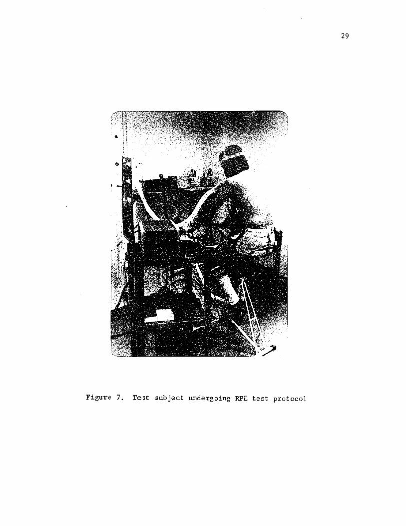
Figure 7. Test subject undergoing RPE test protocol

Figure 8. Subject pointing to the numerical rating
of perceived exertion

ergometer while all testing instruments were quickly disconnected.
Each subject then immediately walked to a desk positioned approxi
mately 15 feet away. After a 2 min. period had elapsed, a blood
sample was drawn by venipucture from the anticubital vein (Figure 9)
to be used in the analysis of venous blood lactate (LA), sodium (Na)
and potassium (K+ ). The samples were drawn by an exercise physiolo
gist from the Department of Health, Physical, and Recreation Educa
tion at Louisiana State University, Baton Rouge, Louisiana.
Gas Collection Procedures
An open circuit gas collection system was utilized for the
collection and evaluation of the respiratory metabolic data. Gas
samples were collected during the exercise bo> t and delivered to the
Beckman O2 and CO2 analyzers via the spirometer. While the subjects
were exercising the metabolic gases were continuously analyzed and
the data were recorded during each 30 sec. interval of the 6 min.
exercise bout. All O2 and CO2 data were recorded in percent concen
tration and later used to calculate VO2 and VCO2 in 1/min. and in
ml/kg/min. The Respiratory Quotient (RQ) and Oxygen Pulse (OXP) were
then calculated using the VO2 , VCO2 , and HR data.
Blood Lactate Collection Procedures
The blood samples were drawn into a 12 cc syringe utilizing a
21 gauge needle without stasis and with as little trauma as possible.
After the sample of whole blood was collected in the syringe (approxi
mately 6 cc) the syringe was passed to the experimenter for depro-
teinization of a 1 cc sample. At this point the needle of the syringe

Figure 9. Blood sample being drawn from
the anticubital vein.

33
was removed and quickly discarded. The syringe containing the sample
was then relieved of all air bubbles and a small amount of sample was
forced out of the syringe so that 1 cc of whole blood could be
accurately measured. The whole blood was then transferred to a pre
pared centrifuge tube containing 2.0 ml of iced 0.6 M perchloric
acid. Each tube was covered with Parafilm and thoroughly mixed by
inversion.
When the sample turned to a grayish-brown color the mixing
was completed and the centrifuge tube was placed directly into a con
tainer of crushed ice to cool the sample to 4° Centigrade (Bergmeyer,
1970). A second sample (3-4 cc whole blood) was transferred into a
(4 cc) red stopper Becton-Dickenson Vacutainer for the determination
of sodium and potassium concentrations in venous blood (Figure 10).
The vacutainer was then sealed with the stopper and placed on the
table at room temperature. To eliminate the possibility of error
with the samples, all vials were coded by number. Samples were
analyzed in the School of Veterinary Medicine by the author and
several lab technicians from the Animal Science Department. The time
lag was never greater than four hours for any blood sample.
Training Procedures
Subjects who completed the initial testing phase (N = 12) were
assigned a training schedule to be followed in the weight room in the
Gymnasium Auditorium at Louisiana State University. Subjects trained
three times per week for 5 weeks on the bicycle ergometer. The
subject wore a shirt, shorts and tennis shoes for the training sessions.

34
Figure 10. Container of ice containing numbered centrifuge
tubes for lactic acid samples, with the 4 cc
vacutainers for sodium and potassium samples
situated in front.

35
All subjects began each training period on the bicycle ergometer at
a load setting equal to 80% the load setting that was calculated to
produce a HR-^gQ in 6 min. at a cadence of 60 revolutions per minute.
Each session the subject exercised for a period of 10 minutes
or until volitional exhaustion. Workloads were increased by 10%
each week until a maximum work load of 1207o of the original load was
reached. After each training session the subject recorded his name,
the amount of time spent on the bicycle ergometer and the work load
setting utilized. This was then presented to the supervisor.
After completion of the 5 week training period each subject
reported to the physiology laboratory for a post-test evaluation.
Subjects were scheduled at the same time on the same day of the week
on which they were originally tested so as to eliminate as much
biological rhythm and diurnal variations as possible. All testing
procedures were identical to the pre-test situation. The subject
performed the experiment at the same workload for the same length
of time as during the pre-test evaluation.
Statistical Analysis of the Data
Means and standard deviations were computed on all independent
and dependent variables. Correlation matrices were generated on the
combined data, on pre and post test data and on the first and second
3 minute periods within each pre and post test period. A total of
seven correlation matrices were generated from the data collected.
A stepwise regression analysis was computed for each three minute
time period within each pre and post test evaluation. A (12 X 2) X
(12) split plot analysis of variance was used to assess the
36
physiological changes which took place over time and from pre to post
test (i.e. HR, RPE, VC^ 1/min., VCC>2 1/min., VC>2 ml/kg/min., VCO2
ml/kg/min., RQ and OXP). A 12 X 2 factorial design was utilized to
explore the changes present for the dependent variables BW, NA, K+
and LA from pre test to post test.

CHAPTER III
RESULTS
Means and Standard Deviations
Means and standard deviations for the pre and post test data
are presented in Table 1. Tables B1 and B2 present the means and
standard deviations for the two 3 min. sections of the pre test.
Tables B3 and B4 present the means and standard deviations for the
two 3 min. sections of the post test.
In Table i it can be seen that HR, RPE, RQ, and LA concentration
decreased from the pre test to the post test; VO^ 1/min., VO^ ml/kg/min.,
VCC^ 1/min., VCC>2 ml/kg/min., and OXP increased from the pre to post
test; and Na and k"*" were essentially unchanged.
Correlations
The Pearson r correlations among the dependent variables are
presented in Tables A l , A2, A3, A4, A5, A6, and A7. The combined pre
and post test coefficient demonstrated a subjstatial relationship
(jr = .69) for HR and RPE (Table Al). When correlation coefficients
were computed between HR and RPE for each test separately, an jr = .75
was noted for the pre test while an r = .61 was noted for the post
test (Tables A2 and A3). Both HRs and RPEs (Figures 11 and 12) in
creased linearly during both exercise bouts. Both the RPEs and HRs
were lower in the post test. Respiratory quotient correlated (.55)
with the RPE, while the VCC^ ml/kg/min. correlated (.41) with the RPE
37

38
TABLE 1
MEANS AND STANDARD DEVIATIONS FOR
RPE PRE AND POST TESTS
THE
Variable NPre Test
Mean S.D.Post
MeanTest
S.D.
HR bpm 144* 161.521 14.372 151.840 17.776
RPE 144* 13.847 1.969 11.194 1.940
Ventilation i 144* 166.430 106.093 178.502 109.769
RQ 144 * 1.290 0.194 1.190 0.153
VO2 1/min. 144* 1.872 0.477 2.337 0.488
VO2 ml/kg/min. 144* 25.042 5.538 31.192 5.511
VCO2 1/min. 144 * 2.428 0.710 2.802 0.705
VCO2 ml/kg/min. 144* 32.553 8.796 37.435 8.519
OXP cc 144* 12.000 2.000 16.000 6.000
NA mEq/l 12 146.750 4.079 141.667 1.656
K"*" mEq /l 12 4.367 0.426 4.475 0.515
LA mg/dl 12 73.722 17.448 57.758 19.013
Weight kg 12 74.590 7.363 74.824 6.807
*The N of 144 30 sec. for 6
represents min.
an observation on 12 subjects taken every

39
180
170
160
HR (bpm)150'
140'
130
120 ■
Pre
1--1 I I I I I I I I I I |1 2 3 4 5 6 7 8 9 10 11 12 13
Time Interval
test
test
Figure 11. Mean Heart rates for each time interval during the
pre test and post test.

40
RPE
16
15 -
14
13
12 —
11
10
Pre test
Post test
0 /
os/
6y 0
I.I.M I If M I I TT1 2 3 4 5 6 7 8 9 10 11 12 13
Time Interval
Figure 12. Mean RPE by time plot for each time interval during
the pre test and post test.

in the combined pre and post test matrix (Table Al).
When the pre and post test correlations were viewed separately
RPE correlated with: RQ (.51), VC^ ml/kg/min. (.54), and VCC^
ml/kg/min. (.71) on the pre test (Table A2). The post test correla
tion matrix displayed slightly reduced relationships between RPE and
RQ(.47); RPE and VCO^ ml/kg/min. (.69); and an increased relationship
between RPE and VC>2 ml/kg/min. (.60) (Table A3). The relationships
between RPE and Na, K+ , and LA were very low at both the pre and post
tests (Na jr = -.10 and .24; K+ jr = -.08 and .13; and LA _r = .22 and .30
respectively) (Table A2 & A3).
When the pre and post tests were divided into 3 minute early
stages and 3 minute latter stages respectively, the Pearson r ’s
between RPE and HR were r_ = .67 and _r = .48 for the pre test and
_r = .52 and _r = .49 for the post test (Tables A4, A 5 , A6, and A7).
The correlation between RQ and RPE for the pre test early
stage jc_ = .36 was higher when compared to the pre test latter stage
r = .22 (Tables A4 and A5). Carbon dioxide production in ml/kg/min.
appeared to be substantially related to RPE during the early stage,
_r = .71 (Table A5) but dropped to .43 in the latter stage. Lactic
acid displayed a fair relationship to RPE in the latter stage of the
pre test, jr = .42 (Table A5). The post test correlation coefficients
of RPE with RQ, VC^ ml/kg/min. and VCO^ ml/kg/min. were highest during
the early stage.

42
Analysis of Variance of Ratings of Perceived
Exertion and Cardio-pulmonary Measures
Eight (12 X 2) X (12) split plot analysis of variance were used
to analyze the dependent variables HR, RPE, RQ, OXP, VO^ l/min., VCO^
1/min., VO^ ml/kg/min. and VCC^ ml/kg/min.
Heart Rate
Heart rate was shown to significantly increase over time F
(11,121) - 64.94, £<..01, and there was a significant F ratio for the
pre and post test comparison _F (1,11) = 12.16, £<.,01. No signifi
cant interactions were noted. In Figure 11, it is evident that the
mean HRs steadily increased as the exercise bout progressed. The
figure also illustrates the significantly lower HR for the post test.
Ratings of Perceived Exertion
A significant F for time of exercise JF (11,121) = 150.08,
£ < . 0 1 and for the pre and post tests comparison JF (1,11) = 67.23,
£<,.01 were found for RPE. Figure 12 depicts the linear rise of RPE
over time and the significantly lower post test RPE. Figure 13 dis
plays the comparative training effects on HR and RPE.
Respiratory Quotient
The RQ significantly increased as the exercise bout continued
_F (11,121) = 27.41, £<..01. The F for the pre and post test compari
son was also significant F (1,11) = 7,63, £<.05. A significant test
x time interaction was found F (11,121) = 3.13, £<.,01. Figure 14
illustrates the lower post test RQ and the increases in RQ over time.
The interaction can also be seen in that the increases in RQ over
time were not the same for the two test periods. Pre test mean RQ

43
HR RPE
180
170
160
150
140
17
16
15
14
13
■12
■11
•10
O^.
HR
RPE
1 “pre post
test
Figure 13. Mean HR and RPE changes between the pre and
post tests.

44
1.40 —
1.30 —
1.20 —
1 .1 0 “RQ 1/min.
1 .0 0—
• 9 0 “ .
T i i i i r i i i i i m i —1 2 3 4 5 6 7 8 9 10 11 12 13
Time Interval
Figure 14. Mean RQ for each time interval during the pre test
and post test.
Pre test
"CT-O Post test0/

45
continued to increase while post test RQ decreased.
Oxygen Pulse
Oxygen Pulse measures were shown to be significantly higher
in the post test than the pre test F (1,11) = 67.70, £ <.01. In
Figure 15, it can be seen that OXP increased significantly during the
initial course of the exercise bout F (11,121) = 2.49, £ < . 0 1 and then
leveled off.
Oxygen Consumption in l/min.
Oxygen consumption in liters per minute was significantly
greater in test two than in test one F (1,11) = 169.21, p_<.01. In
Figure 16, it can also be seen that VO2 l/min. significantly increased
as the exercise bout continued F (11,121) = 29.78, £ (.01.
Carbon Dioxide Production in l/min.
Carbon dioxide production in liters per minute was signifi
cantly higher in test two than in test one F (1,11) = 14.31, £<.01.
As seen in Figure 17, VCO2 l/min. significantly increased as the
exercise bout progressed.
Oxygen Consumption in ml/kg/min.
Oxygen consumption in ml/kg/min. increased significantly as the
exercise bout continued F (11,121) = 29.45, £ (.01, Figure 18. The F
for the pre and post test comparison was also significant F (1,11) =
162.41, £ <.01 showing a higher VO2 in the post test than the pre test.
Carbon Dioxide Production in ml/kg/min.
Carbon dioxide production in ml/kg/min. was significantly
larger in test two than in test one F (1,11) = 14.56, £<.01. As seen
in Figure 19, mean VCO2 ml/kg/min. significantly increased as the

46
l/min.
,02 0
.018
,016
,014
,012
,010
008
//
c{
in' I I I r ITTl'TTT1 2 3 4 5 6 7 8 9 10 11 12 13
Post test
Pre test
Time Interval
Figure 15. Mean OXP for each time interval during the pre test
and post test.

47
l/min.
3.0
2.5
2.0
1.5
post test
pre test
1 -0 “1"C.1 M i l I I I I 7 I I I
1 2 3 4 5 6 7 8 9 10 11 12 13
Time Interval
Figure 16. Mean VO 2 l/min. for each time interval during the
pre test and post test.

48
3.0
2.5 —
l/min. 2.0
1.5
1.0
*0 Post
Pre
I I II M I I I I.ITT1 2 3 4 5 6 7 8 9 10 11 12 13
Time Interval
test
test
Figure 17. Mean VC02 l/min. for each time interval during the
pre and post test.

49
35
30
ml/kg/min.25
20
15
- o " or o0>
Post test
/o
i I i i i I I I M I 1 11 2 3 4 5 6 7 8 9 10 11 12 13
Time Interval
Figure 18. Mean VO2 ml/kg/min. for each time interval during
the pre and post test.

50
ml/kg/min.
45
40
35
30
25
20
Post test
Pre test- O
15
n i i i i i i i i i i r1 2 3 4 5 6 7 8 9 10 11 12 13
Time Interval
Figure 19. Mean VCO^ ml/kg/min. for each time interval during
the pre and post test.

51
exercise bout continued.
Analysis of Variance of Sodium, Potassium, and Lactic Acid
In order to ascertain the changes of weight, Na, K+ and LA.
from test one to test two a 12 X 2 factorial design was utilized.
Table 1 contains the mean values for the variables on the pre and
post test.
No significant changes in body weight were evidenced during
the experiment (see Table C5). Sodium blood concentration signifi
cantly decreased from test one to test two F (1,11) = 10.43, £^.01.
Both the pre and post test values were shown to be within normal
ranges. No significant changes in K+ blood concentration were
evidenced (see Table C6). Lactic acid was shown to significantly
decrease from test one to test two F (1,11) = 7.56, p(.05. Table 1
displays the mean LA values for test one and test two.
Stepwise Regression
Stepwise regression was utilized to determine which physio
logical responses contributed the greatest percentage of variance to
RPE. In order to analyze the relative influence of the different
physiological variables on RPE at different stages in the exercise,
the pre and post test exercise bouts were divided into two, 3 minute
periods; the first 3 minute period representing the least stressful
stage of exercise and the last 3 minutes representing the more stress
ful stage of exercise. Two models were developed for each 3 minute
time period for the pre and post test. The models used for the first
3 minutes of each test included (LA) HR, RQ, OXP, VO2 l/min., and
VCO2 l/min. and (2A) HR, RQ, OXP, VO2 ml/kg/min., and VCO2 ml/kg/min.

52
Models used for the last 3 minute intervals of each test were (IB)
HR, OXP, RQ, Na, K+, LA, VO2 l/min., and VCO2 l/min., and (2B) HR,
OXP, RQ, Na, K+, LA, VO2 ml/kg/min., and VCO2 ml/kg/min.
In the analysis of the first 3 minutes of the pre test, the
best regression model for predicting the RPE is shown in Table Dl.
Four dependent variables were entered into the model with an overall
F (4,67) = 33.56, £^.01. All four variables that were entered (HR,
OXP, VO^ l/min., VCO^ l/min.) contributed significantly to the composite
(£(.01). The R for this four-variable model was .83. The R^ was .68
which indicates that approximately 32% of the variance (1.00 - R^) was
unaccounted for by the model. Model 2A for the pre test demonstratedOsimilar results with R = .80 and an R = .64. However, it was noted
that OXP was replaced by RQ in the second model and the remaining
components were, in order, HR, VO2 ml/kg/min., and VCO2 ml/kg/min. An
overall F (4,67) = 30.10, £ (.01 was obtained. In view of the
slightly higher R, model 1A appears to be the better predictor for RPE
during the first 3 minutes of the pre test.
For the last 3 minutes of the pre test, the two models were
identical with regard to the size of the R (R = .76). The regression
equation for IB included HR, VCO2 l/min., Na and LA. Approximately
42%, of the variation is left unaccounted for by both models. However,
several differences were observed in the two regression equations.
Model 2B's equation includes HR, OXP, RQ, N a , and LA. It should be
noted that HR, Na and LA appear in both models.
The regression equations for the first 3 minutes of the post
test were quite different from those found for the initial period of

53
the pre test. In model 1A, three dependent variables emerged as the
most influential in predicting RPE. The variables HR, RQ, and VCC>22l/min. produced an R = .73. An R of .53 showed that almost half of
the variance associated with RPE was left unaccounted for. In Model
2A only 2 dependent variables were identified: HR and VCC^ ml/kg/min.,
R = .71 and R^ =.50.
The regression equations for the last 3 minutes of the post
test are shown in Tables D7 and D8. Model 2B had a slightly higher
R than Model IB, R = .70 and R = .67, respectively. It is noted that
HR did not emerge in Model 2B. Over half of the RPE variance was not
accounted for by the two models.
Alternative Statistical Interpretation
There are perhaps inherent problems arising in the preceding
design of the regression analysis. It was recognized that the data
points for the independent variables are probably correlated and that
the time factor might also be correlated. Certainly it would be
expected that the variables would be affected by time of exercise.
Therefore, the data were also analyzed by regressions computed at each
30 sec. interval. The data for the additional analysis are presented
in Appendix F.
Neither analysis is entirely satisfactory. In the original
analysis, due to the increased df for the error term, the analysis
may have over estimated the importance of each factor that was entered
into the regression model. Furthermore, the ¥_ needed for significance
for each factor was reduced considerably due to the inflated df for

54
error.
The second analysis is limited in that the variance was
undoubtedly reduced because of the clustering of scores that would
be expected for 12 subjects at each 30 sec. interval. Moreover, the
reduction in the df for the error- term may well have masked any
treatment effects because of the magnified error variance. Thus,
this analysis would tend to underestimate the relative influence of
the physiological factors for predicting the RPE. Therefore, it is
suggested that the actual influence of the physiological variables on
RPE are possibly less than those predicted from the first analysis
but greater than those of the second analysis.
CHAPTER XV
DISCUSSION
The Relationship Between RPE, HR, and Metabolic Variables
Gunnar Borg's scale (Borg, 1973) was based on the concept
that RPE and HR possess a linear relationship. As seen in Figure 20,
the results of this study support the linear relationship between the
Ratings of Perceived Exertion and heart rate. This finding is in
agreement with the findings of Bar-or et al. (1972), Borg (1970),
and Borg and Linderholm (1967).
Intercorrelations were computed with the physiological vari
ables and RPE. When all the data were combined in a single correla
tion matrix, the correlation between HR and RPE was _r = .69, a
substantial relationship. The _r of .69 was somewhat lower than those
obtained by Bar-or et al. (1972), Borg (1962), and Skinner et al.
(1970). This could possibly be explained by the number of subjects
(N =12) in the present study. When correlation matrices were com
puted for the pre and post test separately, Pearson rs of .75 and .61
were noted. The lower post test correlation coefficient can be ex
plained due to physiological adjustments which reduced the contribution
of HR to RPE and substituted another variable or variables in its
place. 'Therefore, it indicates that through conditioning, the feelings
of effort are less influenced by HR while performing at the same work
load as before training. Carbon dioxide production (VCO^ ml/kg/min.)
55

56
16
15
14
13
RPE 12
11
10
9
8
Post test
Pre test
/ o
&A , /
/ N /
/
i— — i— “i---1 r130 140 150 160 170 180
HR
Figure 20. Mean Heart Rates plotted against mean RPE's.

(r = .71), VO2 ml/kg/min. ( Jf = .54), and RQ ( = .51) correlated with
RPE during the pre test. At the post test the relationship between
RPE and VC^ increased while the relationships between RPE and RQ and
VCO^ ml/kg/min. decreased. Oxygen consumption (VO2 ml/kg/min.) was
more influential for trained than untrained subjects. Therefore,
training increased the efficiency of VO2 ml/kg/min. for predicting
the RPE. The relationships between RPE and Na, K , and LA increased
from the pre to post test. The above correlations suggest that
(1) the correlation between a trained subject's oxygen consumption
correlates higher than an untrained subject's and (2) that when
trained individuals are evaluated their electrolytes and LA correlate
better than untrained subject's. These observations can be explained
in part due to the efficiency of the physiological adjustments
taking place in an untrained subject vs a trained subject. The
only other high correlation coefficients noted were those
between the metabolic variables. As would be expected, increases
in VC>2 l/min. and VC^ ml/kg/min. were associated with increases
in VCO2 l/min. and VCC>2 ml/kg/min., _r = ,88 and 1: = .86, res
pectively .
Influence of Training on the Physiological
Factors
Heart rates were shown to significantly decrease from the
pre test to post test. This decrease in HR could be attributed
to (1 ) the specificity of the activity, (2) the increased stroke
volume (SV) and (3) decreased sympathetic drive. Since the
subjects who participated in the study were not accustomed to

58
performing on the bicycle, the HR change from the pre to post test
could, at least in part, have been the result of the specificity
of the exercise. It is well established that training will bring
about increases in SV (Mathews & Fox, 1976). Decreased sympa
thetic drive is attributed to two factors (1) intracardiac
mechanisms and (2) extracardiac mechanisms (Mathews & Fox, 1976).
The intracardiac mechanism relies heavily on the increased SV
which in turn decreases sympathetic stimulation. Thus HR and
sympathetic stimulation are decreased while SV is increased with
the same or slightly decreased cardiac output. The extracardiac
mechanism is primarily related to alterations in trained skeletal
muscle. Sympathetic stimulation of the heart can be altered by
nervous impulses from muscles and joints and descending impulses
from the motor cortex. Therefore, a reduction in HR occurs as a
result of training.
Oxygen pulse was shown to change significantly as a result
of training. The OXP is VO^ l/min. divided by HR which represents
the amount of 0^ which can be transported through the circulatory
system by each heart beat. The significant decrease in HR and the
significant increase in OXP logically points to an increase in
stroke volume.
It was found that the mean RQ for the pre test was 1.29
and 1.19 for the post test, which are quite high. However, RQs of
up to 1.5 have been reported (Consolazio, Johnson, & Pecora, 1963).
An RQ exceeding 1.0 could be explained by hyperventilation and the
buffering of lactic acid by sodium bicarbonate. Since Na blood

59
concentrations also changed from pre to post tests, this would give
credence to the explanation. Other explanations for high RQs accord
ing to Consolazio et al. (1963) are: (1) a washing out of C0o due
to the increase in LA and increased ventilation; (2) a change in the
basal metabolic RQ due to the general increase in utilization of
carbohydrate; (3) an indication of the limitations of optimal respira
tory functions and, at the same time, of the optimal cardiovascular
functions; and (4) a spurious indication of contemporary combustion,
spurious because of overbreathing rather than the result of internal
combustion. Because all subjects were exercising at a rigorous
intensity for a period of 6 minutes, any or all of the explanations
presented above could be used to explain the high RQs. Respiratory
Quotients decreased from the pre test to post test as did the Na
and LA concentrations. With improved physical conditioning the Na
buffering was possibly enhanced for the second test, thus decreas
ing LA. One final explanation for the high RQs noted may have been
the specificity of the exercise. Since the leg muscles were
primarily involved, possibly during the first test period some of the
subjects were actually working at work loads exceeding their VO^ max.
Through training and specific physiological adjustments, primarily in
the legs, subjects were able to increase their VO^ max and lower
their HRs during the post test, resulting also in a lowering of the
RQs.
Significant increases in V02 l/min., VC02 l/min., V02
ml/kg/min., and VC02 ml/kg/min. were found from pre test to post
60
test. The increases appear to be a direct result of training. Again,
the specificity of the training could be an important factor (Fox
et al., 1975b; Horstman et al., 1975; Michael & Hackett, 1972;
Michael et al., 1972; Patton et al., 1975). The most logical explana
tion for the increased consumptions of 0^ and increased production of
CO2 lies in the experimental design of the study. All subjects were
tested using individualized work loads which had been designed to
produce HRs equal to 180 bpm at the end of the first 6 minute test.
This was essentially accomplished as the mean HR for the last
minute was 176.8.
Since a HR of 180 is close to maximal, some of the subjects
might have actually reached their physiological limits in the pre
test. Therefore, as a result of exercise and conditioning these
limits were extended, and when tested under identical conditions
subjects were able to elicit higher 0 uptakes with greater CO^
production for the post test. One further explanation for the
increased metabolic activity may have been changes in skeletal
muscle, hypertrophy and increased capillary density primarily in the
leg muscles. Trained subjects have higher VO2 ml/kg/min. when
compared to untrained subjects due to increased capillary density.
(Hermansen and Wachtlova, 1971). Increases in O2 consumption during
maximal exercise has been well documented (Astrand, 1952; Astrand,
Eriksson, Nylander, Engstrom, Korlberg, Saltin & Thoren, 1968;
Ekblom, Astrand, Saltin, Steinberg & Wallstrom, 1968; Fox, 1975; Fox,
Bartels, Billings, O ’Brien, Bason & Mathews, 1975a; Fox et al.,

61
1975b; Saltin, Blomquist, Mitchell, Johnson, Wildenthal & Chapman,
1968). Therefore, since the training period lasted five weeks, 3
days per week, 10 minutes per day at work loads of 80, 90, 100, 110,
and 120 percent the initial estimate and was intensive, the
improved condition may largely explain the increases in 0 consump
tion and CO2 production.
Sodium and LA decreased significantly from the pre test to
the post test. These decreases were perhaps due to a decrease in the
production of LA by the working muscle (Mathews and Fox, 1976) and
an increase in buffering systems (Holloszy, 1973). Karlsson (1971)
demonstrated that high correlations exist between muscle lactath
concentration and the subjective feeling of fatigue. Since plasma
concentration of LA is directly related to muscle concentration
(Gollnich and Hermansen, 1973), data collected in this study indicate
that RPE decreases from pre test to post test (as did LA) suggesting
a causal relationship. Hermansen (1971) reported that LA in non
athletes is higher than in athletes at a specific workload. Therefore,
training should have resulted in lower LA in the second test using
the same work load.
Potassium (K+) evidenced no change from pre test to post test.
Potassium (K+) cubital venous concentrations have been shown to be
less in trained than in untrained subjects (Tibes, Hemmer, Boning, &
Schweigart, 1976). The data collected in this study did not provide
any evidence to support Tibes’ conclusion. The discrepancy in the
finding may be due to the experimental design of this study. In

62
Tibes’ et al. study trained and untrained subjects performed on the
cycle ergometer for 50 minutes at 98W (10 mKp/s) power output at
60 rpms. Subjects in the present study worked for 6 minutes
utilizing an initial workload for both tests at 60 rpms. This
difference in experimental design could possibly explain the lack of
consistency. With increased performance time, one would expect the
venous K+ concentrations to rise significantly due to the expulsion
of K+ from active muscle.
Influences of Physiological Variables in Predicting RPE
Clearly, during the first 3 minutes of the pre test, the
metabolic or respiratory components appeared to be the major influ
encing factors but when the last 3 minute period was viewed, the
anaerobic by-products appeared to be more influential on the RPE.
These findings were in agreement with those of Fox et al. (1975b),
Horstman et al. (1975), Michael et al. (1972), and Patton et al.
(1975) in that the subjective feeling of fatigue was governed by LA,
HR and other anaerobic by-products. It should also be noted that
the R“s account for only 45 to 68 percent of the variance for pre
dicting RPE. Therefore, from 32 to 55 percent of the variance was
left unaccounted for. This variance could possibly be accounted for
if other physiological substrates were measured and/or if certain
psychological characteristics were included in the model.
On the post test, utilizing the identical models as for the
Pre test, marked changes were observed in the variables that were
stepped in for predicting the RPR. Both of the models used on the

63
post test were equally efficient in predicting the RPE using essen
tially the same components. The predictive power of the post test
equations was less than the pre test. It may be that the altering of
the components resulted in less predictive efficiency. Consequently,
other physiological or psychological factors must have accounted for
the percentage of variance lost in the equations as a result of
training.
Summary
This study attempted to investigate the effects of certain
physiological components on the Ratings of Perceived Exertion and to
explore the influence of training on RPE and on the underlying physio
logical responses influencing RPE. Heart rates and RPEs increased
linearly for both tests with a substantial relationship (_r = .69).
The correlation for RPE and HR for the post test was lower than the
pre test (r = .61 and _r = .75). Evidently, the RPEs were reduced
and the physiological components were altered due to training. The
variables of RPE, HR, RQ, Na, and LA were all reduced while VO^
l/min., VCC^ l/min., VC^ ml/kg/min., and VCC^ ml/kg/min. were
increased. No changes were noted in body weight or K+ concentration.
In reviewing the stepwise regression for four models and two
time periods within the pre and post test, the predictor models dis
played Rs of .67 to .83. Therefore, from 45 to 68 percent of the
variance for predicting the RPE was accounted for by the models. RPE
appears to be a multifaceted and multistructural composite of physio
logical and psychological phenomena with perhaps half of the variance

64
of the RPE associated with the physiological state. Possibly, addi
tional portions of the variance could be due to psychological factors.

REFERENCES

REFERENCES
Allen, P. D., & Pandolf, K. B. Rated perceived exertion associatedwith breathing hyperoxic mixture during submaximal work. Medicine and Science in Sports, 1976, 8.(1), 64. (Abstract)
Arstila, M., & Wendelin, H. Comparison of two rating scales in the estimation of perceived exertion in a puls-conducted exercise test. Ergonomics, 1974, 17(5), 577-584.
Astrand, P. 0. Experimental studies of physical working capacity in relation to sex and age. (Published Thesis) Copenhagen,Munksgaard, 1952.
Astrand, P., Eriksson, B., Nylander, I., Engstrom, L., Korlberg, P., Saltin, B., & Thoren, C. Girl swimmers. Acta Physiologica Scandinavica Supplementum 147, 1973.
Bar-or, 0., Skinner, J. S., Buskirk, E. R. , & Borg, G. Physiological and perceptual indicators of physical stress in 41 to 60 year-old men who vary in conditioning level and in body fitness. Medicine and Science in Sports, 1972, 4, 96-100.
Borg, G. A. V. Physical Performance and Perceived Exertion. Lund: Gleerups, 1962.
Borg, G. Perceived exertion as an indicator of sematic stress.Scandinavian Journal of Rehabilitative Medicine, 1970, 2_, 92-98.
Borg, G. A. Perceived exertion: A note on "history" and methods.Medicine and Science in Sports, 1973, .5(2), 90-93.
Borg, G. A., & Linderholm, H. Perceived exertion and pulse rate during graded exercise in various age groups. Acta Medica Scandinavica, 1967, 472, 194-206.
Borg, G. A. V., Sc Noble, B. J. Perceived exertion. In J. H. Wilmore (Ed.), Exercise and Sport Sciences Reviews (Vol. 2). New York: Academic Press, 1974.
Cafarelli, E., & Noble, B. J. The effect of inspired C0£ on perceived exertion. Medicine and Science in Sports, 1975, 7.(1) > 81. (Abstract)
Consolazio, C. F., Johnson, R. E., Sc Pecora, L. J. PhysiologicalMeasurements of Metabolic Functions in M a n . New York: McGraw-HillBook Company, 1963.
66

67
Docktor, R., & Sharkey, B. J. Note on some physiological and subjective reactions to exercise and training. Perceptual and Motor Skills, 1971, 32, 233-234.
Ekblom, B., Astrand, P., Saltin, B., Steinberg, J., & Wallstrom, B. Effect of training on circulatory response to exercise. Journal of Applied Physiology, 1968, 24(4), 518-528.
Ekblom, B., & Goldborg, A. N. The influence of physical training and other factors on the subjective rating of perceived exertion.Acta Physiologica Scandinavica, 1971, 8_3, 399-406.
Frankenhaeuser, M . , Post, B., Nordheden, B., 6c Sjoeberg, H. Physiological and subjective reactions to different work loads. Perceptual and Motor Skills, 1969, 28, 343-349.
Fox, E. Differences in metabolic alterations with sprint versusendurance interval training programs. In H. Howard & J. Poortmans (Eds.), Metabolic Adaption to Prolonged Physical Exercise. Basel, Switzerland, Birkhauser Verlag, 1975.
Fox, E., Bartels, R., Billings, C., O'Brien, R., Bason, R . , 6c Mathews, D. Frequency of duration of interval training programs and changes in aerobic power. Journal of Applied Physiology, 1975, 38(3), 481-484. (a)
Fox, E. L., McKenzie, D. C., 6c Cohen, K. Specificity of training; metabolic and circulatory responses. Medicine and Science in Sports, 1975, Z(^)> 83. (b) (Abstract)
Gollnich, P. D., 6c Hermansen, L. Biochemical adaptions to exercise: Anaerobic metabolism. In J. H. Wilmore (Ed.), Exercise and Sport Science Reviews (Vol. 1). New York: Academic Press, 1973.
Henriksson, J., Knuttgen, H. G., 6c Bonde-Petersen, F. Perceivedexertion during exercise with concentric and eccentric muscular contractions. Ergonomics, 1972, L5, 537-544.
Hermansen, L., 6c Wachtlova, M. Capillary density of skeletal muscle in trained and untrained men. Journal of Applied Physiology,1971, 30(6), 860-863.
Holloszy, J. 0. Biochemical adaptions to exercise: Aerobic metabolism. In J. H. Wilmore (Ed.), Exercise and Sport Science Reviews(Vol. 1). New York: Academic Press, 1973.
Horstman, D. H. , Morgan, W. P., Cymerman, A., 6c Stokes, J. W. Perception of exertion during exercise of different modes.Medicine and Science in Sports, 1975, ,7(1), 81. (Abstract)

68
Kay, C., & Shephard, R. H. On muscle strength and the threshold of anaerobic work. Internationale Zeitzchrift f{ir Angewandte Physiologie. 1969, 27, 311-328.
Kinsman, R. A., ScWeiser, P. C. Subjective symptomatology during work and fatigue. In E. Simonson & P. C. Weiser (Eds.), Psychological Aspects and Physiological Correlates of Work and Fatigue. Springfield: Charles C. Thomas, 1975.
Mathews, D. K . , & Fox, E. L. The Physiological Basis of PhysicalEducation and Athletics. Philadelphia: W. B. Saunders Company, 1976 .
Michael, E. D ., Jr., Durnin, J. V. G. A., Wormsley, J., Whilelaw, S.G., & Norgan, N. G. Selection of a fifteen-minute work load on a treadmill and bicycle. Research Quarterly, 1972, 43(4), 451-45'9.
Michael, E. D., Jr., & Hackett, P. Physiological Variables related to the selection of work effect on a treadmill and bicycle.Research Quarterly, 1972, 43(2), 216-225.
Morgan, W. P. Psychological factors influencing perceived exertion. Medicine and Science in Sports, 1973, 5.(2), 97-103.
Noble, B. J., Metz, K. F., Pandolf, K. B., Bell, C. W., Cafarelli, E.,& Sime, W. E. Perceived exertion during walking and running--II. Medicine and Science in Sports, 1973, .5 (2), 116-120. (a)
Noble, B. J., Metz, K. F., Pandolf, K. B., & Cafarelli, E. Perceptual responses to exercise: A multiple regression study. Medicineand Science in Sports, 1973, 5(2), 104-109. (b)
Pandolf, K. B., Burse, R. L., & Goldman, R. P. Differentiated ratings of perceived exertion during physical conditioning of older individuals using leg weight loading. Perceptual and Motor Skills, 1975, 40, 563-574.
Pandolf, K., Carafelli, E., Noble, B., &Metz, K. Perceptual responses during prolonged work. Perceptual and Motor Skills, 1972, 35, 975-985.
Pandolf, K. B., & Noble, B. J. The effect of pedalling speed and resistance changes on perceived exertion for equivalent power outputs on the bicycle ergoraeter. Medicine and Science in Sports, 1973, 5(2), 132-136.
Patton, J. F., Morgan, W. P., & Vogel, J. A. Perceived exertion of absolute work in active or less active subjects. Medicine and Science in Sports, 1975, 7.(1), 81. (Abstract)

69
Robertson, R. , Hiatt, E., Gillespie, R. , & Rose, K. The influence of sensory augmentation and reduction on perceived exertion.Medicine and Science in Sports, 1975, XC1)* 81-82.
Robertson, R., McCarthy, J., & Gillespie, R. The contribution of regional to overall perceived exertion during cycle ergometer exercise. Medicine and Science in Sports, 1976, 8_(1), 64.(Abstract)
Saltin, B., Bloraquist, G., Mitchell, Jr., Johnson, R., Wildenthal, K . , & Chapman, C. Response to exercise after bedrest and after training. Circulation Supplement 7, 1868.
Sinning, W. E. Experiments and Demonstrations in Exercise Physiology. Philadelphia, Pa.: W. B. Saunders Company, 1975.
Skinner, J. S., Borg, G. A. V., & Buskirk, E. R. Physiological and perceptual reactions to exertion of young men differing in activity and body size. In B. D. Franks (Ed.), Exercise and Fitness. Chicago: Athletic Institute, 1970.
Stamford, B. A., 6c Noble, B. J. Metabolic cost and perception of effort during bicycle ergometer work performance. Medicine and Science in Sports, 1974, £(4), 226-231.
Tibes, U., Hemmer, B. , Boning, D., &. Schweigart, U. Relationships of femoral venous (K+), (H+), P0~, osmolality, and (orthophosphate) with heart rate, ventilation, and leg blood flow during bicycle exercise in athletes and non-athletes. European Journal of Applied Physiology, 1976, 35_, 201-214.
Ulmer, H. V., Janz, U., 6c Lollgen, H. Aspects of the validity ofBorg's scale. Is it measuring stress or strain? In G. Borg (Ed.), Physical Work and Effort. New York: Pergamon Press, 1977.

a p p e n d i c e s

APPENDIX A
CORRELATION COEFFICIENTS

TABLE A1
Combined Pre and Post Test Correlation Coefficients
1 2 3 4 5 6 7 8 9 10 11 12 13
1. HR 1.002. RPE .69 1.003. BW -.14 .02 1.004. HT -.04 .09 .51 1.00
5. NA .11 .35 .07 -.03 1.006 . K+ -.05 -.04 .21 -.06 .18 1.007. LA .27 .42 .23 -.09 .38 -.13 1.008 . RQ .45 .55 .16 -.17 .31 .14 .27 1.009. V02 1/min. .23 .13 .47 .34 -.21 -.05 -.07 -.05 1.00
10. VCO2 1/rain. .41 .39 .36 .23 -.03 .03 .10 .40 1.00
11. V02 ml/kg/min. .31 .14 .10 .16 -.28 .04 -.18 -.01 .91 .83 1.00
12. VC02 ml/kg/min. .49 .41 .01 .06 -.06 .12 .01 .49 .77 .93 .86 1.00
13. OXP .40 -.18 .37 .24 -.20 -.05 -.16 -.19 .62 .47 .54 .37 1.0
ho

TABLE A2
Pre Test Correlation Coefficients
1 2 3 4 5 6 7 8 9 10 11 12
1. HR 1.002. RPE .75 1.003. BW -.34 .02 1.004. HT -.08 .05 .31 1.005. NA -.19 -.10 .09 -.15 1.006 . K+ -.08 -.08 -.02 .04 .49 1.00
7. LA -.04 .22 .14 -.19 .22 -.23 1.008 . RQ .05 .51 -.19 -.14 .12 .06 .15 1.009. V02 1/min. .91 .48 .50 .33 .05 -.05 .07 .02 1.00
10. VO2 ml/kg/min. .52 .54 .11 .15 .02 -.05 .01 .10 .91 1.00
11. VC02 i/min. .49 .67 .35 .23 .12 -.01 .16 .46 .88 .84 1.00
12. VC02 ml/kg/min. .65 .71 .00 .06 .09 .01 .12 .57 .75 .86 .93 1.00
13. OXP .00 .26 .64 .37 .13 .02 .11 -.12 .95 .78 .77 .57
LO

TABLE A3Post Test Correlation Coefficients
1 2 3 4 5 6 7 8 9 10 11 12 13
1 . tlK. I . uu
2. RPE .61 1.00
3. BW .03 .06 1.004. HT .00 .18 .50 1.005. NA .02 .24 .19 .28 1.00
6 . K+ .03 .13 -.39 -.15 .11 1.007. LA .34 .30 .37 .00 .15 .01 1.008 . RQ .37 .47 -.14 -.21 .42 .32 .22 1.00
9. V02 1/min. .48 .54 .53 .40 .21 -.17 .18 .15 1.00
10. V02 ml/kg/min. .55 .60 .09 .21 .14 .00 .02 .23 .89 1.00
11. VC02 1/min. .57 .66 .39 .24 .38 .00 .28 .58 .89 .82 1.00
12. VC02 ml/kg/min. .61 .69 .01 .06 .33 .16 .16 .68 .74 .86 .92 1.00
13. OXP -.47 -.01 .31 .23 .12 -.15 -.04 -.07 .36 .26 .26 .16

TABLE A4
Correlation Coefficients for Pre Test, Time 30 s-econds to 3 minutes
1 2 3 4 5 6 7 8
1. HR 1.00 v
2 . RPE .67 1.00
3. RQ .36 .47 1.00
4. VC>2 1/min. .41 .43 -.01 1.00
5. V(>2 ml/kg/min. .59 .50 .03 .92 1.00
6 . VCO2 1/min. .54 .65 .47 .87 .82 1.00
7. VCO2 ml/kg/min. .69 .71 .53 .77 .85 .94 1.00
8 . OXP .14 .28 -.13 .96 .82 .77 .62 1.00
''- jLn

TABLE A5
Correlation Coefficients for Pretest, Time 3 minutes 30 seconds to 6 minutes
1 2 3 4 5 6 7 8 9 10 11
1. HR 1.002. RPE .48 1.003. NA -.26 -.21 1.004. K+ -.16 -.24 .50 1.00
5. LA .08 .42 .23 -.23 1.00
6 . RQ .15 .22 .22 .10 .35 1.00
7. V02 1/min. -.22 .27 .10 -.08 .15 -.35 1.00
8 . VO2 ml/kg/min. .08 .30 .06 -.10 .09 -.27 .88 1.00
9. VCO2 1/min. -.18 .40 .21 -.02 .38 .11 .89 .80 1.00
10. VC02 ml/kg/min. .14 .43 .20 -.02 .31 .32 .66 .82 .86 1.00
11. OXP -.45 .13 .16 -.04 .15 -.34 .97 .78 .86 .57 1.00
"■■JCT\

TABLE A6
Correlation Coefficients for Post Test, Time 30 seconds to 3i minutes
1 2 3 4 5 6 7 8
1 . HR 1.00
2 . RPE .52 1.00
3. RQ .30 .57 1.00
4. VO2 l/min. .40 .49 .19 1.00
5. VC>2 ml/kg/min. .47 .52 .30 .90 1.00
6 . VC02 1/min. .47 .67 .64 .87 .85 1 .00
7. VCO2 ml/kg/min. .50 .67 .75 .72 .85 .94 1.00
8 . OXP -.65 -.10 -.02 .27 .19 .20 .11 1.00

TABLE A7
Correlation Coefficients for Post Test, Time 3 minutes 30 seconds to 6 minutes
1 2 3 4 5 6 7 8 9 10 11
1. HR 1.002. RPE .49 1.00
3. NA .04 .21 1.00
4. K+ .07 .17 .10 1.00
5. LA .59 .47 .15 .01 1.00
6 . RQ .22 .15 .62 .35 .43 1.00
7. VC>2 1/min. .39 .40 .20 -.16 .25 -.20 1.00
8 . V02 ml/kg/min. .43 .45 .12 .05 .08 -.21 .83 1.00
9. VC02 1/min. .49 .44 .51 .00 .46 .30 .87 .70 1.00
10. VC02 ml/kg/min. .54 .49 .48 .25 .34 .39 .68 .82 .85 1.00
11. OXP .01 .25 .18 -.19 .03 -.31 .93 .74 .74 .51 1.00
'-J00

APPENDIX B
MEANS AND STANDARD DEVIATIONS

TABLE B1
MEANS AND STANDARD DEVIATIONS FOR
PRE TEST, TIME 30 SECONDS TO 3 MINUTES
Variables N Mean S.D.
RPE 72* 12.583 1.489
HR bpm CM 152.708 12.598
Ventilation 1/min. 72* 76.361 45.900
RQ 72* 1.215 0.218
VO2 1/min. 72* 1.708 0.464
VO2 ml/kg/min. 72* 22.882 5.713
VC02 1/min. 72* 2.083 0.676
VC02 ml/kg/min. 72* 27.953 8.637
OXP cc 72* 11.000 3.000
*N is for 6 observations on 12 subjects.

TABLE B2
MEANS AND STANDARD DEVIATIONS FOR PRE TEST,
TIME 3 MINUTES 30 SECONDS TO 6 MINUTES
Variables N Mean S.D.
RPE 72* 15.111 1.534
HR bpm 72* 170.333 9.989
Ventilation 1/min. 72* 256.499 64.107
RQ 72* 1.365 0.131
VO2 1 min. 72* 2.037 0.433
VO2 ml/kg/min. 72* 27.203 4.436
VCO2 1/min. 72* 2.773 0.563
VCO2 ml/kg/min. 72* 37.153 6.188
OXP cc
*CMr*v 12.000 3.000
NA mEq/1 12 146.750 4.093
K+ mEq/I 12 4.367 0.428
LA mg/dl 12 73.722 17.510
*N is for 6 observations on 12 subjects.

TABLE B3
MEANS AND STANDARD DEVIATIONS FOR POST
TEST, TIME 30 SECONDS TO 3 MINUTES
Variables N Mean S.D.
RPE 72* 10.083 1.536
HR bpm 72* 143.764 19.390
Ventilation 1/min. 72* 85.738 49.385
RQ 72* 1.144 0.177
VO2 1/min. 72* 2.167 0.476
VO2 ml/kg/min. 72* 28.904 5.287
VCO2 1/min. 72* 2.504 0.706
VCO2 ml/kg/min. 72* 33.474 8.727
OXP cc 72* 16.000 8.000
*N is for 6 observations on 12 subjects.

TABLE B4
MEANS AND STANDARD DEVIATIONS FOR POST TEST,
TIME 3 MINUTES 30 SECONDS TO 6 MINUTES
Variables N Mean S.D.
RPE 72* 12.306 1.650HR bpm 72* 159.917 11.321
Ventilation 1/min. •XCM 271.267 66.155
RQ 72* 1.235 0.107
VO2 1/min. 72* 2.507 0.441
VO2 ml/kg/min. 72* 33.480 4.755
VCO2 1/min. 72* 3.099 0.568
VCO2 ml/kg/min. 72* 41.397 6.180
OXP cc 72* 16.000 3.000
NA mEq/I 12 141.667 1.662
K"*- mEq /I 12 4.475 0.517
LA mg/dl 12 57.760 19.083
*N is for 6 observations on 12 subjects.

APPENDIX C
ANALYSES OF VARIANCE

TABLE Cl
SUMMARY OF THE SPLIT PLOT ANALYSIS OF VARIANCE FOR THE DEPENDENT
VARIABLES HR AND RPE WITHIN A (12 X 2) X (12) DESIGN
SourceHR RPE
df MS F df MS F
Subject (A) 11 1938.58 3.49* 11 26.80 3.56*
Trial (B) 1 6747.35 12.16** 1 506.68 67.23**
A X B (Error A) 11 555.02 11 7.54
Time (C) 11 3134.06 64.94** 11 50.97 150.08**
B X C 11 69.27 1.44 11 .58 1.69
A X C 121 51.39 1.06 121 .89 2.61**
A X B X C (Error B) 121 48.26 121 .34
* P <.05
** P <.01
00Ln

TABLE C2
SUMMARY OF THE SPLIT PLOT ANALYSIS OF VARIANCE FOR THE DEPENDENT
VARIABLES RQ AND OXP WITHIN A (12 X 2) X (12) DESIGN
SourceRQ OXP
df MS F df MS F
Subject (A) 11 .12 1.29 11 .000112 6.27**
Trial (B) 1 .73 7.63* 1 .001213 67.70**
A X B (Error A) 11 .10 11 .000017
Time (C) 11 .27 27.41** 11 .000038 2.49**
B X C 11 .03 3.13** 11 .000012 0.76
A X C 121 .02 1.46* 121 .000015 0.97
A X B X C (Error B) 121 .01 121 .000016
* P <.05
** P <.01
00On

TABLE C3
SUMMARY OF THE SPLIT PLOT ANALYSIS OF VARIANCE FOR THE DEPENDENT VARIABLES
V02 1/min. AND VC02 1/min. WITHIN A (12 X 2) X (12) DESIGN
SourceV02 1/min. VC02 1/min.
df MS F df MS F
Subject (A) 11 2.73 2 9.70** 11 3.69 5.27**
Trial (B) 1 15.56 169.21** 1 10.03 14.31**
A X B (Error A) 11 .09 11 .70
Time (C) 11 1.68 29.78** 11 6.03 63.77**
B X C 11 .07 1.32 11 .07 0.74
A X C 121 .08 1.39* 121 .13 1.42*
A X B X C (Error B) 121 .06 121 .10
* P (.05
** P <.01
00

TABLE C4
SUMMARY OF THE SPLIT PLOT ANALYSIS OF VARIANCE FOR THE DEPENDENT VARIBALES
V02 ml/kg/min. AND VC02 ml/kg/min. WITHIN A (12 X 2) X (12) DESIGN
SourceV02 ml/kg/min. VC02 ml/kg/min »
df MS F df MS F
Subject (A) 11 202.34 12.07** 11 314.85 2.67
Trial (B) 1 2722.68 162.41** 1 1716.35 14.56**
A X B (Error A) 11 16.77 11 117.90
Time (C) 11 296.24 29.45** 11 1070.17 64.73**
B X C 11 14.24 1.42 11 13.17 0.80
A X C 121 13.94 1.39* 121 22.85 1.38*
A X B X C (Error B) 121 10.06 121 16.53
* P <.05
** P < .01
00CO

TABLE C5
SUMMARY OF THE FACTORIAL ANALYSIS OF VARIANCE FOR THE DEPENDENT
VARIABLES BW AND Na WITHIN A 12 X 2 DESIGN
SourceBW Na
df MS F df MS F
Subject (A) 11 1290.60 78.31** 11 73.59 0.41
Trial (B) 1 3.96 0.24 1 1860.50 10.43**
Error 11 16.48 11 178.32
* P < . 05
** P < .01
00VD

TABLE C6
SUMMARY OF THE FACTORIAL ANALYSIS OF VARIANCE
VARIABLES K* AND LA WITHIN A 12 X 2
FOR THE
DESIGN
DEPENDENT
SourceK+ LA
df MS F df MS F
Subject (A) 11 2.88 0.98 11 6231.24 2.57
Trial (B) 1 .85 0.29 1 18348.98 7.56*
Error 11 - 2 .94 11 2425.69
* P < . 05
MDO

APPENDIX D
STEPWISE REGRESSION ANALYSIS

TABLE D1
SUMMARY OF THE BEST REGRESSION EQUATION FOUND FOR THE
PRETEST, TIME 30 SECONDS TO 3 MINUTES (MODEL 1A)
Source df Approximate F P
Regression 4 33.56 .01
HR 1 23.28 .01
OXP 1 12.24 .01
VO2 1/min. 1 15.05 .01
VCO2 1/min. 1 25.01 •01Error 67
Total 71
R = .83, R2 = .68Factors included in the model: HR, OXP, RQ, VO2 1/min.,
VCO2 1/min.

TABLE D2
SUMMARY OF THE BEST REGRESSION EQUATION FOUND FOR THE
PRE TEST, TIME 30 SECONDS TO 3 MINUTES (MODEL 2A)
Source df Approximate F P
Regression 4 30.10 .01
HR 1 11.77 .01
VO^ ml/kg/min. 1 11.40 .01
VCO^ ml/kg/min. 1 16.91 .01
RQ 1 6.79 .01
Error 67
Total 71
R = .80, R2 = .64
Factors included in the model: HR, OXP, RQ, VO2 ml/kg/min.,
VCO2 ml/kg/min.

TABLE D3
SUMMARY OF THE BEST REGRESSION EQUATION FOUND FOR THE PRE TEST,
TIME 3 MINUTES 30 SECONDS TO 6 MINUTES (MODEL IB)
Source df Approximate F P
Regression 4 22.86 .01
HR 1 31.07 .01
VC02 1/min. 1 25.23 .01
Na 1 8.44 .01
LA 1 11.29 .01
Error 67
Total 71
R = .76, R2 = .58
Factors included in the model: HR, OXP, RQ, VO2 1/min.,
VCO2 1/min., Na, K+, LA

TABLE D4
SUMMARY OF THE BEST REGRESSION EQUATION FOUND FOR THE PRE TEST,
TIME 3 MINUTES 30 SECONDS TO 6 MINUTES (MODEL 2B)
Source df Approximate F P
Regression 5 17.38 .01
HR 1 36.14 .01
OXP 1 22.44 .01
RQ 1 7.24 .01
Na 1 8.45 .01
LA 1 5.24 .01
Error 66
Total 71
R = .76, R2 = .58
Factors included in the model: HR, OXP, VO2 ml/kg/min.,
VCO2 ml/kg/min., RQ, N a , K+ , LA.

TABLE D5
SUMMARY OF THE BEST REGRESSION EQUATION FOUND FOR THE
POST TEST, TIME 30 SECONDS TO 3 MINUTES (MODEL 1A)
Source df Approximate F P
Regression 3 25.79 .01
HR 1 7.68 .01
RQ 1 5.19 .05
VCO2 1/min 1 11.08 .01
Error 68
Total 71
R = .73, R2 = .53
Factors included in the model: HR, OXP, RQ, VO2 1/min.,
VCO2 1/min.

TABLE D6
SUMMARY OF THE BEST REGRESSION EQUATION FOUND FOR THE
POST TEST, TIME 30 SECONDS TO 3 MINUTES (MODEL 2A)
Source df Approximate F P
Regression 2 34.25 .01
HR 1 6.08 .05
VCO^ ml/kg/min. 1 31.86 .01
Error 69
Total 71
R = .71, R2 = .50
Factors included in the Model: HR, OXP, RQ, VO 2 ml/kg/min.,
VCO2 ml/kg/min.

TABLE D7
SUMMARY OF THE BEST REGRESSION EQUATION FOR THE POST TEST,
TIME 3 MINUTES 30 SECONDS TO 6 MINUTES (MODEL IB)
Source df Approximate F P
Regression 6 8.97 .01
HR 1 16.84 .01
OXP 1 12.11 .01
RQ 1 7.14 .01
VC02 1/min. 1 10.97 » O
Na 1 6.32 .05
LA 1 9.00 .01
Error 65
Total 71
R = .67, R2 = .45
Factors included in the model: HR, OXP, RQ, VO2 1/min.,
VCO2 1/min., Na, K+, LA

TABLE D8
SUMMARY OF THE BEST REGRESSION EQUATION FOUND FOR THE POST TEST,
TIME 3 MINUTES 30 SECONDS TO 6 MINUTES (MODEL 2B)
Source df Approximate F P
Regression 6 10.52 .01
OXP 1 3.63 NS
VO2 ml/kg/min. 1 11.47 .01
VCO2 ml/kg/min. 1 8.02 .01
RQ 1 6.00 .05
Na 1 4.08 .05
LA 1 26.04 .01
Error 65
Total 71
R = .70, R2 = .49
Factors included in the model: HR, OXP, VO2 ml/kg/min.,
VC02 ml/kg/min., RQ, N a , K+, LA

APPENDIX E
CALCULATIONS AND LABORATORY PROTOCOL

101
El
CALCULATIONS FOR METABOLIC DATA
All calculations performed on the data were handled through
a SAS program designed by the author. For the convenience of the
reader hand calculations are described below as taken from Consolazio
(1963) and Sinning (1975).
First, the STPD factor was calculated for all time intervals
during the pre and post test. The formula was as follows;
273° PBAR - p H90STPD = --------------- X z<273° " TGAS> 760
Where STPD is the correction factor for standard temperature and
pressure dry; TgA g is the gas temperature in degrees Centigrade;
PBAR t ie atmospheric pressure in mm Hg (barometer reading in inches
then converted to mm) and P ^ O the water vapor pressure in mm Hg
(values for Pj^o can be f°und in Sinning (1975, p. 95).
VE(a t p s ) was then computed by using the formula
\tf. = vt - N(ATPS) (ATPS) *
Where VI(ATPS) -*-s t*ie volume of inspired gas at atmospheric tempera
ture and pressure saturated and N is the percent of nitrogen in the
expired air.
Then, VE^ATpS was converted to VE^STpD by the formula:

102
V^(STPD) = VE (ATPS) X STPD
VEn was then computed as follows:2
FE0VE = VE X 2(STPD) ^
Where FEq^ is the percent concentration of 0 in expired air and
VE(s x p d ) is the volume of expired gas at standard temperature and
pressure dry.
VI(STPD) was comPuted:
VI (STPD) = V I (ATPS) X STPD
Where VI^TPS) is the volume of inspired gas at atmospheric tempera
ture and pressure saturated.
Percent of N2 in expired air (FE^) was calculated by the
formula:
P% 2 = 100 - (FE02 - feC02)
Where is the percent of 0£ in expired air and FEq q^ is the per
cent of CO2 in expired air.
Since heavier people tend to have larger O2 consumption, VO2
1/min. was then converted to VO2 ml/kg/min. where:
VO2 1/min. X 1000 ml/literVO2 ml/kg/min. *» ----------------------------
subject’s weight (kg)
Carbon dioxide production was then calculated (1/min. and
ml/kg/min.) by the use of the following formulas:

103
C02 (STPD) VEC02 (STPD) " VIC02 (STPD)
Where V E ^ (SIpJ)) is the volume of expired CO., at STPD and
is the volume of inspired CC^at STPD.
* Vco2 (STPD)COo 1/min. * ~ “z collection time
_ ^C02 1/min. X 1000 ml/literVC02 ml/kg/min. “ ----- — ---— ;---- r-rr— t.— :—subject s weight (kg)
RQ was calculated by utilizing the following formula;
CO?7„ - .03RQ = z ________N X .265 - 02%
Where C02% is the percent of CO2 in expired air, N is the percent of
nitrogen in expired air and 02 is the percent of 02 in expired air.
OXP was calculated by the following formula;
VO2 1/min.OXP = -----------HR

104
E2
Blood Chemistry
The analysis for determination of sodium and potassium was
conducted at the Department of Animal Sciences, Louisiana State
University, Baton Rouge, Louisiana. All samples were delivered in 4m1
glass vacutainers to the Pathology Laboratory where lab technicians
conducted the analysis.
For the determination of lactic acid, initial preparations
were conducted at the Department of Animal Sciences by the author.
Centrifuge tubes containing the deproteinized blood were used for the
lactate analysis procedure. All samples were placed in a centrifuge and
spun for at least 5 min. at 3000 rpms. After centrifuging was com
pleted the supernatant was removed carefully and placed in a 4 cc glass
vial. The substrate was then discarded.
The supernatant specimens were taken to the School of Veterinary
Medicine Pathology Laboratory where each sample was incubated in a
constant temperature water bath at 30° Centigrade for 5 min. Upon
completion of the pre-incubation period, a 5.0 ml pipette was used to
dispense 2 .9 ml of prepared reagent into a clean, dry cuvet with a 1 cm
light path. Each cuvet was then placed in a Beckman Spectrophotometer
set at 340 nm using a distilled water as a blank. An initial absorbence
measurement was recorded from the spectrophotometer. After completion
of the initial absorbence measurements, .050 ml of the supernatant was
added to the cuvets and mixed quickly by gentle inversion with a square

105
of Parafilm over the mouth of the cuvet. All samples were then re
turned to the constant temperature water bath for 15 min. Upon
completion of the incubation period all cuvets were remeasured for
absorbence changes in the spectrophotometer. The concentration of
lactic acid present in the blood was computed utilizing the formula
A = [A-5 - (Aq X 0.967)] x 1.97 where A = the change in absorbence;
A 15 = the absorbence at the end of the 15 min. period; Aq = the initial
absorbence; .967 = a constant to correct a change in volume due to the
additional .050 ml of supernatant; and 1.97 = the dilution factor for
.050 ml of supernatant. A is then multiplied by 131 in order to
present the lactic acid concentration in mg/dl.

APPENDIX F

107
FI
Alternative Statistical Analysis
Because inherent problems possibly exist in the stepwise
regression analysis an alternative statistical interpretation is
offered. In the original analysis of the data, the 6 min. work
bouts were divided into two, 3 min. periods, one representing an
early period (30 sec. to 3 min.) and one representing a late period
(3 min., 30 sec. to 6 min.). Due to the arrangement of the data,
possible correlations might exist between the data points of indi
vidual subjects as well as a correlation existing between the
variables and time. Therefore, the data were arranged so that
regressions were computed at each 30 sec. interval. This arrangement
of the data was an attempt to eliminate the above correlations. How
ever, it should be noted that by arranging the data in this manner the
variance was greatly reduced between data points. The results of the
alternative analysis are presented in Tables F2 through F9. The step
wise multiple regressions presented in Appendix F contain a total of
11 df (N - 1 df for subjects).

TABLE F2
SUMMARY OF THE SIGNIFICANT REGRESSION EQUATIONS FOUND FOR THE PRE TEST
AT EACH 30 SECOND INTERVAL FOR THE FIRST 3 MINUTES (MODEL 1A)
30 1:00 1:30 2:00 1:30 3:00Variable Order F Order F Order F Order F Order F Order F
HR 1 18.43** 1 12.20** 1 12.72**
OXP 2 17.14**
RQ 2 4.00NS
VO2 1/min. 3 17.50** 2 20.17** 3 5.IONS
VCO2 1/min. 3 30.48** 4 5.63*
R .85 .92 .82
R2 .72 .85 .67
* P C. 05
** P <.01
Factors included in the Model: HR, OXP, RQ, VO2 1/min., VCO2 1/min.

TABLE F3SUMMARY OF THE SIGNIFICANT REGRESSION EQUATIONS FOUND FOR THE PRE TEST
AT EACH 30 SECOND INTERVAL FOR THE FIRST 3 MINUTES (MODEL 2A)
30 1:00 1:30 2:00 2:30 3:00Variable Order F Order F Order F Order F Order F Order F
HR
OXP 1 10.64**
RQ
VO2 ml/kg/min. - 2 37.92**
VCO2 ml/kg/min.
R .91
R2 CM00•
* P <.05
** P <.01
Factors included in the model; HR, OXP, RQ, VO2 ml/kg/min., VCO2 ml/kg/min.

TABLE F4
SUMMARY OF THE SIGNIFICANT REGRESSION EQUATIONS FOUND FOR THE PRE TESTAT EACH 30 SECOND INTERVAL FOR THE LAST 3 MINUTES (MODEL IB)
Variable3:30 4:00 4:30 5:00 5:30 6 :00
Order F Orderm F Order F Order F Order F Order FHR 1 11.24** 1 10.34*OXP 1 5.44* 1 6.26* 2 7.82*RQVO2 1/min. 3 7.06*VGO2 1/min. 2 6 .95*Na 2 11.52** 2 11.47** 4 4.67NSK+ 3 4.97* 3 4.66NSLA 3 3.22NS
R .79 .80 .87 0000•
R2 .62 .64 .76 .77
* P < .05 ** P <.01Factors included in the model: HR, OXP, RQ, VO2 1/min., VCO2 1/min., Na, K+ , LA

TABLE F5
SUMMARY OF THE SIGNIFICANT REGRESSION EQUATIONS FOUND FOR THE PRE TEST
AT EACH 30 SECOND INTERVAL FOR THE LAST 3 MINUTES (MODEL 2B)
3: 30 4:00 4:30 5:00 5: 30 6 :00Variable Order F Order F Order F Order F Order F Order FHR 1 13.58** 1 17.37** 1 13.66**RQ 4 26.55** 4 10.40** 5 4.37NSOXP 2 21.42** 2 9.68** 2 5.49NSVO2 ml/kg/min. 3 8.14*VCO2 ml/kg/min.3 13.30** 3 4.18NS 4 5.47*Na 5 13.61** 6 4.16NSK+LA
R .92 .92 .93R2 .84 00• .86
* P < .05** P < .01
Factors included in the model: HR, RQ, OXP, VO 2 ml/kg/min., VCO2 ml/kg/min., Na, K+ , LA

TABLE F6
SUMMARY OF THE SIGNIFICANT REGRESSION EQUATIONS FOUND FOR THE POST TEST AT EACH
30 SECOND INTERVAL FOR THE FIRST 3 MINUTES (MODEL 1A)
30 1:00 1:30 2:00 2:30 3:00Variable Order F Order F Order F Order F Order F Order FHR
OXP 1 3.86NSRQ 1 6.23* 1 8.39*VO2 1/min.VCO2 1/min. 1 15.37** 2 11.12** 2. 5.23*
R .78 .84 .74 .68R2 .61 .70 .51 .46
* P < .05** p <.01
Factors included in the model: HR, OXP, RQ, VO2 1/min., VCO2 1/min.

TABLE F7
SUMMARY OF THE SIGNIFICANT REGRESSION EQUATIONS FOUND FOR THE POST TEST AT EACH
30 SECOND INTERVAL FOR THE FIRST 3 MINUTES (MODEL 2A)
30 1:00 1:30 2:00 2 ;30 3:00variable Qrder F Order F Order F Order F Order F Order FHR 1 8 .88* 1 7.19*OXP 2 10.55**RQ 3 10.24** 2 6.33*VO2 ml/kg/min.
VC02 ml/kg/min.
R .84 .79R2 .70 .62
* P <.05 ** P < .01
Factors included in the model: HR, OXP, RQ, VO2 ml/kg/min., VCO2 ml/kg/min.
113

TABLE F8
SUMMARY OF THE SIGNIFICANT REGRESSION EQUATION FOUND FOR THE POST TEST AT EACH 30 SECOND INTERVAL FOR THE LAST 3 MINUTES (MODEL IB)
3:30 4:00 4:30 5:00 5:30 6 :00Variable Order F Order F Order F Order F Order F Order FHR 1 6.47*OXP 2 5.70*RQVO2 1/min. 3 5.40*VC02 1/min.NA 4 2.87NSK+
LA
R .84R2 .70
* P <.05** P < .01
Factors included in the model: HR, OXP, RQ, VO^ 1/min., VCO2 1/min. , NA, K"*", LA

TABLE F9
SUMMARY OF THE SIGNIFICANT REGRESSION EQUATIONS FOUND FOR THE POST TEST AT EACH
30 SECOND INTERVAL FOR THE LAST 3 MINUTES (MODEL 2B)
3:30 4:00 4:30 5:00 5:30 6:00Variable Order F Order F Order F Order F Order F Order FHROXPRQ 3 10.77**V02 ml/kg/min. 1 12.29** 1 7.68*VCO2 ml/kg/min. 2 11.27**NAK+LA 4 12.58** 2 5.56*
R .93 .77R2 ' .86 .60
* P {.05** P < .01
Factors included in the model: HR, OXP, RQ, VO2 ml/kg/min, VCO2 ml/kg/min., NA, K+, LA

VITA
The author was born in Port Arthur, Texas on October 5, 1951.
He attended Thomas Jefferson High School in Port Arthur and graduated
in 1970. He attended Lamar University, Beaumont, Texas, graduating
in 1973 with a Bachelor of Science degree in Health and Physical
Education.
The author held an elementary physical education teaching
position at Mont Belview, Texas for one year.
In December, 1974, the Master of Science degree in Health and
Physical Education was earned from Lamar University.
In January, 1975, he entered the Ed.D. program at the
University of Houston in the area of Physical Education. The follow
ing August, he transferred to Louisiana State University to enter the
Ph.D. program in Health, Physical and Recreation Education.
116
EXAMINATION AND THESIS REPORT
Candidate:
Major Field:
Title of Thesis:
David R. Carter
Health, Physical and Recreation Education
The Influence of Various Physiological Responses on Ratings of Perceived Exertion Before and After Training
Approved:
Major Professor and Chairman
Deanll of the Graduate School
E X A M I N I N G COMMITTEE:
Date of Examination:
July 20, 1978