The influence of insulin-like growth factor 1 and its …...In addition, greater potency was...
240
THE INFLUENCE OF' INSULIN.LIKE GROWTH F'ACTOR I AND ITS ANALOGUES ON F'IBROBLASTS AND DERMAL WOLTND HEALING NTCHOLAS JOHN MARSHALL M.B.B.S. (ADEL) Thesis submitted in fulfilment of the requirements for the Degree of I)octorate of Medicine, the University of Adelaide. DEPARTMENT OT'SURGERY FACULTY OF MEDICINE UNIVERSITY OF' ADELAIDE 1998.
Transcript of The influence of insulin-like growth factor 1 and its …...In addition, greater potency was...
THE INFLUENCE OF'
INSULIN.LIKE GROWTH F'ACTOR I AND ITS ANALOGUES
ON F'IBROBLASTS AND DERMAL WOLTND HEALING
NTCHOLAS JOHN MARSHALL M.B.B.S. (ADEL)
Thesis submitted in fulfilment of the requirements for the Degree of
I)octorate of Medicine, the University of Adelaide.
DEPARTMENT OT'SURGERY
FACULTY OF MEDICINE
UNIVERSITY OF' ADELAIDE
1998.
TABLE OF CONTENTS
Dedication
I)eclaration
Acknowledgements
Synopsis
Chapter I Introduction
chapter 2 A study of IGFs in Human wound Fluid and Plasma 27
Chapter 3 IGF-I and its Analogues: In Vitro Responses of Fibroblasts 62
Chapter 4 Application of IGFs to Incisional Models of Wound Repair 101
I
It
Itt
lv
1
Chapter 5 Excisional wounds: Response of Normal and
Diabetic wounds to topical IGF-I and topical Long nf rcr-r 150
Chapter 6 Discussion and SummarY 177
Appendix 188
191Bibliography

ll
DEDICATION
This thesis is dedicated to my wife, Caroline, my children, Henry and Georgina; Caroline for
her enduring love, encouragement, support and patience. To Henry and Georgina who provide
so much wonderment and joy to me every day of my life.
l1r
DECLARATION
This work contains no material which has been accepted for the award of any other degree or
diploma in any university or other tertiary institution and, to the best of my knowledge and
belief, contains no material previously published or written by another person, except where
due reference has been made in the text.
I give consent to this copy of my thesis, when deposited in the University Library, being
available for loan and photocopying.
N Marshall
1998
lv
ACKNO\MLEDGEMENTS
I wish to express my sincere gratitude Mr. Rodney Cooter M.D., senior lecturer in the
Department of Surgery, University of Adelaide for his dedicated supervision and inspiration
in the completion of this thesis.
To Dr. David Belford PhD, Project Leader, Wound Healing Group, Cooperative Research
Centre for Tissue Growth and Repair I extend my sincerest thanks for his guidance in the
basic scientific process. As a supervisor he has provided unerring support and inspiration for
my continued involvement in wound healing research.
Special thanks go to those who provided the technical tuition I required to complete my work.
Firstly to Miss Anna Seamark Research Assistant Child Health Research Institute (CHRI)and
Miss Ingrid Leipe Research Assistant CSIRO for their patience and continued advice as well
as expert technical assistance in the the field of cell and tissue culture and other laboratory
procedures. Secondly to Mrs Kaylene Pickering, Technical Officer, CSIRO, I extend thanks
for her infectious enthusiasm and patience in the supervision of analytical procedures.
I am forever indebted to Miss Xenia Georgiou Technical Assistant, CHzu, and Mrs. Kaylene
Pickering for their dedication, enthusiasm and patience demonstrated during the long days and
evenings of the animal experiments.
I wish to express my gratitude to Ms. Sue Millard, Technical Officer Department of Surgery
Queen Elizabeth Hospital and Miss Olympia Kapinaris, Technical Officer Department of
Surgery, The Queen Elizabeth Hospital for their preparation of the specimens for
immunofl uorescence and immunocytochemical staining.
I wish to also thank Dynek Pty Ltd and 3M Australia for their assistance that allowed some of
this work to be presented in an international forum.
Gratitude is also expressed to Smith and Nephew Pty Ltd for their donation of Opsite@ and
Dr. Judy Owens Department of Obstetrics and Gynaecology, University of Adelaide, for her
gift of rabbit anti-sheep IGFBP-4 antibody.
VI
SYNOPSIS
A comprehension of the wound repair process is central to all surgical practice. The science of
wound healing has progressed dramatically in recent years to facilitate this understanding,
particularly in the area of growth factor biology and offers exciting possibilities for the
manipulation of this process.
One cytokine that has shown particular promise has been insulin-like growth factor-I (IGF-I).
As the second messenger for growth hormone (GH), it is postulated to mediate the majority of
anabolic functions associated with GH. Of clinical importance is the associated reversal of
previously depressed IGF-I levels and reduced protein catabolism when GH is administered to
patients with severe burns. The potential role of IGF-I is further supported by the finding that
IGF-I is found locally at the wound site as well as in measurable concentrations systemically.
It appears, also, to have important vulnerary effects upon fibroblasts and keratinocytes
including cell migration and protein synthesis. Six carrier proteins, known as IGF binding
proteins (IGFBPs), have been identified. Although little is known of how they modulate IGF
biology within a wound it appears that IGFBP-I and -3 can potentiate IGF-I activity.
With the development of recombinant DNA techniques, a fange of IGF variants (analogues)
are now commercially available. The value of these peptides lies in their apparent increased
biological activity in vitro and to a more limited degree in vivo. Primarily, the greater potency
of the analogues is related to decreased aff,rnity for IGFBPs and consequently increased IGF
bioavailability. Therefore they can be used to examine indirectly the influence that IGFBPs
have in wound repair.
In light of this information, the objectives of this study were to investigate:
o the levels of IGFs and the presence of binding proteins in human wound fluid;
vll
o the potency of IGF-I and two analogues in several in vitro models of fibroblast activity;
. the effect of IGF-I and one of the analogues upon healing in both normal and diabetic
rodent wounds.
Characterisation of IGFs and IGFBPs in human wound fluid and plasma was achieved by
obtaining time matched wound fluid samples (from split skin graft donor sites) and plasma.
These were subjected to acid gel permeation chromatography (acid gel HPLC) or acid-ethanol
extraction to separate the binding proteins from the IGFs and then performing RlAs to
quantify the IGFs. The identity of IGFBPs found in the samples was determined using
Western ligand and immunoblotting techniques.
Established models of cell growth (metþlene blue dye absorbance by cultured monolayers
and radiolabelled tþmidine incorporation) were used to examine the potency of a variety of
growth factors. Specifically these included recombinant human IGF-I (rhIGFJ); two IGF-I
analogues (des(l-3) IGF-I and long [A.gt] IGF-I (LN IGF-I)), IGF-II, GH and transforming
growth factor P (TGFB). Experiments examining protein synthesis þroline incorporation) and
the functional activity of fibroblasts (fibroblast populated collagen lattice contraction, FPCL)
were also preformed.
The efficacies of IGF-I, IGFBP-2, IGF-II, GH and the analogue, LN IGF-I were tested using
a rodent incisional model of wound repair. Compromised repair was examined using
streptozotocin-induced diabetes in the same model.
Healing by secondary intention was examined using an established model of excisional
wound repair modified to explore an IGF-I analogue's ability to augment repair in both
normal and diabetic wounds.
vlll
Several important findings were documented. These were
o 'Westem ligand and immunoblots of human wound fluid samples revealed the presence of
IGFBPs -2, -3 and -4. The intensity of these bands was less than those obtained for time
matched samples of plasma. Specifically, wound fluid contained low molecular weight
IGFBP-3 fragments.
o IGF-I and IGF-II concentrations as determined by RIA following either acid gel HPLC or
acid-ethanol extraction were coffelated for each procedure using linear regression. Good
correlation for the extraction techniques was observed for IGF-I analysis, in contrast poor
conelation was seen for IGF-II. This was particularly evident in the wound fluid samples
o FPCL assays and cell growth experiments demonstrated a clearly greater response to IGF-I
compared to GH and importantly a lack of synergism for a combination of GH and IGF-I.
In addition, greater potency was demonstrated by the analogues, LR3 IGF-I and des (1-3)
IGF-I, when applied to the FPCL model. The studies of protein synthesis (3H-proline
incorporation) conf,rrmed the relative potency of the IGF-I analogues compared to rhIGF-I
as well as IGF-II.
o Topically applied GH, IGF-I or the two in combination (concentration of 100¡rg/ml), failed
to produce significant increases in strength in rodent wounds as compared to the vehicle
treated control wounds. However when this was increased to lmg/ml of peptide, IGF-I
treated wounds were up to 40Yo stronger than their paired vehicle treated wounds
(p:0.008).
o Diabetic animals demonstrated weaker wounds than the normal animals and this reduced
wound strength was not restored by rhIGF-I nor LN IGF-I although there appeared to be a
trend in favour of LBÉ IGF-I
IX
Having identified that IGFBP-2 is found in human wound fluid, this binding protein was
applied in combination with IGF-I to incisional wounds using our rodent model. The results
obtained in this model indicated that it did not enhance the effect of rhIGF.
'Wound strength data gathered from the excisional wound study showed that wounds on
normal animals treated with 10¡rg of LR! IGF-I per treatment were significantly weaker than
control wounds however rhIGF-I and LN IGF-I at a dose of 100¡rg did not alter strength
signif,rcantly. In conjunction, no treatment altered the rate of wound contraction however
diabetes slowed the rate of wound closure.
Although IGFs are well documented as mitogens for cell lines involved in wound repair, little
success has been reported demonstrating a similar potency using animal models except where
the peptide is coupled with an IGFBP or in studies of compromised wound healing. The
human wound fluid study conf,rrmed the existence of IGFBPs and the presence of IGFBP
fragments in the acute wound environment.
Despite clearly demonstrated increases in activity of the analogues using tissue culture
models, this potency could not be translated to enhancement of dermal wound repair. The
failure of in vitro success to translate to animal models raises clinically relevant points.
Products that may be suitable as topical wound healing agent should be tested in vivo as well
as in vitro. Secondly these studies suggest that a single agent is unlikely to be effective when
used alone. The timing of treatment application and a myriad of other factors within the
wound, including IGFBPs may be important variables in determining the role of IGF-I in
wound repair.
x
In conclusion, the studies outlined in this thesis provide evidence that:
o IGF-I, IGF-II and their IGFBPs are present in exudate produced by a pafüal thickness
cutaneous wound.
o IGFBPs negatively modulate the activity of IGFs in vitro.
. In contrast IGFs do not necessarily exhibit enhanced activity in vivo at the wound site if
their IGFBP affinþ is decreased. Possible roles of IGFBPs in wound repair are discussed.
CHAPTER 1
INTRODUCTION AND LITERATURE REVIEW
Table of Contents
1.1 Introduction: - Historical Perspective
1.2. Overview of Wound Repair
1.3. Growth Hormone and Insulin-like Growth X'actor-I Axis in'Wound
Healing
1.4. Biological Activity of IGF-I
1.5. Diabetes and'Wound Healing
1.6. Aims and Overall Objectives
2
1.1 Introduction: - Historical Perspective
"Rumour, myth and unsubstantiated opinion have guided wound management since our
forebears achieved upright ambulation.
Reiter, D.1994 (I)
An understanding of wound repair and the restoration of tissue integrity is fundamental to
successful surgical practice. Without attention to the science as well as the art of wound
closure, surgical procedures are likely to be fraught with complications.
Historically, wound healing and its management have undergone various stages of
'revolution' (2). The ancient civilisations of Egypt, Greece, India and Europe appreciated
the necessity for wound debridement and closure, as well as protection of the injured parts
with clean dressings and the avoidance of corrosive agents. A more aggressive attitude was
adopted throughout the Middle Ages and treatments such as burning or boiling oil were
used 'to help the wound to heal'. The caretaker attitude to wound healing was revitalised
serendipitously by the Frenchman, Ambroise Pare. He found that healing of amputation
sites occurred with fewer complications if milder treatments were used. Thus the pendulum
had begun to swing towards the more modern concept of gentle wound care where the
body's response drives the repair process.
3
Medical practice, in an era of aging populations, has become dominated by the management
of chronic and debilitating disease. Despite appropriate mechanical debridement as well as
antibiotic therapy for prevention and treatment of infection, successful uncomplicated
healing frequently does not occur. There are a number of disease processes that impact upon
wound healing in modem western society. This may be directly related to their pathology as
with paraplegia (3), chronic venous stasis (4), vasculitic disorders, peripheral vascular
disease and diabetes mellitus (5). However prescribed treatments also contribute to wound
morbidity. Examples include steroids in chronic airway disease and the effects
chemotherapeutic drugs (6) for the management of malignancies. Consequently, pursuit of
an understanding of the mechanisms involved in wound healing has formed the basis for
intense scientific investigation of the biochemical and cellular pathways associated with the
wound healing response. Further impetus has been provided by recognition at a cellular
level that similarities exist between foetal growth and tumour proliferation (7).
1.2 OVERVIEW OF \üOUND RE,PAIR
Regeneration and repair with scar formation are the means by which animals replace lost
tissue. Phylogenetically regeneration is the more primitive mechanism although it is well
recognised that deer antlers repair in this manner. In humans, damaged skin is repaired
principally by formation of scar covered by a layer of epithelium (8).
The physiological process of wound repair may be divided temporally into three phases as
described by Kanzler et al (9). Kanzler suggests that three broad phases can be identified
namely substrate phase, proliferative phase and remodelling phas¿. Other authors including
4
Schilling (10) have provided more elaborate and detailed divisions of the healing sequence'
While these phases are clearly evident within any wound they do overlap and are subject to
extrinsic and intrinsic influences.
Peptides known as cytokines, mitogens or growth factors modulate much of the cellular
activity in the wound milieu. However much of their physiology is yet to be elucidated,
particularly those mechanisms activated when administered topically or systemically.
Cytokines regulate wound repair by a variety of mechanisms. Some have effects on their
cells of origin (autocrine), some influence neighbouring cells þaracrine) and others tatget
tissues distant from their tissue of origin (endocrine). In the healing wound, these peptides
may originate essentially from four cell lines. These are endothelial, stromal (namely
fibroblasts), epithelial (keratinocytes) and inflammatory cells especially macrophages.
During wound healing, insulin-like growth factor I (IGF-I) is one of many polypeptides to
appear in the wound milieu early and act by such pathways (11-13).
The Substrate Phase
During the substrate phase, complex cellular and cytokine interactions provide the
foundation for subsequent repair. Initially it is characterised by haemostasis and an
inflammatory response (14). Platelet aggregation during haemostasis is associated with the
degranulation of a granules and the subsequent release of important cytokines such as
transforming growth factor beta, platelet derived growth factor and insulin like growth
factors (15). As demonstrated by Kuwano, inhibition of platelet degranulation can impair
wound healing (16) thereby underpinning the importance of platelet function to initiate the
cytokine cascade in wound repair. Other cellular elements include monocytes/ tissue
5
macrophages, neutrophils and, later, reparative cells such as fibroblasts and endothelial
cells. The latter cells akeady preside within the wound. In response to chemoattractants
released by macrophages they migrate through the lattice created by the fibrin plug to
eventually restore skin integrity. Conversion of fibrinogen to fibrin and the deposition of
plasma fibronectin provide a provisional matrix over which cell migration can occur (17).
Along with thrombin these proteins stimulate fibronectin production by macrophages to
create a structure later supplemented by collagen fibrils deposited by fibroblasts positioned
near the wound edge (18).
The inflammatory response is well summarised by Wahl & Wahl (1a). Migration of
neutrophils into the wound is initiated by the release of l}-hydroperoxyeicosatranoic acid
(l2-HPETE) and leukotriene 84 (LTB4). The primary role of neutrophils is to debride the
wound, thereby removing proteinaceous debris including bacteria and damaged cells. Then
follows the appearance of monocyte/macrophages recruited to the wound in response to
specific chemoattractants that include collagen (19), elastin (20), fibronectin (21), activated
thrombin (22) and transforming growth factor B (TGFB) (23). Macrophages appear to
orchestrate the healing response by releasing chemoattractants and mitogenic substances
such as growth factors and members of the interleukin family (24-27) while also performing
phagocytic activities. Therefore their contribution is regarded as essential for effective
wound repair (28) unlike that of neutrophils. Hunt (29) suggested that macrophages also
mediate collagen metabolism and wound vascularisation through cytokines released in
response to tissue hypoxia.
6
The Proliferative Phase
From the substrate phase the wound progresses to the proliferative phase. This is a process
characterised by angiogenesis, fibroplasia and collagen synthesis as well as epithelialisation
(29). Neovascularisation of the wound begins 3 to 5 days after wounding, and enables
delivery of nutrients to the wound (30). Injured tissue is relatively ischaemic and
consequently hypoxic. This low oxygen tension and the associated lactate accumulation
promote capillary bud formation. Angiogenesis can be induced by many factors besides
hypoxia including fibronectin, basement membrane fragments and the various growth
factors that are released by activated macrophages. These include acidic and basic fibroblast
growth factor (FGF) (31), platelet-derived growth factor (PDGF) (32) and heparin. The
endothelial bud is comprised histologically of a leading tip of migrating endothelial cells
originating from pre-existing post-capillary venules and other endothelial cells that
demonstrate proliferation behind this bud. Controlled release of proteolytic enzymes from
advancing endothelial cells allows their migration through the wound by deg¡ading the
basement membrane (33-35). A lumen develops as differentiation proceeds although control
of this process is poorly understood (36).
Migration and proliferation of fibroblasts begin 48 to 72 hours after injury. Collagen
production by these cells restores the mesenchymal matrix. This activity also is closely
modulated by chemoattractants and mitogens produced primarily by macrophages but also
by other fibroblasts (29). The work of Levene eI al (37) suggests that fibroblasts require
stimuli to proliferate that are separate to those which induce collagen synthesise.
Macrophage-derived Tactate appears to be the most significant factor in promoting the latter
process, while growth factors in particular PDGF (33-40) and EGF (41) can stimulate
7
fibroblast migration and proliferation. Accumulated evidence exists showing promotion of
fibroblast activity by PDGF both alone and in combination with other growth factors such
as TGF-cr (42), TGFþ (43,44) FGF (45), insulin or insulin-like growth factors (46).
However the stimuli required to proliferale are probably separate to those which induce
collagen synthesise.
Wound contraction
Fibroblasts contribute to wound closure by mediating wound contraction as well as
producing the collagen required to restore dermal strength. There still exists substantial
debate as to the mechanism of wound contraction. Collagen fibres at the wound site were
once regarded as the source of the effective force. However contraction will still occur
where collagen synthesis is prevented or its normal polymerisation is impaired for example
in larthyrism (47,48). Billingham and Medawar have postulated that the reparative cells
within the wound and not extracellular matrix (ECM) were responsible for the contractile
forces generated (49).
Gabbiani characterised the myofibroblast in 1971 (50,51). This cell was described as having
features common to both smooth muscle cells and fibroblasts. These included
microfilaments that were similar in structure to smooth muscle cell actomyosin (cr smooth-
muscle actin), and which enlarged as the granulation tissue matured. Rudolph et al (52)
observed that these microfilaments extended beyond the cell and formed a fibronexus. They
suggested that this may be a method by which cells attached to collagen and to other cells to
form a transmembranous assembly that allowed the construction of multicellular strands.
8
Therefore the coordinated contractile activity by these collections of cells produces wound
contraction. Other features that these cells have in common with both smooth muscle and
fibroblasts include:
1) the presence of cytoplasmic longitudinal bundles of microfilaments (stress
fibres);
2) dense bodies scattered along the stress fibres;
3) abundant rough endoplasmic reticulum;
4) multiple nuclear membrane folds (53).
Alignment of these cells and their associated filaments may provide the mechanism for a
synchronous contractile response and may therefore be responsible for wound contraction.
However doubt exists about the origin of the myofibroblast and whether this specialised cell
is necessary for wound contraction. Ehrlich et al (54) questioned the existence of
myofibroblasts as a discrete cell population. He proposed that wound contraction resulted
from individual fibroblasts moving through the matrix. This migration led to rearrangement
of the connective tissue matrix. Harris et al demonstrated locomotion in their experiments
using silicone sheeting (55). Their observation was that wrinkling of the silicone developed
beneath the fibroblast as the cells attempted to move. Therefore fibroblasts, working as
individual units to re-organise their surrounding matrix, may generate forces of contraction
through their own locomotion and reorganisation of collagen frbrils (56). These forces are
presumably generated by the sliding of cytoplasmic actin-myosin filaments in response to
adenosine triphosphatase (ATPase) activity (5a). Ehrlich also argues that the myofibroblast
is a transitional state for fibroblasts found in granulation tissue at the periphery of the
I
wognd. The fibroblasts assume this form as they prepare to migrate from the healing wound
during resolution of fibroplasia (55). However contraction can occur in collagen gel lattices
with cells not identifiable morphologically as myofibroblasts (57).
Several investigators have shown that 80-90% of the fibroblasts, observed in the region of a
wound at the time of greatest contractile activity, have features typical of the myofibroblast
(50,51). These cells do not exist solely at the edge of the wound but may be found
throughout the granulation tissue appearing initially at one week and persisting to at least
the fourth week. Their distribution appears to change as they form localised aggregates that
become more evenly distributed throughout the wound by the fourth week. Bouisson et al
(58) used electron microscopy to show that activated fibroblasts and myofibroblasts arise
from resting fibroblasts at the wound edge. These cells will transiently express alpha smooth
muscle actin (59) and may support the wound against surrounding tissue tension. Garana et
al (60) suggest that fibroblasts may not be the only cell type that can exhibit myofibroblast
features, as comeal keratinocytes may do likewise.
Collagen gel lattices provide an in vitro model of wound contraction. Fibroblasts,
keratinocytes and endothelial cells (61,62) can all be used to seed a mix of collagen and
tissue culture medium (57). Besides cell type, gel contraction also depends upon other
factors particularly cell number, collagen concentration and collagen type (56,57,6I).
Regardless of the cell phenotype responsible for contraction, growth factor and cytokine
research has shown that control of this process is complex. Changes during wound healing
occur in the fibroblast/myofibroblast that alter responses to particular cytokines. These
responses relate to the stage of repair from which the cells have been harvested and whether
the fibroblasts used are normal quiescent dermal cells (63) or derived from hypertrophic
10
scar or keloid tissue (64,65). An example of this is the observation that pre-treatment of a
fibroblast populated collagen lattice with b-FGF down regulates the TGF-B responses if
fibroblasts are derived from the early stages of wound repair. However these two growth
factors are synergistic if the cells are derived from older wounds (66). It may ultimately
prove more beneficial to retard or manipulate than to facilitate fibroblast contractile activity
in healing or healed wounds as significant scarring or contracture can be the ultimate
clinical outcome.
Epithelialisation
Mammalian epidermis is separated from the underlying musculoskeletal structures by two
distinct layers. These are the subcutaneous adipose tissue and the dermis. By division of
keratinocytes in the basal layer at a constant rate the epidermis can balance the loss of cells
through desquamation (67). The cycle involves division of the basal cells, migration of the
daughter cells to the granular layer over approximately 14 days and then a further 14 days
bpfore they desquamate. Keratin accumulation which first appears around the nuclei of cells
in stratum spinosum (prickle layer), accompanies the changes in cell morphology that
characterise this process.
Restoration of epithelial integrity during wound repair is accomplished by both keratinocyte
proliferation and migration. Although these mechanisms would seem to be related they have
been shown by Sarret et al (68) to be independent using computer-assisted image analysis.
In this study the authors demonstrated that cell migration can continue despite inhibition of
cellular proliferation. Exposure of keratinocytes to ECM proteins influences the migratory
response. For example, fibrin and fibronectin will stimulate cell movement but the
appearance of laminin, a glycoprotein found in the basement membrane of the dermal-
11
epidermal junction towards the end of keratinocyte migration suggests that laminin alone
may inhibit migration (69). V/oodley et al have postulated therefore that this molecule may
be the 'stop' signal to re-epithelialisation (70).
Keratinocyte migration and proliferation in tissue culture, are regulated partly by growth
factors. In particular epidermal growth factor (EGF), IGF-I and heparin binding epidermal
growth factor (HB-EGF) (71-73) promote keratinocyte activity. A molecular mechanism
substantiating EGF's ability to promote migration is provided by the observation that EGF
promotes B1 integrin subunit expression by fibroblasts and in conjunction with the u2 and
cr5 subunits it appears necessary for keratinocyte migration over fibronectin and type IV
collagen (71,74). Sarret et al (68) dispute this by concluding from their own study that EGF
did not influence keratinocyte migration. Ando et al (7I) highlight the difference in the
composition of the media used in these two studies. In particular, their media contained a
carrier protein for EGF and IGF-I to minimise non-specific losses to test-tube walls, culture
dishes and other laboratory equipment. In addition, TGFBI can retard re-epithelialisation
partly by reducing the normal hyperproliferative response (75,76) although Sarret (68) has
also shown that migration is promoted by TGFB.
While in vitro keratinocyte activity and epidermal outgrowth in skin explants (77) are
clearly influenced by EGF, its use in both human and animal studies to promote re-
epithelialisation (78) has largely been unsuccessful. Thomton (79), using a porcine model,
was unable to accelerate healing with EGF alone or in combination with silver
sulphadiazine (SSD). This was in contradiction to the work of Brown et al (80). In addition,
Arturson (81) could not improve epithelialisation in epidermal and scald wounds using a rat
12
model, although like other investigators (82) he has shown a significant improvement in the
rate of re-epithelialisation of corneal wounds
Clinical studies involving the treatment of wounds with EGF have also produced conflicting
results. Brown et al examined both acute partial thickness wounds (83) and chronic wounds
(84) and demonstrated accelerated healing using EGF. However in a randomised trial
comparing SSD to a mixture of SSD and EGF applied to partial thickness wounds in normal
volunteers, Cohen et al observed no significant difference (78).
These results highlight some of the difficulties in applying growth factors to wound healing
models. These relate principally to the nature of the vehicles and models used in these
experiments as well as incomplete understanding of the biochemical pathways and sites
through which chemical messengers such as EGF exert their effects. Therefore caution is
required when extrapolating in vitro results to animal and human studies.
Remodelling
The final defined phase is that of remodelling. The dermal response to injury is
characterised by both transient and permanent changes in tissue architecture. Once wound
integrity has been restored collagen content will continue to increase, reaching a maximum
2 to 3 weeks after injury (9) and it is then reananged to improve the tensile strength of the
wound. This is characterised by modifications to the calibre of collagen fibres and to their
alignment and degree of crosslinking. The activity directed at the crosslinking and
reorganisation of collagen fibrils along lines of tension represents a balance of collagen and
matrix protein synthesis and lysis (18). Based upon data from animal studies, wound
13
strength is restored by the conversion of Type III collagen (immature) to type I collagen
(85,86). This is accomplished by catabolism by collagenases of old collagen and synthesis
of new protein. Bacterial collagenase, lysosomal protease and tissue collagenase can only
act upon exposed collagen fibrils therefore other proteases and hyaluronidase work upon
noncollagen material to enable collagen digestion to proceed. However this maturation
process has yet to be defined in human healing.
While remodelling continues indefinitely, the scar never returns to the tensile strength of
uninjured tissue. Levenson (87) showed that even after 12 months the wound does not
appear to regain either the architecture or the tensile strength of normal skin, maximum
strength being 80% of the unwounded tissue. These authors also reported that
hydroxl.proline content within the wound strips to correlated well with the tensile strength
results. Levenson concluded that remodelling probably begins when the first new collagen is
laid down in the wound.
Of the cytokines that are involved in this phase of healing, TGF-p appears to be the most
influential. It has been implicated in a multitude of fibrotic disease processes as summarised
by Border and Noble (SS). Studies performed by Shah et al (72) indicated that remodelling
of the dermal matrix can be manipulated using antibodies to TGFB isoforms. Their results
demonstrated that the abnormal architecture associated with adult scar in the neodermis was
eliminated when neutralising anti-TGFB antibodies administered in the first week after
wounding without any effect upon healing time or tensile strength. Therefore as with
epitheliasation and wound contraction, manipulation of the growth factor profiles in a
healing wound may also influence the long term quality of the ECM and hence the wound.
14
1.3. GROWTH HORMONE (GH) AND IGF-I AXIS IN WOUND HEALING
Growth Hormone
Physiologically GH is primarily responsible for linear body growth and increase in organ
size in children until puberty. However its role thereafter is less clear. A direct role for GH
in wound healing is debatable as GH deficient individuals heal normally (89). There is little
doubt that GH does influence dermal and epidermal growth and that its receptor and binding
protein are expressed by skin fibroblasts (90). Immunoreactivity for GH can also be
identified in skin appendages (91). Several experimental studies have demonstrated
beneficial effects of GH upon collagen deposition (92,93) and other healing parameters
including improved abdominal wound bursting strength (94). Zaizen et al demonstrated both
dose dependent and time dependent effects of GH in malnourished animals after
laparotomies compared to normal animals. Unfortunately a normally nourished group that
received GH was not included in the experimental design. Rasmussen et al (95) also
concluded that improved granulation tissue formation and collagen deposition in wound
chambers in response to GH were dose dependent and that GH did not appear to produce
excessive collagen deposition in granulation tissue. Garrel et al (89) provided further
support with their examination of the effect of growth hormone releasing factor (GRF) upon
wound healing indices in rodent incisional wounds and implanted polyvinyl sponges' Their
results revealed elevated GH levels in wound biopsy specimens and thus implicated GH as
an important influence in wound healing physiology. Granulation tissue production can also
be stimulated directly using subcutaneous GH as demonstrated by Steenfos and Jansson
(e6).
15
Growth hormone is able to stabilise fluid distribution in critically ill patients (97) and
reverse the catabolic state associated with severe paediatric thermal injury (98). Subjects
undergoing major abdominal surgery and receiving intravenous parenteral nutrition post-
operatively also demonstrated improved nitrogen balance with biosynthetic human GH (99).
In normal volunteers, protein net balance can also be increased by subcutaneous rh-GH
(100). However hyperglycaemia can be a significant clinical complication following GH
supplementation. To oppose this hyperglycaemic response GH can be administered with
insulin to produce a greater anabolic effect than that seen with insulin alone (101). Besides
its effects upon protein metabolism, GH influences carbohydrate and fat utilisation within
the body by encouraging the mobilisation of fat stores and enhancing conversion of fatty
acids to acetyl coenzyme-A. It also reduces glucose utilisation and promotes glycogen
deposition (I02).
Herndon's study (98) demonstrates the link between GH, IGF-I levels and the catabolic
state of paediatric patients with total body surface atea (TBSA) burns greater than 50o/o.
This study demonstrated that GH administration alleviates depressed IGF-I levels and that
GH promoted earlier healing of skin graft donor sites. This benefit contributed to a
reduction in hospital bed days. The work of M ller et al (103) considered the IGF-I levels in
adult bum patients and confirmed a decline of IGF-I levels in this catabolic state. The
studies by Herndon et al (9S) and Gatzen et al (97) have demonstrated recovery of depressed
IGF-I levels following subcutaneous or intramuscular GH thereby suggesting an important
association between reversal of catabolic states, GH administration and IGF-I levels.
However these studies did not confirm IGF-I as the mediator for the improvement in the
clinical states of these patients nor as the mediator of GH activity. To date GH remains the
only growth factor with accepted efficacy in the management of cutaneous thermal injuries
16
however becaplermin (recombinant human PDGF-BB) has been approved for use in the
United States for the treatment of diabetic ulcers (104).
Insulin-like Growth Factor I
Salmon and Daughaday in 1957 postulated that a'sulphation factor' was largely responsible
for the growth-related effects of GH. These initial observations were based upon studies of
sulphate incorporation of chondroitin sulphate by chondrocytes (105). This relationship
between GH and IGF-I was formalised into the Somatomedin hypothesis in 1972 and the
label somatomedin C was given to this 'sulfation factor' (106). The discovery of IGFs arose
from analyses that suggested the existence of three apparently distinct factors. These factors
firstly mediated GH effects on chondrogenesis (somatomedin); secondly appeared
responsible for non-suppressible insulin-like activity (NSILAs) in serum and thirdly had
multiplication stimulating activity (MSA).
The NSILAs were factors that continued to promote glucose uptake by adipocytes despite
anti-insulin sera, while the MSA, produced in serum-conditioned media, promoted the
proliferation of cultured cells (107). These factors collectively became known as insulin-like
growth factors I and II and account for all the somatomedin/NSILA/MSA activity in plasma
(108). Correlation of changes in serum IGF-I levels with GH levels and the growth rates in
humans provided support for the somatomedin hypothesis in respect to human physiology
(10e).
17
Several extensive reviews have detailed the physiology of systemic IGFs and their effects
on local tissue and their activity in wound healing (109-113). The IGF family consists of the
two peptides, IGF-I and II, their carrier proteins, insulin-like factor binding protein (IGFBP)
-1 to -6 and the IGF receptors. Rinderknecht and Humbel (114) were the first to characterise
IGF-I and II as polypeptides of l0 and 67 amino acids respectively that share -80%
structural homology with the insulin precursor proinsulin. IGF-I is a basic protein of 7.5
kDa that is better known for its place as the second messenger for GH. It circulates in
plasma in significant concentrations and is also released by platelet degranulation
macrophages and into the wound (15). Its levels within the wound are angmented by
fibroblast and keratinocyte synthesis. IGF-II (7.0kDa) is a neutral peptide and is present in
serum at higher concentrations than IGF-I.
Two specific IGF receptors have been identified, types I and II. The activity of IGF-I at the
Iarget tissue level is predominantly mediated through the type I receptor that resides in the
cell membrane. It comprises paired linked cysteine extracellular o subunits linked to paired
transmembranous B subunits that are associated with a tyrosine kinase intracellular domain
(10S). While IGF-I binds preferentially to this receptor, it does exhibit low affinity with
both the insulin receptor and the IGF-type II receptor. The type II receptor also binds
mannose-6-phosphate and appears to activate a GTP-binding protein via a G-protein
activating sequence in the cytoplasmic domain of the receptor (108). The function of this
second receptor is essentially unknown and consequently IGF-II's physiological role
remains unclear.
The IGFBPs are important to IGF physiology. These six proteins modulate IGFs both at a
local tissue level and also within plasma (115,116). The IGFBPs have molecular weights in
18
the range 23 to 46 kDa with considerable conservation of primary structure. They appear to
be expressed diversely in those tissues that respond to IGF-I stimulation and which produce
IGFs. IGFBP-3 is the main carrier of IGFs in the circulation. Its production is partIy under
GH control (117,118) while the others aÍe expressed predominantly at specific
developmental stages. For example, IGFBP-2 appears predominantly in foetal life and in
particular tissues as illustrated by human skin fibroblasts that secrete IGFBP-3, -4, -5 and -6
(116). This dynamic balance possibly influences significantly the activity of IGFs at the
wound site.
1.4. BIOLOGICAL ACTIVITY OF IGF.I
IGF-I is a potent anabolic agent to chondrocytes as well as osteoblasts. It promotes glycogen
synthesis, cell proliferation and differentiation in cell culture while subcutaneous infusions
cause dose related increases in body weight, tibial epiphyseal plate width and thymidine
incorporation in hypophysectomised animals (109,113). However bolus injections of IGF-I
or IGF-II did not significantly alter blood glucose levels in these animals. The IGFs appear
to be important in foetal growth (IGF-II) and growth up to puberty but under normal
circumstances they do not regulate glucose metabolism (109). After puberty their levels
plateau then decrease with age.
The role of IGF-I in the wound healing process is less distinct. In vivo and cell culture
studies show that IGF-I is known to influence the activity of cell types active in wound
repair including macrophages (119), keratinocytes (71), fibroblasts (120) and endothelial
19
cells (112). It is also chemotactic for endothelial cells (121,122). Glllery et al (I23)
demonstrated increased fibroblast activity in a gel lattice. Contraction of the gels was
modestly increased following incubation to IGF-I mixed with low concentrations of foetal
bovine serum.
Cook and co-workers (124), in a series of fibroblast culture assays provided evidence that
GH stimulates IGF-I expression. This local production of IGF-I appeared to induce growth
of the fibroblasts in the presence of a low concentration of GH-deficient serum. Their
conclusions supported the premise that IGF-I provides paracrine stimulation to its farget
cells as well having an endocrine mode of activity. Expression of IGF-I protein, mRNA and
receptors have also been confirmed in experimental wounds (96,125,126) and human skin
(e0).
Few experimental studies have demonstrated efficacy of IGF-I in animal models despite
promising results in tissue culture models and obvious expression of this peptide in wounds
and wound fluid (112). Evidence exists to support its application as a single agent in
compromised states of healing. Using the Hunt-Schilling chamber model, Suh et al (127)
demonstrated that IGF-I infusion (15pg per day) elevated protein and DNA synthesis as
well as hydroxlproline content within granulation tissue in steroid treated animals.
Although appearing relatively inactive in animals with normal healing capabilities when
applied alone, IGF-I is effective when combined with other growth factors particularly
PDGF or with one of the IGFBPs. Certainly the activity of IGF-I in periodontal wounds is
enhanced in the presence of PDGF (128-130). A similar response can be demonstrated for
the closure of excisional wounds in diabetic mice (46,131). The binding proteins may also
positively influence IGF-I activity even though they reduce its free concentration. When
20
applied. in combination with IGFBP-I, IGF-I can increase wound strength up to 33%o (132)
and granulation tissue production by 52Yo in normal animals. A similar increase above
control levels was obtained for ischaemic wounds in a rabbit ear model as demonstrated by
Jennische et al (133). Spencer et al (134) claimed increases in wound healing parameters of
180% using an IGF-I/IGFBP-3 combination. Tsuboi et al (135) examined histological
parameters of wound repair and found that the combination of IGF-I and IGFBP-I produced
more rapid epithelialisation and granulation tissue formation compared to either proteins
alone or their vehicle. They concluded that this combination may have clinical relevance to
the treatment of patients with diabetes and diff,rcult wounds. As hypophysectomised animals
receiving appropriate thyroxine and glucocorticoid replacement exhibit impaired ability to
produce granulation tissue (136) it would seem that IGF-I should facilitate healing if
administered topically or parenterally.
No human studies currently exist to indicate efficacy or safety of IGF-I as a wound healing
agent. However IGF-I has been used in clinical trials as an agent to augment the treatment
of insulin dependent diabetes mellitus (IDDM) (137-139). Unfortunately Jabri and
coworkers documented several adverse effects of the systemically administered IGF-I
including hypoglycaemia, unreliable reduction and control of hyperglycaemia, arthralgia
and peripheral oedema. While no conclusive data exists regarding absorbency and the
influence of factors such as tissue plF-., in yiyo results suggest that topical application may be
an efficacious technique that circumvents these systemic side effects.
The presence and secretion of IGFBPs in tissue culture models can complicate interpretation
of the results of experiments employing IGFs. Recently analogues or variants of the human
IGF-I peptide have been isolated, designed and engineered using recombinant DNA
21
technology (140,141).In particular two that have been extensively tested include a truncated
variant found naturally in the brain known as des(1-3)IGF-I and a fusion peptide known as
long [Arg3] IGF-I (LR3IGF-|. The latter has an amino acid substitution at position 3, an
arginine residue replacing a glutamine residue in addition to an eleven amino acid N-
terminal extension peptide with a bridging Val-Asn dipeptide. These peptides exhibit
increased in vitro and in vivo aclivity compared to the authentic recombinant human IGF-I
(rhIGF-I) peptide despite reduced affinity for the type I receptor. It is their reduced binding
to the IGFBPs that allows greater bioavailability (140,142). Further in vivo studies have
shown these analogues to be equally, or more, potent than rhIGF-I in both normal Q43) and
compromised metabolic rodent models especially in the presence of diabetes and steroid
treatment or malnutrition (144-146). IGF analogues produced a net gain in muscle protein in
steroid treated animals by both retarding proteolysis and increasing protein synthesis (145).
They can also restore growth in animals with streptozotocin induced diabetes (146). Other
consequences of these structural changes in compromised metabolic states include increased
clearance from plasma (I47,748) and accelerated gut growth (149).
Therefore these peptides are particularly suited to the study of IGF-I in the wound
environment as they can allow examination of IGF effects with minimal interference by the
the IGFBPs. Further potential may best be demonstrated in a diabetic model given the
beneficial effect of IGF-I and its analogues in diabetic animals as shown by Tomas et al
(146,150). These authors showed that the analogues, as well as rhIGF-I, should prove to be
useful adjuncts to routine insulin therapy in diabetic patients particularly those that arc
insulin-resistant.
22
1.5. DIABETES AND WOUND HEALING
Diabetes mellitus is the most common endocrine disorder found in modern western society.
It afflicts approximately l5o/o of the population over the age of 65 (151) and is defined by
hyperglycaemia associated with glycosuria. It presents in several forms but may be
categorised primarily as being early onset (insulin-dependent) or mature-onset (non-insulin
dependent), type 1 and type2 respectively.
The complications of diabetes reported include ischaemic heart disease, cerebro-vascular
disease, diabetic nephropathy, accelerated atherosclerosis as well as retinopathy, cataracts
and limb threatening ulceration (152). However hyperglycaemia also predisposes affected
individuals to an increased incidence of infections such as urinary tract infection, cellulitis
and post-operative infections. Diabetics undergoing clean surgical procedures are five times
more likely to develop wound infections than nondiabetics (10.7% compared to 1.8%) (5).
Neuropathies affecting peripheral nerves and the autonomic nervous system are more
commonly seen in diabetics (153). These two factors together contribute to the development
of ulceration in diabetics. Chronic ulceration of the lower extremity and other aberrations of
wound healing are the most common reasons for hospital admission in Australia for
diabetics. Thirty-five to 40o/o of patients undergoing lower limb amputation will be diabetic
(154-156). The cumulative risk over 25 years of any diabetic having an amputation js
approximately llo/o (157) and of those with chronic leg ulcers up to 25%o wlll also have
diabetes (15S). Of importance, even if the ulcers do heal, new ulcers will develop in up to
70o/o within 5 years (159). Up to 20% of hospitalised diabetics will have a lower limb ulcer
(160). In the United Kingdom, the economic cost alone is considerable with diabetic foot
23
problems accounting for 1,.25 million hospital bed-days worth 9_220 millíon annually (156)
These data demonstrate the significance of diabetes in the management of chronic wounds.
Several studies have examined the effect of diabetes in wound repair. Growth factor
production is retarded (161) and wound strength is decreased (162) in both endogenous and
toxin induced models of type 1 diabetes. These changes are accompanied by a decrease in
total collagen content within the skin. The mechanism for these alterations is not fully
understood but it may be related to microvascular ischaemia, impaired inflammatory
response, reduced collagen deposition (163) or generalised increased proteolytic activity
(164).
The application of IGF-I to the management of diabetes has been limited to the treatment of
hyperglycaemia. No studies have demonstrated its efficacy in the treatment of human
diabetic wounds using either topical or systemic administration. While studies such as that
of Morrow et al (138) clearly showed the benefits afforded by rhIGF-I in the management of
insulin resistant syndromes, others raise concerns about its long term systemic effects.
These workers have suggested that IGF-I could ìworsen diabetic retinopathy and
nephropathy by promoting the proliferation of vascular smooth muscle cells and glomerular
mesangial cells (137,165). Several tumours also express and show in vitro growth in
response to IGF-I or IGF-II (166,161).In addition clinically relevant systemic doses of IGF-
I can produce significant adverse effects including myalgias, tachycardia, dyspnoea, oedema
of the hands and face, trismus and facial nerve palsy (139)'
The presence of IGF-I receptors in dermis and epidermal cells, the peptide's documented in
vitro mitogenic effects and the risks of systemic adverse effects with large subcutaneous
24
doses or intravenous infusions would suggest that topical applications of IGF-I may provide
safer and more logical means of delivery of this agent to enhance diabetic wound healing.
1.6. STATEMENT OF OBJECTIVES
The literature underpins the potential of IGF-I to augment the wound repair process
however its activity in vitro is complex and animal experiments demonstrate only limited
efficacy of IGF-I alone. The conditions of its use invariably determine its potency to
improve wound strength, increase collagen deposition or accelerate wound closure.
This study aims to test several hypotheses.:
o 1) IGF-I and IGF-II as well as their binding proteins are identifiable in significant
concentrations in human wound fluid obtained from acute partial thickness dermal
wounds.
o 2) IGF-I activity in vivo and in vitro is augmented by GH.
. 3) Reduction of binding protein affinity confers greater potency to the IGF-I molecule in
models of wound healing.
o 4) Compromised wound repair as illustrated by a model of type I diabetes mellitus can be
improved with the topical application of IGF-I.
25
The objectives are to demonstrate:
o the levels of IGFs and the existence of their binding proteins in acute wound fluid;
o the effects of IGF analogues long [Arg3] IGF-I GR3IGF-I) and des(1-3)IGF-I compared
to native recombinant human IGF-I (IGF-D in various fibroblast culture models;
o the effect of IGF-I and tn3tGp-I upon wound healing in normal and diabetic animals.
26
CHAPTER 2
A STUDY OF IGFS IN HUMAN WOUND FLUID AND
PLASMA
2.1. Introduction
2.2Múerials
2.3 Methods:
2.3.1. Protocol for fluid collection
2.3.2. Procedure for Acid Gel Permeation Chromatography (AGPC)
2.3.3. Acid-Ethanol (AE) Extraction
2.3.4. Radioimmunoassay
2.3.5. 'Western Ligand Blot (WLB) and Immunoblot Analysis
2.3.6. Statistics
2.4. Results
2.4.1. Acid gel permeation chromatography
2.4.2. Acid-Eth anol Extraction
27
2:4.3. Comparison of AE extraction and AGPC for dissociation of IGFBPs and
IGFs
2.4.4. \ilLB and Immunoblot Analysis of \ilhole Human'Wound Fluid and
Plasma
2.5. Discussion
28
2.1. INTRODUCTION
IGF-I and IGF-II levels are readily measured in serum of both humans and experimental
animals. Declining concentrations can be demonstrated with advancing age (168,169) and
malnutrition (170-172) but may be increased by greater physical activity (168). Furthermore
lower systemic IGF-I levels are associated with other abnormalities of metabolism such as
osteoporosis (173), Laron syndrome (174), diabetes mellitus (161) and chronic renal failure
(17s).
The impact of the IGF system upon the recovery of critically ill patients has generated much
interest in the literature since Herndon et al (98) demonstrated that exogenous GH reversed
the catabolic state associated with severe burns. These bumt children exhibited accelerated
donor site healing and increased serum IGF-I levels. Reduction of circulating IGF-I
concentrations and alterations in IGFBPs profiles have been associated with the catabolic
effects of severe burns (176), major elective abdominal surgery Q77) and critically ill
patients (17S). Chronic states such as malnutrition (I72), cirrhosis (179) and diabetes
mellitus (180,181) tend to decrease IGF-I serum concentrations. Concomitantly IGFBP-I
and IGFBP-2 levels can increase in plasma (180,181). However during pregnancy IGF-I
levels are elevated while at the same time IGFBP-3 may be rendered less stable (182).
M ller et al (103) demonstrated that burns of increasing severity were associated with lower
IGF-I levels during the early post-burn period. Increased protein loss from the wounds
possibly contributed to falls in IGF-I as this correlated with decreasing albumin
concentrations. However altered IGFBP profiles associated with acute metabolic stresses
will also significantly influence IGF bioavailability. This may be more relevant in
explaining such changes to IGF-I activity. Surgery influences binding proteins in a variable
29
way as IGFBP-3 appears to increase during the early post-operative phase following catdiac
snrgery (183) and after major abdominal surgery it falls (177,184).IGFBP-I increases in
response to fasting or critical illness (I77 ,185), but this recedes with adequate nutritional
support. However it is diffrcult to accurately compare the reported IGF levels in these
studies as the procedures to extract IGFs from their IGFBPs are so varied. Similarly this
applies also to the measurement of IGFs in these samples after dissociation of IGFs from
their binding proteins.
Drain fluid from mastectomy and abdominoplasty wounds or wound fluid from pressure
sores have been used as models for collection human 'wound fluid' (173,186). Spencer et al
(112) demonstrated the presence of IGFs in mastectomy wound fluid and quantitated IGF-I
by RIA after acid-ethanol extraction of the samples. Another model available is that of a
suction induced blister model described by Xu et al (187). This was employed to determine
the concentration of IGF-I and the IGFBPs in interstitial wound fluid (187). However
comparison of values obtained in these studies is not possible due to the varying techniques
used to measure IGFs. To address this issue, the 3rd International Symposium on Insulin-
like Growth Factor published guidelines detailing detailed procedures to validate these
technique (188). These included:
o Parallelism between the IGF reference curve and dilution curves of IGF extracts of
representative samples. This is a requirement but does not exclude interference by
IGFBPs.
o Acidification of sample extracts at pH <2.8 followed by acid size exclusion
chromatography with IGF measurement performed by actual RIA of neutralised
30
fractions. Interference of residual IGFBPs is detected as apparent immunoactivity in the
elution volume corresponding to IGFBPs.
¡ Recovery of unlabelled IGF pre-incubated with representative samples before separation
and IGF RIA. The separation technique must produce low variability and high recovery
oftracer.
. Comparison of measurements in representative samples by the IGF assay to be validated
with measurements after complete separation of IGFs from IGFBPs by acid size
chromatography using the same RIA (samples should represent the expected extremes of
IGF to IGFBP ratios where problems are to be expected).
o It may also be necessary to directly assay the extracted sample for residual IGFBPs by
RIA or Western ligand blotting or immunoblotting. However these techniques may not
comprehensively identify all IGFBPs or their fragments.
Further support is gained from animal models for the premise that IGF-I plays an important
role in wound repair as well as providing important systemic anabolic effects. These have
been used to demonstrate significant concentrations of IGF-I in wound fluid (96,112,133) as
well as detect mRNA and receptor expression in cells involved in healing (96,125,I89).
This study was designed to document and describe the changes of IGF concentrations and
IGFBP expression within the wound fluid exudate produced from a healing acute wound.
31
Aims:
The aims of this series of experiments were to
. demonstrate the presence of IGFs at the wound site by assessing their concentrations in
wound fluid over the time course of healing of partial thickness wounds and compare
these values to time matched plasma samples;
. compare the values of IGF-I and IGF-II obtained using RIA following treatment of the
samples by two methods of extraction of IGFs from IGFBPs;
detect and identify IGFBPs within wound fluid using 'Western ligand and
immunoblotting techniques.
a
32
2.2NI'ATF.RIALS
Recombinant human IGF-I (1Ong/ml) and rhIGF-II (1Ong/ml) standards were obtained from
GroPep Pty Ltd. Ovine anti rabbit IgG was purchased from Silenus Laboratory. (Hawthorn,
VIC, Australia). Rabbit IgG, rabbit anti-human IGF-I antibody and rabbit anti human IGF-II
polyclonal antibody were obtained from GroPep Pty Ltd, (Adelaide, SA, Australia). Rabbit
polyclonal antibodies to IGFBPs -1 to -3 and IGFBP -5 were purchased from Upstate
Biotechnology Incorporated (Lake Placid, NY, USA). Rabbit anti-sheep IGFBP-4 was a gift
from Dr. J. Owens (Dept of Obstetrics and G¡maecology, University of Adelaide, SA).
Horseradish peroxidase conjugated with goat anti-rabbit IgG was obtained from Dakopath
A/S Dako corporation, Denmark. Reagents for HPLC mobile phase and RIA buffer were
purchased from AnalaR Chemicals. RIA buffer was made in 4 litre quantities and contained
800 mg protamine sulphate, 14.89 g EDTA, 18.72 g sodium dihydrogen phosphate, 2.0 ml
Tween 20,I.0 g sodium azide which was adjusted to pH 7.5 with NaOH. High perfonnance
liquid chromatography (HPLC) was performed using a Protein-Pak 125 gel permeation
column (Waters, Sydney, NSW, Australia) mounted on a HPLC pump (GBC)
Recombinant human IGF-I and rhIGF-II obtained from GroPep Pty Ltd (Adelaide, SA,
Australia) were iodinated with tttl to a specific activity of approximately 50-80 ¡tCil¡"g
using the Chloramine-T reaction as described by Van Obberghen-Schilling and Poussegur
(190). TEMED (N,N,N',N'-Tetrametþletþlenediamine) arrd 40%io Bis/acrylamide were
purchased from BioRad Laboratories (Hercules, CA, USA) while ammonium persulphate
was obtained from Eastman Kodak Co (Rochester, NY, USA). Opsiterv was donated by
Smith & Nephew Pty Ltd (Hertfordshire, UK). Bovine serum albumin was obtained from
33
Sigma. Western saline was preparedin2litre volumes containing 0.01M NaCl and 0'015M
Tris, solutionpHT.4.
34
2.3 METHODS
2.3.1. Protocol for fluid collection
Approval for this study was given by the Human Ethics Committees of the Queen Elizabeth
Hospital (Woodville, SA) and the University of Adelaide (Adelaide, SA). All patients were
enlisted into this project after providing informed consent.
Listed below are the inclusion and exclusion criteria for participation in the study:
Inclusion Criteria
Aged 20-80 years
Procedure requiring the harvesting of a split skin graft
Able to provide informed consent
Exclusion Criteria
Deemed not suitable by treating physician
Refusal to sign consent or unable to provide informed consent
Sensitivity to OpsiterM
Withdrawal Criteria
Excessive leakage from beneath dressing
Wound infection
Patient non-compliance
Discretion of treating physician or desire of patient to withdraw
from trial
Post-operative complications
35
Patients presenting for skin grafting to the Plastic Surgery service at the Queen Elizabeth
Hospital rvere approached for enlistment into the study. Informed consent was obtained and
data were collected concerning medical history, medications and illnesses that may
influence wound repair or affect plasma IGF-I levels (see appendix A).
Twenty-one patients were recruited to the study with five patients completing the time
course of sampling. Patients were withdrawn for several reasons including leakage of
wound fluid from beneath the dressing, pain or post-operative complications. One elderly
patient was withdrawn from the study after he experienced a myocardial infarction24 hours
after his surgery. This poor recruitment to completion of study ratio reflected a number of
issues relating to study design as well as patient compliance. Opsite, once punctured can be
diffrcult to reseal adequately; nursing care of the dressings was variable, some patients
found the dressing too uncomfortable and withdrew from the study. In six cases their was
insufficient wound fluid at day 5 to permit sampling, These were therefore withdrawn.
All patients were female aged 32 to 76 years with no important co-morbidities except one
who was a non-insulin dependent diabetic. 'Wound fluid was collected post-operatively with
time matched samples of plasma on day 1, 3 and 5. A baseline plasma specimen was
obtained pre-operatively on the day of surgery. Once collected all samples were placed in a
heparinised 125 IU/10m1 vial and stored at 4"C until centrifuged and aliquoted into 50 pl
volumes for storage at -20"C.
Samples were labelled in numerical order with the prefix 'Q'. They also received the codes
'WF' for wound fluid samples or 'Pl' for plasma samples. The third part of the code
36
denoted the day of sampling from the time of the operation for example a sample taken on
day 1 after surgery were given the code 'dl'.
2.3.2. Procedure for Acid Gel Permeation Chromatography (AGPC)
Samples were prepared by adding 40 ¡rl of wound fluid or plasma to 260 ¡rl of deionised
water and 100 ¡rl of 4 x mobile phase (200mM acetic acid and 50mM trietþlamine, 0.05%
Tween-20, pH 2.5). Delipidation of each sample solution was performed using a Freon
extraction method (191). A Protein-Pak gel permeation column was equilibrated in the
mobile phase and 300 ¡rl of each fraction was loaded and chromatographed at 0.25 mVmin
as described by Owens et al (1,92) AProtein-Pak gel permeation column was equilibrated in
the mobile phase and 300 ¡rl of each fraction was loaded and chromatographed at 0.25
ml/min as described by Owens et al (192) at pH 2.5. Tracer recovery runs were performed
before and after each extraction procedure and ranged from 89o/o to 95Yo. Fractions were
then pooled and IGFs quantitated by radioimmunoassay.
2.3.3. Acid -Ethanol (AE) Extraction
Wound fluid and plasma samples were also treated using the acid-ethanol extraction
technique as originally described by Daughaday et al (193) to extract IGFs from the
IGFBPs.
Briefly, sample volumes of 40 ¡rl were mixed with 160 pl of acid-ethanol (875% ethanol
and 12.5o/o 2N HCI). The solutions were mixed thoroughly and allowed to stand at room
37
temperature for 30 minutes before being centrifuged at 1800 x g at 4oC for another 30 min.
The supematants (100 ¡rl) were transferred to poþropylene tubes and 40 ¡rl 0.855M Tris
base was added. This was then diluted ten fold with RIA buffer.
2.3.4. Radioimmunoassay
Measurement of IGF-I by RIA was performed by addition of 100 ¡rl of HPLC fractions,
mobile phase alone or standards in mobile phase to 60 ¡.rl 0.4 M Tris followed by 200 pl
RIA buffer, 50 ¡.rl anti IGF-I antibody (final dilution 1:80000), 50 pl ¡I1251IGF-I ftacer (25 x
103 to 30 x 103 cpm) to produce a 460 pl solution. These were then incubated overnight at
4oC before adding ovine anti rabbit IgG and rabbit IgG. After incubating another 30 min at
4"C, I ml of 0.9Y, polyethylene glycol (PEG) was added. The samples were finally
centrifuged at 4000 x g for 20 min at 4"C before aspirating the supematant and measuring
radioactivity of the pellet using a gamma counter (Wallace LI<B 126l Multigamma). All
standards and controls were tested in triplicate for each extract.
IGF-II levels were measured using this same technique with the following alterations:-
o ¿tssÍry volume was 380 ¡.rl (50 pl of HPLC fraction, 30 pl of 0.4 mol/l Tris);
o anti IGF-II polyclonal serum replaced anti IGF-I antibody (1:7500 dilution);
. ¡I1251IGF-II (20x 103 to 25 x 103 cpm) was used as the tracer.
38
Following AE extraction, 100 ¡rl of extract sample, 100 ¡r1 of RIA buffer, 50 ¡rl of either
anti-IGF-I antibody (1/80000 dilution) or 50 ¡.rl of anti IGF-II polyclonal serum (117500
dilution) and 50 prl of the appropriate tracer 1¡tl2s1-rhtcf-I or [I125]-rhIGF-II) were mixed to
provide the RIA incubation solution.
2.3.5. Western Ligand Blot (\üLB) and Immunoblot Analysis
The use of V/LB to document IGFBP profiles is well described (194,195). The samples used
in these experiments were diluted in non-reducing sample buffer. This dilution was l:4 and
the plasma 1:19 for the wound fluid samples. After incubating at 65'C for 15 min the
samples along with ¡14c1-labelled molecular weight markers (Amersham Rainbow Markers,
Amersham Intemational, Aylesbury, U.K.) were electrophoresed on a I2.5o/o wt/vol SDS-
polyacrylamide slab gel aI a constant current. A selection of acid-ethanol extracts was
diluted 1 :1 with loading buffer before also being separated on a 12.5o/o polyacrylamide gel.
Electrotransfer to nitrocellulose membranes (0.45pm; Scleicher &. Schuell, Dassel,
Germany) was performed using a semi-dry blotter (Multiphor II Novablot, Pharmacia) for 1
hour. The membranes were probed with [125I1-IGF-II (1x10⁶ cpm) for two hours after being
blocked with 3% BSA/westem saline overnight,. The IGF/IGFBP complexes \Mere
visualised autoradiographically (RX medical film, Fuji Photo Film Co., Tokyo, Japan). The
resultant IGFBP bands were identified using western immunoblots. The membranes were
probed with monoclonal anti-IGFBP -I, -2, -3, -4 and -5 antibodies (1 in 5000 dilution). The
separated complexes were visualised using the enhanced chemiluminescence detection
39
system (Amersham International, Aylesbury, U.K.) and by exposing the treated
nitro cellulo se membranes to radio graphic film.
2.3.6. Statistics
The results for AE and AGPC methods were compared using linear regression analysis. As
the data were not normally distributed the Spearman correlation coefficient was calculated
to determine the significance of the relationship between the two techniques for IGF-I and
IGF-II levels in both wound fluid and plasma. A p-value less than 0.05 was deemed
significant. The statistical software package Sigmastat version 1.0 (Jandel Corporation, San
Ramon, CA., USA) was used for all analyses.
40
2.4. RESULTS
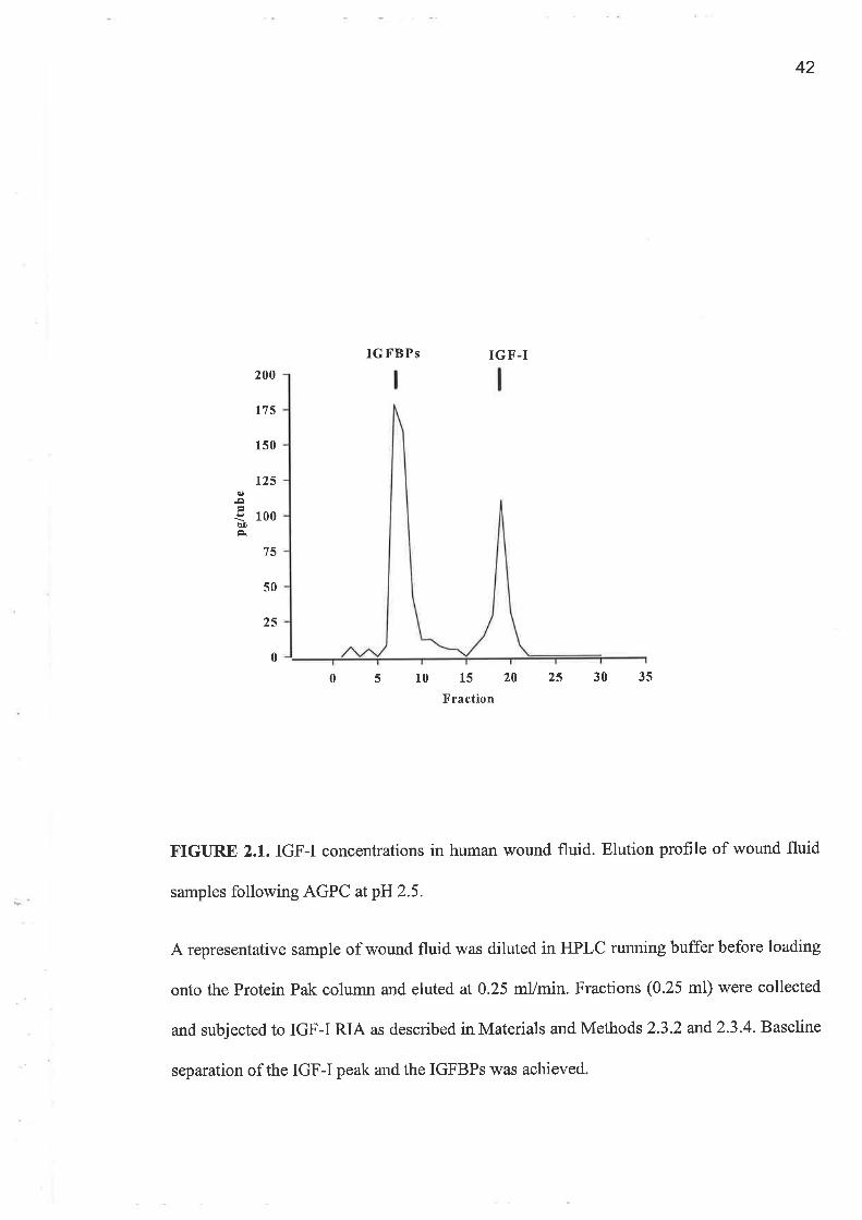
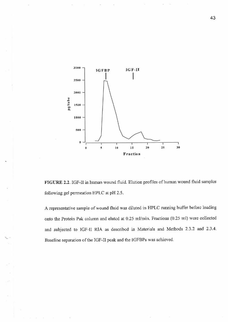
2.4.1. Acid gel permeation chromatography
AGPC which was used to separate IGFs from the IGFBPs was validated using WF and
plasma samples from patient 16 taken on the fifth day after operation. IGF-I and IGF-II
immunoactivity in the fractions recovered after AGPC are shown in figures 2.1' and 2.2
respectively. They demonstrate that baseline separation in wound fluid was achieved for
both peptides. Serum IGFBP and IGF-I separation had previously been demonstrated in our
laboratory using human samples (data not published). Each sample was subsequently
fractionated into four pools at a rate of 0.25 ml/min : pool 1 (2 mls collected at 6.5 min to
8.5 min) containing the IGFBP peak; pool 2 (0.5 ml at 8.5 to 9.0 min); pool 3 (2 mls at 9
min to 11 min) containing IGF-I and IGF-II and pool 4 (0.5 ml at pool at 11 min to 11.5
min).
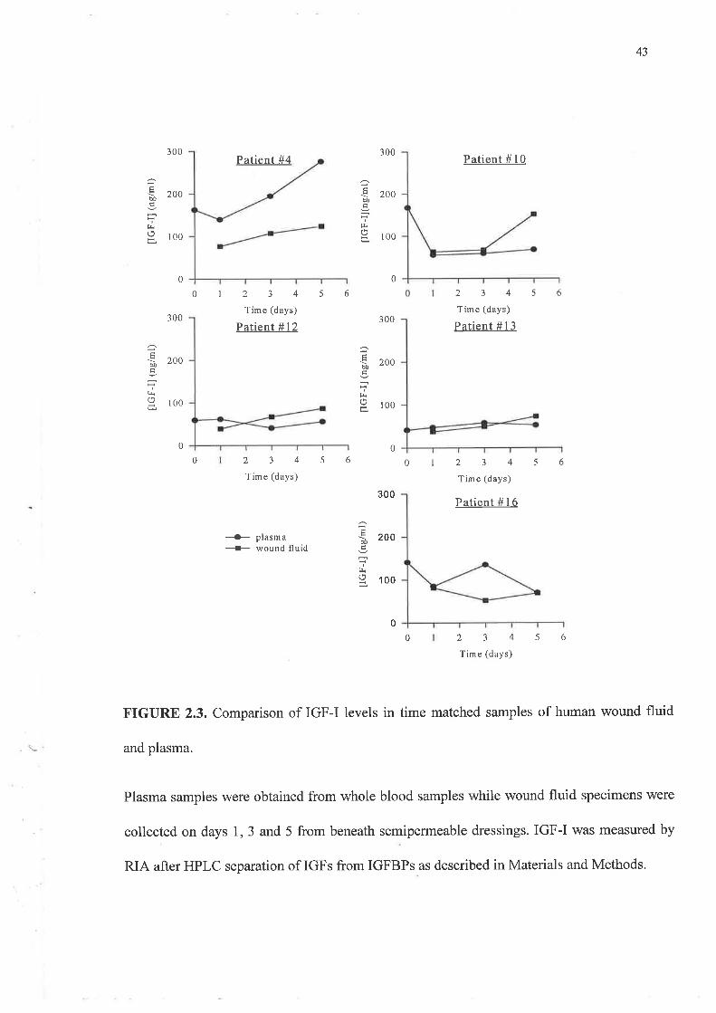
Figure 2.3 illustrates IGF-I accumulation in wound fluid samples collected during the first 5
days of healing. The values obtained were generally lower than those obtained for plasma
samples and lay between 50 and 100 nglml. Matched plasma IGF-I levels did not change
dramatically over the sampling period while IGF-I levels in the wound fluid rose marginally
during the same period.
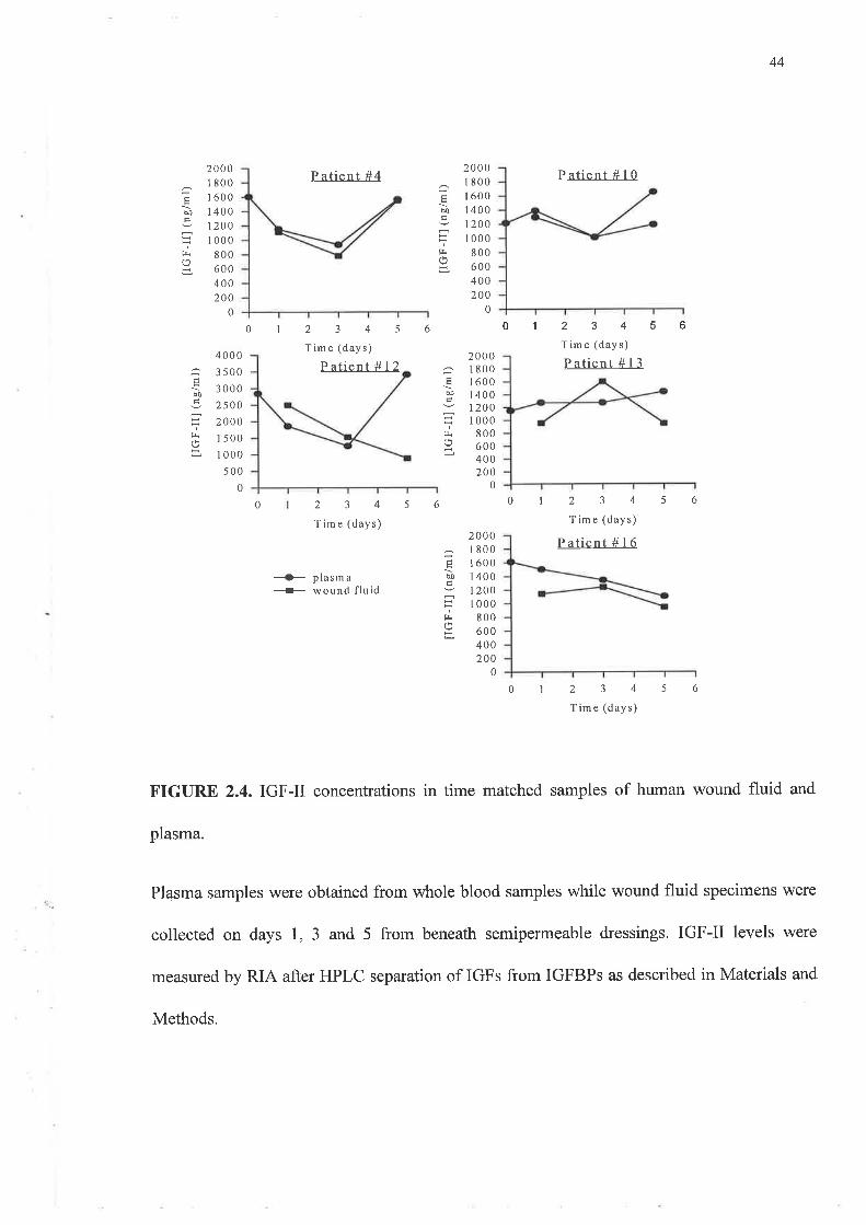
The IGF-II levels were between 5 and 7 fold higher than those observed for IGF-I by RIA.
These also showed no consistent pattem except that in 4 of the 5 patients the final plasma
levels were as high as or higher than those detected at day 0. This contrasted with the wound
fluid concentrations in that IGF-II levels declined between day 1 and day 5 samples. In 3 out
41
of 5 patients IGF-I levels showed no consistent time related pattem in wound fluid and
againthe plasma levels generally changed very little between day 0 and day 5.
42
IGFBPS IGF'-I
q)¡Þ¡)À
200
L75
150
125
100
50
25
0
75
05101520253035Fraction
FIGURE 2.1. IGF-I concentrations in human wound fluid. Elution profile of wound fluid
samples following AGPC atp}J25.
A representative sample of wound fluid was diluted in HPLC running buffer before loading
onto the Protein Pak column and eluted at 0.25 mVmin. Fractions (0.25 ml) rù¡ere collected
and subjected to IGF-I RIA as described in Materials and Methods 2.3.2 and2.3.4. Baseline
separation of the IGF-I peak and the IGFBPS was achieved.
43
3000
2500
2000
1500
1 000
500
o¡àt)È
IGFBP IG F-II
15
X'' raction
0
5 l0 20 25 30
FIGURE 2.2.IGF-IIin human wound fluid. Elution profiles of human wound fluid samples
following gel permeation HPLC atpE2.5.
A representative sample of wound fluid was diluted in HPLC running buffer before loading
onto the Protein Pak column and eluted at 0.25 mVmin. Fractions (0.25 ml) were collected
and subjected to IGF-II RIA as described in Materials and Methods 2.3.2 and 2.3.4'
Baseline separation of the IGF-II peak and the IGFBPs was achieved'
43
300 300
200
100
èD@
EI loo
Patient #4
1234Time (days)
Patient #12
234Time (days)
---ro- plasma----r- wound fluid
Patient # l0
2345Time (days)
Patient # l3
234Time (days)
Patient #16
234Time (days)
0 0
0 56
56
300
6
Ð
fr9 loo
u)
f!
300
200
100
0 0
0 0l 56
56
300
020
100
0
d)
f¡
0
FIGURE 2.3. Comparison of IGF-I levels in time matched samples of human wound fluid
and plasma.
Plasma samples were obtained from whole blood samples while wound fluid specimens were
collected on days 1, 3 and 5 from beneath semipermeable dressings. IGF-I was measured by
RIA after HPLC separation of IGFs from IGFBPs as described in Materials and Methods.
44
Éd)
fr
c
ilÉ
f¡
2000I 800r 600I 400I 2001000
800600400200
0
2000I 800I 6001 400I 200I 000
800600400200
0
P atient #4
2345Time (days)
P aiient #12
Patient #1 0
2345Time (days)
Patient #13
6
56
01
0
6
u
400035003000
25002000I 500
I 000
s000
20001 800I 6001 400I 200I 000
800600400200
0
01
0
EÐ
Éd)
1234T im e (days)
Patient # I 6
56234Time (days)
--O- plasma----¡- wound fluid
20001 800I 600I 4001200I 000
800600400200
0
01234s6Time (days)
FIGURE 2.4. IGF-II concentrations in time matched samples of human wound fluid and
plasma.
Plasma samples were obtained from whole blood samples while wound fluid specimens were
collected on days 1, 3 and 5 from beneath semipermeable dressings. IGF-II levels were
measured by RIA after HPLC separation of IGFs from IGFBPs as described in Materials and
Methods.
45
2.4.2. Acid Ethanol Extraction
The values for IGF-I in plasma and wound fluid following AE extraction to separate IGFs
from IGFBPs are illustrated in figures 2.5 and 2.6. These were about twice those observed
following HPLC separation. The levels observed ranged from approximately 100 up to 800
ng/ml for both biological fluids. Plasma and wound fluid levels of IGF-I using this technique
appeared to show greater similarity in both their variability and their actual levels compared to
similar samples subjected to HPLC. However greater variability was observed for IGF-II
levels during the same period. Interestingly the IGF-II values obtained in samples subjected to
AE extraction proved to be 4 to 7 times lower than samples subjected AGPC. This contrasted
with the results obtained for IGF-I where AE extracted values were higher than AGPC treated
values.
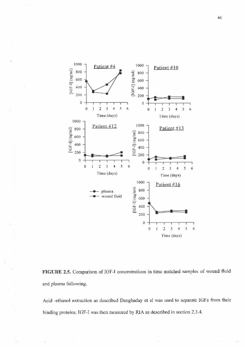
46
000
800
600
400
200
Patient#4
123456Time (days)
Patient #10
123456Time (days)
1 000
I sooÞ
þ uoo
=¿ 400o=, 200
à0
If¡r
1000
800
600
400
200
1000
E 8ooöI)
-9 oooIt: 400
É200
0
0 0
0123 4 5 6
Time (days)
Patient #12 Patient # l3
1234s6Time (days)
Patient #16+- plasma* wound fluid
012345Time (days)
FIGURE 2.5. Comparison of IGF-I concentrations in time matched samples of wound fluid
and plasma following.
Acid -ethanol extraction as described Daughaday et al was used to separate IGFs from their
0
1 000
àf)
If¡ro
â soo
S ooo
f, +oo
t1 2oo
00
00
6
binding proteins. IGF-I was then measured by RIA as described in section 2.3.4.
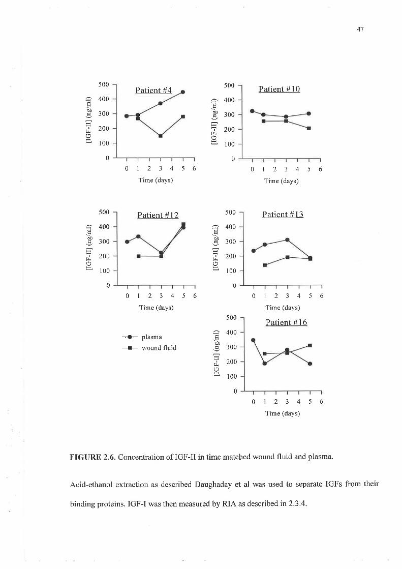
47
500
â 400
P 3oo
f 200Hr¡11 1oo
Òo
I
trr()
s00
400
300
200
100
0
Patient#4
01234s6Time (days)
Patient#12
0123456Time (days)
500 Patient #13
Patient #10
0123 4 5 6
Time (days)
0123456Time (days)
Patient #16
0
H
bo
IlrL,,
500
400
300
200
100
0
òt)
I
trr
400
300
200
100
0
--G- plasma
{- rüound fluid
0123 4 5 6
Time (days)
FIGURE 2.6. Concentration of IGF-II in time matched wound fluid and plasma.
Acid-ethanol extraction as described Daughaday et al was used to separate IGFs from their
500
400
300
200
100
0
öfJ
I
frr()
binding proteins. IGF-I was then measured by RIA as described in2.3.4
48
2.4.3. Comparison of AE Extraction and AGPC for Dissociation IGFBPs and IGFs.
The IGF values in plasma and wound fluid concentrations following AE extraction and AGPC
were compared using linear regression (figures 2.7 and2.8)
IGF-I values for the plasma samples demonstrated good correlation between concentrations
obtained using the two different extraction methods (p<0.001; r:0.84). 'When a similar
analysis was performed upon the results for IGF-II in human plasma no clear-cut relationship
was observed (1t:0.2; r0.54; frgve 2.7).
A similar pattern emerged for wound fluid, (figure 2.8) where IGF values following AE
extraction correlated well with those after AGPC (p<0.05, r:0.56). However the values for
IGF-II following the two extraction procedures correlated poorly (p:0.783; r:-0.075).
Electrophoresis and western ligand blots (V/LB) were performed upon wound fluid and
plasma samples that had been treated by acid-ethanol extraction to determine if IGFBPs
remained within the extracts. Illustrated in figure 2.9 are the residual IGFBPs in AE extracts
of both wound fluid and plasma samples. IGFBPs were detected in all samples except day I
wound fluid from patient 13 (Ql3WFd1) although band intensity varied dramatically between
patients and time points. All bands detected were of 30kDa or less and therefore consistent
with low molecular weight species of binding proteins. The specimens of Q12WFd3 and
Q12WFd1 also showed bands that were clearly within the region of l4kD. Plasma samples
retained higher quantities of low molecular weight IGFBPs than wound fluid.
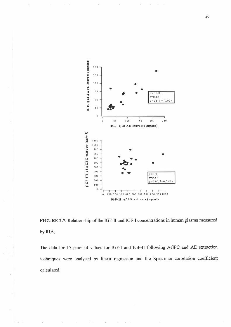
49
èoÉ
G
0
O
ê
t¡i
a
t!b¡É
alE
x
U
o
tsr
300
250
200
150
100
50
0
o
oa
a
oa
oa
p<u.uu I
f=0 84y:28.1 + 1.03x
.*
50
tIGF-Il of AE extracts (ng/m l)
I 100
I 000
900
800
700
600
500
400
300
200
100
0
0 100 200 300 400 500 600 700 800 900 1000
tIGI'-IIl of AE extracts (ng/ml)
FIGURE 2.7. Relationship of the IGF-II and IGF-I concentrations in human plasma measured
by RIA.
The data for 15 pairs of values for IGF-I and IGF-II following AGPC and AE extraction
techniques were analysed by linear regression and the Spearman correlation coefficient
calculated.
0 100 150 200 250
oa
a
-
a
.l-'aaa
aa
47 0 5+0 .266x
0
r=0 54
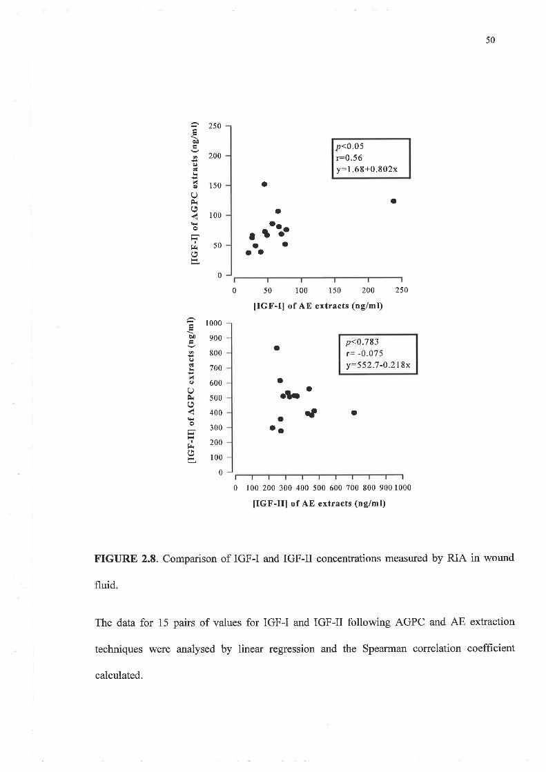
50
a
200
50
EÞ¡
ooçl
XQ)
QÈo
I
t¡(,
aO
èo
oq)cl
Nq)
(,)È(.'
o
I
t¡r(,
250
150
100
a
50 100 150 200
UGF-Il of AE extracts (nglmt)
p<0.05r:0.5 6
y:1,68+0.802x
a
250
p<0.783r= -0.075y--552.7-0.218x
. ¡tïaaaO
0
0
a
ol¡a ct o
aa
0 100 200 300 400 500 ó00 700 800 900 1000
UGF-III of AE extracts (ng/ml)
FIGURE 2.8. Comparison of IGF-I and IGF-II concentrations measured by RIA in wound
fluid.
The data for 15 pairs of values for IGF-I and IGF-II following AGPC and AE extraction
techniques were analysed by linear regression and the Spearman correlation coefficient
1 000
900
800
700
600
500
400
300
200
100
0
calculated.
FIGURE 2.9 V/estern Ligand Blot of Acid Ethanol extracts of matched human wound
fluid and serum from five patients undergoing skin grafting procedures.
Samples were incubated in acid-ethanol after Daughaday et al (190). The supernatant
was removed and freeze-dried after separation. Freeze dried protein was reconstituted
with 50 pl sample buffer and 40 pl of the solution was loaded into each well and
electrophoresed on 12.5% SDS-PAGE gels. After electrotransfer of protein to
nitrocellulose f,rlters the filters were probed with ¡I"¡-IGF-II and the bands visualised
autoradiographically after exposure at -70" C for 12 days.
(¡,\U¡t
(¡,\UÞ
t¡FUÞ
Ql3PIDO
Ql3PlDl
Q13PrD3
Ql6PIDO
Q16PlD1
Q16PrD3
Q16WfD1
Q16WFD3
Q1OF"WD3
Ql0WFD
Q12PlDû
Q12PrD1
Q12WTD3
Q12WFD1
Q12PrD3
Q4PtD0
Q4WFD1
Q4PIDl
Q4WFD3
Q4PrD3
Q10PrD0
Q1OPID1
Q10PrD3
FIGURE 2.10. WLB of raw wound fluid and plasma.
Wound fluid and plasma were collected as described in Methods. One microlitre of
plasma or 4 ¡ú of wound fluid were mixed with non-reducing sample buffer and
electrophoresed on 12.5% SDS-PAGE gels. Proteins were transfered to nitrocellulose
filters which were then probed with ¡125I1-tGf-IL The bands were visualised
autoradiographically after exposure at -70"C for 7 days.
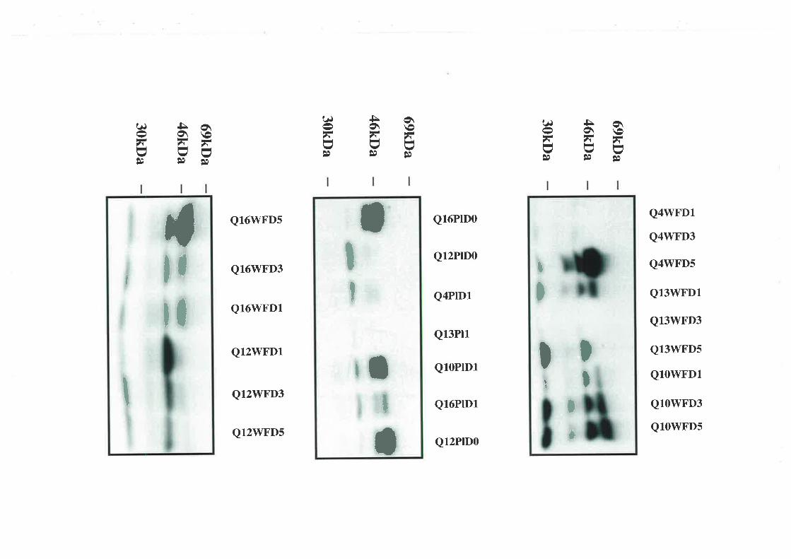
o\\o,\UÊl
5o\-ar-v!)
(¿J
,\I\JÊe
o\\o,\-vÞ
5o\,\UÞ,
?¿J
,\UÞ
àeFF\J \JÞÊl
t¿)
,\I\Jte
tI ll
tl
II
Ql6WFDs
Q16WFD3
Q16WFD1
Q12WFD1
Q12WFD3
Ql2WFDs
Q16PrD0
Q12PrD0
Q4PrD1
Q13Pl1
Q10PrD1
Q16PrD1
Q12PrD0
Q4WFD1
Q4WFD3
Q4\ryFDs
Q13WFD1
Q13\ilFD3
Ql3WFDs
Q1OWFD1
Q1OWFD3
QlOWFDs
t
It
a
Ir
I
$
t
I
IIt
It
I*
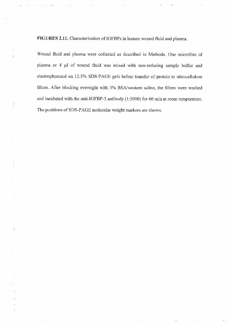
FIGURES 2.11. Characterisation of IGFBPs in human wound fluid and plasma.
'Wound fluid and plasma were collected as described in Methods. One microlitre of
plasma or 4 pl of wound fluid was mixed with non-reducing sample buffer and
electrophoresed on 12.5% SDS-PAGE gels before transfer of protein to nitrocellulose
filters. After blocking overnight with 3yo BSA/western saline, the filters were washed
and incubated with the anti-IGFBP-3 antibody (1:5000) for 60 min at room temperature.
The positions of SDS-PAGE molecular weight markers are shown.
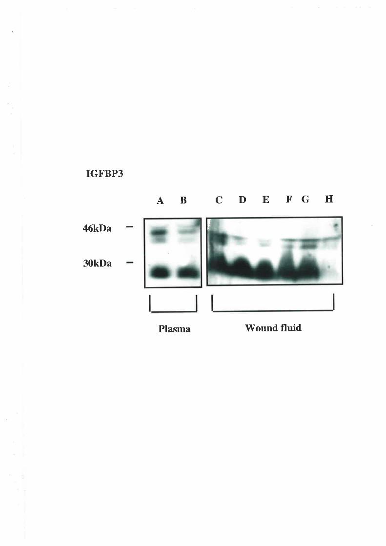
IGFBP3
46kDa
30kDa
A B C D E FG H
Plasma Wound fluid
F
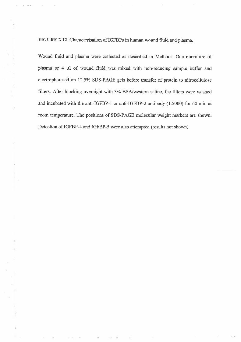
FIGURE 2.12. Characterisation of IGFBPs in human wound fluid and plasma.
Wound fluid and plasma were collected as described in Methods. One microlitre of
plasma or 4 ¡rl of wound fluid was mixed with non-reducing sample buffer and
electrophoresed on 125% SDS-PAGE gels before transfer of protein to nitrocellulose
filters. After blocking overnight with 3Yo BSA/western saline, the hlters were washed
and incubated with the anti-IGFBP-l or anti-IGFBP-2 antrbody (1:5000) for 60 min at
room temperature. The positions of SDS-PAGE molecular weight markers are shown.
Detection of IGFBP-4 and IGFBP-5 were also attempted (results not shown).
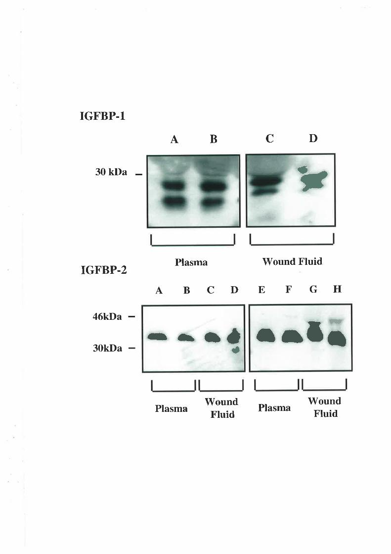
IGFBP.l
30 kDa
IGFBP.2
46kDa r¡t !7
I
AB D
Plasma Wound Fluid
ABCDEFGH
C
30kDa
PlasmaWoundFluid
PlasmaWoundFtuid
Y
-{b¡ úa
57
2.5. DISCUSSION
This series of experiments has sought to quantify changes in IGF-I and II levels in human
wound fluid harvested from partral thickness dermal wounds and to compare these results
with temporally matched peripheral blood samples. IGFBPs within wound fluid were
identifred along with changes in their profiles during the time course of sampling. These
results demonstrated that IGFs are found in measurable concentrations in human wound
fluid.
Minimising the impact of systemic factors was important in choosing our model to measure
the concentrations of IGFs in wound fluid so that realistic conclusions could be drawn about
the changes in homeostasis of the IGF system relating to the healing of a partial thickness
wound. Hov/ever, IGF-I reaches the wound from a variety of sources including platelet
degranulation (15) and leakage through damaged capillaries. Fibroblasts and other cell lines
also synthesise IGFs and their binding proteins. Exudates from mastectomy drains (112) and
wound cylinders (96,126,191) have previously been analysed for IGF-I content. In those
studies IGF-I levels increased transiently. Alternatively, as described by Xu et al (187),
'suction' blisters allow collection of interstitial fluid that can then be analysed for IGFs and
their binding proteins. However this method tends to separate epidermis from basement
membrane and the dermis may not be exposed. Therefore any aspirate may not accurately
reflect dermal growth factor production.
The extraction method employed to separate IGFs from their binding proteins may
influence significantly the final values obtained for IGFs measured by RIA. Many
techniques have been devised to measure IGF concentrations. Bioassays were initially used,
5B
however because of the complexity of IGF physiology, the results obtained with these
techniques were unreliable and difficult to reproduce (196). Several studies and reviews
have been published in recent years emphasising the short comings of the various
techniques and provide answers to the problem of IGFBP interference in measuring IGF-I
and II concentrations in various biological fluids (173,188,197-200). Despite a consensus
Ihat acid gel permeation chromatography, originally described by Horner et al in 1978, is
the most effective method of IGFBP extraction, other methods continue to be used in the
endocrine and surgical literature. Using our model, we concluded that IGF-I and IGF-II
levels measured by RIA after either AE or AGPC extraction procedures generally did not
alter dramatically during the healing of relatively minor wounds such as a partial thickness
skin graft donor site wound. These data were consistent with Antoniades et al (189) who
demonstrated little change in IGF-I receptor and IGF-I mRNA expression in acute wounds
that heal primarily by re-epithelialisation.
Western ligand blots and immunoblots identified IGFBP-1 , -2, -3 and -4 in raw wound fluid
and plasma samples. In addition, low molecular weight fragments of IGFBP-3 and IGFBP-3
were found in wound fluid samples. Various proteases such as collagenase, gelatinase and
other metalloproteinases are expressed in cutaneous wounds (201) and excreted into wound
fluid (202,203). IGFBP-3 proteolytic activity has also been described (187,191). These
proteases while not specifically assayed, may be responsible for the lower molecular weight
immunoactivity that was observed.
'Westem ligand blots were also performed on acid-ethanol extracts examining for residual
IGFBP. Residual IGFBPs were detected in the extracts thus invalidating this method as a
technique for dissociating IGFs completely from IGFBPs in human wound fluid and plasma.
59
Linear regression analysis was used to compare these results with those values obtained
using acid gel permeation HPLC. Correlation of AE extract levels of IGFs with AGPC
extracted samples demonstrated the problems of different separating techniques and the
influence upon IGF levels as measured by RIA.
Acidification of samples in a solution pH 2.5 is critical to adequate separation of the IGFs
from IGFBPs as found by Owens et al (192). That particular paper detailed clearly the
protocol for acid gel permeation HPLC and compared porcine IGF-I levels obtained with
this technique and those following AE extraction. Interestingly a similar trend was found
with AE extracted IGF-I concentrations by Owens et al and this study. This method of
extraction produced IGF-I values lower than those following the HPLC technique. Adequate
acidification \ ¡as especially applicable to IGF-II as this will totally dissociate from the
IGFBPs at a pH of less than 2.8. Consequently our laboratory conducts HPLC assays at a
pH of 2.5 to maximise dissociation. Owens et al (I92) and Mesiano et al (204) suggest that
failure to maintain the samples at this pH may precipitate re-association of residual IGFBPs
with IGFs when the solution was neutralised.
Neutralisation of the solution occurred with the addition of 0.855M Tris base before RIA
when AE extraction was used. The HPLC method avoided this problem by fractionating the
samples into pools that contain IGFBPs (pool 1) or IGFs (pool 3) so that the elutates were
physically separated into pools containing IGFBPs or IGFs. Low molecular weight binding
proteins or their degraded fragments may still fractionate into pool 3 and subsequently lead
to inaccuracies. However most IGFBPs are detectable above 241<Da and would be excluded
in this way from the IGF pool. The WLB of whole wound fluid and plasma suggested that
BPs greater than 30kDa comprised the majority of IGFBPs in the samples tested and
60
therefore supported the above argument for the use of AGPC in preference to AE.
Furthermore, results in this study showed that despite acid conditions and the precipitation
of the pellet the resultant supematant can still contain IGFBPs. This clearly demonstrates
the inadequacy of the unmodifred AE extraction technique. Therefore its use can lead to
inaccuracies in the measurement of IGF levels, particularly IGF-II, in human biological
fluids that contain low molecular weight binding proteins.
While correlation of AGPC derived IGF-I levels with those obtained following AE
extraction suggested that these differences may not be marked, this is not true for IGF-II.
This was most evident when measuring IGF-II in wound fluid. This disparity along with the
reasonable correlation in IGF-I values could be related to the greater affinity IGF-II has for
the IGFBPs overall and IGFBP-2 specifically. Clearly the separation profiles for IGF-I and
IGF-II in wound fluid imply larger quantities of binding protein in the IGF-II assays than
the immunoreactivity of IGF-I would suggest (figures 2.1. and2.2).
Consideration of the complex set of equilibria that are established in performing an RIA
may provide a clearer understanding of the lower values generally seen for both IGFs but
particularly IGF-II. If a method is used that dissociates IGFBPs from IGFs less efficiently
than another it will produce less free IGF to compete against ¡125t1-IGn for binding sites on
the primary antibody. Bound radioactivity is quantified and used to calculate the amount of
IGF present in the pellet. Increasing radioactivity reflects lower levels of free unlabelled
IGF in the extract. Three other factors can influence the amount of free unlabelled IGF:
1) the pH of the solution;
2) the overall concentration of the IGFBPs; and
61
3) whether they bind to free IGF-I when the solution is neutralised.
As IGF-II has a higher affinity for IGFBPs, IGF-II assays are likely to be more significantly
affected. Two methods have been proposed to circumvent this effect. One method requires
saturation of the extracted samples with IGF-I if IGF-II is being assayed and vice versa if
IGF-I is being measured (204). The other method uses an IGF analogue with minimal
binding affinity for IGFBPs, as the tracer (199).
In conclusion, this study has demonstrated the presence of levels of IGF-I and IGF-II in
wound fluid taken from a pafüal thickness wound that were generally similar for time
matched plasma samples. The measurement of IGFs in biological fluids may be confounded
by incomplete separation of these peptides from their binding proteins as demonstrated by
the comparison of IGF values obtained for the two different extraction techniques described
in Materials and Methods. The presence of these IGFBPs has been confirmed using WLBs
and they have been identified using immunoblots. WLBs of AE extracted samples
confirmed the presence of residual low molecular weight binding proteins. Correlation of
values obtained following AGPC or AE extraction supported the conclusion that IGF-II
levels determined following AE extraction will prove to be inaccurate compared to results
obtained following extraction using AGPC.
The presence of IGF-I in physiological concentrations prompted a series of studies aimed at
investigating the role of IGFs in wound healing and to examine the influence of altering the
affinity of IGF-I for its binding proteins.
62
!
CHAPTER 3
IGF-I AND ITS ANALOGT]ES:
IN WTRO RESPONSES OF F'IBROBLASTS
3.1. INTRODUCTION
3.1.1.'lVound Contraction
3.1.2. Growth Factors and Fibroblast Activity
3.1.3. Objectives
3.2. MATERIALS
3.3. METHODS
3.3.1. Harvesting of Fibroblasts
3.3.2. Fibroblast Growth Assay
3.3.3. Protein Synthesis in Response to IGFs
3.3.4. The Fibroblast Populated Collagen Lattice
3.3.5. Incorporation of 13n¡-ttrymidine: A Model of DNA Synthesis
3.3.6. Statistics
63
3.4. RESIILTS
3.4.1. The fnfluence of GH and IGF-I upon Fibroblast Activity
3.4.2. Skin Fibroblast Responses to IGF-I Analogues
3.4.3. DNA Synthesis: Comparison of Growth factors
3.5. DISCUSSION
64
3.1. INTRODUCTION
The final quality of a healed wound is determined by the scar produced. Fibroblast activity
within the wound is responsible for the deposition and maturation of the collagen matrix
that ultimately forms this scar Q}s).Early in healing, fibroblasts also aid in the production
of granulation tissue and wound edge contraction. Granulation tissue is composed of
proliferating fìbroblasts, ground substance proteins and proliferating endothelium and
provides the foundation upon which re-epithelialisation will occur (18).
Migration of fibroblasts into a newly formed wound commences at about day three post-
wounding. Chemoattractants that promote fibroblast migration include thrombin Q06),
tissue plasminogen activator (t-PA), ECM fragments (20), fibronectin (207), cytokines such
as PDGF (208) and TGFB (209), C5a from the complement cascade (210), histamine
released from mast cells (211), interleukins as well as components of fibroblast and
epithelial conditioned media Ql2).Proliferation of fibroblasts within the wound is also
stimulated by a variety of influences including the relative hypoxia found in the centre of
wounds (9) and growrh factors such as IGF-I, PDGF Ql3-217) and TGFB (65).
The proliferative phase features phenotypic changes to the fibroblast population (218) as
well as deposition of a fibronectin and collagen matrix. The appearance and regression of
myofibroblasts within wounded tissue are associated with the commencement and
completion of wound contraction Ql9). As a presumed fibroblast derivative was first
characterised by Gabbiani (50,51) in 1971, it is the cell believed responsible for wound
65
contraction. Myofibroblast activities are regulated by various factors including ECM
components (56,220) and growth factors, for example TGFB Q2l).
Bell demonstrated with an in vitro model of dermal contraction known as the fibroblast
populated collagen lattice, that the contractile response of the fibroblast may be influenced
by its proliferative potential as well as its phenotype (57). Normal dermal flrbroblasts, being
quiescent cells, do not appear to exhibit the same contractile activity as fibroblasts derived
from a wound (63). This may also be related to collagen maturity as this too has been shown
to modulate fibroblast mediated contraction as shown by Ehrlich's studies of gel contraction
which utilised various forms of collagen (89,220). Ehrlich's results indicate that matrix
composed of the more immature type III collagen will contract faster than one rich in type I
collagen.
Finesmith et al (66) have provided further evidence that the age of wounds influences cell-
mediated wound contraction. These authors examined the contraction of collagen gels
seeded with fibroblasts derived from granulation tissue that had been sampled at various
time points in a chronic healing wound. Their findings indicated that rates of gel contraction
responses to TGF-B increased in a dose dependent fashion and these responses were even
more pronounced with cells extracted from older granulation tissue. Growth factor
responses in these studies were also influenced by the timing of peptide application as
demonstrated by pretreatment of the gels with bFGF. Furthermore these authors showed that
this tended to retard TGF-p mediated contraction in those gels seeded with cells derived
from younger granulationtissue. Aphenotypic change is therefore required forthese cells to
induce wound contraction. Such a change increases the complexity of this dynamic process
66
which requires the interaction of cells, ECM and growth factors to generate forces of
contraction that can be attributed to fibroblast locomotion (52,56).
3.1.3. Growth Factors and Fibroblast Activity
Many growth factors have been linked with the activity of fibroblasts generally but also
specifically within the wound (222). They may be chemotactic, mitogenic or induce
synthesis of components of the extracellular matrix. Their influences may be effected by
paracrine or autocrine mechanisms (17,223,224) and those growth factors produced by the
fibroblasts themselves may act similarly upon other cell types, for example keratinocytes
(22s).
GH has been shown to promote growth in children with growth retardation (226) and it can
reverse catabolic states (227) and it has mitogenic effects upon mesenchymal tissues. Well
recognised for its ability to promote growth of cartilage and cell proliferation in osteoblast
and chondrocyte cultures, it also has anabolic efTects upon soft tissue in general (101,228-
233). Until recently GH was the only growth factor to have an important clinical application
in wound healing (98,104). As with most of its anabolic activities, GH probably acts
indirectly in wound repair to influence healing as target tissues exhibit upregulation of IGF-I
protein and mRNA production in response to GH (96).
The mitogenic activity of IGF-I Q34),its role as the second messenger for GH in fibroblast
cultures (124) and its presence in wound fluid (112) provide evidence that this growth factor
has a signiflrcant role in wound repair. Iin vitro it has the capacity to regulate endothelial
proliferation (11), collagen synthesis by fibroblasts (123) and to promote keratinocyte
67
migration and growth (146,223) in a paracrine fashion. IGF-I affects cellular activity in a
paracrine malìner independently of GH in other systems such as chondrocyte cultures Q35)
Much of the literature surrounding IGF-I induced fibroblast activity in vitro indicates that
some other factor is also required to maximise the response to IGF-I. The binding proteins,
IGFBP-I (236) and IGFBP-3 (237,238), PDGF (214), TNF-cr and TGF-pl Q38) and even
sodium chloride (239) influence the growth and contractile responses of fibroblasts to IGF-L
TGF-PI is renowned for its capacity to promote collagen synthesis and to influence matrix
reorganisation. It is associated with keloid scars (65,88,240,241), adult-type scar formation
(242,243) and has been implicated as a modulator of IGF-I bioactivity by upregulating
IGFBP mRNA expression (23S) Like IGF-I, TGF-P may also play a key role in wound
contraction either through its influence upon IGFBP physiology or by direct action on
fibroblasts as suggested by Pena et al and Montesano and Orci Q44,245).In animal studies
PDGF has been shown to be important in activating an IGF-I related response and generally
this is gteater than that achieved with either IGF-I or PDGF alone (46,128,129,134,246).
Consequently, IGF-I has earned the reputation as a 'þrogression factor" rather than a
"competence factor", a concept initially proffered by Van Wyk (21a) As a progression
factor it promotes cell growth and activity rather than simply rendering cells competent to
enter the cell growth cycle.
The aspect of IGF physiology that presents an obstacle to assessing IGF-I's potency as a
mitogen has been its family of carrier proteins, the IGFBPs. These peptides are produced by
cultured cells as well as existing in vivo. Their influence upon in vitro models is
unpredictable as they may either potentiate or inhibit IGF activity Q47).The development
of recombinant techniques for the production of peptides such as IGF-I has resulted in the
68
design of new growth factors or analogues of IGF-I and -II (741 ,248). The potencies of the
analogues, long Arg3 IGF-I (LR3 IGF-I) and des(1-3)IGF-I have been assessed in cell
cultures by examining [3H]Jeucine incorporation by L6 myoblasts, hepatoma cells and
growth of murine fibroblasts (BALB cl3T3 fibroblasts) (140,249). These peptides exhibit
greater activity in vitro than their parent peptides due largely to the reduced affinity they
demonstrate for the IGFBPs. The effect of LR3 IGF-I in cultures of human fibroblasts is not
affected by the addition of IGFBP-3 nor is its effect influenced by other growth factors that
increase IGFBP concentrations in tissue fluid as demonstrated by Yateman et al (238). This
increased activity is not necessarily associated with greater affinity for the type I IGF
receptor as LR3IGF-I actually has less affinity for this receptor than IGF-I (140). However,
little information is available about their comparative effrcacy in models incorporating
human skin fibroblasts.
3.1.4. Objectives
As the second messenger for GH, it is reasonable to assume that IGF-I's activity in vitro
would be equal or better than GH and consequently IGF analogues known to have reduced
afñnity for IGFBPs should exhibit greater potency than IGF-I. Therefore tissue culture
assays were performed to examine:
o The activity of GH in models of fibroblast growth and fibroblast related gel contraction;
o The relative response of IGF-I compared to GH,
. The relative potency of IGF-I, IGF-II, GH and two IGF analogues, namely des(1-3) IGF-
I, LRfIGF-I.
69
3.2. MATERIALS
All IGF-I and -II peptides (recombinant human proteins and analogues) were obtained from
GroPep Pty Ltd (Adelaide, SA" Australia) and were reconstituted in 0.01M HCI from
lyophilised powders. Growth hormone was purchased from Bresatec (Adelaide, SA
Australia). All peptides were of receptor grade purity (95% by IIPLC) for all in vitro
studies. TGF-PI and bFGF were purchased from Austral Biologicals (San Ramon, CL
USA), PDGF-AB from UBI (Upstate Biotechnology Incorporated, Lake Placid, NY, USA)
and EGF was supplied by Chiron Corporation, (Emeryville, CA" USA). Bovine serum
albumin (BSA; Sigma, Fraction V, RIA grade) was purchased from Sigma Chemicals (St
Louis, MO, USA). Dulbecco',s modified Eagle's medium (DMEM; Gibco BRL,
Gaithersburg, MD, USA) and foetal bovine serum (FBS; Cytosystems, Melbourne, VIC,
Australia) were supplemented with benzylpenicillin (6}mgll, CSL, Australia.), streptomycin
(100¡rg/ml; Jurox, Silverwater, NSW, Australia) and amphotericin-B (lmg4; Bristol-
Meyers-Squibb Pharmaceuticals, Noble Pk, \IIC, Australia.). Radiolabelled isotopes (L-
l2))Hf-proline, [3H]-inulin and methyl [3f{-thymidine) were obtained from Amersham
(Little Chalfort, Buckinghamshire, U.K.). Serum free medium (SFM) was prepared using
DMEM and O.lYo wtlvol BSA. All reagents for phosphate buffered saline (PBS) were
obtained from AnalaR BDH Chemicals.
Two human diploid dermal fibroblast cell lines were obtained from the Women's and
Children's Hospital, North Adelaide, South Australia. These were derived from a one day
old neonate who had died with congenital abnormalities (SF3l69) and from a 7 month old
70
infant who had died from a respiratory illness (SF1967) All stocks were maintained in
75cnf flasks (Corning Inc, Corning, NY, USA).
71
3.3. METHODS
3.3.1. Ilarvesting Protocol for Fibroblasts
Fibroblast cultures were used at passages I I to 14 and grown to confluence in lOYo
FBS/DMEM in Costar Tzs tissue culture flasks in a humidified atmosphere of 5Yo CO¿ at
37"C.
The cells were harvested when confluent using 0.025yo trypsiniO.OlM EDTA. Ten per cent
FBS/DMEM was added after two minutes to neutralise the trypsin. The suspension of cells
was next transferred to 10ml eppendorf tubes and centrifuged at 4'C, 1500 rpm for five
minutes. The pellet was washed twice more using SFM before determining cell numbers
and viability using a haemocytometer with trypan blue exclusion. The suspension was again
centrifuged and the pellet mixed with lml of SFM. An appropriate fraction was removed for
use in the tissue culture experiments described.
All steps involving cell harvesting, preparation of solutions, plating of cells and formation
of FPCLs, were performed under aseptic conditions in a laminar flow hood. A1l solutions
apart from collagen were passed through a 22¡tm x 47mm filter (Millipore) prior to use in
tissue culture experiments.
3.3.2. Fibroblast Growth Assay
Passage 12 SF 3169 human fibroblasts were subcultured at a density of 4 x10a cells in 100p1
of 10% FBS/DMEM per well of 96-well tissue culture plates (Nunclon, Denmark). The
72
plates were incubated overnight to encourage cell attachment. On the second day the plates
were washed twice with SFM and incubated for a further two hours in SFM. Treatment
solutions were added to the fibroblast monolayer after aspiration of the wash medium.
Following a further 48 hour incubation, cell growth was assessed using the methylene blue
dye binding assay described by Oliver et al (250). Briefly, methanol (100%) was used to fix
the cells prior to staining with methylene blue in 0.01M borate buffer, pH 8.5. Following
five washes with borate buffer, the methylene blue bound to the cell monolayers was eluted
with acidified ethanol (1:1 v/v 0.1N HcVethanol). Photometric analysis was performed
using an automated vertical light path photometer (Dynatec). These results provided an
index of optical density at the filtered wavelength of 630nm (Aøro) that reliably reflected
changes in the mass of cells in the monolayers
3.3.3. Protein Synthesis in Response to IGFs.
Confluent monolayers of passage 12 dermal fibroblasts (SF3169), cultured in24-well tissue
culture plates (Nunclon, Denmark), were washed with SFM and the cells incubated
overnight in SFM prior to addition of treatments. L-[2,3-3l{-proline was diluted in SFM and
the equivalent of 0.5 ¡rCi/ml of treatment was added with each solution to individual wells.
The plates were incubated for 24hoursbefore harvesting the newly synthesised protein'
The cell monolayers were washed twice with Hanks' balanced salt solution (FIBSS), once
with 5Yo solution of trichloroacetic acid (TCA) to precipitate the cellular protein and finally
with deionised water. Synthesised protein was solubilised using O.7Yo Triton X-100/0'5 M
73
NaOH. Duplicate 100p1 subsamples from each well were each dilutedin2ml of scintillation
fluid (Ultima GoldrM LSC, Packard), before measuring the radioactivity on a B-counter
3.3.4. The Fibroblast Populated Collagen Lattice
Type I collagen was acid-extracted from rat tail tendons using the technique described by
Bell et al (57) This model has also been applied to other combinations of cell types such as
endothelial cells and keratocytes (67,62) or other ECM proteins such as fibronectin (251). A
modification of this model has been devised in our laboratory that incorporates [3f!-inulin
into the gel as a marker of the contractile response. It remains inert and it is not incorporated
by the cells. It therefore tends to be expelled from the gel as it contracts. Residual
radioactivity compared to that of control treated gels provides an index of the potency of a
reagent to promote fibroblast-mediated contraction. This modification to the FPCL model
measures residual radioactivity after a 48 hour incubation. This time point was chosen as the
rate of gel contraction is maximal in this period and generally declines thereafter
(57,64,220).
A standard gel mixture was prepared as follows:
The rat tail derived collagen was added to deionised (Milli-Q) water and 2 x DMEM in a
volumetric ratio of 2:3:5 at 4"C. [3H]-inulin (1 ¡rCilml) was added according to the formula:
Volume [3H]-inulin (frl):6000 x number of gels required x 20
counts/¡rl inulin stock
74
IMNaOH (1.25¡úlmlgel mixture) was used to promote polymerisation of the collagen. The
gel mixture (50OpVwell) was seeded with76-20 x 104 human dermal fibroblasts (SF3169)
per ml of gel and poured into 48-well tissue culture plates (Costar, Cambridge, MA, USA).
The plates were incub ated at 37"C in a humidified atmosphere of 5Yo COz until the gels had
polymerised. 'Rimming' of the gels with a25FG hypodermic syringe needle released them
from the well surface and allowed them to float freely in the treatment media. Doubling
dilutions of the peptides were added in final volumes of 500 pl SFM to each well.
After incubating for another 48 hours the FPCLs were transferred to 2 ml of scintillant
(Ultima GoldrM LSC, Packard), shaken and allowed to dissolve for another 48 hours to
release the residual radioactivity. Each concentration of peptide was tested in triplicate and
each experiment repeated to confirm initial results. Gel size was consistent in all
experiments and their preparation was carried out under the same conditions.
75
3.3.5. Incorporation of 13n¡-tnymidine: A Model of DNA Synthesis
Fibroblasts were subcultured at a density of 2 x 10⁴ cells/ well (l6mm diameter) in serum
supplemented medium and cultured for 72 hours af 37"C in an atmosphere of 5Yo COz. Once
the monolayers \¡/ere almost confluent they were washed with SFM and incubated for 24
hours in SFM. The growth factor dilutions were added at a final volume of 100 pVwell and
the cultures pulsed 6 hours later with the [3U]-thymidine (0.5 pCTml). After 18 hours the
plates were washed twice with PBS, once with l0% TCA at 4"C and the cells lysed using
100 pl 0.5M NaOH. Samples of 85¡rl were taken from each well and placed in scintillant
(2ml) prior to reading of the radioactivity on the p-counter.
3.3.6. Statistics
All results represented pooled data from duplicate experiments and are expressed as mean f
SEM. All analyses were conducted using Sigmastat v.2, (landel Scientihc, San Ramon, CA
USA). One way analysis of variance was performed where multiple treatment solutions
were compared. Levels of significance were p< 0.05 compared with serum free medium
unless otherwise indicated.
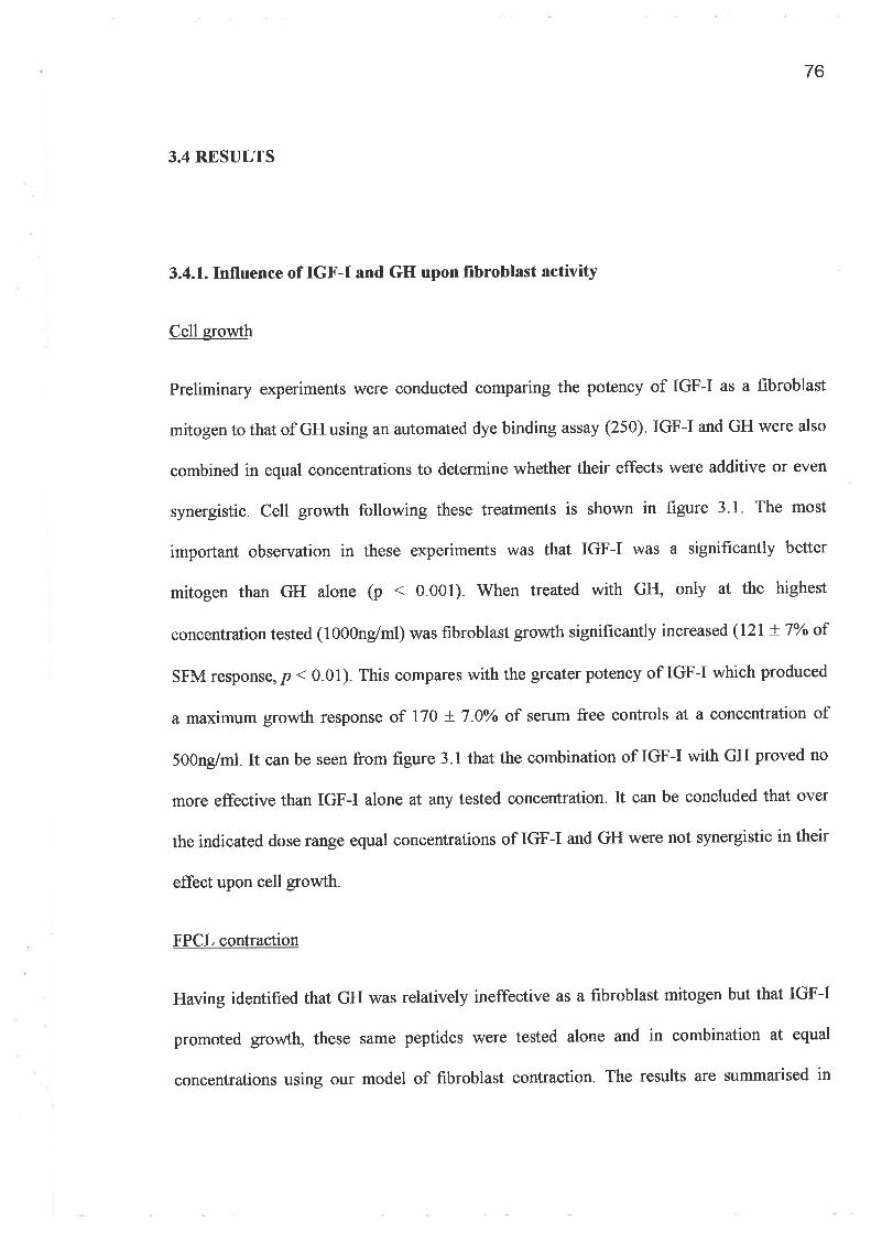
76
3.4 RESULTS
3.4.1. fnfluence of IGF-I and GH upon fibroblast activity
Cell growth
Preliminary experiments u/ere conducted comparing the potency of IGF-I as a fibroblast
mitogen to that of GH using an automated dye binding assay (250). IGF-I and GH were also
combined in equal concentrations to determine whether their effects were additive or even
synergistic. Cell growth following these treatments is shown in figure 3.1. The most
important observation in these experiments v/as that IGF-I was a significantly better
mitogen than GH alone (p < 0 001). When treated with GH, only at the highest
concentration tested (1000ng/ml) was fibroblast growth significantly increased (121 + 7%o of
SFM response, p < 0.01). This compares with the greater potency of IGF-I which produced
a maximum growth response of 170 + 1.OYo of serum free controls at a concentration of
500ng/ml. It can be seen from figure 3.1 that the combination of IGF-I with GH proved no
more effective than IGF-I alone at any tested concentration. It can be concluded that over
the indicated dose range equal concentrations of IGF-I and GH were not synergistic in their
effect upon cell growth.
FPCL contraction
Having identified that GH was relatively ineffective as a fibroblast mitogen but that IGF-I
promoted growth, these same peptides were tested alone and in combination at equal
concentrations using our model of fibroblast contraction. The results are summarised in
77
figure 3.3. When applied at increasing concentrations, the residual radioactivity in the gel
after 48 hours decreased across all IGF-I concentrations indicating significant contraction of
the gel in response to IGF-I. GH failed to significantly influence the contractile response
when used alone. Its maximal effect was noted at a concentration of 250 nglml (contraction
to gOYo of residual radioactivity of SFM treated gels). In comparison, IGF-I produced
signicant gel contraction with treated gels having a minimum residual radioactivity that was
72% of control gels after 48 hours. Furthermore, GH and IGF-I mixed in equal
concentrations were no more effective than IGF-I alone indicating no synergistic
relationship between the two peptides in this model.
Similar experiments testing the concentration range of 7.8 ng/ml to 250 nglml once again
demonstrated the weak activity of GH in the FPCL model with maximal gel contraction to
88yo+ 2Yo at" 125 nglml.In contrast IGF-I was able to induce contraction of the gels to 60%
of the size of time matched controls within 48 hours. The relative inactivity of GH in this
range confirmed GH to be a weak fibroblast mitogen at physiological concentrations.
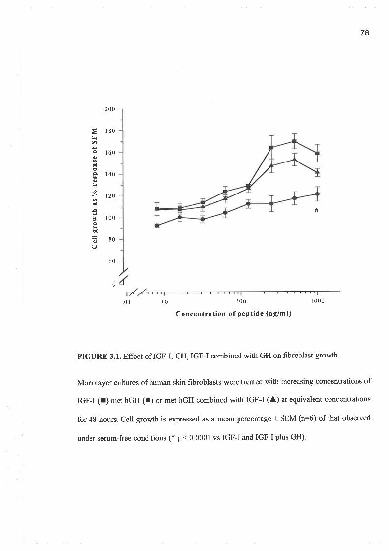
78
h(t)
ooooÊoq.)
socË
Èoè0
(,)(J
200
180
160
t40
120
100
80
60
*
0
.01 10 1000
IrIGURE 3.1. Effect of IGF-I, GH,IGF-I combined with GH on fibroblast growth.
Monolayer cultures of human skin fibroblasts were treated with increasing concentrations of
IGF-I (r) met hGH (o) or met hGH combined with IGF-I (A) at equivalent concentrations
for 48 hours. Cell growth is expressed as a mean percentage + SEM (n:6) of that observed
under serum-free conditions (* p < 0.0001 vs IGF-I and IGF-I plus GH).
100
Concentration of peptide (ng/ml)
FIGURE 3.2: Colour plate of FPCL in wells after incubation for 48 hours in peptide
supplemented SFM
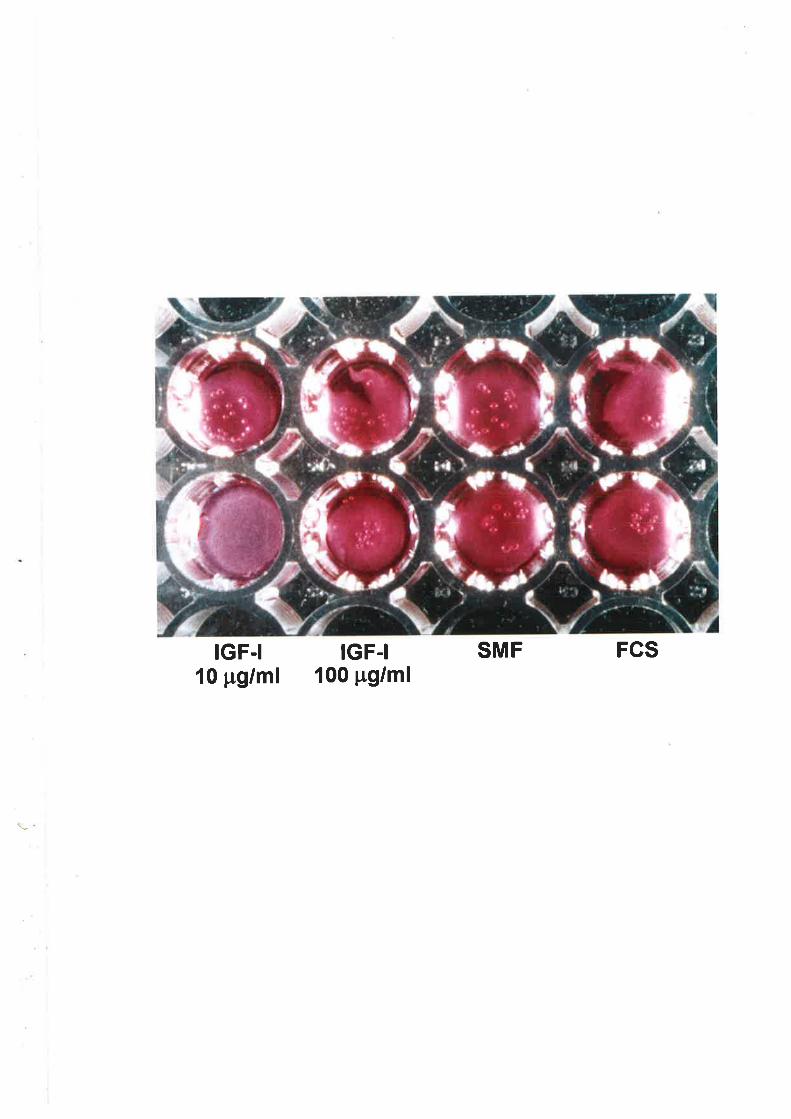
IGF.I10 pg/ml
SMF FCSIGF-I100 pg/ml
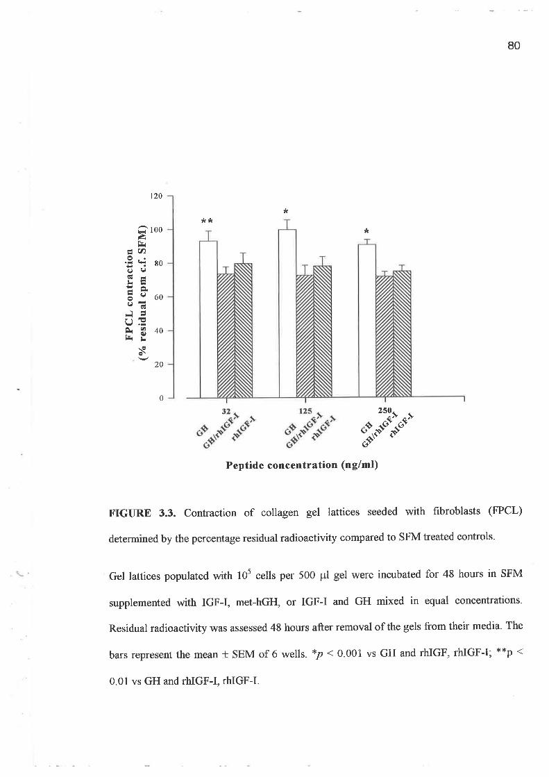
80
120
¡trk*
*Q tooàl=
É, u)
5T 8099cËdLË
ãe60eãrr tlF340l¡ ¡r
20
0
S*r'-t-"%,"--t- $'"11tPeptide concentration (nglml)
FIGIIRE 3.3. Contraction of collagen gel lattices seeded with fibroblasts (FPCL)
determined by the percentage residual radioactivity compared to SFM treated controls.
Gel lattices populated with 105 cells per 500 pl gel were incubated for 48 hours in SFM
supplemented with IGF-I, met-hGH, or IGF-I and GH mixed in equal concentrations.
Residual radioactivity was assessed 48 hours after removal of the gels from their media- The
bars represent the mean + SEM of 6 wells. xp < 0 001 vs GH and rhIGF, rhIGF-I; **p (
0.01 vs GH and rhIGF-I, rhIGF-I.
81
3.4.2. Skin Fibroblast responses to IGF Analogues
Experiments in 3.4.1 clearly demonstrated that fibroblasts will both grow and induce
contraction of a collagen matrix in response to IGF-I but the response to GH is weak. As
their activity was not synergistic, consideration was given to examining other aspects of
IGF-I physiology that could enhance its activity. Experiments were designed to examine the
influence of reduced binding protein affrnity upon activity of IGF-I in contraction of a
collagen gel using the IGF-I analogues, des(1-3)IGF-I and I-n3 ICF'-I. Having established
that IGF-I does promote cell growth, two other assays that examine specific aspects of
growth, namely DNA synthesis ([3ff]-thymidine incorporation) and protein production
([3H]-proline incorporation) were used to assess the relative potencies ofthe IGFs.
Fibroblast contraction
When the gels \¡i/ere suspended in media containing the analogues, a difference in potency of
up to a factor of 10 was demonstrated when compared to rhIGF-I (figure 3.4). Half maximal
effective concentrations (ED5¡) were 40 n{ml, 6.2 nglml, 4 nglml for native IGF-I, des(l-
3)IGF-I and LR3 IGF-I respectively. Data obtained from contraction studies performed
using a raîge of doses from 0.78 ng/ml to 50 nglml revealed that these analogues were
active in concentrations as low as lng/ml in this model (data not shown).
Fibroblast-populated lattice contraction was also examined comparing des(1-3)IGF-I with
PDGF-AB, TGF-81, bFGF, EGF and 10% FBS. Maximal gel contraction using TGF-PI
occurred with a concentration of 2O nglml (S8% + 4.7yo of the serum free controls)
producing a result which was not statistically significant. In contrast PDGF and des(l-
3)IGF-I significantly reduced gel size compared to controls (p < 0.0001). This result
82
demonstrated that the contraction induced by these two growth factors was as great as that
exhibited by gels exposed to l0%o FBS (figure 3.5).
The poor effrcacy demonstrated by TGF-PI prompted examination of the contraction
response of two separate fibroblast lines (SF1967 and SF3169) to treatment with TGFBI.
Similar studies were conducted to provide a comparison with IGF-I. The results obtained
revealed that the contraction of gels demonstrated previously by both IGF-I and TGFpI
were not specific to the fibroblast line (SF3 169) that was used for the majority of the
experiments (data not shown).
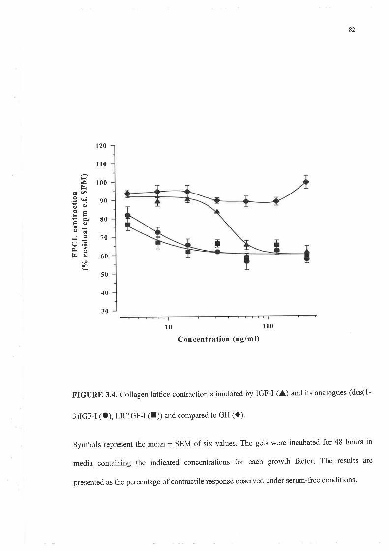
T70
!làtrÅ rt)
.9 t*iËa;Ãt-¡rtr:(J
¡ rl)l{ d)Batr
s
82
120
110
100
90
80
60
50
30
10 100
Concentration (ng/ml)
FIGURE 3.4. Collagen lattice contraction stimulated by IGF-I (A) and its analogues (des(l-
3)IGF-I (O), LR3IGF-I (f)) and compared to GH (l).
Symbols represent the mean t SEM of six values. The gels 'were incubated for 48 hours in
media containing the indicated concentrations for each growth factor' The results are
presented as the percentage of contractile response observed under serum-free conditions.
40
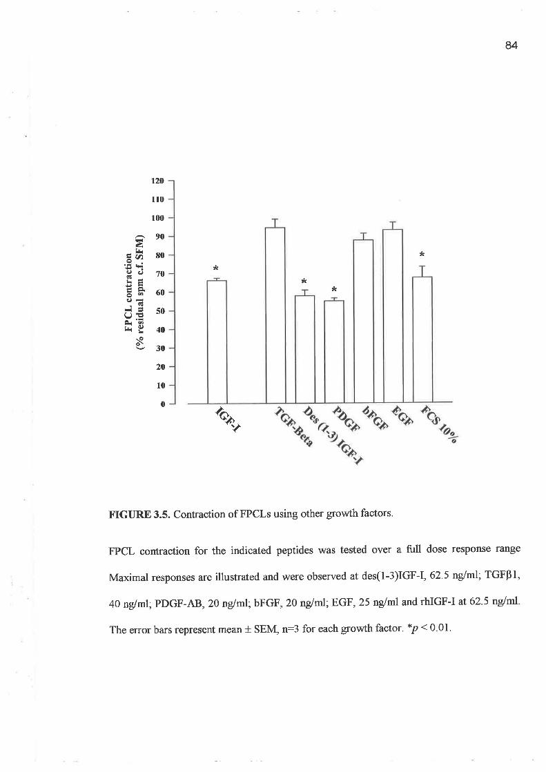
84
120
110
100
*
'tkt(
-90gÀ
â4 80'5-äe 70
ã* 60ñiã so
Y.åt-i g 40
.\<i 30
20
10
0
*
ô,r¿
tr.IGURE 3.5. Contraction of FPCLs using other growth factors.
FPCL contraction for the indicated peptides was tested over a full dose response range
Maximal responses are illustrated and were observed at des(1-3)IGF-I, 62.5 nglml' TGFBI,
4O nglml;PDGF-AB ,20 nglml;bFcF, 2O nglml;EcF, 25 ng/ml and rhIGF-I at' 62.5 ng/ml
The error bars represent mean + SEM, n:3 for each growth factor. *p < 0.01.
85
3.4.3. CelI Growth in response to the IGF Analogues
Having performed experiments comparing the potency of IGF-I and GH as fibroblast
mitogens more specific assays were employed to examine fibroblast protein and DNA
synthesis in response to IGF-I and its analogues.
Protein Synthesis - Incorporation of Tritiated Proline
Protein synthesis by fibroblast in response to stimulation by IGF-I and its analogues was
examined by assessing the incorporation of [3H]-proline into cellular protein. This activity
was further compared with that observed for IGF-II (figure 3.6).
IGF-I stimulated protein synthesis as measured by proline incorporation approximately
240yo more than the baseline levels observed for controls. The IGF-I analogues, particularly
LRf IGF-I, proved even more potent as anabolic agents. The flattening of the dose response
curve and an associated shift to the left of the dose response curve was an important feature
of the observed results. This suggests that there may be a more direct relationship between
dose and availability of free peptide for receptor binding of the IGFs to their target cells.
The order of potency observed was LR3IGF-I > des (1-3) IGF-I > IGF-I > IGF-tr.
DNA Synthesis - fibroblast incorporation oftritiated thymidine
Des(l-3) IGF-I, rhIGF-I and rhIGF-II were applied to monolayer cultures of fibroblasts
exposed to tritiated thymidine tracer. These results are represented in figure 3.7 . The
maximum response for all three peptides was approximately 220Yo of the level observed for
controls. The greater potency of the IGF-I analogues was once again demonstrated by a shift
86
to the left of the dose response curve for des(l-3)IGF-I as well as flattening of the curve
when compared with the recombinant peptide.
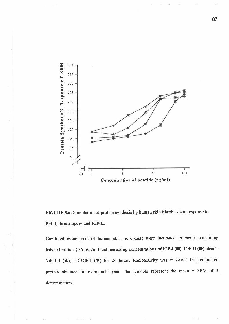
87
FIàl=(t)t:dq¿an
oÈu)í)ús
ar)
u)é)
(t)
é)
o¡rÈ
300
27s
250
225
200
175
150
125
100
75
50
0
1-l.01 .1 10 100
Concentration of peptide (nglml)
¡.IGURE 3.6. Stimulation of protein synthesis by human skin flrbroblasts in response to
IGF-I, its analogues and IGF-II
Confluent monolayers of human skin fibroblasts were incubated in media containing
tritiated proline (0.5 pCi/ml) and increasing concentrations of IGF-I (I), IGF-II (O), des(l-
3)IGF-I (A), LR3IGF-I (V) for 24 hours. Radioactivity was measured in precipitated
protein obtained following cell lysis. The symbols represent the mean + SEM of 3
determinations.
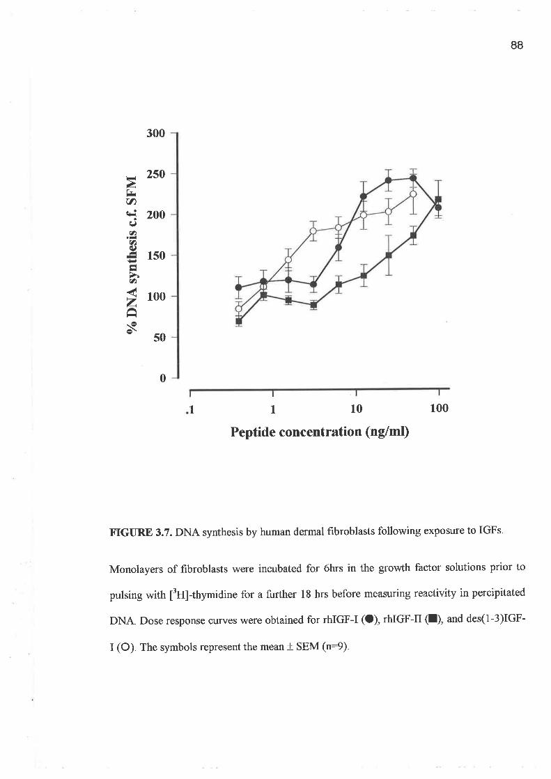
88
1 10 100
Peptide concentration (ng/ml)
F.IGURE 3.7. DNA synthesis by human dermal fibroblasts following exposure to IGFs.
Monolayers of fibroblasts were incubated for 6hrs in the growth factor solutions prior to
pulsing with ¡3U1-thymidine for a further l8 hrs before measuring reactivity in percipitated
DNA. Dose response curves were obtained for rhIGF-I (O), rhIGF-II (l), and des(l-3)IGF-
I (O) The symbols represent the mean + SEM (n:9)
300
2so
200
1s0
100
50
r=rÀ7U)+i
CJ
a.-U)oÁ-+.I
a
zâs
0I
I
89
The anabolic activity of IGF-I is these particular assays can be compared with the less
specific assay of cell growththatrelies on optical density of monolayer cultures (see 3.4.1).
This assay provided a maximal IGF-I response of ITOY' which contrasts with that of 240Yo
for protein synthesis and about 250yo for DNA synthesis. This highlights the methylene
blue absorbance assay as a good screening assay for cellular growth but does not allow
accurate interpretation as to whether the observed response is related to an increase in cell
numbers or if it is also due to increased cell size.
3.4.4. DNA synthesis: Comparison of Growth Factors
The activity of IGF-I was compared with several growth factors in fibroblast monolayer
cultures. All of these peptides are recognised mitogens in fibroblast cultures. The group of
growth factors tested included PDGF-AB, bFGF, EGF and TGFPI. They were compared
with IGF-I and des(1-3)IGF-I. Maximal responses for each growth factor are illustrated in
flrgure 3.8 and were attained at the following concentrations: PDGF-AB 20 nglml; TGFPr
1.25 ng/ml and bFGF 0.625 nglml; IGF-I 50 nglml; des(1-3)IGF-I 50 nglml and EGF 12.5
nglml. PDGF demonstrated greatest activtty at the highest concentration of 2O nglml while
des(l-3)IGF-I and IGF-I both demonstrated greater potency than the remaining growth
factors in this assay. TGFÊr elevated DNA synthesis only 5OYo above that of SFM treated
controls while IGF-I was able to produce a25O%o increase compared to controls.
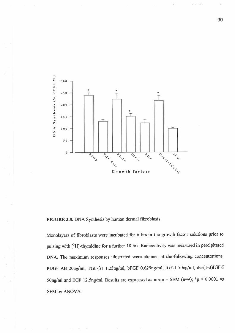
90
af¡(â
s
aO
çn
zÊ
300
250
200
150
100
50
* * *
*
0 ?aà-àò@.oG Q¿
<ò'Qâ cd
.%4.
'ð Þ,'hn
f¿
Growth factors
FIGURE 3.S. DNA Synthesis by human dermal fibroblasts.
Monolayers of fibroblasts were incubated for 6 hrs in the growth factor solutions prior to
pulsing wittr ¡3fq-thymidine for a further 18 hrs. Radioactivity was measured in percipitated
DNA. The maximum responses illustrated were aftained at the ffibwing concentrations:
PDGF-AB 2lnglml, TGF-81 L}5n/ml, bFGF O.625nglml, IGF-I 5Ong/ml, des(1-3)IGF-I
5Ong/ml and EGF l2.5nglml. Results are expressed as mean + SEM (n:9); *p < 0.0001 vs
SFM by ANOVA.
91
The relative inactivity of TGFFr was confirmed in assays using another fibroblast line
(SF1967). These data complemented that obtained for FPCL contraction and suggested that
TGFP1 may only be a weak promoter of fibroblast activity in these assays (data not shown).
92
3.5. DISCUSSION
IGF-I can modulate the behaviour of a variety of cells important to wound healing in a
paracrine (223,224), endocrine or autocrine (11,225) manner at the wound site. Following
injury platelet and tissue macrophages release IGF-I into the wound (15), supplementing
the IGF-I derived from the systemic circulation where it is largely bound to IGFBP-3.
Physiologically, IGF-I activity, expression and regulation is closely linked to GH (105).
Uncertainty exists as to whether GH directly regulates mitogenesis or whether its effects are
dependent upon the presence andlor local production of IGF-L Epiphyseal plate growth in
particular responds to IGF-I stimulation (230,232,252) and systemic GH regulates local
tissue expression of the IGF-I gene Q53) This theory is supported by the expression of GH
receptors by chondrocytes juxtaposed to chondrocytes from the more mature proliferative
zones of epiphyseal cartllage.
Proliferation of human lung fibroblasts appears to be modulated in a paracrine fashion by
IGF-I produced by epithelial cells (120) as well as by autocrine pathways. In skin IGF-I is
produced by melanocytes and fibroblasts but not by keratinocytes, however IGF-I is
regarded as cell survival factor for epidermal cells (90,223).IGF-I has been shown to induce
cell proliferation and protein synthesis by foreskin fibroblasts where GH has not been
effective (254). Eming et al (12) demonstrated this principle of paracrine IGF-I activity in
the skin using genetic modification of human diploid keratinocytes. In this study only those
keratinocytes that had been genetically modified secreted IGF-I while endogenous IGF-I
93
was not detected in unmodified keratinocytes. Ewing study demonstrated that IGF-I was
required for keratinocyte growth. Given that IGF-I is not produced by normal keratinocytes,
this effect must be a paracrine phenomenon presumably associated with dermal derived
IGF-I.
The synthesis of DNA by fibroblasts is a physiological event that is at best only minimally
stimulated by GH supplemented SFM (124,254). Using the metþlene blue absorbance
assay, met-hGH \Mas shown in figure 3.7 to only weakly stimulate growth at
supraphysiological concentrations (maximum concentration of l000ng/ml). Cook et al(124)
showed a similar effect where fibroblast DNA synthesis was increased only marginally
above baseline with SFM supplemented with up to 100ng/ml hGH. The authors reported
that when applied in the presence of O.5Yo HPS (hypopituitary serum), GH at 1000 nglml
increased tþmidine incorporation fïve fold. ft can be concluded that stimulating growth of
dermal fibroblasts by GH requires some other factor to produce significant biological effect
in vitro. The cell growth and FPCL data for GH in 3.4.1 support this finding.
A similar minor effect was reported by Cook et al for IGF-I. However this was only at a
concentration of 10ng/ml. IGF-I in SFM remains capable of inducing cell growth (l70Yo of
SFM at a concentration of 500ng/ml) but growth is only weakly enhanced by GH in these
same serum depleted conditions. Similarly these cells exhibit significant responses under
serum free conditions for thymidine incorporation into cellular DNA (figure 3.7) and
synthesis of cellular protein (figure 3.6). In both of these assays IGF-I had an EDso of
l0mg/ml.
Results for cell growth and gel contraction indicated that IGF-I potentiates fibroblast
activities in the absence of hGH while hGH is ineffective when used alone. GH was not
94
synergistic \ryith IGF-I under the conditions of the models used. These results are consistent
with Salmon and Daughaday's hypothesis (105) and complements the work by Cook et al
Q2\ who demonstrated that GH activity in fibroblast cultures is primarily mediated by the
IGF-L Other cell lines, in particular chondrocytes and osteoblasts, exhibit similar growth
related activity with IGF-I (233). However Clemmons and Van Wyk (21a) reported that
pre-incubation or co-administration of other growth factors augmented IGF-I activity. They
introduced the concept of a conditioned medium containing competence factors one of
which was considered to be PDGF. Several animal studies involving dermal and periodontal
wounds have demonstrated that this synergism is not isolated to tissue culture
(46,129,137,134). The influence of competence factors such as PDGF has not been fully
addressed in this study although this is an important issue in understanding the cellular
events associated with IGF-I. The contraction exhibited by fibroblasts populated gels in
response to IGF-I may be paftly related to priming of the fibroblasts by factors present in
10% FBS/DMEM. The sequence of harvesting of fibroblasts and lattice construction
requires that the cells be primed by FBS however the assays were conducted in solutions of
SFM containing IGFs or GH as the only additives.
The incorporation of [3H]-proline into precipitable protein and [3U]-thymidine into DNA
were used to measure other fibroblast-related events in response to IGF-I and its analogues.
In these experiments the influence of FBS and priming of cells were minimised by
incubating the subcultures for 24 hovrs in SFM before addition of treatment peptide
solutions. IGF-I was able to increase protein production up to 25}yo more than that for
serum free conditions as shown in figure 3.6. The analogues, LRf IGF-I and des(1-3)IGF-I,
lilere more potent than IGF-I as demonstrated by the shift to the left of the dose response
curve. This result was consistent with other data relating to the activity of these peptides in
95
L6 rat myoblasts cultures (140). This response is neither species nor cell line specific with
several studies supporting the potential use of these analogues in vivo (143,745,146,255) as
anabolic agents with greater potency than IGF-I. Protein synthesis and FPCL contraction
were also assessed using the IGF-II analogues, nu tCf'-tl and des (1-6) IGF-II (data not
included). IGF-II and its analogues were effective in both models but all were less effective
than IGF-I. However the analogues were no more potent than the native IGF-II. This
demonstrated that the structural changes to the N-terminus of the IGF-II molecule weakl¡ if
at all, affected biological activity of IGF-II even though IGFBP affrnity is reduced to 7Yo of
IGF-II by such alterations. These results were consistent with those observed for protein
synthesis inL6 rat myoblast (2a\ and contrast dramatically with the markedly different
potencies that similar structural changes can have for IGF-L The reduced activity of IGF-II
compared to IGF-I and its analogues may be explained by the lower binding affrnity that
IGF-II has to the type I IGF receptor Q56) compared to IGF-I and the effect of the IGF-II
receptor mopping-up free IGF-II (personal communication G. Francis)-
The functional activity of fibroblasts in response to various mitogens was extensively
investigated using the FPCL model. The collagen gel lattice is a versatile in vitro model of
wound contraction that canbe used to explore cell-cell and cell-matrix interactions as well
as to study growth factors alone or in various combinations. The change in gel size has
traditionally been recorded as residual arca (57). Assouline et al (61) chose to use optical
transmission coupled with residual gel area to quantitate a change in the collagen density
and size, presumably to correlate this with gel contraction. Our modification incorporates a
small amount of radioactivity (¡3ft1-inulin) into the gel and allows residual gel volume to be
measured and expressed as a function of residual radioactivity. Thus it also allows
assessment of contraction of floating gels in three dimensions. Other factors that have been
96
shown to influence the results in this model include number of cells seeded into the gel, the
type of collagen used in its composition and whether or not they are allowed to float freely
in their medium (56,57,220).
Several important findings were apparent from this study of FPCL contraction.
o In vitro IGF-I did not require FBS even in small concentrations to activate human dermal
fibroblasts to contract collagen lattices although priming of the cells prior to harvesting
cannot be entirely disregarded.
o Gel contraction was not markedly improved when met-hGH was added with rhIGF-I nor
did met-hGH have any great influence upon contraction by itself. These observations are
important in light of the work of Gillery et al (723) where IGF-I was added in the
presence of varying amounts of FBS and its effect upon collagen lattice contraction was
only noticeable when low concentrations of FBS were used. The significance of even a
small amount of serum lies in the provision of other cytokines to the medium which may
influence the IGF-I response.
o The analogues demonstrated even greater abilþ to enhance contraction than IGF-I' This
was illustrated by a shift to the left of the dose response curve and straightening of the
curve for the more potent peptides. This suggests that as the ratio of free IGF-I like
peptide to IGFBP bound peptide increased the level of IGF receptor activation probably
also increased. Therefore a smaller concentration of peptide is required to produce a
comparable response to rhIGF-I.
The exposure of this model to IGF analogues is unique to this study. IGFBP influences can
therefore be examined indirectly. In addition, these peptides have been shown to promote

97
DNA synthesis and cell proliferation in other cell types, including H35 hepatoma cells and
L6 rat myoblasts (140). In neonatal fibroblasts cultures they are up to 20 fold more potent
than IGF-I (238). The type 1 IGF receptor is the primary receptor mediating IGF activity
including IGF-II related events. Similarity of the maximal responses of the IGF-I species
implies that they are all exerting their effects through the same receptor type rather than
through for example the type II receptor or even the insulin receptor.
The mechanisms by which IGFBPs modulate IGF-I effects are not clearly defined. They
may augment IGF-I activity invivo Q57) butthey impede smooth muscle cell migration and
inhibit embryonic fibroblast proliferation in tissue culture preparations (237,258). Fibroblast
cultures are also known to produce endogenous IGFBPs Q59) and their production may be
modulated to some extent by other growth factors Q38).Therefore it is useful to examine
the IGF related effects in such a way that their influence is minimised. This has been
achieved using IGFs with low affrnity for the binding proteins. The results described using
analogues with known affrnity of less Ihan 5Yo for the IGFBPs (142) suggest that binding
proteins produced endogenously may restrict contraction stimulated by IGF-I. This
suggestion concurs with the work of Yateman et al (235) who demonstrated that the
proliferative activity of neonatal fibroblasts was more responsive to Lrtr IGF-I than IGF-I.
The differences observed between naturallcF-I and its analogues are possibly partly due to
the greater aflinity of IGF-I for IGFBPs and endogenous production ofthese proteins., It has
been postul ated thatthe N-terminus and particularly Glu3 are important for IGFBP binding
as modifications in this region provide the structural differences between the analogues and
IGF-I (140,742,260).
98
TGFPI has long been associated with scar contraction and has been shown by other
investigators to be active in gel contraction models (66,245,267,262). However in my
studies it produced only weak gel contraction and was only weakly mitogenic also. The
range of concentrations used in these experiments was 0.7 nglml to 20 ng/ml which are
similar to those doses used by Finesmith et al (66) and MacNeil et al Q62).In these two
studies TGFP produced significant gel contraction. The marked differences in response may
be related to several factors including the time allowed for contraction to occur and the
preparation of the gel lattice, particularly the cell numbers seeded into the gels. In
MacNeil's stud¡ contraction Ìvas measured over a 24 hour period and 5x10⁵ cells were
seeded into 1 ml gels while 1 x 10⁵ cells/0.5ml gel lattice were used in my experiments. In
the experiments by Pena et al, 0.5m1 gels were used however they were inocculated with
250,000 rabbit dermal fibroblasts per gel. Piscatelli et al seeded 100,000 fetal wound
fibroblasts into a Zml lattice that also incorporated the gowth factor additive unlike my
experiments where the gels were allowed to float in their treatment solutions.
Timing and methods of assessing contraction may be influenced by observer error when
determining diameter of the contracted gel and if gels are allowed to contract over a
prolonged period of time the influence of endogenous factors and possibly proliferation of
cells within the lattice may come to bear. The relatively short period allowed for contraction
in our study minimises the influence of endogenous produced peptides while permitting
observation of a significant response. In addition 48 hours appears to be the time point at
which thet majority of contraction has occured (245,261).
Finesmith et al (66) used large gels in petri dishes prepared the day before the experiments
and quantitated gel contraction in relation to the uncontracted area deternined from
99
photographs. In addition their cell inocculated gels were preincubated for up to 24 hours in
10% FCS priorto addition of treatment solutions. My impression is that this would prime
the fibroblasts and therefore influence the final response to the growth factors being tested.
Cellular DNA synthesis (tþmidine incorporation) is accentuated by TGFB in certain
mesenchymal cells (241) including fibroblasts and smooth muscle cells, but was not as
pronounced as that observed for IGF-I in this study TGFPI is known to have inhibitory
influences upon proliferation and differentiation primarily in epithelial cell cultures (248).
Therefore the biological responses prompted by TGFP1 may be cell line dependent. A
possible explanation for my results is that TGF-PI is not a potent mitogen in either of the
human skin fibroblasts used for my experiments. Therefore the function of TGFP1 in the
wound may relate more to fibroblast recruitment to the wound and regulation of ECM
protein secretion and reorganisation (263). Application of TGFpr neutralising antibodies
influence these parameters lending support to such a supposition (72,243,264).In this study,
TGF-Pr was not as mitogenic as IGF-I in two different human skin fibroblast lines
suggesting that IGF-I may play a greater role than TGF-PI in fibroblast proliferation and
wound contraction in the wound milieu. Another possible explanation is provided by the
work of Ghahary et al (265) who have demonstrated that IGF-I is actually esponsible in part
for the induction of TGF-B expression and activity in dermal fibroblasts.
As collagen is one of the fibroblast's principle synthetic products, the magnitude of the
protein production in response to IGF-I suggests this peptide may be more important in
collagen production and remodelling than TGF-81. Comparison with other isoforms of TGF
ß may have been useful to examine difference in the TGF family response in the FPCLs but
this was beyond the scope of the study.
100
In conclusion, in these models of in vitro fibroblast behaviour it has been shown that GH is
largely impotent while IGF-I stimulates dramatic fibroblast growth and protein synthesis
and may do so in the absence of other so-called competence factors such as PDGF.
Secondly, fibroblast associated gel contraction and protein synthesis responses indicate that
the IGF-I analogues are more potent than IGF-I while once again GH was unable to
influence gel contraction to any significant degree. There was also an absence of any
additive effect of GH and IGF-L Finally, stimulation of both gel contraction and DNA
synthesis by IGF-I and the truncated analogue des(l-3)IGF-I were only surpassed by PDGF
when the IGFs were compared with a range of growth factors known to be active in the
wound milieu. This implies that IGF-I is a potent fibroblast mitogen and that the IGFBPs
may tend to blunt the magnitude of this effect.
101'.ì
CIIAPTER 4
APPLICATION OF'IGFs TO INCISIONAL MODELS OF
WOT]ND RBPAIR
4.1. INTRODUCTION
4.1.1. The IGF-I Axis and In Wvo Studies
4.1.2. Diabetes
4.1.3. Aims
4.2. MATERIALS
4.3. METHODS
4.3.1. Surgery and Care of Animals
4.3.2. Peptide Preparation
4.3.2í) GH and IGFs
4.3.2 ä\ rGFBP-2
4.3.3. Confirmation of IGF-I Afflrnity for IGFBP-2
to2
4.3.4. Development of the Model of Streptozotocin-induced Diabetes Mellitus
4.3.4. i) Administration of Streptozotocin
4.3.4. ii) Blood Glucose Testing
4.3.4. iii) Animal Care
4.3.5. IGF-I an¿ l,ttSlGf-I in Diabetic Wounds
4.3.6.Influence of IGF-I and IGFBP-2 on Wound Strength in Diabetic Rats
4.3.7. Statistics
4.4. REST]LTS
4.4.1. Met-hGH and rhIGF-I as Topical Agents in Wound Repair
4.4.2.Influence of IGtr'BP-2 on Wound Strength
4.4.3.I)evelopment of a Rodent Model of Diabetic Wound Healing
4.4.3.i) Weight Loss and Hyperglycaemia
4.4.3.i1) Wo und strength
4.4.4.IGF-I and LR3IGF-I in diabetic wounds
4.4.5. The influence of IGF-I and IGFBP-2 on wound strength in diabetic rats

103
4.5. DISCUSSION
4.6. SUMMARY
104
4.1. INTRODUCTION
Experiments in chapter 3 demonstrated that cultured fibroblasts exhibit greater growth,
synthetic and contractile activity following exposure to the IGF analogues compared to
rhIGF-I or GH. These fibroblast responses may be important in vivo. This chapter explores
the effects of IGF-I and its analogue I-nflCf'-f in animal models of healing.
Models of incised wounds allow examination of fibroblast activity within the healing
wound, particularly extracellular matrix deposition following topical or systemic
administration of treatment preparations (93,94,725,127,132,266-269). The data collected
are generally presented in either histologic or tensiometric formats. Various tensiometry
parameters have been described. The basic parameter is the 'breaking strength' that is the
elongating force required to disrupt a given sample. This can be converted to the 'tensile
strength' by incorporating into the analysis the cross-sectional area of the specimen at the
point of disruption (87). Furthermore during the elongation of a strip of tissue the force
absorbed up to breaking point can be used to calculate the amount of energy absorbed prior
to wound dehiscence known as 'the yield energy'. Levenson (87) reported that uncertainties
of wound thickness measurements made the exact tensile strength curve less precise than the
breaking strength curve. However using groups of animals with paired control and treatment
wounds, Jyung et al suggested that the skin thickness becomes less important for
comparison of wound strength data within treatment groups (132). Therefore breaking
strength remains as valid a parameter as tensile strength. That particular paper compared
105
IGF-I alone and in combination with IGFBP-I as topical wound healing agents and utilised
paired wounds on individual animals.
Examination of histological sections from wounds allows non-parametnc daia to be
obtained relating to fibroblast responses within the wound. Relative amounts of newly
produced to old collagen can be depicted using image analysis technology. These features
can be correlated with tensiometric data (119,267). Immunohistochemical staining, in situ
hybridisation and protein extraction techniques can also generate datathat can be similarly
compared Q7O).
4.1.1. The IGF Axis and In Vivo Studies
Few papers have been published that examine the effects of IGF-I in animal wound healing
models (46,179,727,247,271-274). Greenhalgh et al(46) used a murine model of excisional
wound repair to examine PDGF combined with IGF-I or IGF-IL The authors reported
accelerated healing using IGF-II alone, but that PDGF potentiated both IGF-I and IGF-II
activity. Mueller et al (119) demonstrated that infused IGF-I administered using osmotic
pumps inserted subcutaneously, increased wound macrophage populations and collagen
synthesis in hypophysectomised animals. Suh et al (127) reported a similar result for
collagen synthesis using steroid affected rats.
The effrcacy of IGF-I as a topical wound healing agent appears to rely on the presence of
IGFBPs (732,247,271,275) or another growth factor, such as PDGF (46,134,274). It has
been reported to be effective in models of compromised healing when administered alone
106
(119,127,273). Other reports have demonstrated that the IGFBPs, particularly BP-1 and BP-
3 can potentiate the activity of IGF-I in vivo (732,247,271,273,276). Little is known about
the influence of IGFBP-2inthe wound environment except thatit exists in wound fluid (see
figure 2.11) and, it appears to inhibit the in vitro aclivity of IGF-I (277).
Altering the afünity of IGF-I for its binding proteins also influences the in vitro activity of
IGF-I as was shown in chapter 3 using the analogues, des(1-3)IGF-I and I-t€IGf-I. IGF
analogues have not previously been tested in wound repair models but they have exhibited
greafer potency than rlùGF-I in promoting gastro-intestinal growth (149) and general
somatic glowth (255,278). Their increased bioavailability may also provide some advantage
overthe native peptide where levels of specific IGFBPs are increased (180).
4.1.2. Diabetes
Diabetes is the most common endocrine disorders afflicting humans. Wound healing
complications in diabetics account for substantial numbers of hospital admissions
worldwide despite improved methods for controlling hyperglycaemia (752,156,279).
Ferguson et al (280) reported on the pathology and healing patterns of diabetic ulcers. They
noted features of retarded epidermal migration, acute on chronic inflammation and changes
in pericapillary fibrin deposition. However the authors concluded that local ischaemia from
'small vessel' occlusion was not an important feafure.
Several models exist that allow investigation of the influence of diabetes upon wound
healing parameters. These include toxin-induced and genetically selected models.
to7
Streptozotocin (STX), the broad spectrum antibiotic produced by Streptomyces
øcromogenes (281), has been used as an agent to induce experimental diabetes for many
years. It is toxic to the insulin producing B cells of the islets of Langerhans found in the
pancreas, causing degranulation and necrosis Q82) and thereby causing a clinical state
similar to type I insulin dependent diabetes mellitus. Limitations of this model include
regeneration of the B cells, which reverses the hyperglycaemia or the development of
pancreatic adenomata (162) which secrete insulin, causing hypoglycaemia. Phillips et al
(283) and Masiello et al Q8\ have reported that animal weight and age also affect the
induction of diabetes by STX. Extrapancreatic effects of STX have been reported to reduce
bile salt-independent bile flow (285) and induce nephrotoxicity and ketoacidosis that can
progress to acute renal failure.
4.1.3. Aims
The aims of the in vivo experiments were to:
1) Develop a reliable rodent model of type I insulin dependent diabetes mellitus that
could be used to examine the effects of the IGFs on impaired healing using
incisional and excisional wound models,
2) Examine for differences in wound strength after topical applications of GH and
rhIGF-I applied in several different concentrations,
3) Test for any advantagethat may be conferred by IGF-I analogues;
108
4) Determine the effect on wound strength of coapplication of IGFs with IGFBP-2
109
4.2.MATERIALS
Recombinant human IGF-I, (rhIGF-I, animal grade peptide >75yo pure by I{PLC), rhIGF-II
and tniIGf-I (receptor grade peptide; >95yo pure by HPLC) were obtained from GroPep
Pty Ltd, Adelaide, SA and formulated as described in Chapter 3. Recombinant met-human
GH (GH) was purchased from Bresatec (Thebarton, SA) and stored at 4"C after being
solubilised in sterile deionised water. Type I collagen was acid-extracted from rat-t¿ils
tendons as described by Bell (57) for use as the peptide delivery vehicle. Streptozotocin
(STX), 500 mg (Sigma Chemicals, St. Louis, MO) was dissolved in 10 ml of 0.9Yo NaCl in
initial experiments and later in citrate buffer (pH a.5). All other reagents were obtained from
AnalaR Chemicals or Sigma Chemicals, St. Louis, MO. Male Sprague-Dawley rats, aged 7
to 8 weeks (250 to 300 g) were purchased from the Central Animal House, the University of
Adelaide, Adelaide, SA. For the diabetic studies, body weights of 300-350 g atthe time of
arrival were selected. Glucose reagent strþs (Ames Glucostix@, Bayer Diagnostics,
Mulgrave, Vic.) were used to measure blood glucose levels. Gaseous anaesthesic agents
used were halothane (Fluothanet, Zeneca, Macclesfield, Cheshire, U.K.) 2.5yo, mixed with
a2.l ratio of oxygen (Oz; Linde, Fairfield, NS\Ð and nitrous oxide (NrO; Linde, Fairfield,
NSW). Dynek Australia Pty Ltd (Adelaide, SA" Aust.) supplied suture material (4/0 silk;
Dysilk@). Wound peak loads (breaking strengths) were measured using a tensiometer (M
1000 E, Mecmesin, U.K. with calibration package model MFG 25).
ll0
4.3. METHODS
Experiments were conducted using an incised wound model. The primary objective was to
examine the influence of IGF-I upon the dermal healing response as reflected by wound
breaking strength. General aspects of the studies will first be described before detailing
specific experiments.
4.3.1. Surgery and Care of the Animals
The animals were delivered one week prior to surgery to permit acclimatisation to
individual cages. They were fed water and rat chow ad libitum while housed in an
environment with a 14 hourllO hour light/dark cycle and an atmospheric temperature of
22"C. Approval for all animal studies was granted by the Animal Ethics Committees of The
Queen Elizabeth Hospital (Woodville, SA) and The University of Adelaide (Adelaide, SA
Australia).
The rats were weighed individually on the day of surgery (day 0), then anaesthetised in a
clear perspex box and the dorsal fur clipped before placing the snout into a hood connected
in series with the induction box to maintain anaesthesia (figure 4.1). Two 7cm full thickness
parasagittal incisions (including panniculus) equidistant from the midline were made on the
back of each animal after cleaning the operaling field with a l:l solution of BetadinerM and
TOYo ethanol (see figure 4.2).
lll
The animals were randomly allocated to treatment groups (n:8 unless otherwise stated)
after ranking the animals according to weight on the day before surgery. Treatment
preparations were applied using a pipette set at a volume of 100 pl to one wound and 100 pl
of the vehicle (collagen) was applied to the contralateral wound. The surgeon was blinded to
which treatment preparation he applied. The wounds were closed with simple interrupted
4/0 silk sutures. Recovery from anaesthesia was observed by the operating team prior to the
animal's return to its cage. Post-operatively all rats were weighed at day 1 and every third
day thereafter.
Seven days after surgery the animals were sacrificed using COz inhalation and cervical
spine dislocation. The dorsal pelt was removed from the carcass and each wound was
divided into four parallel strips 4 mm to 5mm wide at right angles to the wound (see figure
4.2.) These strips were cut out and individually placed into the clamps of the tensiometer.
Each wound strip was distracted at a rate of 10 mm/min until disruption to determine the
wound breaking strength which was recorded in kilograms (kg).
Samples at the cranial end of the wound were taken for histological studies and fixed in
methacarn or TOYo formalin for future analysis. In the diabetic studies, samples were fixed in
Bouin's fixative for immunohistochemical studies.
FIGURE 4.1. Apparatus for providing general anaesthesia for all experiments
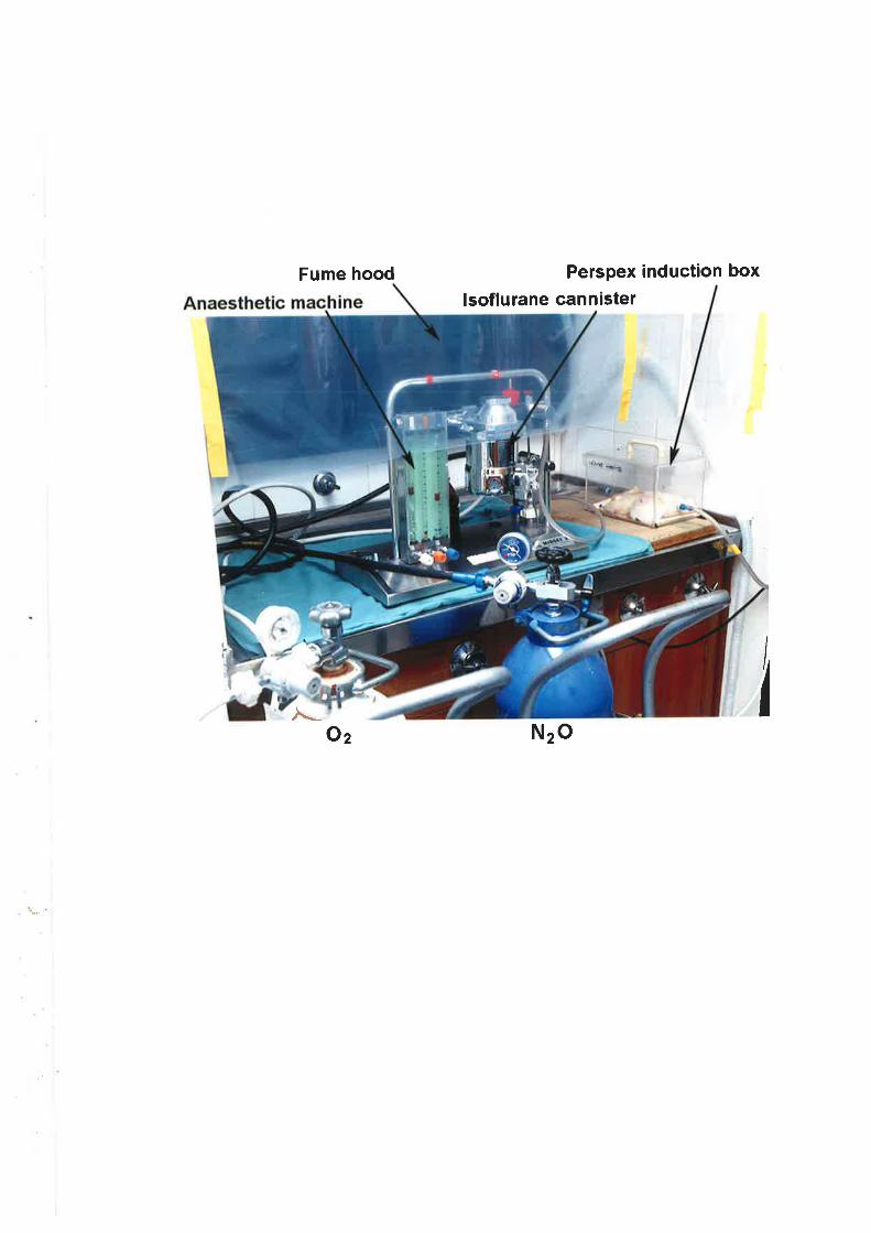
Fume hood
Oz
lsoflurane cannister
Nzo
Perspex induction box
,[_
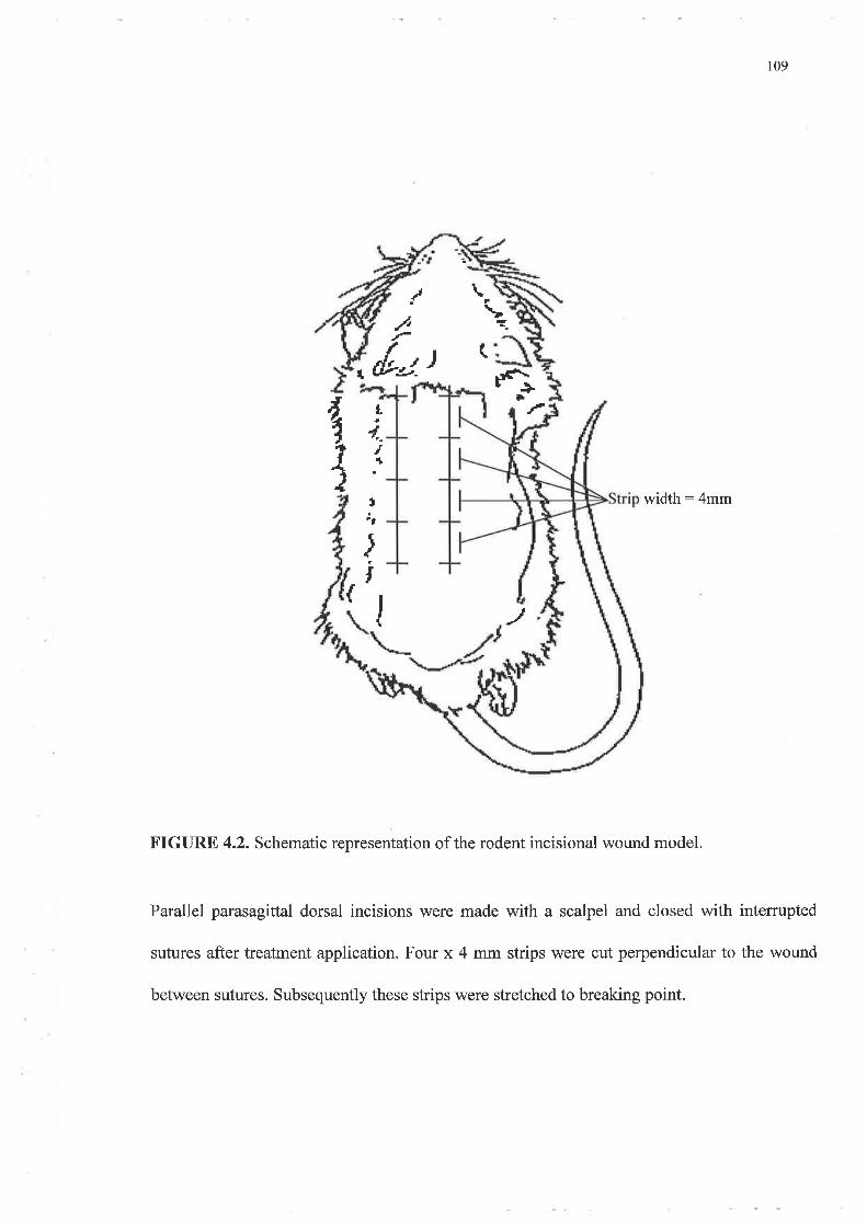
109
.t\ Ln
I t¿t ( +È
width:4mm
J
FIGURE 4.2. Schematic representation of the rodent incisional wound model.
Parallel parasagittal dorsal incisions were made with a scalpel and closed with intemrpted
sutures after treatment application. Four x 4 mm strips were cut perpendicular to the wound
tt
4.T
.-
ttÊ
f{i
{
inlïJ
between sutures. Subsequently these strips were stretched to breaking point.
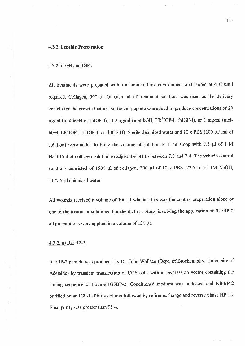
rl4
4.3.2. Peptide Preparation
4 3 2. i) GH and IGFs
A1l treatments were prepared within a laminar flow environment and stored at 4'C until
required. Collagen, 500 ¡rl for each ml of treatment solution, was used as the delivery
vehicle for the growth factors. Sufficient peptide was added to produce concentrations of 20
pelml (met-hGH or rhIGF-I), 100 ¡rglml (met-hGH, lR3tGf-I, rhIGF-I), or 1 mglml (met-
hGH, UfIGf-I, rhIGF-I, or rhIGF-II). Sterile deionised water and 10 x PBS (100 ¡rVlml of
solution) were added to bring the volume of solution to I ml along with 7.5 pl of 1 M
NaOFVml of collagen solution to adjust the pH to between 7 .0 and 7 .4. The vehicle control
solutions consisted of 1500 ¡rl of collager¡ 300 pl of 10 x PBS, 22.5 ¡l of lM NaOH,
1177.5 ¡rl deionised water.
All wounds received a volume of 100 ¡r1 whether this was the control preparation alone or
one of the treatment solutions. For the diabetic study involving the application of IGFBP-2
all preparations were applied in a volume of 120 pl.
4.3.2. ii) rGFBP-2
IGFBP-2 peptide was produced by Dr. John Wallace (Dept. of Biochemistry, University of
Adelaide) by transient transfection of COS cells with an expression vector containirlg the
coding sequence of bovine IGFBP-2. Conditioned medium was collected and IGFBP-2
purified on an IGF-I affrnity column followed by cation exchange and reverse phase I{PLC.
Final purity was greater than95Yo.
l15
IGFBP-2 was dissolved in 10 mM HCI before being mixed with rhrGF-I or ltt'IGr-I in a
1:1 molar ratio. Treated wounds received l2}gtl of solution containing rhIGF-I 10 pg.
IGFBP-2 4o Vgper wound, rhIGF-I 10 pg only, IGFBP-} 40 ¡rg only, f.nfICn-f 10 ¡rg only
ortnÉIGP-I 1o ¡rg : IGFBP-2 40 ve.
4.3.3. Confirmation of IGFBP-2 binding of IGF I
WLB analysis was performed using 12.5o/o SDS-PAGE under reducing conditions as
described in chapter 2 (see 2.3.4). After electrophoresis the nitrocellulose membranes were
probed with ¡r2tI1-tGr-I and ¡l25t1-tnftcr-t for one hour before being visualised
autoradiographicallY.
4.3.4.I)evelopment of the Model of sTX-induced Diabetes
4.3.4. i) Induction of Diabetes
STX was administered intramuscularly to the thigh muscle of the right hind leg' It was
dissolved in its vial in o.gyoNaCl to a concentration of 50 mglml. The solvent was changed
to citrate buffer (pH 4.5) in later experiments as the stability of STX is better maintained in
this medium than in physiological saline. The required doses were calculated using the
formula;
Volume of STX (*l): Weight x [dose]/5O mglml
116
4.3.4. ii) Blood elucose testing
Blood samples for testing glucose concentration \ryere obtained by sectioning the terminal 1-
2 mmof the tail tip. Suffrcient blood was then expressed and applied to a Glucostix reagent
strip for analysis in an Ames Glucometert M blood glucose meter. These strips rely upon a
glucose oxidase reaction to produce changes in colour of two pads. This system allowed the
blood sugar levels (BSL) to be measured within araîge of 2-22 mmol/I.
4.3.4 iii) Preliminary Experiment
Animal weights and BSL were recorded prior to this procedure. The body weights were
recorded every second or third day. Detection of glycosuria was attempted once the animals
developed polyuria (around day 3). BSLs were determined on the seventh day and
subsequently twice weekly to confirm the development of hyperglycaemia' Diabetes was
confirmed by recording three consecutive high BSLs (>11 mmol/dl) over a period of 2
weeks with associated polyuria and weight loss'
On the fourteenth day single incisions were created as described in 4.3.1. Body weights and
BSLs ìÀ/ere fecorded on the day of surgery and on the day of sacrifice (see 4'3'1)' Four
transverse wound strips from the pelts of each animal were subjected to wound strength
analysis. The pancreas from one representative animal in each group was also removed and
processed for immunohistochemical staining of insulin and glucagon content within the
pancreatic islets using a commercially available kit (Histostain-DsrM Kit, Zymed
Laboratories, South San Francisco, CA, USA)
r17
4.3.4. Ð Animal Care
Animals were acclimatised to individual wire-bottomed cages for one week during which
time they were weighed twice and fed standard laboratory rat chow. The sixteen animals
were divided into four groups. Each group received a different dose of STX (0 mg/kg
(controls orvehicle only,45 mg/kg (group I), 55 mglke @roup II), and 65 mglkg (group
III)). These values were determined following communication with Dr Frank Tomas
(Cooperative Research Centre for Tissue Growth and Repair) and from a review of relevant
literature ( 1 5 0, I 6 l, I 62,1 64,180,283,28 4,286,287 ) -
4.3.5. rhIGF-I and Ln3IGn-I in Diabetic Wounds
Diabetes was induced using STX 55 mglkg as an intramuscular injection. BSLs were
conducted only weekly while weights were recorded each second or third day' The animals
were allocated to either the diabetic or normal group depending upon their weight with the
heaviest animals placed in the diabetic group. The controls received a weight calculated
volume of buffer/vehicle (see 4.3.4.).
Three weeks after being rendered diabetic all animals were again randomised on the basis of
weight to two treatment groups. Recombinant human IGF-I and f-nffCf-I were applied at a
concentration of I mglml. The surgery and recovery were undertaken in the same mannef
described in4.3.7
118
4.3.6. The Influence of rhtGF-I and IGFBP-2 upon Wound Strength in Diabetic
Wounds
Animals were allocated to gfoups of eight and surgery was performed over two days'
Anaesthesi a and wounds were produced as previously described and wound closure
completed with 4lO silk. One group received only rhIGF-I and were \À/ounded on the first
day and the other two groups 2 werewounded on the second day. These received solutions
containing IGFBp-2. After seven days the wounds were harvested and analysed in the
standard manner (4.3. 1).
4.3.7. Statistics
The values of the individual strips were pooled for each treatment to give a meaî value * 1
sEM in kilograms (kg). ofily dehisced strips (-5% of all specimens) were excluded from
analysis. Analysis of results was performed using paired student t-tests comparing treated
and paired contralateral control wounds. Statistical analysis was performed using Sigmastat
v2.Ol software package (Jandel corporation, San Ramon cA! usA).
119
4.4. RESULTS
4.4.l.Met-hGH and rhIGF-I as Topical agents in wound Repair.
Rats with normal wound healing properties were used to assess the effect of met-hGH and
rhIGF-I as topical wound healing agents where healing was not compromised. Preliminary
experiments using a concentration of 2O pg of peptide/ml failed to influence wound
breaking strength for any treatment compared to their controls (data not shown)' The effect
of met-hGH, rhIGF-I and Ln3fCf-I were subsequently tested at doses of 100 þglml and I
myml. The results for those experiments that tested a dose of 100 Pglml (10 ¡rglper wound)
are illustrated in figure 4.3.
Two experiments were used to test LRfIGF-I (n:8), met-hGH (n:8), rhIGF-I (n:8) and a
combination of these last two (n:7) Met-hGH and rhIGF-I were repeated but results have
not been pooled as vehicle treated wounds were significantly different between the two
experiments. Data for met-hGH and rhIGF-I have been presented from the first experiment
and are representative ofthe results (figure 4.3).
Wounds that received 10 ¡rglwound met-hGH (79.5 g + 6), rtrIGF-l (77'4 g + 6) and
rnfrCr'-t (7a.g g+ 7) were no stronger Íhantheir paired control wounds. The combination
of met-hGtVrhIGF-I (76 g t 6) also did not improve wound strength after 7 days than their
paired vehicle treated wounds. This was confirmed statistically using a paired t-test.
r20
A separate set of experiments was used to test met-hGH, rhIGF-I, LßfIGF-I and rhIGF-II at
concentrations of I mglml. The first two experiments tested met-hGH, rhIGF-I and ln'-TGn-
I (n : 15 animals/treatment) (figure 4.4). The results for these two experiments were
combined as there was no significant difference in weights or vehicle treated wounds
between the two studies. The potency of rhIGF-II (1 mglml) was also tested and compared
with rhIGF-I and met-hGH (n: 8 per group) (figure 4'5)'
Topical met-hGH failed to improve wound breaking strength significantly compared to
paired vehicle treated wounds. This response remained consistent between experiments
regardless of met-hGH concentration. In contrast rhIGF-I (79'a g+ 8) at a concentration of
1 mglml applied as a volume of 100 pl to incisional wounds produced significantly stronger
wounds compared to the matched controls (56 g t 5; p < 0.01). This represented a 4lYo
improvement in wound breaking strength. nÉfCf-f did not influence wound breaking
strength (70.9 g t 7). Coll agen treated wounds did not reveal a statistically significant
difference between groups (one way ANOVA)'
when rhIGF-II was applied in the same concentration (1 mglml) it did not demonstrate any
influence upon wound breaking strength (see figure 4'4)'
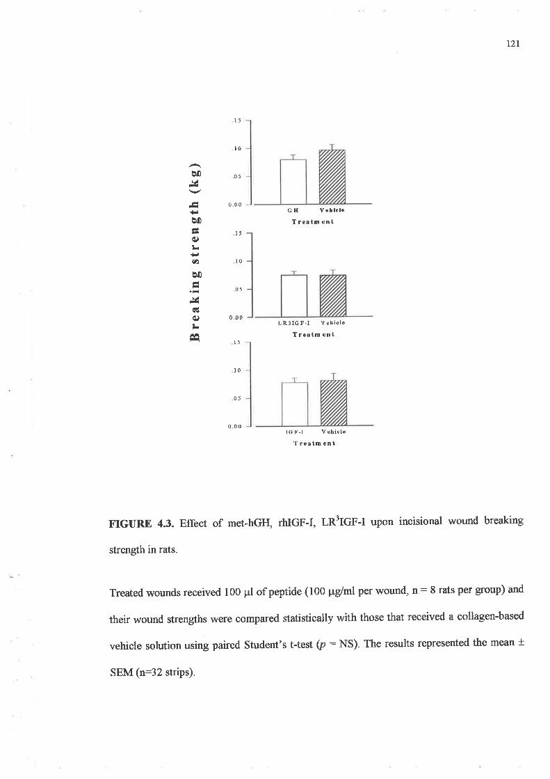
t2l
lo
Þ0r¿
--t+¡ê0ÉOL+¡aÞ0É,¿
cË
c)LÉ
.05
0 00
15
10
-05
0 00
l5
10
05
o 00
GH Vehlcle
T restm ent
LR3IGF-I Vehicle
T reatm ent
IGF-l V ehicle
T ¡eatm ent
IrIGURE 4.3. Effect of met-hGH, rhIGF-I, I,nftCf-t upon incisional wound breaking
strength in rats.
Treated wounds received 100 pl of peptide (100 ¡rglml per wound, û: I rats per group) and
their wound strengths were compared statistically vrith those that received a collagen-based
vehicle solution using paired Student's t-test (p: NS). The results represented the mean *
SEM (n:32 strips).
l5
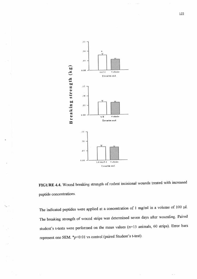
r22
Þ0
'¡lI+.Þ0Éé)L{.)ah
ê0I
&cl()LÉ
l5
l0
05
000
15
10
05
0,0 0
15
_10
05
0.0 0
IGF-I V ehicle
T re¿ tm ent
c H V ehiole
T rea tm ent
LRSIGF-I V ehicle
T reatm ent
FIGURE 4.4. Wound breaking strength of rodent incisional wounds treated with increased
peptide concentrations.
The indicated peptides were appli ed at aconcentration of I mg/ml in a volume of 100 pl'
The breaking strength of wound strips was determined seven days after wounding' Paired
student,s t-tests were perfbrmed on the mean values (n:15 animals, 60 strips)' Error bars
represent one SEM. *p<0.01 vs control (paired Student's t-test)'
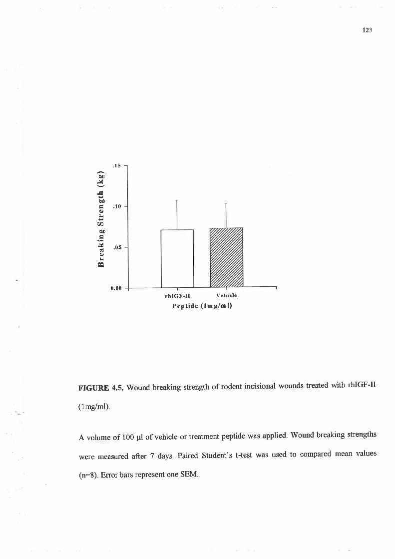
123
0.00rhIGF-II Vehicle
Peptide (1m g/m l)
¡r¡GIIRE 4.5. Wound breaking strength of rodent incisional wounds treated with rhIGF-II
(1mg/ml)
A volume of 100 pl of vehicle or treatment peptide was applied. Wound breaking strengths
were measured after 7 óays. Paired Student's t-test was used to compared mean values
(n:8) Error bars represent one SEM.
.15
q0
ooÉ .10í)tr(t)qo
ã .os(¡)LE
t24
4.4.2. The Influence of IGFBP-2 upon wound strength
As IGFBP-2 is thought to inhibit IGF-I related responses, experiments were performed
comparing the influence of IGFBP-2 upon IGF-I effects where binding affinity of IGFBP-2
to IGF-I is normal and also where binding affrnity is reduced such as with LR:3IGF-I.
IGF-I binding to IGFBP-2was confirmed using WLB (figure 4.6). Comparison of the blots
incubated with ¡tt5t1tR'IGF-I or ¡I25I1IGF-I clearly demonstrated the presence of a band in
the region of 30kDa on the membrane exposed to ¡125I1IGF-I. This band is consistent with
the molecular weight for IGFBP -2 and its absence in the gel incubated with I-n3tCf-I
indicated little if any binding of the analogue to IGFBP-2. Good affrnity of rhIGF-I for the
binding protein was confirmed by these result
Forty rats divided randomly into five groups (mean weight at time of surgery was 316.5 g;
range Zgg.3 g - 367 g), were used for this study. Each group received collagen to one wound
and rhIGF-I, LR3IGF-I, IGFBP-2, IGF-I/IGFBP-2 or rn3tcr-I /IGFBP-2 to the other (see
4.3.2. for peptide concentrations) to determine whether the addition of IGFBP-Z to an
incised wound significantly altered the effect of IGFs on wound healing variables. The
results are summarised in frgare 4.7.
\ilhile rhIGF-I 10 pg alone marginally increased wound strength compared to paired
controls, this was not significant when analysed using Student's paired t-test. Breaking
strengths in the other groups showed no statistically significant difference between peptide
treated wounds and paired vehicle treated wounds. IGFBP-2, 40 ¡rglwound, did not
significantly influence rhIGF-I activity in this model. However a small trend towards
t2s
inhibition of incisional wound healing was observed when IGFBP-2 was applied with
rhIGF-I but when used alone it had no effect.
FIGURE 4.6. Confirmation of IGFBP-2 binding of IGF-I.
WLB using 12.5% SDS-PAGE were performed and proteins were electrotransfened to
nitrocellulose sheets before visualising the IGFBP-2 bands by probing overnight with
¡r2tI1LRÉtGn-I or [125I]IGF-I. Lanes l, 5, 6 and 10 represent molecular weight markers,
lanes2,3, 4 were loaded with IGFBP-2 5 ¡tg in 100 ¡rl of sterile water and probed with
[125I]IGF-I while lanes 7,8, 9 were loaded with IGFBP-2 andprobed with ¡125I1Ln'tCp-
I. IGFBP-2/IGF-I complexes were visualised while the absence of binding proteirV
LR3IGF-I confirms the reduced affinþ of IGFBP-2 for the analogue.
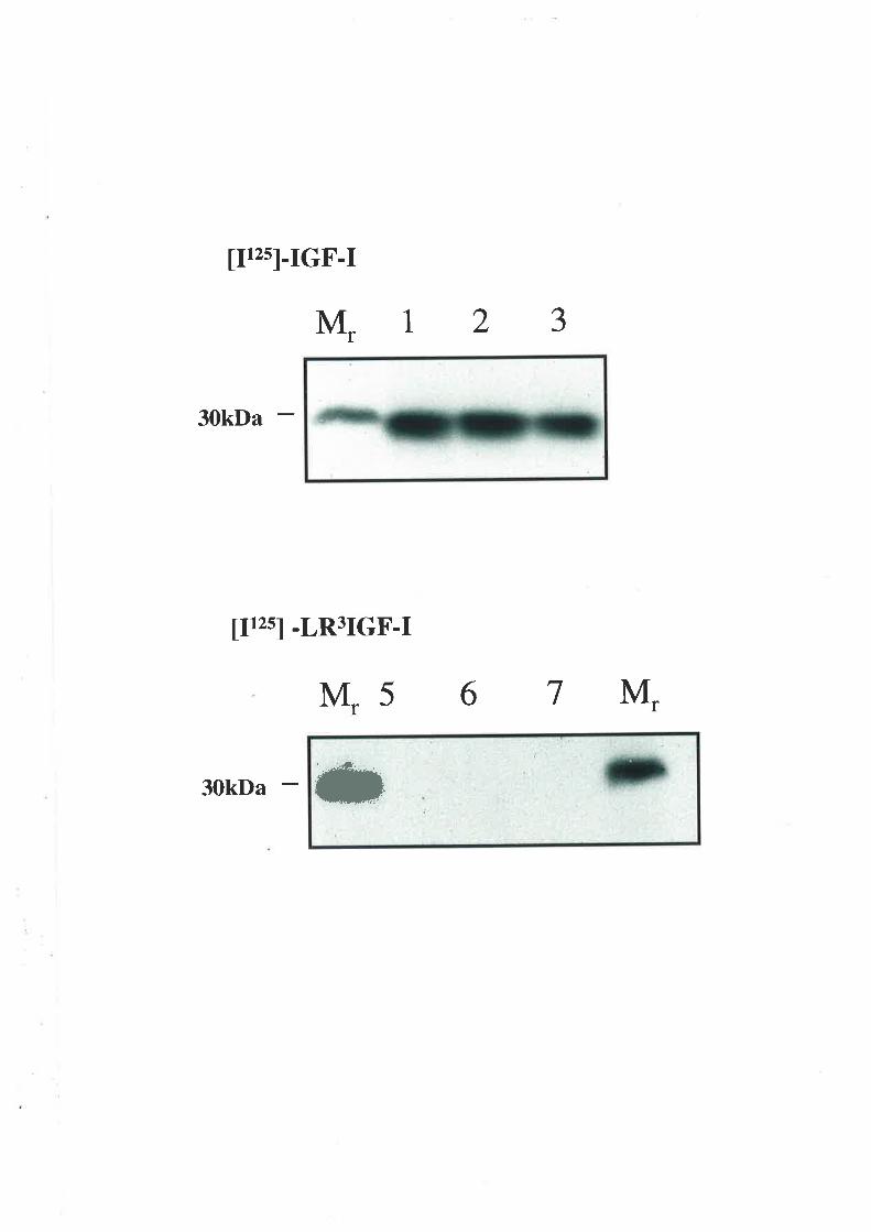
lF2sl-IGF-I
M, 123
30kDa
[rzs1 -LR3IGF-I
M,5 6 7 M.
30kDa
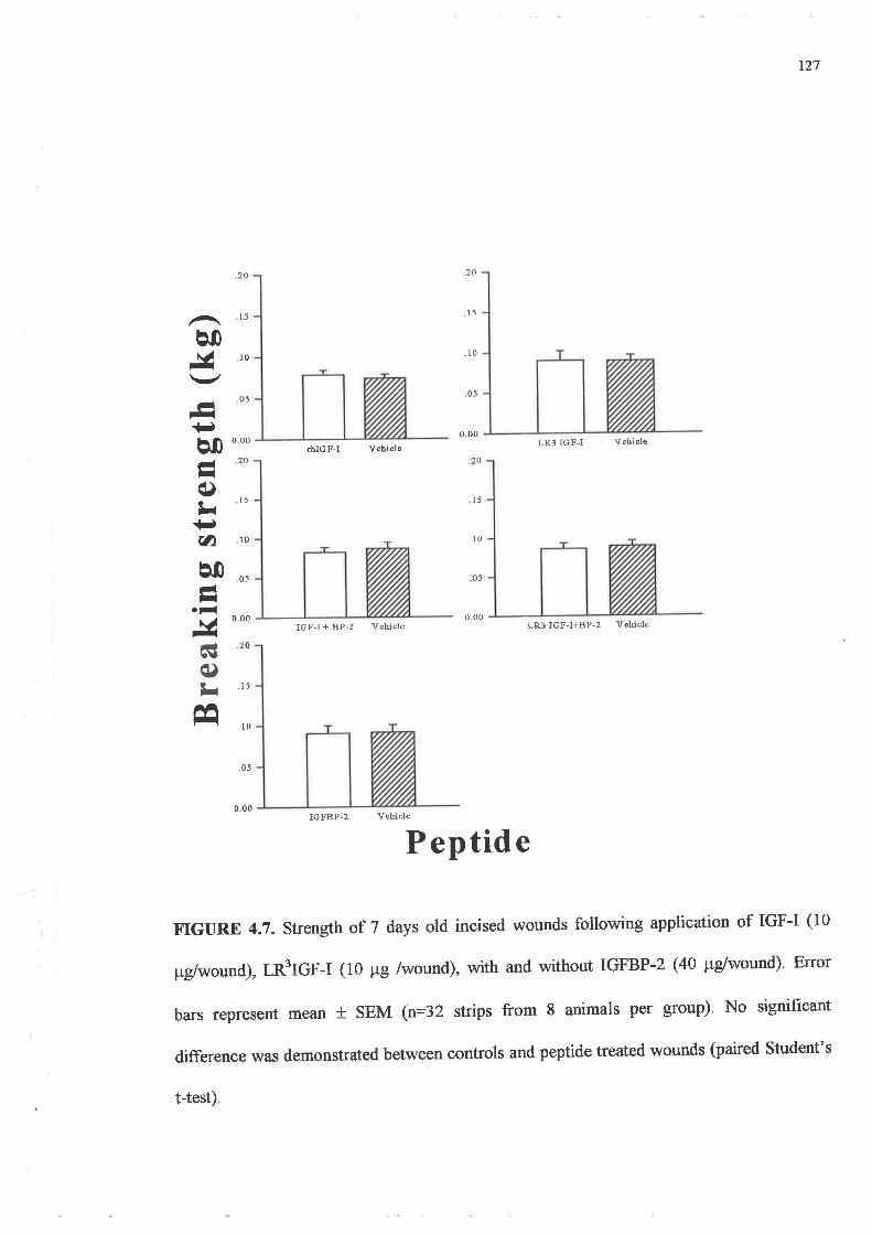
127
cg 20
()¡r '15
â0,!vÁ
-+.q0Å-q)¡i+¡au0Á-.-&
É
15
10
l5
10
000
10
20
o5
05
000
.70
.15
.10
.05
o_00
20rhIGF-I Vehiole
IGF-I + BP-z Vehicle
IGFB?-2 Vehiole
000
.20
15
,10
_05
000
LR3 IGF-I Vehiole
LR3 IGF-I+BP-2 Vehiole
o5
P eptid e
FIGURE 4.7. Strength of 7 days old incised wounds following application of IGF-I (10
¡rglwound), tnftcF-t (10 pg /wound), with and without IGFBP-2 (40 Pglwound)' Eror
bars represent mean t sEM (n:32 strips from 8 animals per group)' No significant
difference was demonstrated between controls and peptide treated wounds (paired Student's
t-test).
r28
4.4.3. Development of a Rodent Model of Diabetes Impaired Wound Healing
4.4.3. i) Weight loss and Hyperglycaemia:
Sixteen rats divided into four equal groups were used for this experiment. Diabetes is
defined clinically in humans as the combination of hyperglycaemia and glycosuria where
random blood glucose levels are greater than 11 mmol/l or fasting glucose concentrations
are greater than 8 mmoVl. The normal raîge of blood glucose concentrations for rats is
l1g.4 mgldl + 47.1 (10.4 mmoVl + 2.3) (288). Accumulated results using the Ames@
Glucometer indicate that the normal raîge of blood sugar levels (BSL) in male rats of 8 to
12 weeks of age between is 4.5 and I mmol/I. The criteria adopted for confirmation of
diabetic state in this rodent model were:
a) failure to thrive characterised by weight loss or reduced gain in weight compared
to age-matched controls;
b) BSL greater than 13 mmoVl;
ò) polydipsia, polyuri a and hyperphagia. Metabolic cages were not used therefore
urine volumes and the quantity of food consumed by the animals were not accurately
measured, however the diabetic animals were obviously polyuric judged by the
accumulated moisture of their bedding.
The BSLs recorded for those animals (n:4 per group) injected with STX at day 2l wete
21.5 mmol/dl t 1.1 for group I (STX 45 m{kg),20.9 mmolldl + I'3 for group II (STX 55
a
129
melke) and27.9 mmol/dl + 1.1 for group III (STX 65 mglkg) (figure 4.8). This compared
with the BSLs of the control group taken just prior to being sedated on the day of surgery
(7.35 + 0.8mmol/1). The technique used for measuring the glucose levels in these rats
produced arange of measurements between 2.0 and 22.l mmol/I. BSLs that registered as a
..higir-' reading (>22.1) were recorded as 22.1 tended to skew the results towards a lower
average. No "lor¡/' results were obtained in any of the four groups. All animals that were
injected with STX developed hyperglycaemia and no deaths were recorded in this trial.
Change in animal body weights are summarised in figure 4.9. Over the first three weeks' all
the animals given STX lost weight whereas age-matched controls continued to gain weight'
Weight loss was most dramatic in the first week and slowed as the trial proceeded' The body
weights of the group II plateaued early after the first week. The magnitude of total weight
change correlated well with the dose of STX administered (n : 76 r :0'969)'
The effect of STX on P cells of the pancreatic islets \¡/as confirmed using
immunofluorescent and immunohistochemical staining for glucagon and insulin in a
representative pancreas of each group. Standard H & E stained sections revealed reduced
cellularity of the islets along with evidence of pyknosis (plates A and B, figure 4'10)' When
stained with anti-insulin and anti-glucagon antibodies, the sections demonstrated that there
was a diminution of insulin storage and production (plates C and D). The colour plates also
demonstrate the lack of fluorescence obtained in a STX affected pancreas (plates E and F)'
This can be interpreted as indicating either 'drop-out' of insulin producing cells or near
absence of insulin production by the pancreatic islets, resulting in a hyperglycaemic state
similar to Type I diabetes mellitus.
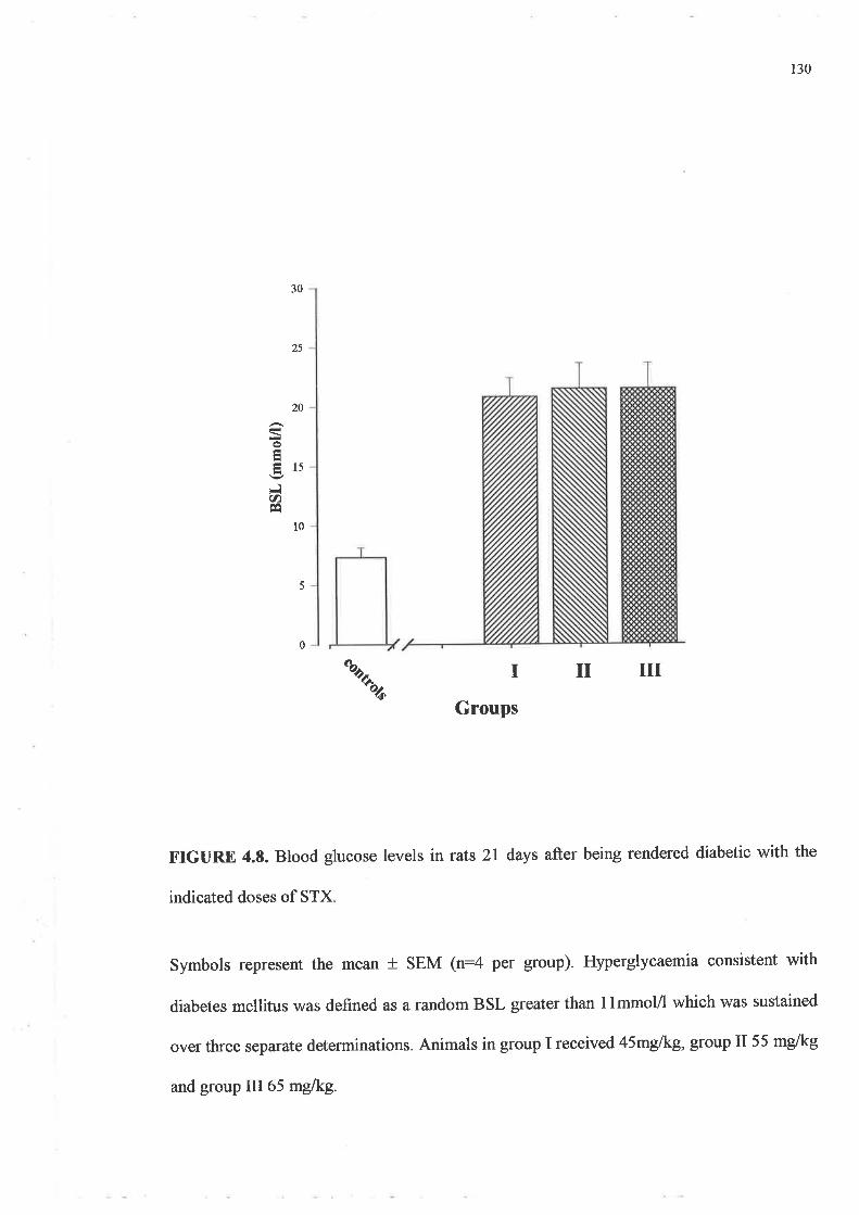
130
30
25
20
o
ÉlsFIU)É
l0
5
0
\I
Groups
IIIIr
FIGURE 4.8. Blood glucose levels in rats 2l days after being rendered diabetic with the
indicated doses of STX
Symbols represent the mean t SEM (n=4 per group). Hyperglycaemia consistent with
diabetes mellitus was defined as a random BSL greater than 1lmmol/l which was sustained
over three separate determinations. Animals in group I received 45mglkg, group II55 mglkg
and group III65 mdkg.
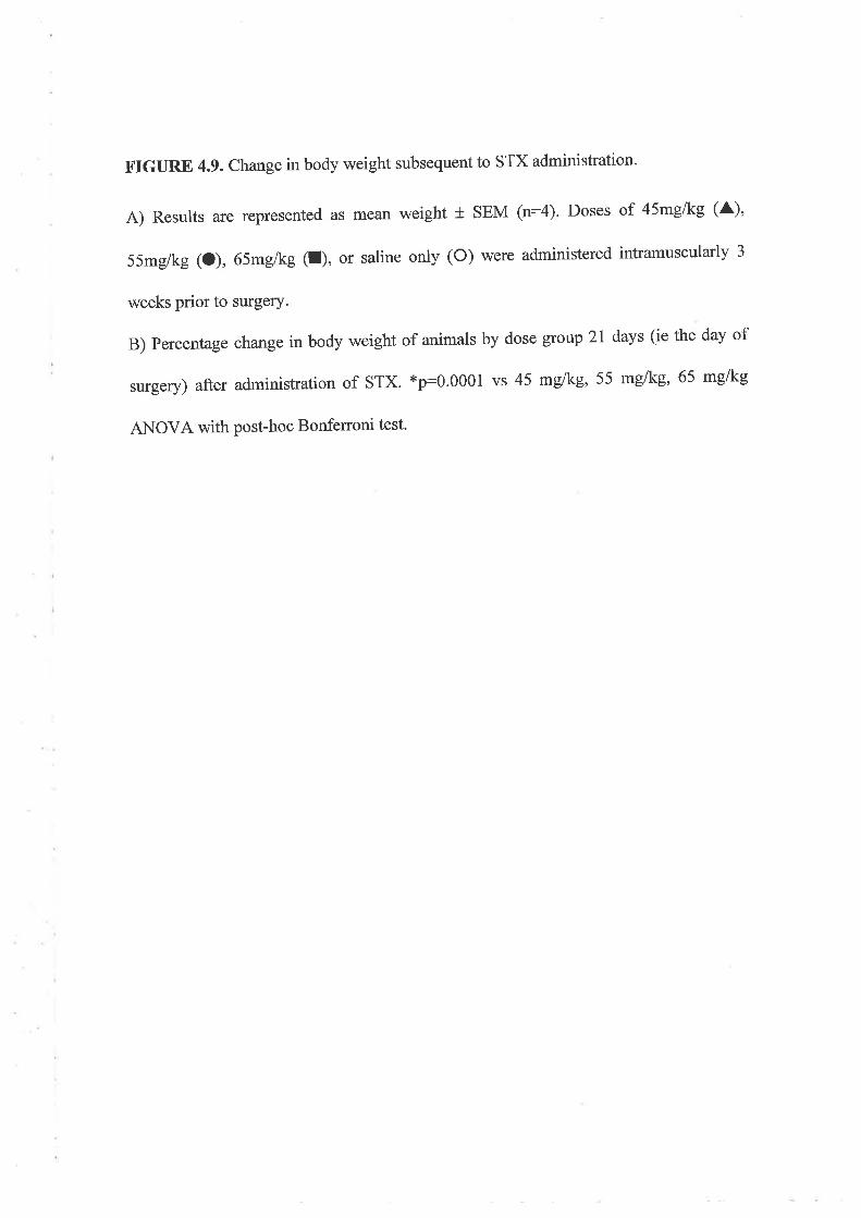
FIGURE 4.9. Change in body weight subsequent to sTX administration'
A) Results are represented as mean weight I SEM (n:4). Doses of 45mglkg ( ),
55mg/kg (O), 65mg/kg (I), or saline only (O) were administered intramuscularly 3
weeks prior to surgery.
B) Percentage change in body weight of animals by dose group 2! days (ie the day of
surgery) after administration of sTX. *p:0.0001 vs 45 mg/kg, 55 mg/kg, 65 mglkg
ANOVA with post-hoc Bonferroni test.
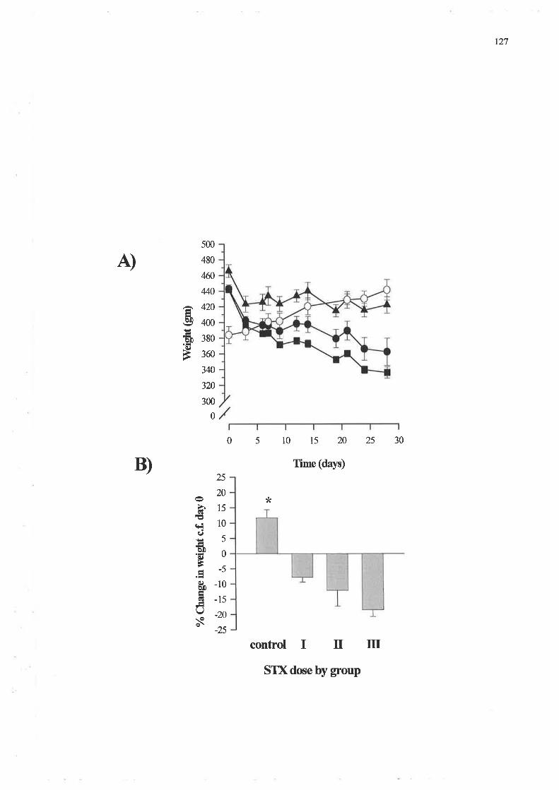
127
500
480
4(fr
4/+0
â 420Ëeg 4oo
å 3Bo.o)
È 360
340
320
300
0
25
20
Þ15;rode5.HD oÈtr
s -10
õ -15ñY -zox' -25
A)
051015202530
Time (da¡n)
*
control I tr m
B)
STX dose by group
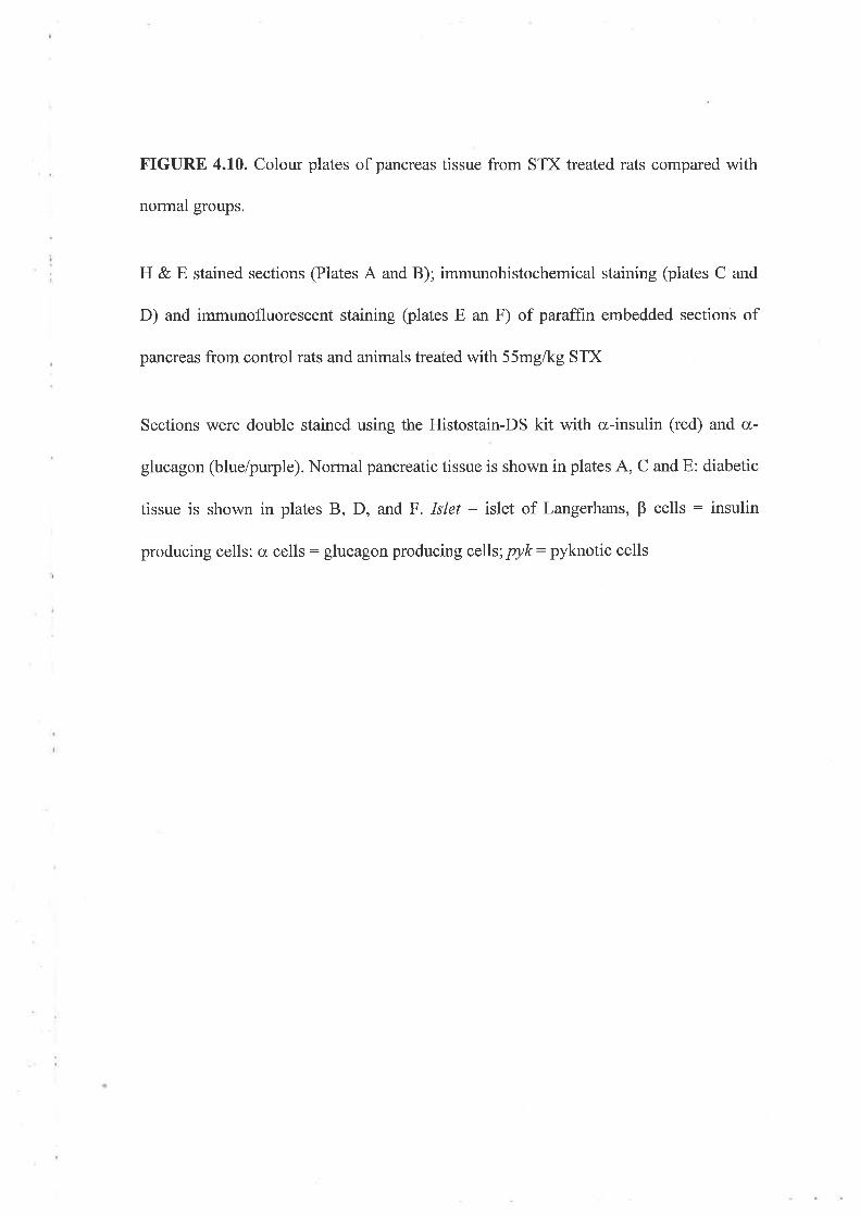
FIGURE 4.10. Colour plates of pancreas tissue from STX treated rats compared with
normal groups.
H & E stained sections (Plates A and B); immunohistochemical staining (plates C and
D) and immunofluorescent staining þlates E an F) of paraffin embedded sections of
pancreas from control rats and animals treated with 55mg/kg STX
Sections were double stained using the Histostain-DS kit with a-insulin (red) and a-
glucagon (blue/purple). Normal pancreatic tissue is shown in plates A, C and E: diabetic
tissue is shown in plates B, D, and F. Islet : islet of Langerhans, B cells : insulin
producing cells: cr cells: glucagon producing cells; pyk: pyknotic cells
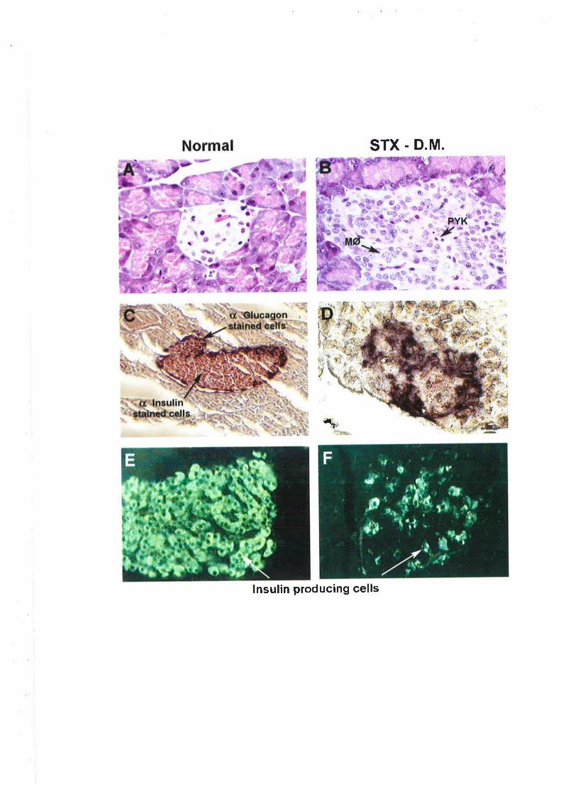
STX - D.M.
t"1,
lnsulin producing cells
tl'I
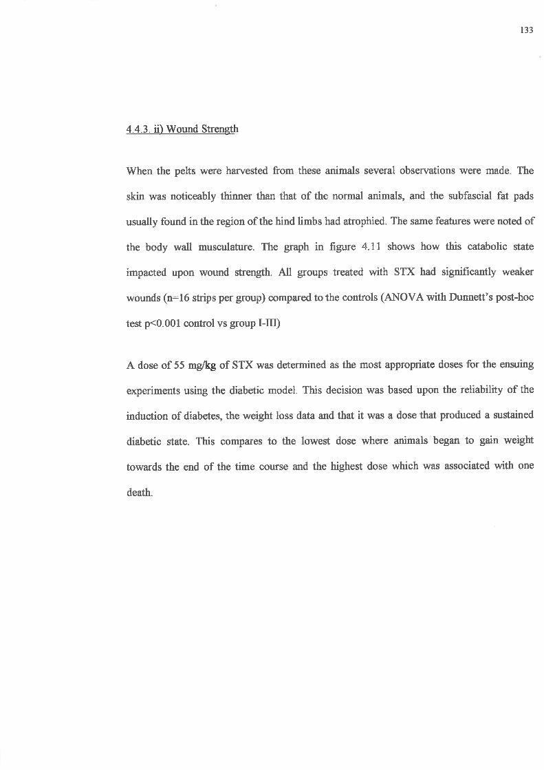
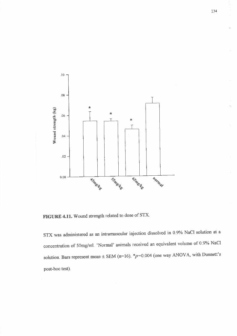
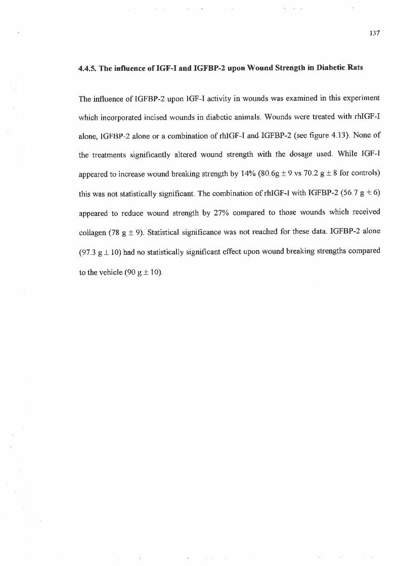
t37
4.4.5. The influence of IGF-I and IGFBP-2 upon Wound Strength in Diabetic Rats
The influence of IGFBP-2 upon IGF-I aúivity in wounds was examined in this experiment
which incorporated incised wounds in diabetic animals. Wounds were treated with rhIGF-I
alone, IGFBP-2 alone or a combination of rhIGF-I and IGFBP-2 (see figure 4.13). None of
the treatments significantly altered wound strength with the dosage used. While IGF-I
appeared to increase wound breaking strength by 14% (80.69 + 9 vs 70.2 g* 8 for controls)
this was not statistically significant. The combination of rhIGF-I with IGFBP-2 (56.7 g x 6)
appeared to reduce wound strength by 27% compared to those wounds which received
collagen (78 g t 9). Statistical significance was not reached for these data. IGFBP-2 alone
(97.3 g+ l0) had no statistically significant effect upon wound breaking strengths compared
to the vehicle (90 g t l0).

141
Normal subjects exhibit gains of net skeletal muscle proteinwhen given GH (100,101). If
administered via subcutaneous or intravenous routes, exogenous GH produced stronger
wounds in laboratory rats (268) as well as increased collagen content of both intact skin (93)
and granulation tissue (92,93).It has also been reported to increase the strength of incisional
wounds in hypophysectomised animals (259) and animals affected by catabolic states
including diabetes Q90) and malnutrition (94). Zaizen et al used the bursting strength of
laparotomy wounds in rats as their model and reported a dose dependent relationship
between subcutaneous GH and wound strength which peaked at 500ng of GIVday with the
greatest advantage seen in the first week.
Immunoreactivity for GH receptor is found in all layers of the skin, however it appears to be
most pronounced in the epidermis and particularly the epidermal appendages as described
by Lobie et al (91). Tavakkol et al (90) used polymerase chain reaction (PCR) techniques to
better localise GH receptor transcript to melanocytes and dermal fibroblasts and
specifically noted very little expression by keratinocytes. It can be hypothesised that GH
directly influences hair growth and melanocyte differentiation or proliferation but its effect
upon the keratinocytes is at best indirect. A proposed pathway would involve activation of
dermal fibroblast proliferation induced by IGF-I secreted in response to systemic GH. IGF-I
may activate keratinocytes in a paracrine fashion to promote epidermal cellular proliferation
and differentiation.
Results from this study demonstrated that a single dose of topical GH did not influence the
strength of incisional wounds when assessed seven days after application (see 4.4.1 and
4.4.2). This contrasts with the findings of Zaizen et al (94) and may reflect inadequate
activation by a single topical dose of the cellular mechanisms that mediate GH responses.
t42
Alternatively GH principally promotes IGF-I mRNA expression and particularly hepatic
IGF-I protein production. However GH acts in selected tissues such as growing bone (291)
and granulation tissue (95,96)to influence IGF-I secretion by target cells. Granulation tissue
produced within Hunt-Schilling chambers expressed increased IGF-I mRNA in response to
GH stimulation. However such expression has not been examined using incisional wound
models. In addition it is unclear whether the changes seen in healing dermis are truly
reflected by formation of granulation tissue within Hunt-Schilling chambers where the
newly formed tissue is bathed in 'wound fluid'.
The minimal mitogenic response to GH documented in this study and also reported by
Tavakkol et al (90) and Cook et al (124) provided evidence that GH is only a weak
fibroblast mitogen. In addition IGF-I is more potent when used alone or where its secretion
within a target tissue is promoted by systemic GH. The wound strength data cleatly
demonstrated the inactivity of GH in vivo when used topically (see 4.4.1)
Breaking strengths of wounds treated with IGF-I (100 ¡rglml (100 pVwound) were similar to
those of the controls. Topical application of IGF-I improved wound strength at the higher
dose of 1 mg/ml, 100 p/wound. This represented a breaking strength about 4OYo greater
than controls. Enhancement of breaking strength using IGF-I as a single large topical dose
has not previously been demonstrated to be effective in normal animals (132,247,273,276).
In states of impaired healing, such as diabetes or steroid affected animals, other parameters
including hydroxyproline content and DNA synthesis are improved with IGF-I alone
(46,727). However both studies used different models. Suh et aI (127) utilised the Hunt-
Schilling chamber and Greenhalgh et al (46) primarily studied responses in excisional
wounds. In this study IGF-I has been used at concentrations that are higher than in previous
143
reports. Therefore variations in techniques of treatment administration and dosage may
contribute to contradictory evidence between studies.
Replacing IGF-I with one of its analogues, LRfIGF-I, did not replicate the effect obtained
with the native peptide in normal animals. This contrasts with data obtained in vitro (see
Chapter 3) and other animal studies that have shown systemic analogues of IGF-I's to have
a greater general anabolic effect than IGF-I, particularly in metabolically impaired animals
(143,145,146,150,255,292). Scheiwiller et al (256) demonstrated that infusions of IGF-I
restored growth in insulin deficient diabetic rats as reflected by weight gain and tibial bone
growth. Other studies have reported that N-terminus modified analogues of IGF-I, when
administered systemically and continuously are able to restore growth in diabetic rats and
therefore show greater potency than the native molecule (146). Similar effects are noted
with the catabolic state induced by high dose systemic steroids (145) where weight loss can
be reversed. Read et al (149) described the analogues as being several fold more potent than
IGF-I in their observations of intestinal growth in rats pretreated with high dose
dexamethasone. Tomas et al (150) suggested the effects of IGF-I and their analogues in
diabetes are complimentary to those of insulin and may in fact facilitate to insulin activity
by increasing tissue sensitivity to insulin. Elevated IGFBP concentrations, particularly
serum IGFBP-3, provided an explanation for the difference between analogue and native
peptide. They reduce the amount of free IGF-I but the analogues with their diminished
affinity for IGFBP's, will not be affected in this manner.
The nature of IGFBPs in the wound and their roles may be important in finding an
explanation forthe effrcacy of analogues in diabetic but not normal wounds. While IGFBPs
are present in rat wound fluid their function at the wound site remains unclear (191)- If they
144
protect IGF-I or help bind IGFs to the ECM Q93) or if one of their fi¡nctions is to present
the peptide to cell surface receptors, then reduction of the binding affrnity for these proteins
may have several effects upon IGF activity invivo. Firstly, IGF half life may be reduced due
to the reduced protection from proteolytic enzymes within the wound. Secondly they may
be cleared more rapidly because IGFBPs associated with ECM proteins will be unable to
retain the peptide within the wound environs. In addition, IGFBPs -1, -2 and -3 contain
within their sequence the arginine-glycine-D-asparagine (RGD) sequence which is the
recognition sequence for integrins Q57,294). These are a group of cell surface receptors
which generally bind extracellular matrix molecules such as laminin, fibronectin and
collagen and are believed to be associated with cell locomotion (70).
The attachment of an IGF/IGFBP complex to an integrin may enable presentation of the
IGF peptide to its own cell surface receptor. This may potentiate the IGF response but may
be an IGFBP dependent activity. Alternatively, as demonstrated by Jones et al (294), they
may stimulate cellular events such as cell migration by themselves by using an RGD-
mediated mechanism. While integrin binding is related to the RGD recognition sequence for
IGFBP-I and -3,IGFBP-2 is not known to bind integrin family members Q95).Galiano et
al (295) examined the significance of the RGD sequence in the IGFBPs and reported that
IGFBP-2 did not influence healing in a rabbit ear dermal ulcer model either when applied
alone or in combination with IGF-I. Further evidence for IGFBP-2 being an inhibitor of
IGF-I activity has been produced by in vitro studies where it has been shown to inhibit IGF-
I migration of smooth muscle cells (258) possibly by preventing IGF-I binding the type I
IGF receptor. These reports correlate well with the present study's results for incisional
wounds where neither diabetic nor normal wounds were influenced by the combination of
IGFBP-2 and rhIGF-I.
145
Gockerman et al (25S) has demonstrated that IGF-I analogues exhibited greater aúivrty in
vitro than native peptide in circumstances where there was an excess of IGFBP-2. In this
study this situation was replicated in two ways by either adding exogenous IGFBP-2 or
inducing an increase in endogenous IGFBP-2 which is a feature of diabetes (180,296-298).
Lower molecular weight binding proteins (LMW-BPs) can be detected at increased
concentrations in diabetic rat wound fluid (1S0) while diabetic wound fluid has
characteristics which inhibit fibroblast growth (178). In both normal and diabetic
experiments, the combination of IGF-VIGFBP-2 had no effect upon wound healing and
neither did IGF-I when used alone in the diabetic model. I-n3fCf-f improved wound
strength in a model that was known to be associated with excess IGFBP-2 production.
The experiments described in this chapter confirmed impairment of the healing response in
STX-diabetic rats. This was reflected by the reduced wound breaking strength of those
animals rendered diabetic in the first experiment. Bitar and Labbad (161) similarly found
that wound strengh and collagen content were STX dose dependent. All doses of STX
caused a significant reduction in wound strength compared with the normal animals. BSL
and body weight data confirmed the diabetogenic effect of the STX. This was further
supported by immunofluorescent staining of sections of pancreas taken from representative
animals in each group. These sections confirmed the loss of p cells from the islets of
Langerhans. The effect of insulin supplementation in these animals was not established as
this would possibly confound the results obtained with the IGFs in the ensuing studies- The
severity of diabetes ie weight loss that the animals could tolerate without insulin had to be
determined. This preliminary study confnmed that while weight loss can exceed 20Yo of
original weight over five weeks, this was tolerated by the animals. The catabolic state
induced by STX and the consequent biomechanical changes might be construed as being
146
secondary to STX however previous investigators have shown that these effects are
prevented by insulin Q99).
Experimental and clinical studies have documented factors which contribute to impaired
healing in diabetics. They include clinically large vessel arterial disease, neuropathies (156)
and a reduced collagen content (180,299). This reflects lower collagen synthesis and
glycosaminoglycan (GAG) content as well as increased proteolytic activity within the skin
(164). There may also be deficiencies of growth factors particularly IGFs and TGFp (300).
Application of IGFs and other growth factors to experimental diabetic wounds is not
unprecedented. Greenhalgh et al @6) examined the effect of IGF-I, IGF-II alone and in
combination with PDGF-BB in a murine model of inherited diabetes. Their major finding
was the synergism observed between IGF-tr and PDGF-BB in healing and that all three
factors improved the rate of healing. This same model has also been used to demonstrate
eflicacy for the application of acidic FGF (301).
Comparison of the recombinant protein with its analogue suggested that both were capable
of improving wound strength and that I.nffCf-f was more potent in reversing the
detrimental effects of diabetes than IGF-I. The wounds treated with the analogue exhibited
significantly increased wound strength compared to the paired control wounds. This
suggested that the reduced binding protein affrnity of fn3IGf-I was beneficial at enhancing
IGF-I effrcacy in diabetic states. This finding contrasted with bhat observed in
uncompromised wounds where IGF-I was more potent.
The influence of one of the IGFBPs (IGFBP-2) was tested in the diabetic model. The results
suggested that this particular binding protein neither impairs nor augments the activity of
t47
IGF-I effrcacy in diabetic states. This finding contrasted with that observed in
uncompromised wounds where IGF-I was more potent.
The influence of one of the IGFBPs (IGFBP-2) was tested in the diabetic model. The results
suggested that this particular binding protein neither impairs nor augments the activity of
IGF-I in these wounds. However given the effect of I-rr-3fGf-I it may be postulated that the
accepted increase in IGFBP-2 concentrations in uncontrolled diabetes may inhibit IGF-I
activity and this may account for the lack of response to rhIGF-I or rhIGF-I combined with
IGFBP-2. In contrast other IGFBPs, BP-l and -3, will augment the effects of IGF-I in
compromised (276) and uncompromised wound environments Q47). These effects appear
to be molar ratio and model dependent (247,258).
148
4.6. SUMMARY
In summary GH alone was inactive in this model of wound repair where a single dose was
applied at the time of wound closure. In contrast IGF-I was able to increase breaking
strength. The lack of GH effect may be explained by the observation that in other studies
multi-dosing of GH may be required to stimulate expression of IGF-I which then exerts
autocrine and paracrine effects upon dermal and epidermal elements. Therefore GH may
exert its effects in a fundamentally different manner in peripheral tissues compared to its
influence in the liver where it promotes the synthesis and release of IGF-I into the systemic
circulation which in turn acts in an endocrine fashion. The presence of GH receptors and
binding protein on dermal fibroblasts supports this hypothesis. However this cell line does
not exhibit proliferative or synthetic responses when isolated and cultured in serum free
media containing only GH (see chapter 3).
Manipulation of the binding affrnity of IGF-I to its binding proteins has been explored in
normal and compromised rodent models of incisional wound repair. Only in normal animals
and at a high dose (100 ¡rglwound, 1 mglml) can exogenous IGF-I improve the breaking
strength of incisional wounds. In compromised metabolic states where IGFBPs are
probably elevated compared to normal animals the analogue demonstrated its potential as a
more effective topical agent. This suggested that endogenous IGFBPs impact upon the
potency of exogenous IGFs perhaps by 'soaking up' the IGF-I thus preventing them from
having an immediate efTect at the cellular level. The functions of IGFBP-2 may not include
presentation of IGF-I to cells therefore it may not promote receptor binding and activation
149
by the IGFs. For the normal wounds large exogenous doses of IGF-I may have allowed
sufficient free IGF-I to reach the cell surface. The lack of effect of fn3fCf-I in the normal
model remains diffrcult to explain. It may partly relate to the role some IGFBPs have in
binding the IGF/IGFBP complex to cell surface integrins and then presenting the IGF
molecule to the IGF receptor.
Diabetes does affect incsional wound repair in rodents and this effect is partly reversed by
LRÍIGF-I but not IGF-I. This effect can probably be explained through a knowledge of the
balance of IGFBPs reported in this model. As previously discussed IGFBP-2 is inhibitory to
IGF function and its concentration is elevated in diabetes. Therefore exogenous rhIGF-I
with its greater affrnity for IGFBPs may be less effective than the analogue, since the
response to IGF-I was further depressed when administered with exogenous IGFBP-2
although not significantly
While some implications of this series of studies at a clinical level remain unclear, LR:IGF-
I may have a place as a topical agent to augment healing of incised wounds in diabetics.
150
CIIAPTER 5
EXCISIONAL WOUNDS: RESPONSE OF NORMAL ANI)
DIABETIC WOT]NDS TO TOPICAL IGF.I AND TOPICAL
LONG nt rcr-r
5.l INTRODUCTION
5.1.1. Objectives
5.2 MATERIALS
5.3 METHODS
5.3.1. Preparation of Treatment Solutions
5.3.2. Induction of Diabetes
5.3.3. Surgical Technique and Post-operative Care
5.3.4. Data Collection and Monitoring of animals
5.3.5. Statistics
151
5.5. RESULTS
5.4.1. Blood Sugar Levels and Weight Change
5.4.2. Wound Healing Data
5.5. DISCUSSION
152
5.1. INTRODUCTION
Excisional wound models have been developed to examine granulation tissue formation,
wound contraction and wound epithelialisation. Previous chapters have discussed IGF
physiology and examined the effect of IGFs on compromised incised wounds in both
normal and compromised healing states. Also the effects of IGF-I and its analogues have
been explored using cultured fibroblasts in models of lattice contraction, cell proliferation
and protein synthesis. In particular the FPCL contraction studies demonstrated that IGF-I
and especially the analogues can promote fibroblast contractile activity. These results
encouraged further investigation of the effects of IGF-I and tn3tGp-I on healing by
secondary intention in both normal and diabetic rats.
While treatment of diabetic excisional wounds with growth factors including IGF-I
(46,135), acidic FGF (301) and PDGF (46) has been described in the literature, obese
genetically diabetic mice were used rather than rats that have a chemically induced diabetic
state. One of the principal findings with these studies is that the use of topical agents, in
particular aFGF alone and IGF-I or IGF-II combined with PDGF, can accelerate wound
closure in a diabetic animal however the rate of healing is not returned to that of normal
animals.
States of impaired healing due to steroids, diabetes, malnutrition or vascular insufficiency
cause significant morbidity and consume health care resources (302). Aberrations in IGF
physiology due to diabetes include altered IGFBP profiles, particularly increases in IGFBP-
153
2 (180) and reduced levels of 46 kDa species (consistent with IGFBP-3) in conjunction with
reduced systemic IGF-I concentrations (161). Collagen metabolism is also impaired
resulting in reduced collagen content in the skin of diabetic animals Q99).
Given the tissue culture results presented in chapter 3 demonstrating the anabolic activity of
IGFs, particularly the analogues and the effect of diabetes upon the bioactivity of IGF-I
exogenous analogues of IGF-I may potentially reverse impaired healing responses
associated with diabetes.
5.1.1. Objectives
The aim of this study was to examine the effect of topical IGF-I and its analogue LFfIGF-I
on excisional wound healing in both normal and diabetic animals.
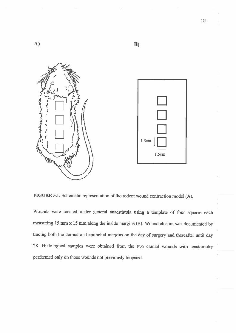
t54
5.2. MATERIALS
Sprague-Dawley rats were obtained from the Central Animal House, University of
Adelaide, and divided into two groups with weight ranges of 200-250 g and 250-300 g on
arrival. The animals in the heavier group were given STX.
Collagen, STX, IGF-I and I.nftCf-I were obtained from the same sources as chapter 4.
Isoflurane (Forthane, Abbott Australasia, Sydney, Australia) mixed with NzO: Oz was used
to provide general anaesthesia for the days of shaving, wounding and tracing of wounds.
Povidone-iodine (Betadine, Mundipharma, Switzerland) and TOYo ethanol were used to
clean the dorsal skin. A semipermeable polyurethane dressing, Opsite@, was supplied by
Smith & Nephew, Hertfordshire, U.K. Wound strips were stretched using a Mecmesin
tensiometer (Model PCM 25OOF,L, Mecmesin, U.K ).
155
5.3. METHODS
5.3.1. Preparation of Solutions
LK^3IGF-I solutions \¡/ere prepared in concentrations of 100¡rg/ml and lmglml mixed with
collagen while rhIGF-I was prepared at a concentration of lmglml The control solution
(collagen only) and the peptides were applied in volumes of 100p1 to each wound on
alternate days for 7 days.
5.3.2.Induction of Diabetic State
One week before surgery the animals were divided into two g[oups and the heaviest 32
animals were allocated to the diabetic group. The rats in this group were injected
intramuscularly with STX, 55mg/kg (5Omg/ml in citrate buffer, pH 4.5), while the control
group received an injection of citrate buffer vehicle. Weights and BSLs were recorded on
the day before surgery after which these groups were fuither randomised into four treatment
subgroups as detailed in table 5.1.
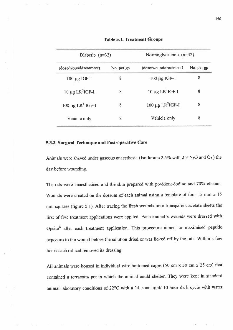
156
Table 5.L. Treatment Groups
Diabetic (w32) Normoglycaemic Q=32)
(dose/wound/treatment) No. per gl (dose/wound/treatment) No. per g¡l
100 ¡rg IGF-I 8
8
8
8
loo ¡rg rnfrCr'-r
100 pg IGF-1
1o pg LK-3IGF-I 1o ¡rg l-n3tC¡-f
100 pg rnÉ ICF-I
Vehicle only Vehicle only
I
8
I
8
5.3.3. Surgical Technique and Post-operative Care
Animals were shaved under gaseous anaesthesia (Isoflurane 2.5Yo with 2:3 NzO and Oz ) the
day before wounding
The rats were anaesthetised and the skin prepared with povidone-iodine and 7oo/o ethanol.
Wounds were created onthe dorsum of each animal using atemplate of four 15 mm x 15
mm squares (figure 5.1). After l';racingthe fresh wounds onto transparent acetate sheets the
first of five treatment applications were applied. Each animal's wounds were dressed with
Opsite@ after each treatment application. This procedure aimed to maximised peptide
exposure to the wound before the solution dried or was licked offby the rats. Within a few
hours eachrat had removed its dressing.
All animals were housed in individual wire bottomed cages (50 cm x 30 cm x 25 cm) that
contained a terracotta pot in which the animal could shelter. They were kept in standard
animal laboratory conditions of 22"C with a 14 hour lightl 10 hour dark cycle with water
157
and food provided ad libitum. Adequate water supply was ensured with a 600 ml water
bottle one per cage.
5.3.4. Data Collection and Monitoring of Animals
All tracings were made under general anaesthesia and treatments were applied on days 0, 1,
3, 5,7 from time of surgery. Body weights were recorded on each day of surgery and the
wounds were traced prior to application of treatment solutions. Tracing of wounds was
conducted on days O, 7,3, 5,7, g, 12, 14,76, 19,21,23,26,28 and 29. These tracings
included both the wound and the advancing epithelial edges once it was apparent. Wound
strengths were determined on day 28 or 29.
The collected wound tracings were scanned using a Color One Scanner (Apple Computer
Inc, Cupertino, Cd USA) and Ofoto v2.0 software (Light Source Computer Images Inc,
Marin County, CA, USA). These images were colour coded and digitised using the Prism
Image Analysis software system (Dapple systems Inc, Sunnyvale, CA, USA) to allow
computation ofwound image areas.
Curves were plotted using Sigmaplot Scientific Graphing Software v 2.07 (Jandel Scientiflrc
Inc, San Ramon, CÀ USA).
Sampling for histology v/as performed using four animals per group on days 3,7 , 14, 27 and
28 or 29. A single wound on each of these animals was used only on days 3, 7 and 74 to
assemble a chronological representation of the wounds. Once wounds were biopsied they
were excluded from tensiometric analysis and further wound area analysis. Tissue
specimens were fixed in 4Yo wt/vol paraformaldehyde/PBS solution.
158
BSLs \üere recorded weekly for the diabetic groups and at the time of surgery and on the
day of sacrifice in normoglycaemic animals with samples being obtained as described in
4.3.t.
On kill days all animals were weighed and blood obtained for BSLs prior to death as
described in 4.3.1. The dorsal pelts were harvested, four wounds/animal excised for
tensiometric studies except where biopsies had been taken. These biopsy wounds were
excluded from statistical analysis and tensiometry.
5.3.5. Statistics
Data were expressed as mean values + 1 SEM and were analysed using Sigmastat v.2.0
(Jandel Scientific, San Ramon, CA USA). Body weight and BSL data for the diabetic
animals and the normoglycaemic rats were compared using unpaired Students t-test and the
effects of the treatment preparations by two-way ANOVA partitioned according to diabetes
and treatment with Student-Newman-Keuls post-hoc multiple comparison analysis.
158
Wound epithelialis ation
The rate of wound re-epithelialisation was calculated using the same formula as that used for
contraction. The results for wound epitheliasation and contraction were similar for the first 9
days therefore it can be concluded that epithelialisation did not contribute significantly to
wound closure until day 9 post-operatively. This component of wound healing appeared to
account for the final20Yo of total wound closure although it commenced earlier when closure
was 65Yoto 70Yo complete
In contrast to contraction, epithelialisation was not retarded by diabetes in this model (p : NS,
two way ANOVA) nor was it influenced by topical IGF-I or LNIGF-I. Those diabetic
animals that received LR3IGF-I 10¡rg/wound/application re-epithelialised their wounds faster
than the diabetic animals which received collagen only (p < 0.05, Students t-test). However it
needs to be emphasised that there were only 6 animals in this group as one died and another
failed to become diabetic. Therefore confirmation of the apparent acceleration in re-
epithelialisation noted for the group of diabetes x LR3IGF-I I)¡tg/wound/application would be
required to exclude a falsely positive result.
160
5.4. RESULTS
5.4.1. Blood Sugar Levels and Weight Change
Conversion to a diabetic state was confirmed using the same criteria used in the
incisional wound studies (4.4.2). BSLs forthe time course of the study are shown in
figure 5.2. The BSLs for the STX-treated rats rose dramatically by the end of the first
week after they were injected with STX. The BSLs remained elevated for the duration
of the study confirming, established diabetes. One rat succumbed within the first week
and two others failed to develop hyperglycaemia.
Body weight dala are shown in figure 5.3 and these confirmed the effect of diabetes
upon growth rates. Growth of STX-treated rats exhibited a plateau which was
maintained for the time course of the study. Another important observation was the
effect that multiple anaesthetics had upon the normal animals. These animals
exhibited a slowing of their growth with each anaesthetic. Therefore protein
malnutrition may be another feature of this particular model.
5.4.2. Excisional Wound Closure:
This is divided principally into three sections.
o wound contraction;
. wound re-epithelialisation;
o wound strength.
161
Wound contraction and epithelialisation data are presented as a ratio of the residual
wound area divided by the original wound area recorded on the day of surgery and
expressed as a percentage (Yo Areas ). A similar technique has been utilised by
Matuszewska et al (301).
potential sources of error arise in the actual tracing of wounds both the remaining
ulcerated area and the rim of neoepithelium. By having the same person perform this
task this error of interpretation would be consistent. Its accuracy was enhanced by
having the animals anaethetised unlike Cross et al (303) and Teo (304). Digitalising
the tracings provides an other important source of observer effor but again the same
operator was used and therefore the error was consistent.
Means with one SEM at each time point for each group of animals were plotted
(figure 5.4).
I(ound contraction
Wound contraction was retarded in those animals rendered diabetic. This was most
apparent between day 3 and day 72. Statistical comparison using two way analysis of
variance revealed significant retardation in all diabetic animals regardless of the
treatment peptide at days 5,7 and 9 (p<0001 daysT andg diabeticvs normal) This
significant retardation of wound contraction represented a delay of approximately 2
days in the healing of diabetic wounds when compared with those wounds on the
backs of normal animals. Importantly, wound contraction appeared to plateau between
day 11 and day 13 of healing leaving a residual percentage of original wound area (x)
that closed principally by re-epithelialisation. In all treated and control groups this
value was approximately 2OYo of Areaú.
t62
Topical administration of rhIGF-l or LR3IGF-I at either dose did not influence the rate
of wound contraction in either diabetic or normal animals
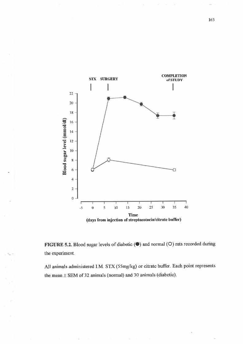
163
STX SI]RGERYCOMPLETION
Of STT]DY
-5 40
FIGURtr 5.2. Blood sugar levels of diabetic (O) and normal (O) rats recorded during
the experiment.
All animals administered I.M. STX (55mg/kg) or citrate buffer. Each point represents
the mean t SEM of 32 animals (normal) and 30 animals (diabetic).
22
20
l8
!16o814
(¡) t¿c)
L10ÉËè0
å8oo6É
4
2
0
05101520253035Time
(days from injection of streptozotocin/citrate buffer)
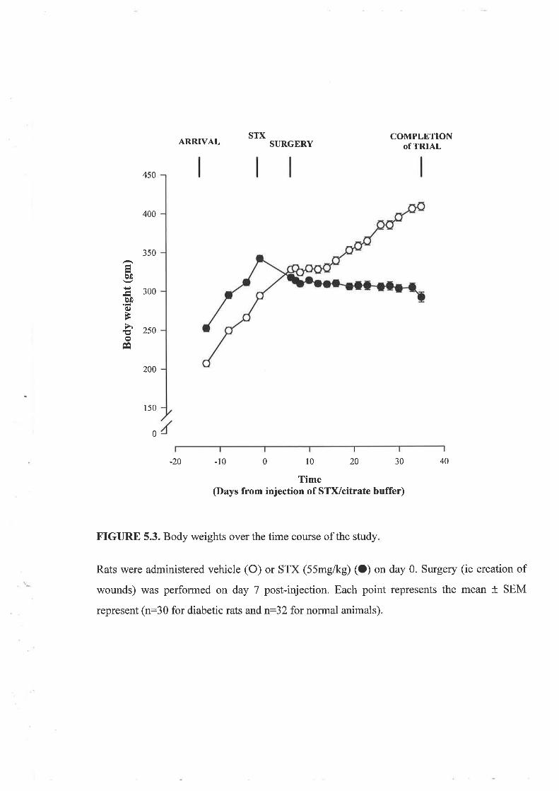
STX COMPLETIONOfTRIAL
40
ARRIVAL SURGERY
-10 0 10 20 30
Time(Days from injection of STVcitrate buffer)
350
a0
Ë 3oobooÈ
å 2soo
Êe
450
400
200 -
150
0
-20
FIGURE 5.3. Body weights over the time course of the study
Rats were administered vehicle (O) or STX (55mg/kg) (O) on day 0. Surgery (ie creation of
wounds) was performed on day 7 post-injection. Each point represents the mean + SEM
represent (n:30 for diabetic rats and tr32 for normal animals).
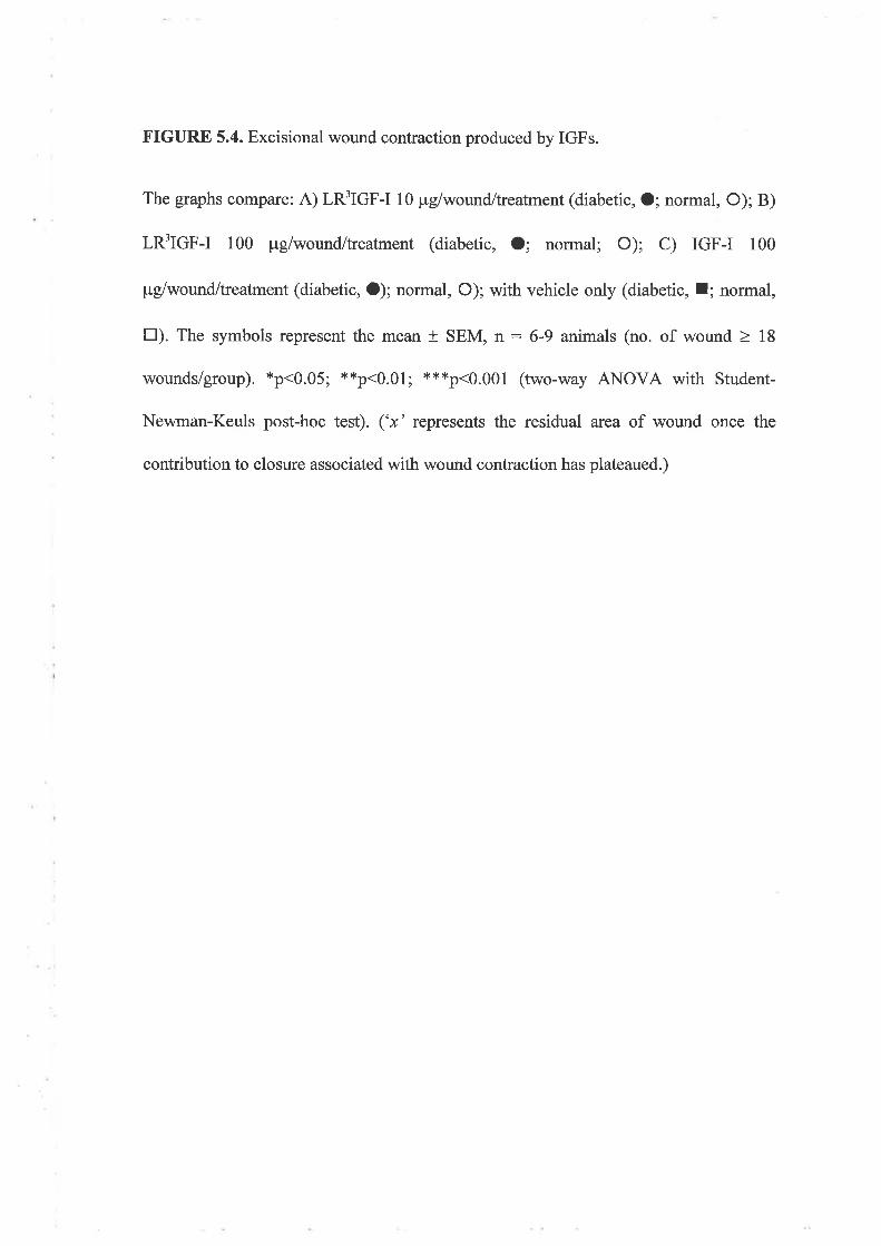
FIGURE 5.4. Excisional wound contraction produced by IGFs.
The graphs compare: A) LR3IGF-I 10 ¡rglwound/treatment (diabetic, O; normal, O); B)
LR3IGF-I 100 pglwound/treatment (diabetic, O; normal; O); C) IGF-I 100
¡rg/wound/treatment (diabetic, O); normal, O); with vehicle only (diabetic, l; normal,
tr). The symbols represent the mean + SEM, n: 6-9 animals (no. of wound > l8
wounds/group). *p<0.05; **p<0.01; ***p<0.001 (two-way ANOVA with Student-
Newman-Keuls post-hoc test). ('x' represents the residual area of wound once the
contribution to closure associated with wound contraction has plateaued.)
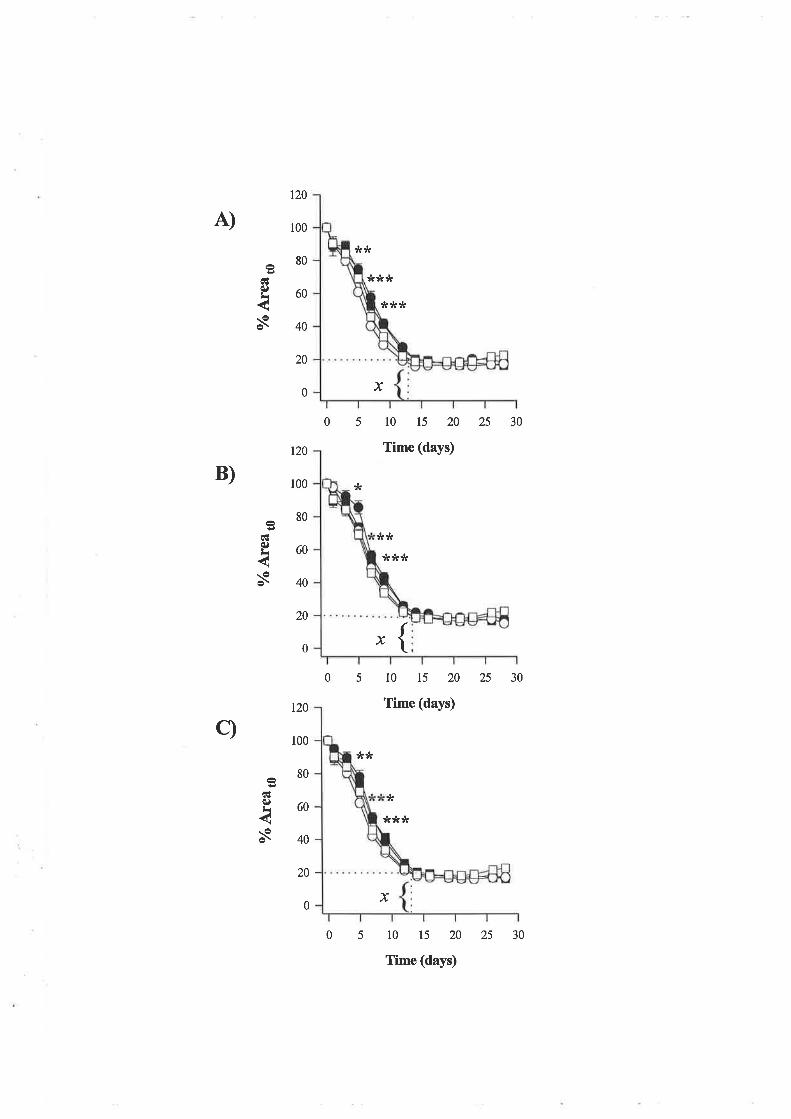
A)
B)
c)
120
100
80
60
40
20
cÉq)¡i
s
0
t20
100
80
60
40
20
êcËq)L
s
?ktf
120
r00
80
60
40
20
cËq)L
s
05
?t ?k tr
x
:r****
x
30
?k ?t
tÉ
trtÉ ?k
l0 15 20 25
Time (days)
0 {50 l0 15 20 25 30
Time (days)
tr
**JÉ
x
05 10 15 20
Time (days)
0
25 30
FIGIIRE 5.5. V/ound epithelialisation following 5 treatment applications over 7 days.
The graphs represent the mean values + SEM (n: 6 - 9) comparing A) LRfIGF-I 10
pglwound/treatment (diabetic, O; normal; O); B) LRfIGF-I 100 pglwound/treatment
(diabetic, O; normal, O); C) IGF-I 100 pgiwound/treatment (diabetic, O; normal, O);
with vehicle only (diabetic, I; normal, tr). Data were statistically compared using two-
way ANOVA with Student-Newman-Keuls post-hoc test.
A)
B)
c)
0
t20
100
80
60
40
20
t20
100
80
60
40
20
05
5
05
cf(Сr
s
120
100
80
60
40
20
(f(l)¡r
s
l0 15 20
Time (days)
l0 15 20
Time (days)
l0 15 20
Time (days)
25 30
25 30
0
0
GIq)L
s
0
25 30
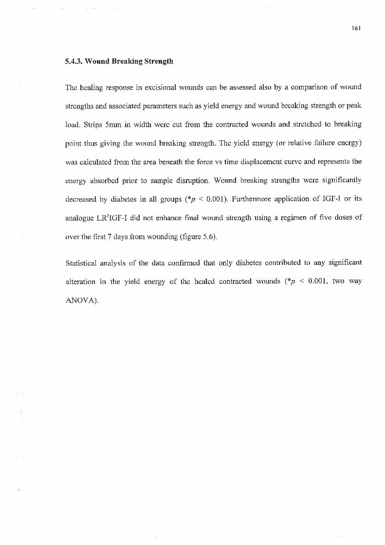
161
5.4.3. Wound Breaking Strength
The healing response in excisional wounds can be assessed also by a comparison of wound
strengths and associated parameters such as yield energy and wound breaking strength or peak
load. Strips 5mm in width were cut from the contracted wounds and stretched to breaking
point thus giving the wound breaking strength. The yield energy (or relative failure energy)
was calculated from the areabeneath the force vs time displacement curve and represents the
energy absorbed prior to sample disruption. Wound breaking strengths were significantly
decreased by diabetes in all groups (*p < 0.001). Furthermore application of IGF-I or its
analogue LR3IGF-I did not enhance final wound strength using a regimen of five doses of
over the first 7 days from wounding (figure 5.6).
Statistical analysis of the data conhrmed that only diabetes contributed to any significant
alteration in the yield energy of the healed contracted wounds (*p < 0.001, two way
ANOVA).
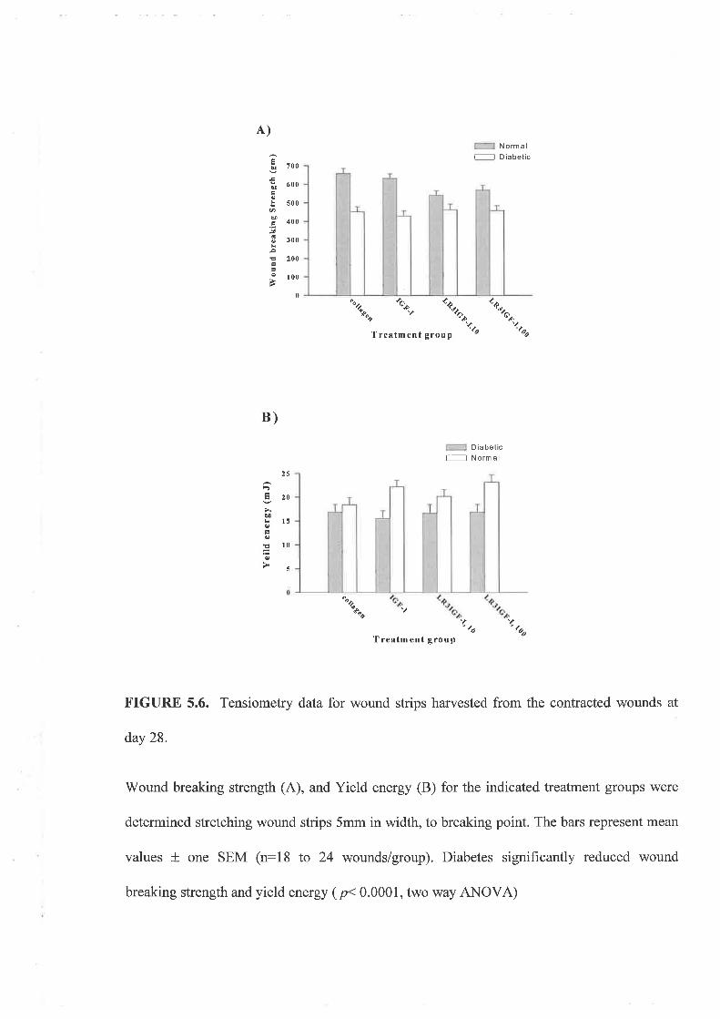
A)
B)
É;¡ 7oo
¡ru 600É
! s00Ø
$ loo.i(3 ¡oo
! 200
.- 100
Normalf l D¡abetic
\ .\ ..,,% t"*
'¿ao
0
'loTrefltment group
D ¡abet¡ctr-----l Normal
ts20Ð! l5otroìã loo'5
\'ooTreatment group
FIGURE 5.6. Tensiometry data for wound strips harvested from the contracted wounds at
day 28.
Wound breaking strength (A), and Yield energy (B) for the indicated treatment groups were
determined stretching wound strips 5mm in width, to breaking point. The bars represent mean
values * one SEM (n:18 to 24 wounds/group). Diabetes significantly reduced wound
\?o
breaking strength and yield energy (p< 0.0001, two way ANOVA)
171
5.5. DISCUSSION
Andreassen et al (305) and Greenwald et al (306) have previously shown that
streptozotocin induced diabetes is associated with weakened incisional wounds. This
has been confirmed by the studies reported in chapter 4. Therefore diabetes was
induced in a model of healing by secondary intention.
As in chapter 4, STX induction of diabetes produced sustained hyperglycaemia and
weight loss for the 5 week period of the study. Therefore it can be concluded that this
model is reliable even up to 5 or 6 weeks after administration of STX. This model of
diabetes has not been described in the wound healing literature for excisional wound
studies. Previous studies involving STX-induced diabetes have used this model to
examine healing within three to four weeks from the time of STX administration and
these have predominantly been incisional wound studies (305-307). Where animal
models have been employed to examine the effect of growth factors upon wound
contraction and re-epithelialisation a genetic murine model of diabetes has been
utilised (45,46,135,301). While reliable and convenient the mouse model is limited by
the size and the number of wounds that can be created on each animal. The same
criticism can be levelled at the rat model in comparison to larger animal models.
However this model is more convenient, allows multiple wounds and can be
manipulated easily if systemic agents such as insulin are to be administered- This
study has clearly demonstrated that untreated diabetes impairs wound healing by not
only reducing the final strength of wounds but also retarding wound contraction.
Therefore this model can be used successfully as one of impaired healing by
secondary intention.
t72
Wound closure was completed in both diabetic and normal rats within 15 to 18 days
from surgery. All groups demonstrated maximum contraction by day 14 with
complete epithelialisation by day 18. While this did not represent a delay in total
wound closure associated with diabetes, contraction from the first to the ninth day for
diabetic wounds was retarded by 2 days compared to normal wounds. This effect was
related statistically to the state of diabetes and not to any of the applied treatments.
The contraction of normal rat wounds is most pronounced during the first ten days
after wounding (205,308) as previously observed by Cross et al (303) in the validation
of their model.
'Wound contraction takes place during the proliferative phase of wound repair and is
characterised by increasing fibroblast/myofibroblast population size (309). Factors at
a biochemical and cellular level that may contribute to the delay in contraction of the
diabetic wounds include altered leucocyte function during the inflammatory and
proliferative phases (5) and reduced collagen synthesis in this model (299,310). Other
factors in this model that may influence contraction include reduced migration of
fibroblasts and inflammatory cells into and around the wound (45) as well as absolute
reductions in fibroblast numbers. An important distinction to make between this rat
model and the genetic model used by Greenhalgh et al (45,46) is that the mouse
model is obese and as stated by Greenhalgh wound contraction accounts for only 30 -
40Yo of closure compared to the 80olo documented in this study. Therefore the
contribution to wound closure that can be attributed to epithelialisation becomes
diffrcult to assess accurately as it is relatively small. While fibroblast and
inflammatory cell numbers have not been compared in this current experiment it
remains the subject of ongoing research and analysis of histological sections in our
laboratory.
t73
Wound strength is determined principally by two factors;
(i) the density of newly synthesised collagen and
(ii) the reorganisation and remodelling of that collagen from type III collagen
to type I (205)
Therefore wound strength is influenced by the intensity of fibroblastic activity which
tends to markedly increase towards the end of the first week of healing while collagen
remodelling continues for months after complete closure of the wound as
demonstrated by Levenson et al (S7). Wound strength is therefore a function of the
maturity of collagen remodelling and the amount of newly formed collagen. Collagen
cross-linking may have been retarded significantly by diabetes as wound strengths in
the diabetic animals r /ere generally lower than their normal counterparts. Collagen
deposition may also be reduced by any retardation of fibroblast and inflammatory cell
migration into the wound (45). Increased collagenase activity, increased glycosylation
of collagen and reduced hydroxyproline concentrations have also been implicated
(311). Certainly in vivo studies exist to demonstrate reduced collagen content in intact
skin (299) and reduced collagen synthesis in incised wounds (306,307).In addition,
diabetic serum can inhibit collagen production by fibroblasts (178).
In excisional wounds treated five times over seven days neither IGF-I nor LICIGF-I
affected wound healing parameters. Failure of the growth factor treatments to
influence wound closure may be related to several factors. The adherent coagulum
that exists in the first three to five days on excisional wounds could provide an
impervious barrier to peptide absorption. While occlusive dressings were used to
maximise treatment exposure to the wound and attempt the simulation of a moist
wound healing environment the dressings could not be maintained more for than 6
t74
hours on the backs of the animals. This subsequently allowed the formation of the
coagulum. It is also possible that a significant amount of the treatment preparation
may have adhered to the dressing even though the collagen was allowed to form a gel
before the dressings were applied.
While technical factors may compromise treatment absorption other more
fundamental physiological factors may also explain the lack of response to IGFs. The
expression of cell surface receptors for IGF-I and the secretion of IGFBPs will also
affect the response at a cellular level. Antonaides et al (189) showed that in acutely
wounded explants of porcine skin IGF-I mRNA expression and IGF type I receptor
were present before and after injur¡ but were not up-regulated in either the dermis or
the epidermis following wounding. Even though IGFs improve excisional wound
olosure in diabetic mice, (46),the lack of IGF receptor up-regulation related to PDGF
and reduced endogenous IGF-I expression, may have contributed to our failure to
demonstrate a positive IGF-I effect. Therefore a competence factor such as PDGF
may be required to maximise any IGF response in our model including the response to
the analogues.
The influence of IGF-I may not be directed primarily to the dermis. Several studies
have shown that IGF-I mRNA is expressed by fibroblasts originating from the dermis
(125) and from granulation tissue (126). In addition epithelial cells, namely
keratinocytes, express IGF-I receptors during healing (3L2,313).IGF-I produced by
fibroblasts has been demonstrated by Barreca el al Q23) to be partly responsible for
keratinocyte growth when cultured fibroblasts are employed as a 'feeder' layer. These
studies indicate a paracrine activity of IGF-I. Therefore it is possible that IGF-I exerts
its major influence upon re-epithelialisation rather than wound contraction. As up to
80 yo of wound closure in this model is attributable to contraction, any delay in re-
epithelialisation may be masked by the large contribution that contraction has to the
healing response. A tight skin mouse model exists to examine re-epithelialisation of
full thickness wounds (205). Therefore the ability of IGFs to promote epithelialisation
may be better examined in such a model.
The complex modulation of IGF activity in the wound is increased by the known
existence of IGFBPs in rat wound fluid (191) and alteration of their profiles with
diabetes (180). IGFBPs, along with proteases found in wound fluid
(180,791,202,203), influence the balance of free IGF-I within the wound milieu by
protecting it from degradation and providing a slow release pool of active peptide.
They may simultaneously prevent IGFs binding to cell surface receptors which
contributes to inactivation of the peptide. The issue surrounding IGFBPs is especially
relevant to diabetic models where IGFBP-2 is known to be proportionally increased in
experimental insulin dependent diabetes mellitus Q57,314) and is recognised as an
inhibitor of IGF-I activity Q77)
5.6. SUMMARY
In summary this chapter presents a combination of two rodent models, the excisional
wound and the STX-induced diabetic rat. The data show that diabetes will persist in
these animals for nearly six weeks and therefore this excisional wound model of
diabetes can be used to investigate effects of diabetes on open wound healing. This
has relevance to human diabetic ulcers or acute wounds that have been surgically
176
debrided. That the rat is a valid and useful diabetic model is reflected in the wound
contraction delay of 2 days when compared to normal animals
Tissue culture studies (chapter 3) showed some promise for IGFs as agents to promote
excisional wound healing. However other factors including mechanisms of vehicle
delivery, the presence of proteolytic enzymes, receptor expression and levels of
various IGFBPs may play a role in determining the net effect attributable to IGF-I. A
greater understanding of anabolic events within the wound would be gained by
examination of processes involved in the induction of wound derived IGF peptides
and receptor mRNA transcripts by exogenous peptides. This is especially important in
metabolically compromised states such as diabetes.
t77
CHAPTER 6
DISCUSSION AND SUMMARY
6.1. IGF PHYSIOLOGY
6.2.IGFS A¡TD IGTBPS IN WOUND F'LUil)
6.3.,rN VTVO WOI]ND HEALING AI\ID THE IGF'S
6.4 CONCLUDING REMARIG
178
This study has examined the ability of IGF-I to facilitate dermal healing as a topical
agent with a focus on diabetic wounds.
6.1.IGF PHYSIOLOGY
The anabolic characteristics of IGF-I QS9), its activity in those cell lines associated
with wound repair (71,90,133) and its expression by fibroblasts at the wound site
(313) all suggest that IGF-I could play an important role in wound repair. These
characteristics are similar to those of insulin (109) in that under normal circumstances
both insulin and the IGFs have anabolic effects but insulin has a much gteater
influence upon glucose homeostasis.
The bioavailability and production of IGF-I is regulated by a wide variety of
influences including GH (253), nutrition (315), metabolic status and the IGFBPs. It
exhibits endocrine, paracrine and autocrine activity. IGF-I is produced by the liver
and complexed with IGFBPs in the systemic circulation. IGFBPs have several
important roles including prolonging the half lives of IGF-I and II as well as
providing a means for distributing IGFs from the intravascular to the interstitial
spaces (257). Of the six known binding proteins, IGFBP-3 carries tne majority of IGF
peptide systemically Q57). The plasma levels of IGF-I corelate with GH secretion
179
and activity. Thus IGF-I has earned a reputation as the second messenger for GH and
therefore accounts for the majority of physiological responses associated with GH.
The somatomedin hypothesis proposed in7972 by Daughaday et al was an attempt to
define somatomedins (IGFs) and their relationship with GH (106). It states that "a
GH-dependent plasma factor stimulates in cafülage not only the incorporation of
sulfate into chondroitin sulfate but also the incorporation of thymidine into DNd
proline into hydoroxyproline of collagen and uridine into RNÂ"' (106). GH is the
major stimulus of IGF-I production particularly by the liver. However other factors
influence IGF production by target tissues including nutritional state (316) and
conditions specific to the model used to investigate expression of IGF such as
monolayers of endothelial cells (11) or granulation tissue obtained from implanted
wound chambers as described by Steenfos et al (96,126).
6.2. IGFS AND IGFBPS IN WOUND FLUID
IGFs have been measured in most body fluids, including wound fluid exudate (112).
Therefore the identification of IGFs in reasonable concentrations in the wound fluid
of skin graft donor sites was not unexpected. It was interesting to discover that no
discernible pattern of IGF concentration existed in any of the five patients over the
time course of this current study. Greater numbers and segregation by age group may
provide a better indication of any trend.
180
By time matching wound fluid and plasma samples it was possible to show that the
IGF levels in both were similar. It is diffrcult to draw any conclusion from this study
as to whether the main source of IGFs in wound fluid was local production or plasma
transudation. It does provide an avenue for further research exploring the transport of
IGFs from blood to the wound, expression of IGF mRNA as well as protein
production in this model. Questions also arise from this curent study concerning
clearance of IGFs and the effects of tissue metalloproteinases, such as gelatinase and
collagenase, upon the equilibrium of IGFs at a wound site. As discussed in chapter 2,
the technical exercise of measuring IGF levels will also impact upon the recorded
concentration of IGFs. Correlation of the two techniques described clearly indicates
that complete removal of IGFBPs from the samples is important if one technique is to
be used reliably in place of another to measure IGFs. This applies particularly to IGF-
II.
WLB and immunoblots confirmed the presence of low molecular weight IGFBPs in
raw samples including proteolytic fragments of IGFBP-3. Low molecular weight
binding proteins such as these moieties tend to be retained by many IGFBP extraction
procedures thus interfering with measurement of IGF-I concentration (317).
Furthermore, WLBs of the acid-ethanol extracts revealed residual IGFBPs in almost
all samples.
As with the IGF levels, inter-patient variation made it diffrcult to determine if any
specific relationships existed between the IGFBPs detected, IGF levels and time of
healing. IGFBP-I, -2, and -3 were the main species found in wound fluid, however
IGFBP-4 was also detected but less reliably
181
6.3.IN WVO WOUI\D HEALING AND THE IGFS
Experimental wounds have provided a means of exploring the effects, expression and
fate of IGF-I at various sites. Models ofwound contraction, granulation tissue growth,
wound fluid production and wound epithelialisation have been utilised to identify
IGFs in the wound as well as mRNA expression and receptor localisation
(96,125,126,133,189). A hybrid model of toxin induced diabetes and excisional
wound healing has been introduced in this study to examine the influence of IGFs in a
compromised healing state.
In vitro effects attributable to IGF-I include fibroblast mitogenesis, collagen
production as well as keratinocyte proliferation and migration. For these activities
IGF-I appears to act via both autocrine (fïbroblast-fibroblast) and paracrine
(fibroblast-keratinocyte) mechanisms. The results in Chapter 3 corroborate with these
previous findings. In addition IGF-I was shown to induce fibroblast-mediated
contraction of collagen lattices. Cell growth and FPCL contraction suggest that IGF-I
is the growth factor responsible for the majority of GH's physiological effects and
that GH alone is relatively ineffectual as a mitogen for fibroblasts. Other studies have
demonstrated that the influence of GH in fibroblast culture is largely due to
endogenous IGF-I production (124) although these experiments were conducted in the
presence of 0.5Yo hypopituitary serum. This contrasts with those results observed in
Chapter 3 where the cell growth and contraction assays were performed under serum-
182
free conditions. The failure of GH to augment IGF-I's effect would suggest that the
observed responses of IGF-I combined with GH were due to IGF-I alone. This lack of
GH response was also observed in the incisional wound repair studies.
Wound strength was improved above paired controls where a suffrciently high IGF-I
concentration v/as administered. Whereas no significant response was observed for an
equal concentration of GH. Such a finding contrasts with those of several other
studies where GH has been shown to increase granulation tissue production,
deposition of collagen as well as to strengthen wounds (89,95). Porcine data from our
laboratory support the improvement in wound healing parameters produced by
systemic GH use (318). However a major difference exists between these studies and
those in Chapter 3. GH was administered subcutaneously or given as an infusion or, in
the case of Garrel's study (89), growth hormone releasing hormone (GHRH) was
used. 'Whereas in the current study, a single dose of GH was administered directly into
the wound as a topical preparation (refer Chapter 3).
Several factors may contribute to the observed absence of GH effect. Firstly, a single
dose may be insuffrcient to activate cell surface receptors for GH in the skin and
thereby induce IGF-I activity. Secondly GH 'related effects' must be mediated by
hepatic-derived IGF-I and skin lacks GH receptors. Thirdly GH is denatured within a
fresh wound before it can exert any effect. The first proposal would presuppose a time
lag between GH stimulation, IGF-I production and IGF-I secretion by the local tissue.
Certainly this is supported by evidence that IGF-I mRNA expression and peptide
concentrations within adipose and granulation tissue increase following GH
administration (126,319) while serum and extracellular levels of IGF-I can also
improved (126,192). The weakness of the second proposal is that rat dermal
183
fibroblasts do express GH receptors. The third proposal may have merit given the
intense proteolytic activity that occurs within a new wound.
If tissue proteases are responsible for the lack of GH effect, a similar influence upon
IGF-I should have been observed. However the homeostatic mechanisms protecting
IGF-I within the ECM may also protect it from degradation. This is especially
applicable to IGFBP-I and IGFBP-2 which contain the Arg-Gly-Asp (RGD) sequence
within their structures allowing them to bind to cell membrane integrin receptors and
to the ECM (320). Thus IGFBPs can increase the half life of IGFs within both the
systemic circulation and target tissues.
Further support is provided by the relative lack of response in wound strength noted
with the analogue I,n3fCn-f. In vitro experiments presented in the current study
suggest that this peptide has greater anabolic potency than rhIGF-I. However it
appears that reduction of binding afninity to IGFBPs may offset this greater potency in
wound healing models because reduced binding to IGFBPs leaves a gteater
proportion of the analogue exposed to be denatured by tissue. Alternativety I-n3tCf-f
may simply be cleared from the tissues intact by transport mechanisms or degraded by
tissue proteases at the wound site. The ability of the binding proteins to provide a
means of slow release of active growth factor may be also important in ensuring a
biological response. However, in vitro studies do not truely replicate the in vivo
response, as IGFBPs differ in their ability to inhibit or facilitate IGF activity in vitro
and their influence may be dictated by the environment in which they are expressed.
The increased potency of IGF-I analogues upon cultured fibroblasts was expected as
previous studies have demonstrated these peptides' greater ability to induce cell
184
proliferation and protein synthesis in a variety of other cells. In addition, studies of
somatic gfolilth indicate that this potency is not confined to cell culture
(145,146,255,292). However in experiments using IGF-II analogues, these peptides
v/ere no more effective than rhIGF-II in the models described (results not shown).
This suggests that while the IGF-I analogues provide more free IGF-I-like peptide to
bind cell surface receptors, the same characteristic for IGF-II analogues may allow
more 'free' IGF-II to bind to the type II receptor for which the physiological response
is not known.
The influence of binding protein affrnity in incisional wound repair was investigated
by combining IGF-I and tlt-3tcf-I with IGFBP-2 and applying these combinations to
wounds in normal rats. The strength of wounds treated with IGF-I combined with
IGFBP-2 was not significantly different to that of their controls. The results obtained
suggest that the IGFBP-2 could possibly inhibit exogenous IGF-I within the wound.
Other, investigators have used IGF-I in compromised states of wound repair producing
improved wound strenEt¡ new collagen deposition and rates of excisional wound
closure (46,127,273,276). However the common finding is that IGF-I influences
wound repair greatest when administered in combination with either an IGFBP
(132,247,275) ot another growth factor. PDGF has been found to be the most
efïective of these in promoting the ability of IGF-I and IGF-II to close full-thickness
skin wounds in mice (46), periodontal wounds in primates and beagles (134,246) as
well as partial thickness porcine wounds Q74). Previous studies differ from those
described in Chapter 3 as the models are different and parameters other than wound
strength were measured. It is possible that IGF-I is capable of improving wound
185
strength by itself as described in chapter 3 but may require other factors to promote
more rapid wound closure.
As models of impaired healing the diabetic models provide two significant obstacles
in the assessment of topical IGF-I treatment. The proteol¡ic enzyme content of the
wounds (161) and circulating levels of IGFBP-2 (167,780,296) are increased in acute
untreated diabetes (181). The influence of exogenous insulin in models of controlled
diabetes must be considered as it can activate the type 1 receptor and consequently
mimic any lGF-related effect. For this reason insulin was not given to the diabetic
animals used in this study. Again it was postulated that in a diabetic wound the
reduced binding protein affinity of fR3fC¡'-I would improve wound strength more
than the recombinant peptide. The results imply that in a diabetic model the analogue
may confer some benefit over the native peptide and lhat exogenous IGFBP-2 added
to IGF-I will tend to reduce its effect or even inhibit the restoration of wound strength.
These results support IGF-I's potential as an agent to promote wound repair, however
the analogues may be better utilised in compromised states where increased
concentrations of binding proteins tend to down-regulate IGF activity. The net effect
of this can be to increase wound strength as shown in Chapter 4. This is further
supported by the in vitro findings of greater protein production and fibroblast
contraction observed in the cell culture studies. Exogenous IGFBPs may also be
useful as agents to modulate IGF-I activity produced within the wound whether this is
related to exogenous or endogenous IGF-L
Further studies of collagen physiology, epithelialisation, inflammatory cell
infiltration, and fibroblast modulation within a wound by IGFBP-2,IGF-I or their
186
combination are required. Their binding to glysoaminoglycan molecules and heparin
may be important to understanding why their combination was ineffective (321).
Indeed recent evidence produced by Hoflich et al would suggest that IGFBP-2 does
sequester IGFs thereby minimising their effects. These observations lend support to
the impressions formed by this thesis that IGFBP-2 inhibits IGF-I activity in would
healing as demonstratedby the lack of rhIGF-I in diabetic animals where IGFBP-2
levels are elevated and in those experiments where IGF-I was used in combination
with IGFBP-2 in normal animals In addition, other objective indices of IGF-I activity
and collagen deposition are required for complete interpretation of the responses
observed. These would provide a better insight into IGF-I's role in dermal wound
repair. This information would be important in answering the question as to whether
IGF-I facilitates collagen maturation rather than promoting collagen production and
deposition. Some evidence for this has been observed in studies examining the
response of fibrobalsts in ligaments where IGF-I can increase the ratio of Type I to
type III collagen (323).If the latter was true then this growth factor may contribute to
hypertrophic scarring as postulated by Ghahary et al Q2$.
6.4 CONCLUDING REMARKS
Despite clearly demonstrated increases in activity of the analogues using tissue
culture models, this activity could not be translated to the enhancement of dermal
wound repair. The failure of in vitro success to translate to animal models raises
187
clinically relevant points. It underpins the well accepted edict that any product being
investigated as a topical wound healing agent should be tested in vivo as well as in
vitro. Secondly, these studies suggest that many single agents may be ineffective
when used alone although success has been observed with the use of rh-PDGF-BB for
lower extremity diabetic ulcers (104). The timing of treatment application and a
myriad of other factors within the wound, fr&y be important variables in determining
the role of IGF-I in wound repair. In wound fluid, the presence of low molecular
weight binding activity for IGF-I in the form of IGFBP fragments suggests that
proteolysis within the wound maybe an important influence upon growth factor
responses generally.
Topical wound healing agents that are effective in reversing the aberrations
encountered in chronic wounds, can only be developed with a better understanding of
the signalling mechanisms that regulate wound repair including growth factor
physiology. While the success of rh-PDGF-BB has not evolved from absolute
understanding of the pathways it is certainly recognised for its activity in cell cultures
of fibroblasts and its activity in animal models both alone and in conjunction with
IGF-I (40,45,262,325-329). IGF-I is one of many such agents that has shown
potential that is yet to be realised because of its complex physiology.
188
Demosraohic Irata Sheet X'or IGF-I ConcÆntrat¡on Protocol
Unit Record Number: Date of birth'. Sex: M_ F_
lfeight:_
Reøson For Current Admission:
Condition of site to be grafted at the time of admission:
Unwounded
rWounded
Compomised
PastMedical History:
1. Peripheral Vascular Disease
2. Diabetes mellitus
3. Deep Venous Thrombosis
YN
189
4. Varicose Veins
5. Chronic Airways disease
6. Rheumatoid Arthritis
7. Hypertension
8. Congestive Cardiac Failure
9. Malþancy
10. Neuropathy
I 1. Cerebro-vascular disease
12. Sickle Cell Disease/ Trait
Medicqtions:
Smoking history:
1. Steroids
2. Other Immunosuppressive Agents
3. Chemotherapeutic Agerús
4. Antibiotics
5. Insulin
6. Oral Hypoglycaemics
7 . Anfiplatelø drugs/ anticoagulants
Reformed
Alcohol:
Amountperweek
190
Date of Operation:
Site of Donor Site
Size of Donor site: Day 0 Day I
Microbiologt:
Volume Aspirated (mls)
Total Vol. Aspirøted:
Needfor Change of Dressing: Y
Day 3 Day 7
N No. of Changes.'_
No. of blood Samples Obtained: _
191
BIBLIOGRAPHY
l. REITER D (1994) Methods and materials for wound management. Otolaryngol Head NeckSurg ll0:550-556
2. MADDEN JW, AREM AJ (1991) Wound Healing - Biological and clinical features. In:Textbook of Surgery - The Biological Basis of Modern Surgical Practice, edited by SabistonDCJ. W.B. Saunders Company, Philadelphia,pp 164-177
3. LEIGH IH, BENNET G (1994) Pressure ulcers: prevalence, etiology, and treatmentmodalities. Am J Surg 167:255-305
4. BURTON CS (1994) Venous ulcers. Am J Surg 167.375-475
5. GOODSON \ryH3, HUNT TK (1979) Wound Healing and the Diabetic Patient. Surg GynecolObstet 149:600-608
6. FERGUSON MW (1982) The effect of antineoplastic agents on wound healing. Surg GynecolObstet 154:421-429
7. SPORN MB, ROBERTS AB (1986) peptide growth factors and inflammation, tissue repairand cancer. J CIin Invest 78.329-332
8. GOSS RI (1992) Regeneration versus repair. In: Wound Healing Biochemical and ClinicalAspects, edited by Cohen IK, Diegelmann RF, Lindbald WJ. W.B. Saunders Company,philadelphia,pp 2O-39
9. KANZLER MH, CTORSULOV/SKY DC, SWANSON NA (1986) Basic mechanisms in the
healing cutaneous wound. J Dermatol Surg Oncol72.7756-1164
10. SCHILLING JA (1976') Wound healing. Surg Clin Nth Am 56.859-874
11. TAYLOR W& ALEXANDER RW (1993) Autocrine control of wound repair by insulinJikegrowth factor I in cultured endothelial cells. Am J Physiol265:C801-C805
12. EMING SA, SNOW RG, YARMUSH ML, MORGAN JR (1996) Targeted expression ofinsulinlike growth factor to human keratinocytes: Modification of the autocrine control ofkeratinocyte proliferation. J Inve st Dermqtol 707 : I 13 -l2O
13. TAYLOR WR, ALEXANDER RW (1993) Autocrine control of wound repair by IGF-I incultured endothelial cells. Am J Physio 263:801-805
192
14. WAHL LM, WAHL SM (1992) Inflammation. In: Wound Healing Biochemical and ClinicalAspects, edited by Cohen IK, Diegelmann RF, Lindblad \ryJ. W.B. Saunders, Philadelphia, pp40-62
15. KAREY KP, SIRBASKU DA (1989) Human platelet derived mitogens II: Subcellularlocalisation of insulinJike growth factor I to the alpha-granule and release in response tothrombin. B lood 7 4: 1093 -l l0O
16. KI.IWANO H, YANO K, OHNO S, IKEBE M, KITAMURAK, TOH Y, MORI M,SUGIMACHI K (1994) Dipyridamole inhibits earþ wound healing in rat skin incisions. J SurgRes 56:267-270
17. JULIANO RL, HASKILL S (1993) Signal transduction from the extracellular matrix. J CellBiol120:577-585
18. KIRSNER RS, EAGLSTEIN WH (1993) The wound healing process. Dermatologic Clinics77.629-639
19. POSTLETHWAITE AE, KANG AH (1976) Collagen and collagen peptide-induced
chemotaxis of human bloood monocytes. J Exp Med 143 1299-1307
20. SENIORRM, GRIFFIN GL, MECHAMRP (1980) Chemotactic activity of elastin-derivedpeptides. J Clin Invest 66:859-862
21. CLARK ¡rqF (1988) Potential roles of fibronectin incutaneous wound repair. ArchDermatol 124.201-206
22. BAR-SHAVIT R, KAHN 4 FENTON JW, WILNER GD (1983) Chemotactic response ofmonocytes to thrombin. J Cell8io196.282-285
23. WAHL SM, HUNT DA5 \A/AKEFIELD LM, MCCARTNEY-FRANCIS NL' WAHL LM'ROBERTS AB, SPORN MB (1937) Ttransformin growth factor type beta induces monocyte
chemotaxis and growth factor production. Proc Natl Acad Sci USA 84:5788-5792
24. O'SULLIVAìù C, LEWIS CE, HARRIS AL, O'D MCGEE I (1993) Secretion of epidermal
growth factor by macrophages associated with breast carcinoma. Lancet 342.148-749
25. SUNDERKÖTTER C, STEINBRINK K GOEBELER I\4 BHARDWAJ R. SORG C
(1994) Macrophages and angiogenesis. J Leukocyte Biol 55.470-422
26. APPLETON I, TOMLINSON,\ COLVILLE NASH P& WLLOUGI{BY DA (1993)
Temporal and spatial immunolocalizationof cytokines in murine chronic granulomatous tissue
193
Implications for their role in tissue development and repair processes fsee comments]. LabInvest 69:4O5-474
27. BARBUL A(1992) Role of the immune system. In: Wound Healing Biochemical andClinical Aspects, edited by Cohen IK, Diegelmann RF, Lindblad WJ. W.B. Saunders Company,Philadelphi a, pp 282-29 I
28 LEIBOVICH S, ROSS R (1975) The role of the macrophage in wound repair: study withhydrocortisone and anti-macrophage serum. Am J Path 78:71-100
29. HUNT TK (1988) The physiology of wound healing. Ann Emerg Med 17.1265-1273
30. IM MJC, HOOPES IE (1970) Energy metabolism in healing skin wounds. J Surg Res
70:459
31. GLASS DJ, YANCOPOULOS GD (1993) The neurotrophins and their receptors. Trends
Cell Biol3:262-268
32. RISAU W, DREXLER H, MIRONOV V, SMITS A5 SIEGBAHN A' FUNA K, HELDINC-IJ (1992) Platelet-derived growth factor is angiogenic in vivo. Growth Factors 7:261-266
33. PEPPERMS, SAPPINO AP, MONTESANO R" ORCI L, VASSALLI!-D (1992)Plasminogen activator inhibitor-l is induced in migrating endothelial cells. J CelI Physiol153:129-139
34. PEPPER MS, SAPPINO AP, STOCKLIN & MONTESAì{O R, ORCI L, VASSALLI J-D
(1993) Up-regulation of urokinase receptor expression on migrating endothelial cells. J Cell Biol122.673-684
35. CLARK RAF, DELLAPELLA P, MANSEAU E, LA}IIGA}I JM, DVORAK [IF, COLVINRB (1982) Blood vessel fibronectin increases in conjunction with endothelial cell proliferation
and capillary ingrowth during wound healing. J Invest Dermatol T9:269-276
36. WHALEN GF, ZETTERBR (1992) Angiogenesis. In: 'Wound Healing Biochemical and
clinical aspects, edited by Cohen IK, Diegelmann RF, Lindblad WJ. \V.8. Saunders,
Philadelphia,pp 77-95
37. LEVENE CI, ALEO JJ, PRYNNE CJ (1974) The activation of the protocollagen proline
hydroxylation by ascorbic acid in cultured 3T6 fibroblasts. Biochem Biophys Acta 338:29
38. FELDMAN SR, TROJAì{OWSKA M, SMITH EA, LEROY EC (1993) Differential
responses of human papillary and reticular fibroblasts to growth factors. Am J Med Sci 305:203-
207
194
39. KOYAMAN, WATANABE S, TEZUKAI\4 MORISAKI N, SAITO Y, YOSHIDA S
(1994) Migratory and proliferative effect of platelet-derived growth factor in rabbit retinalendothelial cells: Evidence of an autocrine pathway of platelet-derived $ourth factor. J CeIIPhysiol l53:l-6
40. HAYNES JTI, JOHNSON DE, MAST BAÞ DIEGELMANN RF, SALZBERG DA" COHENIK, KRUMMEL TM (1994) Platelet-derived growth factor induces fetal wound fibrosis. -IP ediatr Surg 29 :1 405- I 408
41. FROESCH ER" HUSSAIN M (1993) Therapeutic potential of rhIGF-I in diabetes and
conditions of insulin resistance. J Intern Med234:567-570
42, BIRD AR, CROOM WJ, JR., BRO\ryN B& DANIEL LR (1994) The effect of long nrinsulin-like gowth factor-I on jejunal glucose absorption in mice. Nutr Res 14ll0l-7112
43. PSARRAS S, KLETSAS D, STATFIAKOS D (1994) Restoration of down-regulated PDGFreceptors by TGF-! in human embryonic fibroblasts: Enhanced response during cellular in vitroaging. FEBS Lett 339:84-88
44. LAWRENCE \ryT, SPORN MB, GORSCFIBOTH C, NORTON JA, GROTENDORST GR(1985) The reversal of an adriamycin induced healing impairment with chemoattractants and
growth factors. Ann Surg 203.142-147
45. GREENIIALGH DG, SPRUGEL KH, MURRAY MJ, ROSS R (1990) PDGF and FGF
stimulate wound healing in the genetically diabetic mouse. Am J Pathol 136.1235-1246
46. GREENHALGH DG, HUMMEL RP, III, ALBERTSON S, BREEDON MP (1993)
Synergistic actions of platelet-derived growth factor and the insulinJike growth factors in vivo.
Wourd Rep Reg l'.69-81
47. ABERCROMBIE M, FLINT MH, JAMES DH (1956) Wound contraction in relation tocollagen formation in scorbutic guinea pigs. J Embryol Exp Morph 4.167-175
48. CRAVEN fL 0970) Wound contraction in lathyritic rats. Arch Path89.526-530
49. BILLINGHAM RE, MEDAWAR PB (1954) Contraction and intussusceptive growth in the
healing of extensive wounds in mammalian skin. J Anøt 89.174-123
50. GABBIANI G, HIRSCFIEL BJ, RYAN GB, MAINO G (1972) Granulation tissue as a
contractile organ: A study of structure and function. J Exp Med 135.719-735
195
51. GABBIANI G, RYAN GB, MAINO G (1971) Presence of modified fibroblasts ingranulation tissue and possible role in wound contraction . Experimentio 27:549
52. RUDOLPH R" VANDE BERG J, EHRLICH I{P (1992) Wound Countration and Scar
Contracture. In: Wound Healing Biochemical and Clinical Aspects, edited by Cohen IK,Diegelmann RF, Lindblad WJ. W.B. Saunders, Philadeþhia, pp 96-114
53. HEBDAPA COLLINS MA5 THARP MD (1993) Mast cell and myofibroblast in wound
healing. Dermatologic Clinics 1 1 :685-695
54. EHRLICH I{P, RrUAIìATNAN4 JRM (1990) Cell locomotion forces versus cell contraction
forces for collagen lattice contraction. an invitro model of wound contraction. Tissue and Cell22:4O7-417
55. HARRIS AL, WILD P, STOPAK D (1980) silicone rubber substrate: anew wrinHe in the
study of cell locomotion. Science 280.177-779
56. EHRLICH HP (19SS) Wound Closure: Evidence of the cooperation between fibroblasts and
collagen matrix. Eye 2.149-157
57.Bp¡LLE, IVARSSON B, MERRILL C (1979) Production of a tissue-like structure by
contraction of collagen lattices by human fibroblasts of different proliferative potential in vitro.
Proc NatI Acad Sci USA 76'.1274-1278
58. BOUISSON H, PIRRAGI M, JULIAN M (1988) Fibroblasts in dermal tissue repair:
Electron microscopic and immunohistochemical study. Int J Dermatol2l:564
59. DARBY I, SKALLI O, GABBIANI G (1990) Alpha-smooth muscle actin is transiently
expressed by myofibroblasts during experimental wound healing. Lqb Invest 63.27-29
60. GARANA RiVlR" PETROLL WM, CHEN W-T, HERMAN IM, BARRY P, ANDREWS P,
CAVANAGH HD, JESTER IV (1992) Radial keratotomy: II. Role of the myofibroblast in
corneal wound contraction.Invest Ophthalmol Vis Sci 33:3271-3282
61. ASSOULINE M, CIü,W SK THOMPSON HW, BEUERMAN RW (1992) Effect ofgrowth factors on collagen lattice contraction by human keratocytes. Invest Ophthalmol Vis Sci
33:1742-1755
62. GOTO F, GOTO K WEINDEL K, FOLKMAN J (1993) Synergistic effects of vascular
endothelial growth factor and basic fibroblast growth factor on the proliferation and cord
formation of bovine capillary endothelial cells within collagen gels. Lab Invest 69-508-517
196
63. GERMAIN L, JEAN A5 AUGER FAb GARREL DR (1994) Human wound healing
fibroblasts have greater contractile properties than dermal fibroblasts. J Surg Res 57:268-273
64. GARNERWL, RITTENBERG T, EHRLICH IIP (1995) Hypertrophic scar fibroblasts
accelerate collagen gel contraction. Wound Rep Reg 3:185-191
65. BETTINGER DA YAGER DR, DIEGELMANN RF, COHEN IK (1996) The effect ofTGF-beta on keloid fibroblast proliferation and collagen synthesis. Plast Reconstr Surg 98:827-
833
66. FINESMITH TH, BROADLEY KN, DAVIDSON JM (1990) Fibroblasts from wounds ofdifferent stages of repair vary in their ability to contract a collagen gel in response to growth
factors. J Cell Physiol144:99-107
67. STENN KS, MALHOTRA R (1992) Epithelialization. In: lVound Healing Biochemical and
Clinical Aspects, edited by Cohen IK, Diegelmann RF, Lindblad WJ. W.B.Sauders Company,
Philadelphi a, pp 175 -727
68. SARRET KS, WOODLEY DT, GRISBY K, WYNN KC, O'KEEFEEI (1992) Human
keratinocyte locomotion: the effects of selected cytokines. J Invest Dermqtol9S:12-76
69. CLARK RAF, LANIGAN JM, DELLAPELLA P, MANSEAU E, DVORAK I{F., COLVINRB (1982) Fibronectin and fibrin provide a provisional matrix for epidermal cell migration
during wound re-epithelialisation. J Inve st Dermato I 7 9 :264-268
70. WOODLEY DT, CHEN JD, KIM JP, SARRET Y, IWASAKI, KIM YTI, O'KEEFE EJ
(1993) Re-epithelializalion;Human Keratinocyte Locomotion. Dermqtologic Clinics 1l:641-664
71. AI{DO Y, JENSEN PJ (1993) Epidermal growth factor and insulinJike growth factor Ienhance keratinocyte migration. J Invest Dermatol 10O:633-639
72. SHAH M, FOREMAN DM, FERGUSON MWJ (1994) Neutralising antibody to TGF-!r,z
reduces cutaneous scarring in adult rodents. J Cell Sci 7071737-1757
73. MARIKOVSKY M, VOGT P, ERTKSSON E, RUBIN JS, TAYLOR WG, SASSE J,
KLAGSBRLIN M (1996) Wound fluid-derived heparin-binding EGFlike growth factor QIB-EGF) is synergistic with insulinJike growth factor-I for Balb/Ir4K keratinocyte proliferation. -/Invest Dermatol lO6.616-621
74. CI{EN JD, KIM JP, ZTIANG K, SARRET Y, WYNN KC, KRAMER RH, WOODLEY DT
(1993) Epidermal growth factor (EGF) promotes human keratinocyte locomotion on collagen by
increasing the ll2 integrin subunit. Exp Cell Res 209.216-223
197
75. GARLICK JA' TAICHMAN LB (1994) Effect of TGF-! I on re-epithelialization of human
keratinocytes in vitro'. An organotypic model . J Invest Dermatol 103.554-559
76. GARLICK JA5 PARKS WC, \ryELGUS HG, TAICHMAN LB (1996) Re-epithelialisation ofhuman oral keratinocytes in vitro.. J Dent Res 75:972-978
77. AOYAGI T, KATO N, SITYA H, MUIRA Y, YOSHIDA T, OHURA T (1984) Effects ofmouse and human epidermal growth factor on the outgrowing epidermis of pig and human
explants. J Dermatol ll:5l9-522
78. COHEN IK, CROSSLAND MC, GARRETT ,\ DIEGELMANN RF (199s) Topical
application of epidermal growth factor onto partial-thickness wounds of human volunteers does
not enhance reepitheliahzation. Plast Reconstr Surg 96:251-25
79. THORNTON JW, I{ESS CA' CASSINGHAM V, BARTLETT RH (1981) Epidermal
growth factor in the healing of second degree burns: a controlled animal study. Burns 8:150-160
80. BROWN GL, CURTSINGER L, BRIGHTWELL J& ACKERMAN DM, TOBIN G&POLK HC, NASCIMENTO GC, VALENZUELA P, SHULTZ GS (1986) Enhancement ofepidermal regeneration by biosynthetic epidermal growth factor. J Exp Med 163:7319-1324
81. ARTURSON G (1984) Epidermal growth factor in the healing of corneal wounds, epidermal
wounds andpafüalthickness scalds. Scand J Plast Reconstr Surg Hand Surg 18:33-37
32. GOSPODARO\UCZ D, GREENBURG G (1979) The effects of epidermal growth
factorand fibroblast growth factors on the repair of corneal endothelial wounds in bovine
corneas maintained in organ culture. Exp Eye Røs 38:147-157
83, BRO\ryN GL, NANNEY LB, GRITT'EN J, CRAMER AB, YANCEY JM, CURTSINGERLI,IdIOLTZfN L, SHULTZ GS, JURKIEWICZMJ,LYNCH JB (1989) Enhancement ofwoundhealing by topical treatment with epidermal growth factor. N Engl J Med 321.76-79
84. BROWN GL, CURTSINGER L, JURKIEWICZM!,NAHAI F, SCHULTZ G (1991)
Stimulation of healing of chronic wounds by epidermal growth factor. Plast Reconstr Surg
88.189-194
85. CLARK ¡rçr (1935) Cutaneous tissue repair: Basic biological considerations. J Am Acqd
Dermatol 13.701-725
86. CLORE JN, COHEN IK, DIEGELMANN RF (1979) Quantitation of collagen types I and
III during wound healing in rat skin. Proc Soc Exp Biol Med 167.337-340
198
87. LEVENSON SM, GEEVER EF, CRO\ryLEY LV, OATES JF, BERARD CW ROSEN H(1965) The healing of rat skin wounds. Ann Surg I6L.293-308
88. BORDER WAb NOBLE NA (1994) Transforming growth factor beta in tissue fibrosis. ilEngl J Med 33 l: 1286-1292
89. GARREL DR, GAUDREAU P, ZHANG L, REEVES I, BRAZEAU P (1991) Chronic
administration of growth hormone-releasing factor increases wound strength and collagen
maturation in granulation tissue. J Surg Res 51.297-302
90. TAVAKKOL A., ELDER JT, GRIFFITHS CEIYI, COOPER KD, TALWAR H, FISHER GJ,
KEANE KM, FOLTD{ SK VOORI{EES JJ (1992) Expression of growth hormone receptor,
insulinJike growth factor 1 (IGF-l) and IGF-I receptor mRNA and proteins in human skin. ./Invest Dermatol 99 .3 43 -3 49
91. LOBIE PE, BREIPOHL W, LINCOLN D, ET AL (1990) Localization of growth hormone
receptor/binding protein in skin. J Endocrinol 126:467 -472
92. JORGENSEN PIl ANDREASSEN TT (1987) A dose-response study ofthe effects ofbiosynthetic human growth hormone on formation and strength of granulation tissue. Endo
721:1637-7641
93. JORGENSEN pll ANDREASSEN TT, JORGENSEN KD (1989) Growth hormone
influences collagen deposition and mechanical strength of intact rat skin. Acta Endocrinol
(Copenh) 12O:767-772
94. ZAIZF,N l., FORD EG, COSTIN G, ATKINSON JB (1990) The effect of perioperative
exogenous growth hormone on wound bursting strength in normal and malnourished rats' "/Pediatr Surg25:70-74
95. RASMUSSEN LH, GARBARSCH C, SCHUPPAN D, MOE D, HORSLEV-PETERSEN
K, GOTTRUP F, STEENFOS HH (1994) Influence of human growthhormone on granulation
tissue formation, collagen deposition, and the aminoterminal propeptide of collagen type III inwound chambers in rats. Wound Rep Reg 2:3I-36
96. STEENFOS HIl JANSSON I-O (1992) Growth hormone stimulates granulation tissue
formation and insulin-like growth factor-I gene expression in wound chambers in the rat. JEndocrinol 1f2.293 -298
199
97. GATZEN C, SCHELTINGA IVIR" KIMBROUGH TD, JACOBS DO, V/ILMORE DW(1992) Growth hormone attenuates the abnormal distribution of body water in critically illsurgical patients. Surgery 112:787-187
93. HERNDON DN, BARROW RE, BROEMELING LD, KUNKEL K& RUTAN R.L. (1990)Effects of recombinant human growth hormone on donor site healing in severely burnedchildren. Ann Surg 2l:424-429
99. PONTING G,\ HALLIDAY D, TEALE JD, SIM AJM (1988) Postoperative positivenitrogen balance with intravenous hyponutrition and growth hormone. Lancet ä:438-439
100. KUPFER SR" UNDERWOOD LE, BAXTER RC, CLEMMONS DR (1993) Enhancernent
of the anabolic effects of growth hormone and insulinJike growth factor I by use of both agents
simultaneously. -/ Clin Invest 97:397-396
1OI. WOLF RF, HESLIN MT, NEWMAN E, PEARLSTONE DB, GONENNE A' BRENNANMF (1992) Growth hormone and insulin combine to improve whole-body and skeletal muscle
protein kinetics. Surgery 112.284-292
102. KOSTYO J.L., REAGAN CR (1976) Thebiology of growth hormone. Pharmacol Ther
2.262
103. MOLLER S, JENSEN M, SVENSSON P, SKAKKEBAEK NE (1991) InsulinJike growthfactor I (IGF-D in burn patients. Burns 17:279-281
104. XY Z (1999) becaplermin. Wound Rep Reg 72.72-74
105. SALMON \ryD, DAUGHADAY WH (1957) A hormonally controlled serum factor which
stimulates sulfate incorporation by cartilage in vitro. J Lqb Clin Med 49:825-836
106. DAUGHADAY WH, HALL K, RABEN MS, SALMON WD, VAN DEN BRANDE JL,
VAN IVYK II (1972) Somatomedin. Proposed designation for sulphation factor. Nature
235:107
107. PIERSON R, TEMIN H(1972) The partial purification from calf serum of a fraction withmultiplication stimulating activþ for chicken fibroblasts in cell culture and with non-suppressible
insulinJike activity . J C ell P hysiol 7 9 :3 79 -330
108. D'ERCOLE AJ (1996) InsulinJike growth factors and their receptors in growth.
Endocrinol Metqb Clin Nth Am 25.573-590
709. ZAPF J, FROESCH ER (1986) InsulinJike growth factors/somatomedins: Structure,
secretion, biological actions and physiological role. Horm Res 24.127-730
200
110. LUND PK (1994) Insulinlike growth factors. In: Gut peptides: biochemistry andPhysiology, edited by \i/alsh JH, Dockray GJ. Raven Press Ltd, New York, pp 587-672
1ll. COHICKWS, CLEMMONS DR (1996) The insulinJike growth factors. Annu Rev Physiol55:13 1-153
112. SPENCER EM, SKOVER G., HUNT TK (1983) Somatomedins: Do they play a pivotalrole in wound healing? In: Growth Factors and other aspects of wound healing; Biological and
clinical implications, edited by Barbul Ao Pines E, Caldwell MD. Alan R Liss Inc, New Yorþ pp103-1 16
113. DAUGHADAY WH, ROT\ryEIN P (1989) Insulin-like growth factors I and II: Peptide,messenger ribonucleic acid and gene strucfure serum and tissue concentrations. EndocrineReview 10:68-89
114. RINDERKNECI{T E, HUMBEL RE (1978) The amino acid sequence of human insulin-
like growth factor I and its structural homology to proinsulin. J Biol Chem 253:2769-2776
115. BAXTER RC (1993) Circulating binding proteins for the insulinlike growth factors. Trends
Endacrinol Metab 4:9 I -96
116. BAXTER RC (1994) Insulin-like growth factor binding proteins in the human circulation:
A review. Horm Res 42.140-744
117. BREIER BH, MILSOM S& BLUM WF, SCHWANDER J, GALLAHER BW,GLUCKMAN PD (1993) Insulin-like growth factors and their binding proteins in plasma and
milk after growth hormone-stimulated galactopoiesis in normally lactatingwomen. ActaEndacrinol (Copenh ) 729.427 -435
11S. JOHANSSON AG, FORSLLIND À HAMBRAEUS L, BLUM WF, LJUNGHALL S
(1994) Growth hormone-dependent insulin-like growth factor binding protein is a major
determinant of bone mineral density in healtþ men. J Bone Miner Res 9:915-921
119. MUELLER RV, HLINT TK TOKLINAGA A5 SPENCER EM (1994) The effect ofinsulinlike growth factor I on wound healing variables and macrophages in rats. Arch Surg
129:262-265
120. CAMBREY AD, KWON OJ, GRAY AJ, HARRISONNK, YACOUB M BARNES PJ,
LAURENT GJ, CHUNG KF (1995) InsulinJike growth factor I is a major fibroblast mitogen
produced by primary cultures of human airway epithelial cells. Clin,Sci 89:611-617
201
12I. GRANT MB, MAMES RN, FITZGERALD C, ELLIS E,\ ABOUFRIEKHA M, GUY J
(1993) Insulin-like growth factor I acts as an angiogenic agent in rabbit cornea and retina:Comparative studies with basic fibroblast growth factor. Diabetologia36:282-291
122. GRANT l\4 JERDAN J,\ MERIMEE TJ (1987) Insulinlike growth factor modulatesendothelial chemotaxis. Journal of Clinical Endocrinologt andMetøbolism 65:370-371
123. GILLERY P, LEPERRE A, MAQUART F-X, BOREL I-P (1992) InsulinJike growthfactor-I (IGF-I) stimulates protein synthesis and collagen gene expression in monolayer and
lattice cultures of fibroblasts. J Cell Physiol 752:389-396
124. COOK JJ, HAYNES KM, WERTTIER GA (1938) Mitogenic effects of growth hormone incultured human fibroblasts; evidence for action via local insulin-like growth factor I production.J Clin Invest 8I.206-212
125. GARTNER MH, BENSON JD, CALDWELL MD (1992) InsulinJike growth factors I and
II expression in the Healing'Wound. J Surg Res 52.389-394
126. STEENFOS HH, JANSSON I-O (1992) Gene expression of insulinlike growth factor-Iand IGF-I receptor during wound healing in rats. Eur J Surg 158:327-337
127. StItI DY, HUNT TK, SPENCER EM (1992) InsulinJike growth factor-I reverses the
impairment of wound healing induced by corticosteroids in rats. Endo 737:2399-2403
128. LYNCH SE, NIXON JC, COLVIN RB, ANTONIADES HN (1987) Role of platelet-
derived growth factor in wound healing: Synergistic effects with other growth factors. Proc NatlAcad Sci USA 84.7696-7700
129. LYNCH SE, COLVIN RB, ANTONIADES HN (1939) Growth factors in wound healing:
Single and synergistic effects on partial thickness porcine skin wounds. J Clin Invest 84:64O-646
130. LYNCH SE (1991) Interactions of growth factors in tissue repair. Prog Clin Biol Res
365.347-357
131. KIRITSY CP, ANTONIADES HN, CARLSON Nß, BEAI.JLIEU MT, D'ANDREA M,LYNCH SE (1995) Combination of platelet-derived growth factor-BB and insulin-like growth
factor-I is more effective than platelet-derived growth factor-bb alone in stimulating complete
healing of full-thickness wounds in "older" diabetic mice. I(ound Rep Reg 3:340-350
132. yJNG RW, MUSTOE TAb BUSBY WIL CLEMMONS DR (1994) Increased wound-
breaking strength induced by insulinJike growth factor I in combination with insulin-like growth
factor binding protein-l . Surgery 715:233-239
202
133. JENMSCHE E, SKOTTNER A' HANSSON H-A (1987) Dynamic changes in InsulinJikegrowth factor I immunoreactivity correlate to repair events inrat ear after freeze-thaw injury.Exp Mol Pathol 47 :193-207
I34. LYNCH SE, DE CASTILLA GR. \ryLLIAMS RC, KIRITSY CP, HOWELL TH,REDDY MS, ANTONIADES HN (1991) The effects of short-term application of a combinationof platelet-derived and insulin-like growth factors on peridontal wound healing. J Periodontol62:458-467
135. TSUBOI & SHI C, SATO C, COX GN, OGAWAH (1995) Co-administration of Insulin-like growth factor (IGF-I) and lGF-binding protein-l stimulates wound healing in animalmodels. J Invest Dermatol 104:799-203
136. STEENFOS HH, SPENCEREI\A HUNT TK (1989) Insulinlike growth factor-I has a
major role in wound healing Surg Forum 4O.68-70
I3T.KOLACZYNSKI JW, CARO JF (1994) Insulin-like growth factor-l therapy in diabetes
Physiologic basis, clinical benefits, and risks. Ann Intern Med 120 47-55
138. MORROV/ L4 O'BRIEN MB, MOLLER DE, FLIER JS, MOSES AC (1994)Recombinant human insulinJike growth factor-I therapy improves glycemic control and insulinaction in the type A syndrome of severe insulin resistance. J Clin Endocrinol Metab 79:205-210
139. JABRI N, SCHALCH DS, SCHWARTZ SL, FISCHER JS, KIPNES MS, RADNIK BJ,
TURMANNJ, MARCSISIN VS, GULERH-P (1994) Adverse effects of recombinant human
insulinlike growth factor I in obese insulin-resistant type II diabetic patients. Diabetes 43.369-374
140. FRANCIS GL, ROSS M, BALLARD FJ, MILNER SJ, SENN C, MCNEIL KAÞ
WALLACE JC, KING R, \ryELLS IRE (1992) Novel recombinant fusion protein analogues ofinsulinlike growth factor (IGF)-I indicate the relative importance of IGF-binding protein and
receptor binding for enhanced biological potency. J Mol Endocrinol 8.213-223
141. BALLARD FJ, FRANCIS GL, ROSS M, BAGLEY CJ, WALLACE JC (1987) Natural
and synthetic forms of InsulinJike growth factor-l (IGFJ) and the potent derivative,destripeptide IGF-I: Biological activities and receptor binding. Biochem Biophys Res Commun
149:398-404
142. KING & WELLS JRE, KREIG P, SNOS\ryELL I\4 BRAZIER ¿ BAGLEY CJ,
\ryALLACE JC, BALLARD FJ, ROSS M, FRANCIS GL (1992) Production and
characterisation of recombinant insulin-like growth factor-I (IGF-I) and potent analogues ofIGF-I, with Gly or Arg substituted for Glu3, following their expression in Escherichia coli as
fusion proteins. J Mol Endocrinol 8.29-41
203
143. LEMMEY AB, BALLARD FJ, MARTIN AAÞ TOMAS FM, HOWARTH GS, READ LC(1994) Treatment with IGF-I peptides improves function ofthe remnant gut following small
bowel resection in rats. Growth Factors lO:243-252
144. TOMAS FM, KNO\ryLES SE, OWENS PC, READ LC, CHANDLER CS, GARGOSKYSE, BALLARD FJ (1991) Effects of fullJength and truncated insulin-like gfowth factor-I onnitrogen balance and muscle protein metabolism in nitrogen-restricted rats. J Endocrinol 128.97-105
145. TOMAS FM, KNOWLES SE, O\ryENS PC, CHANDLER CS, FRANCIS GL, READ LC,BALLARD ßJ (1992) InsulinJike growth factor-I (IGF-I) and especially IGF-I variants are
anabolic in dexamethasone-treated rats. Biochem J 282.97-97
146. TOMAS FM, KNO\ryLES SE, O\ryENS PC, CHANDLER CS, FRANCIS GL, BALLARDFJ (1993) Insulinlike growth factor-I and more potent variants restore growth of diabetic rats
without inducing all characteristic insulin effects. Biochem J 291:781-786
147. BASTIAN SEP, WALTONPE, \ryALLACE JC, BALLARD FJ (1993) Plasma clearance
and tissue distribution of labelled insulin-like growth factor I (IGF-I) and I-nÉfCf-I in pregnant
rats. J Endocrinol 138'.327 -336
148. BALLARD FJ, KNO\ryLES SE, \ryALTON PE, EDSON K, OWENS PC, MOHLER MÁ!FERRAIOLO BL (1991) Plasma clearance and tissue distribution of labelled insulinJike growth
factor-I (IGF-I), IGF-II and des(1-3)IGF-I in rats. J Endoøinol 128:197-204
149. READ LC, TOMAS FM, HOWARTH GS, MARTIN AA, EDSON K, GILLESPIE CIVT,
OÌI/ENS PC, BALLARD FJ (1992) Insulinlike growth factor-I and its N-terminal modified
analogues induce marked gut growth in dexamethasone-treated rats. J Endocrinol 133:421-431
I5O. TOMAS FM, KNOWLES SE, O\ryENS PC, BURGOYNE JL, CHANDLER CS,
BALLARD FJ (1996) Conjoint IGF-I and insulin infusion shows diverse interactive effects in
diabetic rats. Diobetes 45 .77 0-17 7
151. CAVANAGHPR" SIMONEAU GG, ULBRECHT JS (1993) IJlceratior¡ unsteadiness and
uncertainty: the biomechanical consequences of diabetes mellitus. J Biomech 26.23-40
152. WAUGH NR (1988) Amputations in diabetic patients: A review of rates, relative risks, and
resource use. Community Med 7O:279
153. ISHII DN (1995) Implication of insulin-like growth factors in the pathogenesis of diabetic
neuropatþ. Brain Res Brain Res Rev 2O:47-67
204
154. TSENG CH, TAI TY, CHEN CJ, LIN BJ (1994) Ten year clinical analysis of diabetic legamputees. Journal of the Formosa Medical Association 93.388-392
155. SOLOMON C, VAN RIJ AM BARNETT R" PACKER SG, LEWrS-BARNED NJ (1994)Amputations in the surgical budget. New Zeøland Journal of Medicine lO7 .78-80
156. LAING P (1994) Diabetic foot ulcers. Ar.n J Surg 167:315-365
157. HUMPHREY LL, PALUMBO PJ, BUTTERS MA9 HALLETT JWJ, CHU CP,
O'FALLON WM, BALLARD DJ (1994) The contribution of non-insulin-dependent diabetes tolower-extremity amputation in the community. Arch Intern Med 125.885-892
158. NELSEN O, BERGQVIST D, LINDHAGEN A (1993) High prevalence of diabetes inchronic leg ulcer patients: a cross sectional population study. Diab Med lO:345-350
159. APELQVIST J, LARSSON J, AGARDH CD (1993) Long term prognosis for diabeticpatients with foot ulcers. Jourrnl of Intenwl Medicine 233:485-491
160. BOUTERKP, STORM AJ, DE-GROOT R& UITSLAGERR, ERKSELEN DW,DIEPERSLOOT RI (1993) The diabetic foot in Dutch hospitals: epidemiological features and
clnical outcome. Eur Surg Res 2.215-218
161. BITAR MS, LABBAD ZN (1996) Transforming growth factor-! and insulinJike growthfactor-l in relation to diabetes-induced impairment of wound healing. J Surg Res 6l:113-1 l9
162, GREENWALD DP, SHUMWAY S, ZACHARY LS, LABARBERA M, ALBEAR P,
TEMANER M, GOTTLIEB Ll (1993) Endogenous versus toxin-induced diabetes in rats: Amechanical comparison oftwo skinwound-healing models. Plast Reconstr Surg 91.1087-1093
163. NAGY S, REDEL 4 KORADY S (1961) Studies on granulation tissue production in
alloxan diabetic rats. J Endocrinol22:143
164. CECHOWSKA-PASKO M, PALKA J, BANKOWSKI E (1996) Decrease in theglycosaminoglycan content in the skin of diabetic rats. The role of IGF-I, IGF-binding proteins
and proteolytic activity. Mol Cell Biochem 154:7-8
IÍI.KOLACZYNSKI JW, CARO JF (1994) Insulin-like growth factor I therapy for diabetes
mellitus. Diabetes Care l7:92-96
166. GLICK RP, UNTERMAN TG, VAN DER WOUDE M ZOLLNER BLAYDES L (1992)
Insulin and insulin-like growth factors in central nervous system tumors. Part V: Production ofinsulin-like growth factors I and tr in vitro. J Neurosurg 77:445-450
205
167.LEF. AV, YEE D (1995) Insulinlike growth factors and breast cancer. BiomedP har mac other 49 : 41 5 -421
168. AMBROSIO NTR, VALENTINI A5 TRASFORINI G, MINUTO F, GHIGO E' CELLA S'
MARGUTTI A PANSINI R" UBERTI ECD (1996) Function of the GFVIGF-I axis in healthy
middle-aged male runners. Neuroendocrinology 63:498-503
169. BENNET AE, WAHNER HW, RIGGS BL, HINTZ RL (1984) InsulinJike growth factors
I and II: aging and bone density in women. J Clin Endocrinol Metab 5.701-704
170. O\ryENS JA KIND KL, CARBONE F, ROBINSON JS, O\ryENS PC (1994) Circulatinginsulin-like growth factors-I and -II and substrates in fetal sheep following restriction ofplacental gro\ rth. J Endocrinol l4O:5-13
171. HUA KM, HODGKINSON SC, BASS JJ (1995) Differential regulation of plasma levels ofinsulinJike growth factors-I and -II by nutrition, age and growth hormone treatment in sheep. -/Endocrinol 747 : 507 -5 16
172. CWWIJ-S WJ, BISTRIAN BR (1991) Role of exogenous growth hormone and insulinlikegrowth factor I in malnutrition and acute metabolic stress: a hypothesis. Crit Care Med 79:1377-
7322
173. MOFIAN S, BAYLINK DJ (1995) Development of a valid simple method for the complete
removal of insulin-like growth factor (IGF)- binding proteins from IGFs in human serum and
other biological fluids: comparison with acid- ethanol treatment and Cre Sep-Pak separation. .IClin Endocrinol Metab 80.637-647174. WALKER JL, VAN WYK JJ, LINDERWOOD LE (1992) Stimulation of statural growth
by recombinant insulinJike growth factor I in a child with growth hormone insensitivity
syndrome (Laron type). J Pediatr 721:641-646
t75.L¡,1¡.DY, PARK SK, KIM JS (1996) InsulinJike growth factor-I (IGF-I) and IGF-binding
proteins in children with nephrotic syndrome. J CIin Endocrinol Metqb 81.1856-1860
176. LANG CH, FAN J, FROST R4 GELATO MC, SAKURAI Y, HERNDON DN, WOLFE
RR (1996) Regulation of the insulinlike growth factor system by insulin in burn patients. J Clin
Endocrinol Metab 87 :247 4-2480
177. COTTERILL AIVI, MENDEL P, HOLLY JMP, TIMMINS AG, CAMACHO-HÚBNER C,
HUGFIES SC, ROSS RMJ, BLUM WF, LANGFORD RM (1996) The differential regulation ofthe circulating levels of the insulinJike growth factors and their binding proteins (IGFBP)I, 2
and3 after elective abdominal surgery. Clin Endocrinol (Oxf ) 44:91-101
206
178. SPANIIEII\{ER RG (1991) Inhibition of collagen production by diabetic rat serum:Response to insulin and insulinJike growth-I added in vitro. Endo 129.3078-3026
I79. ROSS RIM, CHEW SL, LI LD, YATEMAN M, RODRIGUEZ-ARNAO J, GIMSON 4HOLLY J, CAMACHO-HUBNER C (1996) Expression of IGF-I and IGF-binding proteingenes in cirrhotic liver. J Endocrinol149:209-276
180. PALKA J, BANKOWSKI E, \ryOLANSKAM (1993) Changes in lGF-binding proteins inrats with experimental diabetes. Ann Biol Clin 5O7Ol-706
181. BEREKET A, LANG CH, BLETHEN SL, WILSON TA (1995) InsulinJike growthfactor-binding protein-Z and insulin: Studies in children with type I diabetes mellitus andmaturity-onset diabetes of the young. J Clin Endocrinol Metqb 80:3647-3652
182. GARGOSKY SE, OWENS PC, WALTON PE, OWENS JA, ROBINSON JS, WALLACEJC, BALLARD FJ (1991) Most ofthe circulating insulinJike growth factors-I and -II arepresent in the l50kDa complex during human pregnancy. J Endocrinol 13l:497-497
I83. CWYFAN HUGHES SC, COTTERILL AM, MOLLOY A& CASSELL TB, BRAUDE N,HINDS CJ, WASS JAH, HOLLY JMP (1992) The induction of specific proteases for insulin-like growth factor-binding proteins following major heart surgery. J Endouinol735.135-145
184. DAVENPORT ML, ISLEY \ryL, PUCILOWSKA JB, PEMBERTON LB, LYMAN B,UNDERWOOD LE, CLEMMONS DR (1992) Insulin-like growth factor-binding protein-3proteolysis is induced after elective surgery. J Clin Endocrinol Metab 75'.590-595
185. TIMMINS AG, COTTERILL AM, CWYFAN HUGI{ES SC, HOLLY JMP, ROSS RJM,BLUM \ /, HINDS CJ (1996) Critical illness is associated with low circulating concentrations ofinsulin-like growth factors-I and -II, alterations in insulinlike growth factor binding proteins,
and induction of an insulinJike growth factor binding protein 3 protease. Crit Cøre Med24:7460-7466
186. YAGER D& ZIIANG L-Y, LI^{}{G H->L DIEGELMANN RF, COHEN IK (1996)
Wound fluids from human pressure ulcers contain elevated matrix metalloproteinase levels and
activity compared to surgical wound fluids. J Invest Dermatol107.743-748
187. XU S, C\ryYFAN HUGHES SC, VAN DER STAPPEN JW, SA}.ISOM J, BURTON JL,
DONNELLY M HOLLY JM (1995) Insulin-like growth factors (IGFÐ and lGF-bindingproteins in human skin interstitial fluid. J Clin Endocrinol Metab 80.2940-2945
207
188. BANG P (1995) Valid measurements of total IGF concentrations in biological fluids.
Recommendations from the 3rd International Symposium on Insulin-like Growth Factors. Endo136:816-817
189. ANTONIADES HN, GALANOPOULOS T, NEVILLE-GOLDEN J, KIRITSY CP,
LYNCH SE (1993) Expression of growth factor and receptor mRNAs in skin epithelial cells
following acute cutaneous injury. Am J Pathol 142:1099-1110
190. VAN OBBERGHEN-SCHILLING E, POIJYSSEGUR J (1983) Mitogen-potentiatingaction and binding characteristics of insulin and insulinlike growth factors in Chinese hamster
fibroblasts. Exp Cell Res 207-274
191. ROBERTSON JG, PICKERINGI(I, BELFORD DA (1996) Insulin-like growth factor I(IGF-I) and IGF-Binding proteins in rat wound fluid. Endo 737.2774-2781
192. OWENS PC, JOHNSON RI, CAMPBELL RG, BALLARD FJ (1990) Growth hormone
increases insulin-like growth factor-I (IGF-I) and decreases IGF-II in plasma of growing pigs. JEndocrinol 124:269 -27 5
193. DAUGHADAY \ryH, PARKERKA BOROWSKY S, TRIVEDI B, KAPADIAM (1982)
Measurement of somatomedin-related peptides in fetal, neonatal and maternal rat serum by
insulinJike growth factor (IGF) I radioimmunoassay, IGF-II radioreceptor assay (RRA), and
multþlication-stimulating activity RRA after Acid-ethanol extraction. Endo 110:575-581
194. HOSSENLOPP P, SEURIN D, SEGOVIA-QUINSON B, HARDOUIN S, BINOI'X M(1986) Analysis of serum insulinJike growth factor binding proteins using Western blotting: Use
of the method of titration of the binding proteins and competitive binding studies. Analyticøl
Bi ochemistry 1 5 4:138-1 43
195. GARGOSKY SE, WALTONPE, V/ALLACE JC, BALLARD FJ (1990) Charactetizaf\on
of insulinlike growth factor-binding proteins in rat serum, lymplU cerebrospinal and amniotic
fluids, and in media conditioned by liver, bone and muscle cells. J Endocrinol127 -391-400
196. HOLLY JMP, CWYFAN HUGHES SC (1994) Measuring insulin-like growth factors:
why, where and how? J Endocrinol 14O:165-169
197. ROSENFELD RG, GARGOSKY SE (1996) Assays for insulin-like growth factors and
their binding proteins: Practicalities and pitfalls. J Pediatr .128:552-557
198. BREIER BIL GALLAHER BW, GLUCKMAN PD (1991) Radioimmutoassay for insulin-
like growth factor-I: solutions to some potential problems and pitfalls. J Endocrinol128347-357
208
199. BANG P, ERIKSSON U, SARA V, \ryIVALL I, HALL K (1991) Comparison of acid-ethanol extraction and acid gel filtration prior to IGF-I and IGF-II radioimmunoassays:Improvement of determinations in acid ethanol extracts by the use of truncated IGF-I as
radioligand . Acto Endocrinologica 124 62O-629
200. FREY RS, HATHAWAY IVIR, DAYTON WR (1994) Comparison of the effectiveness ofvarious procedures for reducing or eliminating insulin-like growth factor-binding proteininterference with insulin-like growth factor-I radioimmunoassays on porcine sera. J Endocrinoll4O:229-237
2OI. VAI'J-AMO M, \ryECKROTH M, PUOLAKKAINEN P, KERE J, SAARINEN P,
LAUHARANTAJ, SAARIALHO-KERE UK (1996) Patterns of matrix metalloproteinase andTIMP-I expression in chronic and normally healing human cutaneous wounds. Br J Dermatol735:52-59
202. MOSES MA, MARIKOVSKY M, HARPER JW, VOGT P, ERIKSSON E,KLAGSBRUN lt[ LANGER R (1996) Temporal study of the activity of matrixmetalloproteinases and their endogenous inhibitors during wound healing. J CelI Biochem6O:379-386
203. \ryECKROTH M, VAHERI A5 LAUHARANTA J, SORSA T, KONTTINEN YT (1996)Matrix metalloproteinases, gelatinase and collagenase, in chronic leg ulcers. J Irwest Dermatol106:1119-1124
204. MESIANO S, YOUNG IR, BROWNE CA, THORBURN GD (1988) Failure of acid-ethanol treatment to prevent interference by binding proteins in radioligand assays for the
insulin-like growth factors. J Endocrinol 119:453-460
205. EHRLICHHP, KRUMMEL TM (1996) Regulation of wound healing from a connective
tissue perspective. Wound Rep Reg 4.203-270
206. DAWES KE, GRAY AJ, LAURENT GJ (1993) Thrombin stimulates fibroblast chemotaús
and replication. Eur J Cell Biol6l'.126-130
207. HORIBA K FUKUD AY (1994) Synchronous appearance of fibronectin, integrin alpha 5
beta 1, vinculin and actin in epithelial cells and fibroblasts during rattracheal wound healing.
Virchow s Arch 425 :425 -43 4
20S. SOMA Y, TAKEIIARAK ISHIBASHI Y (1994) Alteration of the chemotactic response
of human skin fibroblasts to PDGF by growth factors. Exp Cell Res 212:274-277
209. GUNDERSEN GG, KIM I, CHAPIN CJ (1994) Induction of stable microtubules in 3T3
fibroblasts by TGF-I and serum. J Cell Sci lO7:645-659
209
210. SENIOR RM, GRIFFIN GL, PEREZIÐ, WEBSTER RO (1988) Human C5a and C5 des
arg exhibit chemotactic activþ for fibroblasts. J Immunol 741:3570-3574
211. KUPIETÃ<Y A5 LEVI-SCHAFFER F (1996) The role of mast cell-derived histamine inthe closure of an in vitro wound. Inflamm Res 45:176-180
212. GRIERSON I, MILLER JM, DAY JE (1983) Wound Repair: The fibroblast and the
inhibition of scar formation. Eye 2:135-148
213. ROY F, DEBLOIS C, DOILLON CJ (1993) Extracellular matrix analogs as carriers forgrowth factors: In vitro fibroblast behavior. J Biomed Moter Res 27:389-397
214. CLEj\¡Iù\{IONS D& VAN WYK JJ (1931) Somatomedin-C and Platelet-derived gfowthfactor stimulate human fibroblast replication. J Cell Physiol TO6:361-367
2ts. MAEDA A, HIYAMA K YAMAKIDO It ISHIOKA S, YAMAKIDO M (1996)
Increased expression of platelet-derived growth factor A and insulinJike growth factor-I in BALcells during the development of bleomycin-induced pulmonary fibrosis in mice. Chest 109:780-
786
216. LASSARRE C, BINOUX M (1994) Insulinlike growth factor binding protein-3 is
functionally altered in pregnancy plasma. Endo 134:1254-1262
217. WANG HL, PAPPERT TD, CASTELLI IWA9 CHIEGO DJ, JR., SFTYR Y' SMITH BA(lgg4) The effect of platelet-derived growth factor on the cellular response of the periodontium:
an autoradiographic study on dogs. J Periodontol65.429-436
218. CLARK ¡rçr (1993) Regulation of fibroplasia in cutaneous wound repair. Am J Med Sci
306:42-48
219. SCHMITT-GRAFF A5 DESMOULIERE Al GABBIANI G (1994) Heterogeneity ofmyofibroblast phenotypic features: An example of fibroblastic cell plasticity. Virchows Arch IntJ Pathol425.3-24
220. EHRLICH I{P (1988) The modulation of fibroblast populated collagen lattices by types I,II, and III collagen. Tissue ond Cell20:47-50
221. DESMOULIERE 4 GEINOZ A5 GABBIAIII F, GABBIAIII G (1993) Transforming
growth factor-11 induces !-smooth muscle actin expression in granulation tissue
myofibroblasts and in quiescent and growing cultured fibroblasts. J Cell Biol 122:703-11l
210
222. BENNETT NT, SCHULTZ GS (1993) Growth factors and wound healing :Biochemicalproperties of growth factors and their receptors. Am J Surg 765:728-737
223. BARRECA A. DE LUCAM, DEL MONTE P, BONDA}IZA S, DAMONTE G,CARIOLA G, DI MARCO E, GIORDAI{O c, CANCEDDA & MINU'TOF (1992) In vitroparacrine regulation of human keratinocyte growth by fibroblast-derived insulinlike growthfactors. J Cell Pþsiol 15l'262-268
224. SN'{OLAIT TTIIEKÖffBn G, FUSENIGNE (1993) Mutual induction of growth factorgene expression by epidermal-dermal cell interactio¡. J Cell Biol 122:417-429
225. STEENFOS HH (1994) Growth factors and wound healing. Scand J Plast Reconstr SurgHand Surg28:95-lO5
226. ONO T, KANZAKI S, SEINO Y, BAYLINK DJ, MOHAN S (1996) Growth hormone(GH) treatment of GH-deficient children increases serum levels of insulinJike growth factors(IGFs), IGF-binding protein-3 and -5, and bone alkaline phosphatase isoenzyme. J CIinEndocrinol Metqb 8l .21 I I -21 16
227. CLEIvfrvIONS DR, UNDERWOOD LE (1992) Role of insulin-like growth factors andgrowth hormone in reversing catabolic states. Horm.Røs 38 Suppl. 2:37-40
228.MAOR G, HOCI{BERG Z, SILBERMANN M (1993) InsulinJike growth factor Iaccelerates proliferation and differentiation of cartilage progenitor cells in cultures of neonatalmandibular condyles. Acta Endoøinol (Copenh ) 128:56-64
229. KASSEM M BLUM W, RISTELLI J, MOSEKILDE L, ERIKSEN EF (1993) Growthhormone stimulates proliferation and differentiation of normal human osteoblast-like cells lzvitro. Colcif Tissue Int 52:222-226
230. HUNZIKER EB (1994) Mechanism of longitudinal bone growth and its regulation bygrowth plate chondrocytes. Microsc Res Tech 28:505-519
231. OGLE GD, ROSENBERG AR" CALLIGEROS D, KAINER G (1994) Effects of growthhormone treatment for short stature on calcium homeostasis, bone mineralisatior¡ and bodycomposition. Horm Res 4l:76-20
L3L.IIUNZIKER EB, WAGNER J, ZAPE J (1994) Differential effects of insulinlike growthfactor I and growth hormone on developmental stages of rat growth plate chondrocytes in vivoJ Clin Invest 93:1078-1086
211
233. OHLSSON C, NILSSON Ab ISAKSSON oG, LINDAHL A (1992) Effect of growrhhormone and insulinJike growth factor-I on DNA synthesis and matrix production in ratepiphyseal chondrocytes in monolayer culture. J Endocrinol 133:291-300
234. OBERBAUER AM PENG R (1995) Fractionation of growth plate chondrocytes.Differential expression of IGF-I and growth hormone and IGF-I receptor mRNA in purifiedpopulations. Connect Tissue Res 3l:179-187
235. MATSUMOTO T, TSIJKAZAKI T, ENOMOTO TI, IWASAKI K, YAMASHITA S
(1994) Effects of interleukin-l! on insulinJike growth factor-I autocrineþaracrine axis incultured rat afücular chondrocytes. Ann Rheum Dis 53.128-733
236. LEE Y, OSHITA Y, TSUBOI & OGAWA H (1996) Combination of insulin-like growthfactor (IGF)-I and IGF-binding protein -l promotes fibroblast-embedded collagen gelcontraction . Endo 137 :5278-5283
237 . LALOU C, LASARRE C, BINOLIX M (1996) A proteolytic fragment of insulin-likegrowth factor (IGF) binding protein -3 that fails to bind IGFs inhibits the mitogenic effects ofIGF-I and insulin. Endo 137 .32O6-3212
238. YATEMAN ME, CLAFFEY DC, CWYFAN HUGHES SC, FROST VJ, WASS JAH,HOLLY JMP (1993) Cytokines modulate the sensitivþ of human fibroblasts to stimulation withinsulinJike growth factor-I (IGF-I) by altering endogenous lGF-binding protein production. -IEndocrinol 137 :l 5 1 - I 59
239 MOSES AC, USHER p, IKARI N, KING pp, TRAMONTANO D, FLIER JS (1989)Multiple Factors influence insulin-like growth factor I binding to human skin fibroblasts. Endo125.867-875
240. BABU M, DIEGELMANN R" OLIVERN (1992) Keloid fibroblasts exhibit an alteredresponse to TGF-! . J lwest Dermatol99:650-655
241. MARTIN P, HOPKINSON-WOOLLEY J, MCCLUSKEY J (1992) Growth factors and
cutaneous wound repair. Prog Growth Fqctor Res 4.25-44
242. @LLING G, LI LH, PEPLING M, STEBBINS M, GERGEN JP (1996) Drosophilahomologs ofthe proto-oncogene productPEBP2/CBF! regulate the DNA-binding properties ofRunt. Mol Cell Biol 16:932-942
243. FERGUSON MWJ, WHITBY DJ, SHAH M, ARMSTRONG J, SIEBERT JW,LONGAKER MT (1996) Scar formation: The spectral nature of fetal and adult wound repairPlsst Reconstr Surg 97:854-860
212
244. MONTESANO R" ORCI L (1938) Transforming growth factor beta stimulates collagen-
matrix contraction by fibroblasts: implications for wound healing. Proc Natl Acad Sci USA
85 4894-4897
245. PENA RA JERDAN JA GLASER BM (1994) Effects of TGF-D and TGF-! neutralizing
antibodies on fibroblast-induced collagen gel contraction: Implications for proliferative
vitreoretinopathy. Inve st Ophthalmol Vi s Sci 3 5 .2804-2808
246. GIANNOBLE WV, ÌIERDANDEZ R4 FINKELMAN RD, RYAN S, KIRITSY CP,
D'ANDREA M, LYNCH SE (1996) Comparative effects of platelet derived growth factor-BBand insulin-like growth factor I, individually and in combination, on peridontal regeneration in
Møcaca fasciculqri s. J P eriodont Res 3 1 .307 -3 12
T47.MiLIELLEF.RV, SPENCER EM, SOMMER A MAACK CA, SUH D, HUNT TK (1991)
The role of IGF-I and IGFBP-3 in wound healing. In: Modern Concepts of InsulinJike Growth
Factors, edited by Spencer EM. Elsevier, New York, pp 185-192
24t. OIJY, MûLLER HL, ZFIANG H, LING N, ROSENFELD RG (1993) Synthesis and
charactenzation of IGF-II analogs: Applications in the evaluation of IGF receptor function and
IGF-independent actions of IGFBPS . Adv Exp Med Biol343:41-54
249- FRA}ICIS GL, APLIN SE, MILNER SJ, MCNEIL KA5 BALLARD FJ, \ryALLACE JC
(1993) Insulinlike growth factor (IGF)-II binding to IGF-binding proteins and IGF receptors is
modified by deletion of the N-terminal hexapeptide or substitution of arginine for glutamate-6 in
IGF-II. Biochem J 293:713-719
250. OLIVER MH, HARRISON NK BISHOP JE, COLE PJ, LAURENT GJ (1989) A rapid
and convenient assay for counting cells cultured in microwell plates: application for assessment
of growth läctors. J Cell Sci 92.5L3-518
251. GILLERY P, MAQUART FX, BOREL J-P (1986) Fibronectin dependence of the
contraction of collagen lattices by human skin fibroblasts. Exp Cell Res 167:29-37
252. OBERBAUER AM, PENG R (1995) Growth hormone and IGF-I stimulate cell function in
distinct zones of the rat epiphyseal growth plate. Connect Tissue Res 31:189-195
253. MATHEWS LS, NORSTEDT G, PALMITERRD (1986) Regulation of insulin-like
growth factor I gene expression by growth hormone. Proc Natl Acad Sci USA 83:9343-9347
254. DyKSTRA KD, PAYNE AM, ABDELRAHIM M, FRAIICIS GL (1993) Insulinlikegrowth factor 1, but not growth hormone, has in vitro prollferative effects on neonatal foreskin
fibroblasts without affecting 5-!-reductase or androgen receptor activity. J Androl T4:73-78
213
255. TOMAS FM, KNOWLES SE, CHANDLER CS, FRANCIS GL, OWENS PC, BALLARDFJ (1993) Anabolic effects of insulinlike growth factor-I (IGF-I) and an IGF-I variant in normalfemale rats. J Endocrinol 137 .413 -427
256. BENNETT NT, SCHULTZ GS (1993) Growth factors and wound healing: Biochemicalproperties of growth factors and their receptors. Am J Surg 165.728-737
257 .I<ELLEY KM, OH YM, GARGOSKY SE, GUCEV Z, MATSUMOTO T, HWA V, NGL, SIMPSON DM, ROSENFELD RG (1996) InsulinJike growth factor-binding proteins(IGFBPs) and their regulatory dynamics. Int J Biochem Cell Biol28.619-637
258. GOCKERMAN A! PREVETTE T, JONES JI, CLEMMONS DR (1995) Insulinlikegrowth factor(IGF)-binding proteins inhibit the smooth muscle cell migration responses to IGF-Iand IGF-II. Endo 136.4168-4173
259. CAMACHO-HUBNER C, BUSBY WH, JR., MCCUSKER RII, \ryRIGHT G,
CLEMMONS DR (1992) Identification ofthe forms of insulin-like growth factor-bindingproteins produced by human fibroblasts and the mechanisms that regulate their secretion. J BiolChem 267.71949-11956
260. BAGLEY CJ, MAY BL, SZABO L, MCNAMARA PJ, FRANCIS GL, BALLARD FJ,
WALLACE JC (1989) Biochem J293.713-719
261. PISCATELLI SJ, MICHAELS BM, GREGORY P, JENNINGS RW, LONGAKER MT,I{ARRISON híR" SIEBERT JW (1994) Fetal fibroblast contraction of collagen matrices invitro. the effects of epidermal growth fäctor and transforming growth factor-beta. Ann PlastSurg33.38-45
262. MACNEIL RL, D'ERRICO J, STRAYHORN C, PICKRUM H, SOMERMAN MJ (1996)
Agents with periodontal regenerative potential regalate cell-mediated collagen lattice contraction
in vitro.. J Dent Res 75:903-911
263. HOM DB, MAISEL RH (1992) Angiogenic growth factors: their effects and potential in
soft tissue wound healing. Ann Otol Rhinol Inryngol1O7.349-354
264. SHAH It[ FOREMAN DM FERGUSON MW (1992) Control of scarring in adult wounds
by neutralising antibody to transforming growth factor Z. Lancet 339:213-214
214
265. GHAFIARY A5 SHEN Q, SHEN YJ, SCOTT PG, TREDGET EE (1998) Induction oftransfroming growth factor beta 1 by insulinJike growth factor -1 in dermal fibroblasts. J CellPhysiol 174:301-309
266. SOMMER A! MAACK CA! SPRATT SK, MASCARENHAS D, TRESSEL TJ, RHODESET, LEE R" ROUMAS M, TATSUNO GP, FLYNN JA, GERBERN, TAYLOR J, CUDNEYH, NANNYL, HUNT TK, SPENCEREM (1991) Molecular genetics and actions ofrecombinant insulinlike growth factor binding protein-3. In: Modern Concepts of InsulinJikeGrowth Factors, edited by Spencer EM. Elsevier, New York, pp 7L5-728
267. MUSTOE T4 PIERCE GF, THOMASON Al GRAMATES P, SPORN MB, DEUEL TF
(1987) Accelerated healing of incisional wounds in rats induced by transforming growth factor-ll . Science 237 :1333 -1336
268. HOLLANDER DM DEVERE{IX DF, MARAFINO BJJ, HOPPE H (1984) Increased
wound breaking strength in rats following treatment with synthetic human growth hormone.
Surg Forum 35:672-674
269. HAYWARD PG, ROBSON MC (1991) Animal models of wound contraction. In: Clinical
and Experimental Approaches to Dermal and Epidermal Repair: Normal and Chronic Wounds,
edited by Barbul A" Caldwell MD, Eaglstein WH, Hunt TK, Marshall D, Pines E, Skover G.
Wiley-Liss, Inc. New York, pp 3Ol-312
270.FU XB, TIAII Inv1 SHIHHOW H, WANG DW SHENG ZY (1996) In vivo effects oftumor necrosis factor-D on incised wound and gunshot wound healing. J Traumø Iniury Infect
Crit Core 40:S140-5143
271. KRATZ G,LAI<EM, GIDLUND M (1994) Insulin like growth factor-l and -2 and their
role in reepithelialisation of wounds: Interactions with insulin like growth factor binding protein
type 1. Scand J Plast Reconstr Surg Hand Surg 28.707-712
Z7Z. SFiLVTGKA! WIKESJO Ulv[, BOGLE GC, FINKELMAN RD (1994) Impaired early bone
formation in periodontal fenestration defects in dogs following application of insulin-like growth
factor (II). Basic fibroblast gro\¡vth factor and transforming growth factor beta l- J Clin
Periodontol 21 .380-3 85
273. RENAVILLE & MASSART S, SNEYERS M, FALAKT M, GENGLERN, BI.]R}[Y At
PORTETELLE D (1996) Dissociation of increases in plasma insulinlike growth factor I and
testosterone during the onset of puberty in bulls. J Reprod Fertil lO679-86
274. LYNCH SE, COLVIN RB, ANTONIADES HN (19S9) Growth factors in wound healing:
Single and synergistic effects on partial thickness porcine skin wounds. J Clin Invest 84.640-646
215
}7'.}JOWELLM, ORSKOV H, FRYSTYK J, FLYVBJERG A, GRONB ÆKFI, FOEGH M(1gg4)Lanreotide, a somatostatin analogue, reduces insulinJike growth factor I accumulation in
proliferating aortic tissue in rabbits in vivo. A preliminary study Acta Endocrinol (Copenh )130.422-425
276. ZHAO LL, GALIANO RD, COX GN, ROTH SI, MUSTOE TA (1995) effects of insulin-
like growth factor-I and insulin-like grwoth factor binding protein-l on wound healing in a
dermal ulcer model. Wound Rep Reg 3'.316-321
277. PETERKOFSKY B, GOSIEWSKA A, KIPP DE, SHAH V, \ryILSON S (1994)
Circulating insulinJike growth factor binding proteins (IGFBPs) 1 and2 induced in vitamin C-
deficient or fasted guinea pigs inhibit IGF-I action in cultured cells. Growth Factors lO:229-247
278. TOMAS FM, LEMMEY AB, READ LC, BALLARD FJ (1996) Superior potency ofinfused IGF-I analogues which bind poorþ to lGF-binding proteins is maintained when
administered by injecti on. J Endoøinol 7 50.7 7 -84
27g.PIilIPS TJ, DOVER JS (1991) Leg tfcers . J Am Acad Dermotol25:965-987
280. FERGUSON MWJ, HERRICK SE, SPENCER MJ, BOULTON AJM, SLOAN P (1996)
The histology of diabetic foot ulcers. Diab Med 13:S30-S33
281. SCHEIN pS, COONEY DA! MCMENAMIN MG, ANDERSON T (1973) Streptozotocin
diabetes - Further studies on the mechanism of depression of nicotinamide adenine dinucleotide
concentrations in mouse pancreatic islets and liver. Biochem Pharmacol22:2625-2631
282. RERUP CC (1970) Drugs producing diabetes through damage ofthe insulin secreting cells
Pharmacol Rev 33 .485 -518
283. PHILLtr'S LS, YOLINGHS (1976) Nutrition and Somatomedin. IL Serum somatomedin
activity and cartilage growth activity in streptozotocin-diabetic rats. Diabetes 25'-516-527
284. MASIELLO p, DE POOLI A! BERGMINI E (1975) Age-dependent changes in the
sensitivity ofthe ratto a diabetogenic agent (streptozotocÐ. Endo 96'.2129-2139
285. CARNOVALE CE, ROMA MG, MONTI JAÞ RODRIGUEZ-GARAY EA (1991) StUdiES
on the mechanism ofbile salt independent bile flow impairment in streptozocin-induced
hepatotoxi city . Toxi cologt 68:207
286. SCHEIWILLER E, GULER H-P, MERRY\ATEATI{ER J, SCANDELLA C, MAERKI WZASF J, FROESCH ER (1936) Growth restoration of insulin-deficient diabetic rats by
recombinant insulinlike growth factor I. Nature 323'.769-177
216
287. FALANGA V (1993) Growth Factors and wound healing. Dermatologic Clinics 7l:667-675
288. MATSUZ^WAT, NOMURAI\4 UNNO T (1993) Clinical pathology reference ranges oflaboratory animals. J Vet Med Sci 55:351-362
289. SKOTTNER A" ARRHENruS-NYBERG V, KANJE M, FRYKLUND L (1990) Anabolicand tissue repair functions of recombinant insulin-like growth factor-I. Acta Paediatr Scandßuppl) 367:63-66
290. SEYER-HANSEN M, ANDREASSEN TT, JORGENSEN pH, O)OUND H (1993)Influence ofbiosynthetic human growth hormone on the biomechanical strength development inskin incisional wounds of diabetic rats. Eur Surg Res 25:162-168
291. ISGAARD J, CARLSSONL, ISAKSSON OGP, JANSSON J-O (1988) Regulation ofinsulinJike growth factor messenger ribonucleic acid in rat growth plate by growth hormone.Endo 122.1575-1520
292.TOMAS FM, KNOWLES SE, OWENS PC, READ LC, CHANDLER CS, GARGOSKYSE, BALLARD FJ (1996) Increased weight gain,rntrogen retention and muscle proteinsynthesis following treatment of diabetic rats with insulinJikegrowth factor(IGF)-I and des(l-3)IGF-I. Biochem J 276.547-554
293. PARKER À CLARKE JB, BUSBY WII, JR., CLEMMONS DR (1996) Identification ofthe extracellular matrix binding sites for insulinlike growth factor-binding protein 5. J BiolChem 271:13523-13529
294. JONES JI, GOCKERMAN A5 BUSBY WH, JR., \ryRIGHT G, CLEMMONS DR (1993)Insulin-like growth factor binding protein I stimulates cell migration and binds to lsnr intregrinby means of its Arg-Gly-Asp sequence. Proc Natl Acad Sci USA 90.10553-10557
295. GALTAI\IO RD, ZHAO LL, CLEMMONS D& ROTH SI, LIN X, MUSTOE TA (1996)Interaction between the insulin-like growth factor family and the integrin receptor family intissue repair processes: Evidence in a rabbit ear dermal ulcer model. J CIin Invest 98:2462-2468
296. RODGERS BD, BAUTISTARM, NICOLL CS (1995) Regulation of insulinlike growthfactor-binding proteins in rats with insulin-dependent diabetes mellitus. Proc Soc Exp Biol Med210.234-241
297. UNTERMAN TG, PATEL K MAHATHRE \rK, RAJAMOI{AN G, OEHLERDT,BECKER RE (1990) Regulation of low molecular weight insulinJike growth factor bindingproteins in experimental diabetes mellitus. Endo 126.2674-2624
217
298. OOI GT, ORLOV/SKI CC, BROWN AL, BECKER RE, UNTERMAN TG, RECHLERMM (1990) DifFerent tissue distribution and hormonal regulation of messenger RNAs encodingrat insulinJike growth factor -binding proteins-l and -2. Mol Endocrinol4:321-328
299. ANDREASSEN TT, SEYER-HANSEN K, OXLUND H (1981) Biomechanical changes inconnnective tissues induced by experimental diabetes. Actq Endocrinologica 98.432-436
300 TONNERE, BEATTIE J, FLINT DJ (1995) Production of insulinJike growth factorbinding protein (IGFBP), IGFBP-3 protease, and expression of IGF-I receptors by cells ofthesheep immune system. Eur J Endocrinol132:118-722
301. MATUSZE\rySKA B, KEOGANI M, FISTIER DM, SOPER KA5 HOE C-I\A HUBER AC,BONDI JV (1994) Acidic fibroblast growth factor: Evaluation of topical formulations in adiabetic mouse wound healing model. Phorm Res 11.65-71
302. HUSE DM, OSTER G, KILLEN AR, LACEY MJ, COLDITZ GA(1989) The economiccosts of non-insulin dependent diabetes mellitus. Journal of the American Medical Association262.2708-2713
303. CROSS SE, NAYLOR IL, COLEMAN R4 TEO TC (1995) An experimental model toinvestigate the dynamics of wound contraction. Br J Plast Surg 48:189-197
304. TEO T.C., NAYLOR I.L. (1996) Modifications to the rate of wound contraction byallopurinol. Br J PIo^c Surg 48:798-202
305. ANDREASSEN TT, OXLUND H (1987) The influence of experimental diabetes andinsulin treatments on the biochemical properties of rat incisional wounds. Actø ChirugicøScandinøvia 153:405
306. GREENWALD D.P., SHUM\4/AY S., ZACHARY L.S., LABARBERA M., TEMANERM., CTOTTLIEB L.I. (1993) Endogenous versus toxin-induced diabetes in rats: a mechanicalcomparison of two skin wound-healing models. Plast Reconstr Surg 91:1087-1093
307. GOODSONWH3, HUNT TK(1977) Studies ofwound healing in experimental diabetesmellitus. J Surg Res 22:221-227
308. BILLINGI{AM RE, RUSSELL PS (1956) Studies on wound healing with special referenceto the phenomenon of contracture in experimental wounds in rabbit sktn. Ann Surg 744:961-981
309. MCGRATH MIl HUNDAHL SA (1982) The spatial and temporal quantification ofmyofibrobl asts. P lqst Reconstr Sur g 69:97 5 -983
218
310 SCHNEIR M, BOWERSOX T, RAMAMURTHY N, YAVELOW J, EDLIN-FOLZE,GOLUB L (1979) Response of rat connective tissues to streptozotocin-diabetes: Tissue-specificeffects on collagen metabolism. Biochimica et Biophysicq Acta 583:95-102
311. HENNESSEYPJ, BLACK CT, ANDRASSYRI (1990) Epidermal growth factor andinsulin act synergistically during diabetic healing. Arch Surg 125:926-929
312. KRANE JF, MURPHY DP, CARTER DI\[ KRUEGER JG (1991) Synergistic effects ofepidermal growth factor (EGF) and insulin-like growth factor Vsomatomedin C (IGF-I) onkeratinocyte proliferation may be mediated by IGF-I transmodulation ofthe EGF receptor. JInvest Dermatol 96.419-424
313. BATCH J,\ MERCURI FA, EDMONDSON SR, WERTIIER GA (1994) Localization ofmessenger ribonucleic acid for insulinJike grourth factor-binding proteins in human skin by lnsituhybndnation. J Clin Endocrinol Metab 79:1444-1449
314. UNTERMAN TG, JENTEL JJ, OEHLER DT, LACSON RG, HOFERT fn (1993) Effectsof glucocorticoids on circulating levels and hepatic expression of insulinJike growth factor(IcF)-binding proteins and IGF-I in the adrenalectottzed streptozotocin-diabetic rat. Endo733.253t-2539
315. IINDERWOOD LE, THISSEN J-P, LE}''{OZY S, KETELSLEGERS J-M, CLEMMONSDR (1994) Hormonal and nutritional regulation of IGF-I and its binding proteins. Hivm Res42:145-757
316. MCCUSKER RH, CAMPION D& JONES WK, CLEMMONS DR (1989) The insulinJikegrowth factor-binding proteins of porcine serum: endocrine and nutritional regulation. Endo725 501-5O9
317. DAUGHADAY \ryH, KAPADIA M, MARZ ID (1987) Serum somatomedin bindingproteins: Physiologic significance and interference in radioligand assay. J Løb CIin Med709:355-363
3I8. ROBERTSON JG, WALTON P, DUNSHEA F, DUNAISKI V, BALLARD FJ,BELFORD DA (1997) Growth Hormone but not InsulinJike growth factor-I improves woundstrength in pigs. lVound Rep Reg 5:168-174
319. PETER l\4 WINTERHALTER K, ZAPF F (1993) Regulation of IGF-I and lGF-bindingproteins by GH in white adipose tissue. Endo 733:2624-2631
320. RUOSLAHTI E, PIERSCI{BACIIER MD (1994) New perspectives in cell adhesions.RGD and integrins. Science 238.497-497
219
321. ARAI T, BUSBY W, JR., CLEMMONS DR (1996) Binding of InsulinJike growth factor(IGF) Ior tr to IGF-binding protein-2enable it to bind to heparin and extracelllular matrix.Endocrinolog¡,t 137 .457 l-457 5
3LL.HOFLICH A, LAHM H, BLUM W, KLOW H, WOLF E, InsulinJike growth factorbinding protetn-2 inhibits proliferation of human embryonic kidney fibroblasts and IGF-responsive colon carcinoma cell lines FEBS Letters 434 (3) 329-34,1998.
323. DESROUSIERS EA! YAHAI L, RIVARD CH (1996) Poliferative and matrix response ofcanine anterior cruciate ligament fibroblasts submitted to combine growth factors. J Orthop Res
79:2QQ-208
324. GHARARY A, SHEN yJ, NEDELEC B, WANG & SCOTT pG, TREDGET EE (1996)Collagenase production üs lower in post-burn hypertrophic scarfibroblasts than normalfibroblastsand is reduced by insulin-like growth factor I. J lrwest Dermatol706.478-487
3Z5.TZ[KAS TL, BURGESS LpA, PICKETT Bp, BRAMMERRE, VOSSOUGHI J (1996)'Wound
healing: Effects of closing tension, Zyplast(TM), and platelet-derived growth factor.Loryngo s c op e 7 O 6 .3 22 -3 27
326. BARTOLD Pl\[ RABEN A (1996) Growth factor modulation of fibroblasts in simulatedwound healing. J Periodont Res 3l:205-216
327. MIURAM LI S-\ry, DUMENIL G, BASERGAR (1994) Platelet-derived growth factor-induced expression of messenger RNA for the proliferating cell nuclear antigenrequires a
functional receptor for the insulin-like growth factor I. Cancer Res 54:2472-2477
328. YAMAMOTO M, YAMAMOTO K (1994) Growth regulation in primary culture of rabbitarterial smooth muscle cells by platelet-derived growth factor, insulin-like growth factor-I, and
epidermal growth factor. Exp CelI Res 212.62-68
329. GREENHALGH DG, HUMMEL RP, ALBERTSON S, BREEDEN MP (1993)Synergistic actions of platelet-derived growth factor and the insulin-like growth factors in vivoWoundRep Reg 1.69-81