The impact of temperature, pH and environmental ...
236
University of New Mexico UNM Digital Repository Biology ETDs Electronic eses and Dissertations 12-1-2015 e impact of temperature, pH and environmental heterogeneity on prokaryotic diversity in Yellowstone National Park thermal springs Xiaoben Jiang Follow this and additional works at: hps://digitalrepository.unm.edu/biol_etds is Dissertation is brought to you for free and open access by the Electronic eses and Dissertations at UNM Digital Repository. It has been accepted for inclusion in Biology ETDs by an authorized administrator of UNM Digital Repository. For more information, please contact [email protected]. Recommended Citation Jiang, Xiaoben. "e impact of temperature, pH and environmental heterogeneity on prokaryotic diversity in Yellowstone National Park thermal springs." (2015). hps://digitalrepository.unm.edu/biol_etds/57
Transcript of The impact of temperature, pH and environmental ...

University of New MexicoUNM Digital Repository
Biology ETDs Electronic Theses and Dissertations
12-1-2015
The impact of temperature, pH and environmentalheterogeneity on prokaryotic diversity inYellowstone National Park thermal springsXiaoben Jiang
Follow this and additional works at: https://digitalrepository.unm.edu/biol_etds
This Dissertation is brought to you for free and open access by the Electronic Theses and Dissertations at UNM Digital Repository. It has beenaccepted for inclusion in Biology ETDs by an authorized administrator of UNM Digital Repository. For more information, please [email protected].
Recommended CitationJiang, Xiaoben. "The impact of temperature, pH and environmental heterogeneity on prokaryotic diversity in Yellowstone NationalPark thermal springs." (2015). https://digitalrepository.unm.edu/biol_etds/57

i
Xiaoben Jiang
Candidate
Biology
Department
This dissertation is approved, and it is acceptable in quality and form for publication:
Approved by the Dissertation Committee:
Cristina Takacs-Vesbach, Chairperson
Robert Sinsabaugh
Diana Northup
Laura Crossey

ii
THE IMPACT OF TEMPERATURE, PH AND ENVIRONMENTAL
HETEROGENEITY ON PROKARYOTIC DIVERSITY IN YELLOWSTONE
NATIONAL PARK THERMAL SPRINGS
by
XIAOBEN JIANG
B.S., Biotechnology, Shanghai Fisheries University, 2000
M.S., Biology, Brigham Young University, 2010
DISSERTATION
Submitted in Partial Fulfillment of the
Requirements for the Degree of
Doctor of Philosophy
Biology
The University of New Mexico
Albuquerque, New Mexico
December 2015

iii
DEDICATION
To my parents

iv
ACKNOWLEDGMENTS
With sincere gratitude, I thank my advisor, Dr. Cristina Takacs-Vesbach, for her
guidance and support throughout the process of my dissertation work. Her broad
knowledge, deep insight and great enthusiasm promoted the development and
accomplishment of my dissertation projects. Not only has she enhanced my research
experience, but my future career in science as well. The four years working with Dr.
Takacs-Vesbach will benefit me all through my life.
I am indebted to my committee members, Dr. Robert Sinsabaugh, Dr. Diana
Northup and Dr. Laura Crossey for their advice and support. I mostly owe thanks to
Daniel Colman, who has provided his time, advice, enlightenment and encouragement
and has been my best friend since we met. I would like to specially thank: David Van
Horn, Jordan Okie, George Rosenberg and Heather Buelow.
I also want to extend thanks to my mother and father for their unending love,
support and encouragement, throughout the pursuit of my PhD.

v
The Impact of Temperature, pH and Environmental Heterogeneity on Prokaryotic
Diversity in Yellowstone National Park Thermal Springs
by
Xiaoben Jiang
ABSTRACT
Yellowstone National Park (YNP) is one of the largest and most diverse
hydrothermal areas on Earth. Extensive culture-independent studies in YNP thermal
springs have shown dramatic taxonomic and metabolic diversity in microbial
communities. We conducted a survey of bacterial communities along temperature
gradients in three alkaline springs with similar geochemistries at the local scale. With
these data, we investigated the influence of environmental variables on bacterial
community diversity and assemblages along a broad temperature range using high
throughput 454 pyrosequencing. Previous studies have suggested that pH is the driver of
microbial diversity in thermal springs among geographical regions or at the global scale,
whereas the temperature is an important factor controlling microbial diversity in springs
within a similar pH range or within an individual geographical region. Our results
revealed that temperature was the most important environmental variable to shape the
structure of bacterial communities at the local scale. Similar community structure was
observed in the sites with similar temperatures, irrespective of the origin of the thermal
spring. The results of this study expand our current knowledge of the role of temperature
in controlling community structure in YNP alkaline thermal springs.

vi
In addition to temperature, pH is also a crucial factor in determining microbial
assemblages in thermal springs. Although thermal springs in YNP are characterized by a
broad range of pH (1-10), the pH distribution is bimodal. Most of springs are either
vapor-dominated acidic springs or water-dominated neutral to slightly alkaline springs.
Yet the intermediate pH (e.g., pH 4-5) habitats have been overlooked. The dearth of
information on microbial communities in the intermediate pH sites has still hindered an
understanding of the whole picture of microbial diversity in YNP thermal springs. We
conducted the first metagenomic investigation of microbial phylogenetic and functional
diversity in a pH 4 and low temperature site. We uncovered a high proportion of
Chloroflexi, Bacteroidetes, Proteobacteria and Firmicutes in this site. Functional
comparison indicated that the community was enriched in the COG functions related to
energy production and conversion, transcription and carbohydrate transport, possibly to
result from high microbial dynamics. This is the first study to examine a pH 4 and low
temperature habitat, which is a key step towards an understanding of the whole picture of
microbial diversity in YNP thermal springs.
Local environmental heterogeneity may also be responsible for the assemblages
and distribution of some microbial groups. We examined microbial diversity and
community composition in ten filamentous thermal springs with similar physiochemical
properties in the Shoshone area by using the barcoded amplicon pyrosequencing
approach. Despite all these samples from the same type of springs, statistical analyses
suggest that the relatively small variation of environmental variables such as temperature,
pH and conductivity among the springs can shape microbial composition. Additionally,
we conducted a metagenomic analysis on a previously uninvestigated high temperature

vii
and pH filamentous habitat. The results indicated that the site was exclusively dominated
by the family Aquificaceae, and functions related to aerobic respiration and amino acid
synthesis were enriched.
The research presented here is an in-depth investigation of thermal springs,
enhanced by high-throughput community sequencing and a combination of
environmental data. The work will fill the knowledge gap for microbial communities in
uninvestigated habitats and offer a basis for understanding microbial ecology in global
thermal springs.

viii
Table of Contents
Chapter 1 ............................................................................................................................. 1
Microbes and Microbial Ecology in Thermal Springs of Yellowstone National Park ....... 1
Background ......................................................................................................................... 1
References ........................................................................................................................... 6
Chapter 2 ........................................................................................................................... 17
Influence of Temperature on Bacterial Community Structure in Three Alkaline Thermal
Springs of Yellowstone National Park .............................................................................. 17
Abstract ............................................................................................................................. 18
Materials and Methods ...................................................................................................... 21
Results ............................................................................................................................... 25
Discussion ......................................................................................................................... 28
Conclusions ....................................................................................................................... 36
References ......................................................................................................................... 38
Tables and Figures ............................................................................................................ 55
Chapter 3 ........................................................................................................................... 70
Taxonomic and Functional Insights of a Low Temperature pH 4 Thermal Spring
Microbial Community in Yellowstone National Park ...................................................... 70
Abstract ............................................................................................................................. 71
Introduction ....................................................................................................................... 72
Materials and Methods ...................................................................................................... 74
Results ............................................................................................................................... 79
Discussion ......................................................................................................................... 83
Conclusions ....................................................................................................................... 91
References ......................................................................................................................... 93
Tables and Figures .......................................................................................................... 104
Chapter 4 ......................................................................................................................... 129
Changes in the Microbial Community Structure in Response to Environmental
Heterogeneity in Filamentous Thermal Springs, Yellowstone National Park ................ 129
Abstract ........................................................................................................................... 130
Introduction ..................................................................................................................... 131

ix
Materials and Methods .................................................................................................... 133
Results ............................................................................................................................. 139
Discussion ....................................................................................................................... 143
Conclusions ..................................................................................................................... 152
References ....................................................................................................................... 154
Tables and Figures .......................................................................................................... 188
Chapter 5 ......................................................................................................................... 224
Summary ......................................................................................................................... 224

1
Chapter 1
Microbes and Microbial Ecology in Thermal Springs of Yellowstone National Park
Background
Microbial organisms are the most abundant and diverse organisms on Earth and
can colonize every possible niche with an exploitable source of energy, accounting for
most of the evolutionary diversity on Earth. Over the past century, the discovery of
extreme environments and their microbial inhabitants have provided opportunities to
study the extent to which physicochemical settings control the distribution, diversity and
physiology of life. Microbial life in extreme environments attracts broad scientific
interests. Owing to the analogous environments found on other celestial bodies such
Mars, Europa and Titan (Cavicchioli 2002), extreme environments on Earth have made it
more plausible to search extraterrestrial life and also to shed light on the possibility of
panspermia (Rothschild and Mancinelli 2001).
Important members of extreme environments, terrestrial thermal springs are
formed as the result of geothermally heated groundwater emerging from the Earth’s crust.
Terrestrial thermal springs are globally distributed. The earliest observation and
description of the life in thermal springs of Yellowstone National Park (YNP) can date
back to the late 1890s (Davis 1897). However, microbial life and microbial communities
in thermal springs were not in the spotlight until the discovery and isolation of
hyperthermophiles by Thomas D. Brock from thermal springs of YNP in the late 1960s
(Brock and Brock 1978). Research on the extent of microbial diversity was sorely
restricted by traditionally culture-dependent approaches, because only less than 1% of
microbes in environments can be cultivated (Pace 1997; Rappé and Giovannoni 2003;

2
Schoenborn et al. 2004). During the past two decades, scientists have made great progress
in understanding of the microbial life and communities in high temperature
environments, due to the great leap in culture-independent molecular methods. For
instance, recent advances in utilizing DNA sequencing and bioinformatics technologies
are highlighted by our nascent understanding the diversity of microbial communities.
Therefore, microbial communities in thermal springs have been studied throughout
different continents, such as those in YNP, USA (Barns et al. 1996; Barns et al. 1994; De
León et al. 2013; Hall et al. 2008; Hugenholtz et al. 1998; Inskeep et al. 2013a; Inskeep et
al. 2013b; Inskeep et al. 2010; Klatt et al. 2013; Kozubal et al. 2012; Kozubal et al. 2013;
Macur et al. 2013; Meyer-Dombard et al. 2005; Miller et al. 2009; Mitchell 2009; Pace
1997; Swingley et al. 2012; Takacs-Vesbach et al. 2013), Great Boiling Spring, USA
(Cole et al. 2013), Lassen Volcanic National Park, USA (Siering et al. 2013; Wilson et al.
2008), El Tatio Geyser Field, Chile (Engel et al. 2013), Japan (Everroad et al. 2012;
Nakagawa and Fukui 2002; Otaki et al. 2012), Tengchong and Tibet, China (Huang et al.
2011; Jiang et al. 2012; Lau 2007; Lau et al. 2009a; Pagaling et al. 2012; Song et al.
2010; Song et al. 2013; Valverde et al. 2012; Wang et al. 2013; Yim et al. 2006),
Uttaranchal Himalaya, India (Kumar et al. 2004), Gedongsongo and Kamojang,
Indonesia (Aditiawati et al. 2009; Aminin et al. 2008), Sungai Klah and Ulu Slim,
Malaysia (Drammeh 2010), Philippines (Huang et al. 2013), Bor Khlueng, Thailand
(Kanokratana et al. 2004), White Island, New Zealand (Donachie et al. 2002),
Kamchatka, Russia (Bonch-Osmolovskaya et al. 1999; Burgess et al. 2012; Reigstad et
al. 2009), Iceland (Kvist et al. 2007; Marteinsson et al. 2001; Mirete et al. 2011;
Skirnisdottir et al. 2000; Takacs et al. 2001), Vulcano Island, Italy (Rogers and Amend

3
2005), Romania (Coman et al. 2013), Azores, Portugal (Hamamura et al. 2013), Tunisia
(Sayeh et al. 2010), Limpopo, South Africa (Tekere et al. 2011), and Algerian
(Amarouche-Yala et al. 2014). In conjunction with molecular techniques, these studies
not only enable us to see more diverse microbial communities in extreme environments
(Barns et al. 1994; Bryant et al. 2007; Hugenholtz et al. 1998), but also provide
promising opportunities to gain insights into primitive life forms (Barns et al. 1996;
Brochier-Armanet et al. 2012; Elkins et al. 2008; Kozubal et al. 2013).
Additionally, microorganisms living in extreme environments are also important
sources for enzymes with unusual properties and desirable applications. For example,
biotechnological applications of thermophiles (Topt ≥ 50 ºC) and hyperthermophiles (Topt
≥ 65 ºC) (Mesbah and Wiegel 2008) are drawing increasing attention from
microbiologists because of their thermostabilization. A notable commercial application is
thermostable DNA polymerases that were originally isolated from the YNP hyperthermal
isolate, Thermus aquaticus. Extracellular enzymes including amylases, proteases,
xylanases and pullulanases isolated from thermophiles and hyperthermophiles indicate
highly thermal stability, making them attractive for broad biotechnological applications
in detergent, paper bleaching, baking, brewing and biotechnology industries as well as
bioremediation (Fujiwara 2002; Huber and Stetter 1998). Given these applications, we
can anticipate that (hyper) thermophilic archaea and bacteria will play more important
roles in future biotechnological and industrial processes.
Yellowstone National Park is one of the largest and most diverse hydrothermal
areas in the world and it provides the most accessible and pristine geothermal fields for
microbial ecological studies. Yellowstone National Park harbors more than 10,000

4
geothermal features covering a broad range of environmental gradients such as
temperature (40-92 °C) and pH (1-10) (Fournier 1989; Fournier 2005; Rye and Truesdell
2007). Previous studies (Barns et al. 1994; Boyd et al. 2007; Hall et al. 2008; Hugenholtz
et al. 1998; Inskeep et al. 2013a; Inskeep et al. 2013b; Inskeep et al. 2010; Klatt et al.
2013; Kozubal et al. 2012; Kozubal et al. 2013; Macur et al. 2013; Meyer-Dombard et al.
2005; Mitchell 2009; Reysenbach et al. 2000b; Takacs-Vesbach et al. 2013; Ward et al.
1998a) have shown that YNP thermal springs harbor diverse chemotrophic and
phototrophic thermophilic microbial communities, reflecting a wide range of
geochemistries. While temperature has long been known to control microbial diversity,
previous attempts to classify and measure the biodiversity across temperature gradients
were based on either traditional molecular methods (Nakagawa and Fukui 2002; Sievert
et al. 2000) or certain groups of bacteria (e.g. Cyanobacteria and Chloroflexi) (Miller et
al. 2009). Thus, understanding the influence of temperature variation on the structures of
microbial communities and interaction among different microbial communities is
impeded by lack of available complete data generated from new techniques such as
pyrosequencing.
In addition to temperature, pH is also a crucial factor in determining microbial
assemblages in thermal springs. The bimodal distribution of pH values leads to the
majority of the research on YNP thermophilic microbial communities focusing on two
types of widely distributed springs (Brock et al. 1972; Jackson et al. 2001; Madigan
2003; Papke et al. 2003; Reysenbach et al. 2000b; Reysenbach et al. 1994; Shock et al.
2005; Ward et al. 1998a), which are vapor-dominated acidic springs and water-dominated
circumneutral to slightly alkaline springs (Fournier 1989). Yet the intermediate pH (e.g.,

5
pH 4-5) habitats have been overlooked because of only few sites within a range of the
intermediate pH. The lack of information on microbial communities in intermediate
springs has still hindered an understanding of the whole picture of microbial diversity in
YNP thermal springs.
Last, local environmental heterogeneity may also be responsible for the
assemblages and distribution of some microbial populations. For example,
Synechococcus spp., a member of thermophilic Cyanobacteria, from a single YNP
thermal spring mat, has highly diverse ecotypes that adapt to different temperatures and
photic micro-gradients (Ramsing et al. 2000; Ward et al. 1998a; Ward et al. 1990).
Micro-gradients in light intensity, oxygen, and sulfide concentration also affect the
distribution of some green non-sulfur bacteria such as Chloroflexus and Roseiflexus (van
der Meer et al. 2005). Further studies on other microbial taxa are required to investigate
the environmental heterogeneity controlling over microbial community assembly at the
local scale and to unravel the forces that govern microbial diversity.
My dissertation research has concentrated on the study of microbial communities
in YNP thermal springs, with the goal of determining how temperature, pH and
environmental heterogeneity influence their structure, diversity and potential function in
situ. My work addressed the following questions: i) In Chapter 2, how do temperature
gradients affect bacterial community structure? ii) In Chapter 3, what are dominant
microbial groups in pH 4 sites and what are their functional roles? iii) In Chapter 4, how
does environmental heterogeneity impact on taxonomic and functional diversity in the
same type of springs (neutral to slightly alkaline springs)? I summarize my important
findings and suggest future research in Chapter 5.

6
References
Aditiawati, P., Yohandini, H., Madayanti, F., and Akhmaloka (2009) Microbial diversity
of acidic hot spring (kawah hujan B) in geothermal field of kamojang area, west java-
indonesia. Open Microbiol J 3: 58-66.
Amarouche-Yala, S., Benouadah, A., El Ouahab Bentabet, A., and López-García, P.
(2014) Morphological and phylogenetic diversity of thermophilic cyanobacteria in
Algerian hot springs. Extremophiles 18: 1035-1047.
Aminin, A.L.N., Warganegara, F.M., Aditiawati, P., and Akhmaloka (2008) Simple
enrichment and independent cultures to expand bacterial community analysis from
gedongsongo hot spring. Journal of Bioscience and Bioengineering 106: 211-214.
Barns, S.M., Fundyga, R.E., Jeffries, M.W., and Pace, N.R. (1994) Remarkable archaeal
diversity detected in a Yellowstone National Park hot spring environment. Proceedings of
the National Academy of Sciences of the United States of America 91: 1609-1613.
Barns, S.M., Delwiche, C.F., Palmer, J.D., and Pace, N.R. (1996) Perspectives on
archaeal diversity, thermophily and monophyly from environmental rRNA sequences.
Proceedings of the National Academy of Sciences 93: 9188-9193.
Bonch-Osmolovskaya, E.A., Miroshnichenko, M.L., Slobodkin, A.I., Sokolova, T.G.,
Karpov, G.A., Kostrikina, N.A. et al. (1999) Biodiversity of anaerobic lithotrophic
prokaryotes in terrestrial hot springs of Kamchatka. Microbiology 68: 343-351.
Boyd, E.S., Jackson, R.A., Encarnacion, G., Zahn, J.A., Beard, T., Leavitt, W.D. et al.
(2007) Isolation, characterization, and ecology of sulfur-respiring Crenarchaea inhabiting
acid-sulfate-chloride-containing geothermal springs in Yellowstone National Park.
Applied and Environmental Microbiology 73: 6669-6677.

7
Brochier-Armanet, C., Gribaldo, S., and Forterre, P. (2012) Spotlight on the
Thaumarchaeota. ISME J 6: 227-230.
Brock, T., Brock, K., Belly, R., and Weiss, R. (1972) Sulfolobus: a new genus of sulfur-
oxidizing bacteria living at low pH and high temperature. Archives of Microbiology 84:
54-68.
Brock, T.D., and Brock, T.D. (1978) Thermophilic microorganisms and life at high
temperatures. New York: Springer-Verlag
Bryant, D.A., Costas, A.M.G., Maresca, J.A., Chew, A.G.M., Klatt, C.G., Bateson, M.M.
et al. (2007) Candidatus Chloracidobacterium thermophilum: An Aerobic Phototrophic
Acidobacterium. Science 317: 523-526.
Burgess, E., Unrine, J., Mills, G., Romanek, C., and Wiegel, J. (2012) Comparative
geochemical and microbiological characterization of two thermal pools in the Uzon
caldera, Kamchatka, Russia. Microbial Ecology 63: 471-489.
Cavicchioli, R. (2002) Extremophiles and the search for extraterrestrial life. Astrobiology
2: 281-292.
Cole, J.K., Peacock, J.P., Dodsworth, J.A., Williams, A.J., Thompson, D.B., Dong, H. et
al. (2013) Sediment microbial communities in Great Boiling Spring are controlled by
temperature and distinct from water communities. ISME J 7: 718-729.
Coman, C., Drugă, B., Hegedus, A., Sicora, C., and Dragoş, N. (2013) Archaeal and
bacterial diversity in two hot spring microbial mats from a geothermal region in Romania.
Extremophiles 17: 523-534.
Davis, B.M. (1897) The vegetation of the hot springs of Yellowstone Park. Science 6:
145.

8
De León, K.B., Gerlach, R., Peyton, B.M., and Fields, M.W. (2013) Archaeal and
bacterial communities in three alkaline hot springs in Heart Lake Geyser Basin,
Yellowstone National Park. Frontiers in Microbiology 4.
Donachie, S., Christenson, B., Kunkel, D., Malahoff, A., and Alam, M. (2002) Microbial
community in acidic hydrothermal waters of volcanically active White Island, New
Zealand. Extremophiles 6: 419-425.
Drammeh, I. (2010) Biodiversity of thermophiles in Sungai Klah and Ulu Slim hot
springs. In Faculty of Biosciences and Bioengineering Universiti Teknologi Malaysia
Elkins, J.G., Podar, M., Graham, D.E., Makarova, K.S., Wolf, Y., Randau, L. et al. (2008)
A korarchaeal genome reveals insights into the evolution of the Archaea. Proceedings of
the National Academy of Sciences of the United States of America 105: 8102-8107.
Engel, A.S., Johnson, L.R., and Porter, M.L. (2013) Arsenite oxidase gene diversity
among Chloroflexi and Proteobacteria from El Tatio Geyser Field, Chile. FEMS
Microbiology Ecology 83: 745-756.
Everroad, R.C., Otaki, H., Matsuura, K., and Haruta, S. (2012) Diversification of
bacterial community composition along a temperature gradient at a thermal spring.
Microbes and Environments 27: 374-381.
Fournier, R.O. (1989) Geochemistry and dynamics of the Yellowstone National Park
hydrothermal system. Annual Review of Earth and Planetary Sciences 17: 13-53.
Fournier, R.O. (2005) Geochemistry and dynamics of the Yellowstone National Park
hydrothermal system. In Geothermal biology and geochemistry in Yellowstone National
Park. Inskeep, W.P., and TR, M. (eds). Bozeman, MT: Thermal Biology Institute,
Montana State University, pp. 4-30.

9
Fujiwara, S. (2002) Extremophiles: developments of their special functions and potential
resources. Journal of Bioscience and Bioengineering 94: 518-525.
Hall, J.R., Mitchell, K.R., Jackson-Weaver, O., Kooser, A.S., Cron, B.R., Crossey, L.J.,
and Takacs-Vesbach, C.D. (2008) Molecular characterization of the diversity and
distribution of a thermal spring microbial community by using rRNA and metabolic
genes. Appl Environ Microbiol 74: 4910-4922.
Hamamura, N., Meneghin, J., and Reysenbach, A.L. (2013) Comparative community
gene expression analysis of Aquificales-dominated geothermal springs. Environ
Microbiol 15: 1226-1237.
Huang, Q., Jiang, H., Briggs, B.R., Wang, S., Hou, W., Li, G. et al. (2013) Archaeal and
bacterial diversity in acidic to circumneutral hot springs in the Philippines. FEMS
Microbiology Ecology 85: n/a-n/a.
Huang, Q., Dong, C., Dong, R., Jiang, H., Wang, S., Wang, G. et al. (2011) Archaeal and
bacterial diversity in hot springs on the Tibetan Plateau, China. Extremophiles 15: 549-
563.
Huber, H., and Stetter, K.O. (1998) Hyperthermophiles and their possible potential in
biotechnology. Journal of Biotechnology 64: 39-52.
Hugenholtz, P., Pitulle, C., Hershberger, K.L., and Pace, N.R. (1998) Novel division
level bacterial diversity in a Yellowstone hot spring. Journal of Bacteriology 180: 366-
376.
Inskeep, W.P., Jay, Z.J., Tringe, S.G., Herrgard, M.J., and Rusch, D.B. (2013a) The YNP
metagenome project: environmental parameters responsible for microbial distribution in
the Yellowstone geothermal ecosystem. Front Microbiol 4: 67.

10
Inskeep, W.P., Jay, Z.J., Herrgard, M.J., Kozubal, M.A., Rusch, D.B., Tringe, S.G. et al.
(2013b) Phylogenetic and functional analysis of metagenome sequence from high-
temperature archaeal habitats demonstrate Linkages between metabolic potential and
geochemistry. Front Microbiol 4: 95.
Inskeep, W.P., Rusch, D.B., Jay, Z.J., Herrgard, M.J., Kozubal, M.A., Richardson, T.H.
et al. (2010) Metagenomes from high-temperature chemotrophic systems reveal
geochemical controls on microbial community structure and function. PLoS ONE 5:
e9773.
Jackson, C.R., Langner, H.W., Donahoe-Christiansen, J., Inskeep, W.P., and McDermott,
T.R. (2001) Molecular analysis of microbial community structure in an arsenite-oxidizing
acidic thermal spring. Environmental Microbiology 3: 532-542.
Jiang, H., Dong, C.Z., Huang, Q., Wang, G., Fang, B., Zhang, C., and Dong, H. (2012)
Actinobacterial diversity in microbial mats of five hot springs in central and central-
eastern Tibet, China. Geomicrobiology Journal 29: 520-527.
Kanokratana, P., Chanapan, S., Pootanakit, K., and Eurwilaichitr, L. (2004) Diversity and
abundance of Bacteria and Archaea in the Bor Khlueng Hot Spring in Thailand. Journal
of Basic Microbiology 44: 430-444.
Klatt, C.G., Inskeep, W.P., Herrgard, M.J., Jay, Z.J., Rusch, D.B., Tringe, S.G. et al.
(2013) Community structure and function of high-temperature chlorophototrophic
microbial mats inhabiting diverse geothermal environments. Front Microbiol 4: 106.
Kozubal, M.A., Macur, R.E., Jay, Z.J., Beam, J.P., Malfatti, S.A., Tringe, S.G. et al.
(2012) Microbial iron cycling in acidic geothermal springs of yellowstone national park:

11
integrating molecular surveys, geochemical processes, and isolation of novel fe-active
microorganisms. Front Microbiol 3: 109.
Kozubal, M.A., Romine, M., Jennings, R.d., Jay, Z.J., Tringe, S.G., Rusch, D.B. et al.
(2013) Geoarchaeota: a new candidate phylum in the Archaea from high-temperature
acidic iron mats in Yellowstone National Park. ISME J 7: 622-634.
Kumar, B., Trivedi, P., Kumar Mishra, A., Pandey, A., and Palni, L.M.S. (2004)
Microbial diversity of soil from two hot springs in Uttaranchal Himalaya.
Microbiological Research 159: 141-146.
Kvist, T., Ahring, B.K., and Westermann, P. (2007) Archaeal diversity in Icelandic hot
springs. FEMS Microbiology Ecology 59: 71-80.
Lau, C.-y. (2007) Ecology of natural thermophilic communities in the Tibet autonomous
region (China). In Ecology and Biodiversity. Hong Kong: University of Hong Kong.
Lau, M., Aitchison, J., and Pointing, S. (2009) Bacterial community composition in
thermophilic microbial mats from five hot springs in central Tibet. Extremophiles 13:
139-149.
Macur, R.E., Jay, Z.J., Taylor, W.P., Kozubal, M.A., Kocar, B.D., and Inskeep, W.P.
(2013) Microbial community structure and sulfur biogeochemistry in mildly-acidic
sulfidic geothermal springs in Yellowstone National Park. Geobiology 11: 86-99.
Madigan, M.T. (2003) Anoxygenic phototrophic bacteria from extreme environments.
Photosynthesis research 76: 157-171.
Marteinsson, V.T., Hauksdóttir, S., Hobel, C.F.V., Kristmannsdóttir, H., Hreggvidsson,
G.O., and Kristjánsson, J.K. (2001) Phylogenetic diversity analysis of subterranean hot
springs in Iceland. Applied and Environmental Microbiology 67: 4242-4248.

12
Mesbah, N., and Wiegel, J. (2008) Life at extreme limits. In Incredible anaerobes: from
physiology to genomics to fuels, pp. 44-57.
Meyer-Dombard, D.R., Shock, E.L., and Amend, J.P. (2005) Archaeal and bacterial
communities in geochemically diverse hot springs of Yellowstone National Park, USA.
Geobiology 3: 211-227.
Miller, S.R., Strong, A.L., Jones, K.L., and Ungerer, M.C. (2009) Bar-coded
pyrosequencing reveals shared bacterial community properties along the temperature
gradients of two alkaline hot springs in Yellowstone National Park. Applied and
Environmental Microbiology 75: 4565-4572.
Mirete, S., de Figueras, C.G., and González-Pastor, J.E. (2011) Diversity of Archaea in
Icelandic hot springs based on 16S rRNA and chaperonin genes. FEMS Microbiology
Ecology 77: 165-175.
Mitchell, K.R. (2009) Controls on microbial community structure in thermal
environments : exploring bacterial diversity and the relative influence of geochemistry
and geography. In Biology: University of New Mexico.
Nakagawa, T., and Fukui, M. (2002) Phylogenetic characterization of microbial mats and
streamers from a Japanese alkaline hot spring with a thermal gradient. The Journal of
general and applied microbiology 48: 211-222.
Otaki, H., Everroad, R.C., Matsuura, K., and Haruta, S. (2012) Production and
consumption of hydrogen in hot spring microbial mats dominated by a filamentous
anoxygenic photosynthetic bacterium. Microbes Environ 27: 293-299.
Pace, N.R. (1997) A molecular view of microbial diversity and the biosphere. Science
276: 734-740.

13
Pagaling, E., Grant, W., Cowan, D., Jones, B., Ma, Y., Ventosa, A., and Heaphy, S.
(2012) Bacterial and archaeal diversity in two hot spring microbial mats from the
geothermal region of Tengchong, China. Extremophiles 16: 607-618.
Papke, R.T., Ramsing, N.B., Bateson, M.M., and Ward, D.M. (2003) Geographical
isolation in hot spring cyanobacteria. Environmental Microbiology 5: 650-659.
Ramsing, N.B., Ferris, M.J., and Ward, D.M. (2000) Highly ordered vertical structure of
Synechococcus populations within the one-millimeter-thick photic zone of a hot spring
Cyanobacterial mat. Applied and Environmental Microbiology 66: 1038-1049.
Rappé, M.S., and Giovannoni, S.J. (2003) The unclutured microbial majority. Annual
Review of Microbiology 57: 369-394.
Reigstad, L.J., Jorgensen, S.L., and Schleper, C. (2009) Diversity and abundance of
Korarchaeota in terrestrial hot springs of Iceland and Kamchatka. ISME J 4: 346-356.
Reysenbach, A.L., Wickham, G.S., and Pace, N.R. (1994) Phylogenetic analysis of the
hyperthermophilic pink filament community in Octopus Spring, Yellowstone National
Park. Applied and Environmental Microbiology 60: 2113-2119.
Reysenbach, A.L., Ehringer, H., and Hershberger, K. (2000) Microbial diversity at 83°C
in Calcite Springs, Yellowstone National Park: another environment where the
Aquificales and "Korarchaeota" coexist. Extremophiles 4: 61-67.
Rogers, K.L., and Amend, J.P. (2005) Archaeal diversity and geochemical energy yields
in a geothermal well on Vulcano Island, Italy. Geobiology 3: 319-332.
Rothschild, L.J., and Mancinelli, R.L. (2001) Life in extreme environments. Nature 409:
1092-1101.

14
Rye, R.O., and Truesdell, A.H. (2007) The question of recharge to the deep thermal
reservoir underlying the geysers and hot springs of Yellowstone National Park. In
Integrated Geoscience Studies in the Greater Yellowstone Area: Volcanic, Tectonic, and
Hydrothermal Processes in the YellowstoneGeoecosystem. al., L.A.M.e. (ed): U.S. Geol.
Surv. Prof. Pap., pp. 1717, 1235-1270.
Sayeh, R., Birrien, J., Alain, K., Barbier, G., Hamdi, M., and Prieur, D. (2010) Microbial
diversity in Tunisian geothermal springs as detected by molecular and culture-based
approaches. Extremophiles 14: 501-514.
Schoenborn, L., Yates, P.S., Grinton, B.E., Hugenholtz, P., and Janssen, P.H. (2004)
Liquid serial dilution is inferior to solid media for isolation of cultures representative of
the phylum-level diversity of soil bacteria. Appl Environ Microbiol 70: 4363-4366.
Shock, E.L., Holland, M., Meyer-Dombard, D.A.R., and Amend, J.P. (2005)
Geochemical sources of energy for microbial metabolism in hydrothermal ecosystems:
Obsidian Pool, Yellowstone National Park. In Geothermal Biology and Geochemistry in
Yellowstone National Park. Inskeep, W.P., and McDermott, T.R. (eds). Bozeman, MT:
Montana State University, pp. 95-109
Siering, P.L., Wolfe, G.V., Wilson, M.S., Yip, A.N., Carey, C.M., Wardman, C.D. et al.
(2013) Microbial biogeochemistry of Boiling Springs Lake: a physically dynamic,
oligotrophic, low-pH geothermal ecosystem. Geobiology.
Sievert, S.M., Ziebis, W., Kuever, J., and Sahm, K. (2000) Relative abundance of
Archaea and Bacteria along a thermal gradient of a shallow-water hydrothermal vent
quantified by rRNA slot-blot hybridization. Microbiology 146: 1287-1293.

15
Skirnisdottir, S., Hreggvidsson, G.O., Hjörleifsdottir, S., Marteinsson, V.T., Petursdottir,
S.K., Holst, O., and Kristjansson, J.K. (2000) Influence of sulfide and temperature on
species composition and community structure of hot spring microbial mats. Applied and
Environmental Microbiology 66: 2835-2841.
Song, Z.-Q., Chen, J.-Q., Jiang, H.-C., Zhou, E.-M., Tang, S.-K., Zhi, X.-Y. et al. (2010)
Diversity of Crenarchaeota in terrestrial hot springs in Tengchong, China. Extremophiles
14: 287-296.
Song, Z.-Q., Wang, F.-P., Zhi, X.-Y., Chen, J.-Q., Zhou, E.-M., Liang, F. et al. (2013)
Bacterial and archaeal diversities in Yunnan and Tibetan hot springs, China.
Environmental Microbiology 15: 1160-1175.
Swingley, W.D., Meyer-Dombard, D.A.R., Shock, E.L., Alsop, E.B., Falenski, H.D.,
Havig, J.R., and Raymond, J. (2012) Coordinating environmental genomics and
geochemistry reveals metabolic transitions in a hot spring ecosystem. PLoS ONE 7:
e38108.
Takacs-Vesbach, C., Inskeep, W.P., Jay, Z.J., Herrgard, M.J., Rusch, D.B., Tringe, S.G.
et al. (2013) Metagenome sequence analysis of filamentous microbial communities
obtained from geochemically distinct geothermal channels reveals specialization of three
aquificales lineages. Front Microbiol 4: 84.
Takacs, C.D., Ehringer, M., Favre, R., Cermola, M., Eggertsson, G., Palsdottir, A., and
Reysenbach, A.-L. (2001) Phylogenetic characterization of the blue filamentous bacterial
community from an Icelandic geothermal spring. FEMS Microbiology Ecology 35: 123-
128.

16
Tekere, M., Lotter, A., Olivier, J., Jonker, N., and Venter, S.N. (2011) Metagenomic
analysis of bacterial diversity of Siloam hot water spring, Limpopo, South Africa.
Valverde, A., Tuffin, M., and Cowan, D. (2012) Biogeography of bacterial communities
in hot springs: a focus on the actinobacteria. Extremophiles 16: 669-679.
van der Meer, M.T.J., Schouten, S., Bateson, M.M., Nübel, U., Wieland, A., Kühl, M. et
al. (2005) Diel variations in carbon metabolism by green nonsulfur-like bacteria in
alkaline siliceous hot spring microbial mats from Yellowstone National Park. Applied
and Environmental Microbiology 71: 3978-3986.
Wang, S., Hou, W., Dong, H., Jiang, H., Huang, L., Wu, G. et al. (2013) Control of
temperature on microbial community structure in hot springs of the Tibetan Plateau.
PLoS ONE 8: e62901.
Ward, D.M., Weller, R., and Bateson, M.M. (1990) 16S rRNA sequences reveal
numerous uncultured microorganisms in a natural community. Nature 345: 63-65.
Ward, D.M., Ferris, M.J., Nold, S.C., and Bateson, M.M. (1998) A natural view of
microbial biodiversity within hot spring cyanobacterial mat communities. Microbiology
and Molecular Biology Reviews 62: 1353-1370.
Wilson, M., Siering, P., White, C., Hauser, M., and Bartles, A. (2008) Novel Archaea and
Bacteria Dominate Stable Microbial Communities in North America’s Largest Hot
Spring. Microbial Ecology 56: 292-305.
Yim, L.C., Hongmei, J., Aitchison, J.C., and Pointing, S.B. (2006) Highly diverse
community structure in a remote central Tibetan geothermal spring does not display
monotonic variation to thermal stress. FEMS Microbiology Ecology 57: 80-91.

17
Chapter 2
Influence of Temperature on Bacterial Community Structure in Three Alkaline
Thermal Springs of Yellowstone National Park
XIAOBEN JIANG1†, DANIEL R. COLMAN1, JORDAN G. OKIE2 and CRISTINA
TAKACS-VESBACH1*
1Department of Biology, University of New Mexico, Albuquerque, New Mexico,
87131, United States of America
2School of Earth and Space Exploration, Arizona State University, Tempe, Arizona
85287, United States of America

18
Abstract
One of the fundamental goals in ecology is to elucidate how environmental
variables control biodiversity. In this study, we used 16S rRNA gene pyrosequencing
to investigate the influence of environmental variables on bacterial community
structure along the thermal gradients in three alkaline thermal springs of Yellowstone
National Park. Our results indicated that temperature was the most important
environmental variable to shape the structure of bacterial communities. Community
dissimilarity between samples increased as temperature differences increased.
UPGMA cluster analysis showed four distinct groups and the succession of
predominant bacterial phyla in each group was systematically correlated with
temperature. At high temperatures (81-87ºC), Aquificae and EM3 predominated,
whereas the temperature range of photosynthesis upper limit (73-75ºC) was
dominated by Deinococcus-Thermus and Armatimonadetes. Intermediate
temperatures (63-68ºC) and low temperatures (40-57ºC) both favored Cyanobacteria
and Chloroflexi, but bacterial communities at low temperatures were more diverse. In
addition, Chao1 richness and phylogenetic diversity in phototrophic communities
were significantly higher (Chao1, P=0.014 and phylogenetic diversity, P=0.009,
respectively) than in non-phototrophic communities. We observed nonrandom
distribution patterns of the Cyanobacteria and Chloroflexi populations. This pattern
may be shaped by temperature and species interactions (e.g., cross-feeding,
competition for limited resources). The results of this study expand our current
knowledge of the role of temperature in controlling community structure in YNP
alkaline thermal springs and offer a basis for understanding microbial ecology in
global thermal springs.

19
Microbes can colonize myriads of hostile environments on earth, such as
terrestrial thermal springs (Barns et al. 1996; Hugenholtz et al. 1998; Inskeep et al.
2013b), hot deserts (Robinson et al. 2014), Antarctic cold deserts (Schwartz et al.
2014; Van Horn et al. 2014; Van Horn et al. 2013), hypersaline ponds (Ley et al.
2006), and even possibly other celestial bodies (Cavicchioli 2002; Rothschild and
Mancinelli 2001). Diversification in microbial communities is determined by a variety
of environmental variables, such as temperature, pH and salinity (Lozupone and
Knight 2007). Depending on the environments and geographical scales, the extent that
each environmental variable contributes to the diversification of microbial
communities can vary with different habitats and geographical scales (Acosta-
Martínez et al. 2008; Dequiedt et al. 2009b; Everroad et al. 2012; Lauber et al. 2009;
Lozupone and Knight 2007; Nacke et al. 2011; Segawa et al. 2010; Signori et al.
2014; Van Horn et al. 2013; Wang et al. 2013).
Yellowstone National Park (YNP) is one of the largest and most diverse
hydrothermal areas on Earth. Extensive culture-independent studies (Barns et al.
1994; Hall et al. 2008; Hugenholtz et al. 1998; Inskeep et al. 2013a; Inskeep et al.
2013b; Inskeep et al. 2010; Klatt et al. 2013; Kozubal et al. 2012; Kozubal et al. 2013;
Macur et al. 2013; Meyer-Dombard et al. 2005; Mitchell 2009; Reysenbach et al.
2000b; Takacs-Vesbach et al. 2013; Ward et al. 1998a) have shown that YNP thermal
springs harbor diverse chemotrophic and phototrophic microbial communities.
Temperature and pH have the significant influence on a wide variety of
physicochemical properties of water and on the kinetics and stability of biomolecules,
resulting in the variation of microbial community assemblages. Previous studies have
demonstrated that pH is a primary driver of microbial community composition in
thermal springs among geographical regions or at the global scale (Boyd et al. 2013;

20
Boyd et al. 2010; Dequiedt et al. 2009a; Inskeep et al. 2013b; Song et al. 2013; Xie et
al. 2014). In contrast, the influence of temperature on the structures of microbial
communities has been normally observed in springs within a similar pH range or
within an individual geographical region (Everroad et al. 2012; Wang et al. 2013; Xie
et al. 2014). For example, studies on YNP alkaline springs have previously revealed
that chemolithoautotrophic bacteria such as Aquificae are dominant primary producers
at high temperature (>75ºC) (Graber et al. 2001; Huber and Stetter 2001; Jahnke et al.
2001; Reysenbach et al. 2005). As temperature decreases below 75ºC, thermophilic
phototrophs such as Cyanobacteria and Chloroflexi become predominant at the
moderate temperatures (e.g., 60–75ºC) (Brock and Brock 1978; Cox et al. 2011).
Heterotrophic bacteria become important when temperature decreases below 60ºC
(Zhang et al. 2004).
However, previous attempts to study the influence of environmental variables
on microbial diversity in YNP thermal springs were either based on pilot surveys at
large geographical scales (i.e., sampling one or two sites from different springs) or
focused on a narrow temperature range within one or two springs. Not many
investigations have been performed for the same type of YNP thermal springs along a
broad temperature range, and the microbial diversity and interactions along
temperature gradients are still not well understood. The objectives of the present study
were 1) to expand our current knowledge of the influence of environmental variables
on bacterial community diversity and assemblages along a broad temperature range,
and 2) to compare bacterial community composition within the same spring and
among different springs. To accomplish these two objectives, samples from three
thermal springs and a broad temperature range (40-87ºC) were examined by using
high throughput 454 pyrosequencing.

21
Materials and Methods
Site description and sample collection
Microbial filament and mat samples were collected aseptically along the
outflow channels of three alkaline thermal springs located in the Lower Geyser Basin
of YNP: Red Terrace (RT, 44°33’54”N, -110°51’36”W), Bison Pool (BP,
44°34’11”N, -110°51’54”W) and Octopus Spring (OS, 44°32’03”N, -110°47’53”W)
(Fig. S1). The sampling sites spanned distances of approximately 31 m at RT, 27 m at
BP and 51 m at OS (Table 1). At each sampling location, water temperature and pH
were measured using a Thermo Orion 290A+ meter and electrical conductivity was
measured with a WTW meter with temperature correction. Hydrogen sulfide was
measured in the field using the methylene blue method (Hach method 8131) and a
hand-held colorimeter (Hach DR/850). After field measurements, biomass was
collected into 2-mL microcentrifuge tubes using either a sterile syringe or sterilized
forceps and preserved in sucrose lysis buffer (Giovannoni et al. 1990). The samples
were stored on ice upon return from the field and stored in the laboratory at -80ºC
until DNA extraction. Spring water was collected and analyzed as described
previously (McCleskey 2005). Total dissolved ions were determined using inductively
coupled plasma (ICP) spectrometry and ion chromatography in the laboratory. The
geochemical data of BP and OS were obtained from published data by YNP Research
Coordination Network (http://www.rcn.montana.edu) listed in Table S1. The
geochemistries of YNP thermal springs are stable and lack annual and seasonal
variations (Ball et al. 2010; Ball et al. 2002; Ball et al. 1998).
DNA extraction and 454 pyrosequencing
Total DNA was extracted from 200 µL of preserved samples following bead-
beating disruption in a CTAB buffer and subsequent phenol-chloroform purification
22
steps as described previously (Mitchell and Takacs-Vesbach 2008). Briefly, 2
volumes of 1% CTAB buffer and proteinase K (final concentration 100 µg mL-1) were
added to the samples, which were then incubated for one hour at 60ºC. SDS (final
concentration 2%) and 0.1 mm diameter Zirconia/Silica beads (BioSpec products)
were added. Samples were bead beaten for 45 s at 50 strokes per second. After
incubating for one hour at 60ºC, genomic DNA was extracted once with phenol-
chloroform-isoamyl alcohol (25:24:1), and twice with chloroform. The DNA was then
precipitated with 95% ethanol after the addition of 0.1 volume 3 M sodium acetate.
Finally, the DNA was washed once with 70% ethanol, air-dried and resuspended in 50
µL 10mM Tris, pH 8.0.
Barcoded amplicon pyrosequencing of 16S rRNA genes was performed as
described previously (Dowd et al. 2008a; Van Horn et al. 2013). The V1-V3 variable
region of bacterial 16S rRNA gene was amplified with the universal bacterial primers
28F (5‘-GAGTTTGATCNTGGCTCAG-3’) and 519R (5‘-
GTNTTACNGCGGCKGCTG-3’). PCR conditions for amplification were as follows:
initial cycle of 95ºC for 5 min, 30 cycles of 95ºC for 30 s, 54ºC for 40 s and 72ºC for
1 min, and a final elongation for 10 min at 72ºC. Amplification products were
confirmed by agarose gel electrophoresis. Triplicate reaction mixtures per sample
were combined and subsequently purified with an UltraClean® GelSpin® DNA
Extraction Kit (MoBio Laboratories, Carlsbad, CA, USA). The purified DNA was
quantified using a Nanodrop ND-2000c spectrophotometer and equimolar
concentrations were pooled for 454 pyrosequencing using the GS FLX Titanium
platform (454 Life Sciences, Branford, CT, USA), as previously described (Dowd et
al. 2008a; Van Horn et al. 2013).
Pyrosequencing data processing and analysis

23
Raw 16S rRNA gene sequences were quality filtered, denoised and checked
for chimeras using AmpliconNoise (Quince et al. 2011) integrated into QIIME (Ver.
1.7.0) (Caporaso et al. 2010b). Adapters, multiplex identifiers and primers were
trimmed from denoised data. Denoised sequences were clustered into operational
taxonomic units (OTUs) at the 97% DNA sequence similarity level using UCLUST
(Edgar 2010) in QIIME. The most abundant sequence was picked from each OTU as
a representative sequence (pick_rep_set.py) and aligned using the PyNAST aligner
(Caporaso et al. 2010a) and Greengenes core set (GG 13_5) database (DeSantis et al.
2006) in QIIME. The aligned sequences were given taxonomic assignment using the
Ribosomal Database Classifier program (Wang et al. 2007). To standardize for
varying sequencing efforts across samples, all measurements of bacterial community
structure were performed with randomly drawn subsets of 750 sequences from each
sample.
All statistical analyses were performed using either QIIME or the Vegan
package (Oksanen et al.) in R (Team 2011). Alpha diversity (Chao1 richness, Good’s
coverage, observed number of OTUs, Faith’s phylogenetic diversity (Faith 1992), the
Shannon and Simpson index) and beta diversity (Bray-Curtis dissimilarity) were
calculated at the 97% DNA sequence similarity level. Linear regression was
performed between the observed OTUs and temperature in each site. The unweighted
pair group method with arithmetic mean (UPGMA) was used for sample clustering
(based on Bray-Curtis dissimilarity) and jackknife confidence analysis. ANOSIM
(Clarke 1993), Adonis (Anderson 2001), and MRPP (Mielke et al. 1981), which
perform nonparametric multivariate statistical tests using distance matrices, were used
to test the significance of cluster groups. Non-metric multidimensional scaling
(NMDS) analysis was performed on Bray-Curtis dissimilarity matrix. The metaMDS

24
and envfit functions in ‘vegan’ package were used to test environmental variables
correlated with bacterial community composition by fitting the four environmental
variables (i.e., temperature, pH, conductivity and hydrogen sulfide) measured in the
field to the ordination. The BIO-ENV procedure (Clarke and Ainsworth 1993) was
used to reveal any correlation between community data and major environmental
variables. A Mantel test was also performed to check for any correlation between the
community dissimilarity and the differences in temperature between samples.
SIMPER (similarity percentage) analysis (Clarke 1993) was used to rank the top ten
genera that accounted for the difference between any two adjacent temperature
clusters from the results of UPGMA clustering.
Null model analysis on phototrophic communities
To better understand co-occurrence patterns between Cyanobacteria and
Chloroflexi, we employed checkerboard score (C score), a presence/absence-based
measurement of the co-occurrence of taxa, under a null model (Stone and Roberts
1990) using software EcoSim (Entsminger 2012) with the default settings. A
standardized effect size (SES) for each matrix was calculated, which measures the
difference between the observed C score and the mean of the simulated null model
normalized by the standard deviation of the null distribution. Briefly, SES is
calculated as the number of standard deviations. That is, the observed score is above
or below the mean co-occurrence index for the simulated communities [SES = (Iobs -
Isim)/Ssim]. Where Iobs is the C score of the observed incidence matrix, Isim is the mean
of 5,000 C scores generated from the simulated null model matrices, and Ssim is the
standard deviation of 5,000 simulated communities (Horner-Devine et al. 2007). If the
observed C score is significantly higher than the C score generated from the simulated
null distribution, this indicates a higher degree of nonrandom structure (i.e., there is

25
segregation among species). By contrast, if the C score is significantly lower than the
C score generated from the simulated null distribution, this indicates that a high
degree of random structure exists (i.e., there is aggregation among species).
Nucleotide sequence accession numbers
All raw sequencing data from this research are available through the NCBI
Sequence Read Archive as SRP049962. The individual sff files from this study were
assigned the accession numbers SRX761204, SRX761207, SRX761248 - SRX761254
and SRX761314 - SRX761319 under Bioproject PRJNA267597.
Results
Geochemical characterization of sample locations
The ranges of temperature and pH for each spring were from 54ºC to 84ºC and
8.9 to 9.1, 40ºC to 81ºC and 7.5 to 8.6, and 47ºC to 87ºC and 7.6 to 8.3 (RT, BP, OS,
respectively) (Table 1). Other field measurements such as conductivity and hydrogen
sulfide were also listed in Table 1. The major ion geochemistries of RT, BP and OS
were compared in the Piper diagram (Fig. S2), indicating similarity in these
geochemical variables in the three springs.
Overview of 454 pyrosequencing data and bacterial diversity
A total of 53,872 16S rRNA gene sequences (mean = 3,591 16S rRNA
sequences/site) were obtained from 15 sites after denoising and removal of low
quality sequences, spurious sequences and chimeras in AmpliconNoise. DNA
sequences clustered into 374 OTUs at the level of 97% DNA sequence similarity.
Good’s coverages (Good 1953) of all samples were more than 90% (Table 2),
indicating that a majority of OTU richness was detected in all samples. Bacterial 16S
rRNA gene sequences retrieved from the three thermal springs represented the
following phyla and candidate divisions: Acidobacteria, Actinobacteria, Aquificae,

26
Armatimonadetes, Bacteroidetes, Chlorobi, Chloroflexi, Cyanobacteria,
Deinococcus-Thermus, Elusimicrobia, EM19, EM3, Firmicutes, Nitrospirae, OD1,
OP1, OP9, OP11, Planctomycetes, Proteobacteria, Spirochaetes,
Thermodesulfobacteria, Thermotogae, Unknown Bacteria (Fig. 1B and S4).
UPGMA cluster analysis
The UPGMA cluster analysis indicated that samples clustered into four
distinct groups correlated with different temperature ranges (Fig. 1A): high
temperature (HT, 81-87ºC), photosynthesis upper limit (PUL, 73-75ºC), intermediate
temperature (IT, 63-68ºC) and low temperature (LT, 40-57ºC). Significant clustering
of sample community composition by temperature was confirmed by ANOSIM
(R=0.7699 and P=0.001), Adonis (R2=0.54134 and P=0.001) and MRPP (δ=0.6234
and P=0.001) tests. Microbial community composition among different springs was
not significantly different based on ANOSIM, Adonis and MRPP tests (R=-0.012 and
P=0.481, R2=0.14401 and P=0.416, δ=0.8625 and P=0.495, respectively) because of
similar geochemical environments in these springs (Fig. S2). The group scheme
detected in UPGMA cluster analysis was also verified by the NMDS ordination (Fig.
2B). Overall, HT and PUL groups were non-phototrophic communities, while IT and
LT groups were phototrophic communities. Alpha diversity in phototrophic
communities (<73 ºC) was significantly higher than in non-phototrophic communities
(>73 ºC), based on the non-parametric two sample t test with 1000 Monte Carlo
permutations (observed OTUs, P=0.02; Chao1, P=0.014; phylogenetic diversity,
P=0.009; respectively)
Temperature was the strongest correlate among all measured environmental
variables in structuring bacterial communities (r2=0.8760, P<0.001). pH was also
moderately correlated with the NMDS ordination (r2=0.4419, P=0.03). Conductivity

27
and hydrogen sulfide were not significant at the 0.05 alpha level. Although both
temperature and pH were significantly correlated with the NMDS ordination, the
BIO-ENV analysis did not suggest that additional environmental factors, other than
temperature, would improve the correlation between bacterial community structure
and environmental factors (Table S2). A significant inverse linear relationship was
found between the site temperature and the number of observed OTUs at the 97%
OTU level (R2=0.7, P<0.001, Fig. S3A). Additionally, the result of the mantel test
showed that the dissimilarity between bacterial communities increased as the
temperature differences between samples increased (r=0.6279, p=0.001, Fig. S3B),
suggesting the importance of temperature in shaping the structure of microbial
communities.
Bacterial community composition
Bacterial community composition varied dramatically among different
temperature ranges within the same spring, but was relatively consistent at the same
temperature range among different springs (Fig. 1B). Distinct bacterial groups
dominated at different temperature ranges (Fig. S4). Aquificae, EM3 and
Deinococcus-Thermus were the predominant phyla in the HT group (52.1%, 24.3%
and 14.9%, respectively), while Deinococcus-Thermus, Armatimonadetes and
Aquificae were dominant phyla in the PUL group (50.0%, 29.6% and 7.9%,
respectively). As temperature decreased below 73ºC, Cyanobacteria, Chloroflexi and
Armatimonadetes were abundant in the IT group (49.5%, 21.9% and 9.8%,
respectively), while Cyanobacteria, Chlorobi and Chloroflexi were the predominant
phyla within all LT samples (49.9%, 14.9% and 11.0%, respectively). The top ten
genera that accounted for the dissimilarity among the temperature groups were
identified by the SIMPER test (Table 3). Taxa such as Aquificae (unclassified genus

28
from Family Aquificaceae and Hydrogenothermaceae), Thermotogae (Genus
Fervidobacterium) and Thermodesulfobacteria (Genus Geothermobacterium) were
only found in the HT and PUL groups (>73ºC), whereas other taxa such as
Cyanobacteria (Genus Gloeobacter, Pseudanabaena and Synechococcus) and
Chloroflexi (Genus Chloroflexus) were only present in the IT and LT groups (<73ºC).
Within the phototrophic temperature range of 73-75°C, the relative abundance of
Cyanobacteria was negatively correlated with Chloroflexi abundance (r=-0.9036,
p<0.001) (Fig. 3). Null model species co-occurrence analysis confirmed a nonrandom
structure between these taxa (Cyanobacteria, Pobs>exp = 0.001; Chloroflexi, Pobs>exp =
0.03; i.e., there was segregation of taxa).
Discussion
The impact of temperature gradients on microbial communities in general
Environmental variables have been considered to exert primary control on
microbial community assemblages (Hanson et al. 2012). The primary objective of this
project was to examine the influence of environmental variables on bacterial
communities in alkaline thermal springs of YNP along a broad temperature range.
Temperature was the strongest correlate to the bacterial community composition of all
three springs. Microbial richness and diversity declined with increasing temperature,
suggesting that temperature plays a significant role in shaping the distribution of
microbial taxa. Similar patterns have been observed in a variety of terrestrial thermal
springs on different continents, including Great Boiling Spring (Cole et al. 2013) and
YNP springs (Miller et al. 2009) in North America, Nakabusa springs (Everroad et al.
2012) in Asia and Iceland springs (Tobler and Benning 2011) in Europe. Additionally,
temperature has been found to play a more important role in determining microbial
community diversity than geography, when communities are analyzed at several

29
hundred meter to kilometer scales (Miller et al. 2009). Our results strongly support
this idea. We detected four temperature related groups (HT, PUL, IT and LT) at meter
scales, where sites from the same temperature range showed a tendency to cluster
together, irrespective of the origin of the thermal spring.
Bacterial community composition was significantly different among different
temperature groups. In hotter temperatures (HT and PUL groups), samples contained
a high abundance of non-photosynthetic bacteria, such as the phyla Aquificae,
Deinococcus-Thermus, and Thermodesulfobacteria. In contrast, phototrophic bacteria
such as Cyanobacteria and Chloroflexi were important in cooler temperature sites (IT
and LT groups). The in situ temperature of these bacterial phyla is consistent with
their distribution temperature from other global thermal spring surveys (Hou et al.
2013; Lau et al. 2009b; Otaki et al. 2011; Purcell et al. 2007; Reysenbach et al. 2005).
The transition from non-phototrophic communities to phototrophic communities
occurred at 73°C, which is in agreement with the previously established transition
temperature (73 - 75°C) in alkaline thermal springs (Cox et al. 2011).
Bacterial communities in the High Temperature group
Aquificae, mostly Aquificales dominated in the HT group (Fig. S4). The
Aquificales are widely found to inhabit both marine and terrestrial hydrothermal
systems (Ferrera et al. 2007). Our data demonstrated that the Aquificales were only
present above 66ºC (Fig. 1A), which is similar to previous reports on Nakabusa spring
(Nakagawa and Fukui 2002) and Tibetan springs (Wang et al. 2013). However,
previous study on Nakabusa spring revealed that some members of Aquificales (e.g.,
Sulfurihydrogenibium sp.) occurred across all temperatures due to continuous
availability of electronic donors for growth. The order of Aquificales has two
important families, the Hydrogenothermaceae and Aquificaceae, which are both

30
widespread in various geothermal features of YNP (Reysenbach et al. 2005). Most of
the OTUs associated with Aquificales in this study were classified into the family
Aquificaceae (Table 3), which could explain the discrepancy in the distribution of the
Aquificales. Aquificales, when present, are considered important primary producers in
hydrothermal environments (Blank et al. 2002; Burgess et al. 2012; Eder and Huber
2002; Harmsen et al. 1997; Inagaki et al. 2003; Jahnke et al. 2001; Reysenbach et al.
2000b; Reysenbach et al. 1999; Spear et al. 2005; Takacs-Vesbach et al. 2013;
Yamamoto et al. 1998; Zhang et al. 2004), because many representatives of the
Aquificales are chemoautotrophs that can use a variety of electron donors and
acceptors, and obtain carbon source from environmental CO2 through the reductive
tricarboxylic acid (rTCA) cycle rather than from organic carbon sources (Beh et al.
1993; Ferrera et al. 2007; Hugler et al. 2007; Reysenbach et al. 2009; Shiba et al.
1985; Takacs-Vesbach et al. 2013). The dominance of Aquificae at high temperatures
throughout hydrothermal systems around the world collectively suggests that this
group is of pivotal importance for the productivity of high temperature ecosystems. In
addition to enhancing the ability of the whole community to fix carbon via the rTCA
pathway, this alternative autotrophic pathway can produce a diverse array of
metabolic intermediates and expand available niches in thermal springs, which may
also contribute to in situ productivity and diversity (Everroad et al. 2012).
The candidate division EM3, the dominant component in Sample 10ymid57, is
particularly intriguing because Sample 10ymid57 was the only non-Aquificae
predominant sample within the HT group (Fig. 1B). The EM3 are often found in
thermal springs (Meyer-Dombard et al. 2011; Zhang et al. 2013) and wastewaters
(Hatamoto et al. 2007; Sekiguchi 2006) and has been described as a non-dominant
component in some YNP sites. However, these sites are all dominated by Aquificales

31
and the temperatures are relatively lower (65 ~ 80ºC) (Meyer-Dombard et al. 2011),
whereas the sample temperature at which 10ymid57 was collected was 87ºC.
Molecular phylogenetic studies suggest that the candidate division EM3 is closely
related to the phylum Thermotogae (Dunfield et al. 2012; Rinke et al. 2013), yet little
is known about the metabolic potential and the ecology of the EM3 due to the lack of
cultivated representatives.
Bacterial communities in the Photosynthesis Upper Limit group
The break between the HT group and the PUL group was at 73-75ºC, below
which is considered suitable for photosynthesis. The temperature range of
photosynthesis upper limit (73-75ºC) has long been acknowledged (Brock 1967).
However, microbial communities at 73-75ºC were previously treated as a part of the
phototrophic communities (Everroad et al. 2012; Wang et al. 2013). The microbial
assemblages in the transition fringe between the chemosynthetic zone and
photosynthetic zone have been neglected. Our cluster analysis indicated that microbial
communities at 73-75ºC clustered into an individual group (Fig. 1 and 2).
The PUL group was dominated by the genus Thermus within the phylum
Deinococcus-Thermus, while Aquificae became less important (Fig. 1B, S4 and Table
3). The occurrence of Thermus in the PUL group mirrors widespread distribution of
this group in global thermal springs (Boomer et al. 2009; Cole et al. 2013; Costa et al.
2009; Gumerov et al. 2011; Lin et al. 2002; Purcell et al. 2007; Tobler and Benning
2011; Wang et al. 2013). However, the genus Meiothermus, another important genus
within Deinococcus-Thermus, which commonly inhabits thermal springs, was
completely absent at 73-75ºC, suggesting distinct ecological niches between these two
genera. Although both Meiothermus and Thermus can inhabit the temperature above
50ºC and circumneutral pH, most species within genus Meiothermus are aerobes with

32
an optimum growth temperature of 50-65ºC. On the other hand, the Thermus are
nitrate-respiring anaerobes with an optimum growth temperature of 65-75ºC (Nobre et
al. 1996). Given that Thermus are normally chemoorganotrophs, the dominance of
Thermus may coincide with the abundance of organic carbon as observed elsewhere
(Wang et al. 2013), indicating organic carbon availability in this transition fringe.
In this study, Armatimonadetes were ubiquitous in all temperature groups and
were large components of the PUL group, accounting for approximately 29.6% (Fig.
S4). Armatimonadetes were originally discovered in Obsidian Pool of YNP and were
named after candidate division OP10 (Hugenholtz et al. 1998). Despite only a few
strains isolated to date (Boomer et al. 2009; Gumerov et al. 2011; Im et al. 2012; Lee
et al. 2011), the phylum Armatimonadetes is estimated to comprise 12 groups
(Dunfield et al. 2012), occurring in a wide diversity of environments, such as
geothermal soils (Boomer et al. 2009), ginseng farm soils (Im et al. 2012) and a
metal-rich lake (Stott et al. 2008). We detected the Armatimonadetes within a
temperature range of 47-84ºC. In contrast, other previous studies reached different
conclusions that the Armatimonadetes were present within narrow temperature ranges.
The Armatimonadetes were only found to inhabit below 60-66ºC in a Japanese
thermal spring (Nakagawa and Fukui 2002), while the Armatimonadetes were
detected at 72°C in Alvord Hot Spring, Oregon (Connon et al. 2008). The disparate
temperature distribution of the Armatimonadetes suggests that this phylum consists of
diverse species with respective physiologies.
Bacterial communities in the Intermediate Temperature group and the Low
Temperature group
Members of the ecologically diverse groups of Cyanobacteria and Chloroflexi
are often detected below 73°C in circumneutral to alkaline thermal springs (Inskeep et

33
al. 2013b; Klatt et al. 2011). The majority of Cyanobacteria sequences in this study
were represented by two orders, the Gloeobacterales (e.g., Gloeobacter) and the
Pseudanabaenales (e.g., Pseudanabaena) (Fig. 3B). Sequences affiliated with the
Gloeobacterales (e.g., Gloeobacter) were widely distributed at temperatures below
73°C in this study (Fig. 3B). The presence of the order Pseudanabaenales was also
observed at low temperatures in Tibetan thermal springs (Wang et al. 2013). The
phylum Chloroflexi detected here consisted of the class Anaerolineae (e.g., uncultured
group WCHB1-50) and the class Chloroflexi (e.g., genus Chloroflexus) (Fig. 3B). The
former is a chemoorganotroph obtaining energy from fermentation (Yamada et al.
2006), whereas the latter is an anoxygenic phototroph. The co-occurrence of these two
classes in the IT and LT groups is in agreement with the previous study in Tibetan
thermal springs (Wang et al. 2013). The successive additions of anoxygenic and
oxygenic phototrophs in IT and LT group is expected to increase of photosynthesis
and carbon fixation activities. The Cyanobacteria in thermal springs are considered to
increase productivity, and availability of oxygen and extracellular organic
compounds, which contribute to the association of various heterotrophs (Everroad et
al. 2012). As was expected, bacterial communities at low temperatures were
characterized by appearance and increase of members of heterotrophic bacterial
groups including Acidobacteria, Bacteroidetes, Chlorobi, Firmicutes, Planctomycetes
and Proteobacteria (Fig. 1B). This positive relationship between the productivity and
biodiversity was observed at both mesophilic temperatures and thermophilic
temperatures in freshwater ecosystems (Everroad et al. 2012; Fukami and Morin
2003).
The Cyanobacteria are important oxygenic phototrophs and play major roles
in increasing productivity. Oxygen and organic compounds produced by the

34
Cyanobacteria are important nutrient and energy sources for a variety of heterotrophs
in thermal springs. The Chloroflexi are major components of photosynthetic microbial
mats widely distributed in different types of YNP thermal springs (Bauld and Brock
1973; Boomer et al. 2002; Giovannoni et al. 1987; Nubel et al. 2002; Ward et al.
1998b). Our analyses suggest that Cyanobacteria and Chloroflexi are not simple
passive or transient colonizers in ‘filamentous streamer’ springs, but rather appear to
be adapted to the optimal temperatures associated with phototrophs. For example,
within the temperature range of 40-73°C, we observed that the relative abundance of
Cyanobacteria was negatively correlated with that of Chloroflexi (Fig. 3). A previous
trend between these two phyla was also observed within the temperature range of 38-
73°C elsewhere in YNP (Miller et al., 2009). Additionally, this pattern of taxonomical
segregation was also corroborated by Null model analysis. According to the neutral
theory (Hubbell 2001), stochasticity is the primary drive force shaping communities.
If the role of stochastic processes is more important, the pattern of species co-
occurrence is random. In contrast, if environmental variables and species interaction
play major roles, community structures are not random (i.e., there is segregation of
taxa). Microbial community assemblages reflecting the transition of critical
environmental variables such as moisture (Van Horn et al. 2014), salinity (Lozupone
and Knight 2007), organic resources (Van Horn et al. 2014) and pH (Fierer and
Jackson 2006) have been previously noted. Our null model analysis revealed this
nonrandom distribution pattern of the Cyanobacteria and the Chloroflexi. The
observed nonrandom structures may result from environmental filtering or species
interactions. Temperature was a pronounced environmental factor in filtering the
communities based on the BIO-ENV results (Table S2). If the nonrandom community
structure is exclusively indicative of differences temperature tolerances among taxa,

35
we would expect to see a random structure for data pooled from the samples with
similar temperatures, irrespective of the origin of thermal spring. To rule out
alternative environmental factors, we pooled and sampled from similar temperatures
from different springs. We observed random structure (Pobs>exp>0.05) in all cases:
10mid49 (OS, 66°C), 10ylow16 (RT, 67°C) and 10ylow28 (BP, 68°C); 03ylow18
(RT, 57°C), 03ylow17 (RT, 62°C) and 10ylow27 (BP, 63°C); 10ylow25 (BP, 40°C),
03ymid47 (OS, 47°C) and 03ylow19 (RT, 54°C). Thus, temperature is the most
important environmental factor in shaping the structure of these phototrophic
communities. The distribution of Chloroflexus tended to reflect the adaptation of
temperature observed previously. For example, Chloroflexus aurantiacus Y-400 is
only detected in high temperature mats from alkaline springs, while other uncultivated
Chloroflexus have broader temperature tolerance (Ruff-Roberts et al. 1994).
Additionally, species interactions such cross-feed and competition may also
contribute to the species distribution. Stable carbon isotope analysis has suggested
that some members in the Chloroflexi such as Chloroflexus-like organisms can grow
photoheterotrophically and photoautotrophically (van der Meer et al. 2003). When
Chloroflexus and Cyanobacteria occur in photosynthetic microbial mats, Chloroflexus
normally switch to photoheterotrophic metabolism via cross-feeding the organic
compounds produced by Cyanobacteria rather than compete with Cyanobacteria for
inorganic carbon, which is particularly true of mats in alkaline springs, where CO2 is
limited (van der Meer et al. 2003). We observed that the relative abundance of
Cyanobacteria was higher than that of the Chloroflexi in most of sites (Fig. 3), which
may result from the photoautotrophic Chloroflexi outcompeted by the Cyanobacteria.
Only Chloroflexi species that successfully switch to photoheterotrophic carbon
metabolism can survive, which may explain the relatively low relative abundance in

36
most sites. When using the phototrophic metabolic strategy, the organic carbon
sources largely depend on the primary producers, the Cyanobacteria. Given the
organic carbon loss in springs or it consumed by other heterotrophic organisms, it is
not surprising to observe a relatively low abundance of the Chloroflexi. Furthermore,
species competition for limited resources or may also contribute to community
organization. Light is considered the primary limiting resource and a master control
for phototrophic communities such as plant communities (Gotelli and McCabe 2002).
The isolates of the cyanobacterial genus Synechococcus from YNP thermal springs
indicate broader temperature range distributions than environmental Synechococcus
OTUs (Allewalt et al. 2006; Miller and Castenholz 2000; Miller et al. 2009). The
difference between fundamental and realized niche width results from competition for
limited resources such as light (Weltzer and Miller 2013).
Conclusions
The current study highlights that temperature plays a key role in controlling on
the bacterial community structure and diversity in alkaline thermal springs of YNP.
Despite previous efforts made in examining the microbial communities along the
outflow channels in YNP thermal springs, the broader temperature range and a good
coverage of the barcoded pyrosequencing adopted in this survey lent strong support to
our results. Bacterial community structure and diversity sampled from three
geochemically similar springs did not indicate any site-specific variations. Instead, we
observed communities clustered into four distinct group related to temperature.
Bacterial richness and diversity increased with decreasing temperature. In aggregate,
our results demonstrate that temperature acts as a strong environmental stressor in
alkaline springs at the local scale. In addition, temperature is of pivotal importance for
determining community functions of geothermal ecosystems. In this study, as

37
temperature decreased along thermal gradients, transitions to downstream cooler
communities were represented by the succession of chemolithotrophs,
chemoorganotrophs, anoxygenic phototrophs and oxygenic phototrophs.

38
References
1. Acosta-Martínez, V., S. Dowd, Y. Sun, and V. Allen. 2008. Tag-encoded
pyrosequencing analysis of bacterial diversity in a single soil type as affected
by management and land use. Soil Biol. Biochem. 40:2762-2770.
2. Allewalt, J.P., M.M. Bateson, N.P. Revsbech, K. Slack, and D.M. Ward. 2006.
Effect of temperature and light on growth of and photosynthesis by
synechococcus isolates typical of those predominating in the octopus spring
microbial mat community of yellowstone national park. Appl. Environ.
Microbiol. 72:544-550.
3. Anderson, M.J. 2001. A new method for non-parametric multivariate analysis
of variance. Austral Ecol. 26:32-46.
4. Ball, J.W., D.K. Nordstrom, K.M. Cunningham, M.A.A. Schoonen, Y. Xu,
and J.M. DeMonge. 1998. Water-chemistry and on-site sulfur-speciation data
for selected springs in yellowstone national park, wyoming, 1994-1995. U.S.
Geological Survey Open-File Report:98-574.
5. Ball, J.W., R.B. McCleskey, D.K. Nordstrom, J.M. Holloway, and P.L.
Verplanck. 2002. Water-chemistry data for selected springs, geysers, and
streams in yellowstone national park, wyoming, 1999-2000. U.S. Geological
Survey Open-File Report:02-382.
6. Ball, J.W., R.B. McCleskey, and D.K. Nordstrom. 2010. Water-chemistry data
for selected springs, geysers, and streams in yellowstone national park,
wyoming, 2006–2008. U.S. Geological Survey Open-File Report 2010–
1192:109 p.
7. Barns, S.M., R.E. Fundyga, M.W. Jeffries, and N.R. Pace. 1994. Remarkable
archaeal diversity detected in a yellowstone national park hot spring

39
environment. Proceedings of the National Academy of Sciences of the United
States of America 91:1609-1613.
8. Barns, S.M., C.F. Delwiche, J.D. Palmer, and N.R. Pace. 1996. Perspectives
on archaeal diversity, thermophily and monophyly from environmental rrna
sequences. Proceedings of the National Academy of Sciences 93:9188-9193.
9. Bauld, J., and T. Brock. 1973. Ecological studies of chloroflexis, a gliding
photosynthetic bacterium. Archiv. Mikrobiol. 92:267-284.
10. Beh, M., G. Strauss, R. Huber, K.-O. Stetter, and G. Fuchs. 1993. Enzymes of
the reductive citric acid cycle in the autotrophic eubacterium aquifex
pyrophilus and in the archaebacterium thermoproteus neutrophilus. Arch.
Microbiol. 160:306-311.
11. Blank, C.E., S.L. Cady, and N.R. Pace. 2002. Microbial composition of near-
boiling silica-depositing thermal springs throughout yellowstone national park.
Appl. Environ. Microbiol. 68:5123-5135.
12. Boomer, S.M., D.P. Lodge, B.E. Dutton, and B. Pierson. 2002. Molecular
characterization of novel red green nonsulfur bacteria from five distinct hot
spring communities in yellowstone national park. Appl. Environ. Microbiol.
68:346-355.
13. Boomer, S.M., K.L. Noll, G.G. Geesey, and B.E. Dutton. 2009. Formation of
multilayered photosynthetic biofilms in an alkaline thermal spring in
yellowstone national park, wyoming. Appl. Environ. Microbiol. 75:2464-2475.
14. Boyd, E., T. Hamilton, J. Wang, L. He, and C. Zhang. 2013. The role of
tetraether lipid composition in the adaptation of thermophilic archaea to
acidity. Frontiers in Microbiology 4.

40
15. Boyd, E.S., T.L. Hamilton, J.R. Spear, M. Lavin, and J.W. Peters. 2010.
[fefe]-hydrogenase in yellowstone national park: Evidence for dispersal
limitation and phylogenetic niche conservatism. Isme j 4:1485-1495.
16. Brock, T.D. 1967. Life at high temperatures. Science 158:1012-1019.
17. Brock, T.D., and T.D. Brock. 1978. Thermophilic microorganisms and life at
high temperatures. Springer-Verlag New York.
18. Burgess, E., J. Unrine, G. Mills, C. Romanek, and J. Wiegel. 2012.
Comparative geochemical and microbiological characterization of two thermal
pools in the uzon caldera, kamchatka, russia. Microb. Ecol. 63:471-489.
19. Caporaso, J.G., K. Bittinger, F.D. Bushman, T.Z. DeSantis, G.L. Andersen,
and R. Knight. 2010. Pynast: A flexible tool for aligning sequences to a
template alignment. Bioinformatics 26:266-267.
20. Caporaso, J.G., J. Kuczynski, J. Stombaugh, et al. 2010. Qiime allows analysis
of high-throughput community sequencing data. Nat Methods 7:335-336.
21. Cavicchioli, R. 2002. Extremophiles and the search for extraterrestrial life.
Astrobiology 2:281-292.
22. Clarke, K., and M. Ainsworth. 1993. A method of linking multivariate
community structure to environmental variables. Marine Ecology-Progress
Series 92:205-205.
23. Clarke, K.R. 1993. Non-parametric multivariate analyses of changes in
community structure. Aust. J. Ecol. 18:117-143.
24. Cole, J.K., J.P. Peacock, J.A. Dodsworth, A.J. Williams, D.B. Thompson, H.
Dong, G. Wu, and B.P. Hedlund. 2013. Sediment microbial communities in
great boiling spring are controlled by temperature and distinct from water
communities. ISME J 7:718-729.

41
25. Connon, S.A., A.K. Koski, A.L. Neal, S.A. Wood, and T.S. Magnuson. 2008.
Ecophysiology and geochemistry of microbial arsenic oxidation within a high
arsenic, circumneutral hot spring system of the alvord desert. FEMS Microbiol.
Ecol. 64:117-128.
26. Costa, K., J. Navarro, E. Shock, C. Zhang, D. Soukup, and B. Hedlund. 2009.
Microbiology and geochemistry of great boiling and mud hot springs in the
united states great basin. Extremophiles 13:447-459.
27. Cox, A., E.L. Shock, and J.R. Havig. 2011. The transition to microbial
photosynthesis in hot spring ecosystems. Chemical Geology 280:344-351.
28. Dequiedt, S., J. Thioulouse, C. Jolivet, et al. 2009. Biogeographical patterns of
soil bacterial communities. Environmental microbiology reports 1:251-255.
29. Dequiedt, S., J. Thioulouse, C. Jolivet, et al. 2009. Biogeographical patterns of
soil bacterial communities. Environmental Microbiology Reports 1:251-255.
30. DeSantis, T.Z., P. Hugenholtz, N. Larsen, et al. 2006. Greengenes, a chimera-
checked 16s rrna gene database and workbench compatible with arb. Appl.
Environ. Microbiol. 72:5069-5072.
31. Dowd, S., T. Callaway, R. Wolcott, Y. Sun, T. McKeehan, R. Hagevoort, and
T. Edrington. 2008. Evaluation of the bacterial diversity in the feces of cattle
using 16s rdna bacterial tag-encoded flx amplicon pyrosequencing (btefap).
BMC Microbiol. 8:125.
32. Dunfield, P.F., I. Tamas, K.C. Lee, X.C. Morgan, I.R. McDonald, and M.B.
Stott. 2012. Electing a candidate: A speculative history of the bacterial phylum
op10. Environ Microbiol 14:3069-3080.

42
33. Eder, W., and R. Huber. 2002. New isolates and physiological properties of
the aquificales and description of thermocrinis albus sp. Nov. Extremophiles
6:309-318.
34. Edgar, R.C. 2010. Search and clustering orders of magnitude faster than blast.
Bioinformatics 26:2460-2461.
35. Entsminger, G. 2012. Ecosim professional: Null modeling software for
ecologists, version 1. Acquired intelligence inc., kesey-bear, & pinyon
publishing. Montrose, co 81403.
http://www.garyentsminger.com/ecosim/index.htm.
36. Everroad, R.C., H. Otaki, K. Matsuura, and S. Haruta. 2012. Diversification of
bacterial community composition along a temperature gradient at a thermal
spring. Microbes Environ. 27:374-381.
37. Faith, D.P. 1992. Conservation evaluation and phylogenetic diversity. Biol.
Conserv. 61:1-10.
38. Ferrera, I., S. Longhorn, A. Banta, Y. Liu, D. Preston, and A.L. Reysenbach.
2007. Diversity of 16s rrna gene, its region and aclb gene of the aquificales.
Extremophiles 11:57-64.
39. Fierer, N., and R.B. Jackson. 2006. The diversity and biogeography of soil
bacterial communities. Proceedings of the National Academy of Sciences of
the United States of America 103:626-631.
40. Fukami, T., and P.J. Morin. 2003. Productivity-biodiversity relationships
depend on the history of community assembly. Nature 424:423-426.
41. Giovannoni, S.J., N.P. Revsbech, D.M. Ward, and R.W. Castenholz. 1987.
Obligately phototrophic chloroflexus: Primary production in anaerobic hot
spring microbial mats. Arch. Microbiol. 147:80-87.

43
42. Giovannoni, S.J., E.F. DeLong, T.M. Schmidt, and N.R. Pace. 1990.
Tangential flow filtration and preliminary phylogenetic analysis of marine
picoplankton. Appl. Environ. Microbiol. 56:2572-2575.
43. Good, I.J. 1953. The population frequencies of species and the estimation of
population parameters. Biometrika 40:237-264.
44. Gotelli, N.J., and D.J. McCabe. 2002. Species co-occurrence: A meta-analysis
of jm diamond's assembly rules model. Ecology 83:2091-2096.
45. Graber, J.R., J. Kirshtein, M. Speck, and A.-L. Reysenbach. 2001. Community
structure along a thermal gradient in a stream near obsidian pool, yellowstone
national park, p. 81-91, Thermophiles: Biodiversity, ecology, and evolution.
Springer, New York.
46. Gumerov, V.M., A.V. Mardanov, A.V. Beletsky, E.A. Bonch-Osmolovskaya,
and N.V. Ravin. 2011. Molecular analysis of microbial diversity in the
zavarzin spring, uzon caldera, kamchatka. Microbiology 80:244-251.
47. Hall, J.R., K.R. Mitchell, O. Jackson-Weaver, A.S. Kooser, B.R. Cron, L.J.
Crossey, and C.D. Takacs-Vesbach. 2008. Molecular characterization of the
diversity and distribution of a thermal spring microbial community by using
rrna and metabolic genes. Appl Environ Microbiol 74:4910-4922.
48. Hanson, C.A., J.A. Fuhrman, M.C. Horner-Devine, and J.B.H. Martiny. 2012.
Beyond biogeographic patterns: Processes shaping the microbial landscape.
Nat Rev Micro 10:497-506.
49. Harmsen, H., D. Prieur, and C. Jeanthon. 1997. Group-specific 16s rrna-
targeted oligonucleotide probes to identify thermophilic bacteria in marine
hydrothermal vents. Appl. Environ. Microbiol. 63:4061-4068.

44
50. Hatamoto, M., H. Imachi, Y. Yashiro, A. Ohashi, and H. Harada. 2007.
Diversity of anaerobic microorganisms involved in long-chain fatty acid
degradation in methanogenic sludges as revealed by rna-based stable isotope
probing. Appl. Environ. Microbiol. 73:4119-4127.
51. Horner-Devine, M.C., J.M. Silver, M.A. Leibold, et al. 2007. A comparison of
taxon co-occurrence patterns for macro- and microorganisms. Ecology
88:1345-1353.
52. Hou, W., S. Wang, H. Dong, et al. 2013. A comprehensive census of
microbial diversity in hot springs of tengchong, yunnan province china using
16s rrna gene pyrosequencing. PLoS ONE 8:e53350.
53. Hubbell, S.P. 2001. The unified neutral theory of biodiversity and
biogeography vol. 32. Princeton University Press.
54. Huber, R., and K.O. Stetter. 2001. Aquificales, Els. John Wiley & Sons, Ltd.
55. Hugenholtz, P., C. Pitulle, K.L. Hershberger, and N.R. Pace. 1998. Novel
division level bacterial diversity in a yellowstone hot spring. J. Bacteriol.
180:366-376.
56. Hugler, M., H. Huber, S.J. Molyneaux, C. Vetriani, and S.M. Sievert. 2007.
Autotrophic co2 fixation via the reductive tricarboxylic acid cycle in different
lineages within the phylum aquificae: Evidence for two ways of citrate
cleavage. Environ Microbiol 9:81-92.
57. Im, W.-T., Z.-Y. Hu, K.-H. Kim, S.-K. Rhee, H. Meng, S.-T. Lee, and Z.-X.
Quan. 2012. Description of fimbriimonas ginsengisoli gen. Nov., sp. Nov.
Within the fimbriimonadia class nov., of the phylum armatimonadetes.
Antonie Van Leeuwenhoek 102:307-317.

45
58. Inagaki, F., K. Takai, H. Hirayama, Y. Yamato, K.H. Nealson, and K.
Horikoshi. 2003. Distribution and phylogenetic diversity of the subsurface
microbial community in a japanese epithermal gold mine. Extremophiles
7:307-317.
59. Inskeep, W.P., D.B. Rusch, Z.J. Jay, et al. 2010. Metagenomes from high-
temperature chemotrophic systems reveal geochemical controls on microbial
community structure and function. PLoS ONE 5:e9773.
60. Inskeep, W.P., Z.J. Jay, M.J. Herrgard, et al. 2013. Phylogenetic and
functional analysis of metagenome sequence from high-temperature archaeal
habitats demonstrate linkages between metabolic potential and geochemistry.
Front Microbiol 4:95.
61. Inskeep, W.P., Z.J. Jay, S.G. Tringe, M.J. Herrgard, and D.B. Rusch. 2013.
The ynp metagenome project: Environmental parameters responsible for
microbial distribution in the yellowstone geothermal ecosystem. Front
Microbiol 4:67.
62. Jahnke, L.L., W. Eder, R. Huber, J.M. Hope, K.-U. Hinrichs, J.M. Hayes, D.J.
Des Marais, S.L. Cady, and R.E. Summons. 2001. Signature lipids and stable
carbon isotope analyses of octopus spring hyperthermophilic communities
compared with those of aquificales representatives. Appl. Environ. Microbiol.
67:5179-5189.
63. Klatt, C.G., J.M. Wood, D.B. Rusch, et al. 2011. Community ecology of hot
spring cyanobacterial mats: Predominant populations and their functional
potential. ISME J 5:1262-1278.

46
64. Klatt, C.G., W.P. Inskeep, M.J. Herrgard, et al. 2013. Community structure
and function of high-temperature chlorophototrophic microbial mats
inhabiting diverse geothermal environments. Front Microbiol 4:106.
65. Kozubal, M.A., R.E. Macur, Z.J. Jay, J.P. Beam, S.A. Malfatti, S.G. Tringe,
B.D. Kocar, T. Borch, and W.P. Inskeep. 2012. Microbial iron cycling in
acidic geothermal springs of yellowstone national park: Integrating molecular
surveys, geochemical processes, and isolation of novel fe-active
microorganisms. Front Microbiol 3:109.
66. Kozubal, M.A., M. Romine, R.d. Jennings, Z.J. Jay, S.G. Tringe, D.B. Rusch,
J.P. Beam, L.A. McCue, and W.P. Inskeep. 2013. Geoarchaeota: A new
candidate phylum in the archaea from high-temperature acidic iron mats in
yellowstone national park. ISME J 7:622-634.
67. Lau, M.C., J.C. Aitchison, and S.B. Pointing. 2009. Bacterial community
composition in thermophilic microbial mats from five hot springs in central
tibet. Extremophiles 13:139-149.
68. Lauber, C.L., M. Hamady, R. Knight, and N. Fierer. 2009. Pyrosequencing-
based assessment of soil ph as a predictor of soil bacterial community
structure at the continental scale. Appl. Environ. Microbiol. 75:5111-5120.
69. Lee, K.C.-Y., P.F. Dunfield, X.C. Morgan, et al. 2011. Chthonomonas
calidirosea gen. Nov., sp. Nov., an aerobic, pigmented, thermophilic micro-
organism of a novel bacterial class, chthonomonadetes classis nov., of the
newly described phylum armatimonadetes originally designated candidate
division op10. Int. J. Syst. Evol. Microbiol. 61:2482-2490.

47
70. Ley, R.E., J.K. Harris, J. Wilcox, et al. 2006. Unexpected diversity and
complexity of the guerrero negro hypersaline microbial mat. Appl. Environ.
Microbiol. 72:3685-3695.
71. Lin, L., C. Chen, Q. Peng, K. Ben, and Z. Zhou. 2002. Thermus rehai sp. Nov.,
isolated from rehai of tengchong, yunnan province, china. J Basic Microbiol
42:337-344.
72. Lozupone, C.A., and R. Knight. 2007. Global patterns in bacterial diversity.
Proceedings of the National Academy of Sciences 104:11436-11440.
73. Macur, R.E., Z.J. Jay, W.P. Taylor, M.A. Kozubal, B.D. Kocar, and W.P.
Inskeep. 2013. Microbial community structure and sulfur biogeochemistry in
mildly-acidic sulfidic geothermal springs in yellowstone national park.
Geobiology 11:86-99.
74. McCleskey, R.B. 2005. Water-chemistry data for selected hot springs, geysers,
and streams in yellowstone national park, wyoming, 2001-2002. US
Geological Survey.
75. Meyer-Dombard, D.R., E.L. Shock, and J.P. Amend. 2005. Archaeal and
bacterial communities in geochemically diverse hot springs of yellowstone
national park, USA. Geobiology 3:211-227.
76. Meyer-Dombard, D.R., W. Swingley, J. Raymond, J. Havig, E.L. Shock, and
R.E. Summons. 2011. Hydrothermal ecotones and streamer biofilm
communities in the lower geyser basin, yellowstone national park. Environ
Microbiol 13:2216-2231.
77. Mielke, P.W., K.J. Berry, P.J. Brockwell, and J.S. Williams. 1981. A class of
nonparametric tests based on multiresponse permutation procedures.
Biometrika 68:720-724.

48
78. Miller, S.R., and R.W. Castenholz. 2000. The evolution of thermotolerance in
hot spring cyanobacteria of the genus synechococcus. J. Phycol. 36:48-48.
79. Miller, S.R., A.L. Strong, K.L. Jones, and M.C. Ungerer. 2009. Bar-coded
pyrosequencing reveals shared bacterial community properties along the
temperature gradients of two alkaline hot springs in yellowstone national park.
Appl. Environ. Microbiol. 75:4565-4572.
80. Mitchell, K., and C. Takacs-Vesbach. 2008. A comparison of methods for
total community DNA preservation and extraction from various thermal
environments. J. Ind. Microbiol. Biotechnol. 35:1139-1147.
81. Mitchell, K.R. 2009. Controls on microbial community structure in thermal
environments : Exploring bacterial diversity and the relative influence of
geochemistry and geography. University of New Mexico.
82. Nacke, H., A. Thürmer, A. Wollherr, C. Will, L. Hodac, N. Herold, I.
Schöning, M. Schrumpf, and R. Daniel. 2011. Pyrosequencing-based
assessment of bacterial community structure along different management
types in german forest and grassland soils. PloS one 6:e17000.
83. Nakagawa, T., and M. Fukui. 2002. Phylogenetic characterization of microbial
mats and streamers from a japanese alkaline hot spring with a thermal gradient.
The Journal of general and applied microbiology 48:211-222.
84. Nobre, M.F., H.G. Truper, and M.S.D. Costa. 1996. Transfer of thermus ruber
(loginova et al. 1984), thermus silvanus (tenreiro et al. 1995), and thermus
chliarophilus (tenreiro et al. 1995) to meiothermus gen. Nov. As meiothermus
ruber comb. Nov., meiothermus silvanus comb. Nov., and meiothermus
chliarophilus comb. Nov., respectively, and emendation of the genus thermus.
Int. J. Syst. Bacteriol. 46:604-606.

49
85. Nubel, U., M.M. Bateson, V. Vandieken, A. Wieland, M. Kuhl, and D.M.
Ward. 2002. Microscopic examination of distribution and phenotypic
properties of phylogenetically diverse chloroflexaceae-related bacteria in hot
spring microbial mats. Appl Environ Microbiol 68:4593-4603.
86. Oksanen, J., F.G. Blanchet, R. Kindt, et al. Vegan: Community ecology
package, 2012. R package version:2.0-5.
87. Otaki, H., R.C. Everroad, K. Matsuura, and S. Haruta. 2011. Production and
consumption of hydrogen in hot spring microbial mats dominated by a
filamentous anoxygenic photosynthetic bacterium. Microbes and
environments/JSME 27:293-299.
88. Purcell, D., U. Sompong, L.C. Yim, T.G. Barraclough, Y. Peerapornpisal, and
S.B. Pointing. 2007. The effects of temperature, ph and sulphide on the
community structure of hyperthermophilic streamers in hot springs of northern
thailand. FEMS Microbiol. Ecol. 60:456-466.
89. Quince, C., A. Lanzen, R.J. Davenport, and P.J. Turnbaugh. 2011. Removing
noise from pyrosequenced amplicons. BMC Bioinformatics. 2011 Jan 28
12:38.
90. Reysenbach, A.-L., A. Banta, S.C. Daly, K. Mitchel, S. Lalonde, K.
Konhauser, A. Rodman, K. Rusterholtz, and C. Takacs-Vesbach. 2005.
Aquificales in yellowstone national park. Geothermal Biology and
Geochemistry in YNP 1:129-142.
91. Reysenbach, A.-L., N. Hamamura, M. Podar, et al. 2009. Complete and draft
genome sequences of six members of the aquificales. J. Bacteriol. 191:1992-
1993.

50
92. Reysenbach, A.L., S. Seitzinger, J. Kirshtein, and E. McLaughlin. 1999.
Molecular constraints on a high-temperature evolution of early life. Biological
Bulletin 196:367-372.
93. Reysenbach, A.L., H. Ehringer, and K. Hershberger. 2000. Microbial diversity
at 83°c in calcite springs, yellowstone national park: Another environment
where the aquificales and "korarchaeota" coexist. Extremophiles 4:61-67.
94. Rinke, C., P. Schwientek, A. Sczyrba, et al. 2013. Insights into the phylogeny
and coding potential of microbial dark matter. Nature 499:431-437.
95. Robinson, C.K., J. Wierzchos, C. Black, et al. 2014. Microbial diversity and
the presence of algae in halite endolithic communities are correlated to
atmospheric moisture in the hyper‐ arid zone of the atacama desert. Environ.
Microbiol.
96. Rothschild, L.J., and R.L. Mancinelli. 2001. Life in extreme environments.
Nature 409:1092-1101.
97. Ruff-Roberts, A.L., J.G. Kuenen, and D.M. Ward. 1994. Distribution of
cultivated and uncultivated cyanobacteria and chloroflexus-like bacteria in hot
spring microbial mats. Appl. Environ. Microbiol. 60:697-704.
98. Schwartz, E., D.J. Van Horn, H.N. Buelow, J.G. Okie, M.N. Gooseff, J.E.
Barrett, and C.D. Takacs-Vesbach. 2014. Characterization of growing
bacterial populations in mcmurdo dry valley soils through stable isotope
probing with 18o-water, vol. 89.
99. Segawa, T., N. Takeuchi, K. Ushida, H. Kanda, and S. Kohshima. 2010.
Altitudinal changes in a bacterial community on gulkana glacier in alaska.
Microbes and environments / JSME 25:171-182.

51
100. Sekiguchi, Y. 2006. Yet-to-be cultured microorganisms relevant to methane
fermentation processes. Microbes Environ. 21:1-15.
101. Shiba, H., T. Kawasumi, Y. Igarashi, T. Kodama, and Y. Minoda. 1985. The
co2 assimilation via the reductive tricarboxylic acid cycle in an obligately
autotrophic, aerobic hydrogen-oxidizing bacterium, hydrogenobacter
thermophilus. Arch. Microbiol. 141:198-203.
102. Signori, C.N., F. Thomas, A. Enrich-Prast, R.C.G. Pollery, and S.M. Sievert.
2014. Microbial diversity and community structure across environmental
gradients in bransfield strait, western antarctic peninsula. Frontiers in
Microbiology 5:647.
103. Song, Z.-Q., F.-P. Wang, X.-Y. Zhi, et al. 2013. Bacterial and archaeal
diversities in yunnan and tibetan hot springs, china. Environ. Microbiol.
15:1160-1175.
104. Spear, J.R., J.J. Walker, T.M. McCollom, and N.R. Pace. 2005. Hydrogen and
bioenergetics in the yellowstone geothermal ecosystem. Proceedings of the
National Academy of Sciences of the United States of America 102:2555-
2560.
105. Stone, L., and A. Roberts. 1990. The checkerboard score and species
distributions. Oecologia 85:74-79.
106. Stott, M.B., M.A. Crowe, B.W. Mountain, A.V. Smirnova, S. Hou, M. Alam,
and P.F. Dunfield. 2008. Isolation of novel bacteria, including a candidate
division, from geothermal soils in new zealand. Environ. Microbiol. 10:2030-
2041.
107. Takacs-Vesbach, C., W.P. Inskeep, Z.J. Jay, et al. 2013. Metagenome
sequence analysis of filamentous microbial communities obtained from

52
geochemically distinct geothermal channels reveals specialization of three
aquificales lineages. Front Microbiol 4:84.
108. Team, R.D.C. 2011. R: A language and environment for statistical computing.
R Foundation for Statistical Computing, Vienna, Austria.
109. Tobler, D.J., and L.G. Benning. 2011. Bacterial diversity in five icelandic
geothermal waters: Temperature and sinter growth rate effects. Extremophiles
15:473-485.
110. van der Meer, M.T.J., S. Schouten, J.S. Sinninghe Damsté, J.W. de Leeuw,
and D.M. Ward. 2003. Compound-specific isotopic fractionation patterns
suggest different carbon metabolisms among chloroflexus-like bacteria in hot-
spring microbial mats. Appl. Environ. Microbiol. 69:6000-6006.
111. Van Horn, D.J., M.L. Van Horn, J.E. Barrett, M.N. Gooseff, A.E. Altrichter,
K.M. Geyer, L.H. Zeglin, and C.D. Takacs-Vesbach. 2013. Factors controlling
soil microbial biomass and bacterial diversity and community composition in a
cold desert ecosystem: Role of geographic scale. PLoS ONE 8:e66103.
112. Van Horn, D.J., J.G. Okie, H.N. Buelow, M.N. Gooseff, J.E. Barrett, and C.D.
Takacs-Vesbach. 2014. Soil microbial responses to increased moisture and
organic resources along a salinity gradient in a polar desert. Appl. Environ.
Microbiol. 80:3034-3043.
113. Wang, Q., G.M. Garrity, J.M. Tiedje, and J.R. Cole. 2007. Naïve bayesian
classifier for rapid assignment of rrna sequences into the new bacterial
taxonomy. Appl. Environ. Microbiol. 73:5261-5267.
114. Wang, S., W. Hou, H. Dong, et al. 2013. Control of temperature on microbial
community structure in hot springs of the tibetan plateau. PLoS ONE
8:e62901.

53
115. Ward, D.M., M.J. Ferris, S.C. Nold, and M.M. Bateson. 1998. A natural view
of microbial biodiversity within hot spring cyanobacterial mat communities.
Microbiol. Mol. Biol. Rev. 62:1353-1370.
116. Ward, D.M., M.J. Ferris, S.C. Nold, and M.M. Bateson. 1998. A natural view
of microbial biodiversity within hot spring cyanobacterial mat communities.
Microbiol Mol Biol Rev 62:1353-1370.
117. Weltzer, M., and S. Miller. 2013. Division-specific differences in bacterial
community assembly of two alkaline hot spring ecosystems from yellowstone
national park. Microb. Ecol. 65:537-540.
118. Xie, W., C.L. Zhang, J. Wang, et al. 2014. Distribution of ether lipids and
composition of the archaeal community in terrestrial geothermal springs:
Impact of environmental variables. Environ. Microbiol.:n/a-n/a.
119. Yamada, T., Y. Sekiguchi, S. Hanada, H. Imachi, A. Ohashi, H. Harada, and Y.
Kamagata. 2006. Anaerolinea thermolimosa sp. Nov., levilinea saccharolytica
gen. Nov., sp. Nov. And leptolinea tardivitalis gen. Nov., sp. Nov., novel
filamentous anaerobes, and description of the new classes anaerolineae classis
nov. And caldilineae classis nov. In the bacterial phylum chloroflexi. Int. J.
Syst. Evol. Microbiol. 56:1331-1340.
120. Yamamoto, H., A. Hiraishi, K. Kato, H.X. Chiura, Y. Maki, and A. Shimizu.
1998. Phylogenetic evidence for the existence of novel thermophilic bacteria
in hot spring sulfur-turf microbial mats in japan. Appl. Environ. Microbiol.
64:1680-1687.
121. Zhang, C., J. Wang, J. Dodsworth, A. Williams, C. Zhu, K.-U. Hinrichs, F.
Zheng, and B.P. Hedlund. 2013. In situ production of branched glycerol

54
dialkyl glycerol tetraethers in a great basin hot spring (USA). Frontiers in
Microbiology 4.
122. Zhang, C.L., B.W. Fouke, G.T. Bonheyo, A.D. Peacock, D.C. White, Y.
Huang, and C.S. Romanek. 2004. Lipid biomarkers and carbon-isotopes of
modern travertine deposits (yellowstone national park, USA): Implications for
biogeochemical dynamics in hot-spring systems. Geochim. Cosmochim. Acta
68:3157-3169.

55
Tables and Figures
Table 1. Temperature and in situ water chemistry parameters for sampling sites.
Table 2. Alpha diversity calculations at the 97% similarity OTU level.
Table 3. Results of SIMPER analysis displaying top ten taxa (at the 97% similarity
OTU level) accounting for the most dissimilarities of between adjacent temperature
groups (i.e., HT vs. PUL, PUL vs. IT and IT vs. LT)
Fig. 1. The similarity of samples based on the bacterial community composition at the
phylum level. (A) Cluster analysis using Bray-Curtis dissimilarity, all nodes are
supported by jackknife values >99.0%. (B) Bar charts showing the community
composition of each sample. Only the phyla with >1% abundance are displayed. The
phyla with <1% abundance are included as ‘Others’. The numbers after bar chart
represent the temperatures (ºC) of sampling locations.
Fig. 2. The similarity of samples based on the bacterial community composition at the
phylum level. (A) Cluster analysis using Bray-Curtis dissimilarity, all nodes with
jackknife values >99.0%. (B) Non-metric multidimensional scaling (NMDS)
ordination analysis constructed with Bray-Curtis dissimilarity. Color coding of
samples from Red Terrace (diamonds), Bison Pool (squares) and Octopus Spring
(triangles) indicates the temperatures of sampling locations.
Fig. 3. Distribution of Cyanobacteria and Chloroflexi within the temperature range
(40-75°C). (A) The relative abundance of total Cyanobacteria and Chloroflexi. (B)
The relative abundance of major genera within Cyanobacteria and Chloroflexi as a
function of temperature. Each point represent an individual sample. For both phyla,
only genera with relative abundance of >5% were included. The sum of these genera
within Cyanobacteria and Chloroflexi accounted for mostly negative correlations
between the two phyla.

56
Supplementary figures
Table S1. Detailed geochemical data and site information for all springs. The ion data
of BP and OS are from YNP research coordination network
(http://www.rcn.montana.edu)
Fig. S1. Geographic maps showing sampling locations
Fig. S2. Piper diagram of the major anions (right ternary plot) and cations (left ternary
plot). Both anions and cations in the lower triangles are projected up onto the
diamond.
Fig. S3. (A) Regression between temperature and the number of observed OTUs for
each site collected at the 97% similarity OTU level. (B) Decay of bacterial
community similarity with increased difference in temperature. X-axis represents the
pairwise temperature difference between samples. Y-axis represents the community
dissimilarity using Bray-Curtis distance.
Fig. S4. Pie charts show the relative proportions of bacterial phyla in each
temperature group. Only the phyla with >1% abundance are displayed. The phyla with
<1% abundance are included as ‘Others’.

57
Table 1.
Sample ID Spring name
Distance from source
(m)
Temp
(ºC) pH Conductivity (µS cm-1) Hydrogen sulfide (mg L-1)
03ylow14 Red Terrace 7 84 8.9 1391 0.321
03ylow16 Red Terrace 18 67 9 1431 0.194
03ylow17 Red Terrace 23 62 9 1454 0.148
03ylow18 Red Terrace 28 57 9.1 1481 0.097
03ylow19 Red Terrace 31 54 9.1 1481 0.084
10ylow35 Bison Pool 2 81 7.5 1452 0.155
10ylow29 Bison Pool 11 73 7.9 1504 0.020
10ylow28 Bison Pool 14 68 7.6 1670 0
10ylow27 Bison Pool 18 63 7.7 1664 0.030
10ylow25 Bison Pool 27 40 8.6 1460 0.045
10ymid57 Octopus Spring 3 87 7.7 1515 0.010
10ymid54 Octopus Spring 9 82 7.6 1525 0.025
10ymid51 Octopus Spring 18 75 7.8 1534 0
10ymid49 Octopus Spring 25 66 8 1532 0.045
10ymid47 Octopus Spring 51 47 8.3 1530 0.000

58
Table 2.
Sample ID
Good's coverage
(%)
Richness
(Chao1)
NO. observed
OTUs
Shannon’s
index
Simpson
index
Phylogenetic
Diversity
03ylow14 99% 39 25 2.69 0.77 2.30
03ylow16 97% 63 39 1.82 0.43 4.40
03ylow17 98% 66 35 2.43 0.70 3.80
03ylow18 95% 132 58 2.34 0.60 5.22
03ylow19 94% 172 91 3.94 0.80 8.52
10ylow25 95% 145 71 4.16 0.90 7.73
10ylow27 98% 45 27 1.92 0.53 3.14
10ylow28 99% 33 24 2.38 0.68 2.66
10ylow29 99% 34 21 2.53 0.78 2.06
10ylow35 99% 31 22 2.19 0.62 1.97
10ymid47 93% 165 101 4.64 0.91 9.63
10ymid49 97% 124 38 2.72 0.69 3.62
10ymid51 98% 51 29 2.36 0.66 2.19
10ymid54 99% 48 20 1.76 0.51 2.30
10ymid57 99% 19 14 1.89 0.66 1.34

59
Table 3.
HT vs. PUL
Taxona Genus/Familyb Contr. (%)c Avg.d HT (%) Avg. PUL
Aquificae Aquificaceae 21.0 49.8 7.9
Deinococcus-Thermus Thermus 17.6 14.8 49.9 Armatimonadetes unidentified Armatimonadetes 13.3 2.9 29.6
EM3 unidentified EM3 10.9 24.3 3.9
Chlorobi unidentified Chlorobi 3.3 0.0 6.7 Thermodesulfobacteria Geothermobacterium 1.5 2.9 0.0
Aquificae Hydrogenothermaceae 1.1 2.2 0.0
OP9 unidentified OP9 0.5 1.0 0.0 Thermotogae Fervidobacterium 0.3 0.7 0.0
Acidobacteria unidentified Acidobacteria 0.2 0.0 0.5
PUL vs. IT
Taxona Genus/Familyb Contr. (%)c Avg.d PUL (%) Avg. IT Cyanobacteria Gloeobacter 24.7 0.0 49.5
Deinococcus-Thermus Thermus 21.5 49.9 6.9
Armatimonadetes unidentified Armatimonadetes 10.1 29.6 9.7 Chloroflexi unidentified Chloroflexi 8.3 0.0 16.7
Aquificae Aquificaceae 3.3 7.9 1.7
Chlorobi unidentified Chlorobi 3.0 6.7 1.5 Chlorobi SM1B02 2.8 0.0 5.5
Chloroflexi Chloroflexus 1.9 0.0 3.9
EM3 unidentified EM3 1.5 3.9 1.0 Chloroflexi Roseiflexus 0.5 0.0 1.1
IT vs. LT
Taxona Genus/Familyb Contr. (%)c Avg.d IT (%) Avg. LT
Cyanobacteria Gloeobacter 15.9 49.5 30.4 Chloroflexi unidentified Chloroflexi 7.9 16.7 4.8
Chlorobi SM1B02 6.0 5.5 13.4
Armatimonadetes unidentified Armatimonadetes 4.6 9.7 0.6 Cyanobacteria Pseudanabaena 4.3 0.0 8.5
Deinococcus-Thermus Thermus 3.5 0.0 7.0
Acidobacteria unidentified Acidobacteria 3.4 6.9 0.1 unknown Bacteria unknown Bacteria 2.8 1.0 6.6
Cyanobacteria Pseudanabaenaceae 2.3 0.0 4.7
Cyanobacteria Synechococcus 2.1 0.0 4.2 aPhylum level.
bGenus was displayed; if genus was not available, family was displayed.
cContribution of OTU to overall dissimilarity between groups.
dAverage abudance of OTU in each group.

60
Fig. 1.

61
Fig. 2.

62
Fig. 3.

63
Table S1. Temperature and in situ water chemistry parameters for sampling sites
Sample ID 03ylow14 03ylow16 03ylow17 03ylow18 03ylow19 10ylow25 10ylow27 10ylow28
Spring name Red Terrace Red Terrace Red Terrace Red Terrace Red Terrace Bison Pool Bison Pool Bison Pool
Distance from source (m) 7 18 23 28 31 27 18 14
Temp (ºC) 84 67 62 57 54 40 63 68
pH 8.9 9 9 9.1 9.1 8.6 7.7 7.6
Conductivity (µS cm-1) 1391 1431 1454 1481 1481 1460 1664 1670
Hydrogen sulfide (mg L-1) 0.321 0.194 0.148 0.097 0.084 0.045 0.030 0
Dissolved oxygen (mg L-1) NA NA NA NA NA 4.8 1.3 1.1
Al (ppm) 0 0 0 0 0 0 NA NA
Alkalinity as HCO3- (ppm) 307 313 315 319 318 337 NA NA
As(III) (ppm) 1 1 1 1 1 NA NA NA
As(T) (ppm) 1 1 1 1 1 1 NA NA
B (ppm) 3 4 4 4 4 4 NA NA
Ba (ppm) 0 <0.0008 0 <0.0008 <0.0008 BD NA NA
Be (ppm) 0 0 0 0 0 BD NA NA
Br (ppm) 1 1 1 1 1 1 NA NA
Ca (ppm) <0.4 <0.4 <0.4 <0.4 <0.4 0 NA NA
Cd (ppm) <0.001 <0.001 <0.001 <0.001 <0.001 BD NA NA
Cl (ppm) 266 262 256 266 263 189 NA NA
Co (ppm) <0.002 <0.002 <0.002 <0.002 <0.002 BD NA NA
Cr (ppm) <0.002 <0.002 <0.002 <0.002 <0.002 BD NA NA
Cu (ppm) <0.003 <0.003 <0.003 <0.003 <0.003 BD NA NA
F (ppm) 26 28 28 29 29 23 NA NA
Fe(II) (ppm) 0 0 0 0 0 BD NA NA
Fe(T) (ppm) 0 0 0 0 0 BD NA NA
K (ppm) 10 10 10 10 11 12 NA NA
Li (ppm) 2 2 2 2 2 NA NA NA
Mg (ppm) <0.04 <0.04 <0.04 <0.04 <0.04 BD NA NA
Mn (ppm) 0 <0.001 <0.001 <0.001 <0.001 BD NA NA
Mo (ppm) 0 0 0 0 0 BD NA NA
NO2- (ppm) 0 0 0 0 0 BD NA NA
Na (ppm) 305 302 281 302 309 301 NA NA
NH4+ (ppm) 0 1 0 0 0 0 NA NA
Ni (ppm) 0 0 0 0 0 BD NA NA
NO3- (ppm) <0.1 <0.1 <0.1 <0.1 <0.1 BD NA NA
Pb (ppm) <0.008 <0.008 <0.008 <0.008 <0.008 BD NA NA
PO43- (ppm) <0.3 <0.3 <0.3 <0.3 <0.3 BD NA NA
S2O32- (ppm) <0.1 0 0 0 0 NA NA NA
Se (ppm) <0.04 <0.04 <0.04 <0.04 <0.04 BD NA NA
SiO42- (ppm) 276 249 244 294 308 141 NA NA
SO42- (ppm) 15 16 16 17 17 17 NA NA
Sr (ppm) <0.0003 <0.0003 <0.0003 <0.0003 <0.0003 BD NA NA
V (ppm) <0.005 <0.005 <0.005 <0.005 <0.005 BD NA NA
Zn (ppm) <0.004 <0.004 0 <0.004 <0.004 BD NA NA

64
Sample ID 10ylow29 10ylow35 10ymid47 10ymid49 10ymid51 10ymid54 10ymid57
Spring name Bison Pool Bison Pool
Octopus
Spring
Octopus
Spring
Octopus
Spring
Octopus
Spring
Octopus
Spring
Distance from source (m) 11 2 51 25 18 9 3
Temp (ºC) 73 81 47 66 75 82 87
pH 7.9 7.5 8.3 8 7.8 7.6 7.7
Conductivity (µS cm-1) 1504 1452 1530 1532 1534 1525 1515
Hydrogen sulfide (mg L-1) 0.020 0.155 0.000 0.045 0 0.025 0.010
Dissolved oxygen (mg L-1) 1.0 0.3 3.3 0.9 0.9 0.6 0.3
Al (ppm) NA NA 0 NA NA NA NA
Alkalinity as HCO3- (ppm) NA NA 344 NA NA NA NA
As(III) (ppm) NA NA NA NA NA NA NA
As(T) (ppm) NA NA 1 NA NA NA NA
B (ppm) NA NA 3 NA NA NA NA
Ba (ppm) NA NA BD NA NA NA NA
Be (ppm) NA NA BD NA NA NA NA
Br (ppm) NA NA 1 NA NA NA NA
Ca (ppm) NA NA 0 NA NA NA NA
Cd (ppm) NA NA BD NA NA NA NA
Cl (ppm) NA NA 202 NA NA NA NA
Co (ppm) NA NA BD NA NA NA NA
Cr (ppm) NA NA BD NA NA NA NA
Cu (ppm) NA NA BD NA NA NA NA
F (ppm) NA NA 21 NA NA NA NA
Fe(II) (ppm) NA NA BD NA NA NA NA
Fe(T) (ppm) NA NA BD NA NA NA NA
K (ppm) NA NA 15 NA NA NA NA
Li (ppm) NA NA NA NA NA NA NA
Mg (ppm) NA NA BD NA NA NA NA
Mn (ppm) NA NA BD NA NA NA NA
Mo (ppm) NA NA BD NA NA NA NA
NO2- (ppm) NA NA BD NA NA NA NA
Na (ppm) NA NA 310 NA NA NA NA
NH4+ (ppm) NA NA 0 NA NA NA NA
Ni (ppm) NA NA BD NA NA NA NA
NO3- (ppm) NA NA 0 NA NA NA NA
Pb (ppm) NA NA BD NA NA NA NA
PO43- (ppm) NA NA BD NA NA NA NA
S2O32- (ppm) NA NA NA NA NA NA NA
Se (ppm) NA NA BD NA NA NA NA
SiO42- (ppm) NA NA 117 NA NA NA NA
SO42- (ppm) NA NA 18 NA NA NA NA
Sr (ppm) NA NA BD NA NA NA NA
V (ppm) NA NA BD NA NA NA NA
Zn (ppm) NA NA BD NA NA NA NA

65
Table S2. BIO-ENV results showing correlation between microbial community
composition and each subset of environmental variables
Size Correlation coefficient
Temp 1 0.8562
Temp pH 2 0.6446
Temp pH H2S 3 0.4308
Temp pH H2S Conductivity 4 0.2785

66
Fig. S1.

67
Fig. S2.

68
Fig. S3.

69
Fig. S4.

70
Chapter 3
Taxonomic and Functional Insights of a Low Temperature pH 4 Thermal Spring
Microbial Community in Yellowstone National Park
Xiaoben Jiang · Cristina Takacs-Vesbach

71
Abstract
In this study, we used 16S rRNA gene pyrosequencing to investigate microbial
community structure sampled from four different temperature pH 4 thermal springs in
Yellowstone National Park (YNP), USA. Our results suggest that temperature exerts a
strong control on taxonomic assemblages. The taxonomic composition and metabolic
functional potential of the microbial community from a low temperature (55 ºC) and pH 4
site in the Seven Mile Hole region was investigated using shotgun metagenome
sequencing. The taxonomic classification, based on 372 Mbp of unassembled
metagenomic reads and 16S rRNA barcoded amplicon sequencing, indicated that this
community included a high proportion of Chloroflexi, Bacteroidetes, Proteobacteria and
Firmicutes. Functional comparison with other YNP metagenomic datasets indicated that
this community was enriched in the COG functions related to energy production and
conversion, transcription, and carbohydrate transport. Analysis of genes involved in the
nitrogen metabolism revealed the presence of assimilatory and dissimilatory nitrate
reduction. We uncovered that genes involved in sulfur metabolism were mostly related to
the reduction of sulfate to adenylylsulfate, sulfite and H2S.

72
Introduction
Yellowstone National Park (YNP) is one of the largest and most diverse
hydrothermal areas on Earth and it harbors more than 10,000 thermal springs that are
characterized by a broad range of tempeature (40-92 ºC), pH (1-10) and geochemical
properties (Fournier 1989; Rye and Truesdell 2007). YNP thermal springs often have
abundant and diverse electron donors (e.g., H2, sulfide, S0, thiosulfate and Fe2+) and
electron acceptors (e.g., dissolved O2, S0 and Fe3+), which may serve to increase potential
niches. As a consequence, thermal springs support microbial communities that comprise
a diverse array of metabolisms including photoautotrophs, photoheterotrophs,
chemolithotrophs and chemoorganotrophs (Amend and Shock 2001).
pH is a primary environmental factor that directly influences microbial
community composition in thermal springs at the regional and global scale (Boyd et al.
2013; Boyd et al. 2010; Dequiedt et al. 2009a; Inskeep et al. 2013b; Song et al. 2013; Xie
et al. 2014). While the range of pH in YNP thermal springs is broad (1-10), the majority
of thermal springs in YNP can be classified into two categories by pH – the vapor-
dominated acid system and the water-dominated circumneutral to alkaline system
(Fournier 1989). The vapor-dominated springs, often with little discharge of liquid water,
contain H2S that oxidizes to H2SO4 when it contacts air in perched pools of ground water.
In contrast, the water-dominated systems discharge significant amounts of circumneutral
or alkaline water enriched in chloride (Fournier 1989). As a consequence, there are few
thermal features in Yellowstone with intermediate pH in the range of 4 to 5. For example,
of the more than 7000 thermal featuers inventoried by the National Park Service (data
available online at http://www.rcn.montana.edu/Default.aspx), only ~ 5 % of the entries

73
have a pH between 4 and 5. Microbial communities in the two pH systems common in
YNP (acidic and circumneutral to alkaline) have been extensively studied and are known
to each harboring distinct communities. For example, circumneutral to alkaline springs
often include microbial communities that are dominated by members of the Aquificales,
Chlorobi, Chloroflexi and Cyanobacteria (Inskeep et al. 2013b; Inskeep et al. 2010;
Madigan 2003; Meyer-Dombard et al. 2005; Meyer-Dombard et al. 2011; Reysenbach et
al. 2000b; Reysenbach et al. 1994; Ward et al. 1998a). Although the Archaea are present
in circumneutral springs, they are estimated to be a lesser fraction of the total biomass
(Inskeep et al. 2013b). Conversely, Archaea dominate the microbial communities in
acidic vapor dominated springs (Brock et al. 1972; Jackson et al. 2001; Meyer-Dombard
et al. 2005; Whitaker et al. 2003). A bimodal pH distribution among terrestrial themal
springs has been noted for thermal areas worldwide (Brock 1971). For example, previous
surveys of microbial communities from other geothermal hotspots around the world
including El Tatio, Chile (Engel et al. 2013), Kamojang, Indonesia (Aditiawati et al.
2009), Nakabusa, Japan (Everroad et al. 2012), Odisha, India (Sen and Maiti 2014) and
Tibet, China (Song et al. 2013; Wang et al. 2013) mainly forcused on acidic springs with
pH below 3 or slight-neutural to alkaline springs with pH above 6. Thus, given the
limited number of intermediate pH springs in YNP, little is known about their microbial
ecology.
The goals of this study were to (1) investigate taxonomic profiles of four YNP
springs with intermediate pH (pH 4.05 to 4.35) using 16S rRNA amplicon
pryosequencing, and (2) characterize the metabolic potential of one of these sites, a low
temperature spring that contained a novel microbial community. To our knowledge, the

74
present study is the first survey on microbial taxonomic and functionl diversity of pH 4
springs by applying a combined 16 rRNA amplicon and metagnomic sequencing
approaches.
Materials and Methods
Site description and Sample Collection
Four geothermal areas (Norris, Mary Bay Area, Mud Kettles and Seven Mile
Hole) in YNP were selected for field measurements and sample collection (Table S1 and
Fig. 1). In the field, water for geochemical analysis was filtered through a 0.02 µm
Sterivex filter using sterile 50-mL syringes and preserved as appropriate for the analysis
to be performed (McCleskey et al. 2005). Macroscopically visible sediment, mat or
filament samples were collected aseptically into 2-mL microcentrifuge tubes and
preserved in sucrose lysis buffer (SLB; 20 mM EDTA, 200 mM NaCl, 0.75 M sucrose,
50 mM Tris-HCl, pH 9.0). Samples were stored at ambient temperature (~10 to 26 °C)
for up to 1 days before they were stored at -80 °C. Previous experiments indicated that
storage of samples in SLB without freezing did not lead to a loss of DNA or microbial
diversity in the samples relative to samples immediately frozen in liquid nitrogen
(Mitchell and Takacs-Vesbach 2008). Once in the laboratory, samples were stored at -80
ºC until DNA extraction.
Geochemical Analyses
At each sampling location, water temperature and pH were measured using a
Thermo Orion 290A+ meter and electrical conductivity was measured with a WTW
meter with temperature correction. Hydrogen sulfide was measured using a hand held
colorimeter (Hach DR/850) in the field. Geochemical analyses, including anions and

75
cations, were conducted using standard USGS methods (McCleskey et al. 2005).
DNA extraction
Total DNA was extracted from preserved samples following bead-beating
disruption in a CTAB buffer (1 % CTAB, 0.75 M NaCl, 50 mM Tris pH 8, 10 mM
EDTA) and subsequent phenol-chloroform purification steps as described in (Mitchell
and Takacs-Vesbach 2008). Briefly, 2 volumes of 1 % CTAB buffer and proteinase K
(final concentration 100 µg mL-1) were added to the samples, which were then incubated
for one hour at 60 ºC. SDS (final concentration 2 %) and 0.1 mm diameter Zirconia/Silica
beads were added. Samples were bead beated for 45 s at 50 strokes per second. After
incubating for one hour at 60ºC, DNA was extracted once with an equal volume
phenol:chloroform:isoamyl alcohol (25:24:1), followed by two extractions with an equal
volume chloroform. Finally, the DNA was precipitated in 95 % ethanol, washed with 70
% ethanol, dried by speed-vac and reconstituted with 50 µl of filter-sterilized, autoclaved
10 mM Tris pH 8.0. DNA extracts were quantified using a Nanodrop ND-2000c
spectrophotometer.
16S rRNA pyrosequencing
Barcoded amplicon pyrosequencing of 16S rRNA genes was performed as
described previously (Van Horn et al. 2013). Briefly, DNA isolated from each sample
was amplified using the universal bacterial primers 28F (5‘-
GAGTTTGATCNTGGCTCAG-3’) and 519R (5‘-GTNTTACNGCGGCKGCTG-3’),
and archaeal primers Arch349F (5’-GYGCASCAGKCGMGAAW-3’) and Arch806R
(5’-GGACTACVSGGGTATCTAAT-3’) targeting the 16S rRNA genes as described
previously (Colman et al. 2015; Rhoads et al. 2012). PCR was performed as follows:

76
initial cycle of 95 ºC for 5 min, 30 cycles of 95 ºC for 30 s, 54 ºC for 40 s and 72 ºC for 1
min, and a final elongation for 10 min at 72 ºC. Successful amplification was confirmed
by agarose gel electrophoresis. Triplicate reaction mixtures per sample were combined
and subsequently purified with an UltraCleanTM GelSpinTM DNA Extraction Kit (MoBio
Laboratories, Carlsbad, CA, USA). The purified DNA was quantified using a Nanodrop
ND-2000c spectrophotometer (Thermo Fisher Scientific, Wilmington, DE, USA).
Amplicons from all samples were pooled at equimolar concentrations for pyrosequencing
on a 454 GS FLX (454 Life Sciences, Branford, CT, USA) using Titanium reagents
according to the manufacturer’s protocol.
16S rRNA pyrosequencing data processing
Raw sequences obtained from pyrosequencing were denoised to correct for
sequencing errors and remove low quality sequences and potential sequencing chimeras
using AmpliconNoise (Quince et al. 2011) integrated into QIIME (Ver. 1.8.0, Caporaso
et al. 2010b). Adapters, multiplex identifiers and primers were trimmed from denoised
data. Operational taxonomic units (OTUs) were identified at the 97 % DNA similarity
level using UCLUST (Edgar 2010) in QIIME. The most abundant sequence from each
OTU was picked as a representative sequence and aligned using the PyNAST aligner
(Caporaso et al. 2010a) and the Greengenes database (GG 13_5, DeSantis et al. 2006).
Taxonomic assignments were made using the Ribosomal Databse Classifier program
(Wang et al. 2007). All alpha and beta diversity estimates (e.g., rarefaction curves,
Good’s coverage) were performed with randomly drawn subsets of 800 sequences per
sample to standardize for varying sequencing efforts across samples.
Shotgun metagenome sequencing

77
The Seven Mile Hole (04YSMH020, Table S1) sample was selected for further
characterization by metagenomic sequencing because it contained a relatively unique
microbial community compared to the other samples described here. Approximately 500
ng of DNA was used for library construction. Metagenome library preparation and
sequencing was performed on one-half of a picotiter plate according to manufacturer’s
protocol on a 454 GS FLX Titanium platform (454 Life Sciences, Branford, CT, USA).
Metagenomic assembly, comparison and analysis
Metagenomic sequencing reads were quality-filtered and assembled using
Newbler 2.6 (Margulies et al. 2005) using default settings. Contigs and singleton reads
were submitted to the JGI IMG/M annotation pipeline (Markowitz et al. 2012). In order
to determine the type of geobiological ecosystem in the low temperature pH 4 site,
Clusters of Orthologous Group (COG, Tatusov et al. 2000) functions of the 04YSMH020
metagenome assembly were compared to those of other YNP metagenomes (Table S2,
Inskeep et al. 2013b). For each metagenome, data were normalized by total number of
COG functions detected, weighted by contig depth if assembly information was
available. For unassembled singleton reads, a contig depth of one was assumed. A Bray-
Curtis dissimilarity matrix was built based on the COG function abundance table. COG
functions were subsequently classified into COG categories on IMG/M and a Bray-Curtis
dissimilarity matrix based on the COG category abundance table was also constructed.
The COG category and function dissimilarity matrices were subjected to hierarchical
agglomerative cluster analysis. Principal Coordinates Analysis (PCoA) was performed
with both COG categories and functions to confirm that observed patterns were stable
regardless of hierarchical level. A two-way hierarchical clustering was done on COG

78
category abundance. All multivariate comparisons and ordinations were performed using
R (Team 2011) statistical software with ‘vegan’ (Oksanen et al.) and ‘cluster’ (Maechler
et al. 2013) packages. In addition, unassembled raw reads were also submitted for
annotation on the metagenomics analysis server, MG-RAST (Meta Genome Rapid
Annotation using Subsystem Technology, v3.3, Glass et al. 2010), using the default
quality control pipeline. Microbial composition analyses were conducted via the MG-
RAST best hit classification tool against the GenBank (NCBI-nr), M5NR (M5 non-
redundant protein) and RefSeq databases using the minimum identity of 60 %, e-value
cutoff of 10-5 and minimum alignment length of 50 bp.
COG function enrichment analysis of the site 04YSMH020
For an indication of what functions might be unusually prominent in a low
temperature pH 4 site, we used the “Function Comparison” tool on IMG/M to compare
the metagenome of 04SMYH020 against seven of the most similar metagenomic datasets
detected through cluster analyses. The relative abundance of COG function was
calculated based on normalized gene counts and expressed as D-scores, which were used
to calculate the standard variation from the null hypothesis (i.e., relative gene counts in
metagenome A = relative gene counts in metagenome B). For each comparison, the P
value cutoff for significant D-scores was determined using a false discovery rate of 0.05.
Metabolic mapping of energy metabolism
After metagenome library adapters and low quality ends were trimmed from the
unassembled raw reads, individual reads were annotated by BLASTX against to NCBI
non-redundant (NR) protein database (Altschul et al. 1997) using the e-value cutoff of
10-5. The results of BLASTX were imported into MEtaGenome ANalyzer software

79
(MEGAN v4.70.4, Huson et al. 2007) and were taxonomically classified using the least
common ancestor (LCA) algorithm based on the top 10 BLAST alignments for each read.
Energy metabolism pathways were annotated using KEGG database (Ogata et al. 1999).
The sequences in each pathway (oxidative phosphorylation, methane metabolism,
nitrogen metabolism, carbon fixation pathways in prokaryotes, carbon fixation in
photosynthetic organisms, sulfur metabolism and photosynthesis) were given
taxonomical assignments at the phylum level. The pathways related to nitrogen and sulfur
metabolism were mapped and reconstructed using KEGG identifiers.
Sequence Data Submission
The metagenome is publicly available on IMG/M (04YSMH020: IMG
submission ID 13526) and MG-RAST (04YSMH020: ID 4523620.3). All raw sequencing
data from this research are available through the NCBI Sequence Read Archive as
SRP058441. The individual sff files from this study were assigned the accession numbers
SRX1031281 - SRX1031284 under Bioproject PRJNA284196.
Results
Taxonomic profiles of 16S rRNA amplicon sequencing
A total of 6,432 bacterial 16S rRNA gene sequences were obtained from the four
pH 4 sites after denoising and removing low quality or chimeric sequences. We solely
focused on diversity of the Bacteria, because no archaeal 16S rRNA gene sequences were
amplified with the Arch349F and Arch806R primers. The Bacterial 16S rRNA gene
sequences clustered into 226 OTUs at 97 % DNA similarity and a majority of rarefaction
curves approached saturation (Fig. S2). Good’s coverages (Good 1953), which provide
the estimate of sampling completeness, ranged from 91.1 % to 98.5 %, with an average of

80
95.9 %. The majority of bacterial phyla in the two high temperature sites (Table S1),
03YNOR021 and 03YMRY047, were dominated by phototrophic bacteria. For example,
Cyanobacteria, Proteobacteria and Chloroflexi were the predominant bacterial phyla
within 03YNOR021 (38.5 %, 23.4 % and 7.9 % of total 16S rRNA gene sequences,
respectively, Fig. 5A), whereas Cyanobacteria, Chlorobi and Chloroflexi were the three
most abundant bacterial groups within 03YMRY047 (82.5 %, 7.9 % and 5 % of total 16S
rRNA gene sequences, respectively, Fig. 5A). In contrast, 03YMKL049 was comprised
mostly of Aquificae (93.5 % of total 16S rRNA gene sequences), while 04YSMH020 was
dominated by Armatimonadetes, Chloroflexi and Bacteroidetes (52.8 %, 18.6 % and 18
% of total 16S rRNA gene sequences, respectively, Fig. 5A).
Metagenome sequencing, coverage and overview of microbial community groups
Metagenome sequencing generated 848,583 reads, with 438 bp mean length,
totaling 372 Mbp, for 04YSMH020 (Table 1). Assembly of this metagenomic sequence
dataset yielded 19,346 contigs. An N50 contig length of 4,303 bp was obtained from
04YSMH020.
Hierarchical agglomerative cluster analyses based on COG functional categories
and COG functions indicated that the 21 YNP metagenomes compared here were
clustered into three distinct groups that could be characterized by their dominant
members as: (1) archaeal communities; (2) Aquificales communities; and (3)
phototrophic communities (Figs. 3, S1). The grouping detected through hierarchical
cluster analyses was also confirmed by PCoA ordination based on COG functional
categories (Fig. 4A) and on COG functions (Fig. 4B).
Taxonomic profiles of metagenomics

81
In this study, rarefaction curves were performed based on the taxonomic
information retrieved from annotation results on MG-RAST, suggesting a majority of
individual genomes were sampled and covered in the metagenome (Fig. 2). Overall
community structure analysis performed with the M5NR (M5 non-redundant protein)
database on MG-RAST indicated that 04YSMH020 was dominated by Bacteria (81.04
%, Table S3). The remaining sequences from 04YSMH020 matched with Archaea (6.36
%), Eukaryota (0.21 %), and unclassified sequences, or were unassigned (12.38 %). A
closer look at the metagenome of 04YSMH020 revealed the taxonomic distribution of
numerically abundant microbial phyla derived from the metagenome of 04YSMH020,
indicating that Chloroflexi (~17.8 %), Bacteroidetes (~17.7 %), Proteobacteria (~13.5 %)
and Firmicutes (~12.0 %) were the top four most abundant phyla, according to GenBank,
M5NR and RefSeq database (Fig. 6). The class Ktedonobacteria was the dominant class
(~58.0 %) in phylum Chloroflexi based on all three databases. All of the sequences
affiliated with the order Ktedonobacterales were assigned to Ktedonobacter racemifer
(Fig. 7A). Sphingobacteria was the dominant class in the phylum Bacteroidetes (~52.4 %,
Fig. 6), with all sequences related to the order Sphingobacteriales (Fig. 7B), that included
Chitinophaga pinensis (~73.2 %, Fig. 7B). Sequences assigned to Deltaproteobacteria
were dominated by two orders, Myxococcales (~38.6 %) and Desulfuromonadales (~32.0
%, Fig. 7C). At the class level, Clostridia accounted for ~ 66.8% of all Firmicutes reads,
followed by Bacilli (~28.8 %), Negativicutes (~4.2 %) and Erysipelotrichi (~0.3 %).
Within the class Clostridia, a large proportion of sequences were assigned to Clostridiales
(~58.7 %) represented by a variety of genera (Fig. 7D).
Gene functions enriched in the metagenome of 04YSMH020.

82
The metagenome of 04YSMH020 provided the first information on the functional
capabilities of microbial community in a low temperature pH 4 site. Cluster analyses
showed that the functional profile of the 04YSMH020 metagenome was similar to
metagenomes of phototrophic communities (Figs. 3, 4, S1). A total of 27 COG functions
were significantly overrepresented in the 04YSMH020 dataset in at least six of the seven
comparisons (Table S4). “Energy production and conversion”, “Transcription” and
“Carbohydrate metabolism and transport” were the top three most abundant COG
categories among these functions (25.9 %, 18.5 % and 11.1 %, respectively) and were
dominant among those with highest enrichment D-scores (Fig. 8).
Energy metabolism mapping
Clearly, the functional assignment of the unassembled 04YSMH020 metagenomic
dataset provided information about possible functions in this community. A total of
14,957 reads were assigned to energy metabolism using BLASTX against the NCBI-nr
database, and the majority of reads were related to the domain Bacteria (~ 92 %). Among
the bacterial reads, most were mapped to Bacteroidetes, Chloroflexi, Firmicutes,
Proteobacteria and Planctomycetes involved in diverse pathways, such as oxidative
phosphorylation, methane metabolism, nitrogen metabolism, carbon fixation pathways in
prokaryotes, carbon fixation in photosynthetic organisms, sulfur metabolism and
photosynthesis (Fig. 9). A total of 2,239 and 638 reads were mapped to nitrogen and
sulfur metabolism, respectively. Major KEGG function categories and unique hits
assigned to each category are listed in Tables S5 and S6. Genes involved in nitrate
reduction were among the abundant categories associated nitrogen metabolism (Table

83
S5). Genes encoding sulfate adenylyltransferase, cysteine synthase and sulfite reductase
were also highly enriched in the metagenome of 04YSMH020 (Table S6).
Discussion
In this study, we employed several approaches and databases to examine
microbial diversity. First, 16S rRNA sequencing results suggest that temperature exerts a
strong control on taxonomic assemblages. Diverse community assemblages were
recovered at sites with different temperatures (Fig. 5A), although the pH values were
close (Table S1). For example, the Aquificae were dominant in the site 03YMKL049
(Fig. 5A), one of the high temperature sites of pH 4 (Table S1), which is consistent with
previous studies in YNP. The Aquificae are considered thermophilic microorganisms and
are one of the basal lineages of Bacteria domain (Takacs-Vesbach et al. 2013). The
phylum Aquificae normally predominates in high temperature springs with temperature
above 70 ºC (Inskeep et al. 2013b), or high temperature reaches within a spring where
photosynthesis is limited (Cole et al. 2013; Everroad et al. 2012; Hall et al. 2008; Huber
and Stetter 2001). In contrast, diverse bacterial groups (e.g., Armatimonadetes,
Bacteroidetes and Chloroflexi) were uncovered from the low temperature site
04YSMH020 (Fig. 5A). Second, the metagenomic analysis based on the M5NR database
indicated that the relative abundance of Bacteroidetes and Chloroflexi in 04YSMH020
was consistent with their proportions retrieved in 16S rRNA gene sequencing (Fig. 5B).
The annotations based on M5NR database indicated that Archaea were present in very
low abundance in 04YSMH020 (Figs. 5C), which could explain why we were unable to
amplify archaeal 16S rRNA gene using archaeal specific primers. Interestingly, compared
to a large number of sequences retrieved from 16S rRNA gene sequencing,

84
Armatimonadetes sequences were not detected from the metagenomic annotation,
although similar relative abundance of Chloroflexi and Bacteroidetes was found in both
metagenomic and 16S rRNA sequencing data (18.6% and 18.1% of total of bacterial
sequences according to M5NR, respectively; 18.6% and 18% of total 16S rRNA gene
sequences, respectively, Fig. 5B). Instead, additional prevalent bacterial groups detected
from 04YSMH020 were affiliated with Acidobacteria, Actinobacteria, Firmicutes,
Planctomycetes and Proteobacteria (Fig. 5B). The phylum Armatimonadetes is a new
described phylum and is estimated to comprise 12 groups, occurring in a variety of
environments (Dunfield et al. 2012). However, only several strains of Armatimonadetes
have been isolated to date (Dunfield et al. 2012). The lack of isolates and classification
can lead to inaccurate annotations and incongruent results. Unexpectedly, our 16S rRNA
based results showed that phototrophic organisms (e.g., Cyanobacteria) were present in
03YNOR021 and 03YMRY047 (Fig. 5A), where temperatures were both above 75 ºC
(Table. S1). Generally, the temperature limit for photosynthesis is considered ~75 ºC
(Rothschild and Mancinelli 2001). Samples collected from site 03YNOR021 and
03YMRY047 belong to sediment layers, where dead cells from allochthonous
phototrophic microorganisms might deposit and accumulate.
We compared the metagenome of a pH 4 site, 04YSMH020, to 20 YNP
metagenomes that were publicly available through IMG/M (Inskeep et al. 2013b). The
observation of three main geobiological ecosystem types is in agreement with previously
metagenomic surveys on YNP thermal springs (Inskeep et al. 2013a; Inskeep et al.
2013b; Klatt et al. 2013; Takacs-Vesbach et al. 2013). Branch lengths appeared to be
short in the clustering using the highest COG categories, suggesting overall

85
compositional similarity among the metagenomes (Fig. 3A). In contrast, branch lengths
in the clustering using lowest COG functions were longer, revealing the specificities
among metagenomes (Fig. 3B). Most interestingly, site 04YSMH020 and the archaeal
sites did not fall within the same group, although pH values across these sites were close
(Tables S1, S2). Instead, site 04YSMH020, representing the low temperature pH 4
habitat, appeared to cluster together with phototrophic sites at both COG hierarchical
levels (Fig. 3), despite large differences in pH (Tables S1, S2).
Of significance is that, unlike the other phototrophic communities that were
dominated by one or two phyla such as Cyanobacteria and Chloroflexi (Inskeep et al.
2013b), site 04YSMH020 was dominated by more diverse bacterial groups at the phylum
level (Fig. 6), which may lead to in situ functional distinction and metabolic
diversification. A close look into the deep taxonomic distribution of 04YSMH020 can
provide the linkage between the taxonomic diversity and metabolic diversity.
Ktedonobacter racemifer was the most frequently encountered species in the phylum
Chloroflexi (3,429 assigned reads using GenBank database, Fig. 7A). K. racemifer is a
mesophilic aerobic heterotroph often found in soils (Cavaletti et al. 2006; Weber and
King 2010) and the only isolated strain DSM 44963 produces H2S but cannot reduce
nitrate in vitro (Cavaletti et al. 2006; Chang et al. 2011). Despite being assigned to the
strain DSM 44963, the K. racemifer uncovered from our environmental sample displayed
differences in functional gene content. Apart from genes (e.g., cysC) related to sulfur
metabolism (Table S6), the assimilatory nitrate reduction gene (e.g., nasA) was detected
in association with the K. racemifer from site 04YSMH020, suggesting the in situ
metabolic versatility of K. racemifer. Not surprisingly, several common genera within

86
class Chloroflexi were also detected in site 04YSMH020, which were previously
implicated in the primary production of phototrophic communities, including the
following bacteria previously isolated from YNP thermal springs or detected through
culture-independent approaches: Roseiflexus spp. and Chloroflexus spp. (Pierson and
Castenholz 1974; Ward et al. 1990). The presence of order Sphingobacteriales within
class Sphingobacteriia (phylum Bacteroidetes, Figs. 6, 7B) is not surprising, as this order
has also been uncovered from alkaline thermal springs in Africa using 16S rRNA
amplicon pyrosequencing (Tekere et al. 2011). Of the order Sphingobacteriales, the most
abundant species found in site 04YSMH020, Chitinophaga pinensis, can degrade the
chitin and grow at pH between 4 to 10 (Del Rio et al. 2010; Sangkhobol and Skerman
1981). Temperature and pH in site 04YSMH020 are suitable for C. pinensis growth.
Additionally, the thick clumpy mat structure in site 04YSMH020 (Table S1) can provide
additional niches and available organic compounds for the growth of C. pinensis. Within
Deltaproteobacteria, the order Myxococcales was the predominant order (Fig. 7C) with a
few of sequences related to Anaeromyxobacter. Members of genus Anaeromyxobacter
are found to utilize a wide spectrum of electron donors and acceptors (Sanford et al.
2002; Wu et al. 2006). For example, Anaeromyxobacter dehalogenans Strain 2CP-C
detected in this metagenome possesses two Ni-Fe-type hydrogenases using inorganic
(e.g., hydrogen) or organic (e.g., acetate) as electronic donors and it can also utilize Fe
(III), U (VI), nitrate, nitrite, fumarate or oxygen as electron acceptors. Bacterial
respiratory versatility may be fueled by the continuous supply of electron donors from
thermal spring water and diverse electron acceptors from the spring sediments. Sequences
assigned to the order Clostridiales within class Clostridia (Fig. 7D) are commonly found

87
in global thermal springs (Almarsdottir et al. 2010; Fardeau et al. 2010; Meyer-Dombard
et al. 2005; Sayeh et al. 2010; Tekere et al. 2011). Genus Thermaerobacter, one of the
dominant genera within the Clostridiales (Fig. 7D), represents aerobic and thermophilic
heterotrophs previously detected in both acidic and alkaline terrestrial thermal springs
through 16S rRNA analyses (Burgess et al. 2012; Inagaki et al. 2001), growing in the
presence of organic substrates such as cellulose, chitin, sugars and amino acids
(Spanevello et al. 2002). Overall, despite the short length of reads for the unassembled
metagenome and the limitation of public databases that are biased toward readily cultured
representatives, the results of the unassembled 04YSMH020 metagenome reveal a
general pattern of the taxonomic profile, which is consistent with part of our 16S rRNA
amplicon sequencing results (Figs. 5A, 6). The microbial community of 04YSMH020
supports a diverse array of phototrophic, heterotrophic and chemotrophic
microorganisms, which can contribute to the metabolic potential and diversity in a low
temperature pH 4 environment.
The metagenome of 04YSMH020 provides the first picture of the functional
profile in a low temperature pH 4 site of YNP. Additionally, comparison of 04YSMH020
metagenomic data to those from other phototrophic sites offers information on prominent
functional capabilities of a low temperature pH 4 microbial community. Among these
overrepresented functions, many were responsible for various energy production and
conversion such as redox reactions and electron transports. For example, some enriched
COG functions (Table S4), such as coenzyme F420 (COG 2141), heme Cu oxidase
(COG1622) and carbon monoxide dehydrogenase (COG1529, COG2080 and COG1319)
and proteins (COG3794, COG0723) involved in electron transportation can be related

88
directly to the microbial dynamics and metabolic potentials. For example, COG 2141
associated with coenzyme F420-dependent 5,10-methylenetetrahydromethanopterin
reductase belongs to the family of oxidoreductases responsible for the redox reaction in
many Actinobacteria and methanogenic Archaea (Deppenmeier 2002). Heme-copper-
type oxidases (COG 1622) representing the terminal energy-transfer enzymes of
respiratory chains play a significant role in aerobic metabolism (García-Horsman et al.
1994a). In addition, housekeeping functions associated with transcription were well
represented in 04YSMH020 (COG1595, COG1522, COG1846, COG0789 and
COG5662), suggesting that the microbial community has the potential to reproduce
quickly. Among the overrepresented functions, we observed many relevant to
carbohydrate metabolism and transport. For instance, COG2814 belongs to the family of
“arabinose efflux permease”. Proteins of this COG function belong to the major
facilitator superfamily (MFS) that can transport a diverse array of substrates, such as,
amino acids, drugs, ions and sugars across the membrane (Law et al. 2008). Another
overrepresented function COG2271, which is responsible for transport sugar derived
from the environment, is also affiliated with MFS. The enrichment of functions involved
in transportation in site 04YSMH020 suggests higher microbial activities and metabolic
dynamics than the other phototrophic sites. More intermediates produced by higher
microbial activities may expand available niches in the community and increase the
matter and energy flux in the whole system, thus facilitating the observed increase of
microbial diversity and competition. For example, the functions (e.g., COG 1131 and
COG 0841) associated with multidrug resistance with high abundance are known to
export antibiotics and toxic molecules (Piddock 2006). Bacteria bearing these functions

89
can defend against toxic compounds produced by competitors. The functions (COG1629
and COG1914) related to inorganic ion transport and metabolism, were significantly
abundant in 04YSMH020. Bacteria in this site are expected to encounter heavy metals
and possess the genes involved in heavy metal transport (e.g., iron transporters), because
site 04YSMH020 has considerable concentration of iron (Table S1).
Pathways involved in nitrogen and sulfur metabolism are known to be important
in thermal springs, where non O2 electronic acceptors such as arsenate, CO2, element
sulfur, ferric iron, nitrate, sulfate and thiosulfate normally play more important roles
(Hall et al. 2008; Inskeep et al. 2010; Jiménez et al. 2012). Approximately 81.1% of
genes detected in association with nitrogen metabolism were related to Bacteroidetes,
Chloroflexi, Firmicutes, Proteobacteria and Planctomycetes. Genes coding for
dissimilatory nitrate reductases (e.g., narG, narH, narI, narJ) and nitric oxide reductases
(e.g., norB, norC, norD, norQ) were prevalent in site 04YSMH020 (Table S5, Fig.S3).
The presence of genes coding for these two types of enzymes have been previously
identified in a high temperature and acidic system of YNP (e.g., Joseph’s Coat hot spring,
Inskeep et al. 2010). Additionally, the gene coding for nitrite reductase (nirK) was also
uncovered (Table S5, Fig. S3), which is an indispensable step of the dissimilatory nitrate
reduction pathway. According to the models of dissimilatory nitrate reduction in bacteria
(Gonzalez et al. 2006; Moreno-Vivián et al. 1999; Richardson et al. 2001), a nitrite
reductase (nirK or nirS) is the requisite for producing NO, which serves as a substrate for
nitric oxide reductase to produce N2O. The sequences related to gene nosZ coding for
nitrous oxide reductase (associated with class Aquificae, Bacteroidetes, Flavobacteriia,
Ignavibacteriae, Sphingobacteriia and Thermomicrobia) are important for the last step of

90
denitrification, which converts N2O to N2 (Table S5, Fig. S3). It has been suggested that
the in situ process of denitrification largely depends on the environmental factors such as
temperature, pH, oxygen, nitrate availability and organic matter content (Jiménez et al.
2012). The discovery of all the important genes associated with denitrification pathways
has important implications for the N cycle in low temperature pH 4 sites. Compared to
high denitrification activities, the absence of nifK, a gene involved in the synthesis of
molybdenum dependent nitrogenase, suggests that nitrogen fixation is not an important
metabolic pathway in 04YSMH20 (Dos Santos et al. 2012). The lack of detection of
genes (e.g., amoA) coding for ammonium monooxygenase is consistent with the previous
report in YNP thermal springs (Inskeep et al. 2010), suggesting that this may not be a
predominant metabolic process in this site. Based on the taxonomic composition of the
assigned reads, assimilatory reduction of nitrate was performed mostly by bacterial
classes associated with Acidobacteria, Actinobacteria and Chloroflexi, while
dissimilatory reduction of nitrate was carried out by microorganisms related to
Actinobacteria, Betaproteobacteria, Deltaproteobacteria, Firmicutes, Nitrospirae and
Archaea (Table S5, Fig S3).
Most of the genes involved in the sulfur metabolism were related to the
conversion of sulfate into adenylylsulfate and to the subsequent production of sulfite and
H2S (Table S6, Fig. S4), similar to what has been reported previously in other thermal
springs (Jiménez et al. 2012). Genes responsible for cysteine synthase A (cysK) and B
(cysM) are implicated in formation of adenylylsulfate (Table S6, Fig. S4). The
environmental aprA and aprB sequences coding for adenosine-5’-phosphosulfate (APS)
reductase (Apr) exhibited closest matches to members of Betaproteobacteria and

91
Thermoprotei (Table S6, Fig. S4). Based on the current model of dissimilatory sulfate
reduction and sulfur oxidation in prokaryotes, Apr is a pivotal enzyme. During the
process of sulfate reduction, the function of Apr is to convert APS to sulfite, once sulfate
is activated to APS by ATP-sulfurylase at the expense of ATP. Sulfite is subsequently
reduced to sulfide by dissimilatory sulfite reductases (DSRs, Meyer and Kuever 2008).
The alpha subunits of Apr enzymes is considered to be ubiquitous in all known sulfate
reducing and most of sulfur oxidizing prokaryotes (Meyer and Kuever 2008). For
example, environmental aprA reads found in site 04YSMH020 showed excellent identity
to those annotated in the Thiobacillus plumbophilus and Caldivirga maquilingensis
genomes (e-value < 10-35, Table S6, Fig. S4). Thiobacillus plumbophilus requires H2S as
an electron donor (Drobner et al. 1992), whereas Caldivirga maquilingensis respires
sulfur, thiosulfate or sulfate (Itoh et al. 1999). Genes coding for the reduction of
adenylylsulfate to sulfite (e.g., aprA, aprB, cysH) and the subsequent reduction of sulfite
to H2S (e.g., cysI, sir) observed in site 04YSMH020 suggest that sulfate and sulfite
reduction pathways are dominant processes in the environment studied here. However,
enzymes such as sulfite oxidase and sulfide quinone oxidoreductase were not detected in
our dataset, either due to not enough sequencing depth was accomplished or because they
may be absent or be present in very low abundance.
Conclusions
In the present study, microbial community structure and diversity in pH 4 springs
were investigated using 16S rRNA pyrosequencing. In doing so, we provided the first
information on the microbial communities of pH 4 springs with different temperatures,
which indicated that temperature was an important factor in controlling microbial

92
assemblages. Additionally, we assessed the functional profiles of the microbial
community in a low temperature pH 4 spring that was previously unexplored, using
shotgun metagenome sequencing. Functional cluster analyses revealed that this
unexplored geobiological ecosystem type belonged to phototrophic communities.
However, taxonomically, this spring community exhibited the diverse and atypical
community composition, which was very different from other phototrophic communities.
Apart from Chloroflexi that were commonly found in phototrophic communities,
Bacteroidetes, Proteobacteria and Firmicutes were abundant in this spring. The
taxonomic diversity resulted in metabolic diversity (e.g., chemotrophs, heterotrophs), as
observed here. Compared to other YNP phototrophic metagenomes, the metagenome of
04YSMH020 indicated that the enrichment functions were involved in energy production
and conversion, transcription and carbohydrate transport. The identification of genes
coding for nitrogen and sulfur cycling revealed that the microbial population was
involved in assimilatory and dissimilatory reduction of nitrate, and conversion of sulfate
into adenylylsulfate, sulfite and H2S. Microbial communities in pH 4 springs deserve
more attention in YNP, and we expect that the results generated from this research will
provide a foundation for understanding microbial communities in these less common
springs.

93
References
Almarsdottir AR, Taraceviz A, Gunnarsson I, Orlygsson J (2010) Hydrogen production
from sugars and complex biomass by Clostridium species, AK14, isolated from
Icelandic hot spring. Icelandic Agricultural Sciences:61-71
Altschul SF, Madden TL, Schäffer AA, Zhang J, Zhang Z, Miller W, Lipman DJ (1997)
Gapped BLAST and PSI-BLAST: a new generation of protein database search
programs. Nucleic Acids Res 25:3389-3402
Amend JP, Shock EL (2001) Energetics of overall metabolic reactions of thermophilic
and hyperthermophilic Archaea and Bacteria. FEMS Microbiol Rev 25:175-243
Ball JW, McCleskey RB, Nordstrom DK (2010) Water-chemistry data for selected
springs, geysers, and streams in Yellowstone National Park, Wyoming, 2006–
2008. US Geological Survey Open-File Report 2010–1192:109 p
Ball JW, McCleskey RB, Nordstrom DK, Holloway JM, Verplanck PL (2002) Water-
chemistry data for selected springs, geysers, and streams in Yellowstone National
Park, Wyoming, 1999-2000. US Geological Survey Open-File Report:02-382
Ball JW, Nordstrom DK, Cunningham KM, Schoonen MAA, Xu Y, DeMonge JM (1998)
Water-chemistry and on-site sulfur-speciation data for selected springs in
Yellowstone National Park, Wyoming, 1994-1995. US Geological Survey Open-
File Report:98-574
Brock T, Brock K, Belly R, Weiss R (1972) Sulfolobus: a new genus of sulfur-oxidizing
bacteria living at low pH and high temperature. Arch Microbiol 84:54-68

94
Burgess E, Unrine J, Mills G, Romanek C, Wiegel J (2012) Comparative geochemical
and microbiological characterization of two thermal pools in the Uzon caldera,
Kamchatka, Russia. Microb Ecol 63:471-489
Cadillo-Quiroz H, Didelot X, Held NL, Herrera A, Darling A, Reno ML, Krause DJ,
Whitaker RJ (2012) Patterns of gene flow define species of thermophilic Archaea.
PLoS Biol 10:e1001265
Caporaso JG, Bittinger K, Bushman FD, DeSantis TZ, Andersen GL, Knight R (2010a)
PyNAST: a flexible tool for aligning sequences to a template alignment.
Bioinformatics 26:266-267
Caporaso JG, Kuczynski J, Stombaugh J, Bittinger K, Bushman FD, Costello EK, Fierer
N, Pena AG, Goodrich JK, Gordon JI, Huttley GA, Kelley ST, Knights D, Koenig
JE, Ley RE, Lozupone CA, McDonald D, Muegge BD, Pirrung M, Reeder J,
Sevinsky JR, Turnbaugh PJ, Walters WA, Widmann J, Yatsunenko T, Zaneveld J,
Knight R (2010b) QIIME allows analysis of high-throughput community
sequencing data. Nat Methods 7:335-336
Cavaletti L, Monciardini P, Bamonte R, Schumann P, Rohde M, Sosio M, Donadio S
(2006) New lineage of filamentous, spore-forming, gram-positive bacteria from
soil. Appl Environ Microbiol 72:4360-4369
Cavicchioli R, Amils R, Wagner D, McGenity T (2011) Life and applications of
extremophiles. Environ Microbiol 13:1903-1907
Chang Y-J, Land M, Hauser L, Chertkov O, Del Rio TG, Nolan M, Copeland A, Tice H,
Cheng J-F, Lucas S (2011) Non-contiguous finished genome sequence and

95
contextual data of the filamentous soil bacterium Ktedonobacter racemifer type
strain (SOSP1-21T). Standards in genomic sciences 5:97
Cole JK, Peacock JP, Dodsworth JA, Williams AJ, Thompson DB, Dong H, Wu G,
Hedlund BP (2013) Sediment microbial communities in Great Boiling Spring are
controlled by temperature and distinct from water communities. ISME J 7:718-
729
Del Rio TG, Abt B, Spring S, Lapidus A, Nolan M, Tice H, Copeland A, Cheng J-F,
Chen F, Bruce D (2010) Complete genome sequence of Chitinophaga pinensis
type strain (UQM 2034T). Standards in genomic sciences 2:87
Deppenmeier U (2002) Redox-driven proton translocation in methanogenic Archaea.
CMLS, Cell Mol Life Sci 59:1513-1533
DeSantis TZ, Hugenholtz P, Larsen N, Rojas M, Brodie EL, Keller K, Huber T, Dalevi D,
Hu P, Andersen GL (2006) Greengenes, a chimera-checked 16S rRNA gene
database and workbench compatible with ARB. Appl Environ Microbiol 72:5069-
5072
Dos Santos PC, Fang Z, Mason SW, Setubal JC, Dixon R (2012) Distribution of nitrogen
fixation and nitrogenase-like sequences amongst microbial genomes. BMC
Genomics 13:162
Drobner E, Huber H, Rachel R, Stetter KO (1992) Thiobacillus plumbophilus spec. nov.,
a novel galena and hydrogen oxidizer. Arch Microbiol 157:213-217
Dunfield PF, Tamas I, Lee KC, Morgan XC, McDonald IR, Stott MB (2012) Electing a
candidate: a speculative history of the bacterial phylum OP10. Environ Microbiol
14:3069-3080

96
Edgar RC (2010) Search and clustering orders of magnitude faster than BLAST.
Bioinformatics 26:2460-2461
Everroad RC, Otaki H, Matsuura K, Haruta S (2012) Diversification of bacterial
community composition along a temperature gradient at a thermal spring.
Microbes Environ 27:374-381
Fardeau M-L, Barsotti V, Cayol J-L, Guasco S, Michotey V, Joseph M, Bonin P, Ollivier
B (2010) Caldinitratiruptor microaerophilus, gen. nov., sp. nov. isolated from a
French hot spring (Chaudes-Aigues, Massif Central): a novel cultivated
facultative microaerophilic anaerobic thermophile pertaining to the
Symbiobacterium branch within the Firmicutes. Extremophiles 14:241-247
Fournier RO (1989) Geochemistry and dynamics of the Yellowstone National Park
hydrothermal system. Annual Review of Earth and Planetary Sciences 17:13-53
García-Horsman JA, Barquera B, Rumbley J, Ma J, Gennis RB (1994) The superfamily
of heme-copper respiratory oxidases. Journal of Bacteriology 176:5587-5600
Giovannoni SJ, DeLong EF, Schmidt TM, Pace NR (1990) Tangential flow filtration and
preliminary phylogenetic analysis of marine picoplankton. Appl Environ
Microbiol 56:2572-2575
Glass EM, Wilkening J, Wilke A, Antonopoulos D, Meyer F (2010) Using the
metagenomics RAST server (MG-RAST) for analyzing shotgun metagenomes.
Cold Spring Harbor Protocols 2010:pdb.prot5368
Gonzalez P, Correia C, Moura I, Brondino C, Moura J (2006) Bacterial nitrate reductases:
molecular and biological aspects of nitrate reduction. Journal of inorganic
biochemistry 100:1015-1023

97
Good IJ (1953) The population frequencies of species and the estimation of population
parameters. Biometrika 40:237-264
Hall JR, Mitchell KR, Jackson-Weaver O, Kooser AS, Cron BR, Crossey LJ, Takacs-
Vesbach CD (2008) Molecular characterization of the diversity and distribution of
a thermal spring microbial community by using rRNA and metabolic genes. Appl
Environ Microbiol 74:4910-4922
Huber R, Stetter KO (2001) Aquificales. In: eLS. John Wiley & Sons, Ltd,
Huson DH, Auch AF, Qi J, Schuster SC (2007) MEGAN analysis of metagenomic data.
Genome Res 17:377-386
Inagaki F, Motomura Y, Doi K, Taguchi S, Izawa E, Lowe DR, Ogata S (2001) Silicified
microbial community at Steep Cone hot spring, Yellowstone National Park.
Microbes Environ 16:125-130
Inskeep WP, Jay ZJ, Herrgard MJ, Kozubal MA, Rusch DB, Tringe SG, Macur RE,
Jennings R, Boyd ES, Spear JR, Roberto FF (2013a) Phylogenetic and functional
analysis of metagenome sequence from high-temperature archaeal habitats
demonstrate Linkages between metabolic potential and geochemistry. Front
Microbiol 4:95
Inskeep WP, Jay ZJ, Tringe SG, Herrgard MJ, Rusch DB (2013b) The YNP metagenome
project: environmental parameters responsible for microbial distribution in the
Yellowstone geothermal ecosystem. Front Microbiol 4:67
Inskeep WP, Rusch DB, Jay ZJ, Herrgard MJ, Kozubal MA, Richardson TH, Macur RE,
Hamamura N, Jennings Rd, Fouke BW, Reysenbach A-L, Roberto F, Young M,
Schwartz A, Boyd ES, Badger JH, Mathur EJ, Ortmann AC, Bateson M, Geesey

98
G, Frazier M (2010) Metagenomes from high-temperature chemotrophic systems
reveal geochemical controls on microbial community structure and function.
PLoS ONE 5:e9773
Itoh T, Suzuki K-i, Sanchez PC, Nakase T (1999) Caldivirga maquilingensis gen. nov.,
sp. nov., a new genus of rod-shaped crenarchaeote isolated from a hot spring in
the Philippines. Int J Syst Bacteriol 49:1157-1163
Jackson CR, Langner HW, Donahoe-Christiansen J, Inskeep WP, McDermott TR (2001)
Molecular analysis of microbial community structure in an arsenite-oxidizing
acidic thermal spring. Environ Microbiol 3:532-542
Jiménez DJ, Andreote FD, Chaves D, Montaña JS, Osorio-Forero C, Junca H, Zambrano
MM, Baena S (2012) Structural and functional insights from the metagenome of
an acidic hot spring microbial planktonic community in the Colombian Andes.
PLoS ONE 7:e52069
Klatt CG, Inskeep WP, Herrgard MJ, Jay ZJ, Rusch DB, Tringe SG, Niki Parenteau M,
Ward DM, Boomer SM, Bryant DA, Miller SR (2013) Community structure and
function of high-temperature chlorophototrophic microbial mats inhabiting
diverse geothermal environments. Front Microbiol 4:106
Konhauser KO, Jones B, Reysenbach A-L, Renaut RW (2003) Hot spring sinters: keys to
understanding Earth's earliest life forms. Canadian Journal of Earth Sciences
40:1713-1724
Law CJ, Maloney PC, Wang D-N (2008) Ins and outs of major facilitator superfamily
antiporters. Annu Rev Microbiol 62:289

99
Madigan MT (2003) Anoxygenic phototrophic bacteria from extreme environments.
Photosynthesis Res 76:157-171
Maechler M, Rousseeuw P, Struyf A, Hubert M, Hornik K (2013) Cluster: cluster
analysis basics and extensions.
Margulies M, Egholm M, Altman WE, Attiya S, Bader JS, Bemben LA, Berka J,
Braverman MS, Chen YJ, Chen Z, Dewell SB, Du L, Fierro JM, Gomes XV,
Godwin BC, He W, Helgesen S, Ho CH, Irzyk GP, Jando SC, Alenquer ML,
Jarvie TP, Jirage KB, Kim JB, Knight JR, Lanza JR, Leamon JH, Lefkowitz SM,
Lei M, Li J, Lohman KL, Lu H, Makhijani VB, McDade KE, McKenna MP,
Myers EW, Nickerson E, Nobile JR, Plant R, Puc BP, Ronan MT, Roth GT,
Sarkis GJ, Simons JF, Simpson JW, Srinivasan M, Tartaro KR, Tomasz A, Vogt
KA, Volkmer GA, Wang SH, Wang Y, Weiner MP, Yu P, Begley RF, Rothberg
JM (2005) Genome sequencing in microfabricated high-density picolitre reactors.
Nature 437:376-380
Markowitz VM, Chen I-MA, Chu K, Szeto E, Palaniappan K, Grechkin Y, Ratner A,
Jacob B, Pati A, Huntemann M, Liolios K, Pagani I, Anderson I, Mavromatis K,
Ivanova NN, Kyrpides NC (2012) IMG/M: the integrated metagenome data
management and comparative analysis system. Nucleic Acids Res 40:D123-D129
McCleskey RB (2005) Water-chemistry data for selected hot springs, geysers, and
streams in Yellowstone National Park, Wyoming, 2001-2002. US Geological
Survey,
McCleskey RB, Ball JW, Nordstrom DK, Holloway JM, Taylor HE (2005) Water-
chemistry data for selected hot springs, geysers, and streams in Yellowstone

100
National Park, Wyoming 2001-2002. US Geological Survey Open-File Report
2004-1316
Meyer-Dombard DR, Shock EL, Amend JP (2005) Archaeal and bacterial communities
in geochemically diverse hot springs of Yellowstone National Park, USA.
Geobiology 3:211-227
Meyer-Dombard DR, Swingley W, Raymond J, Havig J, Shock EL, Summons RE (2011)
Hydrothermal ecotones and streamer biofilm communities in the Lower Geyser
Basin, Yellowstone National Park. Environ Microbiol 13:2216-2231
Meyer B, Kuever J (2008) Homology modeling of dissimilatory APS reductases (AprBA)
of sulfur-oxidizing and sulfate-reducing prokaryotes. PloS one 3:e1514
Mitchell K, Takacs-Vesbach C (2008) A comparison of methods for total community
DNA preservation and extraction from various thermal environments. J Ind
Microbiol Biotechnol 35:1139-1147
Moreno-Vivián C, Cabello P, Martínez-Luque M, Blasco R, Castillo F (1999)
Prokaryotic nitrate reduction: molecular properties and functional distinction
among bacterial nitrate reductases. J Bacteriol 181:6573-6584
Oksanen J, Blanchet FG, Kindt R, Legendre P, Minchin PR, O’Hara R, Simpson GL,
Solymos P, Stevens MHH, Wagner H Vegan: community ecology package, 2012.
R package version:2.0-5
Piddock LJ (2006) Multidrug-resistance efflux pumps - not just for resistance. Nat Rev
Microbiol 4:629-636
Pierson B, Castenholz R (1974) A phototrophic gliding filamentous bacterium of hot
springs, Chloroflexus aurantiacus, gen. and sp. nov. Arch Microbiol 100:5-24

101
Quince C, Lanzen A, Davenport RJ, Turnbaugh PJ (2011) Removing noise from
pyrosequenced amplicons. BMC Bioinformatics 2011 Jan 28 12:38
Reysenbach AL, Ehringer H, Hershberger K (2000) Microbial diversity at 83°C in Calcite
Springs, Yellowstone National Park: another environment where the Aquificales
and "Korarchaeota" coexist. Extremophiles 4:61-67
Reysenbach AL, Seitzinger S, Kirshtein J, McLaughlin E (1999) Molecular constraints
on a high-temperature evolution of early life. Biological Bulletin 196:367-372
Reysenbach AL, Wickham GS, Pace NR (1994) Phylogenetic analysis of the
hyperthermophilic pink filament community in Octopus Spring, Yellowstone
National Park. Appl Environ Microbiol 60:2113-2119
Rhoads DD, Wolcott RD, Sun Y, Dowd SE (2012) Comparison of culture and molecular
identification of bacteria in chronic wounds. International Journal of Molecular
Sciences 13:2535-2550
Richardson D, Berks B, Russell D, Spiro S, Taylor C (2001) Functional, biochemical and
genetic diversity of prokaryotic nitrate reductases. CMLS, Cell Mol Life Sci
58:165-178
Rothschild LJ, Mancinelli RL (2001) Life in extreme environments. Nature 409:1092-
1101
Rye RO, Truesdell AH (2007) The question of recharge to the deep thermal reservoir
underlying the geysers and hot springs of Yellowstone National Park. In: al.
LAMe (ed) Integrated Geoscience Studies in the Greater Yellowstone Area:
Volcanic, Tectonic, and Hydrothermal Processes in the
YellowstoneGeoecosystem. U.S. Geol. Surv. Prof. Pap., pp 1717, 1235-1270

102
Sanford RA, Cole JR, Tiedje JM (2002) Characterization and Description of
Anaeromyxobacter dehalogenans gen. nov., sp. nov., an Aryl-Halorespiring
Facultative Anaerobic Myxobacterium. Appl Environ Microbiol 68:893-900
Sangkhobol V, Skerman VBD (1981) Chitinophaga, a new genus of chitinolytic
myxobacteria. Int J Syst Bacteriol 31:285-293
Sayeh R, Birrien J, Alain K, Barbier G, Hamdi M, Prieur D (2010) Microbial diversity in
Tunisian geothermal springs as detected by molecular and culture-based
approaches. Extremophiles 14:501-514
Spanevello MD, Yamamoto H, Patel BKC (2002) Thermaerobacter subterraneus sp.
nov., a novel aerobic bacterium from the Great Artesian Basin of Australia, and
emendation of the genus Thermaerobacter. Int J Syst Evol Microbiol 52:795-800
Takacs-Vesbach C, Inskeep WP, Jay ZJ, Herrgard MJ, Rusch DB, Tringe SG, Kozubal
MA, Hamamura N, Macur RE, Fouke BW, Reysenbach AL, McDermott TR,
Jennings R, Hengartner NW, Xie G (2013) Metagenome sequence analysis of
filamentous microbial communities obtained from geochemically distinct
geothermal channels reveals specialization of three aquificales lineages. Front
Microbiol 4:84
Takacs-Vesbach C, Mitchell K, Jackson-Weaver O, Reysenbach A-L (2008) Volcanic
calderas delineate biogeographic provinces among Yellowstone thermophiles.
Environ Microbiol 10:1681-1689
Team RDC (2011) R: a language and environment for statistical computing. R
Foundation for Statistical Computing, Vienna, Austria

103
Tekere M, Lotter A, Olivier J, Jonker N, Venter SN (2011) Metagenomic analysis of
bacterial diversity of Siloam hot water spring, Limpopo, South Africa.
Wang Q, Garrity GM, Tiedje JM, Cole JR (2007) Naïve Bayesian classifier for rapid
assignment of rRNA sequences into the new bacterial taxonomy. Appl Environ
Microbiol 73:5261-5267
Ward DM, Ferris MJ, Nold SC, Bateson MM (1998) A natural view of microbial
biodiversity within hot spring cyanobacterial mat communities. Microbiol Mol
Biol Rev 62:1353-1370
Ward DM, Weller R, Bateson MM (1990) 16S rRNA sequences reveal numerous
uncultured microorganisms in a natural community. Nature 345:63-65
Weber CF, King GM (2010) Distribution and diversity of carbon monoxide-oxidizing
bacteria and bulk bacterial communities across a succession gradient on a
Hawaiian volcanic deposit. Environ Microbiol 12:1855-1867
Whitaker RJ, Grogan DW, Taylor JW (2003) Geographic barriers isolate endemic
populations of hyperthermophilic archaea. Science 301:976-978
Wu Q, Sanford RA, Löffler FE (2006) Uranium(VI) reduction by Anaeromyxobacter
dehalogenans strain 2CP-C. Appl Environ Microbiol 72:3608-3614
Xie W, Zhang CL, Wang J, Chen Y, Zhu Y, de la Torre JR, Dong H, Hartnett HE,
Hedlund BP, Klotz MG (2014) Distribution of ether lipids and composition of the
archaeal community in terrestrial geothermal springs: impact of environmental
variables. Environ Microbiol:n/a-n/a

104
Tables and Figures
Table 1. 454 pyrosequencing and Newbler assembly metrics of the metagenomic DNA
sample from site 04YSMH020
Parameter 04YSMH020
Total number of reads 848,583
Mean read length 438 bp
Metagenome size (unassembled reads) 372 Mbp
Metagenome size (assembled reads) 38 Mbp (10.2%)
Number of reads in contigs 641,401
Number of contigs 19,346
Reads/contig 31.15
Largest contig (bp) 187,560
Mean contig length (bp) 2,554
N50 contig length (bp) 4,303
Number of singletons 129,660

105
Table 2. Features of the thermal spring metagenomes based on MG-RASTa and IMG/Mb annotations
Annotation Platform MG-RAST IMG/M
Metagenome/Features 04YSMH020 04YSMH020
Total number of reads post MG-RAST quality control 730,387 -
Total DNA scaffolds post IMG/M data processing - 19,293
Average GC content 52 ± 10 % -
Protein coding sequences 426,466 53,332
Protein coding sequences with function prediction 185,985 (43.6%) 32,441 (60.16%)
rRNA genes 402 63
aFeatures from unassembled reads that passed MG-RAST quality control.
bFeatures from Newbler assembled reads post IMG/M data processing.
-, Not applicable or not determined.

106
Table 3. Top 30 (by sequence count) COG functions represented in 04YSMH020 metagenomic assembled sequences
COG category COG ID COG Name sequence count ranking
M COG0438 Glycosyltransferase 247 1
G COG2814 Arabinose efflux permease 173 2
K COG1595 DNA-directed RNA polymerase specialized sigma subunit, sigma24 homolog 169 3
TK COG0745 Response regulators consisting of a CheY-like receiver domain and a winged-helix DNA-binding domain 161 4
R COG0673 Predicted dehydrogenases and related proteins 146 5
IQR COG1028 Dehydrogenases with different specificities (related to short-chain alcohol dehydrogenases) 145 6
RTKL COG0515 Serine/threonine protein kinase 142 7
H COG2226 Methylase involved in ubiquinone/menaquinone biosynthesis 141 8
V COG1131 ABC-type multidrug transport system, ATPase component 138 9
NU COG2165 Type II secretory pathway, pseudopilin PulG 124 10
R COG4783 Putative Zn-dependent protease, contains TPR repeats 122 11
NU COG3063 Tfp pilus assembly protein PilF 119 12
M COG0463 Glycosyltransferases involved in cell wall biogenesis 117 13
T COG2204 Response regulator containing CheY-like receiver, AAA-type ATPase, and DNA-binding domains 109 14
C COG1529 Aerobic-type carbon monoxide dehydrogenase, large subunit CoxL/CutL homologs 108 15
MG COG0451 Nucleoside-diphosphate-sugar epimerases 107 16
E COG0747 ABC-type dipeptide transport system, periplasmic component 107 17
I COG1960 Acyl-CoA dehydrogenases 105 18
P COG1629 Outer membrane receptor proteins, mostly Fe transport 93 19
E COG0531 Amino acid transporters 92 20
T COG0642 Signal transduction histidine kinase 91 21
I COG1024 Enoyl-CoA hydratase/carnithine racemase 90 22
O COG1225 Peroxiredoxin 89 23
R COG0596 Predicted hydrolases or acyltransferases (alpha/beta hydrolase superfamily) 88 24
TK COG2197 Response regulator containing a CheY-like receiver domain and an HTH DNA-binding domain 88 25
C COG1012 NAD-dependent aldehyde dehydrogenases 86 26
IQ COG0318 Acyl-CoA synthetases (AMP-forming)/AMP-acid ligases II 84 27
V COG0841 Cation/multidrug efflux pump 82 28
T COG2205 Osmosensitive K+ channel histidine kinase 78 29
C COG2141 Coenzyme F420-dependent N5,N10-methylene tetrahydromethanopterin reductase and related flavin-
dependent oxidoreductases
75 30

107
Fig. 1. Geographic maps showing sampling locations.

108
Fig. 2. Rarefaction curves of the total number of species annotations (MG-RAST M5NR
database) as a function of the number sequences.

109
Fig. 3. Hierarchical clustering of functional gene groups of 21 YNP metagenomes. (A)
Cluster analysis based on COG categories; (B) cluster analysis based on COG functions.

110
Fig. 4. Principal coordinates analysis (PCoA) of 21 YNP metagenomes. (A) Cluster
analysis based on COG categories; (B) cluster analysis based on COG functions.

111
Fig. 5. Taxonomic classification and comparison of 16S rRNA and metagenomic reads.
(A) Taxonomic classification based on 16S rRNA amplicon pyrosequencing using the
Greengenes database; (B) Comparison of 04YSMH020 taxonomic profiles between 16S
rRNA amplicon pyrosequencing and metagenomics, based on the Green genes database
and MG-RAST M5NR database, respectively.

112
Fig. 6. Comparison of the taxonomic assignment of unassembled 04YSMH020
metagenomic sequences based on GenBank (NCBI-nr), M5NR and RefSeq databases

113
Fig. 7. Taxonomic comparison within most abundant classes based on GenBank (NCBI-
nr), M5NR and RefSeq databases. A) Ktedonobacteria, B) Sphingobacteria, C)
Deltaproteobacteria and D) Clostridia.

114
Fig. 8. Overrepresented COG functions in the metagenome of 04YSMH020 relative to
seven other phototrophic metagenomes. These represent the 20 most enriched functions
from a total of 27 COG functions that received significant enrichment scores in six of
seven comparisons (Table S2). Average D-score represents the mean enrichment score
over all seven comparisons. Letters above graphs represent COG category

115
Fig. 9. Taxonomic assignment of metagenomic reads from the site 04YSMH020 related
to energy metabolism (KEGG identifiers)

116
Table S1. Geographic and geochemical parameters for the four sampling sites in YNP
Sample area Norris Mary Bay Area Mud Kettles Seven Mile Hole
Sample ID 03YNOR021 03YMRY047 03YMKL049 04YSMH020
GPS Location 44.732423/ 44.553361/ 44.634664/ 44.754916/
(N/W) -110.709777 -110.3045805 -110.6045546 -110.4158659
Physical Context Sediment, pool
edge Sediment, pool edge
Sediment, gas turbulent pool
bottom
Clumpy mat, thermal
creek
Temperature (ºC) 84 80 72 55
pH 4.34 4.32 4.35 4.05
Conductivity (µS/cm) 2130 1260 1170 1538
DOC (mg L-1) 0.3 0 1.7 0
Ca (mg L-1) 4.3 6 46 88.8
K (mg L-1) 44 4.2 23 30
Na (mg L-1) 382.2 4.6 137.6 152.5
Mg (mg L-1) 0.02 4 19 29.1
Alkalinity as HCO3- (mg L-1) 0 0 0 0
SO42- (mg L-1) 73.3 542 571 306
Cl- (mg L-1) 599 3.67 1.82 272
Al (mg L-1) 0.91 0.047 0.43 0.263
As(Total) (mg L-1) 2.16 0.038 0.002 0.2
As(III) (mg L-1) 2.16 0.025 0.001 0.005
Ba (mg L-1) 0.019 0.04 0.083 0.073
Be (mg L-1) 0.0038 0.00022 0.00025 0.0009
Cd (mg L-1) 0 0.00006 0 0.00008
Co (mg L-1) 0 0 0.000027 0.00293
Cu (mg L-1) 0.0014 0.00098 0.00063 0.0009
Fe(Total) (mg L-1) 0.033 1.4 0.264 0.009
Fe(II) (mg L-1) 0.031 0.359 0.252 0.003
Li (mg L-1) 3.8 0.0047 0.072 0.611
Mn (mg L-1) 0.0031 0.13 0.29 1.07
Ni (mg L-1) 0 0.0005 0 0.01
P (mg L-1) 0.002 0.017 0.13 0.00002
Pb (mg L-1) 0 0.0005 0.000083 0
Sb (mg L-1) 0.06 0.0017 0.000049 0.00064
Se (mg L-1) 0.0072 0 0 0.0035
SiO2 (mg L-1) 396 237 282 190
Sr (mg L-1) 0.014 0.019 1.1 1.01
V (mg L-1) 0 0.0009 0.00067 0
Zn (mg L-1) 0.016 0.067 0.078 0.0308
NH4- (mg L-1) 7.19 0 33.06 16.9
NO2- (mg L-1) 0.0029 0 0.0013 0.053
NO3- (mg L-1) 0 0.23 0 37
Br (mg L-1) 2.01 0 0 0.907
Cr (mg L-1) 0 0 0 0
F- (mg L-1) 8.7 0.25 0.5 0.518
H2S (mg L-1) 0.327 1.175 1.625 0

117
Table S2. General features of 20 selected YNP metagenomes for functional comparison.
The table adapted from Inskeep et al. 2013b
Site Site abbreviation T (oC) pH DS (µM) S0 Geobiological ecosystem type IMG submission ID
White Creek WC_6 48-50 8.2 <1 N Phototrophic mats 341
Chocolate Pots CP_7 52 6.2 <1 N Phototrophic mats 396
Mushroom Spring MS_15 60 8.2 <1 N Phototrophic mats 485
Fairy Geyser FG_16 36-38 9.1 <1 N Phototrophic mats 891
Bath Lake Vista BLVA_5 56-57 6.2 117 Y Phototrophic mats 340
Bath Lake Vista BLVA_20 54-56 6.2 120 Y Phototrophic mats 605
Obsidian Pool
Prime OPP_17 56 5.7 <1 N Bacterial OP Divisions 572
Dragon Spring DS_9 70-72 3.1 80 Y Aquificales-rich ‘filamentous-streamer’
communities 394
100 Spring Plain OSP_14 72-74 3.5 10 N Aquificales-rich ‘filamentous-streamer’
communities 344
Mammoth Hot
Spring MHS_10 70-72 6.5 70 Y
Aquificales-rich ‘filamentous-streamer’
communities 888
Calcite Springs CS_12 76 7.8 105 Y Aquificales-rich ‘filamentous-streamer’
communities 395
Octopus Spring OS_11 82 7.9 <1 N Aquificales-rich ‘filamentous-streamer’
communities 397
Bechler Spring BCH_13 82 7.8 <1 N Aquificales-rich ‘filamentous-streamer’
communities 343
100 Spring Plain OSP_8 72 3.4 <1 N Crenarchaeota,Mixed, Novel Archaea 342
Crater Hills CH_1 76 2.6 2 Y Archaeal-dominated sediments 392
Nymph Lake NL_2 88 ~4 3 Y Archaeal-dominated sediments 484
Monarch Geyser MG_3 78-80 4 25 Y Archaeal-dominated sediments 875
Cistern Spring CIS_19 78-80 4.4 20 Y Archaeal-dominated sediments 483
Joseph’s Coat
Spring JCHS_4 80 6.1 20 Y Archaeal-dominated sediments 378
Washburn Spring WS_18 76 6.4 160 Y Archaeal-dominated sediments 571

118
Table S3. Domain distribution on the metagenomic sample based on M5NR database
Database M5NR
04YSMH020
% Domain
Archaea
6.36
Bacteria
81.04
Eukaryota
0.21
Viruses
-
Other sequences
0.01
Unassigned
12.32
Unclassified
sequences 0.06

119
Table S4. Overrepresented COG functions in the metagenome 04YSMH020 compared to other YNP metagenomes from
phototrophic communities (MS_15, FG_16, OPP_17, BLVA_20, BLVA_5, WC_6 and CP_7). Grey shading indicates
significant D-scores
Gene counts D-scoresa
COG COG category COG description 04YSMH020 Mean MS_15 FG_16 OPP_17 BLVA_20 BLVA_5 WC_6 CP_7
COG2814 G Arabinose efflux permease 173 7.4 8.73 7.55 5.52 7.43 6.91 9.42 6.07
COG0531 E Amino acid transporters 92 6.6 8.78 6.31 3.62 7.54 6.81 6.66 6.49
COG2141 C Coenzyme F420 75 6.5 8.46 4.28 6.28 7.17 6.45 6.43 6.6
COG5178 A U5 snRNP spliceosome subunit 54 6.0 6.72 5.57 6.02 6.35 5.82 5.27 6.06
COG2165 NU Pseudopilin PulG, type II secretory pathway 124 6.0 3.93 5.15 5.18 6.12 5.64 8.66 6.99
COG1529 C Aerobic-type carbon monoxide dehydrogenase 108 5.7 5.22 5.17 4.55 6.17 5.31 7.21 6.05
COG1629 P Outer membrane receptor proteins, mostly Fe transport 93 5.6 7.35 4.84 4.29 6.83 7.55 4.01 4.45
COG1595 K DNA-directed RNA polymerase specialized sigma subunit 169 5.4 7.44 5.32 5.16 5.96 5.57 4.24 4.11
COG5373 S Predicted membrane protein 65 5.3 5.4 4.29 5.35 5.7 6.42 5.29 4.36
COG2271 G Sugar phosphate permease 68 5.2 5.09 4.79 2.12 7.01 6.62 5.81 5.19
COG3794 C Plastocyanin 30 4.6 5.58 4.5 4.36 5.19 4.94 4.41 3.28
COG1914 P Mn2+ and Fe2+ transporters of the NRAMP family 40 4.5 6.05 5.42 3.33 3.85 4.09 4.88 3.78
COG3387 G Glucoamylase and related glycosyl hydrolases 33 4.3 5.7 3.68 2.68 4.98 3.59 4.7 4.8
COG1522 K Transcriptional regulators 48 4.3 4.63 4.9 3.07 4.29 4.44 4.08 4.64
COG4102 S Uncharacterized protein conserved in bacteria 43 4.2 3.94 2.62 5.32 5.66 5.15 2.96 3.65
COG1672 R Predicted ATPase 30 4.1 3.75 4.5 3.3 4.68 3.94 4.41 4.01
COG1319 C Aerobic-type carbon monoxide dehydrogenase 45 4.0 3.89 3.77 3 4.69 3.17 5.1 4.21
COG1622 C Heme/copper-type cytochrome/quinol oxidases 42 3.8 5.19 3.48 2.92 4.59 4.66 3.03 3.01
COG3209 M Rhs family protein 48 3.8 2.76 4.38 4.78 2.79 2.84 4.6 4.45
COG0789 K Predicted transcriptional regulators 41 3.7 3.99 3.73 3.22 3.95 3.81 2.93 4.08
COG1961 L Site-specific recombinases, DNA invertase Pin homologs 26 3.6 4.39 2.46 3.44 3.06 4.28 4 3.36
COG5662 K Predicted transmembrane transcriptional regulator (anti-sigma factor) 35 3.5 3.42 4.31 2.87 4.72 3.59 3.01 2.91
COG0517 R FOG: CBS domain 36 3.4 3.25 2.69 4.32 3.44 3.7 2.93 3.42
COG0723 C Rieske Fe-S protein 24 3.4 4.59 2.2 3.23 4.02 2.78 3.51 3.42
COG4099 R Predicted peptidase 20 3.4 3.76 3.38 2.5 3.52 3.92 3.02 3.6
COG2080 C
Aerobic-type carbon monoxide dehydrogenase, small subunit CoxS/CutS
homologs 38 3.3 3.79 3.08 1.99 3.65 3.15 3.32 4.25
COG1846 K Transcriptional regulators 44 3.3 5.24 3.03 2.04 2.82 3.56 2.9 3.37
aP-value cutoffs used to identify significant D-scores (False Discovery Rate of 0.05): MS_15, ≤ 3.15e-03; FG_16, ≤ 1.61e-03;
OPP_17, ≤ 1.51e-03; BLVA_20, ≤ 2.82e-03; BLVA_5, ≤ 3.02e-03; WC_6, ≤ 2.28e-03; CP_7, ≤ 1.95e-03.

120
Table S5. Sequences related to specific functions and taxa within nitrogen cycle using
KEGG pathways
Number
of reads
assigned
EC
number
Orthology
KEGG Enzyme name Gene Metagenome best hit organism Class E-value
234 1.7.99.4 K00373
nitrate reductase delta
subunit narJ Kyrpidia tusciae DSM 2912 Bacilli 2e-16
K00372
assimilatory nitrate
reductase catalytic
subunit nasA Streptomyces sp. Actinobacteria 3e-46
K08345
Thermobaculum terrenum n.a. 3e-55
Candidatus Solibacter usitatus Ellin6076 Solibacteres 1e-53
Ktedonobacter racemifer DSM 44963 Ktedonobacteria 6e-64
K00374
nitrate reductase
gamma subunit narI Vulcanisaeta distributa DSM 14429 Thermoprotei 6e-24
Kyrpidia tusciae DSM 2912 Bacilli 6e-34
Salinispora tropica CNB-440 Actinobacteria 9e-17
Sulfobacillus acidophilus TPY Clostridia 4e-21
Metallosphaera yellowstonensis MK1 Thermoprotei 9e-23
Rubrobacter xylanophilus DSM 9941 Actinobacteria 3e-26
Salinispora arenicola CNS-205 Actinobacteria 1e-22
Geobacillus kaustophilus HTA426 Bacilli 2e-32
Geobacillus sp. Y412MC61 Bacilli 1e-32
Bacillus coagulans 36D1 Bacilli 2e-20
Rubrobacter xylanophilus DSM 9941 Actinobacteria 4e-26
Burkholderia pseudomallei 576 Betaproteobacteria 5e-16
Catenulispora acidiphila DSM 44928 Actinobacteria 9e-20
Paenibacillus peoriae KCTC 3763 Bacilli 4e-18
Geobacillus thermodenitrificans NG80-2 Bacilli 1e-29
K00371
nitrate reductase beta
subunit narH Bacillus cytotoxicus NVH 391-98 Bacilli 3e-63
Kyrpidia tusciae DSM 2912 Bacilli 7e-83
Bacillus thuringiensis serovar finitimus YBT-020 Bacilli 3e-82
Bacillus sp. 10403023 Bacilli 2e-55
K00370
nitrate reductase
alpha subunit narG Geobacillus sp. WCH70 Bacilli 7e-26
Vulcanisaeta distributa DSM 14429 Thermoprotei 8e-52
Geobacillus thermodenitrificans NG80-2 Bacilli 4e-65
Kyrpidia tusciae DSM 2912 Bacilli 7e-74
Bacillus selenitireducens MLS10 Bacilli 6e-54
Streptomyces griseoaurantiacus M045 Actinobacteria 3e-46
Paenibacillus terrae HPL-003 Bacilli 2e-60
Geobacillus sp. G11MC16 Bacilli 1e-49
Rubrobacter xylanophilus DSM 9941 Actinobacteria 2e-34
Streptomyces griseoaurantiacus M045 Actinobacteria 4e-29
Acidothermus cellulolyticus 11B Actinobacteria 5e-34
Paenibacillus sp. Aloe-11 Bacilli 1e-58
Desulfitobacterium hafniense DP7 Clostridia 4e-46
Bacillus coagulans 2-6 Bacilli 8e-76
Bacillus azotoformans LMG 9581 Bacilli 3e-59
Desulfobacterium autotrophicum HRM2 Deltaproteobacteria 2e-32
Candidatus Nitrospira defluvii Nitrospira 6e-54
Desulfomonile tiedjei DSM 6799 Deltaproteobacteria 4e-37
Paenibacillus terrae HPL-003 Bacilli 7e-21
Lentibacillus sp. Grbi Bacilli 1e-20
Streptomyces sp. Actinobacteria 4e-26
25 1.7.2.1 K00368 nitrite reductase nirK Psychrobacter sp. 1501(2011) Gammaproteobacteria 2e-24
Actinobacillus pleuropneumoniae serovar 1 str. 4074 Gammaproteobacteria 7e-34
Rhodothermus marinus SG0.5JP17-172 Bacteroidetes 4e-35
Chryseobacterium gleum ATCC 35910 Flavobacteriia 2e-59
Turneriella parva DSM 21527 Spirochaetia 8e-32
Kingella oralis ATCC 51147 Betaproteobacteria 3e-43
Flavobacteriaceae bacterium 3519-10 Flavobacteriia 8e-40
Flavobacterium johnsoniae Flavobacteriia 2e-47
Aequorivita sublithincola DSM 14238 Flavobacteriia 3e-40
104 1.7.99.7 K04748
nitric oxide reductase
NorQ protein norQ Desulfococcus oleovorans Hxd3 Deltaproteobacteria 3e-24
Methanosarcina mazei Go1 Methanomicrobia 5e-59

121
K02305
nitric oxide reductase
subunit C norC Hydrogenobacter thermophilus TK-6 Aquificae 6e-48
K02448
nitric oxide reductase
NorD protein norD Desulfococcus oleovorans Hxd3 Deltaproteobacteria 9e-37
K04561
nitric oxide reductase
subunit B norB Geobacter sp. FRC-32 Deltaproteobacteria 8e-48
Anaeromyxobacter sp. K Deltaproteobacteria 1e-47
Microlunatus phosphovorus NM-1 Actinobacteria 3e-60
Anaeromyxobacter dehalogenans 2CP-C Deltaproteobacteria 6e-61
Sorangium cellulosum 'So ce 56' Deltaproteobacteria 2e-45
Capnocytophaga sp. CM59 Flavobacteriia 5e-38
Rhodococcus equi 103S Actinobacteria 1e-40
Anaeromyxobacter sp. Fw109-5 Deltaproteobacteria 9e-67
Mycobacterium avium subsp. avium ATCC 25291 Actinobacteria 2e-50
Bacillus sp. 1NLA3E Bacilli 3e-61
Allochromatium vinosum DSM 180 Gammaproteobacteria 8e-30
Intrasporangium calvum DSM 43043 Actinobacteria 7e-20
Brachybacterium faecium DSM 4810 Actinobacteria 1e-66
38 1.7.99.6 K00376
nitrous-oxide
reductase nosZ Melioribacter roseus P3M Ignavibacteriae 3e-62
Persephonella marina EX-H1 Aquificae 2e-32
Imtechella halotolerans K1 Flavobacteriia 2e-59
Rhodothermus marinus SG0.5JP17-172 Bacteroidetes 7e-41
Thermomicrobium roseum DSM 5159 Thermomicrobia 1e-50
Niastella koreensis GR20-10 Sphingobacteriia 4e-45
58 1.7.7.1 K00366
ferredoxin-nitrite
reductase nirA Chthoniobacter flavus Ellin428 Verrucomicrobia 4e-51
Paenibacillus sp. Bacilli 5e-37
Acidobacterium capsulatum ATCC 51196 Acidobacteriia 1e-57
Geobacillus sp. WCH70 Bacilli 6e-31
Rhodothermus marinus SG0.5JP17-172 Bacteroidetes 6e-33
Candidatus Koribacter versatilis Ellin345 Solibacteres 3e-58
Corynebacterium aurimucosum ATCC 700975 Actinobacteria 2e-29
Acidothermus cellulolyticus 11B Actinobacteria 8e-35
Nitrolancetus hollandicus Thermomicrobia 2e-61
Brevibacillus sp. BC25 Bacilli 4e-23
Bacillus sp. NRRL B-14911 Bacilli 1e-28
Thermosinus carboxydivorans Nor1 Negativicutes 6e-31
Candidatus Nitrospira defluvii Nitrospira 2e-46
Gloeobacter violaceus PCC 7421 Gloeobacteria 1e-35
31 1.7.7.4 K00362
nitrite reductase large
subunit nirB Planctomyces maris DSM 8797 Planctomycetia 2e-49
Schlesneria paludicola DSM 18645 Planctomycetia 1e-64
Ktedonobacter racemifer DSM 44963 Ktedonobacteria 1e-34
Niastella koreensis GR20-10 Sphingobacteriia 8e-29
Thermobacillus composti KWC4 Bacilli 1e-29
Schlesneria paludicola DSM 18645 Planctomycetia 8e-69
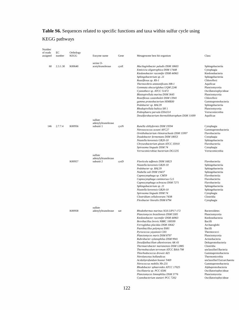
122
Table S6. Sequences related to specific functions and taxa within sulfur cycle using
KEGG pathways
Number
of reads
assigned
EC
number
Orthology
KEGG Enzyme name Gene Metagenome best hit organism Class
E-
value
60 2.3.1.30 K00640
serine O-
acetyltransferase cysE Mucilaginibacter paludis DSM 18603 Sphingobacteriia 1e-47
Emticicia oligotrophica DSM 17448 Cytophagia 1e-61
Ktedonobacter racemifer DSM 44963 Ktedonobacteria 6e-67
Sphingobacterium sp. 21 Sphingobacteriia 3e-59
Roseiflexus sp. RS-1 Chloroflexi 3e-22
Thermovibrio ammonificans HB-1 Aquificae 4e-57
Gemmata obscuriglobus UQM 2246 Planctomycetia 8e-56
Cyanothece sp. ATCC 51472 Oscillatoriophycideae 5e-21
Blastopirellula marina DSM 3645 Planctomycetia 2e-52
Roseiflexus castenholzii DSM 13941 Chloroflexi 1e-26
gamma proteobacterium HIMB30 Gammaproteobacteria 4e-30
Pedobacter sp. BAL39 Sphingobacteriia 8e-30
Rhodopirellula baltica SH 1 Planctomycetia 7e-66
Pedosphaera parvula Ellin514 Verrucomicrobia 8e-45
Desulfurobacterium thermolithotrophum DSM 11699 Aquificae 4e-53
146 2.7.7.4 K00956
sulfate
adenylyltransferase
subunit 1 cysN Runella slithyformis DSM 19594 Cytophagia 1e-37
Nitrosococcus oceani AFC27 Gammaproteobacteria 3e-62
Ornithobacterium rhinotracheale DSM 15997 Flavobacteriia 1e-60
Dyadobacter fermentans DSM 18053 Cytophagia 4e-35
Niastella koreensis GR20-10 Sphingobacteriia 3e-38
Chryseobacterium gleum ATCC 35910 Flavobacteriia 5e-23
Spirosoma linguale DSM 74 Cytophagia 1e-37
Verrucomicrobiae bacterium DG1235 Verrucomicrobia 3e-54
K00957
sulfate
adenylyltransferase
subunit 2 cysD Fluviicola taffensis DSM 16823 Flavobacteriia 1e-29
Niastella koreensis GR20-10 Sphingobacteriia 6e-40
Pedobacter sp. BAL39 Sphingobacteriia 2e-52
Niabella soli DSM 19437 Sphingobacteriia 1e-35
Capnocytophaga sp. CM59 Flavobacteriia 1e-58
Capnocytophaga canimorsus Cc5 Flavobacteriia 3e-57
Capnocytophaga ochracea DSM 7271 Flavobacteriia 2e-37
Sphingobacterium sp. 21 Sphingobacteriia 6e-37
Niastella koreensis GR20-10 Sphingobacteriia 5e-60
Spirosoma linguale DSM 74 Cytophagia 8e-32
Clostridium cellulovorans 743B Clostridia 1e-20
Flexibacter litoralis DSM 6794 Cytophagia 3e-21
K00958
sulfate
adenylyltransferase sat Rhodothermus marinus SG0.5JP17-172 Bacteroidetes 2e-61
Planctomyces brasiliensis DSM 5305 Planctomycetia 5e-63
Ktedonobacter racemifer DSM 44963 Ktedonobacteria 2e-81
Brevibacillus brevis NBRC 100599 Bacilli 5e-32
Ferroglobus placidus DSM 10642 Archaeoglobi 5e-44
Paenibacillus polymyxa E681 Bacilli 2e-46
Pyrococcus yayanosii CH1 Thermococci 4e-43
Planctomyces maris DSM 8797 Planctomycetia 9e-51
Rubrobacter xylanophilus DSM 9941 Actinobacteria 1e-29
Desulfatibacillum alkenivorans AK-01 Deltaproteobacteria 2e-32
Thermaerobacter marianensis DSM 12885 Clostridia 2e-41
Thermobaculum terrenum ATCC BAA-798 unclassified Bacteria 5e-69
Thiorhodococcus drewsii AZ1 Gammaproteobacteria 1e-19
Nitrolancetus hollandicus Thermomicrobia 2e-26
Aciduliprofundum boonei T469 unclassified Euryarchaeota 1e-43
Nitrococcus mobilis Nb-231 Gammaproteobacteria 4e-42
Rhodobacter sphaeroides ATCC 17025 Alphaproteobacteria 4e-62
Oscillatoria sp. PCC 6506 Oscillatoriophycideae 3e-30
Planctomyces limnophilus DSM 3776 Planctomycetia 2e-55
Cyanobacterium stanieri PCC 7202 Oscillatoriophycideae 3e-25

123
Ferroglobus placidus DSM 10642 Archaeoglobi 7e-21
K00955
bifunctional
enzyme
CysN/CysC cysNC Kribbella flavida DSM 17836 Actinobacteria 5e-26
20 1.8.99.2 K00394
adenylylsulfate
reductase, subunit
A aprA Thiobacillus plumbophilus Betaproteobacteria 2e-54
Caldivirga maquilingensis IC-167 Thermoprotei 9e-36
K00395
adenylylsulfate
reductase, subunit
B aprB Sideroxydans lithotrophicus ES-1 Betaproteobacteria 2e-33
60 2.7.1.25 K00955
bifunctional
enzyme
CysN/CysC cysNC Candidatus Nitrospira defluvii Nitrospira 2e-42
Opitutus terrae PB90-1 Verrucomicrobia 3e-21
Kribbella flavida DSM 17836 Actinobacteria 5e-26
K00860
adenylylsulfate
kinase cysC Thermobaculum terrenum ATCC BAA-798 unclassified Bacteria 6e-37
Oscillatoria sp. PCC 6506 Oscillatoriophycideae 2e-25
Haliangium ochraceum DSM 14365 Deltaproteobacteria 7e-46
Ktedonobacter racemifer DSM 44963 Ktedonobacteria 2e-68
Candidatus Solibacter usitatus Ellin6076 Solibacteres 5e-47
Methanohalobium evestigatum Z-7303 Methanomicrobia 2e-42
Gemmata obscuriglobus UQM 2246 Planctomycetia 2e-40
Rubrobacter xylanophilus DSM 9941 Actinobacteria 2e-48
planctomycete KSU-1 Planctomycetia 2e-25
153 2.5.1.47 K12339
cysteine synthase
B cysM Rubrobacter xylanophilus DSM 9941 Actinobacteria 3e-60
Ktedonobacter racemifer DSM 44963 Ktedonobacteria 3e-66
Emticicia oligotrophica DSM 17448 Cytophagia 6e-57
Candidatus Methylomirabilis oxyfera unclassified Bacteria 4e-30
Gloeobacter violaceus PCC 7421 Gloeobacteria 4e-53
Synechococcus sp. JA-3-3Ab Oscillatoriophycideae 1e-47
Thermobaculum terrenum ATCC BAA-798 unclassified Bacteria 3e-42
Mucilaginibacter paludis DSM 18603 Sphingobacteriia 5e-64
Mycobacterium vaccae RIVM Actinobacteria 1e-26
Hydrogenobaculum sp. Y04AAS1 Aquificae 3e-23
K01738
cysteine synthase
A cysK Symbiobacterium thermophilum IAM 14863 Clostridia 1e-48
Bacteroides uniformis ATCC 8492 Bacteroidia 8e-26
Desulfomicrobium baculatum DSM 4028 Deltaproteobacteria 2e-27
Singulisphaera acidiphila DSM 18658 Planctomycetia 9e-55
Cyanobium sp. PCC 7001 Oscillatoriophycideae 1e-51
Neisseria cinerea ATCC 14685 Betaproteobacteria 3e-29
Methylovorus glucosetrophus SIP3-4 Betaproteobacteria 2e-36
Eikenella corrodens ATCC 23834 Betaproteobacteria 7e-33
Gemmata obscuriglobus UQM 2246 Planctomycetia 5e-52
Anaerolinea thermophila UNI-1 Anaerolineae 3e-53
Synechococcus sp. PCC 7335 Oscillatoriophycideae 8e-22
Cyanobacterium stanieri PCC 7202 Oscillatoriophycideae 2e-42
Chroococcidiopsis thermalis PCC 7203 unclassified Cyanobacteria 6e-40
Isosphaera pallida ATCC 43644 Planctomycetia 1e-51
Mycobacterium parascrofulaceum ATCC BAA-614 Actinobacteria 3e-34
Lysinibacillus fusiformis ZC1 Bacilli 1e-33
Acidobacterium capsulatum ATCC 51196 Acidobacteriia 8e-34
Ktedonobacter racemifer DSM 44963 Ktedonobacteria 1e-28
Methylacidiphilum infernorum V4
unclassified
Verrucomicrobia 2e-42
Desulfovibrio alaskensis G20 Deltaproteobacteria 1e-37
Desulfosporosinus acidiphilus SJ4 Clostridia 3e-25
Acaryochloris sp. HICR111A Oscillatoriophycideae 5e-49
Nostoc sp. PCC 7524 Nostocales 1e-28
Cyanothece sp. PCC 7425 Oscillatoriophycideae 2e-30
30 1.8.4.8 K00390
phosphoadenosine
phosphosulfate
reductase cysH Gemmata obscuriglobus UQM 2246 Planctomycetia 5e-74
Sulfolobus islandicus L.S.2.15 Thermoprotei 2e-60
Vulcanisaeta moutnovskia 768-28 Thermoprotei 5e-43
Symbiobacterium thermophilum IAM 14863 Clostridia 5e-54
Pedobacter saltans DSM 12145 Sphingobacteriia 4e-48
Chitinophaga pinensis DSM 2588 Sphingobacteriia 3e-31

124
Rhodothermus marinus DSM 4252 Bacteroidetes 1e-64
Acidothermus cellulolyticus 11B Actinobacteria 4e-24
Flavobacterium frigoris PS1 Flavobacteriia 8e-24
57 1.8.1.2 K00381
sulfite reductase
(NADPH)
hemoprotein beta-
component cysI Sphingobacterium sp. 21 Sphingobacteriia 1e-48
Chitinophaga pinensis DSM 2588 Sphingobacteriia 9e-60
Emticicia oligotrophica DSM 17448 Cytophagia 2e-61
Schlesneria paludicola DSM 18645 Planctomycetia 2e-48
Paenibacillus vortex V453 Bacilli 4e-32
Robiginitalea biformata HTCC2501 Flavobacteriia 7e-21
Emticicia oligotrophica DSM 17448 Cytophagia 2e-59
Gemmata obscuriglobus UQM 2246 Planctomycetia 6e-46
Mucilaginibacter paludis DSM 18603 Sphingobacteriia 1e-40
Sphingobacterium spiritivorum ATCC 33300 Sphingobacteriia 1e-45
4 1.8.7.1 K00392
sulfite reductase
(ferredoxin) sir Gemmata obscuriglobus UQM 2246 Planctomycetia 6e-49

125
Fig. S1. Two way-hierarchical clustering of normalized COG protein family abundance
data averaged across the level of COG categories. The data was standardized (subtract
mean and divide by standard deviation) across sites before clustering so that the color
scale units represent standard deviations from the mean across sites. Red colors
correspond to values that are higher than the site mean and green colors to values that are
lower than the mean

126
Fig. S2. 16S rRNA gene rarefaction curves for four pH 4 samples depicting the observed
OTUs (97% similarity) for randomly drawn subsets of 800 sequences per sample

127
Fig. S3. Partial nitrogen pathways identified by KEGG affiliation of the sequences from the site 04YSHM020. Boxes indicate
the KEGG identifiers and numbers in gray circles indicate the number of sequences assigned to the KEGG function

128
Fig. S4. Partial sulfur pathways identified by KEGG affiliation of the sequences from the site 04YSHM020. Boxes indicate the
KEGG identifiers and numbers in gray circles indicate the number of sequences assigned to the KEGG function

129
Chapter 4
Changes in the Microbial Community Structure in Response to Environmental
Heterogeneity in Filamentous Thermal Springs, Yellowstone National Park
Xiaoben Jianga, Kendra Maasb and Cristina Takacs-Vesbacha
aDepartment of Biology University of New Mexico Albuquerque, NM 87131, USA
bDepartment of Biology, University of British Columbia, Vancouver, V6T1Z4, BC,
Canada

130
Abstract
In this study, we investigated microbial diversity and community composition in
10 filamentous thermal springs with similar physiochemical properties in the Shoshone
area by using the barcoded amplicon pyrosequencing approach. Non-metric
multidimensional scaling ordination based on 16S rRNA genes indicated that the 20
samples fell into three groups. Temperature and pH were the two strongest correlates
environmental variables (R2 = 0.7315, P = 0.001; R2 = 0.7979, P = 0.001; respectively),
followed by conductivity (R2 = 0.6141, P = 0.007). Even all these samples from the same
type of springs, our results revealed that the relatively small variation of environmental
factors could shape microbial community composition. Indicator species analysis
indicated that each group was dominated by different phyla. Functional analysis of the
unique Aquificaceae dominant site indicated that functions related to aerobic respiration
and amino acid synthesis were enriched (P < 0.05), suggesting highly microbial activities
in situ. Energy metabolic pathway analysis showed that rTCA cycle played a dominant
role in energy metabolism. The identification of genes coding for nitrogen and sulfur
cycling indicated that the microbial population was involved in assimilatory and
dissimilatory nitrate reduction, and conversion of sulfate into adenylylsulfate and sulfite.

131
Introduction
One of the fundamental goals of ecology is to understand the distribution of
organisms, the range of their habitats and the factors influencing on their distributions.
Biogeography is the study of the distribution of biodiversity over varying spatial and
temporal scales; research on the biogeography of microorganisms can offer insights into
microbial speciation, extinction and dispersal. A century ago, Baas-Becking proposed the
most famous hypothesis on microbial biogeography: “[E]verything is everywhere, but the
environment selects” (Becking 1934). According to Bass-Becking’s hypothesis,
microorganisms appear to have cosmopolitan distributions due to their vast population
size and enormous capacity for long-distance dispersal (Griffin et al., 2002). This view of
microbial biogeography has been the paradigm for many years and is supported by some
studies, especially for the patterns of the protist distributions (Fenchel and Finlay 2004;
Finlay 2002). However, this paradigm of microbial biogeography has been challenged by
recent studies using molecular techniques. Geographic distances as a barrier to dispersal
do exist at the global scale. For example, globally biogeographic patterns have been
detected in the distribution of some thermophiles such as Sulfolobus and Synechococcus
(Martiny et al. 2006). In addition to geographic barriers, like macro-organisms, a variety
of environmental factors such as temperature (Everroad et al. 2012; Wang et al. 2013),
pH (Fierer and Jackson 2006; Lauber et al. 2009; Van Horn et al. 2013) and salinity
(Kunin et al. 2008; Ley et al. 2006) controlling the distribution microorganisms and
communities have been observed at different habitats and geographical scales.
Yellowstone National Park (YNP) is one of the largest and most diverse
hydrothermal areas in the world, containing more than 14,000 geothermal features that

132
possess a broad range of multiple environmental gradients such as temperature (40-92 °C)
and pH (1-10) (Fournier 1989; Fournier 2005; Rye and Truesdell 2007). It is generally
held that YNP thermal springs have three major community types: (1) phototrophic mats;
(2) Aquificales-rich ‘filamentous-streamer’ communities; and (3) archaeal-dominated
sediments (Inskeep et al. 2013b); the classification is based on dominant species and
geochemical properties. It has been suggested that environmental variables of local
habitats are important in controlling over local distribution microbial taxa and
communities in phototrophic mats (Ramsing et al. 2000; van der Meer et al. 2005; Ward
et al. 1998a; Ward et al. 1990). Filamentous-streamer communities are characterized by
the dominance of Aquificales. However, how environmental heterogeneity influences the
diversification of Aquificales-rich ‘filamentous-streamer’ communities at a local scale is
not clear. Previous studies of 16S rRNA gene-based sequencing (Reysenbach et al.
2005), together with the YNP metagenome survey (Inskeep et al. 2013b), have shed light
on the distribution of the Aquificales. Despite current breakthroughs in understanding the
relationship between environmental variables and metabolic potential in some YNP
geothermal features, several important microbial habitat types (e.g., pH, 5-9; temperature,
>70 °C) are yet to be examined (Inskeep et al. 2013b). Moreover, of the previous pilots
studies that attempted to link environmental variables with microbial community
composition, most concentrated on the vent of each spring and therefore were hindered
by not enough number of representative samples or sequencing depth (Coman et al. 2013;
Huang et al. 2013; Meyer-Dombard et al. 2005; Purcell et al. 2007; Reigstad et al. 2009;
Tobler and Benning 2011). The influence of local environmental heterogeneity

133
(especially, fine temperature and pH variation) on the structure of Aquificales-rich
‘filamentous-streamer’ communities requires further investigation.
The goal of the present study was to expand the current understanding of how the
environmental heterogeneity structures microbial (in particular, bacterial) communities in
filamentous springs of YNP. To address this question, a survey of molecular diversity of
bacteria using 16S rRNA gene-based pyrosequencing on 20 sites from 10 circumneutral
to alkaline filamentous springs was undertaken. In addition to the 16S rRNA gene
marker, we also explored the phylogenetic diversity of the Aquificales using the internal
transcribed spacer (ITS) region between the 16S rRNA and 23S rRNA genes, in order to
assess the diversity of the Aquificales in these springs. Finally, we provided a functional
analysis of metagenomic sequences obtained from a high temperature and pH
filamentous community to fill the knowledge gap for this uninvestigated habitat type.
Materials and Methods
Sampling sites
Ten thermal springs within the Shoshone region of YNP were sampled during
2007 (Figure S1 and Table 1). Within each spring, two visible filamentous samples were
collected. One sample was collected from the highest temperature locality where
filaments existed, whereas the other sample was collected from the lowest temperature
locality where filaments existed. Prior to collecting filaments, spring water for
geochemical analysis was filtered through a 0.02 µm filter (Millipore) using sterile 50-
mL syringes and preserved as appropriate for the analysis to be performed. Geochemical
analyses were carried out using standard USGS methods (McCleskey 2005).
Macroscopically visible filamentous samples were collected aseptically into 2-mL

134
microcentrifuge tubes and preserved in sucrose lysis buffer (Giovannoni et al. 1990).
Samples were stored at ambient temperature (~10 to 26 °C) for up to one days. Once in
the laboratory, they were stored at -80 ºC until DNA extraction.
DNA extraction, barcoded pyrosequencing and shotgun metagenome sequencing
Total DNA was extracted from preserved samples following bead-beating
disruption in a cetyltrimethylammonium bromide (CTAB) buffer (1% CTAB, 0.75 M
NaCl, 50 mM Tris pH 8, 10 mM EDTA) and subsequent phenol-chloroform purification
steps as described before (Mitchell and Takacs-Vesbach 2008). Barcoded amplicon
pyrosequencing of 16S rRNA genes was performed as described previously (Andreotti et
al. 2011; Dowd et al. 2008b; Van Horn et al. 2013) using universal bacterial primers
939F (5‘- TTG ACG GGG GCC CGC AG -3’) and 1492R (5‘-GGT TAC CTT GTT
ACG ACT T -3’). For each sample, barcoded amplicon pyrosequencing was also carried
out using specific ITS primers 1492F (5’-AAG TCG TAA CAA GGT AAC C-3’) and
115R (5’-GGG TTB CCC CAT TCR G-3’) designed for the Aquificales (Ferrera et al.
2007). We focused solely on the bacterial composition and diversity, because Archaea
and Eukarya were not dominant groups in these circumneutral to alkaline thermal
systems of YNP (Miller et al. 2009; Ward et al. 1998a). Triplicate reaction mixtures per
sample were combined and subsequently purified with an UltraCleanTM GelSpinTM DNA
Extraction Kit (MoBio Laboratories, Carlsbad, CA, USA). The purified amplicons were
quantified using a Nanodrop ND-2000c spectrophotometer (Thermo Fisher Scientific,
Wilmington, DE, USA). Amplicons from all samples were pooled with equimolar
concentrations for pyrosequencing, using Roche titanium reagents and titanium
procedures on a Roche 454 GS FLX instrument (454 Life Sciences, Branford, CT, USA).

135
In addition, we selected one sample, 07SHO09A, collected from the high temperature
and pH site for metagenomic sequencing to gain a better understanding of the metabolic
potential of the communities. Approximately 500 ng of quality-checked DNA was used
for library construction. Metagenome sequencing was performed according to
manufacturer’s protocol on a 454 GS FLX Titanium platform (454 Life Sciences,
Branford, CT, USA).
16S rRNA and ITS pyrosequencing data processing
All 16S rRNA gene raw sequences were quality-filtered, denoised and checked
for chimeras using the AmpliconNoise tool (Quince et al. 2011) integrated in the
Quantitative Insights into Microbial Ecology (QIIME, Ver. 1.8.0) (Caporaso et al.
2010b). Adapters, multiplex identifiers and primers were trimmed from denoised data.
The QIIME pipeline was employed to analyze the denoised sequences. Species level
operational taxonomic units (OTUs) were identified at 97% similarity using UCLUST
(Edgar 2010). The most abundant sequence was picked from each OTU as a
representative sequence and aligned using the PyNAST aligner (Caporaso et al. 2010a)
and Greengenes database (DeSantis et al. 2006). The phylogenetic affiliations were
assigned by the Ribosomal Database Classifier program (Wang et al. 2007). The same
settings in QIIME were used for ITS gene sequence data analysis, except representative
sequences were aligned using MUSCLE (Edgar 2004) and given taxonomic assignments
using BLASTN (Altschul et al. 1997) against the NCBI nucleotide (NT) database. In
order to standardize for varying sequencing efforts across samples, all measurements of
community structure were performed with randomly drawn subsets of 867 sequences
from each sample for the 16S rRNA gene sequencing and 824 sequences for ITS gene

136
sequencing from each sample, respectively. Alpha diversity indices, including observed
OTUs, Good’s coverage, Shannon index and phylogenetic diversity, were calculated at
the 97% similarity OTU level for both 16S rRNA gene and ITS gene sequences using
tools implemented in QIIME (Caporaso et al. 2010b).
Statistical analysis of bacterial community
All statistical analyses were conducted using the R (Team 2011) statistical
package with the ‘vegan’ library (Oksanen et al. 2013), unless otherwise noted. For 16S
rRNA sequences, the metaMDS and envfit functions in the ‘vegan’ package were used to
test environmental variables correlated with bacterial community composition by fitting
all environmental variables measured to the ordination. To assess the similarity of the
microbial community structures among the sites, a hierarchical cluster analysis was
performed. The significance of the cluster groups was determined using a permutational
MANOVA test (Anderson 2001). An indicator species analysis was conducted to obtain
taxonomic signatures of each group. Differences in environmental variables among
cluster groups were tested using the one-way ANOVA test. Box plots were also built to
show the significance of environmental variables among cluster groups.
Metagenomic assembly and analysis
Metagenome sequencing reads were quality-filtered and assembled using Newbler
2.6 (Margulies et al. 2005) under default settings. All contigs and remaining singleton
reads were submitted to the JGI IMG/M annotation pipeline (Markowitz et al. 2012).
Unassembled raw reads were also submitted for annotation on the metagenomics analysis
server, MG-RAST (Meta Genome Rapid Annotation using Subsystem Technology,
v3.3.9) (Glass et al. 2010), using its default quality control pipeline. Microbial taxa

137
analysis was conducted via the MG-RAST best-hit classification tool against the
GenBank (NCBI-NR), M5NR (M5 non-redundant protein) and RefSeq databases using
the minimum identity of 60%, e-value cutoff of 10-5 and minimum alignment length of 50
bp.
Metabolic mapping of energy metabolism
Unassembled 07YSHO09A raw reads were annotated by BLASTX (Altschul et
al. 1997) against the NCBI non-redundant (NR) protein database using the e-value cutoff
of 10-5. The results of BLASTX were imported into MEtaGenome ANalyzer software
(MEGAN v5.5.3) (Huson et al. 2011) and were taxonomically classified using the least
common ancestor (LCA) algorithm based on the top 10 BLAST alignments for each read.
The pathways of energy metabolism were recognized using KEGG identifiers (Ogata et
al. 1999). The sequences in each pathway (oxidative phosphorylation, methane
metabolism, nitrogen metabolism, carbon fixation pathways in prokaryotes, carbon
fixation in photosynthetic organisms, sulfur metabolism and photosynthesis) were given
taxonomical assignments at phylum level. In order to detect important electron transfer
processes likely linked to thermal springs, the metabolic pathways involved in carbon
fixation pathways in prokaryotes, nitrogen and sulfur metabolism were identified using
KEGG identifiers.
Comparisons to other YNP metagenomes
A cluster of Orthologous Group (COG) functions of the 07YSHO09A
metagenome assembly was compared to available YNP metagenomes (Table S1)
(Inskeep et al. 2013b) to better characterize the potential community functions of this
sample. Annotated COG functions of these metagenomes were obtained from the IMG/M

138
server. Prior to quantitative characterization, metagenomic data were normalized by the
total number of COG functions detected, weighted by contig depth if assembly
information was available. For unassembled singleton reads, a contig depth of one was
assumed. COG functions were subsequently classified into COG categories on the
IMG/M and a Bray-Curtis dissimilarity matrix was constructed, based on the COG
category abundance table. The COG category dissimilarity matrix was subject to
hierarchical agglomerative cluster analysis. Moreover, the standardized data were
analyzed using a principal coordinates analysis (PCoA). All multivariate comparisons
and ordinations were performed using the R (Team 2011) statistical package with ‘vegan’
(Oksanen et al. 2013) and ‘cluster’ (Maechler et al. 2013) libraries.
We used the “Function Comparison” tool on the IMG/M to compare the
metagenome of 07YSHO09A against metagenomic datasets from other communities, in
purpose to determine whether unusually prominent functions existed in this unexplored
site. The relative abundance of COG function was calculated based on normalized gene
counts and was expressed as D-scores, which were used to calculate the standard
variation from the null hypothesis (i.e., relative gene counts in metagenome A = relative
gene counts in metagenome B). For each comparison, the P value cutoff for significant
D-scores was determined using a false discovery rate of 0.05.
Sequence Data Submission
The metagenome of 07YSHO09A is publicly available on the IMG/M (IMG
submission ID 13525) and the MG-RAST (ID 4523621.3). The individual sff files of 16S
rRNA gene amplicon sequencing were assigned the accession numbers SRX1093556 -
SRX1093565, SRX1093570 - SRX1093571 and SRX1093579 - SRX1093586 under

139
Bioproject PRJNA289700. The individual sff files of ITS rRNA gene amplicon
sequencing were assigned the accession numbers SRX1100239 - SRX1100258 and
SRX1100241 - SRX1100250 under Bioproject PRJNA290194.
Results
Water chemistry of the studied sites
A total of 20 filamentous samples were collected from 10 different thermal
springs. Water temperature of the sampled thermal springs ranged from 62 to 87 °C. The
pH was from circumneutral to alkaline (6.21-8.88). The conductivity was from 1060 to
1475 (µS/cm). The concentration of hydrogen sulfide in these springs ranged from 0.035
to 4.45 mg L-1. The concentration of major anions detailed in Table 1.
Microbial community composition of 16S rRNA and ITS amplicons
Collectively, 120,662 16S rRNA gene sequences (mean = 6,033 sequences/site
and 80,332 ITS gene sequences (mean = 4016 sequences/site) were obtained after
denoising, respectively. Good’s coverage calculation indicated that the analyzed
sequences covered the diversity of bacteria (Table 3). The Shannon diversity index of
16S rRNA gene sequences ranged from 0.062 to 3.815, with an average of 2.099 (Table
3). The Shannon diversity index of ITS gene sequences ranged from 0.158 to 1.746, with
an average of 0.857 (Table 3). The Shannon diversity index, based on 16S rRNA gene
sequences, clearly demonstrated that samples collected from low temperature sites had
about 79.5% higher species richness than those collected from high temperature sites (P =
0.005, one-tailed t-test). However, there was no significant differences in the Shannon
diversity between high and low temperature sites (P = 0.308, one-tailed t-test), based on
ITS gene sequences.

140
Bacterial communities of all samples was composed of sequences related to seven
major phyla and candidate divisions (Figure 1 and Figure 3A). Candidate division EM3,
Aquificae, Proteobacteria and Deinococcus-Thermus were the four most dominant phyla,
contributing 33.67%, 24.17%, 19.08% and 14.22% of the total 16S RNA sequences,
respectively. The cluster analysis revealed three distinct clusters (groups A, B and C;
Figure 3A and Figure 3B). The cluster groups were confirmed by the permutational
MANOVA test (R2 = 0.78728, P < 0.001). In contrast, microbial communities among
springs were not significantly different, according to the permutational MANOVA test
(R2 = 0.5318, P = 0.264). Each cluster significantly affiliated with one or several phyla,
according to the indicator species analysis (P < 0.05). For instance, Aquificae and EM3
were associated with group A and group C, respectively, whereas Armatimonadetes,
Cyanobacteria, Deinococcus-Thermus were closely related to group B.
Correlation between bacterial communities and environmental variables
Non-metric multidimensional scaling (nMDS) ordination based on 16S rRNA
gene sequences was shown in Figure 4 (2D stress = 0.1). Several environmental variables
were significantly correlated to the first two axes of the ordination (Figure 4).
Temperature and pH were the two strongest correlates among all environmental variables
(R2 = 0.7315, P = 0.001; R2 = 0.7979, P = 0.001; respectively), followed by conductivity
(R2 = 0.6141, P = 0.007). Significant correlations were not detected between ordination
and other environmental variables (P > 0.05), including H2S, F-, Cl-, NO2-, NO3
- SO42-
and PO43-. Significant differences in temperature and pH among groups were found,
according to the ANOVA test (Both P<0.05), but the difference in conductivity among
the three groups was close to the marginally significant level (P = 0.06). Generally, group

141
A represented the low temperature and pH habitat, and group B represented the low
temperature and high pH habitat, in contrast to group C exhibiting high temperature and
pH (Figure 5).
Metagenome sequencing summary, coverage and classification of the microbial
community
Metagenomic pyrosequencing generated 765,145 reads, with 435 bp by mean
length, totaling 333 Mbp from site 07YSHO09A (Table 2). Assembly of these reads
yielded 26,440 contigs, with the N50 contig length of 1,162 (Table 2). The result of
hierarchical clustering analysis mirrored the previous characterization of YNP thermal
springs (Inskeep et al. 2013b), indicating three distinct community groups: (1) archaeal
communities; (2) Aquificales communities; and (3) phototrophic communities (Figure
6A). Site 07YSHO09A affiliated with Aquificales communities. Additionally, the group
scheme of PCoA ordination confirmed the same group pattern (Figure 6B).
The taxonomic profile of metagenomics
As shown in Table S2, taxonomic classification with M5 non-redundant protein
(M5NR) database on MG-RAST detected that Bacteria domain (87.71%) was
predominant in site 07YSHO09A. The remaining sequences from 07YSHO09A were
associated with Archaea (6.36%), Eukaryota (0.01%), Virus (0.05%) and unassigned
sequences (8.71%). Among the Bacteria, the most abundant sequences were associated
with phylum Aquificae (91.45%). This result was similar to that from 16S rRNA gene
sequencing, in which the Aquificae sequences were predominantly retrieved, accounting
for 93.61% of total 16S rRNA gene sequences (Figure S2). BLAST analyses using
different databases revealed detailed taxonomic information with respect to this

142
unexplored site. All our results unequivocally demonstrated that the Aquificae
predominated over other phyla in site 07YSHO09A, according to GenBank (93.64%),
M5NR (90%) and RefSeq (93.31%) database (Figure 7). The family Aquificaceae was
the dominant family (~93%) within order Aquificales based on all three databases (Figure
8). Hydrogenothermaceae (~7%) was also detected in this site (Figure 8). With all
sequences related to the family Aquificaceae, Aquifex aeolicus (36.70%), Thermocrinis
albus (30.79%) and Hydrogenobacter thermophilus (27.89%) were the three most
abundant species (Figure 8). Within the family Hydrogenothermaceae, a large proportion
of sequences were assigned to Sulfurihydrogenibium sp. YO3AOP1 (57.50%), followed
by Sulfurihydrogenibium yellowstonense (29.13%) and Sulfurihydrogenibium azorense
(8.84%) (Figure 8).
Functional comparison with other YNP metagenomes
Both cluster and PCoA analysis unequivocally demonstrated that the functional
profile of 07YSHO09A metagenome was similar to metagenomes from the Aquificales
communities. Therefore, we compared the functional profile of 07YSHO09A to those of
metagenomes from the archaeal group and phototrophic group, respectively. First, we
uncovered that 21 COG functions were overrepresented in the 07YSHO09A dataset in at
least five of the seven comparisons (Table S3). “Energy production and conversion”,
“amino acid transport and metabolism” and “translation, ribosomal structure and
biogenesis” were three most abundant COG categories among these functions (19%,
14.3% and 14.3%, respectively) and were dominant among those with the highest
enrichment D-scores (Table S3 and Figure 9). In addition, 140 COG functions were
enriched within the metagenome of 07YSHO09A in at least five of the six comparisons

143
(Table S4). “Amino acid transport and metabolism”, “translation, ribosomal structure and
biogenesis” and “general function prediction only” were the three most abundant COG
categories (15.7%, 10% and 10%, respectively; Table S4 and Figure 10).
Energy metabolism, rTCA, nitrogen and sulfur metabolism
Functional assignment of the unassembled 07YSHO09A metagenome provided
detailed information about possible functions in this habitat. A total of 59,235 reads were
assigned to functions related to energy metabolism using BLASTX against the NCBI-nr
database. Among the reads of energy metabolism, most were mapped to the phylum
Aquificae and involved in a variety of pathways, such as oxidative phosphorylation,
methane metabolism, nitrogen metabolism and carbon fixation pathways in prokaryotes,
carbon fixation in photosynthetic organisms, sulfur metabolism and photosynthesis
(Figure S3). A total of 14,281, 14,636 and 277 reads were assigned to carbon fixation
pathways in prokaryotes (rTCA), nitrogen metabolism and sulfur metabolism,
respectively. Major KEGG functional categories and the unique hits mapped to each
categories were listed (Table S5, Table S6 and Table S7).
Discussion
Microbial community environmental heterogeneity
The first objective of this study was to examine the effects of local environmental
heterogeneity on microbial communities from filamentous springs in the Shoshone area.
Aquificales have been found to be dominant in high temperature (> 65°C), neutral-
alkaline, filamentous springs in YNP (Hamamura et al. 2013; Meyer-Dombard et al.
2005; Meyer-Dombard et al. 2011; Reysenbach et al. 2000b; Takacs-Vesbach et al.
2013); as well as in other similar terrestrial hydrothermal systems (China, Iceland and

144
Japan) (Kato et al. 2004; Nakagawa and Fukui 2002; Takacs et al. 2001; Wang et al.
2013). The 10 filamentous springs in this project are considered ideal habitats for
Aquificales communities (Inskeep et al. 2013b). However, the order Aquificales is not
the exclusively dominant taxonomic group in these springs, despite the fact that
considerable sequences related to the Aquificales have been previously recovered from
springs in Shoshone (Takacs-Vesbach et al. 2008). Previous reports correlating thermal
springs around the world have shown that pronounced temperature and pH variation can
affect the structure and distribution of microbial communities among springs or within a
spring (Allewalt et al. 2006; Everroad et al. 2012; Inskeep et al. 2010; Miller et al. 2009;
Purcell et al. 2007; Wang et al. 2013). Here, we observed that environmental
heterogeneity contributed to the considerable variation of microbial taxonomic
assemblages and richness, despite all these sites belonging to the same type of springs
within the Shoshone area (Figure 3).
Based on the correlations between the phylogenetic composition and
physicochemical conditions, the 20 sites can be classified into three distinct groups,
designated here as group A, group B and group C (Figure 3A and Figure 3B), which
suggest significant differences in temperature and pH (Figure 5). In group A, microbial
communities were mainly composed of sequences related to the Aquificae (Figure 3A
and Figure 3B). Within the phylum Aquificae, all sequences were related to the family
Hydrogenothermaceae (77.5% of all Aquificae sequences) and Aquificaceae within the
order Aquificales (22.5% of all Aquificae sequences). The sites dominated by the
Aquificales represent relatively low temperature and low pH sites among the 20 sites
(Figure 5). Six of seven sites in group A were dominated by members of the family

145
Hydrogenothermaceae (Figure 3B and Figure 4), with a temperature range of 62 - 78 °C
and a pH range of 6.21-7.10. By contrast, the only site dominated by the family
Aquificaceae was from the 83°C and pH 8.83 site (Table 1, Figure 3B and Figure 4),
accounting for 87.4% of all the Aquificaceae sequences. The further classification using
the ITS gene indicated that genus Sulfurihydrogenibium was the major taxonomic group
in the family Hydrogenothermaceae (Figure 2). Additionally, metagenomic sequencing
revealed that genus Aquifex, Thermocrinis and Hydrogenobacter were three most
abundant bacterial groups in the family Aquificaceae (Figure 8). The genus
Sulfurihydrogenibium associated with the family Hydrogenothermaceae normally
inhabits circumneutral sulfidic springs of YNP (Hugenholtz et al. 1998; Reysenbach et al.
2005; Reysenbach et al. 2000a). The two well-studied groups in the family Aquificaceae,
the Hydrogenobaculum spp. and Thermocrinis-like group, appear to be divergent. These
two groups normally colonize two distinct niches in thermal springs. The
Hydrogenobaculum spp. generally dominate in pH springs (pH<4), whereas the
Thermocrinis-like organisms dominate in higher pH systems (pH 6-9) (Takacs-Vesbach
et al. 2013). Given that the concentration of hydrogen sulfide is not significantly different
among cluster groups by ANOVA test, the differences in temperature and pH among sites
seem to play a more important role in controlling the distribution of the
Hydrogenothermaceae and Aquificaceae in the Shoshone. Furthermore, the distribution
of members from these two families is consistent with previous observed temperature and
pH range elsewhere (Blank et al. 2002; Deckert et al. 1998; Hugenholtz et al. 1998;
Pitulle et al. 1994; Reysenbach et al. 2005; Reysenbach et al. 2000a; Takacs-Vesbach et
al. 2013).

146
In contrast to group A, group B was detected at sites with higher pH. The
bacterial communities in group B contained a high proportion of sequences associated
with the Deinococcus-Thermus (35.6% of total Bacteria in group B, mainly genus
Thermus. The presence of Thermus in group B coincides with the widespread distribution
of this group in global thermal springs with temperature of 50 - 99 °C and circumneutral
to alkaline pH (Boomer et al. 2009; Cole et al. 2013; Costa et al. 2009; Gumerov et al.
2011; Lin et al. 2002; Meyer-Dombard et al. 2011; Purcell et al. 2007; Tobler and
Benning 2011; Wang et al. 2013). Despite the widespread distribution of the Thermus,
the dominance of this genus in terrestrial thermal springs is rarely observed, except for a
few springs in Iceland (Tobler and Benning 2011) and Tibet (Wang et al. 2013). The
dominance of the genus Thermus usually is associated with higher available organic
carbon content (Wang et al. 2013). Given the abundance of Thermus, coupled with the
evidence that the highest abundance of autotrophic bacteria, Cyanobacteria (mainly genus
Gloeobacter) found in group B (Figure 3A), we can reason that sites in group B correlate
with more available organic carbon.
The candidate division EM3 was the indicator of group C (Figure 3A and Figure
4), exhibiting high temperature and pH among these three clustering groups. In group C,
the average temperature is 82 °C and the average pH is 8.01, which mirror the
temperature and pH at Octopus Spring where the EM3 were originally reported
(Reysenbach et al. 1994). However, the EM3 seem to be well adapted to different
environments. Aside from thermal springs (Meyer-Dombard et al. 2011; Zhang et al.
2013), the EM3 are also present in wastewaters (Hatamoto et al. 2007; Sekiguchi 2006).
Previously, molecular phylogenetic studies suggested that the candidate division EM3

147
was closely related to the phylum Thermotogae (Dunfield et al. 2012; Rinke et al. 2013).
The recent study implies that this candidate division is the deepest lineage of domain
Bacteria (Colman et al. in prep). Despite this phylogenetic information, little is known
about the metabolic pathway and the ecology of the EM3 due to the lack of cultivated
representatives. Although one-way ANOVA analysis revealed that the difference in
conductivity among three groups was at the marginally significant level (P = 0.06), the
conductivity was significantly correlated with the first two axes of the ordination (Figure
4). In particular, group C showed a strong linkage to high conductivity (Figure 4).
Conductivity is an important environmental variable to control the global distribution of
bacterial community composition (Lozupone and Knight 2007). Abrupt salinity gradients
often occur in habitats such as estuaries, wetlands, salt marshes and coastal lagoons
(Jeffries et al. 2012). We observed that the higher salinity seemed to favor the EM3,
suggesting that the fine salinity heterogeneity has an important influence on the local
distribution of bacteria.
Functional profile of 07YSHO09A metagenome
The second goal of this research was to characterize the functional profile of the
microbial community in 07YSHO09A. Although our cluster analysis based on 16S rRNA
gene sequencing indicated that 07YSHO09A clustered with sites dominated by the
Aquificales (Figure 3B), it separated itself from the other Aquificales-dominated sites on
nMDS plot (Figure 4) due to the dominance of the family Aquificaceae rather than the
family Hydrogenothermaceae. It is noteworthy that the predominance of the Aquificaceae
is also supported by metagenomic analysis (Figure 8).

148
The metagenome of 07YSHO09A provides the functional profile in a relatively
low elemental sulfur, high temperature and pH site of YNP. In addition, the comparison
of 07YSHO09A metagenomic data to those from sites associated with other communities
can reveal the information concerning prominent functional capabilities of the
07YSHO09A (Figure 9, Figure 10, Table S3 and Table S4). Among all the
overrepresented functions, many were responsible for a variety of energy production and
conversion such as carbohydrate metabolism. Particularly, some functions found
overrepresented in both comparisons (Table S3 and Table S4), such as Succinyl-CoA
synthetase (COG 0045) and NADH:ubiquinone oxidoreductase (COG1007, COG0649,
COG1009), can be related directly to aerobic respiration. Succinyl-CoA synthetase is an
enzyme that catalyzes the reversible reaction of succinyl-CoA to succinate, which plays a
key role as one of the catalysts involved in the citric acid cycle (TCA) (Voet and Voet
2011) and the rTCA cycle (Williams et al. 2006). Moreover, NADH:ubiquinone
oxidoreductase is one of the main entry enzymes for the respiratory chains of myriad
organisms (Friedrich et al. 1998).
Heme copper oxidases (subunit 1 of terminal oxidase complexes, COG0843) were
enriched, when compared to metagenomes from phototrophic communities (Table S4).
Heme copper oxidases that can catalyze the reduction of O2 to H2O are good indicators of
the potential for aerobic respiration (García-Horsman et al. 1994b; Pereira et al. 2004).
This result, together with the predominance of the Aquificaceae populations implies that
the majority of these organisms respire oxygen. The sample 07YSHO09A was collected
from visible filaments. Here, the overabundance of functions contributing to amino acid
synthesis (e.g., COG0019, COG0436, COG0620 and COG0498) and concomitant with

149
the function implicated in cell division (e.g., COG0445) suggest high productivity
existing in the filaments (Table S3 and Table S4). Higher microbial productivity
produces more intermediates, which may expand the available niches and increase the
microbial diversity and competition in the whole spring (Everroad et al. 2012). For
instance, the presence of the function involved in multidrug resistance (COG0841),
which is to export antibiotics and toxic molecules (Piddock 2006) (Table 3), suggests that
microorganisms can defend against toxic compounds produced by competitors. The over
overrepresented COG functions (Table S4) also included functions related to DNA repair
(COG0419 and COG0497). In contrast to phototrophic communities, the high proportion
of these COGs can be indicative of the need to respond to the high temperature and pH
environment of site 07YSHO09A. Among the enriched functions present in
07YSHO09A, we observed the protein involved with trace element detoxification
(COG1055, Na+/H+ antiporter NhaD and related arsenite permeases, Table S4). Arsenic
and mercury are two common toxic constituents originating from YNP geothermal
features (Ball et al. 2002; Boyd et al. 2009; Stauffer and Thompson 1984). The COG1055
coded by gene arsB is responsible for transporting arsenite out of the cell under toxic
conditions (Mukhopadhyay et al. 2002; Stolz et al. 2006). Previous reports have indicated
the widespread distribution of arsB gene in all three main communities of YNP (Inskeep
et al. 2013b; Inskeep et al. 2010). Compared to phototrophic communities, the significant
abundance of COG1055 has implications for higher activities in arsenic efflux in situ.
Potential primary production strategies in thermal springs are of primary interest
because the species richness and primary production in thermophilic environments are
considered to be low compared to mesophilic environments (Brock 1994). Aquificae,

150
mainly Aquificales dominant in site 07YSHO09A, are known for their roles as primary
producers in hydrothermal systems (Blank et al. 2002; Eder and Huber 2002; Harmsen et
al. 1997; Reysenbach et al. 2000a; Reysenbach et al. 1999; Spear et al. 2005; Takacs-
Vesbach et al. 2013; Yamamoto et al. 1998). The rTCA cycle was well represented
compared to the nitrogen cycle and the sulfur cycle (Figure S4, Figure S5 and Figure S6),
according to the KEGG database. The rTCA cycle is the reversed process of the oxidative
TCA cycle, providing an alternative strategy to the Calvin cycle for fixing CO2. During
the rTCA cycle, three molecules of CO2 are fixed, forming acetyl-CoA and subsequently
pyruvate, the precursors of all other central metabolites. Three key enzymes are involved
in the TCA cycle, including ATP citrate lyase, pyruvate ferredoxin oxidoreductase and 2-
oxoglutarate ferredoxin oxidoreductase (He et al. 2013; Hugler et al. 2007). Although
ATP citrate lyase was not detected by KEGG pathway analysis, pyruvate ferredoxin
oxidoreductase and 2-oxoglutarate ferredoxin oxidoreductase were prevalent in
07YSHO09A. In addition, despite the low abundance, ATP citrate lyase was detected
using the COG database (see above discussion). Apart from CO2 fixation via rTCA cycle,
the microbial community in site 07YSHO09A contained the evidence of the reductive
acetyl-CoA pathway for CO2 (marked by the gene ACSS, acetyl-CoA synthetase, Table
S5 and Figure S4). These ACSS sequences were phylogenetically related to Aquificae,
Betaproteobacteria, Chloroflexi, Crenarchaeota, Firmicutes and Thermodesulfobacteria.
The genes coding for rTCA and reductive acetyl-CoA pathway have been previously
reported to be prevalent in other YNP springs (e.g., Crater Hills, Mammoth Hot Springs)
(Inskeep et al. 2010), where the Aquificales predominate.

151
Pathways related to nitrogen and sulfur metabolism are of importance in thermal
springs, where terminal electronic acceptors other than O2 (e.g., CO2, element sulfur,
ferric iron, nitrate, sulfate and thiosulfate) normally play more important roles (Hall et al.
2008; Inskeep et al. 2010; Jiménez et al. 2012). Genes coding for dissimilatory nitrate
reductases (e.g., narI) and nitric oxide reductases (e.g., norB, norC, norQ) occurred in
07YSHO09A (Table S6 and Figure S5). The presence of genes coding for these two
enzymes has been previously reported in an Archaeal-dominated spring of YNP
(Joseph’s Coat Hot Spring) (Inskeep et al. 2010). However, genes coding for nitrite
reductase (nirK or nirS) were not present. During the process of dissimilatory nitrate
reduction, a nitrite reductase (nirK or nirS) is the requisite for producing NO, which
serves as a substrate for nitric oxide reductase to produce N2O (Gonzalez et al. 2006;
Moreno-Vivián et al. 1999; Richardson et al. 2001). The lack of detection of nitrite
reductase in 07YSHO09A, coupled with evidence of the absence of nitrous oxide
reductase (nosZ), which converts N2O to N2, suggests that denitrification might be not an
important metabolic pathway in this site. Most microorganisms, with the exception of a
few members in the Euryarchaeota, can only fix nitrogen up to 64 °C (Mehta and Baross
2006). Given the in situ temperature of site 07YSHO09A (Table 1), not surprisingly,
genes (e.g., nifH) contributing to nitrogen fixation were not detected (Table S6, Figure
S5). The absence of genes (e.g., amoA) coding for ammonium monooxygenase is in
agreement with the previous finding in other YNP thermal springs (Inskeep et al. 2010),
suggesting that biological ammonia oxidation may not be predominant in YNP thermal
springs (Table S6, Figure S5).

152
In the sulfur cycle, genes related to the conversion of sulfate into adenylylsulfate
and to the further generation of sulfite were detected (Table S7 and Figure S6). The
environmental aprA and aprB sequences coding for adenosine-5’-phosphosulfate (APS)
reductase (Apr) exhibited closest matches to members of Thermodesulfobacteria and
Thermoprotei. Based on the current model of dissimilatory sulfate reduction and sulfur
oxidation in prokaryotes, APS reductase (Apr) is a key enzyme. During the process of
sulfate reduction, the function of Apr is to convert APS to sulfite, once sulfate is
activated to APS by ATP-sulfurylase at the expense of ATP (Meyer and Kuever 2008).
The alpha subunits of Apr enzymes are present in all known sulfate reducing and most of
sulfur oxidizing prokaryotes (Meyer and Kuever 2008). For instance, the environmental
aprA reads found in site 07YSHO09A indicate high identity to those in the genome of
hyperthermophilic sulfate reducer (e-value < 10-80), Thermodesulfobacterium geofontis
OPF15 (Table S7), an isolate from the YNP thermal spring (Elkins et al. 2013; Hamilton-
Brehm et al. 2013). Thermodesulfobacterium geofontis can reduce sulfate with an optimal
temperature of 83 °C. Genes coding for the subsequent reduction of sulfite to H2S were
not detected (Table S7 and Figure S6), either because not enough sequencing depth was
accomplished or due to the fact that they were absent or present in very low abundance.
Conclusions
Phylogenetic analysis of amplicon sequencing data from 10 circumneutral to
alkaline thermal springs suggests that the variation of microbial taxonomic assemblages
within the same type of community (i.e., ‘filamentous streamer’ community) can result
from the fine physicochemical heterogeneity of local habitats. Here, we observed that the
bacterial community composition changed in response to the relatively small variation of

153
temperature, pH and conductivity (R2 = 0.7315, P = 0.001; R2 = 0.7979, P = 0.001; R2 =
0.6141, P = 0.007; respectively). Phylogenetic and functional analyses of a high
temperature and pH, Aquificales-dominated site (07YSHO09A) revealed that the family
Aquificaceae was exclusively abundant. Compared to Archaeal-dominated communities
and phototrophic communities in YNP, the metagenome of 07YSHO09A indicated that
functions such as aerobic respiration and amino acid synthesis were enriched, implying
active microbial activities in situ. The identification of genes coding for the rTCA cycle
suggests that rTCA cycle plays a dominant role in in situ energy metabolism. The
identification of genes coding for nitrogen and sulfur cycling indicated that the microbial
population was involved in assimilatory and dissimilatory nitrate reduction, and
conversion of sulfate into adenylylsulfate and sulfite. The results from this study suggest
that the taxonomic diversity in Aquificales-rich ‘filamentous-streamer’ communities may
not be homogenous. The dominant bacterial groups can change in response to the small
variation of environmental factors.
154
References
Acosta-Martínez V, Dowd S, Sun Y, Allen V (2008) Tag-encoded pyrosequencing
analysis of bacterial diversity in a single soil type as affected by management and
land use. Soil Biol Biochem 40:2762-2770
Aditiawati P, Yohandini H, Madayanti F, Akhmaloka (2009) Microbial diversity of
acidic hot spring (kawah hujan B) in geothermal field of kamojang area, west
java-indonesia. Open Microbiol J 3:58-66
Allewalt JP, Bateson MM, Revsbech NP, Slack K, Ward DM (2006) Effect of
temperature and light on growth of and photosynthesis by Synechococcus isolates
typical of those predominating in the Octopus spring microbial mat community of
Yellowstone National Park. Appl Environ Microbiol 72:544-550
Almarsdottir AR, Taraceviz A, Gunnarsson I, Orlygsson J (2010) Hydrogen production
from sugars and complex biomass by Clostridium species, AK14, isolated from
Icelandic hot spring. Icelandic Agricultural Sciences:61-71
Altschul SF, Madden TL, Schäffer AA, Zhang J, Zhang Z, Miller W, Lipman DJ (1997)
Gapped BLAST and PSI-BLAST: a new generation of protein database search
programs. Nucleic Acids Res 25:3389-3402
Amarouche-Yala S, Benouadah A, El Ouahab Bentabet A, López-García P (2014)
Morphological and phylogenetic diversity of thermophilic cyanobacteria in
Algerian hot springs. Extremophiles 18:1035-1047
Amend JP, Shock EL (2001) Energetics of overall metabolic reactions of thermophilic
and hyperthermophilic Archaea and Bacteria. FEMS Microbiol Rev 25:175-243

155
Aminin ALN, Warganegara FM, Aditiawati P, Akhmaloka (2008) Simple enrichment
and independent cultures to expand bacterial community analysis from
gedongsongo hot spring. J Biosci Bioeng 106:211-214
Anderson MJ (2001) A new method for non-parametric multivariate analysis of variance.
Austral Ecol 26:32-46
Andreotti R, Perez de Leon A, Dowd S, Guerrero F, Bendele K, Scoles G (2011)
Assessment of bacterial diversity in the cattle tick Rhipicephalus (Boophilus)
microplus through tag-encoded pyrosequencing. BMC Microbiol 11:6
Ball JW, McCleskey RB, Nordstrom DK (2010) Water-chemistry data for selected
springs, geysers, and streams in Yellowstone National Park, Wyoming, 2006–
2008. US Geological Survey Open-File Report 2010–1192:109 p
Ball JW, McCleskey RB, Nordstrom DK, Holloway JM, Verplanck PL (2002) Water-
chemistry data for selected springs, geysers, and streams in Yellowstone National
Park, Wyoming, 1999-2000. US Geological Survey Open-File Report:02-382
Ball JW, Nordstrom DK, Cunningham KM, Schoonen MAA, Xu Y, DeMonge JM (1998)
Water-chemistry and on-site sulfur-speciation data for selected springs in
Yellowstone National Park, Wyoming, 1994-1995. US Geological Survey Open-
File Report:98-574
Barns SM, Delwiche CF, Palmer JD, Pace NR (1996) Perspectives on archaeal diversity,
thermophily and monophyly from environmental rRNA sequences. Proceedings
of the National Academy of Sciences 93:9188-9193

156
Barns SM, Fundyga RE, Jeffries MW, Pace NR (1994) Remarkable archaeal diversity
detected in a Yellowstone National Park hot spring environment. Proceedings of
the National Academy of Sciences of the United States of America 91:1609-1613
Bauld J, Brock T (1973) Ecological studies of Chloroflexis, a gliding photosynthetic
bacterium. Archiv Mikrobiol 92:267-284
Becking LGMB (1934) Geobiologie, of inleiding tot de milieukunde: met literatuurlijst
en ind. W.P. Van Stockum & Zoon, Hague, Netherlands
Beh M, Strauss G, Huber R, Stetter K-O, Fuchs G (1993) Enzymes of the reductive citric
acid cycle in the autotrophic eubacterium Aquifex pyrophilus and in the
archaebacterium Thermoproteus neutrophilus. Arch Microbiol 160:306-311
Blank CE, Cady SL, Pace NR (2002) Microbial composition of near-boiling silica-
depositing thermal springs throughout Yellowstone National Park. Appl Environ
Microbiol 68:5123-5135
Bonch-Osmolovskaya EA, Miroshnichenko ML, Slobodkin AI, Sokolova TG, Karpov
GA, Kostrikina NA, Zavarzina DG, Prokof'eva MI, Rusanov, II, Pimenov NV
(1999) Biodiversity of anaerobic lithotrophic prokaryotes in terrestrial hot springs
of Kamchatka. Microbiology 68:343-351
Boomer SM, Lodge DP, Dutton BE, Pierson B (2002) Molecular Characterization of
Novel Red Green Nonsulfur Bacteria from Five Distinct Hot Spring Communities
in Yellowstone National Park. Appl Environ Microbiol 68:346-355
Boomer SM, Noll KL, Geesey GG, Dutton BE (2009) Formation of multilayered
photosynthetic biofilms in an alkaline thermal spring in Yellowstone National
Park, Wyoming. Appl Environ Microbiol 75:2464-2475

157
Boyd E, Hamilton T, Wang J, He L, Zhang C (2013) The role of tetraether lipid
composition in the adaptation of thermophilic archaea to acidity. Frontiers in
Microbiology 4
Boyd ES, Hamilton TL, Spear JR, Lavin M, Peters JW (2010) [FeFe]-hydrogenase in
Yellowstone National Park: evidence for dispersal limitation and phylogenetic
niche conservatism. Isme j 4:1485-1495
Boyd ES, Jackson RA, Encarnacion G, Zahn JA, Beard T, Leavitt WD, Pi Y, Zhang CL,
Pearson A, Geesey GG (2007) Isolation, characterization, and ecology of sulfur-
respiring Crenarchaea inhabiting acid-sulfate-chloride-containing geothermal
springs in Yellowstone National Park. Appl Environ Microbiol 73:6669-6677
Boyd ES, King S, Tomberlin JK, Nordstrom DK, Krabbenhoft DP, Barkay T, Geesey GG
(2009) Methylmercury enters an aquatic food web through acidophilic microbial
mats in Yellowstone National Park, Wyoming. Environ Microbiol 11:950-959
Brochier-Armanet C, Gribaldo S, Forterre P (2012) Spotlight on the Thaumarchaeota.
ISME J 6:227-230
Brock T, Brock K, Belly R, Weiss R (1972) Sulfolobus: a new genus of sulfur-oxidizing
bacteria living at low pH and high temperature. Arch Microbiol 84:54-68
Brock TD (1967) Life at high temperatures. Science 158:1012-1019
Brock TD (1971) Bimodal distribution of pH values of thermal springs of the world.
Geological Society of America Bulletin 82:1393-1394
Brock TD (1994) Life at high temperatures. In. Yellowstone National Park, WY:
Yellowstone Association for Natural Science, History & Education, Inc.,

158
Brock TD, Brock TD (1978) Thermophilic microorganisms and life at high temperatures.
Springer-Verlag New York
Bryant DA, Costas AMG, Maresca JA, Chew AGM, Klatt CG, Bateson MM, Tallon LJ,
Hostetler J, Nelson WC, Heidelberg JF, Ward DM (2007) Candidatus
Chloracidobacterium thermophilum: An Aerobic Phototrophic Acidobacterium.
Science 317:523-526
Burgess E, Unrine J, Mills G, Romanek C, Wiegel J (2012) Comparative geochemical
and microbiological characterization of two thermal pools in the Uzon caldera,
Kamchatka, Russia. Microb Ecol 63:471-489
Caporaso JG, Bittinger K, Bushman FD, DeSantis TZ, Andersen GL, Knight R (2010a)
PyNAST: a flexible tool for aligning sequences to a template alignment.
Bioinformatics 26:266-267
Caporaso JG, Kuczynski J, Stombaugh J, Bittinger K, Bushman FD, Costello EK, Fierer
N, Pena AG, Goodrich JK, Gordon JI, Huttley GA, Kelley ST, Knights D, Koenig
JE, Ley RE, Lozupone CA, McDonald D, Muegge BD, Pirrung M, Reeder J,
Sevinsky JR, Turnbaugh PJ, Walters WA, Widmann J, Yatsunenko T, Zaneveld J,
Knight R (2010b) QIIME allows analysis of high-throughput community
sequencing data. Nat Methods 7:335-336
Cavaletti L, Monciardini P, Bamonte R, Schumann P, Rohde M, Sosio M, Donadio S
(2006) New lineage of filamentous, spore-forming, gram-positive bacteria from
soil. Appl Environ Microbiol 72:4360-4369
Cavicchioli R (2002) Extremophiles and the search for extraterrestrial life. Astrobiology
2:281-292

159
Chang Y-J, Land M, Hauser L, Chertkov O, Del Rio TG, Nolan M, Copeland A, Tice H,
Cheng J-F, Lucas S (2011) Non-contiguous finished genome sequence and
contextual data of the filamentous soil bacterium Ktedonobacter racemifer type
strain (SOSP1-21T). Standards in genomic sciences 5:97
Clarke K, Ainsworth M (1993) A method of linking multivariate community structure to
environmental variables. Marine Ecology-Progress Series 92:205-205
Clarke KR (1993) Non-parametric multivariate analyses of changes in community
structure. Aust J Ecol 18:117-143
Cole JK, Peacock JP, Dodsworth JA, Williams AJ, Thompson DB, Dong H, Wu G,
Hedlund BP (2013) Sediment microbial communities in Great Boiling Spring are
controlled by temperature and distinct from water communities. ISME J 7:718-
729
Colman DR, Thomas R, Maas KR, Takacs-Vesbach CD (2015) Detection and analysis of
elusive members of a novel and diverse archaeal community within a thermal
spring streamer consortium. Extremophiles 19:307-313
Coman C, Drugă B, Hegedus A, Sicora C, Dragoş N (2013) Archaeal and bacterial
diversity in two hot spring microbial mats from a geothermal region in Romania.
Extremophiles 17:523-534
Connon SA, Koski AK, Neal AL, Wood SA, Magnuson TS (2008) Ecophysiology and
geochemistry of microbial arsenic oxidation within a high arsenic, circumneutral
hot spring system of the Alvord Desert. FEMS Microbiol Ecol 64:117-128

160
Costa K, Navarro J, Shock E, Zhang C, Soukup D, Hedlund B (2009) Microbiology and
geochemistry of great boiling and mud hot springs in the United States Great
Basin. Extremophiles 13:447-459
Cox A, Shock EL, Havig JR (2011) The transition to microbial photosynthesis in hot
spring ecosystems. Chemical Geology 280:344-351
Davis BM (1897) The vegetation of the hot springs of Yellowstone Park. Science 6:145
De León KB, Gerlach R, Peyton BM, Fields MW (2013) Archaeal and bacterial
communities in three alkaline hot springs in Heart Lake Geyser Basin,
Yellowstone National Park. Frontiers in Microbiology 4
Deckert G, Warren PV, Gaasterland T, Young WG, Lenox AL, Graham DE, Overbeek R,
Snead MA, Keller M, Aujay M, Huber R, Feldman RA, Short JM, Olsen GJ,
Swanson RV (1998) The complete genome of the hyperthermophilic bacterium
Aquifex aeolicus. Nature 392:353-358
Del Rio TG, Abt B, Spring S, Lapidus A, Nolan M, Tice H, Copeland A, Cheng J-F,
Chen F, Bruce D (2010) Complete genome sequence of Chitinophaga pinensis
type strain (UQM 2034T). Standards in genomic sciences 2:87
Deppenmeier U (2002) Redox-driven proton translocation in methanogenic Archaea.
CMLS, Cell Mol Life Sci 59:1513-1533
Dequiedt S, Thioulouse J, Jolivet C, Saby N, Lelievre M, Maron PA, Martin MP,
Prévost‐Bouré NC, Toutain B, Arrouays D (2009a) Biogeographical patterns of
soil bacterial communities. Environmental microbiology reports 1:251-255
Dequiedt S, Thioulouse J, Jolivet C, Saby NPA, Lelievre M, Maron P-A, Martin MP,
Prévost-Bouré NC, Toutain B, Arrouays D, Lemanceau P, Ranjard L (2009b)

161
Biogeographical patterns of soil bacterial communities. Environmental
Microbiology Reports 1:251-255
DeSantis TZ, Hugenholtz P, Larsen N, Rojas M, Brodie EL, Keller K, Huber T, Dalevi D,
Hu P, Andersen GL (2006) Greengenes, a chimera-checked 16S rRNA gene
database and workbench compatible with ARB. Appl Environ Microbiol 72:5069-
5072
Donachie S, Christenson B, Kunkel D, Malahoff A, Alam M (2002) Microbial
community in acidic hydrothermal waters of volcanically active White Island,
New Zealand. Extremophiles 6:419-425
Dos Santos PC, Fang Z, Mason SW, Setubal JC, Dixon R (2012) Distribution of nitrogen
fixation and nitrogenase-like sequences amongst microbial genomes. BMC
Genomics 13:162
Dowd S, Callaway T, Wolcott R, Sun Y, McKeehan T, Hagevoort R, Edrington T (2008a)
Evaluation of the bacterial diversity in the feces of cattle using 16S rDNA
bacterial tag-encoded FLX amplicon pyrosequencing (bTEFAP). BMC Microbiol
8:125
Dowd SE, Sun Y, Wolcott RD, Domingo A, Carroll JA (2008b) Bacterial tag-encoded
FLX amplicon pyrosequencing (bTEFAP) for microbiome studies: bacterial
diversity in the ileum of newly weaned Salmonella-infected pigs. Foodborne
pathogens and disease 5:459-472
Drammeh I (2010) Biodiversity of thermophiles in Sungai Klah and Ulu Slim hot springs.
Universiti Teknologi Malaysia

162
Drobner E, Huber H, Rachel R, Stetter KO (1992) Thiobacillus plumbophilus spec. nov.,
a novel galena and hydrogen oxidizer. Arch Microbiol 157:213-217
Dunfield PF, Tamas I, Lee KC, Morgan XC, McDonald IR, Stott MB (2012) Electing a
candidate: a speculative history of the bacterial phylum OP10. Environ Microbiol
14:3069-3080
Eder W, Huber R (2002) New isolates and physiological properties of the Aquificales and
description of Thermocrinis albus sp. nov. Extremophiles 6:309-318
Edgar RC (2004) MUSCLE: multiple sequence alignment with high accuracy and high
throughput. Nucleic Acids Res 32:1792-1797
Edgar RC (2010) Search and clustering orders of magnitude faster than BLAST.
Bioinformatics 26:2460-2461
Elkins JG, Hamilton-Brehm SD, Lucas S, Han J, Lapidus A, Cheng JF, Goodwin LA,
Pitluck S, Peters L, Mikhailova N, Davenport KW, Detter JC, Han CS, Tapia R,
Land ML, Hauser L, Kyrpides NC, Ivanova NN, Pagani I, Bruce D, Woyke T,
Cottingham RW (2013) Complete genome sequence of the hyperthermophilic
sulfate-reducing bacterium Thermodesulfobacterium geofontis OPF15T. Genome
Announc 1:e0016213
Elkins JG, Podar M, Graham DE, Makarova KS, Wolf Y, Randau L, Hedlund BP,
Brochier-Armanet C, Kunin V, Anderson I, Lapidus A, Goltsman E, Barry K,
Koonin EV, Hugenholtz P, Kyrpides N, Wanner G, Richardson P, Keller M,
Stetter KO (2008) A korarchaeal genome reveals insights into the evolution of the
Archaea. Proceedings of the National Academy of Sciences of the United States
of America 105:8102-8107

163
Engel AS, Johnson LR, Porter ML (2013) Arsenite oxidase gene diversity among
Chloroflexi and Proteobacteria from El Tatio Geyser Field, Chile. FEMS
Microbiol Ecol 83:745-756
Entsminger G (2012) EcoSim Professional: Null modeling software for ecologists,
Version 1. Acquired Intelligence Inc., Kesey-Bear, & Pinyon Publishing.
Montrose, CO 81403. http://wwwgaryentsmingercom/ecosim/indexhtm
Everroad RC, Otaki H, Matsuura K, Haruta S (2012) Diversification of bacterial
community composition along a temperature gradient at a thermal spring.
Microbes Environ 27:374-381
Faith DP (1992) Conservation evaluation and phylogenetic diversity. Biol Conserv 61:1-
10
Fardeau M-L, Barsotti V, Cayol J-L, Guasco S, Michotey V, Joseph M, Bonin P, Ollivier
B (2010) Caldinitratiruptor microaerophilus, gen. nov., sp. nov. isolated from a
French hot spring (Chaudes-Aigues, Massif Central): a novel cultivated
facultative microaerophilic anaerobic thermophile pertaining to the
Symbiobacterium branch within the Firmicutes. Extremophiles 14:241-247
Fenchel TOM, Finlay BJ (2004) The ubiquity of small species: patterns of local and
global diversity. Bioscience 54:777-784
Ferrera I, Longhorn S, Banta A, Liu Y, Preston D, Reysenbach AL (2007) Diversity of
16S rRNA gene, ITS region and aclB gene of the Aquificales. Extremophiles
11:57-64

164
Fierer N, Jackson RB (2006) The diversity and biogeography of soil bacterial
communities. Proceedings of the National Academy of Sciences of the United
States of America 103:626-631
Finlay BJ (2002) Global dispersal of free-living microbial eukaryote species. Science
296:1061-1063
Fournier RO (1989) Geochemistry and dynamics of the Yellowstone National Park
hydrothermal system. Annual Review of Earth and Planetary Sciences 17:13-53
Fournier RO (2005) Geochemistry and dynamics of the Yellowstone National Park
hydrothermal system. In: Inskeep WP, TR M (eds) Geothermal biology and
geochemistry in Yellowstone National Park. Thermal Biology Institute, Montana
State University, Bozeman, MT, pp 4-30
Friedrich T, Abelmann A, Brors B, Guénebaut V, Kintscher L, Leonard K, Rasmussen T,
Scheide D, Schlitt A, Schulte U (1998) Redox components and structure of the
respiratory NADH: ubiquinone oxidoreductase (complex I). Biochimica et
Biophysica Acta (BBA)-Bioenergetics 1365:215-219
Fujiwara S (2002) Extremophiles: developments of their special functions and potential
resources. J Biosci Bioeng 94:518-525
Fukami T, Morin PJ (2003) Productivity-biodiversity relationships depend on the history
of community assembly. Nature 424:423-426
García-Horsman JA, Barquera B, Rumbley J, Ma J, Gennis RB (1994a) The superfamily
of heme-copper respiratory oxidases. Journal of Bacteriology 176:5587-5600
García-Horsman JA, Barquera B, Rumbley J, Ma J, Gennis RB (1994b) The superfamily
of heme-copper respiratory oxidases. J Bacteriol 176:5587

165
Giovannoni SJ, DeLong EF, Schmidt TM, Pace NR (1990) Tangential flow filtration and
preliminary phylogenetic analysis of marine picoplankton. Appl Environ
Microbiol 56:2572-2575
Giovannoni SJ, Revsbech NP, Ward DM, Castenholz RW (1987) Obligately phototrophic
Chloroflexus: primary production in anaerobic hot spring microbial mats. Arch
Microbiol 147:80-87
Glass EM, Wilkening J, Wilke A, Antonopoulos D, Meyer F (2010) Using the
metagenomics RAST server (MG-RAST) for analyzing shotgun metagenomes.
Cold Spring Harbor Protocols 2010:pdb.prot5368
Gonzalez P, Correia C, Moura I, Brondino C, Moura J (2006) Bacterial nitrate reductases:
molecular and biological aspects of nitrate reduction. Journal of inorganic
biochemistry 100:1015-1023
Good IJ (1953) The population frequencies of species and the estimation of population
parameters. Biometrika 40:237-264
Gotelli NJ, McCabe DJ (2002) Species co-occurrence: a meta-analysis of JM Diamond's
assembly rules model. Ecology 83:2091-2096
Graber JR, Kirshtein J, Speck M, Reysenbach A-L (2001) Community structure along a
thermal gradient in a stream near Obsidian Pool, Yellowstone National Park. In:
Thermophiles: Biodiversity, Ecology, and Evolution. Springer, New York, pp 81-
91
Gumerov VM, Mardanov AV, Beletsky AV, Bonch-Osmolovskaya EA, Ravin NV (2011)
Molecular analysis of microbial diversity in the Zavarzin Spring, Uzon Caldera,
Kamchatka. Microbiology 80:244-251

166
Hall JR, Mitchell KR, Jackson-Weaver O, Kooser AS, Cron BR, Crossey LJ, Takacs-
Vesbach CD (2008) Molecular characterization of the diversity and distribution of
a thermal spring microbial community by using rRNA and metabolic genes. Appl
Environ Microbiol 74:4910-4922
Hamamura N, Meneghin J, Reysenbach AL (2013) Comparative community gene
expression analysis of Aquificales-dominated geothermal springs. Environ
Microbiol 15:1226-1237
Hamilton-Brehm S, Gibson R, Green S, Hopmans E, Schouten S, Meer MJ, Shields J,
Damsté JS, Elkins J (2013) Thermodesulfobacterium geofontis sp. nov., a
hyperthermophilic, sulfate-reducing bacterium isolated from Obsidian Pool,
Yellowstone National Park. Extremophiles 17:251-263
Hanson CA, Fuhrman JA, Horner-Devine MC, Martiny JBH (2012) Beyond
biogeographic patterns: processes shaping the microbial landscape. Nat Rev
Micro 10:497-506
Harmsen H, Prieur D, Jeanthon C (1997) Group-specific 16S rRNA-targeted
oligonucleotide probes to identify thermophilic bacteria in marine hydrothermal
vents. Appl Environ Microbiol 63:4061-4068
Hatamoto M, Imachi H, Yashiro Y, Ohashi A, Harada H (2007) Diversity of anaerobic
microorganisms involved in long-chain fatty acid degradation in methanogenic
sludges as revealed by RNA-based stable isotope probing. Appl Environ
Microbiol 73:4119-4127

167
He P, Liu Y, Yue W, Huang X (2013) Key genes expression of reductive tricarboxylic
acid cycle from deep-sea hydrothermal chemolithoautotrophic Caminibacter
profundus in response to salinity, pH and O2. Acta Oceanol Sin 32:35-41
Horner-Devine MC, Silver JM, Leibold MA, Bohannan BJ, Colwell RK, Fuhrman JA,
Green JL, Kuske CR, Martiny JB, Muyzer G, Ovreas L, Reysenbach AL, Smith
VH (2007) A comparison of taxon co-occurrence patterns for macro- and
microorganisms. Ecology 88:1345-1353
Hou W, Wang S, Dong H, Jiang H, Briggs BR, Peacock JP, Huang Q, Huang L, Wu G,
Zhi X, Li W, Dodsworth JA, Hedlund BP, Zhang C, Hartnett HE, Dijkstra P,
Hungate BA (2013) A comprehensive census of microbial diversity in hot springs
of Tengchong, Yunnan Province China using 16S rRNA gene pyrosequencing.
PLoS ONE 8:e53350
Huang Q, Dong C, Dong R, Jiang H, Wang S, Wang G, Fang B, Ding X, Niu L, Li X,
Zhang C, Dong H (2011) Archaeal and bacterial diversity in hot springs on the
Tibetan Plateau, China. Extremophiles 15:549-563
Huang Q, Jiang H, Briggs BR, Wang S, Hou W, Li G, Wu G, Solis R, Arcilla CA,
Abrajano T, Dong H (2013) Archaeal and bacterial diversity in acidic to
circumneutral hot springs in the Philippines. FEMS Microbiol Ecol 85:n/a-n/a
Hubbell SP (2001) The unified neutral theory of biodiversity and biogeography vol 32.
Princeton University Press,
Huber H, Stetter KO (1998) Hyperthermophiles and their possible potential in
biotechnology. J Biotechnol 64:39-52
Huber R, Stetter KO (2001) Aquificales. In: eLS. John Wiley & Sons, Ltd,

168
Hugenholtz P, Pitulle C, Hershberger KL, Pace NR (1998) Novel division level bacterial
diversity in a Yellowstone hot spring. J Bacteriol 180:366-376
Hugler M, Huber H, Molyneaux SJ, Vetriani C, Sievert SM (2007) Autotrophic CO2
fixation via the reductive tricarboxylic acid cycle in different lineages within the
phylum Aquificae: evidence for two ways of citrate cleavage. Environ Microbiol
9:81-92
Huson DH, Auch AF, Qi J, Schuster SC (2007) MEGAN analysis of metagenomic data.
Genome Res 17:377-386
Huson DH, Mitra S, Ruscheweyh H-J, Weber N, Schuster SC (2011) Integrative analysis
of environmental sequences using MEGAN4. Genome Res 21:1552-1560
Im W-T, Hu Z-Y, Kim K-H, Rhee S-K, Meng H, Lee S-T, Quan Z-X (2012) Description
of Fimbriimonas ginsengisoli gen. nov., sp. nov. within the Fimbriimonadia class
nov., of the phylum Armatimonadetes. Antonie Van Leeuwenhoek 102:307-317
Inagaki F, Motomura Y, Doi K, Taguchi S, Izawa E, Lowe DR, Ogata S (2001) Silicified
microbial community at Steep Cone hot spring, Yellowstone National Park.
Microbes Environ 16:125-130
Inagaki F, Takai K, Hirayama H, Yamato Y, Nealson KH, Horikoshi K (2003)
Distribution and phylogenetic diversity of the subsurface microbial community in
a Japanese epithermal gold mine. Extremophiles 7:307-317
Inskeep WP, Jay ZJ, Herrgard MJ, Kozubal MA, Rusch DB, Tringe SG, Macur RE,
Jennings R, Boyd ES, Spear JR, Roberto FF (2013a) Phylogenetic and functional
analysis of metagenome sequence from high-temperature archaeal habitats

169
demonstrate Linkages between metabolic potential and geochemistry. Front
Microbiol 4:95
Inskeep WP, Jay ZJ, Tringe SG, Herrgard MJ, Rusch DB (2013b) The YNP metagenome
project: environmental parameters responsible for microbial distribution in the
Yellowstone geothermal ecosystem. Front Microbiol 4:67
Inskeep WP, Rusch DB, Jay ZJ, Herrgard MJ, Kozubal MA, Richardson TH, Macur RE,
Hamamura N, Jennings Rd, Fouke BW, Reysenbach A-L, Roberto F, Young M,
Schwartz A, Boyd ES, Badger JH, Mathur EJ, Ortmann AC, Bateson M, Geesey
G, Frazier M (2010) Metagenomes from high-temperature chemotrophic systems
reveal geochemical controls on microbial community structure and function.
PLoS ONE 5:e9773
Itoh T, Suzuki K-i, Sanchez PC, Nakase T (1999) Caldivirga maquilingensis gen. nov.,
sp. nov., a new genus of rod-shaped crenarchaeote isolated from a hot spring in
the Philippines. Int J Syst Bacteriol 49:1157-1163
Jackson CR, Langner HW, Donahoe-Christiansen J, Inskeep WP, McDermott TR (2001)
Molecular analysis of microbial community structure in an arsenite-oxidizing
acidic thermal spring. Environ Microbiol 3:532-542
Jahnke LL, Eder W, Huber R, Hope JM, Hinrichs K-U, Hayes JM, Des Marais DJ, Cady
SL, Summons RE (2001) Signature lipids and stable carbon isotope analyses of
Octopus Spring hyperthermophilic communities compared with those of
Aquificales representatives. Appl Environ Microbiol 67:5179-5189

170
Jeffries T, Seymour J, Newton K, Smith R, Seuront L, Mitchell J (2012) Increases in the
abundance of microbial genes encoding halotolerance and photosynthesis along a
sediment salinity gradient. Biogeosciences 9:815-825
Jiang H, Dong CZ, Huang Q, Wang G, Fang B, Zhang C, Dong H (2012) Actinobacterial
diversity in microbial mats of five hot springs in central and central-eastern Tibet,
China. Geomicrobiol J 29:520-527
Jiménez DJ, Andreote FD, Chaves D, Montaña JS, Osorio-Forero C, Junca H, Zambrano
MM, Baena S (2012) Structural and functional insights from the metagenome of
an acidic hot spring microbial planktonic community in the Colombian Andes.
PLoS ONE 7:e52069
Kanokratana P, Chanapan S, Pootanakit K, Eurwilaichitr L (2004) Diversity and
abundance of Bacteria and Archaea in the Bor Khlueng Hot Spring in Thailand. J
Basic Microbiol 44:430-444
Kato K, Kobayashi T, Yamamoto H, Nakagawa T, Maki Y, Hoaki T (2004) Microbial
mat boundaries between chemolithotrophs and phototrophs in geothermal hot
spring effluents. Geomicrobiol J 21:91-98
Klatt CG, Inskeep WP, Herrgard MJ, Jay ZJ, Rusch DB, Tringe SG, Niki Parenteau M,
Ward DM, Boomer SM, Bryant DA, Miller SR (2013) Community structure and
function of high-temperature chlorophototrophic microbial mats inhabiting
diverse geothermal environments. Front Microbiol 4:106
Klatt CG, Wood JM, Rusch DB, Bateson MM, Hamamura N, Heidelberg JF, Grossman
AR, Bhaya D, Cohan FM, Kuhl M, Bryant DA, Ward DM (2011) Community

171
ecology of hot spring cyanobacterial mats: predominant populations and their
functional potential. ISME J 5:1262-1278
Kozubal MA, Macur RE, Jay ZJ, Beam JP, Malfatti SA, Tringe SG, Kocar BD, Borch T,
Inskeep WP (2012) Microbial iron cycling in acidic geothermal springs of
yellowstone national park: integrating molecular surveys, geochemical processes,
and isolation of novel fe-active microorganisms. Front Microbiol 3:109
Kozubal MA, Romine M, Jennings Rd, Jay ZJ, Tringe SG, Rusch DB, Beam JP, McCue
LA, Inskeep WP (2013) Geoarchaeota: a new candidate phylum in the Archaea
from high-temperature acidic iron mats in Yellowstone National Park. ISME J
7:622-634
Kumar B, Trivedi P, Kumar Mishra A, Pandey A, Palni LMS (2004) Microbial diversity
of soil from two hot springs in Uttaranchal Himalaya. Microbiol Res 159:141-146
Kunin V, Raes J, Harris JK, Spear JR, Walker JJ, Ivanova N, von Mering C, Bebout BM,
Pace NR, Bork P, Hugenholtz P (2008) Millimeter-scale genetic gradients and
community-level molecular convergence in a hypersaline microbial mat. Mol Syst
Biol 4
Kvist T, Ahring BK, Westermann P (2007) Archaeal diversity in Icelandic hot springs.
FEMS Microbiol Ecol 59:71-80
Lau C-y (2007) Ecology of natural thermophilic communities in the Tibet autonomous
region (China). University of Hong Kong
Lau M, Aitchison J, Pointing S (2009a) Bacterial community composition in
thermophilic microbial mats from five hot springs in central Tibet. Extremophiles
13:139-149

172
Lau MC, Aitchison JC, Pointing SB (2009b) Bacterial community composition in
thermophilic microbial mats from five hot springs in central Tibet. Extremophiles
13:139-149
Lauber CL, Hamady M, Knight R, Fierer N (2009) Pyrosequencing-based assessment of
soil pH as a predictor of soil bacterial community structure at the continental scale.
Appl Environ Microbiol 75:5111-5120
Law CJ, Maloney PC, Wang D-N (2008) Ins and outs of major facilitator superfamily
antiporters. Annu Rev Microbiol 62:289
Lee KC-Y, Dunfield PF, Morgan XC, Crowe MA, Houghton KM, Vyssotski M, Ryan
JLJ, Lagutin K, McDonald IR, Stott MB (2011) Chthonomonas calidirosea gen.
nov., sp. nov., an aerobic, pigmented, thermophilic micro-organism of a novel
bacterial class, Chthonomonadetes classis nov., of the newly described phylum
Armatimonadetes originally designated candidate division OP10. Int J Syst Evol
Microbiol 61:2482-2490
Ley RE, Harris JK, Wilcox J, Spear JR, Miller SR, Bebout BM, Maresca JA, Bryant DA,
Sogin ML, Pace NR (2006) Unexpected diversity and complexity of the Guerrero
Negro hypersaline microbial mat. Appl Environ Microbiol 72:3685-3695
Lin L, Chen C, Peng Q, Ben K, Zhou Z (2002) Thermus rehai sp. nov., isolated from
Rehai of Tengchong, Yunnan Province, China. J Basic Microbiol 42:337-344
Lozupone CA, Knight R (2007) Global patterns in bacterial diversity. Proceedings of the
National Academy of Sciences 104:11436-11440

173
Macur RE, Jay ZJ, Taylor WP, Kozubal MA, Kocar BD, Inskeep WP (2013) Microbial
community structure and sulfur biogeochemistry in mildly-acidic sulfidic
geothermal springs in Yellowstone National Park. Geobiology 11:86-99
Madigan MT (2003) Anoxygenic phototrophic bacteria from extreme environments.
Photosynthesis Res 76:157-171
Maechler M, Rousseeuw P, Struyf A, Hubert M, Hornik K (2013) Cluster: cluster
analysis basics and extensions.
Margulies M, Egholm M, Altman WE, Attiya S, Bader JS, Bemben LA, Berka J,
Braverman MS, Chen YJ, Chen Z, Dewell SB, Du L, Fierro JM, Gomes XV,
Godwin BC, He W, Helgesen S, Ho CH, Irzyk GP, Jando SC, Alenquer ML,
Jarvie TP, Jirage KB, Kim JB, Knight JR, Lanza JR, Leamon JH, Lefkowitz SM,
Lei M, Li J, Lohman KL, Lu H, Makhijani VB, McDade KE, McKenna MP,
Myers EW, Nickerson E, Nobile JR, Plant R, Puc BP, Ronan MT, Roth GT,
Sarkis GJ, Simons JF, Simpson JW, Srinivasan M, Tartaro KR, Tomasz A, Vogt
KA, Volkmer GA, Wang SH, Wang Y, Weiner MP, Yu P, Begley RF, Rothberg
JM (2005) Genome sequencing in microfabricated high-density picolitre reactors.
Nature 437:376-380
Markowitz VM, Chen I-MA, Chu K, Szeto E, Palaniappan K, Grechkin Y, Ratner A,
Jacob B, Pati A, Huntemann M, Liolios K, Pagani I, Anderson I, Mavromatis K,
Ivanova NN, Kyrpides NC (2012) IMG/M: the integrated metagenome data
management and comparative analysis system. Nucleic Acids Res 40:D123-D129

174
Marteinsson VT, Hauksdóttir S, Hobel CFV, Kristmannsdóttir H, Hreggvidsson GO,
Kristjánsson JK (2001) Phylogenetic diversity analysis of subterranean hot
springs in Iceland. Appl Environ Microbiol 67:4242-4248
Martiny JBH, Bohannan BJM, Brown JH, Colwell RK, Fuhrman JA, Green JL, Horner-
Devine MC, Kane M, Krumins JA, Kuske CR, Morin PJ, Naeem S, Ovreas L,
Reysenbach A-L, Smith VH, Staley JT (2006) Microbial biogeography: putting
microorganisms on the map. Nat Rev Micro 4:102-112
McCleskey RB (2005) Water-chemistry data for selected hot springs, geysers, and
streams in Yellowstone National Park, Wyoming, 2001-2002. US Geological
Survey,
McCleskey RB, Ball JW, Nordstrom DK, Holloway JM, Taylor HE (2005) Water-
chemistry data for selected hot springs, geysers, and streams in Yellowstone
National Park, Wyoming 2001-2002. US Geological Survey Open-File Report
2004-1316
Mehta MP, Baross JA (2006) Nitrogen fixation at 92 degrees C by a hydrothermal vent
archaeon. Science 314:1783-1786
Mesbah N, Wiegel J (2008) Life at extreme limits. In: Incredible anaerobes: from
physiology to genomics to fuels. pp 44-57
Meyer-Dombard DR, Shock EL, Amend JP (2005) Archaeal and bacterial communities
in geochemically diverse hot springs of Yellowstone National Park, USA.
Geobiology 3:211-227

175
Meyer-Dombard DR, Swingley W, Raymond J, Havig J, Shock EL, Summons RE (2011)
Hydrothermal ecotones and streamer biofilm communities in the Lower Geyser
Basin, Yellowstone National Park. Environ Microbiol 13:2216-2231
Meyer B, Kuever J (2008) Homology modeling of dissimilatory APS reductases (AprBA)
of sulfur-oxidizing and sulfate-reducing prokaryotes. PloS one 3:e1514
Mielke PW, Berry KJ, Brockwell PJ, Williams JS (1981) A class of nonparametric tests
based on multiresponse permutation procedures. Biometrika 68:720-724
Miller SR, Castenholz RW (2000) The evolution of thermotolerance in hot spring
cyanobacteria of the genus Synechococcus. J Phycol 36:48-48
Miller SR, Strong AL, Jones KL, Ungerer MC (2009) Bar-coded pyrosequencing reveals
shared bacterial community properties along the temperature gradients of two
alkaline hot springs in Yellowstone National Park. Appl Environ Microbiol
75:4565-4572
Mirete S, de Figueras CG, González-Pastor JE (2011) Diversity of Archaea in Icelandic
hot springs based on 16S rRNA and chaperonin genes. FEMS Microbiol Ecol
77:165-175
Mitchell K, Takacs-Vesbach C (2008) A comparison of methods for total community
DNA preservation and extraction from various thermal environments. J Ind
Microbiol Biotechnol 35:1139-1147
Mitchell KR (2009) Controls on microbial community structure in thermal environments :
exploring bacterial diversity and the relative influence of geochemistry and
geography. University of New Mexico

176
Moreno-Vivián C, Cabello P, Martínez-Luque M, Blasco R, Castillo F (1999)
Prokaryotic nitrate reduction: molecular properties and functional distinction
among bacterial nitrate reductases. J Bacteriol 181:6573-6584
Mukhopadhyay R, Rosen BP, Phung LT, Silver S (2002) Microbial arsenic: from
geocycles to genes and enzymes. FEMS Microbiol Rev 26:311-325
Nacke H, Thürmer A, Wollherr A, Will C, Hodac L, Herold N, Schöning I, Schrumpf M,
Daniel R (2011) Pyrosequencing-based assessment of bacterial community
structure along different management types in German forest and grassland soils.
PloS one 6:e17000
Nakagawa T, Fukui M (2002) Phylogenetic characterization of microbial mats and
streamers from a Japanese alkaline hot spring with a thermal gradient. The
Journal of general and applied microbiology 48:211-222
Nobre MF, Truper HG, Costa MSD (1996) Transfer of Thermus ruber (Loginova et al.
1984), Thermus silvanus (Tenreiro et al. 1995), and Thermus chliarophilus
(Tenreiro et al. 1995) to Meiothermus gen. nov. as Meiothermus ruber comb. nov.,
Meiothermus silvanus comb. nov., and Meiothermus chliarophilus comb. nov.,
respectively, and emendation of the genus Thermus. Int J Syst Bacteriol 46:604-
606
Nubel U, Bateson MM, Vandieken V, Wieland A, Kuhl M, Ward DM (2002)
Microscopic examination of distribution and phenotypic properties of
phylogenetically diverse Chloroflexaceae-related bacteria in hot spring microbial
mats. Appl Environ Microbiol 68:4593-4603

177
Ogata H, Goto S, Sato K, Fujibuchi W, Bono H, Kanehisa M (1999) KEGG: Kyoto
encyclopedia of genes and genomes. Nucleic Acids Res 27:29-34
Oksanen J, Blanchet FG, Kindt R, Legendre P, Minchin PR, O’Hara R, Simpson GL,
Solymos P, Stevens MHH, Wagner H Vegan: community ecology package, 2012.
R package version:2.0-5
Oksanen J, Blanchet FG, Kindt R, Oksanen MJ, Suggests M (2013) Package ‘vegan’.
Community ecology package Version 2:0-0
Otaki H, Everroad RC, Matsuura K, Haruta S (2011) Production and consumption of
hydrogen in hot spring microbial mats dominated by a filamentous anoxygenic
photosynthetic bacterium. Microbes and environments/JSME 27:293-299
Otaki H, Everroad RC, Matsuura K, Haruta S (2012) Production and consumption of
hydrogen in hot spring microbial mats dominated by a filamentous anoxygenic
photosynthetic bacterium. Microbes and environments / JSME 27:293-299
Pace NR (1997) A molecular view of microbial diversity and the biosphere. Science
276:734-740
Pagaling E, Grant W, Cowan D, Jones B, Ma Y, Ventosa A, Heaphy S (2012) Bacterial
and archaeal diversity in two hot spring microbial mats from the geothermal
region of Tengchong, China. Extremophiles 16:607-618
Papke RT, Ramsing NB, Bateson MM, Ward DM (2003) Geographical isolation in hot
spring cyanobacteria. Environ Microbiol 5:650-659
Pereira M, Bandeiras T, Fernandes A, Lemos R, Melo AP, Teixeira M (2004) Respiratory
chains from aerobic thermophilic prokaryotes. J Bioenerg Biomembr 36:93-105

178
Piddock LJ (2006) Multidrug-resistance efflux pumps - not just for resistance. Nat Rev
Microbiol 4:629-636
Pierson B, Castenholz R (1974) A phototrophic gliding filamentous bacterium of hot
springs, Chloroflexus aurantiacus, gen. and sp. nov. Arch Microbiol 100:5-24
Pitulle C, Yang Y, Marchiani M, Moore ER, Siefert JL, Aragno M, Jurtshuk P, Jr., Fox
GE (1994) Phylogenetic position of the genus Hydrogenobacter. Int J Syst
Bacteriol 44:620-626
Purcell D, Sompong U, Yim LC, Barraclough TG, Peerapornpisal Y, Pointing SB (2007)
The effects of temperature, pH and sulphide on the community structure of
hyperthermophilic streamers in hot springs of northern Thailand. FEMS
Microbiol Ecol 60:456-466
Quince C, Lanzen A, Davenport RJ, Turnbaugh PJ (2011) Removing noise from
pyrosequenced amplicons. BMC Bioinformatics 2011 Jan 28 12:38
Ramsing NB, Ferris MJ, Ward DM (2000) Highly ordered vertical structure of
Synechococcus populations within the one-millimeter-thick photic zone of a hot
spring Cyanobacterial mat. Appl Environ Microbiol 66:1038-1049
Rappé MS, Giovannoni SJ (2003) The unclutured microbial majority. Annu Rev
Microbiol 57:369-394
Reigstad LJ, Jorgensen SL, Schleper C (2009) Diversity and abundance of Korarchaeota
in terrestrial hot springs of Iceland and Kamchatka. ISME J 4:346-356
Reysenbach A-L, Banta A, Daly SC, Mitchel K, Lalonde S, Konhauser K, Rodman A,
Rusterholtz K, Takacs-Vesbach C (2005) Aquificales in Yellowstone National
Park. Geothermal Biology and Geochemistry in YNP 1:129-142

179
Reysenbach A-L, Hamamura N, Podar M, Griffiths E, Ferreira S, Hochstein R,
Heidelberg J, Johnson J, Mead D, Pohorille A, Sarmiento M, Schweighofer K,
Seshadri R, Voytek MA (2009) Complete and draft genome sequences of six
members of the Aquificales. J Bacteriol 191:1992-1993
Reysenbach AL, Banta AB, Boone DR, Cary SC, Luther GW (2000a) Microbial
essentials at hydrothermal vents. Nature 404:835
Reysenbach AL, Ehringer H, Hershberger K (2000b) Microbial diversity at 83°C in
Calcite Springs, Yellowstone National Park: another environment where the
Aquificales and "Korarchaeota" coexist. Extremophiles 4:61-67
Reysenbach AL, Seitzinger S, Kirshtein J, McLaughlin E (1999) Molecular constraints
on a high-temperature evolution of early life. Biological Bulletin 196:367-372
Reysenbach AL, Wickham GS, Pace NR (1994) Phylogenetic analysis of the
hyperthermophilic pink filament community in Octopus Spring, Yellowstone
National Park. Appl Environ Microbiol 60:2113-2119
Rhoads DD, Wolcott RD, Sun Y, Dowd SE (2012) Comparison of culture and molecular
identification of bacteria in chronic wounds. International Journal of Molecular
Sciences 13:2535-2550
Richardson D, Berks B, Russell D, Spiro S, Taylor C (2001) Functional, biochemical and
genetic diversity of prokaryotic nitrate reductases. CMLS, Cell Mol Life Sci
58:165-178
Rinke C, Schwientek P, Sczyrba A, Ivanova NN, Anderson IJ, Cheng J-F, Darling A,
Malfatti S, Swan BK, Gies EA, Dodsworth JA, Hedlund BP, Tsiamis G, Sievert
SM, Liu W-T, Eisen JA, Hallam SJ, Kyrpides NC, Stepanauskas R, Rubin EM,

180
Hugenholtz P, Woyke T (2013) Insights into the phylogeny and coding potential
of microbial dark matter. Nature 499:431-437
Robinson CK, Wierzchos J, Black C, Crits‐Christoph A, Ma B, Ravel J, Ascaso C,
Artieda O, Valea S, Roldán M (2014) Microbial diversity and the presence of
algae in halite endolithic communities are correlated to atmospheric moisture in
the hyper‐arid zone of the Atacama Desert. Environ Microbiol
Rogers KL, Amend JP (2005) Archaeal diversity and geochemical energy yields in a
geothermal well on Vulcano Island, Italy. Geobiology 3:319-332
Rothschild LJ, Mancinelli RL (2001) Life in extreme environments. Nature 409:1092-
1101
Ruff-Roberts AL, Kuenen JG, Ward DM (1994) Distribution of cultivated and
uncultivated cyanobacteria and Chloroflexus-like bacteria in hot spring microbial
mats. Appl Environ Microbiol 60:697-704
Rye RO, Truesdell AH (2007) The question of recharge to the deep thermal reservoir
underlying the geysers and hot springs of Yellowstone National Park. In: al.
LAMe (ed) Integrated Geoscience Studies in the Greater Yellowstone Area:
Volcanic, Tectonic, and Hydrothermal Processes in the
YellowstoneGeoecosystem. U.S. Geol. Surv. Prof. Pap., pp 1717, 1235-1270
Sanford RA, Cole JR, Tiedje JM (2002) Characterization and Description of
Anaeromyxobacter dehalogenans gen. nov., sp. nov., an Aryl-Halorespiring
Facultative Anaerobic Myxobacterium. Appl Environ Microbiol 68:893-900
Sangkhobol V, Skerman VBD (1981) Chitinophaga, a new genus of chitinolytic
myxobacteria. Int J Syst Bacteriol 31:285-293

181
Sayeh R, Birrien J, Alain K, Barbier G, Hamdi M, Prieur D (2010) Microbial diversity in
Tunisian geothermal springs as detected by molecular and culture-based
approaches. Extremophiles 14:501-514
Schoenborn L, Yates PS, Grinton BE, Hugenholtz P, Janssen PH (2004) Liquid serial
dilution is inferior to solid media for isolation of cultures representative of the
phylum-level diversity of soil bacteria. Appl Environ Microbiol 70:4363-4366
Schwartz E, Van Horn DJ, Buelow HN, Okie JG, Gooseff MN, Barrett JE, Takacs-
Vesbach CD (2014) Characterization of growing bacterial populations in
McMurdo Dry Valley soils through stable isotope probing with 18O-water vol 89.
vol 2.
Segawa T, Takeuchi N, Ushida K, Kanda H, Kohshima S (2010) Altitudinal changes in a
bacterial community on Gulkana Glacier in Alaska. Microbes and environments /
JSME 25:171-182
Sekiguchi Y (2006) Yet-to-be cultured microorganisms relevant to methane fermentation
processes. Microbes Environ 21:1-15
Sen R, Maiti N (2014) Genomic and functional diversity of bacteria isolated from hot
springs in Odisha, India. Geomicrobiol J 31:541-550
Shiba H, Kawasumi T, Igarashi Y, Kodama T, Minoda Y (1985) The CO2 assimilation
via the reductive tricarboxylic acid cycle in an obligately autotrophic, aerobic
hydrogen-oxidizing bacterium, Hydrogenobacter thermophilus. Arch Microbiol
141:198-203
Shock EL, Holland M, Meyer-Dombard DAR, Amend JP (2005) Geochemical sources of
energy for microbial metabolism in hydrothermal ecosystems: Obsidian Pool,
182
Yellowstone National Park. In: Inskeep WP, McDermott TR (eds) Geothermal
Biology and Geochemistry in Yellowstone National Park. Montana State
University, Bozeman, MT, pp 95-109
Siering PL, Wolfe GV, Wilson MS, Yip AN, Carey CM, Wardman CD, Shapiro RS,
Stedman KM, Kyle J, Yuan T, Van Nostrand JD, He Z, Zhou J (2013) Microbial
biogeochemistry of Boiling Springs Lake: a physically dynamic, oligotrophic,
low-pH geothermal ecosystem. Geobiology
Sievert SM, Ziebis W, Kuever J, Sahm K (2000) Relative abundance of Archaea and
Bacteria along a thermal gradient of a shallow-water hydrothermal vent quantified
by rRNA slot-blot hybridization. Microbiology 146:1287-1293
Signori CN, Thomas F, Enrich-Prast A, Pollery RCG, Sievert SM (2014) Microbial
diversity and community structure across environmental gradients in Bransfield
Strait, Western Antarctic Peninsula. Frontiers in Microbiology 5:647
Skirnisdottir S, Hreggvidsson GO, Hjörleifsdottir S, Marteinsson VT, Petursdottir SK,
Holst O, Kristjansson JK (2000) Influence of sulfide and temperature on species
composition and community structure of hot spring microbial mats. Appl Environ
Microbiol 66:2835-2841
Song Z-Q, Chen J-Q, Jiang H-C, Zhou E-M, Tang S-K, Zhi X-Y, Zhang L-X, Zhang C-L,
Li W-J (2010) Diversity of Crenarchaeota in terrestrial hot springs in Tengchong,
China. Extremophiles 14:287-296
Song Z-Q, Wang F-P, Zhi X-Y, Chen J-Q, Zhou E-M, Liang F, Xiao X, Tang S-K, Jiang
H-C, Zhang CL, Dong H, Li W-J (2013) Bacterial and archaeal diversities in
Yunnan and Tibetan hot springs, China. Environ Microbiol 15:1160-1175

183
Spanevello MD, Yamamoto H, Patel BKC (2002) Thermaerobacter subterraneus sp.
nov., a novel aerobic bacterium from the Great Artesian Basin of Australia, and
emendation of the genus Thermaerobacter. Int J Syst Evol Microbiol 52:795-800
Spear JR, Walker JJ, McCollom TM, Pace NR (2005) Hydrogen and bioenergetics in the
Yellowstone geothermal ecosystem. Proceedings of the National Academy of
Sciences of the United States of America 102:2555-2560
Stauffer RE, Thompson JM (1984) Arsenic and antimony in geothermal waters of
Yellowstone National Park, Wyoming, USA. Geochim Cosmochim Acta
48:2547-2561
Stolz JF, Basu P, Santini JM, Oremland RS (2006) Arsenic and selenium in microbial
metabolism. Annu Rev Microbiol 60:107-130
Stone L, Roberts A (1990) The checkerboard score and species distributions. Oecologia
85:74-79
Stott MB, Crowe MA, Mountain BW, Smirnova AV, Hou S, Alam M, Dunfield PF (2008)
Isolation of novel bacteria, including a candidate division, from geothermal soils
in New Zealand. Environ Microbiol 10:2030-2041
Swingley WD, Meyer-Dombard DAR, Shock EL, Alsop EB, Falenski HD, Havig JR,
Raymond J (2012) Coordinating environmental genomics and geochemistry
reveals metabolic transitions in a hot spring ecosystem. PLoS ONE 7:e38108
Takacs-Vesbach C, Inskeep WP, Jay ZJ, Herrgard MJ, Rusch DB, Tringe SG, Kozubal
MA, Hamamura N, Macur RE, Fouke BW, Reysenbach AL, McDermott TR,
Jennings R, Hengartner NW, Xie G (2013) Metagenome sequence analysis of
filamentous microbial communities obtained from geochemically distinct

184
geothermal channels reveals specialization of three aquificales lineages. Front
Microbiol 4:84
Takacs-Vesbach C, Mitchell K, Jackson-Weaver O, Reysenbach A-L (2008) Volcanic
calderas delineate biogeographic provinces among Yellowstone thermophiles.
Environ Microbiol 10:1681-1689
Takacs CD, Ehringer M, Favre R, Cermola M, Eggertsson G, Palsdottir A, Reysenbach
A-L (2001) Phylogenetic characterization of the blue filamentous bacterial
community from an Icelandic geothermal spring. FEMS Microbiol Ecol 35:123-
128
Tatusov RL, Galperin MY, Natale DA, Koonin EV (2000) The COG database: a tool for
genome-scale analysis of protein functions and evolution. Nucleic Acids Res
28:33-36
Team RDC (2011) R: a language and environment for statistical computing. R
Foundation for Statistical Computing, Vienna, Austria
Tekere M, Lotter A, Olivier J, Jonker N, Venter SN (2011) Metagenomic analysis of
bacterial diversity of Siloam hot water spring, Limpopo, South Africa.
Tobler DJ, Benning LG (2011) Bacterial diversity in five Icelandic geothermal waters:
temperature and sinter growth rate effects. Extremophiles 15:473-485
Valverde A, Tuffin M, Cowan D (2012) Biogeography of bacterial communities in hot
springs: a focus on the actinobacteria. Extremophiles 16:669-679
van der Meer MTJ, Schouten S, Bateson MM, Nübel U, Wieland A, Kühl M, de Leeuw
JW, Sinninghe Damsté JS, Ward DM (2005) Diel variations in carbon metabolism

185
by green nonsulfur-like bacteria in alkaline siliceous hot spring microbial mats
from Yellowstone National Park. Appl Environ Microbiol 71:3978-3986
van der Meer MTJ, Schouten S, Sinninghe Damsté JS, de Leeuw JW, Ward DM (2003)
Compound-specific isotopic fractionation patterns suggest different carbon
metabolisms among chloroflexus-like bacteria in hot-spring microbial mats. Appl
Environ Microbiol 69:6000-6006
Van Horn DJ, Okie JG, Buelow HN, Gooseff MN, Barrett JE, Takacs-Vesbach CD (2014)
Soil microbial responses to increased moisture and organic resources along a
salinity gradient in a polar desert. Appl Environ Microbiol 80:3034-3043
Van Horn DJ, Van Horn ML, Barrett JE, Gooseff MN, Altrichter AE, Geyer KM, Zeglin
LH, Takacs-Vesbach CD (2013) Factors controlling soil microbial biomass and
bacterial diversity and community composition in a cold desert ecosystem: role of
geographic scale. PLoS ONE 8:e66103
Voet D, Voet JG (2011) Biochemistry, 4-th Edition. Wiley & Sons, New York,
Wang Q, Garrity GM, Tiedje JM, Cole JR (2007) Naïve Bayesian classifier for rapid
assignment of rRNA sequences into the new bacterial taxonomy. Appl Environ
Microbiol 73:5261-5267
Wang S, Hou W, Dong H, Jiang H, Huang L, Wu G, Zhang C, Song Z, Zhang Y, Ren H,
Zhang J, Zhang L (2013) Control of temperature on microbial community
structure in hot springs of the Tibetan Plateau. PLoS ONE 8:e62901
Ward DM, Ferris MJ, Nold SC, Bateson MM (1998a) A natural view of microbial
biodiversity within hot spring cyanobacterial mat communities. Microbiol Mol
Biol Rev 62:1353-1370

186
Ward DM, Ferris MJ, Nold SC, Bateson MM (1998b) A natural view of microbial
biodiversity within hot spring cyanobacterial mat communities. Microbiol Mol
Biol Rev 62:1353-1370
Ward DM, Weller R, Bateson MM (1990) 16S rRNA sequences reveal numerous
uncultured microorganisms in a natural community. Nature 345:63-65
Weber CF, King GM (2010) Distribution and diversity of carbon monoxide-oxidizing
bacteria and bulk bacterial communities across a succession gradient on a
Hawaiian volcanic deposit. Environ Microbiol 12:1855-1867
Weltzer M, Miller S (2013) Division-Specific Differences in Bacterial Community
Assembly of Two Alkaline Hot Spring Ecosystems from Yellowstone National
Park. Microb Ecol 65:537-540
Whitaker RJ, Grogan DW, Taylor JW (2003) Geographic barriers isolate endemic
populations of hyperthermophilic archaea. Science 301:976-978
Williams TJ, Zhang CL, Scott JH, Bazylinski DA (2006) Evidence for autotrophy via the
reverse tricarboxylic acid cycle in the marine magnetotactic coccus strain MC-1.
Appl Environ Microbiol 72:1322-1329
Wilson M, Siering P, White C, Hauser M, Bartles A (2008) Novel Archaea and Bacteria
Dominate Stable Microbial Communities in North America’s Largest Hot Spring.
Microb Ecol 56:292-305
Wu Q, Sanford RA, Löffler FE (2006) Uranium(VI) reduction by Anaeromyxobacter
dehalogenans strain 2CP-C. Appl Environ Microbiol 72:3608-3614
Xie W, Zhang CL, Wang J, Chen Y, Zhu Y, de la Torre JR, Dong H, Hartnett HE,
Hedlund BP, Klotz MG (2014) Distribution of ether lipids and composition of the

187
archaeal community in terrestrial geothermal springs: impact of environmental
variables. Environ Microbiol:n/a-n/a
Yamada T, Sekiguchi Y, Hanada S, Imachi H, Ohashi A, Harada H, Kamagata Y (2006)
Anaerolinea thermolimosa sp. nov., Levilinea saccharolytica gen. nov., sp. nov.
and Leptolinea tardivitalis gen. nov., sp. nov., novel filamentous anaerobes, and
description of the new classes Anaerolineae classis nov. and Caldilineae classis
nov. in the bacterial phylum Chloroflexi. Int J Syst Evol Microbiol 56:1331-1340
Yamamoto H, Hiraishi A, Kato K, Chiura HX, Maki Y, Shimizu A (1998) Phylogenetic
evidence for the existence of novel thermophilic bacteria in hot spring sulfur-turf
microbial mats in Japan. Appl Environ Microbiol 64:1680-1687
Yim LC, Hongmei J, Aitchison JC, Pointing SB (2006) Highly diverse community
structure in a remote central Tibetan geothermal spring does not display
monotonic variation to thermal stress. FEMS Microbiol Ecol 57:80-91
Zhang C, Wang J, Dodsworth J, Williams A, Zhu C, Hinrichs K-U, Zheng F, Hedlund BP
(2013) In situ production of branched glycerol dialkyl glycerol tetraethers in a
Great Basin hot spring (USA). Frontiers in Microbiology 4
Zhang CL, Fouke BW, Bonheyo GT, Peacock AD, White DC, Huang Y, Romanek CS
(2004) Lipid biomarkers and carbon-isotopes of modern travertine deposits
(Yellowstone National Park, USA): Implications for biogeochemical dynamics in
hot-spring systems. Geochim Cosmochim Acta 68:3157-3169

188
Tables and Figures
Table 1. Geographic and geochemical parameters for the 20 sampling sites in YNP.
Thermal
Inventory IDa
GPS Location
(N/W) Sample ID
Distance from source pool to
A (cm)
Distance from A
to B (cm)
Temperature
(ºC) pH
Conductivity
(µS/cm)
H2S (mg
L-1)
F- (mg
L-1) Cl- (mg L-1)
NO2- (mg
L-1)
NO3- (mg
L-1)
SO42- (mg
L-1)
PO43-
(mg L-1)
SMMG045 44°21'18"/-
110°47'55" 07YSHO01A 52 284 83a 7.67a 1400a 0.53 18.726 167.7 0 0 44.777 0
07YSHO01B
77 8.31 1348 0.1 n.d. n.d. n.d. n.d. n.d. n.d.
SMMG028 44°21'19"/-
110°47'53"
07YSHO02A 1 741 80 8.69 1334 2.65 18.177 161.2 0 0.54 51.208 0
07YSHO02B
70 8.88 1367 0.53 n.d. n.d. n.d. n.d. n.d. n.d.
SOG003 44°21'11"/-110°47'57"
07YSHO03A 13 15 78 6.76 1195 0.125 17.847 160.7 0 0 46.607 0
07YSHO03B
72 7.04 n.d. n.d. n.d. n.d. n.d. n.d. n.d. n.d.
SOG002 44°21'11"/-
110°47'56"
07YSHO04A 173 259 86 8.06 1475 0.303 22.167 209.5 0 0 48.929 0
07YSHO04B
63 8.39 n.d. 0.057 n.d. n.d. n.d. n.d. n.d. n.d.
SSG016 44°21'14"/-110°48'04"
07YSHO05A 64 530 87 8.49 1367 0.15 19.564 181.1 0 0 44.309 0
07YSHO05B
72 8.41 1435 0.043 n.d. n.d. n.d. n.d. n.d. n.d.
SLGG043 44°21'22"/-
110°47'54"
07YSHO06A 192 193 73 8.21 1260 0.197 17.172 150.4 0 0 40.466 0
07YSHO06B
67 8.15 1288 0.107 n.d. n.d. n.d. n.d. n.d. n.d.
SNG036 44°21'16"/-110°47'58"
07YSHO07A 3 343 80 6.65 1350 0.555 19.62 173.8 0 0 45.451 0
07YSHO07B
71 7.3 1378 0.047 n.d. n.d. n.d. n.d. n.d. n.d.
SNG040 44°21'15"/-
110°47'57"
07YSHO08A 2 71 64 6.47 n.d. 0.483 19.525 169.2 0 0 47.144 0
07YSHO08B
62 6.79 n.d. 0.053 n.d. n.d. n.d. n.d. n.d. n.d.
SLGG034 44°21'06"/-110°47'42"
07YSHO09A 216 638 83 8.83 1303 n.d. 19.091 160.5 0 0 42.004 0
07YSHO09B
74 8.51 1330 0.035 n.d. n.d. n.d. n.d. n.d. n.d.
SLG017 44°21'05"/-
110°47'45"
07YSHO10A 0 645 76 6.21 1060 4.45 18.931 202.968 0 0 50.79 0
07YSHO10B 67 7.1 n.d. n.d. n.d. n.d. n.d. n.d. n.d. n.d.
a Site ID given by Yellowstone Center for Resources in their parkwide survey of Yellowstone thermal features, more info
available at network (http://www.rcn.montana.edu) b n.d. = not determined

189
Table 2. 454 GS FLX Titanium pyrosequencing and Newbler assembly metrics of the metagenomic DNA sample from
07YSHO09A
Parameter 07YSHO09A
Total number of reads 765,145
Mean read length 435 bp
Metagenome size (unassembled reads) 333 Mbp
Metagenome size (assembled reads) 14 Mbp (4.2%)
Number of reads in contigs 552,192
Number of contigs 26,440
Reads/contig 20.88
Largest contig (bp) 17,623
Mean contig length (bp) 1,095
N50 contig length (bp) 1,162
Number of singletons 66,251

190
Table 3. Alpha-diversity indices of 16S rRNA gene and ITS gene; this table was calculated by randomly drawing subsets of
867 sequences per sample from 16S rRNA sequences and subsets of 824 sequences per sample from ITS sequences,
respectively.
16S rRNA gene ITS gene
Sample ID
NO. observed
OTUs Good's coverage Shannon’s index
Phylogenetic
Diversity
NO. observed
OTUs Good's coverage Shannon’s index
Phylogenetic
Diversity
07YSHO01A 26 0.975 0.570 2.232
4 0.999 1.255 0.675
07YSHO01B 43 0.971 2.781 4.728
3 1.000 1.232 0.673
07YSHO02A 20 0.984 0.960 1.908
4 0.999 1.053 0.733
07YSHO02B 43 0.973 2.898 3.652
5 0.998 0.320 0.954
07YSHO03A 19 0.993 2.080 2.203
6 0.996 1.062 1.405
07YSHO03B 31 0.980 1.818 2.509
6 0.999 1.727 1.266
07YSHO04A 12 0.990 0.657 1.269
2 1.000 0.280 0.653
07YSHO04B 33 0.990 3.363 4.055
2 1.000 0.158 0.653
07YSHO05A 5 0.997 0.062 0.728
3 0.999 0.178 0.681
07YSHO05B 36 0.980 3.012 4.168
4 1.000 0.515 1.216
07YSHO06A 43 0.970 2.693 4.748
4 0.999 0.882 0.679
07YSHO06B 54 0.975 3.815 5.793
9 0.998 0.595 1.377
07YSHO07A 21 0.991 1.784 2.442
12 0.996 1.746 1.641
07YSHO07B 43 0.975 3.472 3.815
7 0.999 1.081 1.478
07YSHO08A 90 0.932 2.980 8.184
9 0.995 0.843 1.526
07YSHO08B 39 0.975 2.079 4.627
3 0.999 0.529 1.024
07YSHO09A 16 0.987 0.663 1.966
5 0.996 0.294 0.767
07YSHO09B 8 0.998 1.189 1.455
3 1.000 0.556 0.751
07YSHO10A 34 0.985 2.569 3.733
10 0.998 1.572 1.525
07YSHO10B 46 0.975 2.532 3.633 5 0.998 1.268 0.873

191
Figure 1. Bacterial diversity in different sites based on 16S rRNA sequences. Stacked
bars represent the relative distribution of major phyla. Only phyla with relative
abundance of more than 1% are shown. Phyla with relative abundance of less than 1% are
shown as other Bacteria.

192
Figure 2. The Aquificales diversity in different sites based on ITS sequences. Stacked
bars represent the relative distribution of species level (97% similarity of ITS gene
identity).

193
Figure 3A. The similarity of samples based on the bacterial community composition at
the phylum level. The right dendrogram showing sample clustering at phylum-level using
Bray-Curtis dissimilarity. The right bar charts showing the community composition of
each sample. Only phyla with relative abundance of more than 1% are shown. Phyla with
relative abundance of less than 1% are shown as other Bacteria.

194
Figure 3B. Heatmap of 16S rRNA gene abundance in each sample ordered by taxonomic
classification vertically and sample clustering horizontally. Dendrogram of sample
clustering at phylum-level using Bray-Curtis dissimilarity is shown above the heatmap
with each cluster colored according to the group showed at the node of each cluster.
Rows in the heatmap represent genus-level OTUs (at the 95% similarity OTU level).

195
Figure 4. nMDS ordination depicting the distributions of twenty sites and major
representative genus (solid circles). Environmental variables significantly (P ˂ 0.05)
correlating to ordination positions are given by arrows, indicting the directionality and
relative magnitude of the correlations to the plot. The names of samples are colored by
the groups they fall into based on the cluster analysis, as shown in Figure 3B.

196
Figure 5. Box plot showing the two significant environmental variables (temperature and pH) detected in ANOVA analyses.
Ranges of maximum and minimum are shown as whiskers, with outliners as open circles beyond these values. The median of
each group is shown as a dark line.

197
Figure 6. (A) Hierarchical clustering of COG categories of 21 YNP metagenomes. (B) Principal coordinates analysis (PCoA)
of 21 YNP metagenomes based on COG categories.

198
Figure 7. Comparison of the taxonomic assignment of unassembled metagenomic sequences based on GenBank (NCBI-NR),
M5NR and RefSeq databases.

199
Figure 8. Taxonomic comparison within the most abundant class (Aquificales) based on
GenBank (NCBI-NR), M5NR and RefSeq databases.

200
Figure 9. Overrepresented COG functions in the metagenome of 07YSHO09A relative to
seven other archaeal metagenomes. These represent the 20 most enriched functions from
21 COG functions that received significant enrichment scores in five of seven
comparisons (Table S3). Average D-score represents the mean enrichment score over all
seven comparisons. Letters above graphs denote COG category.

201
Figure 10. Overrepresented COG functions in the metagenome of 07YSHO09A relative
to six other phototrophic metagenomes. These represent the 20 most enriched functions
from 140 COG functions that received significant enrichment scores in five of six
comparisons (Table S4). Average D-score represents the mean enrichment score over all
seven comparisons. Letters above graphs denote COG category.

202
Table S1. General features of 20 selected YNP metagenomes for functional comparison.
The table adapted from Inskeep et al. 2013.
Site Site abbreviation T (oC) pH DS (µM) S0 Geobiological ecosystem type IMG submission ID
White Creek WC_6 48-50 8.2 <1 N Phototrophic mats 341
Chocolate Pots CP_7 52 6.2 <1 N Phototrophic mats 396
Mushroom Spring MS_15 60 8.2 <1 N Phototrophic mats 485
Fairy Geyser FG_16 36-38 9.1 <1 N Phototrophic mats 891
Bath Lake Vista BLVA_5 56-57 6.2 117 Y Phototrophic mats 340
Bath Lake Vista BLVA_20 54-56 6.2 120 Y Phototrophic mats 605
Obsidian Pool
Prime OPP_17 56 5.7 <1 N Bacterial OP Divisions 572
Dragon Spring DS_9 70-72 3.1 80 Y Aquificales-rich ‘filamentous-streamer’
communities 394
100 Spring Plain OSP_14 72-74 3.5 10 N Aquificales-rich ‘filamentous-streamer’
communities 344
Mammoth Hot
Spring MHS_10 70-72 6.5 70 Y
Aquificales-rich ‘filamentous-streamer’
communities 888
Calcite Springs CS_12 76 7.8 105 Y Aquificales-rich ‘filamentous-streamer’
communities 395
Octopus Spring OS_11 82 7.9 <1 N Aquificales-rich ‘filamentous-streamer’
communities 397
Bechler Spring BCH_13 82 7.8 <1 N Aquificales-rich ‘filamentous-streamer’
communities 343
100 Spring Plain OSP_8 72 3.4 <1 N Crenarchaeota,Mixed, Novel Archaea 342
Crater Hills CH_1 76 2.6 2 Y Archaeal-dominated sediments 392
Nymph Lake NL_2 88 ~4 3 Y Archaeal-dominated sediments 484
Monarch Geyser MG_3 78-80 4 25 Y Archaeal-dominated sediments 875
Cistern Spring CIS_19 78-80 4.4 20 Y Archaeal-dominated sediments 483
Joseph’s Coat
Spring JCHS_4 80 6.1 20 Y Archaeal-dominated sediments 378
Washburn Spring WS_18 76 6.4 160 Y Archaeal-dominated sediments 571

203
Table S2. Domain distribution on 07YSHO09A based on M5NR database.
07YSHO09A
Domain %
Archaea
3.52
Bacteria
87.71
Eukaryota
0.01
Viruses
0.05
Unassigned
8.71

204
Table S3. Overrepresented COG functions in the metagenome 07YSHO09A compared to other YNP metagenomes from
archaeal communities (OSP_8, CH_1, WS_18, CIS_19, NL_2, JCHS_4 and MG_3). Grey shading indicates significant D-
scores.
Gene counts D-scoresa
COG COG category COG descirpiton 07YSHO09A Mean OSP_8 CH_1 WS_18 CIS_19 NL_2 JCHS_4 MG_3
COG1009 CP
NADH:ubiquinone oxidoreductase subunit 5 (chain L)/Multisubunit
Na+/H+ antiporter, MnhA subunit 99 5.7 7.12 4.72 7.61 5.24 4.3 5.68 4.94
COG4770 I Acetyl/propionyl-CoA carboxylase, alpha subunit 63 4.5 5.27 2.86 4.67 5.15 3.4 5.54 4.7
COG0612 R Predicted Zn-dependent peptidases 58 4.0 4.41 4.37 4.06 4.46 3.51 3.89 3.62
COG0019 E Diaminopimelate decarboxylase 49 4.0 4.3 4.02 4.67 4.3 2.86 4.29 3.46
COG0863 L DNA modification methylase 64 3.9 4.17 2.68 5.09 4.46 2.64 5.59 2.89
COG0147 EH
Anthranilate/para-aminobenzoate
synthases component I 55 3.9 3.84 3.2 5.32 4.47 3.4 3.32 3.63
COG0013 J Alanyl-tRNA synthetase 83 3.8 5.09 3.53 2.94 4.25 1.99 4.86 3.99
COG0525 J Valyl-tRNA synthetase 74 3.5 4.39 3.8 2.8 3.11 3.02 4.2 3.17
COG1060 HR
Thiamine biosynthesis enzyme
ThiH and related uncharacterized
enzymes 49 3.4 3.38 4.02 4.8 2.89 3.49 3.27 2.04
COG0029 H Aspartate oxidase 38 3.3 3.61 1.99 5.25 3.51 2.71 2.82 3.15
COG0045 C
Succinyl-CoA synthetase, beta
subunit 54 3.2 3.27 2.65 5.22 2.37 3.06 2.89 2.78
COG1007 C NADH:ubiquinone oxidoreductase subunit 2 (chain N) 46 3.1 3.87 2.74 5.13 2.65 1.22 3.25 3.04
COG0621 J 2-methylthioadenine synthetase 66 3.1 4 2.34 2.43 3.54 2.72 3.05 3.37
COG0649 C
NADH:ubiquinone oxidoreductase
49 kD subunit 7 44 3.0 3.71 2.64 4.22 3.1 2.64 2.49 2.25
COG0472 M
UDP-N-acetylmuramyl
pentapeptide
phosphotransferase/UDP-N-acetylglucosamine-1-phosphate
transferase 44 2.8 2.78 2.64 2.59 3.1 2.32 3.09 3.11
COG0436 E
Aspartate/tyrosine/aromatic
aminotransferase 87 2.7 2.43 4.09 3.36 3.06 2.81 1.41 1.87
COG1190 J Lysyl-tRNA synthetase (class II) 43 2.7 2.69 2.58 3.98 2.41 1.65 2.61 3.04
COG2217 P Cation transport ATPase 49 2.7 2.06 2.9 2.87 2.89 1.96 3.07 2.83
COG1331 O Highly conserved protein containing a thioredoxin domain 40 2.6 2.05 0.03 6.56 2.57 1.48 3.66 2.15

205
COG0558 I
Phosphatidylglycerophosphate
synthase 35 2.5 3.12 1.79 2.7 3.03 1.85 2.57 2.42
COG1110 L Reverse gyrase 88 2.0 2.61 1.15 3.23 3.81 -2.04 3.25 2.06 aP-value cutoffs used to identify significant D-scores (False Discovery Rate of 0.05): OSP_8, ≤ 2.38e-02; CH_1, ≤ 1.69e-02;
WS_18, ≤ 2.13e-02; CIS_19, ≤ 2.26e-02; NL_2, ≤ 9.45e-03; JCHS_4, ≤ 2.36e-02; MG_3, ≤ 2.09e-02.
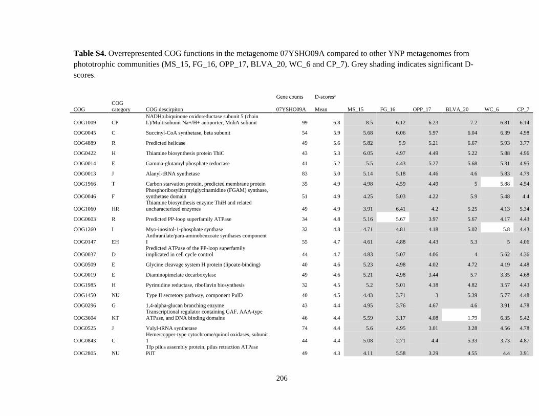
206
Table S4. Overrepresented COG functions in the metagenome 07YSHO09A compared to other YNP metagenomes from
phototrophic communities (MS_15, FG_16, OPP_17, BLVA_20, WC_6 and CP_7). Grey shading indicates significant D-
scores.
Gene counts D-scoresa
COG
COG
category COG descirpiton 07YSHO09A Mean MS_15 FG_16 OPP_17 BLVA_20 WC_6 CP_7
COG1009 CP NADH:ubiquinone oxidoreductase subunit 5 (chain L)/Multisubunit Na+/H+ antiporter, MnhA subunit 99 6.8 8.5 6.12 6.23 7.2 6.81 6.14
COG0045 C Succinyl-CoA synthetase, beta subunit 54 5.9 5.68 6.06 5.97 6.04 6.39 4.98
COG4889 R Predicted helicase 49 5.6 5.82 5.9 5.21 6.67 5.93 3.77
COG0422 H Thiamine biosynthesis protein ThiC 43 5.3 6.05 4.97 4.49 5.22 5.88 4.96
COG0014 E Gamma-glutamyl phosphate reductase 41 5.2 5.5 4.43 5.27 5.68 5.31 4.95
COG0013 J Alanyl-tRNA synthetase 83 5.0 5.14 5.18 4.46 4.6 5.83 4.79
COG1966 T Carbon starvation protein, predicted membrane protein 35 4.9 4.98 4.59 4.49 5 5.88 4.54
COG0046 F
Phosphoribosylformylglycinamidine (FGAM) synthase,
synthetase domain 51 4.9 4.25 5.03 4.22 5.9 5.48 4.4
COG1060 HR
Thiamine biosynthesis enzyme ThiH and related
uncharacterized enzymes 49 4.9 3.91 6.41 4.2 5.25 4.13 5.34
COG0603 R Predicted PP-loop superfamily ATPase 34 4.8 5.16 5.67 3.97 5.67 4.17 4.43
COG1260 I Myo-inositol-1-phosphate synthase 32 4.8 4.71 4.81 4.18 5.02 5.8 4.43
COG0147 EH Anthranilate/para-aminobenzoate synthases component I 55 4.7 4.61 4.88 4.43 5.3 5 4.06
COG0037 D
Predicted ATPase of the PP-loop superfamily
implicated in cell cycle control 44 4.7 4.83 5.07 4.06 4 5.62 4.36
COG0509 E Glycine cleavage system H protein (lipoate-binding) 40 4.6 5.23 4.98 4.02 4.72 4.19 4.48
COG0019 E Diaminopimelate decarboxylase 49 4.6 5.21 4.98 3.44 5.7 3.35 4.68
COG1985 H Pyrimidine reductase, riboflavin biosynthesis 32 4.5 5.2 5.01 4.18 4.82 3.57 4.43
COG1450 NU Type II secretory pathway, component PulD 40 4.5 4.43 3.71 3 5.39 5.77 4.48
COG0296 G 1,4-alpha-glucan branching enzyme 43 4.4 4.95 3.76 4.67 4.6 3.91 4.78
COG3604 KT
Transcriptional regulator containing GAF, AAA-type
ATPase, and DNA binding domains 46 4.4 5.59 3.17 4.08 1.79 6.35 5.42
COG0525 J Valyl-tRNA synthetase 74 4.4 5.6 4.95 3.01 3.28 4.56 4.78
COG0843 C Heme/copper-type cytochrome/quinol oxidases, subunit 1 44 4.4 5.08 2.71 4.4 5.33 3.73 4.87
COG2805 NU
Tfp pilus assembly protein, pilus retraction ATPase
PilT 49 4.3 4.11 5.58 3.29 4.55 4.4 3.91

207
COG0776 L Bacterial nucleoid DNA-binding protein 35 4.3 5.97 5.16 4.28 3.06 3.77 3.58
COG1007 C NADH:ubiquinone oxidoreductase subunit 2 (chain N) 46 4.3 4.96 3.43 3.76 4.5 3.95 5.06
COG4147 R Predicted symporter 33 4.3 4.85 3.64 4.73 4.39 4.61 3.36
COG0123 BQ
Deacetylases, including yeast histone deacetylase and
acetoin utilization protein 46 4.3 3.64 3.95 4.42 4.93 4.38 4.23
COG1206 J
NAD(FAD)-utilizing enzyme possibly involved in
translation 28 4.2 4.09 3.15 3.97 4.94 4.64 4.68
COG1452 M Organic solvent tolerance protein OstA 33 4.2 4.7 4.73 4.07 4.95 3.52 3.36
COG0841 V Cation/multidrug efflux pump 97 4.2 5.8 3.87 2.01 3.44 5.77 4.38
COG0620 E Methionine synthase II (cobalamin-independent) 29 4.2 4.25 4.44 3.85 4.44 4.77 3.45
COG0847 L DNA polymerase III, epsilon subunit and related 3'-5' exonucleases 26 4.1 5.66 4.49 3.98 3.63 3.13 3.96
COG4770 I Acetyl/propionyl-CoA carboxylase, alpha subunit 63 4.1 4.74 2.46 3.86 4.4 4.84 4.5
COG0034 F
Glutamine phosphoribosylpyrophosphate
amidotransferase 40 4.1 4.31 4.16 4.02 4.11 4.35 3.78
COG0095 H Lipoate-protein ligase A 31 4.1 4.88 4.9 2.67 3.44 4.79 3.69
COG0260 E Leucyl aminopeptidase 32 4.1 5.55 3.35 3.96 4.82 3.07 3.61
COG0467 T
RecA-superfamily ATPases implicated in signal
transduction 29 4.0 3.64 4.23 3.63 3.68 4.77 4.31
COG0649 C NADH:ubiquinone oxidoreductase 49 kD subunit 7 44 4.0 4.95 3.2 3.73 4.28 3.88 4.03
COG0067 E Glutamate synthase domain 1 31 4.0 4.4 3.38 3.85 4.5 3.99 3.69
COG0497 L ATPase involved in DNA repair 46 4.0 4.61 4.97 3.3 3.96 2.66 4.23
COG0445 D NAD/FAD-utilizing enzyme apparently involved in cell division 45 4.0 3.82 4.86 3.35 3.98 3.57 4.13
COG0751 J Glycyl-tRNA synthetase, beta subunit 38 4.0 4.96 3.47 3.11 4.02 5.37 2.77
COG0741 M
Soluble lytic murein transglycosylase and related
regulatory proteins (some contain LysM/invasin domains) 40 3.9 4.95 3.42 3 3.01 5.03 4.13
COG2905 T
Predicted signal-transduction protein containing cAMP-
binding and CBS domains 38 3.9 3.44 4.25 4.58 3.87 4.12 3.08
COG0498 E Threonine synthase 49 3.9 2.78 3.88 2.6 3.91 5.29 4.84
COG0317 TK
Guanosine polyphosphate
pyrophosphohydrolases/synthetases 55 3.9 4.42 4.62 3.17 4.18 3.84 3.02
COG0552 U Signal recognition particle GTPase 34 3.9 3.59 4.11 3.2 4 3.99 4.23
COG2084 I 3-hydroxyisobutyrate dehydrogenase and related beta-hydroxyacid dehydrogenases 29 3.8 3.79 3.65 3.21 4.24 3.92 4.09
COG1729 S Uncharacterized protein conserved in bacteria 51 3.8 4.05 4.22 3.34 3.64 3.19 4.4
COG4775 M Outer membrane protein/protective antigen OMA87 62 3.7 5 5.41 3.15 3.77 3.07 2.09
COG0370 P Fe2+ transport system protein B 44 3.7 2.7 4.16 3.57 3.48 3.88 4.7

208
COG2270 R Permeases of the major facilitator superfamily 33 3.7 4.4 3.99 3.27 3.39 3.69 3.73
COG0419 L ATPase involved in DNA repair 39 3.7 3.69 4.69 4.1 4.61 3.03 2.29
COG1353 R
Predicted hydrolase of the HD superfamily (permuted
catalytic motifs) 38 3.7 3.11 4.41 2.95 3.43 5.37 3.08
COG0558 I Phosphatidylglycerophosphate synthase 35 3.7 3.74 4.23 3.68 3.49 4.11 3.07
COG1190 J Lysyl-tRNA synthetase (class II) 43 3.7 3.67 4.34 3.31 2.75 3.77 4.43
COG0050 J GTPases - translation elongation factors 43 3.7 3.89 4.05 3.96 2.75 4.06 3.31
COG0675 L Transposase and inactivated derivatives 75 3.7 3.63 7.83 4.21 5.93 -2.56 2.9
COG1692 S Uncharacterized protein conserved in bacteria 21 3.6 4.18 2.89 3.11 3.5 5.1 3.06
COG0075 E
Serine-pyruvate aminotransferase/archaeal aspartate
aminotransferase 34 3.6 3.47 3.13 3.02 3.52 4.54 4.03
COG0825 I Acetyl-CoA carboxylase alpha subunit 32 3.6 3.82 3.68 3.35 4.82 3.07 2.88
COG0452 H Phosphopantothenoylcysteine synthetase/decarboxylase 35 3.6 3.25 3.73 2.96 4.12 4.11 3.41
COG0492 O Thioredoxin reductase 52 3.5 4.07 3.81 3.16 3.41 3.78 2.98
COG1509 E Lysine 2,3-aminomutase 23 3.5 3.77 3.85 2.89 3.59 3.99 3.1
COG2262 R GTPases 29 3.5 3.09 3.84 3 4.24 3.73 3.25
COG0165 E Argininosuccinate lyase 33 3.5 2.84 2.85 4.28 3.87 4.42 2.83
COG1008 C NADH:ubiquinone oxidoreductase subunit 4 (chain M) 52 3.5 4.07 3.57 3.57 4.74 2.5 2.61
COG0618 R Exopolyphosphatase-related proteins 38 3.5 3.33 3.93 3.81 3.29 3.19 3.4
COG2870 M ADP-heptose synthase, bifunctional sugar kinase/adenylyltransferase 37 3.5 4.55 3.35 3.35 3.74 2.93 2.97
COG0743 I 1-deoxy-D-xylulose 5-phosphate reductoisomerase 31 3.5 3.67 4.48 2.32 3.95 4.37 2.09
COG0029 H Aspartate oxidase 38 3.5 3 3.47 3.46 3.16 3.8 3.91
COG1199 KL Rad3-related DNA helicases 33 3.5 3.97 2.41 2.55 3.87 4.05 3.92
COG1331 O
Highly conserved protein containing a thioredoxin
domain 40 3.4 2.96 3.85 3 3.4 4.35 2.99
COG0495 J Leucyl-tRNA synthetase 62 3.4 4.27 4.79 2.68 1.56 4.15 3.07
COG0021 G Transketolase 36 3.4 4.68 3.69 2.89 2.64 3.25 3.35
COG0677 M UDP-N-acetyl-D-mannosaminuronate dehydrogenase 37 3.4 2.86 3.65 2.05 4.05 3.84 3.99
COG1793 L ATP-dependent DNA ligase 25 3.4 2.99 3.29 2.71 2.58 4.26 4.6
COG0436 E Aspartate/tyrosine/aromatic aminotransferase 87 3.4 4.48 3.49 3.38 4.06 2.24 2.63
COG0026 F
Phosphoribosylaminoimidazole carboxylase (NCAIR
synthetase) 28 3.4 4.26 3.15 3.74 4.94 1.61 2.56
COG0016 J Phenylalanyl-tRNA synthetase alpha subunit 35 3.4 3.74 4.41 2.96 2.78 3.6 2.74

209
COG2265 J
SAM-dependent methyltransferases related to tRNA
(uracil-5-)-methyltransferase 39 3.4 4.68 3.16 3.39 3.7 3.31 1.88
COG0794 M
Predicted sugar phosphate isomerase involved in
capsule formation 23 3.3 3.42 4.58 3.37 2.8 2.89 2.88
COG1912 S Uncharacterized conserved protein 24 3.3 2.26 3.99 3.25 3.34 3.04 3.96
COG0482 J Predicted tRNA(5-methylaminomethyl-2-thiouridylate) methyltransferase, contains the PP-loop ATPase domain 41 3.3 2.99 4.75 2.06 2.75 3.99 3.25
COG1995 H Pyridoxal phosphate biosynthesis protein 26 3.3 3.61 4.49 2.43 3.63 3.33 2.29
COG0312 R
Predicted Zn-dependent proteases and their inactivated
homologs 42 3.3 3.22 5.19 1.11 3.77 3.66 2.77
COG0820 R Predicted Fe-S-cluster redox enzyme 33 3.3 3.08 4.53 2.91 2.51 3.03 3.54
COG0403 E
Glycine cleavage system protein P (pyridoxal-binding),
N-terminal domain 36 3.3 4.01 4.01 2.73 3.18 3.25 2.39
COG0169 E Shikimate 5-dehydrogenase 28 3.2 3.63 4.1 2.49 2.86 3.22 2.93
COG0243 C Anaerobic dehydrogenases, typically selenocysteine-containing 60 3.2 2.98 0.85 3.87 2.5 5.59 3.36
COG1538 MU Outer membrane protein 62 3.2 5.29 3.77 -0.25 3.37 4.04 2.85
COG2896 H Molybdenum cofactor biosynthesis enzyme 25 3.2 3.29 3.49 2.92 3.11 3.39 2.73
COG1640 G 4-alpha-glucanotransferase 29 3.1 3.5 2.95 3.21 3.17 3 3.06
COG0177 L Predicted EndoIII-related endonuclease 28 3.1 3.07 3.52 3.29 3.19 3.41 2.38
COG2067 I Long-chain fatty acid transport protein 20 3.1 3.8 2.94 2.47 3.12 2.85 3.43
COG0345 E Pyrroline-5-carboxylate reductase 24 3.1 2.67 3.77 2.36 3.95 3.04 2.8
COG0647 G Predicted sugar phosphatases of the HAD superfamily 22 3.1 2.61 2.64 3.24 3.03 3.61 3.45
COG0240 C Glycerol-3-phosphate dehydrogenase 25 3.1 3.77 3.91 2.5 3.68 3.59 1.12
COG1459 NU Type II secretory pathway, component PulF 48 3.1 2.66 3.52 1.13 2.97 4.16 4.12
COG2877 M
3-deoxy-D-manno-octulosonic acid (KDO) 8-phosphate
synthase 23 3.1 4.14 3.4 2.66 3.18 2.69 2.45
COG1778 R Low specificity phosphatase (HAD superfamily) 18 3.1 4.31 3.59 2.68 2.78 2.52 2.61
COG0364 G Glucose-6-phosphate 1-dehydrogenase 25 3.1 3.44 2.55 2.71 3.29 2.8 3.6
COG0069 E Glutamate synthase domain 2 30 3.1 2.74 3.08 2.93 3.14 3.49 2.99
COG0351 H
Hydroxymethylpyrimidine/phosphomethylpyrimidine
kinase 23 3.0 2.94 2.8 2.44 3.38 3.31 3.34
COG0161 H Adenosylmethionine-8-amino-7-oxononanoate aminotransferase 25 3.0 4.48 2.05 3.15 2.75 2.62 3.15
COG2041 R Sulfite oxidase and related enzymes 22 3.0 3.77 2.45 2.52 3.88 3.16 2.31
COG0846 K NAD-dependent protein deacetylases, SIR2 family 24 3.0 3.12 3.34 3.02 3.14 3.04 2.39
COG0439 I Biotin carboxylase 25 3.0 3.14 3.69 3.38 3.68 2.44 1.62

210
COG0074 C Succinyl-CoA synthetase, alpha subunit 26 3.0 2.6 2.36 2.84 3.08 3.94 3.07
COG0150 F Phosphoribosylaminoimidazole (AIR) synthetase 28 3.0 2.55 3.71 3.97 3.02 2.06 2.56
COG2890 J Methylase of polypeptide chain release factors 33 3.0 3.2 3.31 2.22 3.71 3.03 2.34
COG1055 P Na+/H+ antiporter NhaD and related arsenite permeases 34 2.9 2.87 3.77 2.33 3.37 3 2.3
COG0138 F
AICAR transformylase/IMP cyclohydrolase PurH (only
IMP cyclohydrolase domain in Aful) 42 2.9 2.63 2.36 4.04 2.28 3.95 2.35
COG0496 R Predicted acid phosphatase 26 2.9 3.61 3.24 2.63 2.73 2.08 3.28
COG0003 P Oxyanion-translocating ATPase 33 2.9 3.84 2.7 3.86 2.65 2.88 1.59
COG0104 F Adenylosuccinate synthase 31 2.9 3.27 3.38 2.67 2.11 3.1 2.94
COG0803 P
ABC-type metal ion transport system, periplasmic
component/surface adhesin 26 2.9 3.16 2.69 3.05 3.26 2.41 2.87
COG0061 G Predicted sugar kinase 28 2.9 2.93 3.33 2.68 3.02 2.7 2.74
COG0129 EG Dihydroxyacid dehydratase/phosphogluconate dehydratase 37 2.9 3.19 2.12 1.47 3.31 4.17 3.13
COG0644 C Dehydrogenases (flavoproteins) 43 2.9 2.67 2.71 2.27 4.9 2.45 2.32
COG2894 D Septum formation inhibitor-activating ATPase 17 2.9 3.88 2.66 2.53 2.83 2.35 2.99
COG0441 J Threonyl-tRNA synthetase 42 2.9 2.82 2.98 2.3 2.99 2.58 3.51
COG1048 C Aconitase A 33 2.9 4.25 2.85 3.09 2.65 1.48 2.83
COG0134 E Indole-3-glycerol phosphate synthase 22 2.8 2.92 3.25 2.3 3.88 1.98 2.52
COG0458 EF
Carbamoylphosphate synthase large subunit (split gene
in MJ) 46 2.8 2.08 3.17 2.3 3.09 3.54 2.64
COG0279 G Phosphoheptose isomerase 19 2.8 4.03 2.15 2.57 2.53 2.91 2.52
COG0263 E Glutamate 5-kinase 22 2.8 2.61 2.45 2.75 3.03 2.74 2.97
COG0352 H Thiamine monophosphate synthase 22 2.8 2.04 2.84 2.75 3.66 2.74 2.52
COG0323 L DNA mismatch repair enzyme (predicted ATPase) 39 2.7 3.24 2.89 2.73 3.15 1.98 2.44
COG0119 E Isopropylmalate/homocitrate/citramalate synthases 52 2.7 2.63 3.69 3.72 2.55 1.47 2.25
COG0182 J
Predicted translation initiation factor 2B subunit, eIF-
2B alpha/beta/delta family 27 2.7 2.39 2.84 2.36 2.88 2.56 3.2
COG0005 F Purine nucleoside phosphorylase 30 2.7 2.16 3.25 2.02 2.98 2.97 2.81
COG0693 R Putative intracellular protease/amidase 20 2.6 2.39 2.52 2.97 2.5 2.63 2.68
COG0340 H Biotin-(acetyl-CoA carboxylase) ligase 21 2.6 3.06 2.68 2.61 2.48 2.19 2.59
COG0343 J Queuine/archaeosine tRNA-ribosyltransferase 34 2.5 2.43 2.98 2.84 1.51 3 2.46
COG1253 R Hemolysins and related proteins containing CBS domains 41 2.5 2.22 2.61 2.34 2.87 2.34 2.8
COG0040 E ATP phosphoribosyltransferase 20 2.5 2.55 2.52 2.71 2.31 2.22 2.68

211
COG0815 M Apolipoprotein N-acyltransferase 23 2.5 1.96 2.42 2.66 2.99 2.69 2.25
COG0315 H Molybdenum cofactor biosynthesis enzyme 17 2.5 2.63 2.9 1.31 3.07 2.35 2.45
COG1053 C
Succinate dehydrogenase/fumarate reductase,
flavoprotein subunit 33 2.3 2.5 2 2.55 2.25 2.29 2.5
COG0209 F Ribonucleotide reductase, alpha subunit 42 2.3 2.35 3.24 2.74 0.46 2.58 2.49 aP-value cutoffs used to identify significant D-scores (False Discovery Rate of 0.05): MS_15, ≤ 1.68e-02; FG_16, ≤ 1.69e-02;
OPP_17, ≤ 1.18e-02; BLVA_20, ≤ 1.42e-02; WC_6, ≤ 1.63e-02; CP_7, ≤ 1.22e-02.

212
Table S5. Sequences related to specific functions and taxa within carbon fixation pathways in prokaryotes (rTCA) cycle using
KEGG pathways.
Number of
reads
assigned
EC
number Orthology KEGG Enzyme name Gene Metagenome best hit organism Class E-value
1045 2.7.9.2 K01007 pyruvate, water dikinase pps Hydrogenobacter thermophilus TK-6 Aquificae 4e-86
Thermocrinis albus DSM 14484 Aquificae 2e-88
40 1.4.1.1 K00259 alanine dehydrogenase ald Dictyoglomus turgidum DSM 6724 Dictyoglomia 6e-48
Dictyoglomus thermophilum H-6-12 Dictyoglomia 2e-52
Thermosediminibacter oceani DSM 16646 Clostridia 5e-25
Solitalea canadensis DSM 3403 Sphingobacteriia 5e-23
Haliangium ochraceum strain DSM 14365 Deltaproteobacteria 2e-8
Nocardiopsis dassonvillei subsp. dassonvillei DSM 43111 Actinobacteria 5e-19
Belliella baltica DSM 15883 Cytophagia 5e-62
Marinilabilia sp. AK2 Bacteroidia 2e-29
Clostridium bartlettii DSM 16795 Clostridia 2e-10
Marivirga tractuosa DSM 4126 Bacteroidetes 3e-54
Chloroherpeton thalassium ATCC 35110 Chlorobi 3e-26
Idiomarina baltica OS145 Gammaproteobacteria 2e-8
Selenomonas noxia ATCC 43541 Negativicutes 6e-37
Alistipes shahii WAL 8301 Bacteroidia 6e-51
Planctomyces brasiliensis DSM 5305 Planctomycetia 4e-10
Parabacteroides goldsteinii CL02T12C30 Bacteroidia 3e-15
2421 1.2.7.1 K00169
pyruvate ferredoxin
oxidoreductase, alpha
subunit porA Thermocrinis albus DSM 14484 Aquificae 5e-68
Hydrogenobacter thermophilus TK-6 Aquificae 3e-82
K00170
pyruvate ferredoxin
oxidoreductase, beta
subunit porB Thermocrinis albus DSM 14484 Aquificae 5e-96
Hydrogenobacter thermophilus TK-6 Aquificae 1e-96
K00171
pyruvate ferredoxin
oxidoreductase, delta
subunit porD Hydrogenobacter thermophilus TK-6 Aquificae 4e-37
Thermodesulfobacterium Thermodesulfobacteria 6e-22
K00172
pyruvate ferredoxin
oxidoreductase, gamma
subunit porG Thermocrinis albus DSM 14484 Aquificae 2e-92
Hydrogenobacter thermophilus TK-6 Aquificae 1e-54
Sulfurihydrogenibium yellowstonense Aquificae 7e-18
1895 6.2.1.1 K01895 acetyl-CoA synthetase ACSS Thermodesulfatator indicus DSM 15286 Thermodesulfobacteria 3e-11
Thermocrinis albus DSM 14484 Aquificae 5e-99
Thauera sp. MZ1T Betaproteobacteria 1e-28
Thermofilum pendens Hrk 5 Thermoprotei 3e-34
Hydrogenivirga sp. 128-5-R1-1 Aquificae 1e-14
Symbiobacterium thermophilum IAM 14863 Clostridia 7e-32
Caldilinea aerophila DSM 14535 Caldilineae 8e-95
Aquifex aeolicus VF5 Aquificae 7e-39

213
681 4.2.1.3 K01681 aconitate hydratase ACO Thermodesulfobacterium Thermodesulfobacteria 5e-24
Hydrogenobaculum sp. HO Aquificae 1e-16
Thermocrinis albus DSM 14484 Aquificae 8e-68
Aquifex aeolicus VF5 Aquificae 2e-74
Hydrogenobacter thermophilus TK-6 Aquificae 2e-43
481 1.1.1.42 K00031
isocitrate
dehydrogenase IDH1, IDH2, icd Hydrogenobacter thermophilus TK-6 Aquificae 4e-67
Thermocrinis albus DSM 14484 Aquificae 9e-67
138 1.2.7.3 K00174
2-oxoglutarate
ferredoxin
oxidoreductase subunit
alpha korA Macrococcus caseolyticus JCSC5402 Bacilli 3e-8
Thermosphaera aggregans DSM 11486 Thermoprotei 3e-52
Chloroflexus aggregans DSM 9485 Chloroflexia 8e-17
Brevibacillus laterosporus Bacilli 2e-9
Aeropyrum pernix K1 Thermoprotei 2e-56
Pyrococcus furiosus DSM 3638 Thermococci 4e-12
Acidilobus saccharovorans 345-15 Thermoprotei 4e-22
Thermodesulfobacterium Thermodesulfobacteria 3e-23
Mahella australiensis 50-1 BON Clostridia 4e-9
Bacillus cellulosilyticus DSM 2522 Bacilli 1e-19
Gemella haemolysans Bacilli 6e-10
Bacillus bataviensis Bacilli 3e-38
Brevibacillus borstelensis Bacilli 5e-21
K00175
2-oxoglutarate
ferredoxin
oxidoreductase subunit
beta korB Thermosphaera aggregans DSM 11486 Thermoprotei 6e-53
Thermaerobacter subterraneus Clostridia 6e-12
Methanothermobacter marburgensis str. Marburg Methanobacteria 2e-11
Caldisphaera lagunensis DSM 15908 Thermoprotei 2e-20
Clostridium ultunense Clostridia 8e-36
Thermoproteus uzoniensis 768-20 Thermoprotei 3e-22
K00177
2-oxoglutarate
ferredoxin
oxidoreductase subunit
gamma korC Thermodesulfobacterium Thermodesulfobacteria 2e-21
1649 6.2.1.5 K01902
succinyl-CoA
synthetase alpha subunit sucD Thermocrinis albus DSM 14484 Aquificae 1e-81
Aquifex aeolicus VF5 Aquificae 1e-72
K01903
succinyl-CoA
synthetase beta subunit sucC Hydrogenobacter thermophilus TK-6 Aquificae 1e-73
Thermocrinis albus DSM 14484 Aquificae 3e-78
1289 1.3.99.1 K00239
succinate
dehydrogenase
flavoprotein subunit sdhA Hydrogenobacter thermophilus TK-6 Aquificae 7e-66
Aeropyrum pernix K1 Thermoprotei 7e-69
Thermocrinis albus DSM 14484 Aquificae 1e-65
K00240
succinate
dehydrogenase iron-
sulfur subunit sdhB Thermocrinis albus DSM 14484 Aquificae 6e-42
Hydrogenobacter thermophilus TK-6 Aquificae 2e-51

214
K00241
succinate
dehydrogenase
cytochrome b556
subunit sdhC Aeropyrum pernix K1 Thermoprotei 2e-33
Thermocrinis albus DSM 14484 Aquificae 2e-8
K00245
fumarate reductase iron-
sulfur subunit frdB Aquifex aeolicus VF5 Aquificae 3e-46
Hydrogenivirga sp. 128-5-R1-1 Aquificae 2e-16
Sulfurihydrogenibium azorense Az-Fu1 Aquificae 2e-7
Hydrogenobacter thermophilus TK-6 Aquificae 7e-32
Carboxydothermus hydrogenoformans Z-2901 Clostridia 2e-38
527 4.2.1.2 K01677
fumarate hydratase
subunit alpha fumA Thermocrinis albus DSM 14484 Aquificae 5e-78
Hydrogenobacter thermophilus TK-6 Aquificae 5e-46
K01678
fumarate hydratase
subunit beta fumB Hydrogenivirga sp. 128-5-R1-1 Aquificae 5e-43
Aquifex aeolicus VF5 Aquificae 6e-54
Thermodesulfobacterium Thermodesulfobacteria 3e-10
Thermocrinis albus DSM 14484 Aquificae 1e-18
K01679
fumarate hydratase,
class II fumC Aeropyrum pernix K1 Thermoprotei 1e-49
Thermus thermophilus JL-18 Deinococci 3e-14
Deferribacter desulfuricans SSM1 Deferribacteres 2e-31
Thermaerobacter marianensis DSM 12885 Clostridia 2e-14
Flexistipes sinusarabici DSM 4947 Deferribacteres 3e-43
Calditerrivibrio nitroreducens DSM 19672 Deferribacteres 1e-23
Rubrobacter xylanophilus DSM 9941 Actinobacteria 2e-37
Bdellovibrio bacteriovorus str. Tiberius Deltaproteobacteria 4e-30
Spirochaeta smaragdinae DSM 11293 Spirochaetia 6e-26
358 1.1.1.37 K00024 malate dehydrogenase mdh Hydrogenobacter thermophilus TK-6 Aquificae 7e-73
Thermocrinis albus DSM 14484 Aquificae 2e-79
Acidobacterium capsulatum ATCC 51196 Acidobacteriia 4e-16
Azospirillum brasilense Sp245 Alphaproteobacteria 3e-20
Desulfurobacterium thermolithotrophum DSM 11699 Aquificae 6e-38
Caldisphaera lagunensis DSM 15908 Thermoprotei 2e-54
Desulfurivibrio alkaliphilus AHT2 Deltaproteobacteria 3e-9
Thermodesulfobacterium Thermodesulfobacteria 3e-29
Gloeocapsa sp. PCC 7428 Chroococcales 8e-15
13 4.1.1.31 K01595
phosphoenolpyruvate
carboxylase ppc Pyrobaculum aerophilum str. IM2 Thermoprotei 8e-59
Pyrobaculum islandicum DSM 4184 Thermoprotei 5e-53
Pyrobaculum sp. 1860 Thermoprotei 5e-65

215
Table S6. Sequences related to specific functions and taxa within nitrogen cycle using KEGG pathways.
Number of
reads
assigned EC number
Orthology
KEGG Enzyme name Gene Metagenome best hit organism Class E-value
7 1.7.99.4 K00372 assimilatory nitrate reductase catalytic subunit nasA Sulfurihydrogenibium yellowstonense SS-5 Aquificae 2e-73
K00374 nitrate reductase gamma subunit narI Pyrobaculum aerophilum str. IM2 Thermoprotei 2e-58
37 1.7.99.7 K04561 nitric oxide reductase subunit B norB Pyrobaculum aerophilum str. IM2 Thermoprotei 8e-66
Pyrobaculum sp. 1860 Thermoprotei 1e-32
K02305 nitric oxide reductase subunit C norC Hydrogenobacter thermophilus TK-6 Aquificae 3e-74
K04748 nitric oxide reductase NorQ protein norQ Thermocrinis albus DSM 14484 Aquificae 5e-86
12 1.7.7.1 K00366 ferredoxin-nitrite reductase nirA Hydrogenobacter thermophilus TK-6 Aquificae 7e-34
Thermocrinis albus DSM 14484 Aquificae 1e-21
1.7.7.4 K00362 nitrite reductase large subunit nirB Thermocrinis albus DSM 14484 Aquificae 6e-84
483 1.13.12.16 K00459 nitronate monooxygenase ncd2 Thermocrinis albus DSM 14484 Aquificae 1e-91
Hydrogenobacter thermophilus TK-6 Aquificae 1e-57

216
Table S7. Sequences related to specific functions and taxa within sulfur cycle using KEGG pathways.
Number
of reads
assigned
EC number Orthology
KEGG Enzyme name Gene Metagenome best hit organism Class E-value
224 2.5.1.47 K10150 cysteine synthase cysO Thermosphaera aggregans DSM 11486 Thermoprotei 3e-60
Aeropyrum pernix K1 Thermoprotei 6e-28
Pyrobaculum aerophilum str. IM2 Thermoprotei 6e-68
Pyrobaculum arsenaticum DSM 13514 Thermoprotei 2e-73
K01738 cysteine synthase A cysK Chlorobium limicola DSM 245 Chlorobia 2e-21
Pyrobaculum arsenaticum DSM 13514 Thermoprotei 6e-39
Pyrobaculum islandicum DSM 4184 Thermoprotei 2e-31
Dehalococcoides ethenogenes 195 Dehalococcoidia 2e-73
K12339 cysteine synthase B cysM Hydrogenobacter thermophilus TK-6 Aquificae 4e-78
Thermocrinis albus DSM 14484 Aquificae 2e-54
Hydrogenivirga sp. 128-5-R1-1 Aquificae 6e-9
Aquifex aeolicus VF5 Aquificae 2e-68
Hydrogenobaculum sp. Y04AAS1 Aquificae 2e-6
6 2.5.1.65 K10150 cysteine synthase cysO Thermosphaera aggregans DSM 11486 Thermoprotei 3e-60
Aeropyrum pernix K1 Thermoprotei 6e-28
Pyrobaculum aerophilum str. IM2 Thermoprotei 6e-68
Pyrobaculum arsenaticum DSM 13514 Thermoprotei 2e-73
11 2.7.7.4 K00958 sulfate adenylyltransferase sat Pyrobaculum arsenaticum DSM 13514 Thermoprotei 1e-88
Thermodesulfatator indicus DSM 15286 Thermodesulfobacteria 3e-64
Pyrobaculum oguniense TE7 Thermoprotei 7e-90
Pyrobaculum aerophilum str. IM2 Thermoprotei 1e-36
22 1.8.99.2 K00394 adenylylsulfate reductase, subunit A aprA Pyrobaculum sp. 1860 Thermoprotei 2e-98
Pyrobaculum arsenaticum DSM 13514 Thermoprotei 1e-58
Thermodesulfobacterium geofontis OPF15 Thermodesulfobacteria 2e-85
K00395 adenylylsulfate reductase, subunit B aprB Thermodesulfobacterium geofontis OPF15 Thermodesulfobacteria 9e-71

217
Pyrobaculum calidifontis JCM 11548 Thermoprotei 1e-31
8 1.8.4.8 K00390 phosphoadenosine phosphosulfate
reductase cysH Pyrobaculum islandicum DSM 4184 Thermoprotei 5e-55
Pyrobaculum oguniense TE7 Thermoprotei 4e-86
Thermosphaera aggregans DSM Thermoprotei 3e-95
Pyrobaculum arsenaticum DSM 13514 Thermoprotei 7e-73
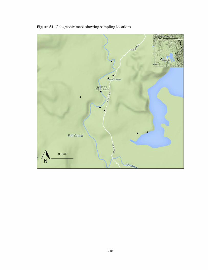
218
Figure S1. Geographic maps showing sampling locations.

219
Figure S2. Taxonomic classification and comparison of 16S rRNA gene and
metagenomic sequences.

220
Figure S3. Taxonomic assignment of metagenomic reads from the site 07YSHO09A
related to energy metabolism (KEGG identifiers). For Bacteria, only phyla with relative
abundance of more than 1% are shown. Phyla with relative abundance of less than 1% are
shown as other Bacteria.

221
Figure S4. Partial carbon fixation pathways in prokaryotes (rTCA) identified by KEGG affiliation of the sequences from the
site 07YSHO09A. Boxes indicate the KEGG identifiers and numbers in gray circles indicate the number of sequences assigned
to the KEGG function.

222
Figure S5. Partial nitrogen pathways identified by KEGG affiliation of the sequences from the site 07YSHO09A. Boxes
indicate the KEGG identifiers and numbers in gray circles indicate the number of sequences assigned to the KEGG function.

223
Figure S6. Partial sulfur pathways identified by KEGG affiliation of the sequences from the site 07YSHO09A. Boxes indicate
the KEGG identifiers and numbers in gray circles indicate the number of sequences assigned to the KEGG function.

224
Chapter 5
Summary
This research is an important complementary YNP survey on the effect of
different environmental variables on microbial taxonomic and functional diversity using
recent high-throughput sequencing technologies. This work expands our current
understanding of microbial ecology in different geothermal communities of YNP. We
assessed the patterns of microbial composition and functions in two thermal spring types
that were previously unexplored from a geobiological viewpoint: the pH 4 and low
temperature site, and the high temperature and pH site.
Our results reported here suggest that the succession of predominant bacterial
phyla is systematically correlated with temperature in circumneutral thermal springs. At
high temperatures (81-87ºC), Aquificae and EM3 predominated, whereas within the
temperature range of photosynthesis upper limit (73-75ºC) microbial communities were
dominated by Deinococcus-Thermus and Armatimonadetes. Intermediate temperatures
(63-68ºC) and low temperatures (40-57ºC) both favored Cyanobacteria and Chloroflexi,
but bacterial communities within the low temperature range were more diverse.
Furthermore, Chao1 richness and phylogenetic diversity in phototrophic communities
were significantly higher than those in non-phototrophic communities were.
225
The wide variety of abiotic factors are known to contribute to microbial diversity
in YNP thermal springs. By examining the metagenomic reads retrieved from the
unexplored pH 4 and low temperature site, we outlined the first scenario regarding the
microbial composition and functions in the pH 4 and low temperature YNP thermal
spring. We found that the ecosystem type of this spring was similar to phototrophic
communities; a high proportion of Chloroflexi, Bacteroidetes, Proteobacteria and
Firmicutes were observed. This community was enriched in the COG functions related to
energy production and conversion, transcription and carbohydrate transport, possibly to
result from high microbial dynamics. The metabolic pathway analysis indicated the
presence of assimilatory and dissimilatory nitrate reduction. Genes involved in the sulfur
metabolism were mostly related to the reduction of sulfate to adenylylsulfate, sulfite and
H2S.
Finally, we investigated microbial diversity and community composition in 10
filamentous springs with similar physiochemical properties in the Shoshone area.
Although all these samples were collected from springs that were geochemically very
similar, we found that the relatively small variation of environmental variables such as
temperature, pH and conductivity among the springs could shape microbial composition.
Metagenomic analysis of the Aquificaceae dominant site indicated that functions related
to aerobic respiration and amino acid synthesis were enriched. The results discussed here
suggest that the rTCA cycle plays a dominant role in in situ energy metabolism.
The culture-independent research of microbial ecology in terrestrial thermal
springs has been largely improved and benefited from the revolution of molecular
techniques during the last decade. In future, metagenomics and metatranscriptomics

226
sequencing needs to be conducted using other high-throughput sequencing technologies
such as PacBio and Illumina to confirm our results generated from 454 pyrosequencing.
Furthermore, the microbial activities and interactions are yet to be further investigated
using stable isotope analysis, metaproteomics and field experiments.