
The Impact of EGDT on Sepsis Mortality in a Single...
9
Research Article The Impact of EGDT on Sepsis Mortality in a Single Tertiary Care Center in Lebanon Christopher El Khuri, 1 Gilbert Abou Dagher, 1 Ali Chami, 1 Ralph Bou Chebl, 1 Tarek Amoun, 1 Rana Bachir, 1 Batoul Jaafar, 2 and Nesrine Rizk 2,3 1 Department of Emergency Medicine, American University of Beirut Medical Center, Beirut, Lebanon 2 Department of Internal Medicine, American University of Beirut Medical Center, Beirut, Lebanon 3 Division of Infectious Diseases, American University of Beirut Medical Center, Beirut, Lebanon Correspondence should be addressed to Nesrine Rizk; [email protected] Received 10 September 2018; Accepted 3 January 2019; Published 15 January 2019 Academic Editor: eodore J. Gaeta Copyright © 2019 Christopher El Khuri et al. is is an open access article distributed under the Creative Commons Attribution License, which permits unrestricted use, distribution, and reproduction in any medium, provided the original work is properly cited. Background. EGDT (Early Goal Directed erapy) or some portion of EGDT has been shown to decrease mortality secondary to sepsis and septic shock. Objective. Our study aims to assess the effect of adopting this approach in the emergency department on in- hospital mortality secondary to sepsis/septic shock in Lebanon. Hypothesis. Implementation of the EGDT protocol of sepsis in ED will decrease in-hospital mortality. Methods. Our retrospective study included 290 adult patients presenting to the ED of a tertiary center in Lebanon with severe sepsis and/or septic shock. 145 patients between years 2013 and 2014 who received protocol care were compared to 145 patients treated by standard care between 2010 and 2012. Data from the EHR were retrieved about patients’ demographics, medical comorbidities, and periresuscitation parameters. A multivariate analysis using logistic regression for the outcome in-hospital mortality aſter adjusting for protocol use and other confounders was done and AOR was obtained for the protocol use. 28-day mortality, ED, and hospital length of stay were compared between the two groups. Results. e most common infection site in the protocol arm was the lower respiratory tract (42.1%), and controls suffered more from UTIs (33.8%). Patients on protocol care had lower in-hospital mortality than that receiving usual care, 31.7% versus 47.6% (p=0.006) with an AOR of 0.429 (p =0.018). Protocol patients received more fluids at 6 and 24 hours (3.8 ± 1.7 L and 6.1 ± 2.1 L) compared to the control group (2.7 ± 2.0 L and 4.9 ± 2.8 L p=<0.001). Time to and duration of vasopressor use, choice of appropriate antibiotics, and length of ED stay were not significantly different between the two groups. Conclusion. EGDT- (Early Goal Directed erapy-) based sepsis protocol implementation in EDs decreases in-hospital mortality in developing countries. Adopting this approach in facilities with limited resources, ICU capabilities, and prehospital systems may have a pronounced benefit. 1. Introduction Sepsis is a life-threatening organ dysfunction caused by a dysregulation of host responses to infection [1]. Despite sig- nificant medical progress, it remains an oſten fatal condition with mortality reaching 25% [2]. Sepsis accounts for a large number of Emergency Department (ED) visits in the United States [3], and leads to more than half of in-hospital mortality [4]. A delay in initiating antibiotic therapy and fluid therapy is detrimental in sepsis management [5, 6]. In 2001, the early goal directed trial EGDT [7] introduced a 6-hour-protocol approach that resulted in a dramatic reduc- tion in mortality from sepsis. is lead to the establishment of the Surviving Sepsis Campaign regularly updating sepsis management guidelines [1]. Recently three trials, ARISE, PROMISE, and Process, challenged the EGDT and showed similar survival with standard care [8–10], and this result was replicated by PRISM, a meta-analysis published in 2016 [11]. ese trials were conducted in developed countries [11, 12] with rare studies exploring sepsis management in developing countries where hospital and human resources are limited [13]. Studies from Africa and the Middle East suggest a decreased mortality with the use of EGDT [14]. e primary outcome of our study is the 72 hours, hospital, and 28-day mortality to evaluate the implications of a structured sepsis protocol derived from the 2001 EGDT in Hindawi Emergency Medicine International Volume 2019, Article ID 8747282, 8 pages https://doi.org/10.1155/2019/8747282
Transcript of The Impact of EGDT on Sepsis Mortality in a Single...

Research ArticleThe Impact of EGDT on Sepsis Mortality in a Single TertiaryCare Center in Lebanon
Christopher El Khuri,1 Gilbert Abou Dagher,1 Ali Chami,1 Ralph Bou Chebl,1
Tarek Amoun,1 Rana Bachir,1 Batoul Jaafar,2 and Nesrine Rizk 2,3
1Department of Emergency Medicine, American University of Beirut Medical Center, Beirut, Lebanon2Department of Internal Medicine, American University of Beirut Medical Center, Beirut, Lebanon3Division of Infectious Diseases, American University of Beirut Medical Center, Beirut, Lebanon
Correspondence should be addressed to Nesrine Rizk; [email protected]
Received 10 September 2018; Accepted 3 January 2019; Published 15 January 2019
Academic Editor: Theodore J. Gaeta
Copyright © 2019 Christopher El Khuri et al. This is an open access article distributed under the Creative Commons AttributionLicense, which permits unrestricted use, distribution, and reproduction in any medium, provided the original work is properlycited.
Background. EGDT (Early Goal DirectedTherapy) or some portion of EGDT has been shown to decrease mortality secondary tosepsis and septic shock.Objective. Our study aims to assess the effect of adopting this approach in the emergency department on in-hospital mortality secondary to sepsis/septic shock in Lebanon.Hypothesis. Implementation of the EGDT protocol of sepsis in EDwill decrease in-hospital mortality.Methods. Our retrospective study included 290 adult patients presenting to the ED of a tertiarycenter in Lebanon with severe sepsis and/or septic shock. 145 patients between years 2013 and 2014 who received protocol carewere compared to 145 patients treated by standard care between 2010 and 2012. Data from the EHR were retrieved about patients’demographics, medical comorbidities, and periresuscitation parameters. A multivariate analysis using logistic regression for theoutcome in-hospital mortality after adjusting for protocol use and other confounders was done and AOR was obtained for theprotocol use. 28-day mortality, ED, and hospital length of stay were compared between the two groups. Results. The most commoninfection site in the protocol arm was the lower respiratory tract (42.1%), and controls suffered more from UTIs (33.8%). Patientson protocol care had lower in-hospital mortality than that receiving usual care, 31.7% versus 47.6% (p=0.006) with an AOR of 0.429(p =0.018). Protocol patients received more fluids at 6 and 24 hours (3.8 ± 1.7 L and 6.1 ± 2.1 L) compared to the control group (2.7± 2.0 L and 4.9 ± 2.8 L p=<0.001). Time to and duration of vasopressor use, choice of appropriate antibiotics, and length of ED staywere not significantly different between the two groups. Conclusion. EGDT- (Early Goal DirectedTherapy-) based sepsis protocolimplementation in EDs decreases in-hospital mortality in developing countries. Adopting this approach in facilities with limitedresources, ICU capabilities, and prehospital systems may have a pronounced benefit.
1. Introduction
Sepsis is a life-threatening organ dysfunction caused by adysregulation of host responses to infection [1]. Despite sig-nificant medical progress, it remains an often fatal conditionwith mortality reaching 25% [2]. Sepsis accounts for a largenumber of Emergency Department (ED) visits in the UnitedStates [3], and leads tomore than half of in-hospital mortality[4]. A delay in initiating antibiotic therapy and fluid therapyis detrimental in sepsis management [5, 6].
In 2001, the early goal directed trial EGDT [7] introduceda 6-hour-protocol approach that resulted in a dramatic reduc-tion in mortality from sepsis. This lead to the establishment
of the Surviving Sepsis Campaign regularly updating sepsismanagement guidelines [1]. Recently three trials, ARISE,PROMISE, and Process, challenged the EGDT and showedsimilar survival with standard care [8–10], and this result wasreplicated by PRISM, a meta-analysis published in 2016 [11].These trials were conducted in developed countries [11, 12]with rare studies exploring sepsis management in developingcountries where hospital and human resources are limited[13]. Studies from Africa and the Middle East suggest adecreased mortality with the use of EGDT [14].
The primary outcome of our study is the 72 hours,hospital, and 28-day mortality to evaluate the implications ofa structured sepsis protocol derived from the 2001 EGDT in
HindawiEmergency Medicine InternationalVolume 2019, Article ID 8747282, 8 pageshttps://doi.org/10.1155/2019/8747282

2 Emergency Medicine International
patients presenting to the emergency department of a singletertiary hospital in Lebanon.
Secondary outcomes includes length of stay, time toantibiotics, percent of ICU admission, amount of IV fluid use,and vasopressor use.
2. Methods
2.1. Study Design and Patient Selection. This is an IRBapproved, single center, retrospective, cohort study con-ducted in a large tertiary care center in Lebanon.We reviewedthe electronic health record using the ICD-9 coding system ofpatients presenting to the emergency department with severesepsis or septic shock. Patients whowere eligible for the studyshould meet the definition of severe sepsis or septic shockas per the 2012 Surviving Sepsis Campaign guidelines defini-tions [15]. Severe sepsis was defined as sepsis with associatedorgan dysfunction with one of the following: SBP <90mmHgor MAP <65mmHg or lactate >2mmol/L after an initial fluidchallenge; INR>1.5 or a PTT>60s; bilirubin>2𝜇mol/L; urineoutput <0.5ml/kg/hr for 2hrs; creatinine>2mg/dl; plateletcount <100,000/mm3; SpO
2<90%. Septic shock was defined
as sepsis with any of the following: SBP <90mmHg or MAP<65mmHg despite 30ml/kg of crystalloid resuscitation (i.e.,vasopressor dependence).We excluded patients younger than18 years of age, pregnant, or presenting with cardiac arrest ortrauma.
We included 145 consecutive patients from January 2013until May 2014 presenting to our EDwith severe sepsis and/orseptic shock who were managed by the EGDT sepsis basedprotocol, and these constituted the intervention arm. For thecontrol cohort, 145 patients with sepsis and/or septic shockpresenting to the ED between January 2010 and December2012 were randomly selected from a large pool of databaseusing computer software for random number generation.The AUBMC-ED volume of patients diagnosed with severesepsis and started on the protocol during the years followingimplementation of a sepsis protocol (2012-2014) was used todetermine the size of the study population. A rough estimateof each group size ranges between 200 and 300 patients.
Medical records were used to retrieve data about patients’demographics including patients’ age, gender and medicalinformation regarding presence/absence of comorbidities,site of infection,microbiology findings, and site of disposition(intensive care unit ICU, general practice unit GPU orhome). Pertinent laboratory blood workup and vital signsupon presentation and the latter 6 hours after resuscitationwere also collected. Antibiotics use and its appropriateness(defined as initial antibiotic regimen used in the first 48hrsof treatment that matches the bacteria later recovered) wereextracted. Data on the requirements of intravenous fluidresuscitation, vasopressor (use and duration) and steroid usewere also retrieved. We also collected the length of stay inthe ED, GPU and ICU along with in-hospital and 72-hrmortality.
2.2. EGDT-Sepsis Based Protocol Definition. For the inter-vention cohort, patients were managed as per our ED sepsis
protocol (Supplementary Materials available here), that is,based on the EGDT recommendations [7].
2.3. Usual Care Definition. For the control cohort, the man-agement was not standardized and was left as per the treatingthe physician, guided by individual preference practice.
2.4. Statistical Analysis. Statistical analyses were performedusing SPSS version 24.0 (Armonk, NY: IBM Corp).
Univariate analysis was carried out between the interven-tion and the control groups for comparison of patients’ char-acteristics, preresuscitation parameters, resuscitation param-eters, and length of hospital stay/mortality outcomes. Com-parison was done between patients’ demographics, comor-bidities, severity of sepsis, site of infection, and microbiol-ogy isolate (Table 1). Preresuscitation parameters includedvital signs and pertinent laboratory findings upon presen-tation (Table 2). As for the parameters of resuscitation, theyincluded patients’ vital signs 6 hours after ED management,requirements of intravenous fluids at 6 and 24 hours, vaso-pressor/inotrope (use, time to start, and duration of usewithin the first 24 hours), CVC placement, and requirementof mechanical ventilation (Table 3). As for length of hospitalstay (in ED, ICU, and GPU and in-hospital), 72hours and28 days mortality were compared between the two arms(Table 4). For continuous variables, an independent t-testcomparing the mean across both groups was done and both,the mean and standard deviation, are shown. For categoricalvariables, a chi-square test was run and data is represented asfrequency percentages.
A multivariate analysis was performed using logisticregression to find the best model that fits the data andexplains the use of the EGDT-sepsis based protocol aspredictor of in-hospital mortality while controlling for allpossible confounders. A backward selection procedure, withsignificance level for removal from the model set at 0.1, wasconducted by fitting in-hospital mortality with all risk factorsfound to be significant in the bivariate level, in additionto those considered as being clinically meaningful. Theincluded variables were protocol use, age, gender, diagnosis(severe sepsis or septic shock), systolic congestive heartfailure (CHF) EF<40%, diabetes mellitus (DM), coronaryartery disease (CAD), hypertension (HTN), cerebrovascularaccidents (CVA), chronic kidney disease (CKD), hemodial-ysis (HD), chronic obstructive pulmonary disease (COPD),central venous catheter (CVC) placement, endotracheal tubeplacement, MAP upon presentation to the ED, BUN, creati-nine, and appropriate use of antibiotics.
3. Results
3.1. Patients Characteristics. Two hundred and ninetypatients were included in the final analysis with 145 patientsin each arm. The protocol arm had a mean age of 71.9 ±14.1 years compared to 72.9 ± 16.3 years in the control arm.51.7% and 52.4% of the protocol group and control group,respectively, were male patients. All baseline demographicssuch as age, gender, and studied comorbidities in addition
Emergency Medicine International 3
Table 1: Patient demographics.
Protocol N= 145 Control N= 145 p-valueContinuous mean ± SDAge (years) 71.9 ± 14.1 72.9 ± 16.3 0.573Categorical no.(%)Male 75 (51.7) 76 (52.4) 0.906Diagnosis
0.280Septic shock 92 (63.4) 83 (57.2)Severe sepsis 53 (36.6) 62 (42.8)
HTN 102 (70.3) 101 (69.7) 0.898DM 71(49.0) 68 (46.9) 0.724CAD 64 (44.1) 59 (40.7) 0.552Systolic CHF: EF<40% 32 (22.1) 29 (20.0) 0.666COPD/Emphysema 13 (9.0) 19 (13.1) 0.261CKD on HD 4 (2.8) 5 (3.4) 0.735CVA 19 (13.1) 20 (13.8) 0.863Site of Infection
<0.001∗
Lung 61 (42.1) 45 (31.0)Gastrointestinal 21 (14.5) 11 (7.6)Urine 52 (35.9) 49 (33.8)Skin 6 (4.1) 19 (13.1)Bile 0 (0.0) 5 (3.4)Liver 0 (0.0) 1 (0.7)Undetermined 5 (3.4) 15 (10.3)
Microbiology IsolateCoNS1 1 (0.7) 2 (2.1) 0.562Staphylococcus aureus 6 (4.1) 3 (2.1) 0.310Escherichia coli 44 (37.9) 61 (42.1) 0.472klebsiella pneumonia 9 (6.2) 11 (7.6) 0.643pseudomonas aeroginosa 12 (8.3) 7 (4.8) 0.235Acinetobacter baumani 8 (5.5) 5 (3.4) 0.395Enterococcus spp. 2 (1.4) 9 (6.3) 0.030∗
Proteus mirabillis 4 (2.8) 7 (4.8) 0.356Streptococcus spp. 9 (6.2) 6 (4.1) 0.426Clostridium spp. 3 (2.1) 1 (0.7) 0.314Others2 3 (2.1) 0 (0.0) 0.281
1Coagulase-negative staphylococci. 2Others included: Bacteroides fragilis, Candida albicans, Citrobacter, Diphteroids spp., Enterobacter cloacae, Haemophilusinfluenzae (type B), Haemophilus parainfluenzae, Legionella pneumophila, Leuconostoc, Morganella morgani, Peptococcus spp., Providncia stuartii, Serratiamarsescens, and Stenotrophomonas maltophilia.∗p=<0.05 considered significant.
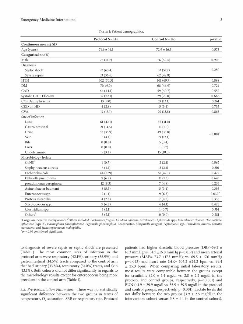
to diagnosis of severe sepsis or septic shock are presented(Table 1). The most common sites of infection in theprotocol arm were respiratory (42.1%), urinary (35.9%) andgastrointestinal (14.5%) tracts compared to the control armthat had urinary (33.8%), respiratory (31.0%) tracts, and skin(13.1%). Both cohorts did not differ significantly in regards tothe microbiology results except for enterococcus being moreprevalent in the control arm (Table 1).
3.2. Pre-Resuscitation Parameters. There was no statisticallysignificant difference between the two groups in terms oftemperature, O
2saturation, SBP, or respiratory rate. Protocol
patients had higher diastolic blood pressure (DBP=59.2 ±16.3 mmHg vs. 54.7 ±16.0 mmHg p=0.019) and mean arterialpressure (MAP= 73.7 ±17.3 mmHg vs. 69.5 ± 17.6 mmHgp=0.043) and heart rate (HR= 106.2 ±24.2 bpm vs. 99.4± 25.3 bpm). When comparing initial laboratory results,most results were comparable between the groups exceptfor creatinine (2.0 ± 1.4 mg/dl vs. 2.8 ± 2.2 mg/dl in theprotocol and control groups, respectively, p=<0.001) andBUN (41.9 ± 29.9 mg/dl vs. 55.9 ± 39.5 mg/dl in the protocoland control groups, respectively, p=0.001). Lactate levels didnot differ between the two groups (3.9 ± 2.5 mg/dl in theintervention cohort versus 3.8 ± 4.1 in the control cohort).

4 Emergency Medicine International
Table 2: Preresuscitation parameters.
Protocol N= 145 Control N= 145 p-valueContinuous mean ± SDSBP (mmHg) 104.7 ± 23.9 100.0 ± 26.0 0.114DBP (mmHg) 59.2 ± 16.3 54.7 ± 16.0 0.019∗
MAP (mmHg) 73.7 ± 17.3 69.5 ± 17.6 0.043∗
HR (bpm) 106.2 ± 24.2 99.4 ± 25.3 0.021∗
O2Saturation (%) 92.8 ± 8.5 93.4 ± 7.1 0.533
Temperature (∘C) 37.7 ± 1.2 37.4 ± 1.4 0.071RR (breath/min) 23.9 ± 7.2 22.9 ± 6.4 0.196Glucose (mg/dl) 169.8 ± 102.9 172.3 ± 116.1 0.860WBC (x109cells/L) 15,238.6 ± 11,683.5 16,412.4 ± 15,648.1 0.470Hemoglobin (g/dL) 11.0 ± 62.2 11.0 ± 2.3 0.935Hematocrit(%) 33.1 ± 6.9 33.1 ± 7.9 0.994Bicarbonate(mmol/L) 20.5 ± 6.0 19.9 ± 8.9 0.515BUN (mg/dL) 41.9 ± 29.9 55.9 ± 39.5 0.001∗
Creatinine (mg/dL) 2.0 ± 1.4 2.8 ± 2.2 <0.001∗
Arterial pH 7.34 ± 0.1 7.34 ± 0.1 0.964INR 1.8 ± 1.1 2.0 ± 1.3 0.397Lactate1 (mmol/L) 3.9 ± 2.5 3.8 ± 4.1 0.787Lactate > 4 mmol/L 18 (12.4) 48 (33.1) 0.1901133 protocol patients had their lactate taken vs 67 controls.∗p=<0.05 considered significant.
Table 2 includes those parameters upon presentation to theED.
3.3. Resuscitation Parameters. All vital signswere comparablebetween the groups after treatment, except for MAP whereprotocol patients had a lower MAP than the control groupat 6 hours (69.8 ± 13.8 mmHg vs. 73.9 ± 14.9 mmHg,respectively, p=0.022). Patients who were managed using thesepsis protocol received more fluids at 6 and 24 hours (3.8± 1.7 L and 6.1 ± 2.1 L) compared to the control group (2.7± 2.0 L and 4.9 ± 2.8 L) p=<0.001. More vasopressors wereinitiated within the first 24 hours in the protocol arm aswell (88 (60.6%) vs. 52 (35.8%) p=<0.001). The interventiongroup had more CVC placed (21.2% vs. 9.9% p=0.010). Bothprotocol and control cohorts have similar rates of mechan-ical ventilation. The time to vasopressor initiation and theduration of vasopressor use were not significantly differentbetween the cohorts. Both cohorts had similar steroid andantibiotic use. The mean time of antibiotics initiation was2.0 ± 3.6 hours in the protocol group compared to 2.8 ± 2.6hours in the control group p=0.054. More antibiotics wereinitiated in the ED in the control arm than the protocolarm (99.3% vs. 93.8% p=0.010). Ninety-one patients (97.8%)in the intervention group received appropriate antibioticscompared to 75 (91.5%) in the control group (p=0.056).Table 3 summarizes the vital signs 6 hours after treatment andthe general parameters of the resuscitation.
3.4. Length of Stay and Mortality Analysis. The length ofstay in the ED was found to be 26.0 ± 29.0 hours vs. 19.4
± 28.8 hours in the protocol and control group, respectively(p=0.051). ICU and GPU LOS did not differ between the twoarms (Table 4). Forty-six (31.7%) protocol patients died dur-ing their hospital stay compared to sixty-nine (47.6%) con-trol patients (p=0.006). When all statistically and clinicallyrelevant variables were controlled for, protocol patients hadan adjusted odds ratio of dying in hospital of 0.429 (Cl 95%0.213-0.864 p =0.018) when compared to the control group(Table 5). The relative risk reduction was found to be 33.3%.
4. Discussion
This study attempts to evaluate the utility and efficacy ofusing EGDT-sepsis based protocol on sepsis managementat a tertiary care centre in Beirut. The primary outcomeis in-hospital mortality; the secondary outcomes were 28-day mortality, ED, and hospital length of stay. After theintroduction of the EGDT in the USA, clinical outcomes ofseptic patients substantially improved [7] and mortality fromsepsis decreased [16, 17]. Novel interventions ranged fromprehospital recognition and management of sepsis [18] to in-hospital optimization of care [6, 19, 20]. In Lebanon, datafrom national registries on mortality secondary to sepsis arestill lacking. And the guidelines from the Surviving SepsisCampaign are not adopted nationally, where the standard ofcare remains ill-defined [21]. To our knowledge, our study isthe first to assess the impact of implementing EGDT sepsisbased protocol in the emergency department on mortalityoutcomes in Lebanon. Our results show lower in-hospitalmortality from sepsis after the introduction of the protocol,

Emergency Medicine International 5
Table 3: Resuscitation parameters.
Protocol N= 145 Control N= 145 p-valueVital signs after 6 hoursContinuous mean ± SDSBP (mmHg) 103.3 ± 19.9 104.7 ± 18.9 0.595DBP (mmHg) 57.2 ± 13.0 59.1 ± 14.5 0.243MAP (mmHg) 69.8 ± 13.8 73.9 ± 14.9 0.022∗
HR (bpm) 92.4 ± 21.0 91.9 ± 20.0 00.834O2 Saturation (%) 96.0 ± 11.2 97.1 ± 6.8 0.394Temperature (∘C) 37.3 ± 0.9 37.4 ± 5.9 0.781RR (breath/min) 21.8 ± 5.5 21.0 ± 4.3 0.166Resources variablesContinuous mean ± SDIV fluid requirements at first 6 hrs (L) 3.8 ± 1.7 2.7 ± 2.0 <0.001∗
IV fluid requirements at first 24 hrs (L) 6.1 ± 2.1 4.9 ± 2.8 <0.001∗
Time to initiation of antibiotics (hrs) 2.0 ± 3.6 2.8 ± 2.6 0.054Time to vasopressor/inotrope use within the 1st 24hrs (hrs) 9.1 ± 22.6 7.7 ± 7.0 0.67Duration of vasopressor/inotrope treatment within 1st 24hrs (hrs) 50.9 ± 649.6 40.0 ± 56.1 0.234Categorical No (%)Vasopressor/inotrope use within 1st 24hrs 88 (60.6) 52 (35.8) <0.001∗
CVC placement 29 (21.2) 14 (9.9) 0.010∗
Mechanical ventilation 31 (21.5) 23 (15.9) 0.217Steroid use 50 (34.5) 54 (37.2) 0.624Antibiotic use 143 (98.6) 145 (100.0) 0.155Appropriate antibiotic use1 91 (97.8)% 75 (91.5) 0.056Antibiotics initiation in the ED 136 (93.8) 144 (99.3) 0.010∗
Antibiotics initiation in the ICU 4 (2.8) 1 (0.7) 0.176Antibiotics initiation in the GPU 3 (2.1) 0 (0) 0.0821Appropriate use of antibiotics was defined as preliminary antibiotic given in the first 48hrs of treatment covering the bacteria grown later in bacteriology.
Table 4: Length of stay and mortality outcomes.
Protocol N= 145 Control N= 145 P-valueLength of Stay (mean ± SD)ED (hours) 26.0 ± 29.0 19.4 ± 28.8 0.051ICU (days) 5.8 ± 6.9 12.0 ± 38.9 0.285GPU (days) 7.02 ± 6.2 7.1 ± 3.8 0.913Hospital1 (days) 10.7 ± 8.7 15.3 ± 29.8 0.148Mortality no.(%)In-hospital 46 (31.7) 69 (47.6) 0.006∗
72-hour 17 (11.7) 11 (7.6) 0.23328-day2 30 (20.7) 47 (32.4) 0.0141Hospital LOS days were calculated only for those that did not expire in hospital (as shorter LOS times may be associated with early deaths). 228 patients(19.3%) and 14 (9.7%) of patients had unknown 28-day mortality in the protocol and control groups, respectively.∗p=<0.05 considered significant.
31.7% compared to 47.6% when on “usual care,” before theprotocol implementation. These findings are similar to thehospital mortality noted in the first EGDT paper (30.5%and 46.5% respectively) but higher than those reportedin the more recent ProCESS, ARISE, and ProMISe trials[11]. Prehospital management of sepsis has a contributingimpact on ultimate outcomes [22] and Lebanon lacks the
infrastructure for prehospital sepsis management due tolimited resources [23]. We assume that mortality is higherin our cohort compared to developed countries becauseprehospital care in Lebanon is not optimized [24].
4.1. Effectiveness of the Bundle. Adherence to the sepsisprotocol was associated with a relative risk reduction (RRR)

6 Emergency Medicine International
Table 5: Multiple logistic regression for hospital mortality.
Protocol N= 145 Control N= 145Adjusted OR2 (CI 95%) P-value
Hospital mortality no.(%)1 46 (31.7) 69 (47.6) 0.429 (0.213-0.864) 0.018∗1Reference group is being the Control group.2While controlling for the following: age, gender, diagnosis (severe sepsis or septic shock), systolic CHF: EF<40%, DM, CAD, HTN, cerebrovascular accidents,CKD, CKD on HD, COPD emphysema, CVC placed, ET tube placed, MAP upon presentation to the ED, BUN, creatinine, and appropriate use of antibiotics.∗p=<0.05 considered significant.
on in-hospital mortality of 33.3%. This is in line with thefindings of the Rivers [7] and others [25] where the RRRranged from 24.3% to 45% [11]. Our protocol-based care wasassociated with an adjusted odds ratio (AOR) for hospitalmortality of 0.429 (Cl 95% 0.213-0.864 p =0.018).
Several studies questioned the effectiveness of the EGDTprotocol versus standard care and showed that alternatestrategies may have equal effect at reducing mortality withoutthe increased costs [8–10]. Those studies highlight the needfor “unbundling” the sepsis protocol to better define whichintervention ismost important while gauging specific clinicalendpoints. A “single intervention” analysis in our paper is notpossible as controlling for other interventions is not feasible.we noticed a more conservative approach to CVC placementin our study in both arms, the intervention and the control,21.2% and 9.9%, respectively, compared to that reported inthe EGDT and other trial were more than 50% of the controlpopulation had a CVC inserted [8]. This lower rate is poten-tially explained by the stipulated endpoints in our protocol:MAP (via non-invasive blood pressure readings) and urineoutput. While CVP measurement via CVC insertion waskept an optional clinical decision, our physicians were mostof the times not driven by the indication of getting a CVPmeasurement to insert a CVC. This approach was supportedby a meta-analysis that included 24 studies looking at therole of CVP measurement on fluid responsiveness and founda poor association [26]. Despite the difference in invasivemonitoring rates in our study and other trials, mortalitywas reduced and at a comparable RRR. This finding mightsupport the “unbundling” approach to invasive monitoringin resources-limited settings where cost and resources maybecome restrictive.
4.2. ED Length of Stay. Patient’s length of stay in the ED anddelayed transfers to the ICU have come under discussion inrecent years [22, 26]. Some proposals have targeted ED times,such as the 4-hour rule [27], and other studies have shownan increased hospital stay and mortality with delayed patienttransfer from the emergency department to the ICU [28, 29].However, the length of stay in the ED in our study in bothgroups, the protocol and the control, was longer than thatin the literature, 26.0 ± 29.0 hours and 19.4 ± 28.8 hours,respectively. This finding can be partially explained by thehospital’s limited capacity of ICU beds [21]. In addition, theprotocol arm had longer ED stay than the control arm, butit was not statistically significant. We explain this observeddifference by the new introduction of the protocol in our ED,as the staff was in the training process to acquire the skills of
the formulated protocol [30]. Moreover, the implementationof the new sepsis bundle was more time consuming, as itrequires the use of more resources such as IV resuscitatingfluids, vasopressors, monitoring, CVCplacement andwaitingfor laboratory and radiology results. Delays in patient transferfrom the ED to ICU in sepsis are well studied and correlateusually with worse outcomes. Despite the observed longerstay in the ED in our study, themortality rate was still reducedat a comparable relative risk reduction to the EGDT trial.
4.3. AntibioticTherapy. Our study showed that fewer patientswere started on antibiotics in the ED in the protocol armdespite a lower mortality in this group.This finding relates toongoing antimicrobial stewardship efforts [30]. It is crucialto promptly initiate antibiotic therapy in septic patients[31]; however, it is vital to apply standards of antimicrobialstewardship [32] as we are losing the battle against multidrugresistant organisms [33]. By following the protocol for sepsismanagement, we circumvented the initiation of unnecessaryantibiotics. Though it did not reach significance, antibioticswere more used appropriately in the protocol arm thanthe control arm 91 (97.8%) vss. 75 (91.5%) (p=0.056). Thisreduction in inappropriate empiric antibiotic use mightreflect positively on the cost, potential side effects, andcomplications. However, this study was not powered to testthis hypothesis and we believe that future research in thisdirection would provide data to the gap in this knowledge.
5. Strengths and Limitations
This is the first study to be conducted in Lebanon assessingthe effect of implementing an EGDT sepsis based protocolin ED on mortality outcome. The retrospective design of thestudy is a main limitation of the study as data collected fromthe electronic health records may sometimes be misinter-preted ormissing. Tominimize the information bias, frequentmeetings were held between the investigators to standardizethe data collection process. In addition, we were not able touse a matched control group. We included all patients whopresented with sepsis between January 2013 and May 2014 toensure a large sample size.
The definition of “usual care” is not standard and wasper the treating physician, thus comparing the managementacross the two arms is limited. The control group in ourstudy did receive timely antibiotics, vasopressors, and IVfluids. Data about the paramedical care was not available;therefore we were not able to adjust for it in our analyses. Thetwo cohorts were not matched, and the control arm subjects
Emergency Medicine International 7
were sicker at baseline, showing worse hemodynamics uponpresentation to the ED. These points might be explainedby the inaccuracy of the non-invasive measurements. Theyhad lower diastolic blood pressure (DBP=54.7 ±16.0 mmHgvs. 59.2 ± 16.3 mmHg p=0.019) and mean arterial pressure(MAP= 73.7 ±17.3 mmHg vs. 69.5 ± 17.6 mmHg p=0.043)when compared to the intervention arm. These findingsmight not be pertinent clinically as both groups had similarlactate and arterial pH. Those markers are indicators of end-organ hypoperfusion [34]. Moreover, despite less fluid andvasopressor use in the control arm, this group had higherMAP after 6 hours of resuscitations (73.9 ± 14.9 mmHgvs. 69.8 ± 13.8 mmHg, respectively, p=0.022). However,patients of the control cohort had more severe kidney injuryat baseline compared to intervention cohort, with highercreatinine (2.8 ± 2.2 mg/dl vs. 2.0 ± 1.4 mg/dl, respectively,p=<0.001) and BUN (55.9 ± 39.5 mg/dl vs. 41.9 ± 29.9mg/dl, respectively, p=0.001). The internal validity of ourstudy is limited by the missing data on the urine output,that was used as an endpoint in the sepsis protocol and theunits of blood transfusions received in both groups. It isunclear which parts of the bundle contribute to the noteddifference in mortality between the two groups. There iscontinued work in developed countries to decipher the mostimpactful parts of the EGDT protocol. In countries withlimited resources, it is potentially more critical to determinethose fundamental factors in order to decrease the effort andresources required to effectively treat sepsis. Finally, our studyis not generalizable, as it is limited to one center in Lebanonand further multicenter studies are needed.
6. Conclusion
In a single tertiary center in Lebanon, the introduction of anEGDT-based sepsis protocol decreased in-hospital mortalityof patients presenting with severe sepsis or septic shock. 28-day sepsis mortality is also reduced after the implementationof the EGDT protocol. Although the utility of EGDT bundleshas been under scrutiny in recent years, their benefits whenused in countries with limited resources, ICU capabilitiesand pre-hospital systems may be pronounced. In conclusion,this study highlights several potentially important points.Theintroduction of a structured approach to sepsis is feasible ina resource limited setting; the results achieved in terms ofreduced mortality are comparable to those demonstrated indeveloped countries. Outcomes could be further improved,especially regarding the baseline mortality by improving theexisting pre-hospital infrastructure to ensure expedited andappropriate treatment of septic patients.
Abbreviations
BP: Blood pressureBUN: Blood urea nitrogenCAD: Coronary artery diseaseCHF: Congestive heart failureCOPD: Chronic obstructive pulmonary diseaseCVC: Central venous catheterCVP: Central venous pressure
DBP: Diastolic blood pressureDM: Diabetes mellitusED: Emergency DepartmentEGDT: Early Goal Directed TherapyEHR: Electronic health recordEM: Emergency medicineET: Endotracheal tubeGPU: General practice unitHR: Heart rateHTN: HypertensionICU: Intensive care unitINR: International normalised ratioLOS: length of stayMAP: Mean arterial pressureRR: Respiratory rateRRR: Relative risk reductionSBP: Systolic blood pressureSSC: Surviving Sepsis Campaign.
Data Availability
The protocol dataset applied in the intervention arm, usedto support the findings of this study, is included within thesupplementary material.
Disclosure
Nisrine Rizk is the corresponding and submitting author.
Conflicts of Interest
Authors declare no conflicts of interest.
Supplementary Materials
Sepsis protocol order set that was used in the case arm.(Supplementary Materials)
References
[1] M. Singer, C. S. Deutschman, C. Seymour et al., “The thirdinternational consensus definitions for sepsis and septic shock(sepsis-3),” Journal of the AmericanMedical Association, vol. 315,no. 8, pp. 801–810, 2016.
[2] M. M. Levy, M. P. Fink, J. C. Marshall et al., “2001 SCCM/ESICM/ACCP/ATS/SIS international sepsis definitions confer-ence,”Critical Care Medicine, vol. 31, no. 4, pp. 1250–1256, 2003.
[3] H. E. Wang, N. I. Shapiro, D. C. Angus, and D. M. Yealy,“National estimates of severe sepsis in United States emergencydepartments,” Critical Care Medicine, vol. 35, no. 8, pp. 1928–1936, 2007.
[4] C. Rhee, R. Dantes, L. Epstein et al., “Incidence and trends ofsepsis in US hospitals using clinical vs claims data, 2009–2014,”Journal of the American Medical Association, vol. 318, no. 13, pp.1241–1249, 2017.
[5] A. Kumar, D. Roberts, K. E. Wood et al., “Duration of hypoten-sion before initiation of effective antimicrobial therapy is thecritical determinant of survival in human septic shock,”CriticalCare Medicine, vol. 34, no. 6, pp. 1589–1596, 2006.

8 Emergency Medicine International
[6] E. P. Riversa, A. K. Jaehne, L. Eichhorn-Wharry, S. Brown, andD. Amponsah, “Fluid therapy in septic shock,”Current Opinionin Critical Care, vol. 16, no. 4, pp. 297–308, 2010.
[7] E. Rivers, B. Nguyen, S. Havstad et al., “Early goal-directedtherapy in the treatment of severe sepsis and septic shock,”TheNew England Journal of Medicine, vol. 345, no. 19, pp. 1368–1377,2001.
[8] ProCESS Investigators, D. M. Yealy, J. A. Kellum et al., “Arandomized trial of protocol-based care for early septic shock,”The New England Journal of Medicine, vol. 370, no. 18, pp. 1683–1693, 2014.
[9] The ARISE Investigators and the ANZICS Clinical TrialsGroup, “Goal-directed resuscitation for patients with earlyseptic shock,”TheNew England Journal of Medicine, vol. 371, no.16, pp. 1496–1506, 2014.
[10] P. R. Mouncey, T. M. Osborn, and G. S. Power, “Trial of early,goal-directed resuscitation for septic shock,” The New EnglandJournal of Medicine, vol. 372, no. 14, pp. 1301–1311, 2015.
[11] H. B. Nguyen, A. K. Jaehne, N. Jayaprakash et al., “Early goal-directed therapy in severe sepsis and septic shock: Insights andcomparisons to ProCESS, ProMISe, and ARISE,” Critical Care,vol. 20, no. 1, 2016.
[12] H. B. Nguyen, S. W. Corbett, R. Steele et al., “Implementationof a bundle of quality indicators for the early managementof severe sepsis and septic shock is associated with decreasedmortality,” Critical Care Medicine, vol. 35, no. 4, pp. 1105–1112,2007.
[13] J. U. Becker, C. Theodosis, S. T. Jacob, C. R. Wira, and N. E.Groce, “Surviving sepsis in low-income and middle-incomecountries: new directions for care and research,” The LancetInfectious Diseases, vol. 9, no. 9, pp. 577–582, 2009.
[14] S. T. Jacob, P. Banura, J. M. Baeten et al., “The impact ofearly monitored management on survival in hospitalized adultUgandan patientswith severe sepsis: A prospective interventionstudy,”Critical CareMedicine, vol. 40, no. 7, pp. 2050–2058, 2012.
[15] D. C. Angus and T. Van Der Poll, “Severe sepsis and septicshock,” The New England Journal of Medicine, vol. 369, no. 21,p. 2063, 2013.
[16] R. P. Dellinger, J. M. Carlet, H. Masur et al., “Surviving SepsisCampaign guidelines for management of severe sepsis andseptic shock,”Critical Care Medicine, vol. 32, no. 3, pp. 858–873,2004.
[17] R. P. Dellinger, M. M. Levy, J. M. Carlet et al., “Surviving sepsiscampaign: international guidelines for management of severesepsis and septic shock: 2008,” Critical Care Medicine, vol. 36,no. 1, pp. 296–327, 2008.
[18] R. A. Band,D. F. Gaieski, J. H.Hylton, F. S. Shofer,M.Goyal, andZ. F. Meisel, “Arriving by emergency medical services improvestime to treatment endpoints for patients with severe sepsis orseptic shock,” Academic Emergency Medicine, vol. 18, no. 9, pp.934–940, 2011.
[19] A. Bastani, B. Shaqiri, S. Mansour, and W. Anderson, “Sepsisscreening clinical decision rule: a novel tool to identify emer-gency department patients who are at high risk for developingsevere sepsis/septic shock,” Annals of Emergency Medicine, vol.62, no. 4, pp. S153–S154, 2013.
[20] M. Capuzzo, M. Rambaldi, G. Pinelli et al., “Hospital staffeducation on severe sepsis/septic shock and hospital mortality:An original hypothesis,” BMC Anesthesiology, vol. 12, 2012.
[21] G. A. Dagher, M. Saadeldine, R. Bachir, D. Zebian, and R. B.Chebl, “Descriptive analysis of sepsis in a developing country,”International Journal of Emergency Medicine, vol. 8, no. 1, 2015.
[22] N. Alam, K. B. N. S. Doerga, T. Hussain et al., “Epidemiology,recognition and documentation of sepsis in the pre-hospitalsetting and associated clinical outcomes: A prospective multi-center study,” Acute Medicine, vol. 15, no. 4, pp. 168–175, 2016.
[23] M. J. El Sayed and J. D. Bayram, “Prehospital emergencymedicalservices in Lebanon: Overview and prospects,” Prehospital andDisaster Medicine, vol. 28, no. 2, pp. 163–165, 2013.
[24] S. Na, W. S. Kuan, M. Mahadevan et al., “Implementation ofearly goal-directed therapy and the surviving sepsis campaignresuscitation bundle in Asia,” International Journal for Qualityin Health Care, vol. 24, no. 5, Article ID mzs045, pp. 452–462,2012.
[25] E. P. Rivers, V. Coba, and M. Whitmill, “Early goal-directedtherapy in severe sepsis and septic shock: A contemporaryreview of the literature,” Current Opinion in Anaesthesiology,vol. 21, no. 2, pp. 128–140, 2008.
[26] D. Junhasavasdikul, P. Theerawit, and S. Kiatboonsri, “Asso-ciation between admission delay and adverse outcome ofemergency medical patients,” Emergency Medicine Journal, vol.30, no. 4, pp. 320–323, 2013.
[27] R. J. Lewis, “Disassembling goal-directed therapy for sepsis afirst step,” Journal of the AmericanMedical Association, vol. 303,no. 8, pp. 777–779, 2010.
[28] G. C. Geelhoed and N. H. de Klerk, “Emergency departmentovercrowding, mortality and the 4-hour rule in Western Aus-tralia,”Medical Journal of Australia, vol. 196, no. 2, pp. 122–126,2012.
[29] S.Mason, E. J.Weber, J. Coster, J. Freeman, andT. Locker, “Timepatients spend in the emergency department: England’s 4-hourrule—a case of hitting the target but missing the point?” Annalsof Emergency Medicine, vol. 59, no. 5, pp. 341–349, 2012.
[30] E. J. Septimus, C. M. Coopersmith, J. Whittle, C. P. Hale, N.O. Fishman, and T. J. Kim, “Sepsis national hospital inpatientquality measure (SEP-1): multistakeholder work group recom-mendations for appropriate antibiotics for the treatment ofsepsis,” Clinical Infectious Diseases, vol. 65, no. 9, pp. 1565–1569,2017.
[31] J. Garnacho-Montero, J. L. Garcia-Garmendia, A. Barrero-Almodovar, F. J. Jimenez-Jimenez, C. Perez-Paredes, and C.Ortiz-Leyba, “Impact of adequate empirical antibiotic therapyon the outcome of patients admitted to the intensive care unitwith sepsis,”Critical CareMedicine, vol. 31, no. 12, pp. 2742–2751,2003.
[32] J. Burston, S. Adhikari, A. Hayen et al., “A Role for Antimi-crobial Stewardship in Clinical Sepsis Pathways: A ProspectiveInterventional Study,” Infection Control and Hospital Epidemiol-ogy, vol. 38, no. 9, pp. 1032–1038, 2017.
[33] M. Bassetti, M. Merelli, C. Temperoni, and A. Astilean, “Newantibiotics for bad bugs: where are we?” Annals of ClinicalMicrobiology and Antimicrobials, vol. 12, no. 1, article 22, 2013.
[34] M. Shankar-Hari, G. S. Phillips, M. L. Levy et al., “Developinga new definition and assessing new clinical criteria for septicshock: for the third international consensus definitions forsepsis and septic shock (sepsis-3),”The Journal of the AmericanMedical Association, vol. 315, no. 8, pp. 775–787, 2016.

Stem Cells International
Hindawiwww.hindawi.com Volume 2018
Hindawiwww.hindawi.com Volume 2018
MEDIATORSINFLAMMATION
of
EndocrinologyInternational Journal of
Hindawiwww.hindawi.com Volume 2018
Hindawiwww.hindawi.com Volume 2018
Disease Markers
Hindawiwww.hindawi.com Volume 2018
BioMed Research International
OncologyJournal of
Hindawiwww.hindawi.com Volume 2013
Hindawiwww.hindawi.com Volume 2018
Oxidative Medicine and Cellular Longevity
Hindawiwww.hindawi.com Volume 2018
PPAR Research
Hindawi Publishing Corporation http://www.hindawi.com Volume 2013Hindawiwww.hindawi.com
The Scientific World Journal
Volume 2018
Immunology ResearchHindawiwww.hindawi.com Volume 2018
Journal of
ObesityJournal of
Hindawiwww.hindawi.com Volume 2018
Hindawiwww.hindawi.com Volume 2018
Computational and Mathematical Methods in Medicine
Hindawiwww.hindawi.com Volume 2018
Behavioural Neurology
OphthalmologyJournal of
Hindawiwww.hindawi.com Volume 2018
Diabetes ResearchJournal of
Hindawiwww.hindawi.com Volume 2018
Hindawiwww.hindawi.com Volume 2018
Research and TreatmentAIDS
Hindawiwww.hindawi.com Volume 2018
Gastroenterology Research and Practice
Hindawiwww.hindawi.com Volume 2018
Parkinson’s Disease
Evidence-Based Complementary andAlternative Medicine
Volume 2018Hindawiwww.hindawi.com
Submit your manuscripts atwww.hindawi.com


![Impact of statin therapy on mortality in patients with sepsis ......respiratory distress syndrome (ARDS), mortality among patients with sepsis-associated ARDS remains high [1]. The](https://static.fdocuments.us/doc/165x107/60a4c7629b40e8516b28d227/impact-of-statin-therapy-on-mortality-in-patients-with-sepsis-respiratory.jpg)

