The Immunoassay guide to successful mass spectrometry
19
The Immunoassay Guide to Successful Mass Spectrometry Orr Sharpe Robinson Lab SUMS User Meeting October 29, 2013
Transcript of The Immunoassay guide to successful mass spectrometry

The Immunoassay Guide to Successful Mass Spectrometry
Orr Sharpe
Robinson Lab
SUMS User Meeting
October 29, 2013

What is it ?
Hey! Look at that! Something is reacting in here! I just wish I knew what it is! Maybe we should mass spec it!
Coffey GP et.al. 2009 JCS 22(3137-44)
anti-phospho-Tyrosine

True or false
1. A big western blot band means I have a LOT of protein
2. One band = 1 protein

Bands are affected mainly by:
Antibody affinity to the antigen
Number of available epitopes
Remember: After the Ag-Ab interaction, you are amplifying the signal by using an enzyme linked to a secondary antibody.
Big band on Western blot
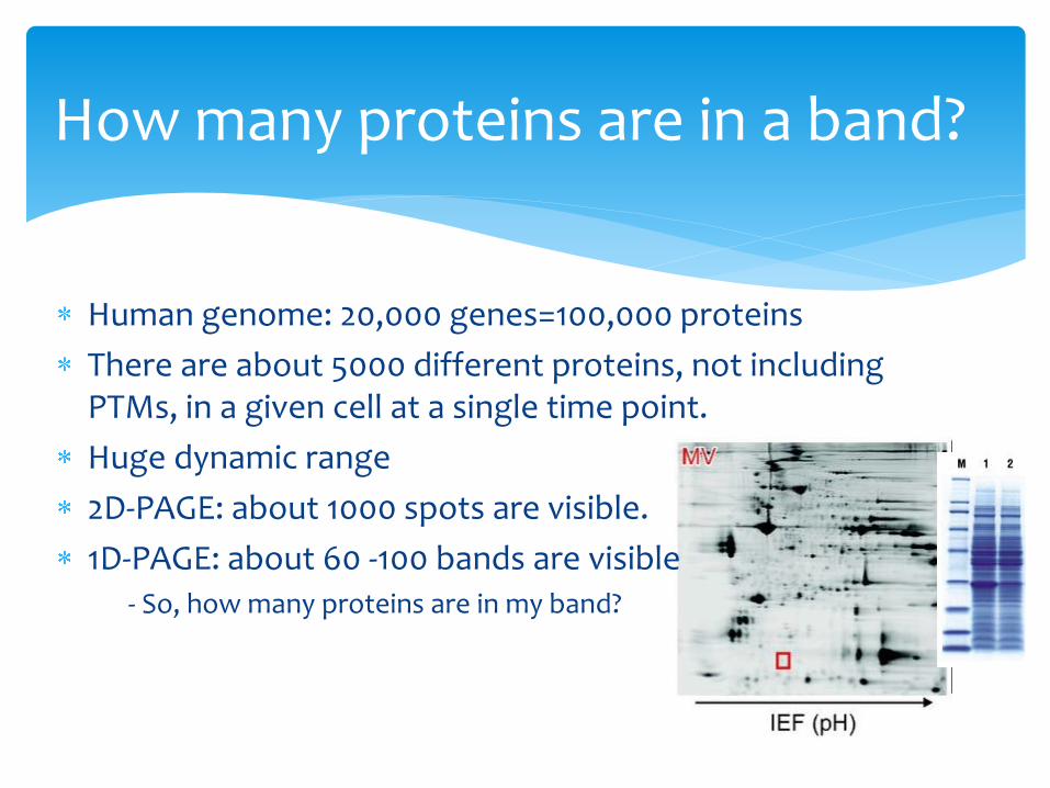
Human genome: 20,000 genes=100,000 proteins
There are about 5000 different proteins, not including PTMs, in a given cell at a single time point.
Huge dynamic range
2D-PAGE: about 1000 spots are visible.
1D-PAGE: about 60 -100 bands are visible - So, how many proteins are in my band?
How many proteins are in a band?

Can you IP your protein of interest?
Can you find other way to help with the separation?
-Organelle enrichment
-PTMs enrichment
-Size enrichment
Have you optimized your running conditions?
Separation is the key!
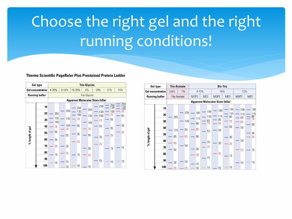
Choose the right gel and the right running conditions!
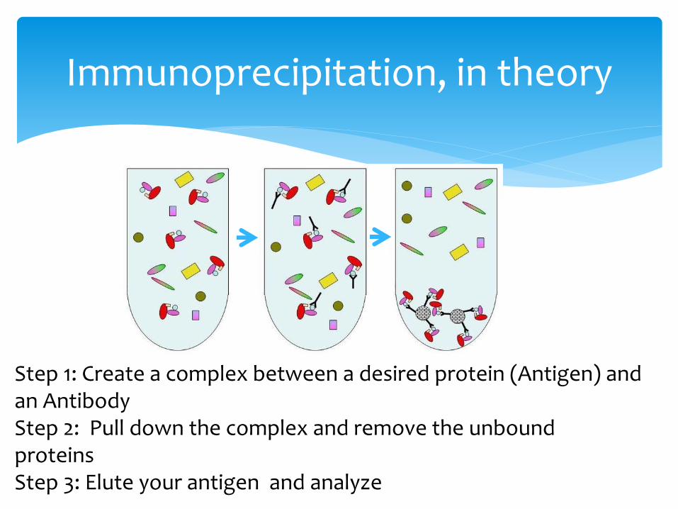
Immunoprecipitation, in theory
Step 1: Create a complex between a desired protein (Antigen) and an Antibody Step 2: Pull down the complex and remove the unbound proteins Step 3: Elute your antigen and analyze
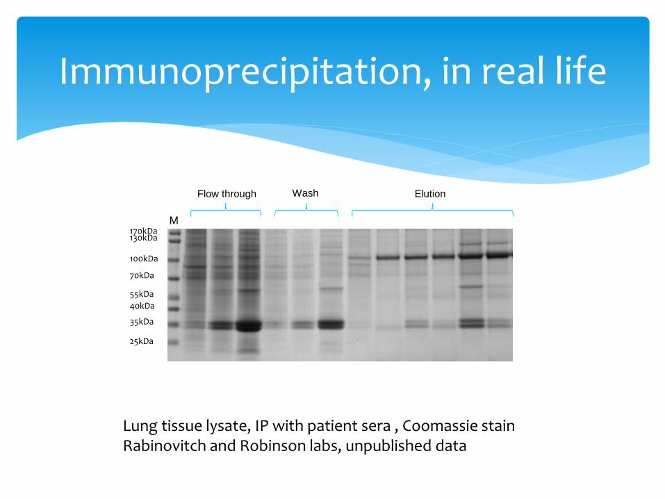
Immunoprecipitation, in real life
170kDa
130kDa
100kDa
70kDa
40kDa
55kDa
35kDa
25kDa
M
Flow through Wash Elution
Lung tissue lysate, IP with patient sera , Coomassie stain Rabinovitch and Robinson labs, unpublished data
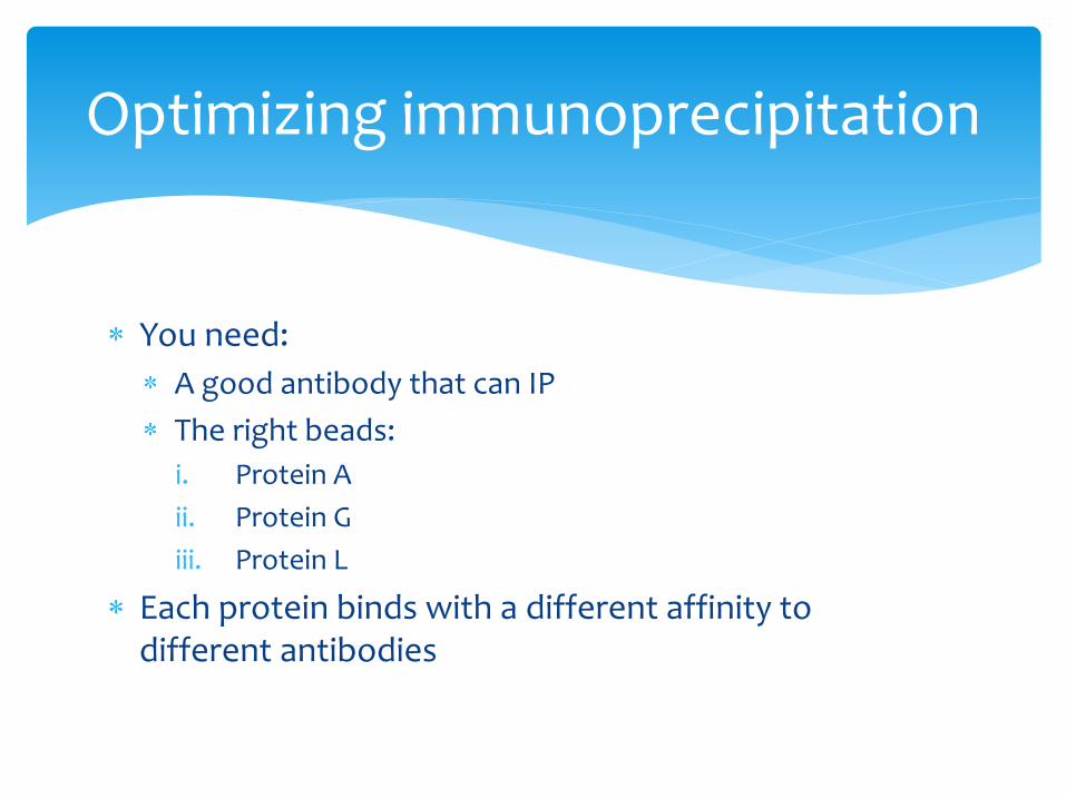
You need:
A good antibody that can IP
The right beads:
i. Protein A
ii. Protein G
iii. Protein L
Each protein binds with a different affinity to different antibodies
Optimizing immunoprecipitation

Choosing the right bead: affinity!
Species Antibody Class Protein A Protein G Protein L
Human Total IgG +++ +++ +++
IgG1 +++ +++ +++
IgG2 +++ +++ +++
IgG3 +/- +++ +++
IgG4 +++ +++ +++
IgM +/- - +++
IgD - - +++
IgA +/- - +++
Fab +/- +/- +++
ScFV +/- - +++
Mouse Total IgG +++ +++ +++
IgM - - +++
IgG1 +/- +/- +++
IgG2a +++ +++ +++
IgG2b +++ +++ +++
IgG3 +++ +++ +++
Rat Total IgG +/- +/- +++
IgG1 +/- +/- +++
IgG2a - +++ +++
IgG2b - +/- +++
IgG2c +++ +++ +++
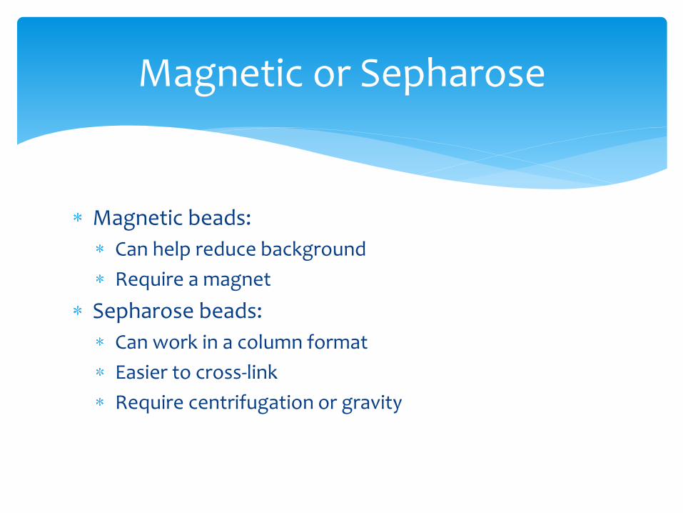
Magnetic beads:
Can help reduce background
Require a magnet
Sepharose beads:
Can work in a column format
Easier to cross-link
Require centrifugation or gravity
Magnetic or Sepharose

Magnetic or Seprhaose
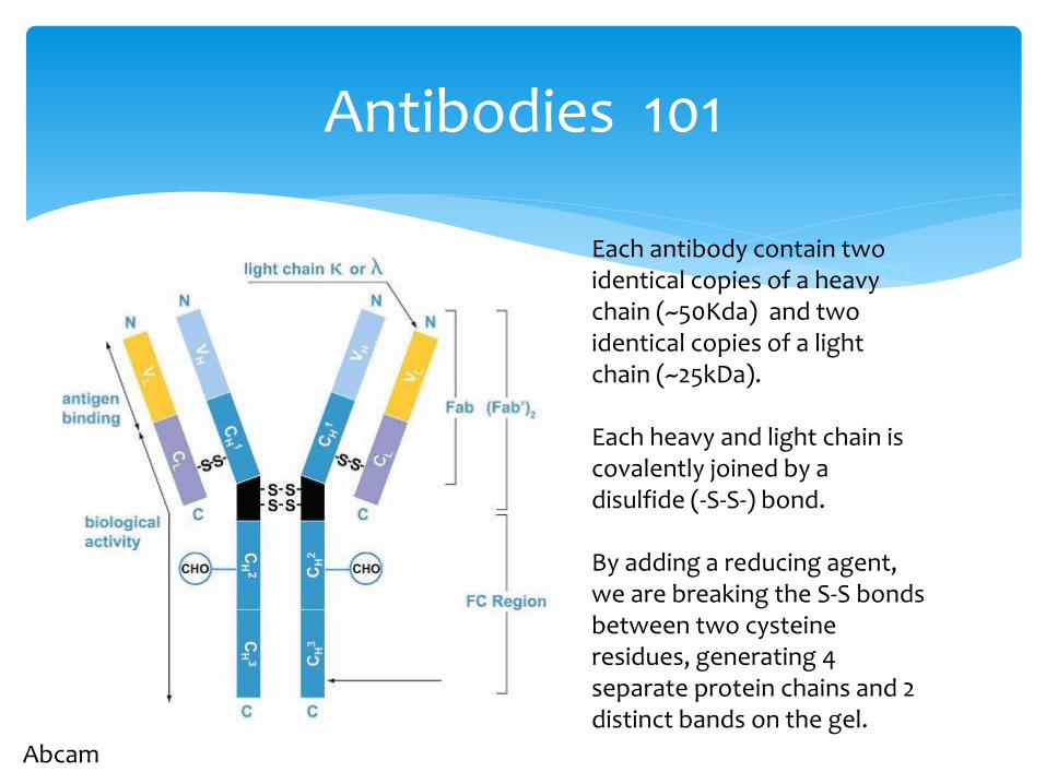
Antibodies 101
Abcam
Each antibody contain two identical copies of a heavy chain (~50Kda) and two identical copies of a light chain (~25kDa). Each heavy and light chain is covalently joined by a disulfide (-S-S-) bond. By adding a reducing agent, we are breaking the S-S bonds between two cysteine residues, generating 4 separate protein chains and 2 distinct bands on the gel.

Crosslinking is the process of chemically joining two or more molecules by a covalent bond. This enables us to :
Capture and identify unknown protein interactors or interaction domains.
Conjugate an enzyme or tag to an antibody or other purified protein.
Immobilize antibodies or other proteins for assays or affinity-purification.
Crosslinking

Pro: - Can stabilize the Ab-Ag interaction - Can be used to immobilize the Ab to beads, thus removing it
completely from the elution product. This can result in a cleaner result.
Cons: - Can bind an innocent bystander, thus creating either a false
positive or increase the background
Choose your crosslinker wisely!
To crosslink or not to crosslink?
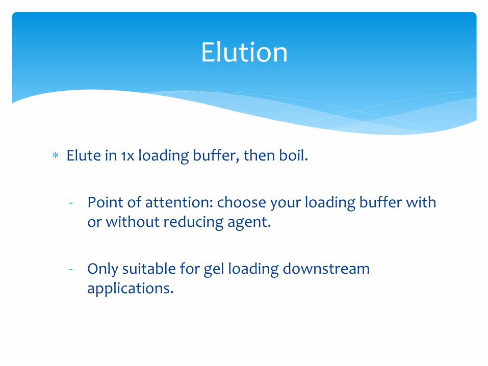
Elute in 1x loading buffer, then boil.
- Point of attention: choose your loading buffer with or without reducing agent.
- Only suitable for gel loading downstream applications.
Elution
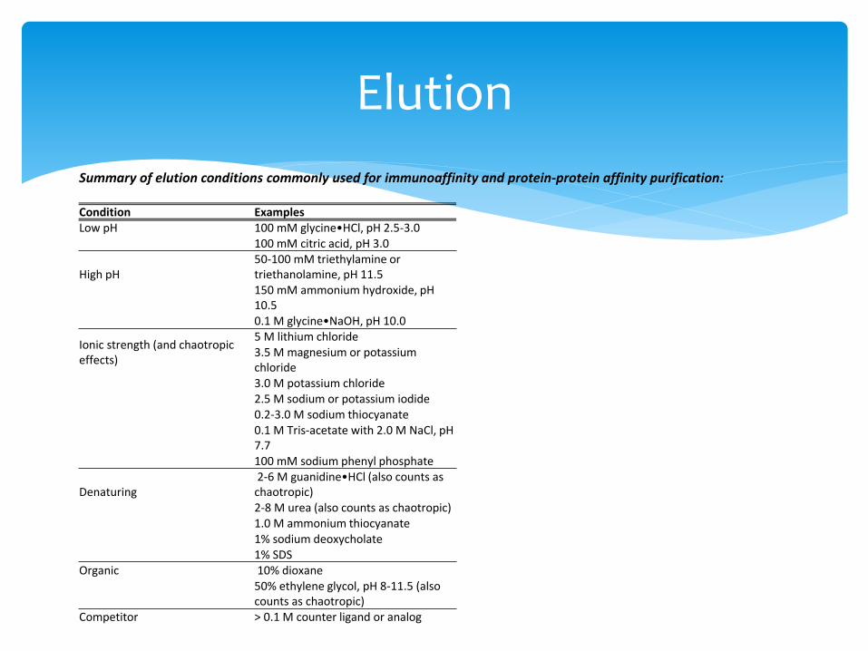
Summary of elution conditions commonly used for immunoaffinity and protein-protein affinity purification: Condition Examples Low pH 100 mM glycine•HCl, pH 2.5-3.0 100 mM citric acid, pH 3.0
High pH 50-100 mM triethylamine or triethanolamine, pH 11.5 150 mM ammonium hydroxide, pH 10.5
0.1 M glycine•NaOH, pH 10.0
Ionic strength (and chaotropic effects)
5 M lithium chloride 3.5 M magnesium or potassium chloride 3.0 M potassium chloride 2.5 M sodium or potassium iodide 0.2-3.0 M sodium thiocyanate 0.1 M Tris-acetate with 2.0 M NaCl, pH 7.7
100 mM sodium phenyl phosphate
Denaturing 2-6 M guanidine•HCl (also counts as chaotropic) 2-8 M urea (also counts as chaotropic) 1.0 M ammonium thiocyanate 1% sodium deoxycholate
1% SDS Organic 10% dioxane
50% ethylene glycol, pH 8-11.5 (also counts as chaotropic)
Competitor > 0.1 M counter ligand or analog
Elution

- The key to successful MS discovery is having a clean, enriched protein
- Know your friends and your enemies
- Try to enrich and separate your protein of interest
- As proteins are very different from one to another, it might take time to
really work out all the fine tuning, so be patient ….
In summary….